/


Text
SECOND
. ,
.
. 'l1li
'lit ....
. ,
.
.
.
..
. .
.
.. ..
.
..
.
" . .
.
.
. .
rr. .
II
. .
,
..
.
Hi
G,
r
Classification of the Metazoa
(with estimated numbers of described living species)
CLASSIFICATION
Phylum Porifera
Class Calcarea
Class Hexactinellida
Class Demospongiae
Phylum Placozoa
Phylum Monoblastozoa
Phylum Rhomobozoa
Phylum Orthonectlda
Phylum Cnidarla
Class Hydrozoa
Class Anthozoa
Class Cubozoa
Class Scyphozoa
Phylum Ctenophora
Phylum Platyhelminthes
Class Turbellaria
Class Monogenea
Class Trematoda
Class Cestoda
Phylum Nemertea
Class Anopia
Class Enopla
Phylum Rotifera
Class Digonota
Class Monogononta
Phylum Gastrotricha
Phylum Kinorhyncha
Phylum Nemata (= Nematoda)
Class Adenophorea (= Aphasmida)
Class Secernentea (= Phasmida)
Phylum Nematomorpha
Phylum Acanthocephala
Phylum Entoprocta
Phylum Gnathostomulfda
Phylum Prlapula
Phylum Loricifera
Phylum Cycllophora
Phylum Sipuncula
Class Phascolosomida
Class Sipunculida
Phylum Echlura
Phylum Annelida
Class Polychaeta
Class Clitellata
Phylum Onychophora
Phylum Tardigrada
Phylum Arthropoda
Subphylum Trilobitomorpha
Subphylum Crustacea
Class Remipedia
Class Cephalocarida
Sponges
Placozoans (Trichoplax)
Monoblastozoans (Salinella)
Rhombozoans
Orthonectids
Cnidarians
Hydroids, hydromedusae, siphonophores,
myxozoans
Sea anemones, sea fans, corals
Cubomedusae
Jellyfish
Ctenophores, sea gooseberries
Flatworms, flukes, tapeworms
Flatworms
Monogenetic flukes
Digenetic and aspidogastrean flukes
Tapeworms
Nemerteans, Ribbon worms
Rotifers
Gastrotrichs
Kinorhynchs
Nematodes
Horsehair worms, hair worms
Acanthocephalans
Entoprocts
Gnathostomulids
Priapulans
Loriciferans
Cydiophorans
Sipunculans, peanut worms
Echiurans, spoon worms
Segmented worms
Fan worms, tube worms, pogonophorans
Earth worms and leeches
Velvet worms
Water bears
Arthropods
Trilobites and their kin
Remipedes
Cephalocarids
DESCRIBED SPECIES
5,500
1
1
70
20
10,000
100
20,000
900
1,800
450
150
25,000
320
1,100
150
80
16
10
1
320
135
16,500
110
800
-1,097,289
extinct
CLASSIFICATION
Class Branchiopoda
Class Malacostraca
Class Maxillopoda
Subphylum Cheliceriformes
Class Chelicerata
Class Pycnogonida
Subphylum Myriapoda
Class Diplopoda
Class Chilopoda
Class Pauropoda
Class Symphyla
Subphylum Hexapoda
Class Entognatha
Class Insecta
Phylum Mollusca
Class Aplacophora
Class Monoplacophora
Class Polyplacophora
Class Gastropoda
Class Bivalvia (= Pelecypod a)
Class Scaphopoda
Class Cephalopoda
Phylum Phoronlda
Phylum Ectoprocta
Class Phylactolaemata
Class Stenolaemata
Class Gymnolaemata
Phylum Brachiopoda
Class Inarticulata
Class Articulata
Phylum Echinodermata
Class Crinoidea
Class Asteroidea
Class Ophiuroidea
Class Echinoidea
Class Holothuroidea
Phylum Chaetognatha
Phylum Hemichordata
Class Enteropneusta
Class Pterobranchia
Class Planctosphaeroidea
Phylum Chordata
Subphylum Urochordata (= Tunicata)
Class Ascidiacea
Class Thaliacea
Class Appendicularia (= Larvacea)
Class Sorberacea
Subphylum Cephalochordata
(= Acrania)
Subphylum Vertebrata
Class Myxini
Class Cephalaspidomorphi
Class Chondrichthyes
Class Osteichthyes
Class Amphibia
Class Reptilomorpha (:; Sauropsida)
Class Mammalia
Fairy shrimps, brine shrimps, tadpole shrimps,
clam shrimps, cladocerans
Crabs, shrimps, pill bugs, beach hoppers, etc.
Barnacles, ostracods, etc.
Horseshoe crabs, spiders, scorpions, mites,
ticks, etc.
Sea spiders
Millipedes
Centipedes
Pauropodans
Symphylans
Springtails, proturans, diplurans
Insects
Molluscs
Caudofoveatans and Solenogasters
Monoplacophorans
Chitons
Snails and slugs
Clams
Tusk shells
Octopuses, squids, Nautilus
Phoronids
Ectoprocts
Brachiopods, lamp shells
Echinoderms
Sea lilies and feather stars
Sea stars and sea daisies
Brittle stars and basket stars
Sea urchins and sand dollars
Sea cucumbers
Chaetognaths, arrow worms
Hemichordates
Acorn, or tongue worms
Pterobranchs
Planctosphaera pelagica
Chordates
Tunicates
Ascidians, sea squirts
Pelagic tunicates, salps
larvaceans
Sorberaceans
Lancelets, amphioxus
Hagfishes
Lampreys
Sharks, skates, rays
Bony fishes
Frogs, salamanders, etc.
Reptiles and birds
Mammals
DESCRIBED SPECIES
93,195
20
4,500
335
7,000
100
85
49,693
Invertebrates
SECOND EDITION
SECOND EDITION
Richard C. Brusca
...
Director of Conservation and Science,
Arizona-Sonora Desert Museum
Gary /. Brusca
.. Late, Professor of Zoology,
Humboldt State University
... ........
....
"
....
with illustrations by Nancy Haver
,\,
Sinauer Associates, Inc., Publishers
Sunderland, Massachusetts 01375
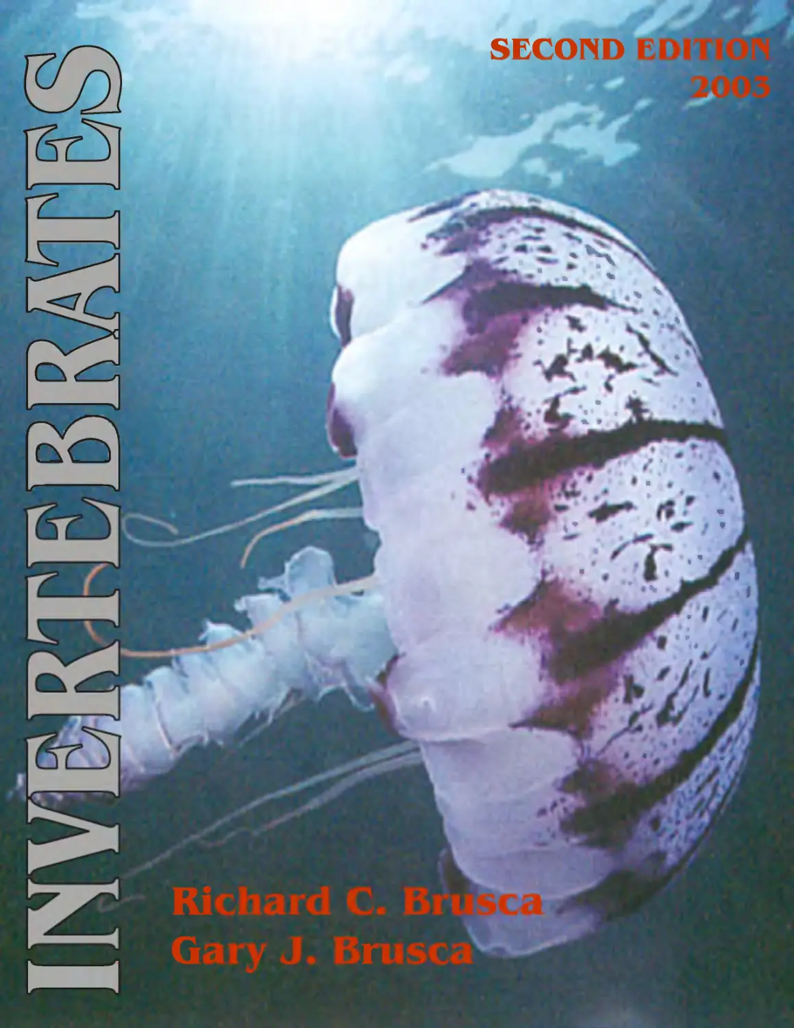
About the cover:
Pelagia, a large semaeostoman medusa.
@D. J. Wrobel/Biological Photo Service
...
- "'
"-:-
Title Page Photo:
Close-up of a polychaete worm, Nereis sp.
Courtesy of Larry Jon Friesen.
-
J1I" ..;
" f
.cQ
".
.....
Invertebrates, Second Edition
Copyright@2003 by Sinauer Associates, Inc.
All rights reserved.
This book may not be reprinted in whole or
in part without permission from the publisher.
For information or to order, address:
I
-
.....
J. ......
Sinauer Associates, Inc.
23 PIumtree Road/PO Box 407
Sunderland, MA 01375 U.S.A.
FAX: 413-549-1118
Email: publish@sinauer.com
www.sinauer.com
Library of Congress Catologing-in-Publication Data
Brusca, Richard C.
Invertebrates / Richard C. Brusca, Gary J. Brusca.-- 2nd ed.
p.cm.
Includes bibliographical references.
ISBN 0-87893-097-3 (hardcover)
1. Invertebrates. 1. Brusca, Gary J. II. Title.
QL362 .B924 2002
592--dc21
2002154089
Printed in U.S.A.
10 9 8 7 6 5 4 3 2
To the Memory of Gary
..,.:::..
:
.c
""
"
Who had a passion for teaching and for invertebrates.
And who loved tide pools on fog-shrouded mornings, a good fishing trip, and a good poker game.
We had some great times.
Brief Contents
1 Introduction 1
2 Classification, Systematics, and Phylogeny 23
3 Animal Architecture and the Sauplan Concept 41
4 Animal Development, Life Histories, and Origins 93
5 The Protists 121
6 Phylum Porifera: The Sponges 179
7 Four Phyla of Uncertain Affinity 209
8 Phylum Cnidaria 219
9 Phylum Ctenophora: The Comb Jellies 269
10 Phylum Platyhelminthes 285
11 Phylum Nemertea: The Ribbon Worms 319
12 Blastocoelomates and Other Phyla 337
.13 Phylum Annelida: The Segmented Worms 389
14 Sipuncula and Echiura 447
.15 The Emergence of the Arthropods: Onychophorans, Tardigrades,
Trilobites, and the Arthropod Bauplan 463
16 Phylum Arthropoda: The Crustacea 513
17 Phylum Arthropoda: The Hexapoda (Insects and Their Kin) 591
18 Phylum Arthropoda: The Myriapods (Centipedes, Millipedes,
and Their Kin) 639
19 Phylum Arthropoda: The Cheliceriformes 655
20 Phylum Mollusca 703
21 Lophophorates 773
22 Phylum Echinodermata 803
23 Other Deuterostomes: Chaetognatha, Hemichordata, Chordata 841
24 Perspectives on Invertebrate Phylogeny 875
Contents
Preface xvii
Acknowledgments xix
Introduction 1
Where Did Invertebrates Come From? 4
The Dawn of Life 4
The Ediacaran Epoch and the Origin of Animals 5
The Paleozoic Era (570-250 mya) 6
The Mesozoic Era (250-65 mya) 7
The Cenozoic Era (65 mya-present) 7
Where Do Invertebrates Live? 9
Marine Habitats 9
Estuaries and Coastal Marshlands 13
Freshwater Habitats 13
Terrestrial Habitats 14
A Special Type of Environment: Symbiosis 14
Some Comments On Evolution 15
Microevolution 15
Macroevolution 16
A Final Introductory Message to the Reader
19
Classification, Systematics, and Phylogeny 23
Biological Classification 24
Nomenclature 24
Systematics 27
Important Concepts and Terms 28
Constructing Phylogenies and Classifications
31
Animal Architecture and the Bauplan Concept 41
Body Symmetry 43
Cellularity, Body Size, Germ Layers, and
Body Cavities 46
Locomotion and Support 49
Ameboid Locomotion 50
Cilia and Flagella 50
Muscles and Skeletons 52
feeding Mechanisms 56
Intracellular and Extracellular Digestion 56
Feeding Strategies 57
Excretion and Osmoregulation 66
Nitrogenous Wastes and Water Conservation 66
Osmoregulation and Habitat 67
Excretory and Osmoregulatory Structures 68
Circulation and Gas Exchange 71
[nternal Transport 71
Circulatory Systems 71
Hearts and Other Pumping Mechanisms 73
VIII CONTENTS
Gas Exchange and Transport 73
Nervous Systems and Sense Organs 77
Invertebrate Sense Organs 77
Sense Organs 78
Independent Effectors 82
Bioluminescence 82
Nervous Systems and Body Plans 82
Hormones and Pheromones 84
Reproduction 84
Asexual Reproduction 85
Sexual Reproduction 86
Parthenogenesis 88
'Jt
, .
.':\..
Animal Development, Life Histories, and Origins 93
Eggs and Embryos 93
Eggs 94
Cleavage 94
The Problem of Cell Fates 98
Blastula Types 98
Gastrulation and Germ Layer Formation 99
Mesoderm and Body Cavities 100
Life Cycles: Sequences and Strategies 102
Classification of Life Cycles 102
Indirect Development 103
Settling and Metamorphosis 104
Direct Development 105
The Protists 121
Taxonomic History and Classification 123
Classification of the Protista 123
The Protist Bauplan 124
Body Structure, Excretion. and Gas Exchange 124
Support and Locomotion 125
Nutrition 125
Activity and Sensitivity 125
Reproduction 127
Phylum Euglenida 129
Support and Locomotion 130
Nutrition 131
Reproduction 131
Phylum Kinetoplastida (The Trypanosomes
and Their Relatives) 131
Support and Locomotion 133
Nutrition 134
Mixed Development 105
Adaptations to Land and Fresh Water 106
Parasite Life Cycles 106
The Relationships between Ontogeny and
Phylogeny 106
The Concept of Recapitulation 107
Heterochrony and Paedomorphosis 108
Origins of Major Groups of Metazoa 108
Origin of the Metazoa 108
Origin of the Bilateral Condition 112
Origin of the Coelomic Condition 112
Reproduction and Life Cycles 135
Phylum Ciliophora (The Ciliates) 135
Support and Locomotion 135
Nutrition 139
Reproduction 142
Phylum Apicomplexa (The Gregarines,
Coccidians, Haemosporidians, and
Piroplasms) 145
Support and Locomotion ] 46
Nutrition 146
Reproduction and Life Cycles 147
Phylum Dinoflagellata 148
Support and Locomotion 151
Nutrition 151
Reproduction 152
Phylum Stramenopila 153
Support and Locomotion 154
Nutrition 154
Reproduction 155
Phylum Rhizopoda (Amebas) 155
Support and Locomotion 157
Nutrition 159
Reproduction 160
Phylum Actinopoda 161
Support and Locomotion 162
Nutrition 163
Reproduction 164
Phylum Granuloreticulosa (Foraminifera and
Their Kin) 165
Support and Locomotion 166
Nutrition 166
Reproduction and Some Life Cycles 167
CONTENTS IX
Phylum Diplomonadida 167
Support and Locomotion 168
Nutrition 169
Reproduction 169
Phylum Parabasilida (The Trichomonads
and Hypermastigotes) 169
Support and Locomotion 170
Nutrition 171
Reproduction 171
Phylum Cryptomonada 172
Phylum Microspora 172
Phylum Ascetospora 173
Phylum Choanoflagellata 173
Phylum Chlorophyta 173
Phylum Opalinida 173
Genus Stephanopogon 174
Protist Phylogeny 174
Phylum Porifera: The Sponges 179
Taxonomic History and Classification 180
The Poriferan Bauplan 182
Body Structure and the Aquiferous System 183
Cell Types 188
Cell Aggregation 191
Support 191
Nutrition, Excretion, and Gas Exchange 192
Activity and Sensitivity 194
Reproduction and Development 196
Some Additional Aspects of Sponge Biology
2'01
Distribution and Ecology 201
Biochemical Agents 201
Growth Rates 202
Symbioses 202
Poriferan Phylogeny 203
The Origin of Sponges 203
Evolution within the Porifera 204
. ..
..
..
..
...
..
. .
.
.,
,
'w
.
. .
Four Phyla of Uncertain Affinity 209
Taxonomic History 209
Mesozoan Bauplans 210
Phylum Placozoa 210
Phylum Monoblastozoa 210
Phylum Rhombozoa 211
Phylum Orthonectida 215
Mesozoan Phylogeny 216
X CONTENTS
yl
-do -,
Taxonomic History and Classification 222
The Cnidarian Hauplan 225
.
The Body Wall 226
Support 236
Movement 239
Cnidae 242
Feeding and Digestion 244
Defense, Interactions, and Symbiosis 246
Circulation, Gas Exchange, Excretion, and
Osmoregulation 250
Nervous System and Sense Organs 250
Reproduction and Development 253
Cnidarian Phylogeny 261
Phylum Ctenophora: The Comb Jellies 269
Taxonomic History and Classification 271
The Ctenophoran Hauplan 274
Support and Locomotion 274
Feeding and Digestion 276
Circulation, Excretion, Gas Exchange, and
Osmoregulation 279
Nervous System and Sense Organs 279
Reproduction and Development 280
Ctenophoran Phylogeny 281
Phylum Platyhelminthes 285
Taxonomic History and Classification 286
The Platyhelminth Hauplan 289
Body Wall 291
Support, Locomotion, and Attachment 294
Feeding and Digestion 295
Circulation and Gas Exchange 299
Excretion and Osmoregulation 299
Nervous System and Sense Organs 300
Reproduction and Development 303
Platyhelminth Phylogeny 313
Phylum Nemertea: The Ribbon Worms 319
Taxonomic History and Classification 321
Classification 321
The Nemertean Hauplan 322
Body Wall 322
Support and Locomotion 322
Feeding and Digestion 323
Circulation and Gas Exchange 327
Excretion and Osmoregulation 328
Nervous System and Sense Organs 329
Reproduction and Development 331
Nemertean Phylogeny 333
CONTENTS XI
Blastocoelomates and Other Phyla 337
Taxonomic History 338
The Blastocoelomate Condition 338
Phylum Rotifera:
The Rotifers 338
General External Anatomy and Details of the
Corona 340
Body Wall, Body Cavity, Support, and Locomotion
341
Feeding and Digestion 341
Circulation, Gas Exchange, Excretion, and
Osmoregulation 343
Nervous System and Sense Organs 343
Reproduction and Development 343
Phylum Gastrotricha:
The Gastrotrichs 345
Body Wall, Support, and Locomotion 346
Feeding and Digestion 347
Circulation, Gas Exchange, Excretion, and
Osmoregulation 347
Nervous System and Sense Organs 347
Reproduction and Development 347
Phylum Kinorhyncha:
The Kinorhynchs 348
Body Wall 349
Support and Locomotion 350
Feeding and Digestion 350
Circulation, Gas Exchange, Excretion, and
Osmoregulation 350
Nervous System and Sense Organs 350
Reproduction and Development 350
Phylum Nemata:
The Nematodes 351
Body Wall, Support, and Locomotion 352
Feeding and Digestion 353
Circulation, Gas Exchange, Excretion, and
Osmoregulation 355
Nervous System and Sense Organs 357
Reproduction, Development, and Life Cycles 357
Life Cycles of Some Parasitic Nematodes 359
Phylum Nematomorpha:
Hair Worms and Their Kin 362
Body Wall, Support, and Locomotion 363
Feeding and Digestion 363
Circulation, Gas Exchange, Excretion, and
Osmoregula tion 363
Nervous System and Sense Organs 365
Reproduction and Development 365
Phylum Priapula:
The Priapulans 365
Body Wall, Support, and Locomotion 367
Feeding and Digestion 367
Circulation, Gas Exchange, Excretion, and
Osmoregulation 368
Nervous System and Sense Organs 368
Reproduction and Development 368
Phylum Acanthocephala:
The Acanthocephalans 368
Body Wall, Support, Attachment, and Nutrition
369
Circulation, Gas Exchange, and Excretion 370
Nervous System 370
Reproduction and Development 370
Phylum Entoprocta:
The Entoprocts 371
Body Wall, Support, and Movement 373
Feeding and Digestion 373
Circulation, Gas Exchange, and Excretion 374
Nervous System 374
Reproduction and Development 374
Phylum Gnathostomulida:
The Gnathostomulids 375
Body Wall, Support, and Locomotion 376
Nutrition, Circulation, Excretion, and Gas
Exchange 376
Nervous System 377
Reproduction and Development 377
Phylum Loricifera:
The Loriciferans 377
Phylum Cycliophora:
The Cycliophorans 378
Some Phylogenetic Considerations 381
<5
,
XII CONTENTS
Phyl m Annelida: The Segmented Worms 387
I
Taxonomic History and Classification 388
The Annelid Bauplan 395
Body Forms 395
Body Wall and Coelomic Arrangement 397
Support and Locomotion 400
Feeding 405
Digestive System 410
Circulation and Gas Exchange 413
Excretion and Osmoregulation 417
Nervous System and Sense Organs 420
Regeneration and Asexual Reproduction 425
Sexual Reproduction and Development 427
Siboglinidae: The Beard Worms 434
Taxonomic History 434
The Tube, Body Wall, and Body Cavity 434
Nutrition 436
Circulation, Gas Exchange, Excretion, and
Osmoregulation 436
Nervous System and Sense Organs 437
Reproduction and Development 437
Annelid Phylogeny 438
Sipuncula and Echiura 445
The Sipunculans 445
Taxonomic History and Classification 447
The Sipunculan Bauplan 447
Body Wall, Coelom, Circulation, and Gas
Exchange 447
Support and Locomotion 449
Feeding and Digestion 449
Excretion and Osmoregulation 450
Nervous System and Sense Organs 450
Reproduction and Development 451
The Echiurans 451
Taxonomic History and Classification 452
The Echiuran Bauplan 453
Body Wall and Coelom 453
Support and Locomotion 453
Feeding and Digestion 454
Circulation and Gas Exchange 455
Excretion and Osmoregulation 456
Nervous System and Sense Organs 456
Reproduction and Development 456
Some Comments on Phylogeny 457
The Emergence of the Arthropods: Onychophorans,
Tardigrades, Trilobites, and the Arthropod Soup/an 461
Phylum Onychophora 463
Phylum Tardigrada 469
An Introduction to the Arthropods 475
Taxonomic History and Classification 475
Synopses of the Five Arthropod Subphyla 476
The Arthropod Bauplan and Arthropodization 476
The Body Wall 478
Arthropod Appendages 479
Support and Locomotion 482
Growth 485
The Digestive System 489
Circulation and Gas Exchange 489
Excretion and Osmoregulation 491
Nervous System and Sense Organs 492
Reproduction and Development 495
The Trilobites (Subphylum Trilobitomorpha)
497
General Body Form 497
Internal Anatomy 499
Development 499
The Evolution of Arthropods 499
The Origin of the Arthropoda 499
Evolution within the Arthropoda 500
Emerging Views of Arthropod Relationships 503
Where Are We Now? 506
CONTENTS XIII
Phylum Arthropoda: The Crustacea 511
\.
Synopses of Crustacean Taxa 517
The Crustacean Bauplan 550
Locomotion 551
Feeding 557
Digestive System 561
Circula tion and Gas Exchange 563
Excretion and Osmoregulation 568
Nervous System and Sense Organs 570
Reproduction and Development 573
Crustacean Phylogeny 579
Classification of the Crustacea 514
Subphylum Crustacea 514
7 Phylum Arthropoda: The Hexapoda (Insects and
eir .) S
Hexapod Classification 593
Subphylum Hexapoda 594
Synopses of Major Hexapod Taxa 594
The Hexapod Bauplan 601
General Morphology 601
Locomotion 607
Feeding and Digestion 610
Circulation and Gas Exchange 617
Excretion and Osmoregulation 618
Nervous System and Sense Organs 619
Reproduction and Development 623
Hexapod Evolution 628
The Origin of the Hexapoda 628
The Origin of Insect Flight 629
Evolution within the Hexapoda 630
Phylum Arthropoda: The Myriapods (Centipedes,
Millipedes, and Their Kin) 637
Myriapod Classification 639
Subphylum Myriapoda 639
Synopses of Myriapod Taxa 639
The Myriapod Bauplan 640
Head and Mouth Appendages 642
Locomotion 642
Feeding and Digestion 644
Circulation and Gas Exchange 644
Excretion and Osmoregulation 646
Nervous System and Sense Organs 646
Reproduction and Development 647
Myriapod Embryogeny 649
Myriapod Phylogeny 649
XIV CONTENTS
Phylum Arthropoda: The Cheliceriformes 653
Subphylum Cheliceriformes 654
Synopses of the Chelicerate Taxa 656
The Chelicerate Bauplan 666
Spinnerets, Spider Silk, and Spider "Webs" 667
Locomotion 670
Feeding and Digestion 672
Circulation and Gas Exchange 678
Excretion and Osmoregulation 679
Nervous System and Sense Organs 680
Reproduction and Development 683
Phylum Mollusca 701
Taxonomic History and Classification 702
The Molluscan Bauplan 715
.
1
.
Loph phora es 771
The Lophophorates: An Overview 771
Taxonomic History 772
The Lophophorate Bauplan 773
Phylum Phoronida 773
The Phoronid Bauplan 774
Body Wall, Body Cavity, and Support 775
The Lophophore, Feeding, and Digestion 776
Circulation, Gas Exchange, and Excretion 776
Nervous System 777
Reproduction and Development 777
Phylum Ectoprocta 778
The Ectoproct Bauplan 779
The Body Wall, Coelom, Muscles, and Movement
783
The Class Pycnogonida 691
The Pycnogonid Bauplan 692
External Anatomy 692
Locomotion 695
Feeding and Digestion 695
Circulation, Gas Exchange, and Excretion 695
Nervous System and Sense Organs 696
Reproduction and Development 696
Cheliceriform Phylogeny 696
The Body Wall 718
The Mantle and Mantle Cavity 719
The Molluscan Shell 720
Torsion, or "How the Gastropod Got Its Twist" 725
Locomotion 728
Feeding 733
Digestion 741
Circulation and Gas Exchange 744
Excretion and Osmoregulation 747
Nervous System 748
Sense Organs 752
Cephalopod Coloration and Ink 754
Reproduction 755
Development 759
Molluscan Evolution and Phylogeny 761
Zooid Interconnections 785
The Lophophore, Feeding, and Digestion 785
Circulation, Gas Exchange, and Excretion 788
Nervous System and Sense Organs 788
Reproduction and Development 789
Phylum Brachiopoda 792
The Brachiopod Bauplan 793
The Body Wall, Coelom, and Support 793
The Lophophore, Feeding, and Digestion 795
Circulation, Gas Exchange, and Excretion 796
Nervous System and Sense Organs 796
Reproduction and Development 796
Lophophorate Phylogeny 798
CONTENTS XV
Phyl m Echin derm ta 8..01
Taxonomic History and Classification 803
The Echinoderm Bauplan 806
Body Wall and Coelom 808
Water Vascular System 810
Support and Locomotion 813
Feeding and Digestion 814
3
Circulation and Gas Exchange 823
Excretion and Osmoregulation 824
Nervous System and Sense Organs 825
Reproduction and Development 826
Echinoderm Phylogeny 830
Other Deuterostomes: Chaetognatha, Hemichordata,
Chordata 839
Taxonomic History and Classification 839
Phylum Chaetognatha: Arrow Worms 841
The Chaetognath Bauplan 841
Phylum Hemichordata:
The Hemichordates 847
The Hemichordate Bauplan 849
Phylum Chordata: The Chordates 854
The Urochordates (Tunicates) 855
The Tunicate Bauplan 857
The Cephalochordates 864
The Cephalochordate Bauplan 865
Phylogenetic Considerations 868
Perspectives on Invertebrate Phylogeny 873
A Word about Characters 876
Metazoan Evolution 876
The Tree 876
Metazoan Roots 876
Evolution within the Metazoa 877
Other Ideas about Animal Phylogeny 882
Molecular Phylogenetics 882
Evolutionary Developmental Biology 884
Appendix A: Common Human Diseases
Transmitted by Insects 890
Appendix B: Data Matrix for Analysis of
Metazoan Phylogeny 892
Illustration Credits 897
Index 903
Preface
It is not now nor will it ever be given to one man
to observe all the things recounted in the
following pages.
Waldo L. Schmitt
Crustaceans, 1 965
During the revision of Invertebrates my brother Gary
passed away. For a while the project was stalled. But,
buoyed by the support of family, friends, and col-
leagues, I eventually returned to the task, which, at
times, seemed overwhelming. The field of invertebrate
biology is so vast, and cuts across so many disciplinary
lines, that even in a book of this size it is necessary to
generalize about some topics and to slight others. As
university instructors, my brother and I realized early
on that the teaching of invertebrate zoology should not
be compartmentalized. Thus, in planning this book we
were concerned about two potential dangers. First, the
text might become an encyclopedic list of "facts" about
one group after another, the sort of "flash-card" ap-
proach that we wanted to avoid. Second, the book
might be a rambling series of stories or vignettes about
randomly selected animals (or "model organisms") and
their ways of life. The first book would be dull, would
encourage rote memorization instead of understanding,
and might give the misconception that there is little left
to discover. The second book might be full of interesting
"gee whiz" stuff but would seem disorganized and
without continuity or purpose to the serious student.
Either approach could fail to present the most important
aspects of invertebrates-their phenomenal diversity,
their natural history, and their evolutionary relation-
ships. We also held to the belief that what we know
about these animals is not as important as how we think
about them. You should be prepared to assimilate much
new material, but you should also be prepared for a
great deal of uncertainty and mystery, as much remains
to be discovered.
To avoid the pitfalls noted above, and to establish
threads of continuity in our discussions about inverte-
brates, we developed our book around two fundamen-
tal themes: unity and diversity. The first theme we ap-
proach by way of functional body architecture, or what
we call the bauplan concept. The second theme we ap-
proach through the principles of phylogenetic biology.
Our hope was that weaving the book tightly to these
themes would provide a meaningfu] flow as readers
move from one phyllll11 to the next. The first four chap-
ters provide background for these themes and thus pro-
vide an important foundation upon which the rest of
the book rests. Please read these chapters carefully and
refer back to them throughout your study.
The bulk of this book (Chapters 5-23) is devoted to a
phylum-by-phylum discussion of invertebrates. Fairly
detailed classifications or taxonomic synopses for each
phylum are included in separate sections of each chap-
ter to serve as references. A consistent organization is
maintained throughout each chapter, although we did
yield to the important and sometimes different lessons
to be learned by investigating the special attributes of
each group of animals. In addition, because of their size
and diversity, some taxa receive more attention than
others-although this does not mean that such groups .
are more "important" biologically than smaller or more
homogeneous ones. (Five chapters are devoted to the
arthropods and their kin.) In certain chapters more than
one phylum is covered. In some cases the phyla covered
are thought to be closely related to one another; in other
cases the phyla merely represent a particular grade of
complexity and their inclusion in a single chapter facili-
tates our comparative approach.
Certain aspects of this book have, of course, been in-
fluenced by our own biases; this is especially true of the
discussions on phylogeny. We use a combination of
phylogenetic trees (cladograms) and narrative discus-
sions to talk about animal evolution. Cladograms are
used when appropriate, because they provide the least
ambiguous statements that can be made about animal
relationships. We always knew that some of you, profes-
sors and students both, would disagree with our meth-
ods and ideas to various degrees-at least we lzaped that
you would. Never placidly accept what you see in a
XVIII
PREFACE
textbook, or anyplace else for that matter, but try to be
critical in your reading.
The book's final chapter is a phylogenetic summary
of the animal kingdom. It reinforces the point that much
remains to be explored and learned about the evolution-
ary relationships of invertebrates. Like all scientific
knowledge, we are dealing here with provisional, tran-
sient "truths" that always remain open to challenge and
revision. And, of course, scientists disagree. It is this dis-
agreement and the constant challenging of hypotheses
that enliven the field and push the frontiers of knowl-
edge forward.
There are a few other things you should know about
this book. A brief historical review of the classification of
each major group is provided. We felt this material was
not only interesting but also served to imbue students
with a sense of the dynamic nature of taxonomy and the
development of our understanding of eacn group.
Unless otherwise indicated, the Classification section in
each chapter deals only with extant taxa. Descriptions of
taxa in these annotated classifications are written in
somewhat telegraphic style to save space; we never ex-
pected these sections to be "read"-they are for refer-
ence. Important new words, when first defined, are set
in boldface type. These boldfaced terms are also indicat-
ed by boldfaced page references in the index; thus the
index can also be used as a glossary. We tried hard to be
consistent in our usage of zoological terminology, but
the existence of similar terms for entirely different struc-
tures in certain groups is notoriously troublesome-
these are noted in the text.
For this second edition of Invertebrates, of course, we
have tried to be as current as possible with the research
literature, but even as this book goes into production
important new publications appear daily. It has been es-
timated that the volume of scientific infonnation is dou-
bling about every 10 years (or faster). A half-million
nonclinical biology papers are published annually. As
Professor George Bartholomew noted, "ll one equates
ignorance with the ratio between what one knows and
what is available to be known...each biological investi-
gator becomes more ignorant with every passing day."
My goal has been to provide sufficient reference materi-
a] to lead the interested student quickly into the heart of
the relevant literature. Most of the references cited in the
text will be found at the end of the corresponding chap-
ter. However, to conserve space and eliminate redun-
dancy, in a number of cases (especially in figure cita-
tions) references of a general nature may be listed only
once, usually in the introductory chapters. You will also
notice citations of a fair number of references that are
quite old, some from the nineteenth century. These are
included not out of whimsy, but because many of these
are benchmark research papers or they stand out as
some of the best available descriptions for the subject at
hand. (It is surprising how many of the illustrations in
modern biology texts can be traced back to origins in
nineteenth-century publications.) It is distressing to see
how commonplace it has become for researchers to ig-
nore the excellent (and important) work of past decades.
For example, many phylogenetic research papers com-
pletely ignore 150 years of careful embryological re-
search that was published, largely in the German and
American literature, in the nineteenth and twentieth
centuries. For some scientists, biological research seems
to be little more than "sound bites" from the past
decade. Sadly, today, this "sound bite research culture"
is often imbued in graduate students-a shocking and
dangerous trend that encourages dilettantes. To under-
stand animals requires a thorough understanding of
their overall biology, and the dedication of a career, not
just dabbling.
Since the first edition of this book, there has been an
explosion of research in the field of molecular biology.
Much of this has been in molecular phylogenetics, but
huge strides are also being made in the area of molecu-
lar developmental biology. Papers in these fields now
appear at such a pace that it is difficult to write about
them in a textbook, for fear the ideas will be obsolete in
six months. There have been many new phylogenetic
hypotheses proposed on the basis of DNA sequence
analyses since the fist edition of this book. Many of the
molecular phylogenetic trees that were published before
2000 were quirky and troublesome, due to the simple
fact that the field is still new and emerging. Because
most of these trees are relatively new and still await rig-
orous testing with independent data, we do not discuss
them all. However, we do discuss molecular-based hy-
potheses that have a growing body of support or have
received widespread attention. But, in general we have
taken a conservative approach in this regard-we are
only just beginning to discover which genes are appro-
priate for different levels of phylogenetic analysis, and
how best to analyze them.
These things being said, I hope you are now ready to
forge ahead in your study of invertebrates. The task
may at first seem daunting, and rightly so. I hope that
this book will make this seemingly overwhelming task a
bit more manageable. If I succeed in enhancing your en-
joyment and appreciation of invertebrates, then my ef-
forts will have been worthwhile.
RC.B.
Tucson, Arizona
December 2002
Acknowledgments
I have benefited immeasurably from the careful and
professional work of many conscientious reviewers for
this second edition (listed below), most of whom went
far beyond simply correcting factual errors. any sent
extended commentaries on difficult points, reprints of
their recent work, and even photographs for the book. [
extend to these reviewers my most sincere gratitude;
this book is far better for their efforts. Where weakness-
es and errors remain, they are of course solely my re-
sponsibility. Some people have taken a special interest
in this book, providing thoughtful discussion and in-
sight over the years, and their contributions are espe-
cially appreciated, including Steve Gould, Todd Haney,
Robert Higgins, Jens H0eg, Reinhar t Kristensen, Jody
Martin, Jim Morin, Diana Lipscomb, and Wendy Moore.
A very special note of appreciation goes to Diana
Lipscomb, wno, witn the help of her student Kristen
Kivimaki, essentially rewrote the protist chapter. Diana
and Kristen devoted many weeks in that effort, and I
am deeply grateful to them.
I cannot list here all of the colleagues who sent pho-
tographs or allowed me to rummage through their files
of slides and prints and never complained about how
long 1 kept their pictures. I thank tnem all, but I am par-
ticularly indebted to Gita Bodner, Judith Connor, Peter
Fankboner, Rainer Foelix, Larry Friesen, Todd Haney,
Jens H0eg, Alex Kerstitch, Wayne Maddison, Gary
McDonald, and Jim Morin. In addition, I am grateful to
the authors and publishers who allowed me to repro-
duce material from their books, journals, and other pub-
lications. A special note of appreciation is due to Katja
Schultz-thanks so much for sharing the results of your
literature searches for phylogenetic papers for the Tree of
Life project. Special thanks also to Ken Rinehart, for all
those unexpected phone calls from sundry airports that
resulted in rendezvous in remote regions of the world to
collect invertebrates. Some authors have strongly influ-
enced my tninking, including Paul Dayton, Niles
Eldredge, Steve Gould, Ed Ricketts, Warren Stau1s, John
Steinbeck, and Jerome Ticlmor.
Most of the original artwork in this text was done from
our own sketches or from other sources by Nancy Haver,
supported by our publisher, Sinauer Associates. Ms.
PREFACE XIX
Haver's extraordinary talent is exceeded only by her pa-
tience with our nit-picking; I am proud to have her out-
standing work in this book. The production staff at
Sinauer truly are magicians with photograpbs and lay-
outs, and they never lost patience with my last-minute
changes and requests-thanks especially to Chris Small
and his expert production team, including Janice
Holabird, who was responsible for turning the manu-
script into beautiful page spreads, to Jefferson Johnson
for his extraordinary design work, and to David
Mcintyre for his exceptional ability to track down hard-
to-find images. All of the editors at Sinauer Associates
have been critical and passionate about their work (seem-
ingly a company policy), and I am indebted to them all,
including Carol Wigg, Kerry Falvey, Nan Sinauer, Sydney
Carroll, Che1sea Holabird, and Kathaleen Emerson. I es-
pecially want to thank Kathaleen, who stewarded this
second edition through to closure with extraordinary
style and good humor. Andy Sinauer originally became a
part of this project in the mid-1980s, at a time when my
brother and I wondered if the book would ever see the
light of day. He has been an unwavering friend since that
day. Were it not for Andy's gentle manner, wise council,
good business sense, and clever prodding, this book
would probably not exist.
I want to offer my apologies to those whose work I
might have missed in my attempts to keep up with the
ever-expanding literature on invertebrates. And I want
to thank those who have been thoughtful enough to
send me their publications over the years. Keeping track
of published research on invertebrates is an all-consum-
ing task.
Finally, I thank my colleagues, students, friends, and
family for encouragement and patience, especially Bob
Edison (Director of the Arizona-Sonora Desert Mu-
seum) who graciously allowed me the freedom from
other obligations to devote time to this project. Most im-
portant of all, of course, has been Wendy Moore, who
helped me in more ways than I can count through the
years of revising this book. Wendy's phylogenetic ex-
pertise and knowledge of arthropods is exceeded only
by her good humor, unwavering patience, and steadfast
friendship.
Scientific Reviewers
In addition to again thanking the reviewers for the first
edition of Invertebrates, I wish to thank the following
people who reviewed parts of this second edition.
GERALD J. BAKUS
University of Southern California, Los Angeles, CA
BILL BIRKY
University of Arizona, Tucson, AZ
NICOLE BOURy-ESNAULT
Centre d'Oceanologie de Marseille, Marseille, France
STEVE CAIRNS
Smithsonian Institution, Washington, D.C.
JOSE LUIS CARBALLO
National University of Mexico
Estaci6n Mazatlan, Mazatlan, Mexico
ANNE COHEN
Bodega Bay, CA
JOHN O. CORLISS
Albuquerque, NM
RICHARD DEMAREE
California State University, Chico, CA
NILES ELDREDGE
American Museum of Natural History, New York, NY
DAPHNE FAUTIN
University of Kansas, Lawrence, KS
PETER GLYNN
University of Miami, Miami, FL
MICHAELJ. GREENBERG
University of Florida, St. Augustine, FL
RICHARD GROSBERG
University of California, Davis, CA
TODD HANEY
Natural History Museum, Los Angeles, CA
GERHARD HAsZPRUNAR
Zoologische Staatssammlung Muenchen, Munich, Gernmny
DAVID J. HORNE
University of Greenwich, Kent, England
M. A. HOUCK
Texas Tech University, Lubbock, TX
KRIsTEN KIvIMAKI
The George Washington University, Washington, o.c.
DIANA LIPSCOMB
The George Washington University, Washington, D.C.
JOEL MARTIN
Natural History Museum, Los Angeles, CA
WENDY MOORE
University of Arizona, Tucson, AZ
JAMES MORIN
Shoals Marine l.11boratory, Cornell University, Ithaca, NY
KATJA ScHULTZ
University of Arizona, Tucson, AZ
WILLIAM SHEAR
Hampden-Sydney College, Hampden-Sydney, VA
ROWLAND SHELLEY
North Carolina State Museum of Natural Sciences,
Raleigh, NC
TIM STEBBINS
Ocean Monitoring Program, San Diego, CA
JEANVACELET
Centre Oceanographie, Marseille, France
JOHANN-WOLFGANG WAGELE
Ruhr-Universitaet Bochum, Germany
GREG WRAY
Duke University, Durham, NC
JILL YAGER
Antioch College, Yellow Springs, OH
1
.....
...
Introduction
For a gentleman should know something of
invertebrate zoology, call it culture or what you will,
just as he ought to know something about painting
and music and the weeds in his garden.
Martin Wells, Lower Animals, 1968
" -,
O ne of the first evolutionary trees of life conceived from a Darwinian
(genealogical) perspective was published by Ernst Haeckel in 1866
(Figure 1.1). Haeckel's famous tree of life began a tradition of depicting
phylogenetic hypotheses as branching diagrams, or trees, a tradition that has per-
sisted since that time. We discuss various ways in which these trees are devel-
oped in Chapter 2. Since Haeckel's day, many names have been coined for the
larger branches that sprout from these trees. We will not burden you with all of
these names, but a few of them need to be defined here, before we launch into our
study of the invertebrates. Some of these names refer to groups of organisms that
are probably natural phylogenetic groups (Le., groups that include an ancestor
and all of its descendants), such as Metazoa (the animal kingdom). Other names
refer to unnatural, or composite, groupings of organisms, such as "microbes"
(i.e., any organism that is microscopic in size, such as bacteria, most protists, and
unicellular fungi) and "protozoa" (a loose assemblage of primarily unicellular
heterotrophic eukaryotes).
The discovery that organisms with a cell nucleus constitute a natural group di-
vided the living world neatly into two categories, the prokaryotes (those organ-
isms lacking membrane-enclosed organelles and a nucleus, and without linear
chromosomes), and the eukaryotes (those organisms that do possess membrane-
bound organelles and a nucleus, and linear chromosomes). Investigations by Carl
Woese and others, beginning in the 1970s, led to the discovery that the prokary-
otes actually comprise two distinct groups, called Eubacteria and Archaea (=
Archaebacteria), both quite distinct from eukaryotes (Box lA). Eubacteria corre-
2 CHAPTER ONE
BOX 1 A
The Six Kingdoms of Life
THE PROKARYOTES (the "domains" Eubacteria and Archaea)Q
Kingdom Eubacteria (Bacteria)
The "true" bacteria, including Cyanobacteria (or blue--green algae) and spirochetes. Never with membrane-enclosed or-
ganelles or nuclei, or a cytoskeleton; none are methanogens; some use chlorophyll-based photosynthesis; with peptidogly-
can in cell wall; with a single known RNA polymerase.
Kingdom Archaea (Archaebacteria)
Anaerobic or aerobic, largely methane-producing microorganisms. Never with membrane-enclosed organelles or nuclei, or a
cytoskeleton; none use chlorophyU-based photosynthesis; without peptidoglycan in cell wall; with several RNA polymerases.
THE EUKARYOTES (the "domain" Eukaryota, or Eukarya)
Cells with a variety of membrane-enclosed organelles (e.g., mitochondria, Iysosomes, peroxisomes) and with a membrane-
enclosed nucleus. Cells gain structural support from an internal network of fibrous proteins called a cytoskeleton.
Kingdom Fungi
The fungi. Probably a monophyletic group that includes molds, mushrooms, yeasts, and others. Saprobic, heterotrophic,
multicellular organisms. The earliest fossil records of fungi are from the Middle Ordovician, about 460 mya. The 72,000 de-
scribed species are thought to represent only 5-10 percent of the actual diversity.
Kingdom Plantae (= Metaphyta)
The multicellular plants. Photosynthetic, autotrophic, multicellular organisms that develop through embryonic tissue layer-
ing. Includes some groups of algae, the bryophytes and their kin, and the vascular plants (about 240,000 of which are flow-
ering plants). The described species are thought to represent about half of Earth's actual plant diversity.
Kingdom Protista
Eukaryotic single-celled microorganisms and certain algae. A polyphyletic grouping of perhaps 18 phyla, including eu-
glenids, green algae, diatoms and some other brown algae, ciliates, dinoflagellates, foraminiferans, amoebae, and others.
Many workers feel that this group should be split into several separate kingdoms to better reflect the phylogenetic lineages
of its members. The 80,000 described species probably represent about 10 percent of the actual protist diversity on Earth
today.
Kingdom Animalia (= Metazoa)
The multicellular animals. A monophyletic taxon, containing 34 phyla of ingestive, heterotrophic, multicellular organisms.
About '.3 million living species have been described; estimates of the number of undescribed spedes range from lows of
10-30 million to highs of 100-200 million.
"Portions of the old "Kingdom Monera" are now included in the Eubacteria and the Archaea. Viruses (about 5,000 described
"species") and subviral organisms (viroids and prions) are not included in this classification.
spond more or less to our traditional understanding of
bacteria. Archaea strongly resemble Eubacteria, but they
have genetic and metabolic characteristics that make
them unique. For example, Archaea differ from both
Eubacteria and Eukaryota in the composition of their ri-
bosomes, in the construction of their cell walls, and in
the kinds of lipids in their cell membranes. Some
Eubacteria conduct chlorophyll-based photosynthesis, a
trait that is never present in Archaea. Not surprisingly,
due to their great age,* the genetic differences among
*The date of the first appearance of life on Earth remains debat-
able. The oldest evidence consists of 3.8-billion-year-old trace fos-
sils from Australia, but these fossils have recently been chal-
lenged, and opinion is now split on whether they are traces of
early bacteria or simply mineral deposits. Uncontestable fossils
occur in rocks 2 billion years old, but these fossils already include
multicellular algae, suggesting that life must have evolved well
before then.
prokaryotes are much greater than those seen among
eukaryotes, even though these differences do not typi-
cally reveal themselves in gross anatomy. Current think-
ing favors the view that prokaryotes ruled Earth for at
least 2 billion years before the modern eukaryotic cell
appeared in the fossil record. In fact, it seems likely that
a significant portion of Earth's biodiversity, at the level
of both genes and species, resides in the "invisible"
prokaryotic world. About 4,000 species of prokaryotes
have been described, but there are an estimated 1 to 3
million undescribed species living on Earth today.
Evolutionary change in the prokaryotes gave rise to
metabolic diversity and the evolutionary capacity to ex-
plore and colonize every conceivable environment on
Earth. Many Archaea live in extreme environments, and
this pattern is often interpreted as a refugiallifestyle-in
other words, these creatures tend to live in places where
they have been able to survive without confronting
I'
('
Jirr I I(
d
l'lalll''''
-
\
. I\. ,. I"
\ "'t-" ,.""".
,, ' -.. ..
L" tt
M .. I
""\' I
I.;
11.""-1>1.....\
I Iild _ P III II 'I II'. "'';''''' I
. Yd.- P x. )" 'I 1.1 \ _I'.
· .;, r.:.,:..1. ;[,; :"" r!, ::': , :,.
w.,,'u»Mlm t:NI...,...t J.I' 101IolllllM....
I- --
Monorh)'lell..her
\h,nrl rs I ' S.ammbaum.... Org.nisn\tll
_mlo'-IILI_1Ii -,..,.,. -v",_A.,,_
roo I 1",.,,11 J/"",-Art ./6111 (M-
d
h
Figure 1.1 Haeckel's Tree of Life (1866).
rompetition with more highly derived life forms. Many
of these "extremophiles" are anaerobic chemoauto-
trophs, and they have been found in a variety of habi-
tats, such as deep-sea hydrothermal vents, benthic ma-
rine cold seeps, hot springs, saline lakes, sewage
treatment ponds, certain sediments of natural waters,
and the guts of humans and other animals. One of the
most astonishing discoveries of the 1980s was that ex-
tremophile Archaea (and some fungi) are widespread in
the deep rocks of Earth's crust. Since then, a community
of hydrogen-eating Archaea has been found living in a
geothermal hot spring in Idaho, 600 feet beneath Earth's
surface, relying on neither sunshine nor organic carbon.
Other Archaea have been found at depths as great as 2.8
km, living in igneous rocks with temperatures as high
as 75°C. Extremophiles include halophiles (which grow
in the presence of high salt concentrations), ther-
mophiles and psychrophiles (which live at very high or
very low temperatures), acidiphiles and alkaliphiles
(which are optimally adapted to acidic or basic pH val-
ues), and barophiles (which grow best under pres-
INTRODUCTION 3
sure).* Molecular phylogenetic studies now suggest that
some of these extremophiles, particularly the ther-
mophiles, lie close to the "universal ancestor" of all life
on Earth.
It has recently been suggested that the three main di-
visions of life (Eubacteria, Archaea, Eukaryota) should
be rerognized at a new taxonomic level, called domains.
However, fundamental questions remain about these
three" domains," including how many natural groups
(kingdoms) exist in each domain, whether the domains
themselves represent natural (= monophyletic) groups,
and what the phylogenetic relationships are among
these domains and the kingdoms they contain. Current
evidence suggests that eukaryotes are a natural group,
defined by the unique trait of a nucleus and linear chro-
mosomes, whereas Eubacteria and Archaea may not be
natural groups.
Courses and texts on invertebrates often include dis-
cussions of two eukaryotic kingdoms, the Animalia (=
Metazoa) and certain "animal-like" (i.e., heterotrophic)
protist phyla loosely referred to as "protozoa." Follow-
ing this tradition, we treat 34 phyla of Metazoa and 18
phyla of protists (many of which have traditionally been
viewed as "protozoa") in this text. The vast majority of
kinds (species) of living organisms that have been de-
scribed are animals. The kingdom Animalia, or
Metazoa, is usually defined as the multicellular, inges-
tive, heterotrophic t eukaryotes. However, its members
possess other unique attributes as well, such as an
acetylcholine/ cholinesterase-based nervous system,
special types of cell-cell junctions, and a unique family
of connective tissue proteins called collagens. Over a
million species of living animals have been described,
but estimates of how many living species remain to be
discovered and described range from lows of 10-30 mil-
lion to highs of 100-200 million} Among the Metazoa
are some species that possess a backbone (or vertebral
column), but most do not. Those that possess a back-
bone constitute the subphylum Vertebrata of the phy-
lum Chordata, and account for less than 5 percent
(about 46,670 species) of all described animals. Those
*One of the most striking examples of a thermophile is Pyrolobus
fumarii, a chemoIithotrophic archaean that lives in oceanic
hydrothermal vents at temperatures of 90°-113°C. (Chemo-
Iithotrophs are organisms that use inorganic compounds as energy
sources.) On the other hand, Polaromonas vacuolata grows optimal-
ly at 4°C. Picrophilus oshimae is an acidiphile whose growth opti-
mum is pH 0.7 (P. oshimae is also a thermophile. preferring tem-
peratures of 60°C). The alkaliphile Natronobacterium gregoryi lives
in soda lakes where the pH can rise as high as 12. Halophilic
microorganisms abound in hypersaline lakes such as the Dead
Sea, Great Salt Lake, and solar salt evaporation ponds. Such lakes
are often colored red by dense microbial communities (e.g.,
Halobacterium). Halobacterium salinarum lives in the salt pans of San
Francisco Bay and colors them red. Barophiles have been found
living at all depths in the sea, and one unnamed species from the
Mariana Trench has been shown to require at least 500 atmos-
pheres of pressure in order to grow.
tHeterotrophic organisms are those that consume other organisms
or organic materials as food.
4 CHAPTER ONE
that do not possess a backbone (the remainder of the
phylum Chordata, plus 33 additional animal phyla)
constitute the invertebrates. Thus we can see that the
division of animals into invertebrates and vertebrates is
based more on tradition and convenience, reflecting a
dichotomy of zoologists' interests, than it is on the
recognition of natural biological groupings. About
10,000 to 13,000 new species are named and described
by biologists each year, most of them invertebrates.
Where Did Invertebrates Come From?
The incredible array of extant (= living) invertebrates is
the outcome of billions of years of evolution on Earth.
Indirect evidence of prokaryotic organisms has been
found in some of the oldest sediments on the planet,
suggesting that life first appeared in Earth's seas almost
as soon as the planet cooled enough for it to exist. fi A re-
markable level of metabolic sophistication had been
achieved by the end of the Archean eon, about 2.5 bil-
lion years ago. Hydrocarbon biomarkers suggest that
. the first eukaryotic cells might have appeared 2.7 bil-
lion years ago. However, we know very few details
about the origin or early evolution of the eukaryotes.
Even though the eukaryotic condition appeared early
in Earth's history, it probably took a few hundred mil-
lion more years for evolution to invent multicellular or-
ganisms. Molecular clock data (tenuous as they are)
suggest that the last common ancestor of plants and an-
imals existed about 1.6 billion years ago-long after the
initial appearance of eukaryotes and long before a de-
+Our great uncertainty about how many species of living organ-
isms exist on Earth is unsettling and speaks to the issue of priori-
ties and funding in biology. We know approximately how many
genes are in organisms from yeast (about 6,000 genes) to humans
(about 10,000 genes), but taxonomic research has lagged behind
other disciplines. At our current rate of species descriptions, it
would take us 2,000-8,000 years to describe the rest of Earth's life
forms. Not all of these new species are invertebrates-in fact, just
between 1990 and 2002, 38 new primate species were discovered
and named. If prokaryotes are thrown into this mix, the numbers
become even larger (one recent estimate suggested that a ton of
soil could contain as many as 4 million species, or "different taxa,"
of prokaryotes). However, dt our current rate of anthropogenic-
driven extinction, an estimated 90 percent of all species could go
extinct before they are ever described. In the United States alone,
at least 5,000 species are threatened with extinction, and an esti-
mated 500 species have already gone extinct since people first
arrived in North America. Globally, the United Nations Environ-
ment Programme estimates that by 2030 nearly 25 percent of the
world's mammals could go extinct.
There are three popular theories on how life first evolved on
Earth. Tile classic "primeval soup" theory, dating from Stanley
Miller's work in the 1950s, proposes that self-replicating organic
molecules first appeared in Earth's early atmosphere and were
deposited by rainfall in the ocean, where they reacted further to
make nucleic acids, proteins, and other molecules of life. More
recently, the idea of the first synthesis of biological molecules by
chemical and thermal activity at deep-sea hydrotl:1ermal vents has
been suggested. The third proposal is that organic molecules first
arrived on Earth from another planet, or from deep space, on
comets or meteorites.
finitive fossil record of metazoans, but in line with trace
fossil evidence. The fossil record tells us that metazoan
life had its origin in the Proterozoic eon, at least 600
million years ago, although trace fossils suggest that
the earliest animals might have originated more than
1.2 billion years ago.
The ancestors of both plants and animals were al-
most certainly protists, suggesting that the phenomenon
of multicellularity arose independently in the Metazoa
and Metaphyta. Indeed, genetic and developmental
data suggest that the basic mechanisms of pattern for-
mation and cell-cell communication during develop-
ment were independently derived in animals and in
plants. In animals, segmental identity is established by
the spatially specific transcriptional activation of an
overlapping series of master regulatory genes, the
homeobox (Hox) genes. The master regulatory genes of
plants are not members of the homeobox gene family,
but belong to the MADS box family of transcription fac-
tor genes. There is no evidence that the animal home-
obox and MADS box transcription factor genes are ho-
mologous.
Although the fossil record is rich with the history of
many early animal lineages, many others have left very
few fossils. Many were very small, some were soft-bod-
ied and did not fossilize well, and others lived where
conditions were not suitable for the formation of fossils.
Therefore, we can only speculate about the abundance
of members of most animal groups in times past.
However, groups such as the echinoderms (sea stars,
urchins), molluscs (clams, snails), arthropods (crus-
taceans, insects), corals, ectoprocts, brachiopods, and
vertebrates have left rich fossil records. In fact, for some
groups (e.g., echinoderms, brachiopods, ectoprocts,
molluscs), the number of extinct species known from
fossils exceeds the number of known living forms.
Representatives of nearly all of the extant animal phyla
were present early in the Paleozoic era, more than 500
million years ago (mya). Life on land, however, did not
appear until fairly recently, by geological standards, and
terrestrial radiations began only about 470 mya.
Apparently it was more challenging for life to invade
land than to first evolve on Earth! The following ac-
count briefly summarizes the early history of life and
the rise of the invertebrates.
The Dawn of Life
It used to be thought that the Proterozoic was a time
of only a few simple kinds of life; hence the name.
However, discoveries over the past 20 years have
shown that life on Earth began early and had a very
long history throughout the Proterozoic. It is estimated
that Earth is about 4.6 billion years old, although the
oldest rocks found are only about 3.8 billion years old.
The oldest evidence of possible life on Earth consists of
3.8-billion-year-old, debated, biogenic traces suspected
to represent anaerobic sulfate-reducing prokaryotes and
perhaps cyanobacterial stromatolites. The first certain
traces of prokaryotic life (secondary chemical evidence
of Cyanobacteria) occur in rocks dated at 2.5 billion
years, although fossil molecular residues of Cyano-
bacteria have been found in rocks 2.7 billion years old.
The first actual fossil traces of eukaryotic life (benthic
algae) are 1.7 to 2 billion years old, whereas the first cer-
tain eukaryotic fossils (phytoplankton) are 1.4 to 1.7 bil-
lion years old. Together, these bacteria and protists ap-
pear to have formed diverse communities in shallow
marine habitats during the Proterozoic eon. Living stro-
matolites (compact layered colonies of Cyanobacteria
and mud) are still with us, and can be found incertain
high evaporation/high-salinity environments in such
places as Shark Bay (Western Australia), Scammon's
Lagoon (Baja California), the Persian Gulf, the Paracas
coast of Peru, the Bahamas, and Antarctica.
The Ediacaran Epoch
and the Origin of Animals
One of the most perplexing unsolved mysteries in biol-
ogy is the origin and early radiation of the Metazoa. We
now know that by 600 mya, at the beginning of a period
in the late Proterozoic known as the Ediacaran epoch, a
worldwide marine invertebrate fauna had already
made its appearance. If any animals existed before this
time, they left no known unambiguous fossil record.
The Ediacaran fauna (600-570 mya) contains the first
evidence of many modem phyla, although the precise
evolutionary relationships of many of these fossils are
still being debated.* The modem phyla thought to
be represented among the Ediacaran fauna include
Porifera, Cnidaria, Echiura, Mollusca, Onychophora,
Echinodermata, a variety of annelid-like forms (includ-
ing possible pogonophorans), and quite probably
arthropods (soft-bodied trilobite-like organisms, anom-
alocarids and their kin, etc.). However, many Ediacaran
animals cannot be unambiguously assigned to any liv-
ing taxa, and these animals may represent phyla or
other high-level taxa that went extinct at the Protero-
zoic-Cambrian transition. t
Ediacaran fossils were first reported from sites in
Newfoundland and Namibia, but the name is derived
from the superb assemblages of these fossils discovered
at Ediacara in the Flinders Ranges of South Australia.
Most of the Ediacaran organisms were preserved as
shallow-water impressions on sandstone beds, but
'During the 1980s, some workers believed that most of the
Ediacaran biota was unrelated to modem phyla-that it was a
"failed experiment" in the evolution of life on Earth. There was
even the suggestion that Ediacaran organisms be referred to a new
phylum or even a new kingdom, the "Vendozoa," which was said
to contain "quilted" organisms that lacked mouths and guts and
presumably received energy by absorbing dissolved organic mole-
cules or by harboring photosynthetic or chemosynthetic sym-
bionts. Today we know that this biota (now sometimes called the
"Vendobionta") actually represents only a portion of the Edia-
caran biota, and the entire fauna includes many species now
viewed as primitive members of extant phyla.
INTRODUCTION 5
some of the 30 or more worldwide sites represent deep-
water and continental slope communities. The Edia-
caran fauna was almost entirely soft-bodied, and there
have been no heavily shelled creatures reported from
these deposits. Even the molluscs and arthropod-like
creatures from this fauna are thought to have had rela-
tively soft (unmineralized, or lightly calcified) skeletons.
A few chitinous structures developed during this time,
such as the jaws of some annelid-like creatures (and the
chitinous sabellid-like tubes of others) and the radulae
of early molluscs} In addition, siliceous spicules of
hexactinellid sponges have been reported from Au-
stralian and Chinese Ediacaran deposits. Many of these
Proterozoic animals appear to have lacked complex in-
ternal organ structures. Most were small and possessed
radial symmetry. However, at least by late Ediacaran
times, large animals with bilateral symmetry had ap-
peared, and some almost certainly had internal organs
(e.g., the segmented, sheetlike Dickinsonia, which
reached a meter in length; Figure 1.2). The Ediacaran
epoch was followed by the Cambrian period and the
great "explosion" of skeletonized metazoan life associ-
ated with that time (see below). Why skeletonized ani-
mals appeared at that particular time, and in such great
profusion, remains a mystery.
Geological evidence tells us that Earth's earliest at-
mosphere lacked free oxygen, and clearly the radiation
of the animal kingdom could not have begun under
those conditions. Free oxygen probably accumulated
over many millions of years as a by-product of photo-
synthetic activity in the oceans, particularly by the
Cyanobacterial (blue-green algae) stromatolites. How-
ever, the evidence on free oxygen levels in the
Proterozoic is still a little murky. Significant atmospher-
ic Ozlevels may have been achieved fairly early in the
Proterozoic, 1.5 to 2.8 billion years ago, or perhaps even
earlier. Proterozoic seas might have been oxic near the
surface, but anoxic in deep waters and on the bottom.
Some workers suggest that the absence of metazoan life
in the early fossil record is due to the simple fact that the
first animals were small, lacked skeletons, and did not
fossilize well, not to the absence of oxygen. The discov-
ery of highly diverse communities of metazoan meio-
fauna fi in the Proterozoic strata of south China and in
deposits from the Middle and Upper Cambrian (e.g.,
tThe largest mass extinctions occurred at the ends of the Proterozoic
era (Ediacaran epoch) and the Ordovician, Devonian, and Permian
periods, and in the Early Triassic, Late Triassic, and end-Cretaceous.
Most of these extinction events were experienced by both marine
and terrestrial organisms. An excellent review of
EdiacaranjCambrian animal life can be found in Lipps and Signor
(1992).
tChitin is a cellulose-like family of compounds that is widely dis-
tributed in nature, especially in invertebrates, fungi, and yeasts, but
it is apparently uncommon in deuterostome animals and higher
plants, perhaps due to the absence of the chitin synthase enzyme.
Meiofauna is usually defined as the interstitial animals that pass
through a 1 mm mesh sieve, but are retained by a 0.1 mm mesh
sieve.
6 CHAPTER ONE
(A)
(B)
the Swedish Orsten fauna) lends support to the idea
that many of the first animals were microscopic. In
thinking about the earliest metazoans, it is not difficult
to imagine a microscopic primordial animal resembling
a colony of choanoflagellate protists whose cell-cell con-
nections were enhanced by metazoan cell junctions and
whose inner and outer cells became separated and spe-
cialized. However, large animals are not uncommon
among the Ediacaran and early Cambrian faunas.
It has also been proposed that the advent of predato-
ry lifestyles was the key that favored the first appear-
ance of animal skeletons (as defensive structures), lead-
ing to the "Cambrian explosion." The rapid appearance
and spread of diverse metazoan skeletons in the early
Figure 1.2 Some Ediacaran (late Proterozoic) animals.
(A) Chornio and Chorniodiscus, two Cnidaria resembling
modern sea pens (Anthozoa, Pennatulacea). (B) A bushlike
fossil of uncertain affinity (suggestive of a cnidarian). (C)
Ediocoro, a cnidarian medusa. (D) Dickinsonio, probably a
polychaete annelid. (E) One of the numerous soft-bodied
trilobites known from the Ediacaran period (some of which
also occurred in the Early Cambrian).
(D)
Cambrian heralded the beginning of the Phanerozoic
eon. The Ediacaran fauna seems to have included pri-
marily passive suspension and detritus feeders; very
few of these animals appear to have been active carni-
vores or herbivores. Only a few Ediacaran species are
known to have spanned the transition to the Cambrian
period. Early Cambrian animal communities, on the
other hand, included most of the trophic roles found in
modem marine communities, including giant predatory
arthropods.
The Paleozoic Era (570-250 mya)
The Phanerozoic eon was ushered in with the almost si-
multaneous appearance in the Lower Cambrian of well-
developed calcareous body skeletons in numerous
groups, including archaeocyathans, molluscs, ecto-
procts, brachiopods, crustaceans, and trilobites. The ap-
pearance of mineralized animal skeletons thus defines
the beginning of the Cambrian, and it was an event of
fundamental importance in the history of life. The
newly skeletonized animals radiated quickly and filled
a multitude of roles in all shallow-water marine envi-
ronments. The other major event at the Protero-
zoic-Cambrian transition was the explosion of bilateral-
ly symmetrical animals. Most of our modem metazoan
phyla and classes were established as distinct lineages at
this time.
Much of what we know about early Cambrian life
comes from the Lower Cambrian Chengjiang fossil de-
posits of the Yunnan Province of southern China and
similarly aged (although less well preserved) deposits
spread across China and the Siberian Platform. The
Chengjiang deposits are the oldest Cambrian occur-
rences of well-preserved soft-bodied and hard-bodied
animals, and they include a rich assemblage of exquis-
itely preserved arthropods, onychophorans, medusae
(Cnidaria), and brachiopods, many of which appear
closely related to Proterozoic Ediacaran species.
In the Middle Cambrian (e.g., the Burgess Shale
fauna of western Canada and similar deposits else-
where; Figure 1.3) polychaetes and tardigrades made
their first positive appearances in the fossil record, and
the first complete echinoderm skeletons appeared. In
the Upper Cambrian (e.g., the Orsten deposits of south-
ern Sweden and similar strata), the first pentastomid
Crustacea and the first agnathan fishes made their ap-
pearances. By the end of the Cambrian, nearly all of the
major animal phyla had appeared.
The early Paleozoic also saw the first xiphosurans,
eurypterids, trees, and teleost fishes (in the Ordovician).
The first land animals (arachnids, centipedes, myri-
apods) appeared in the Upper Silurian. By the middle
Paleozoic (the Devonian), life on land had begun to pro-
liferate. Forest ecosystems became established and
began reducing atmospheric COzlevels (eventually ter-
minating an earlier Paleozoic greenhouse environment).
The first insects also appeared in the middle Paleozoic
fossil record. Insects developed flight in the Lower
Carboniferous, and they began their long history of co-
evolution with plants shortly thereafter (at least by the
mid-Carboniferous, when tree fern galls first appeared
in the fossil record). During the Carboniferous period,
global climates were generally warm and humid, and
extensive coal-producing swamps existed.
The late Paleozoic experienced the formation of the
world supercontinent Pangaea in the Permian period
(about 270 mya). The end of the Permian (250 mya) was
brought about by the largest mass extinction known, in
which 85 percent of Earth's marine species (and 70 per-
cent of the terrestrial vertebrate genera) were lost over a
brief span of a few million years. The Paleozoic reef
corals (Rugosa and Tabulata) went extinct, as did the
once dominant trilobites, never to be seen again. The dri-
ving force of the Permian extinction is thought to have
been a huge asteroid impact, probably coupled with
massive Earth volcanism, and perhaps degassing of
stagnant ocean basins. The volcanism may have been the
same event that created the massive flood basalts known
as the Siberian Traps in Asia. This event may have led to
atmospheric "pollution" in the form of dust and sulfur
particles that cooled Earth's surface or massive gas emis-
sions that led to a prolonged greenhouse warming.
The Mesozoic Era (250-65 mya)
The Mesozoic era is divided into three broad periods:
the Triassic, Jurassic, and Cretaceous. The Triassic began
with the continents joined together as Pangaea. The
land was high, and few shallow seas existed. Global cli-
mates were wann, and deserts were extensive. The ter-
INTRODUCTION 7
restrial flora was dominated by gymnosperms, with an-
giospenns first appearing in the latest part of the period.
The oldest evidence of a flowering plant is from 130
mya. Vertebrate diversity exploded in the Triassic. On
land, the first mammals appeared, as well as the turtles,
pterosaurs, plesiosaurs, and dinosaurs. In Triassic seas,
the diversity of predatory invertebrates and fishes in-
creased dramatically, although the paleogeological data
suggest that deeper marine waters might have been too
low in oxygen to harbor much (or any) multicellular life.
The end of the Triassic witnessed a sizable global extinc-
tion event, perhaps driven by the combination of aster-
oid impact and widespread volcanism that created the
Central Atlantic Magmatic Province of northeastern
South America 200 mya.
The Jurassic saw a continuation of warm, stable cli-
mates, with little latitudinal or seasonal variation and
probably little mixing between shallow and deep ocean-
ic waters. Pangaea split into two large land masses, the
northern Laurasia and southern Gondwana, separated
by a circumglobal tropical seaway known as the Tethys
Sea. Many tropical marine families and genera today are
thought to be direct descendants of inhabitants of the
pantropical Tethys Sea. On land, modem genera of
many gymnosperms and advanced angiosperms ap-
peared, and birds began their dramatic radiation. Leaf-
mining insects (lepidopterans) appeared by the late
Jurassic (150 mya), and other leaf-mining orders ap-
peared through the Cretaceous, coincident with the ra-
diation of the vascular plants.
In the Cretaceous, large-scale fragmentation of Gond-
wana and Laurasia took place, resulting in the forma-
tion of the Atlantic and Southern Oceans. During this
period, land masses subsided and sea levels were high;
the oceans sent their waters far inland, and great epi-
continental seas and coastal swamps developed. As
land masses fragmented and new oceans fonned, glob-
al climates began to cool and oceanic mixing began to
move oxygenated waters to greater depths in the sea.
The end of the Cretaceous was marked by the Cre-
taceous- Tertiary mass extinction, in which an estimated
50 percent of Earth's species were lost, including the di-
nosaurs and all of the sea's rich Mesozoic ammonite di-
versity. There is strong evidence that this extinction
event was driven by a combination of two factors: a
major asteroid impact (probably in the Yucatan region of
modem Mexico) and massive Earth volcanism associat-
ed with the great flood basalts of India known as the
Deccan Traps.
The Cenozoic Era (65 mya-present)
The Cenozoic era dawned with a continuing worldwide
cooling trend. As South America decoupled from
Antarctica, the Drake Passage opened to initiate the cir-
cum-Antarctic current, which eventually drove the for-
mation of the Antarctic ice cap, which in turn led to our
modem cold ocean bottom conditions (in the Miocene).
8 CHAPTER ONE
(A)
(F)
Figure 1.3 Some Cambrian life forms from the Burgess
Shale deposits of Canada. (A) Canadaspis, an early mala-
costracan crustacean. (B) Yohoia, an arthropod of uncer-
tain classification. (C) Two species of Anomalocaris, A.
nathorsti (above) and A. canadensis. Anomalocarids were
once thought to represent an extinct phylum of segment-
ed animals, but are now regarded by many workers as
primitive crustaceans dating back to the Ediacaran.
(D) Wiwaxia, a Burgess Shale animal with no clear affinity
to any known metazoan phylum (although some workers
regard it as a polychaete annelid). (E) Nectocaris, another
creature that has yet to be classified into any known phy-
(B)
. .;.:. \
'l":''''',,<'''
." ....:;t :
....... "1;0.1' i,i...
::J" :" "
/;,., 'f.
":lJ,,1 .,j >J.:.
'0 !Ii!
,
1.
. l 1:;'
J'
..
(D)
'.
.
/'1
i'
i,;, ,l;'i<'
<£plJi
"t/
lum (despite its strong chordate-like appearance).
(F) Dinomischus, a stalked creature with a U-shaped gut
and with the mouth and anus both placed on a radially
symmetrical calyx. Although superficially resembling sever-
al extant phyla, Dinomischus is now thought to belong to
an unnamed extinct phylum of sessile Cambrian animals.
(G) The elusive Odontogriphus, an appendageless flattened
vermiform creature of unknown affinity. (H) One of the
more enigmatic of the Burgess Shale animals, Opabinia;
this segmented creature was probably an ancestral arthro-
pod. Notice the presence of five eyes, a long prehensile
"nozzle," and gills positioned dorsal to lateral flaps.
India moved north from Antarctica and collided with
southern Asia (in the early Oligocene). Africa collided
with western Asia (late Oligocene/early Miocene), sep-
arating the Mediterranean Sea from the Indian Ocean
and breaking up the circumtropical Tethys Sea.
Relatively recently (in the Pliocene), the Arctic ice cap
formed, and the Panama isthmus rose, separating the
Caribbean Sea from the Pacific and breaking up the last
remnant of the ancient Tethys Sea about 3.5 mya.
Modem coral reefs (scleractinian-based reefs) appeared
early in the Cenozoic, reestablishing the niche once held
by the rugose and tabulate corals of the Paleozoic.
This textbook focuses primarily on invertebrate life at
the very end of the Cenozoic, in the Recent (Holocene)
epoch. However, evaluation of the present-day "suc-
cess" of animal groups also involves consideration of
the history of modem lineages, the diversity of life over
time (numbers of species and higher taxa), and the
abundance of life (numbers of individuals). The pre-
dominance of certain kinds of invertebr tes today is un-
questionable. For example, of the 1,335,188 or so de-
scribed species of animals (1,288,518 of which are
invertebrates), 85 percent are arthropods. Most arthro-
pods today are insects, probably the most successful
group of animals on Earth. But the fossil record tells us
that arthropods have always been key players in the
biosphere, even before the appearance of the insects.
Box IB conveys a general idea of the levels of diversity
among the animal phyla today.
INTRODUCTION 9
Where Do Invertebrates Live?
Marine Habitats
Earth is a marine planet-salt water covers 71 percent of
its surface. The vast three-dimensional world of the seas
contains 99 percent of Earth's inhabited space. Life al-
most certainly evolved in the sea, and the major events
described above leading to the diversification of inverte-
brates occurred in late Proterozoic and early Cambrian
shallow seas. Many aspects of the marine world mini-
mize physical and chemical stresses on organisms. The
barriers to evolving gas exchange and osmotic regulato-
ry structures that can function in freshwater and terres-
trial environments are formidable, and relatively few
lineages have escaped their marine origins to do so.
Thus, is not surprising to find that the marine environ-
ment continues to harbor the greatest diversity of high-
er taxa and major body plans. Some phyla (e.g., echino-
derms, sipunculans, chaetognaths, cycliophorans,
placozoans, echiurans, ctenophores) have remained ex-
clusively marine. Productivity in the world's oceans is
very high, and this also probably contributes to the high
diversity of animal life in the sea (the total primary pro-
ductivity of the seas is about 48.7 x 10 9 metric tons of
carbon per year). Perhaps the most significant factor,
however, is the special nature of seawater itself.
Water is a very efficient thermal buffer. Because of its
high heat capacity, it is slow to heat up or cool down.
BOX 18 Approximate Numbers of Known Extant Species
In Various Croups
Specialists in certain groups estimate that the known kinds of organisms probably represent only a small fraction of actual
existing species. Note that we have broken down the phyla Arthropoda and Chordata into their respective subphyla, and
that we have lumped the protist phyla together (see Chapter 5 for a complete classification of the protists). Of the
1,335,188 estimated described species of Animalia (excluding protists), 1,288,518 (96 percent) are invertebrates.
Kingdom Protista (80,000)
Porifera (5,500)
Cnidaria (10,000)
Ctenophora (100)
Placozoa (1)
Monoblastozoa (1)
Rhombozoa (70)
Orthonectida (20)
Platyhe!minthes (20,000)
Nemertea (900)
Gnathostomulida (80)
Rotifera (1,800)
Gastrotricha (450)
Kinorhyncha (150)
Nematoda (25,000)
Nematomorpha (230)
Priapula (16)
Acanthocephala (700)
Cycliophora (1)
Entoprocta (150)
Loricifera (10)
Annelida (16,500)
Echiura (1 35)
Sipuncula (320)
Tardigrada (600)
Onychophora (110)
Arthropoda (1,097,631)
Cheliceriformes (70,000)
Crustacea (68,171)
Hexapoda (948,000: estimates
range from 870,000 to
1,500,000)
Myriapoda (11,460)
Mollusca (93,195)
Brachiopoda (335)
Ectoprocta (4,500)
Phoronida (20)
Chaetognatha (1 00)
Echinodermata (7,000)
Hemichordata (85)
Chordata (49,693)
Urochordata (3,000)
Cephalochordata (23)
Vertebrata (46,670)
10 CHAPTER ONE
r-NERITIC ZONE----,
'" I
I
:g I
: I
to I
:E 0 I
I ...J I
I I I
I
I EUlitttral
zone
SupraliUoral
zone
BENTHIC ZONE
Large bodies of water, such as oceans, absorb and lose
great amounts of heat with little change in actual water
temperature. Oceanic temperatures are very stable in
comparison with those of freshwater and terrestrial en-
vironments. Short-term temperature extremes occur
only in intertidal and estuarine habitats; invertebrates
living in such areas must possess behavioral and physi-
ological adaptations that allow them to survive these
temperature changes, which are often combined with
aerial exposure during low tide periods.
The saltiness, or salinity, of seawater averages about
3.5 percent (usually expressed as parts per thousand,
35%0). This property, too, is quite stable, especially in
areas away from shore and the influence of freshwater
runoff. The salinity of seawater gives it a high density,
which enhances buoyancy, thereby minimizing energy
expenditures for flotation. Furthermore, the various
ions that contribute to the total salinity occur in fairly
constant proportions. These qualities result in a total
ionic concentration in seawater that is similar to that in
the body fluids of most animals, minimizing the prob-
lems of osmotic and ionic regulation (see Chapter 3).
The pH of seawater is also quite stable throughout most
of the ocean. Naturally occurring carbonate compounds
participate in a series of chemical reactions that buffer
seawater at about pH 7.5-8.5. However, today's anthro-
pogenic changes in atmospheric CO 2 threaten to alter
the carbonate buffering capacity of the world's seas.
In shallow and nearshore waters, carbon dioxide,
various nutrients, and sunlight are generally available
in quantities sufficient to allow high leveJs of photosyn-
thesis, either seasonally or continuously (depending on
latitude and other factors). Dissolved oxygen leveJs
Figure 1.4 A schematic cross section of the major habi-
tat regions of the ocean (not drawn to scale).
OCEANIC ZONE
Epipelagic zone
200m
Photic zone
Mesopelagic zone
1,000m
Bathypclagic zone
Aphotic zone
-I.OOOm
Abyssopelagic zone
6,000m
Hadalpelagic zone
Abyssal plain
rarely drop below those required for normal respiration,
except in stagnant waters such as might occur in certain
estuarine or ocean basin habitats, or where anthro-
pogenic activities have created eutrophic conditions.
Because the marine realm is home to most of the ani-
mals discussed in this book, some terms that describe the
subdivisions of that environment and the categories of
animals that inhabit them will be useful. Figure 1.4 illus-
trates a generalized cross section through an ocean. The
shoreline marks the littoral region, where sea, air, and
land meet and interact (Figure 1.5A). Obviously, this re-
gion is affected by the rise and fall of the tides, and we
can subdivide it into zones or shore elevations relative to
the tides. The supralittoral zone, or splash zone, is rarely
covered by water, even at high tide, but it is subjected to
storm surges and spray from waves. The eulittoral zone,
or true intertidal zone, lies between the levels of the high-
est and lowest tides. It can be subdivided by its flora and
fauna, and by mean monthly hours of aerial exposure,
into high, mid-, and low intertidal zones. The sublittoral
zone, or subtidal zone, is never uncovered, even at very
low tides, but it is influenced by tidal action (e.g., by
changes in turbulence, turbidity, and light penetration).
Organisms that inhabit the world's littoral regions
are subjected to dynamic and often demanding condi-
INTRODUCTION 11
tions, and yet these areas commonly are home to excep-
tionally high numbers of species, As noted above, most
animals and plants are more or less restricted to particu-
lar elevations along the shore, a condition resulting in
the phenomenon of zonation. Such zones are visible as
distinct bands or communities of organisms along the
shoreline. The upper elevationallimit of an intertidal or-
ganism is commonly established by its ability to tolerate
Figure 1.5 A few of Earth's major habitat types. (A)
Exposed rocks and algae in the intertidal zone, northern
California. (B) A tidal flat and bordering salt marsh in
northem California, (C) A mangrove swamp at low tide, in
Mexico. (D) A freshwater stream in a tropical wet forest
("rain forest"), Costa Rica, (E) Flowering trees in a tropical
dry forest, Costa Rica. (F) The Sonoran desert.
(B)
(A)
" '. J"-
\
. .
- I:
-
-
(D) ... .- ..
, ..
(C) -c.
-. , ......
,..... 10- " -" . \
." '.
, ... :M. r ..
-
.
. . II"
,. .
t.. -<
. . - I t.
--.
I . - .. ,
: ...
" ""' .. .
I .....
'\" . t« ;"':"
\ .. ...
.. "-
, . .
\tr ...
..
.. .... "
. ..
f '.... ..,.., .
!'
" .
- r . .
-
,.
(E) (F)
. . ....J ,..
..
.,
,
if'.
-t
\ 'f
L . II ."",f:'. 4,. ·
. ... ..... \ ... -.
. \,." Ij '. I'
: . f.h ."' .
. . .X ) - b' \
, . -t\ ' .L t.. ', '
:, . . ir."!)j, .' " . -C
-; - ,-I ' - "
..
:---.:
I
-...
.' .....
I
('
..
.......
.,.
I .
I
:J'
r .
..
..,:..
..
''\0'-
-
-, .
12 CHAPTER ONE
conditions of exposure to air (e.g., desiccation, tempera-
ture fluctuations), whereas its lower elevationallirnit is
often determined by biological factors (competition
with or predation by other species). There are, of course,
many exceptions to these generalizations.
Extending seaward from the shoreline is the conti-
nental shelf, a feature of most large land masses. The
continental shelf may be only a few kilometers wide, or
it may extend up to 1,000 krn from shore (50 to 100 krn is
average for most areas). It usually reaches a depth of
150-200 m. These nearshore shelf areas are among the
most productive environments of the ocean, being rich
in nutrients and shallow enough to permit photosynthe-
sis over much of the area.
The outer limit of the continental shelf-called the
continental edge-is indicated by a relatively sudden
increase in the steepness of the bottom contour. The
"steep" parts of the ocean floor, the continental slopes,
actually have slopes of only 4-6 percent (although the
slope is much steeper arOlmd volcanic islands). The con-
tinental slope continues from the continental edge to the
deep ocean floor, which forms the expansive, relatively
flat abyssal plain. The abyssal plain lies an average of
about 4 krn below the sea's surface, but it is interrupted
by a variety of ridges, sea mounts, abyssal mountain
ranges, trenches, and other formations. The bottoms of
deep-sea trenches can exceed 10 krn in depth.
Organisms that inhabit the water column are known
as pelagic organisms, whereas those living on the sea
bottom anywhere along the entire contour shown in
Figure 1.4 are referred to as benthic organisms. Both the
variety and the abundance of life tend to decrease with
increasing depth, from the rich littoral and continental
shelf environments to the deep abyssal plain. However,
an overgeneralization of this relationship can be mis-
leading. For example, although pelagic biomass de-
clines exponentially with depth, both diversity and bio-
mass increase again near the bottom, in a thick layer of
resuspended sediments called the benthic boundary
layer. Also, shelf and slope habitats in temperate re-
gions are often characterized by low animal density but
high species diversity. In many areas, benthic diversity
increases abruptly below the continental edge (100-300
m depth), peaks at 1,000 to 2,000 m depth, and then de-
creases gradually. Species diversity in the benthic
abyssal region itself may be surprisingly high. The first
impression of early marine scientists-that the deep sea
bed was an environment able to sustain only a few
species in impoverished simple communities-was
simply wrong.
Benthic animals may live on the surface of the sub-
stratum (epifauna, or epibenthic forms, such as most
sea anemones, sponges, many snails, and barnacles) or
burrow within soft substrata (infauna). Infaunal forms
include many relatively large invertebrates, such as
clams and various worms, as well as some specialized,
very tiny forms that inhabit the spaces between sand
grains, termed interstitial organisms (the smallest of
which are meiofauna, animals smaller than 0.5 mrn).
Benthic animals may also be categorized by their loco-
motor capabilities. Animals that are generally quite
motile and active are described as being errant (e.g.,
crabs, many worms), whereas those that are firmly at-
tached to the substratum are sessile (e.g., sponges,
corals, barnacles). Others are unattached or weakly at-
tached, but generally do not move arOlmd much(e.g.,
crinoids, solitary anemones, most clams); these animals
are said to be sedentary.
The region of water extending from the surface to
near the bottom of the sea is called the pelagic zone. The
pelagic region over the continentaJ shelf is called the
neritic zone, and that over the continental slope and be-
yond is called the oceanic zone. The pelagic region can
also be subdivided into increments on the basis of water
depth (Figure 1.4) or the depth to which light pene-
trates. The latter factor is, of course, of paramOlmt bio-
logical importance. Only within the photic zone does
enough sunlight penetrate that photosynthesis can
occur, and (except in a few special circumstances) all life
in the deeper, aphotic zone depends ultimately upon
organic input from the overlying sunlit layers of the sea.
Notable exceptions are the restricted deep-sea hy-
drothermal vent and benthic cold seep communities, in
which sulfur-fixing microorganisms serve as the basis of
the food chain.* The photic zone can be up to 200 m
deep in the clear waters of the open ocean, decreasing to
about 40 m over continental shelves and to as little as 15
m in some coastal waters. Note that some oceanogra-
phers restrict the term" aphotic zone" to depths below
1,000 m, where absolutely no sunlight penetrates; the re-
gion between this depth and the photic zone is then
called the disphotic zone.
Organisms that inhabit the pelagic zone are often de-
scribed in terms of their relative powers of locomotion.
Pelagic animals that are strong swimmers, such as fish-
es and squids, constitute the nekton. Those pelagic
forms that simply float and drift, or generally are at the
mercy of water movements, are collectively called the
plankton. Many planktonic animals (e.g., small crus-
taceans) actually swim very well, but they are so small
that they are swept along by prevailing currents in spite
of their swimming movements, even though those
movements may serve to assist them in feeding or es-
caping predators. Both plants (phytoplankton) and ani-
mals (zooplankton) are included among the plankton,
*In addition to deep-sea hydrothermal vents, nonphotosynthetic
chemoautotroph-based communities have recently been discov-
ered in a cave (Movile Cave in Romania). The base of the food
chain in this unique ecosystem consists of autotrophic microor-
ganisms (bacteria and fungi) thriving in thin mats in and near
geothermal waters that contain high levels of hydrogen sulfide.
These communities, which sustain dozens of microbial and inver-
tebrate species, create pockets of oxygen-poor, CO 2 - and methane-
rich air. It is thought that the hydrogen sulfide originates from a
deep magmatic source, similar to that seen in deep-sea vents.
the latter being represented by invertebrates such as jel-
lyfishes, comb jellies, arrow worms, many small crus-
taceans, and the pelagic larvae of many benthic adults.
Planktonic animals that spend their entire lives in the
pelagic realm are called holoplanktonic animals; those
. whose adult stage is benthic are called meroplanktonic
animals.
Estuaries and Coastal Marshlands
Estuaries usually occur along low-lying coasts and are
created by the interaction of fresh and marine waters,
typically where rivers enter the sea. Here one finds an
unstable blending of freshwater and saltwater condi-
tions, moving water, tidal influences, and drastic sea-
sonal changes. Estuaries receive high concentrations of
nutrients from terrestrial runoff in their freshwater
sources and are typically highly productive environ-
ments. Temperature and salinity vary greatly with tidal
activity and with season. Depending on tides and tur-
bulence, the waters of estuaries may be relatively well
mixed and more or less homogeneously brackish, or
they may be distinctly stratified, with fresh water float-
ing on the denser salt water below.
The amount of dissolved oxygen in an estuary may
also change markedly throughout a 24-hour cycle as a
function of temperature and the metabolism of au-
totrophs. In many cases, hypoxic conditions may occur
on a daily basis, especially in the early morning hours.
Animals inhabiting these areas must be capable of mi-
grating to regions of higher oxygen levels, be able to
store oxygen bound to certain body fluid pigments, or
be able to switch temporarily to metabolic processes
that do not require oxygen-based respiration. Further-
more, vast amounts of silt borne by freshwater runoff
are carried into the waters of estuaries; most of this silt
settles out and creates extensive tidal flats (Figure 1.5B).
In addition to the natural stresses common to estuarine
existence, the inbabitants of estuaries are also subject to
stresses resulting from human activity-pollution, ther-
mal additions from power plants, dredging and filling,
excessive siltation resulting from coastal and upland de-
forestation and development, and storm drain dis-
charges are some examples.
Most coastal swamps and marshlands, such as salt
marshes and mangrove swamps, are characterized by
stands of halophytes (flowering plants that flourish in
saline conditions; Figure 1.5B,C). Salt marshes and man-
grove swamps are alternately flooded and uncovered by
tidal action within the estuary, and are thus subjected to
the fluctuating conditions described above. The dense
halophyte stands and the mixing of waters of different
salinities create an efficient nutrient trap. Instead of
being swept out to sea, most dissolved nutrients entering
an estuary (or generated within it) are utilized there,
yielding some of the most productive regions in the
world. This great productivity does eventually enter the
sea in two principal ways: as plant detritus (mainly from
INTRODUCTION 13
halophyte debris), and via the nektonic animals that mi-
grate in and out of the estuary. The contribution of estu-
aries to general coastal productivity can hardly be exag-
gerated. The organic matter produced by plants of the
Florida Everglades, for example, forms the base of a
major detritus food web that culminates in the rich fish-
eries of Florida Bay. Furthermore, an estimated 60-80
percent of the world's commercial marine fishes rely on
estuaries directly, either as homes for migrating adults or
as protective nurseries for the young. Estuaries and other
coastal wetlands are also of prime importance to both
resident and migratory populations of water birds.
A large number of invertebrates have adapted to life
in these dynamic environments. In generaL animals
have but two alternatives when encountering stressful
conditions: either they migrate to more favorable envi-
ronments, or they remain and tolerate (accommodate
to) the changing conditions. Many animals migrate into
estuaries to spend only a portion of their life cycle,
whereas others move in and out on a daily basis with
the tides. Other species remain in estuaries throughout
their lives, and these species show a remarkable range
of physiological adaptations to the environmental con-
ditions with which they must cope (Chapter 3).
Freshwater Habitats
Because bodies of fresh water are so much smaller than
the oceans, they are much more readily and drastically
influenced by extrinsic environmental factors, and thus
are relatively unstable environments (Figure 1.5D).
Changes in temperature and other conditions in ponds,
streams, and lakes may occur quickly and be of a mag-
nitude never experienced in most marine environments.
Seasonal changes are even more extreme, and may in-
clude complete freezing during the winter and com-
plete drying in the summer. Ponds that hold water for
only a few weeks during and after rainy seasons are
called ephemeral pools (or vernal pools). They typically
contain a unique and highly specialized invertebrate
fauna capable of producing resting, or diapause, stages
(usually eggs or embryos) that can survive for months
or even years without water. As stressful as this sounds,
ephemeral pools contain rich communities of plant and
animal life, especially endemic species of crustaceans
(e.g., King et a1. 1996). Diapause is a form of dormancy
in which invertebrates in any stage of development be-
fore the adult, including the egg stage, cease their
growth and development. Diapause is genetically deter-
mined. Some species are programmed to enter diapause
when certain environmental conditions provide the
proper cues (often a combination of temperature and
length of daylight). Hibernation and aestivation are
two other types of dormancy, but they are not genetical-
ly programmed and may occur irregularly, or not at all,
during any stage of an animal's development. Hiber-
nation is a temporary response to cold, while aestivation
is a temporary response to heat.
14 CHAPTER ONE
The very low salinity of fresh water (rarely more than
1 %0) and the lack of constant relative ion concentrations
subject freshwater inhabitants to severe ionic and os-
motic stresses. These conditions, along with other fac-
tors such as reduced buoyancy, less stable pH, and
rapid nutrient input and depletion produce environ-
ments that support far less biological diversity than the
ocean does. Nonetheless, many different invertebrates
do live in fresh water and have solved the problems as-
sociated with this environment. Special adaptations to
life in fresh water are summarized in Chapter 3 and dis-
cussed in relation to the groups of invertebrates that
have such adapations in later chapters. *
Terrestrial Habitats
Life on land is in many ways even more rigorous than
life in fresh water. Temperature extremes are usually en-
countered on a daily basis, water balance is a critical
problem, and just physically supporting the body re-
quires major expenditures of energy. Water provides a
medium for support, for dispersing gametes, larvae, and
adults, and for diluting waste products, and is a source
of dissolved materials needed by animals. Animals liv-
ing in terrestrial environments do not enjoy these bene-
fits of water, and must pay the price. Adaptations to ter-
restrialliving are discussed in Chapter 3.
Relatively few higher taxa have successfully invaded
the terrestrial world. Invertebrate success on land is ex-
emplified by the arthropods, notably the terrestrial
isopod.s, insects, spiders, mites, scorpions, and other
arachnids. These arthropod groups include truly terres-
trial species that have invaded even the most arid envi-
ronments (Figure 1.5F). Except for some snails and nema-
todes, all other land-dwelling invertebrates, including
such familiar animals as earthworms, are largely restrict-
ed to relatively moist areas. In a very real sense, many
smaller terrestrial invertebrates survive only through the
permanent or periodic presence of water.
A Special Type of Environment: Symbiosis
Many invertebrates live in intimate association with
other animals or plants. This kind of association is
termed a symbiotic relationship, or simply symbiosis.
Symbiosis was first defined in 1879 by German mycolo-
gist H. A. DeBary as "unlike organisms living together."
In most symbiotic relationships, a larger organism
(called the host) provides an environment (its body, bur-
row, nest, etc.) on or within which a smaller organism
(the symbiont) lives. Some symbiotic relationships are
rather transient-for example, the relationship between
*Freshwater habitats are some of the most threatened environ-
ments on Earth. Throughout the United States, people destroy
100,000 acres of wetlands annually. Rare aquatic habitats such as
ephemeral pools and subterranean rivers are disappearing faster
than they can be studied. Underground, or hypogean, habitats are
often aquatic, and these habitats are quickly being destroyed by
pollution and groundwater overdraft.
ticks or lice and their vertebrate host-whereas others
are more or less permanent. Some symbionts are oppor-
tunistic (facultative), whereas others cannot survive
without their host (obligatory).
Symbiotic relationships can be subdivided into sever-
al categories based on the nature of the interaction be-
tween the symbiont and its host. Perhaps the most fa-
miliar type of symbiotic relationship is parasitism, in
which the symbiont (a parasite) receives benefits at the
host's expense. Parasites may be external (ectopara-
sites), such as lice, ticks, and leeches; or internal (en-
doparasites), such as liver flukes, some roundworms,
and tapeworms. Other parasites may be neither strictly
internal nor strictly external; rather, they may live in a
body cavity or area of the host that communicates with
the environment, such as the gill chamber of a fish or
the mouth or anus of a host animal (mesoparasites).
Some parasites live their entire adult lives in association
with their hosts and are permanent parasites, whereas
temporary, or intermittent parasites, such as bedbugs,
only feed on the host and then leave it. Parasites that
parasitize other parasites are hyperparasitic. Temporary
parasites, such as mosquitoes and aegiid isopods, are
often referred to as micropredators, in recognition of the
fact that they usually "prey" on several different host in-
dividuals. Parasitoids are insects, usually flies or wasps,
whose immature stages feed on their hosts' bodies, usu-
ally other insects, and ultimately kill the host. A defini-
tive host is one in which the parasite reaches reproduc-
tive maturity. An intermediate host is one that is
required for parasite's development, but in which the
parasite does not reach reproductive maturity.
A few groups of invertebrates are predominantly or
exclusively parasitic, and almost all invertebrate phyla
have at least some species that have adopted parasitic
lifestyles. Many texts and courses on parasitology pay
particular attention to the effects of these animals on hu-
mans, crops, livestock, and economic conditions. Here
we also try to focus on parasitism from "the parasite's
point of view," that is, as a particular lifestyle suited to a
specific environment, requiring certain adaptations and
conferring certain advantages. It has been estimated
that 50 to 70 percent of the world's species are parasitic,
making parasitism the most common way of life. Since
insects are the most diverse group of organisms on
Earth, and since all insects harbor numerous parasites, it
is fair to say that the most common mode of life on
Earth is that of an insect parasite.
Mutualism is another form of symbiosis that is gener-
ally defined as an association in which both host and sym-
biont benefit. Such relationships may be extremely inti-
mate and important for the survival of both parties; for
example, the bacteria in our own large intestine are im-
portant in the production of certain vitamins and in pro-
cessing material in the gut. In fact, beneficial associations
with specific bacterial symbionts characterize many, if not
all, animal species, although most of these relationships
have not been studied. Another example is the relation-
ship between termites and certain protists that inhabit
their digestive tracts and are responsible for the break-
down of cellu10se into compounds that can be assimilated
by their insect hosts. Other mutualistic relationships may
be less binding on the organisms involved. Cleaner
shrimps, for example, inhabit coral reef environments,
where they establish "cleaning stations" that are visited
regularly by reef-dwelling fishes that present themselves
to the shrimps for the removal of parasites. Obviously,
even this rather loose association results in benefits for the
shrimps (a meal) as well as for the fishes (removal of para-
sites). The mutualistic relationships between plants and
their pollinators are essential to the survival of most flow-
ering plants and their insect partners (and, in some cases,
their bird or nectar-feeding bat partners).
A third type of symbiosis is called commensalism.
This category is something of a catch-all for associations
in which significant harm or mutual benefit is not obvi-
ous. Commensalism is usually described as an associa-
tion that is advantageous to one party (the symbiont) but
leaves the other (the host) unaffected. For instance,
among invertebrates there are numerous examples of
one species inhabiting the tube or burrow of another (in-
quilism); the former obtains protection, food, or both
with little or no apparent effect on the latter. A special
type of commensalism is phoresis, wherein the two
symbionts "travel together," but there is no physiological
or biochemical dependency on the part of either partici-
pant. Usually one phoront is smaller than the other and
is mechanically carried about by its larger companion.
There is a good deal of overlap among the categories
of symbiosis described above, and many animal rela-
tionships have elements of two or even of all the cate-
gories, depending on life history stage or environmental
oonditions. Taken in its broad sense, the concept of sym-
biosis has profound implications for understanding
Earth's biodiversity. It has been said that at least half the
planet's species are symbionts and that all species have
symbiotic partnerships--concepts suggesting that every
individual is an ecosystem.
Some Comments On Evolution
fitness By Any Other Name
Would Be As Loose
A group inept
Might better opt
To be adept
And so adopt
Ways more apt
To wit, adapt.
John Burns
Biograffiti, 1975
This book takes evolution as its central theme. However,
the paradigms that have guided evolutionary biology
INTRODUCTION 1S
for the past 60 years are presently in the midst of a major
reevaluation. This reevaluation has been precipitated by
three phenomena. First is the revolution in molecular bi-
ology, which has produced dramatic discoveries since
the late 1970s and will no doubt continue to do so for
many decades to come. Second is the continuing devel-
opment of an explicit method of inferring phylogenies,
called phylogenetic systematics or cladistics (see
Chapter 2). Third is the development of some very new
and different ideas regarding the operation of evolution
itself. Most students are familiar with Darwin's theory
of natural selection, and with the fundamental genetic
mechanisms that underlie adaptation, but fewer stu-
dents are familiar with more recent ideas that have been
proposed outside the framework of natural selection
and adaptation. We briefly review some of these inter-
esting new thoughts, all of which have implications for
the processes represented by the phylogenetic trees ap-
pearing in the following chapters.
There are three fundamental patterns we see when we
examine evolutionary history: anagenesis, speciation,
and extinction. Anagenesis seems to be driven by those
neo-Darwinian processes often referred to as microevo-
lution-the within-species, generation-by-generation
evolution of populations and groups of populations over
the "lifetime" of a species. Natural selection and adapta-
tion are powerful driving forces at this level. Speciation
is the "birth" of a species, and extinction is the" death"
(termination) of a species. Speciation and extinction en-
gage processes outside the natural selection-adaptation
paradigm-processes often referred to as macroevolu-
tion. The mechanisms that initiate and srulpt each of
these patterns differ. Most college courses today focus
primarily on microevolution, or anagenesis, and most
students reading this book already know a great deal
about population genetics and natural selection.
However, the view that all of evolution can be under-
stood solely on the basis of microevolutionary phenome-
na is being reexamined in light of new ideas regarding
evolutionary change. Consequently, we would like to in-
troduce readers to some ideas with which they might be
less familiar. We will do so by first discussing within-
species processes (presented here under the term "mi-
croevolution"), and then speciation and extinction
(grouped under the heading "macroevolution").
Microevolution
The neo-Darwinian evolutionary model, or so-called
modem synthesis, that resulted from the integration of
Mendelian genetics into Darwinian natural selection
theory dominated evolutionary biology through the
twentieth century. Basically, the neo-Darwinian view
holds that all evolutionary changes result from the ac-
tion of natural selection on variation within populations
(see John Burns's poem above). This view has been
called the "adaptationist paradigm." The theory focuses
on adaptation and deals primarily with genes and
16 CHAPTER ONE
changes in allelic frequencies within populations. These
genetic variations come about primarily by recombina-
tion and mutation, although the random phenomena of
genetic drift and the founder effect are also part of the
neo-Darwinian synthesis.
Evolution by natural selection is viewed as a deter-
ministic process, even though certain elements of
chance are accepted within the theory (e.g., mutation,
random mating, the founder effect). The theory of nat-
ural selection implies that, given a complete under-
standing of the environment and genetics, evolutionary
outcomes should be largely predictable. The theory of
natural selection further implies that virtually all of the
characteristics animals possess are products of adapta-
tions leading to increased fitness (ultimately, to in-
creased reproductive success). An adaptationist view
might lead one to assume that every aspect of an ani-
mal's phenotype is the product of natural selection
working to increase the fitness of a species in a particu-
lar environment.
Microevolution is thus seen to be a deterministic,
within-species phenomenon that affects population ge-
netics on a generation-to-generation basis to produce
changes and patterns in gene frequencies within and
among populations. The modern synthesis deals almost
exclusively with evolution at this level.
Macroevolution
Macroevolution is the focus of some of the most inter-
esting debates among evolutionists today. Macroevo-
lutionary phenomena include such things as the origin
of species and radiations of species lineages (cladogene-
sis), "explosive" radiations that appear to be linked to
the opening up of new ecological arenas or niches,
transgenic events, major shifts in developmental pro-
cesses that might result in new body plans, various
karyotypic alterations (e.g., polyploidy and polyteny),
geological events that profoundly alter the distributions
of species, and mass extinctions (and the subsequent
new biotic proliferations).
Mass extinction events in Earth's history have played
major roles in reshaping the directions of animal evolu-
tion in unpredictable ways. The largest of these extinc-
tion events wiped out a majority of life forms on Earth.
[n the Permian-Triassic event described above, for ex-
ample, an estimated 85 percent of all marine species
went extinct (although no phylum is known to have
gone extinct since the start of the Cambrian). Mass ex-
tinctions thus are profound macroevolutionary events
that can abruptly (in geological time) terminate millions
of species and lineages.
In contrast to microevolution, macroevolution is evo-
lutionary change, often rapid, that produces phyloge-
netic patterns formation above the species level (e.g., the
patterns depicted on the phylogenetic trees in this
book). The fossil record suggests that speciation events
(one species giving rise to one or more new species)
tend to be rapid, or geologically instantaneous. Analysis
of the fossil record also shows that the number of
species has increased, perhaps exponentially, since the
end of the Proterozoic, with this diversification periodi-
cally interrupted by mass extinctions. And mass extinc-
tions have always been followed by periods of rapid
speciation and radiation at higher taxonomic levels (i.e.,
macroevolution).
Newer views suggest that speciation might not be
initiated by natural selection, but rather by processes
outside the natural selection paradigm-most frequent-
ly by purely stochastic processes. Microevolution can be
thought of as a within-species process that maintains ge-
nomic continuity and continually "fine-tunes" popula-
tions and species to their changing environment. A rea-
sonable analogy might be the basic metabolic activities
that keep your own body "fine-tuned" to the environ-
ment-a background process that is always at work
maintaining a level of homeostasis (within your body,
or within a species' gene pool). A macroevolutionary
event, on the other hand, is typically a processes that dis-
rupts genomic, or reproductive, continuity in a species
and may thus initiate speciation events. Following the
above analogy, macroevolutionary events disrupt the
homeostasis of species' gene pools.
One of the most fundamental new approaches to
evolutionary biology is the consideration of stochastic
processes or events-those that occur at random or by
chance. Some examples of stochastic events are de-
scribed below.
The geneticist Goldschmidt, the paleontologist
Schindewolf, and the zoologists Jeannel, Cuenot, and
Cannon all maintained until the 1950s that neither evo-
lution within species nor simple allopatric speciation
could fully explain macroevolution. They advanced an
idea called saltation theory-the sudden origin of
wholly new types of organisms-the "hopeful mon-
sters" of Goldschmidt-in great leaps of change. It has
been proposed that one way such rapid changes might
occur is through transgenic events, involving the "later-
al transfer" of genetic material from one species to an-
other. Two mechanisms of lateral genetic transfer have
been implicated as possible agents of saltation: trans-
posable genetic elements and symbiogenesis.
Transposable elements (TEs) are specialized DNA
segments that move (transpose) from one location to an-
other, either within a cell's DNA, between individuals in
a species, or even between species. They were discov-
ered in maize (Zea mays) by the Nobel laureate Barbara
McClintock in the 1950s, but little was known about
them until recently. With the growth of molecular genet-
ics, hundreds ofTEs now have been identified-over 40
different ones are known from the laboratory fruit fly
Drosophila melanogaster alone. The mechanisms of TE
transfer between organisms are not yet well under-
stood. However, the transfer of genetic elements from
one species to another is suspected to be by way of
viruses, bacteria, arthropod parasites, or other vectors.
There is strong evidence that parasitic mites have been
responsible for the lateral transfer of genetic elements
among Drosophila species.
The movement of a TE within a genome is mediated
by a TE-encoded protein called a transposase, probably
interacting in complex ways with certain cellular fac-
tors. A transposase recognizes the ends of the IE, breaks
the DNA at these ends to release the TE from its original
position, and joins the ends to a new target sequence.
The transposition of some TEs from bacteria to bacteria,
and from bacteria to plant cells, is partially understood,
and we know that the introduction of these DNA seg-
ments can contribute powerful mutagenic qualities to
the new host's genome. Recent work suggests that a
great deal of such "gene swapping" took place during
the eady evolution of the prokaryotes. Although TEs
have been best studied in prokaryotes, they have been
found in most organisms that have been examined, in-
cluding insects, mammals, flowering plants, sponges,
and flatworms. Although we lack specific evidence,
there is reason to suspect that TEs could have been re-
sponsible for some of the major genetic innovations that
have taken place in the history of life.
Another way in which evolutionary novelties can arise
is through symbiosis. The Russian biologist Konstantin
Mereschkovsky (1855-1921) developed the "two-plasm"
(cell within a cell) theory, claiming that chloroplasts origi-
nated from blue-green algae (Cyanobacteria). For this
process, he invented the term symbiogenesis. In Chapter
5, we describe the symbiogenic origin of the eukaryotic
cell, which probably arose by way of incorporation of
once free-living prokaryotes that came to be what we
recognize today as mitochondria, chloroplasts, cilia, fla-
gella, and other organelles. Although symbiogenesis is
an old idea, it was Lynn Margulis who most vigorously
championed it in the twentieth century.
Beyond the origin of the eukaryotic cell, symbiogene-
sis may be at work in many other systems, but we have
little knowledge of how genetic material might be
shared by or influenced among animals in such relation-
ships. In extremely intimate symbiotic partnerships,
however, the two symbionts could have profound effects
on each other's genetic evolution. Many invertebrates
are invovled in such relationships, including the corals
and other cnidarians that serve as hosts for symbiotic di-
noflagellates (called zooxanthellae) that live within their
tissues. Various animals that harbor (and exploit)
tetrodotoxin-secreting bacteria (many chaetognaths, the
blue-ringed octopus, a sea star, and a horseshoe crab,
and certain tetraodontid fishes), and squids with lumi-
nous bacteria, and lichens (an intimate association be-
tween fungi and Cyanobacteria or green algae) are other
examples. That symbionts can affect the evolution of
their hosts in unexpected ways can be seen in parasites
that enhance their own chances for survival by altering
aspects of their host's lives-for example, parasites that
INTRODUCTION 17
increase the likelihood that their intermediate host will
fall prey to their definitive host by changing the interme-
diate host's size, color, biochemistry, or behavior in ways
that make it more vumerable to predation.
Another revelation in our thinking about macroevo-
lution has come from the discovery of homeobox (Hox)
genes. These master regulatory genes modulate other
sets of developmental genes and, in doing so, "select"
the developmental pathways that are followed by di-
viding cells. Hox genes have two functions in the early
development of embryos: (1) they encode short regula-
tory proteins that bind to a particular sequences of bases
in DNA and either enhance or repress gene expression,
and (2) they encode proteins that are expressed in com-
plex patterns that determine the basic geometry of the
organism. The term Hox genes refers specifically to
those genes that are clustered in an array on the chro-
mosome and function primarily in establishing regional
or segmental identities. In all Metazoa that have been
examined, regional or segmental specialization is con-
trolled by the spatially localized expression of these
genes, which play crucial roles in determining body pat-
terns. They underlie such fundamental attributes as an-
terior-posterior differentiation (in both invertebrates
and vertebrates) and the positioning of body wall out-
growths (e.g., limbs).
Hox genes have been conserved to a remarkable de-
gree throughout the animal kingdom, and they are now
known from all animal phyla that have been examined.
There is a striking correlation between the order of these
genes on their chromosomes and the position of their
expression in the developing animal along the main
body axis. Hox proteins regulate the genes that control
the cellular processes involved in morphogenesis. In
doing so, they demarcate relative positions in animals-
they do not specify the precise nature of particular struc-
tures. For example, in arthropods, Hox genes regulate
where body appendages form, and they can either sup-
press limb development or modify it (in concert with
other regulatory genes) to create unique appendage
morphologies. Mutations in Hox genes, and other de-
velopmental genes, can create gross mutations (homeot-
ic mutations or homeosis).
There is a growing body of evidence suggesting that
Hox genes have played major roles in the evolution of
new body plans among the Metazoa. The evolutionary
potential of Hox genes lies in their hierarchical and com-
binatorial nature. We now know that a single Hox gene
can modulate the expression of dozens of interacting
downstream genes, the products of which determine
developmental outcomes. Variation in the output of
these multigene networks can arise at many levels sim-
ply through changes in the relative timing of develop-
mental gene expression (i.e., by heterochrony; see
Chapter 4), or through interactions between genes in the
regulatory network. To understand the profound poten-
tial of Hox genes to drive evolutionary change, consider
18 CHAPTER ONE
that, within the genome of Drosophila, 85-170 different
genes are regulated by the product of the Hox gene
Ultrabithorax (Ubx) alone (Carroll 1995). Changes in the
Ubx protein could potentially alter the regulation of all
these genes! In some families of sea spiders (Pycno-
gonida), Hox gene mutations appear to have produced
spurious segment/leg duplications, creating polymer-
ous lineages (see Chapter 19). Another example of the
potential of Hox genes is seen in the abdominal limbs of
insects. Abdominal limbs ("prolegs") occur on larvae of
various insects in several orders, and they are ubiqui-
tous in the Lepidoptera (e.g., caterpillars). These limbs
were probably present in insect ancestors, hence prolegs
may have reappeared through the de-repression of an
ancestral limb developmental program (i.e., they are a
Hox gene mediated atavism). Proleg formation appears
to involve a change in the regulation and expression of a
single gene (abd-A) during embryogenesis.
In summary, the processes of microevolution (e.g.,
natural selection) act on individuals and populations,
maintain genomic continuity, and create anastomosing
patterns of relationship over time (Figure 1.6). Macro-
evolutionary processes (e.g., speciation and extinction),
on the other hand, act on species and lineages, disrupt
genomic continuity, and create ascending, bifurcating
patterns of relationship over time (Figure 1.6). In a
cladogram of species, the line segments represent the
places where anagenesis (microevolution) is taking
place within a given species. The nodes in the clado-
gram represent macroevolutionary events, speciation
and extinction. Although Darwin titled his book On the
Origin of Species, he dealt primarily with the mainte-
nance of adaptations. In fact, the nature of the relation-
ship between anagenesis and cladogenesis is still not
well understood. Evidence for the disengagement of
natural selection and speciation comes from the fossil
record, which suggests that most species do not change
significantly throughout their existence; rather, they re-
main phenotypically stable for millions of years, then
undergo a rapid change in which they essentially "re-
place themselves" with one or more new and different
species. These new species, in turn, remain phenotypi-
cally static for millions more years. The fossil record
suggests that most species of marine invertebrates per-
sist more or less unchanged for 5-10 million years,
whereas the time required for significant anatomical
change seems to be only a few thousand years or less.
This idea of speciation in rapid bursts, sandwiched be-
Figure 1.6 Microevolution and macroevolution depicted graphically. The
highlighted portion of the cladogram (on the right) is shown in detail in
the drawing to the left.
Microevolution (Within species evolution)
L Individuals and populations linked by gene flow
(e.g., reproductive ties, dispersion)
2. Process produces pattern of reticulation
3. Acts on individuals (e.g. natural selection)
4. Works to maintain genomic continuity
(i.e. evolutionary homeostasis)
5. Creates an anastomosing network
6. Explains anagenesis
Species 1
.
A
BC
D E
FC
H -Individuab
Pop lation #1
Pop lation #2
Macroevolution (Species/clade evolution)
1. Species linked by speciatIOn events
2. Process produces pattern of
bifurcation (" dendrogram")
3. Acts on species
4. DISrupts genomic continuity
5. Creates hierarchical, diverging network
6. Explains cladogen.esis
(origin of clades: species and
species groups)
Species 2
.
Species 1
Species 3
Species 4
, -.....
-Extinction event
(Extinction of
speaes 2)
I 'Il.""'\.
r..'"
,, q;
Anagenesis
(We of species)
"
r-- Speciation event
, (Cladogen.esis)
I
---
tween long periods of species stasis, was explained in
the punctuated equilibrium model of Eldredge and
Gould (1972).
Biologists are still a long way from understanding all
the causes and mechanisms of the evolutionary process.
That evolution has occurred and is occurring is well
documented and consistent with all of the available
data. We are developing excellent methods for analyz-
ing the patterns or the history of evolution (e.g., phylo-
genetics). The current debates concern the process-the
nature of the evolutionary mechanisms themselves. It
seems probable that different processes, working at dif-
ferent levels, have created the patterns we see in the
world today. Despite the many evolutionary questions
currently being discussed, and despite whatever evolu-
tionary processes are now at work (probably all of these
and other processes are), biologists are quite able to con-
tinue their efforts at reconstructing the evolutionary his-
tory of life on Earth, because the processes of evolution
(whatever they entail) result in new organisms that are
distinct by virtue of various unique new characters or
attributes that they have acquired. Their descendants re-
tain these attributes and in time acquire still others,
which are retained by their descendants. In this fashion,
the living world provides us with an analyzable hierar-
chical pattern consisting of nested sets of features recog-
nizable both in fossils and in living organisms. Those
features, in turn, are the data (Le., the "characters") with
which we can reconstruct a history of the descent of life.
We will have much more to say regarding this recon-
struction process in the following chapter, because un-
derstanding what characters are and how they are eval-
uated is fundamental to comparative biology and to an
appreciation of the invertebrate world.
A Final Introductory Message
to the Reader
If you have not already done so, please read the Preface
to this text, which explains this book's limitations, de-
Selected References
General References
Adams, E.1987. Invertebrate collagens. Science 202: 591-598.
Barnes, R D. and E. Ruppert. 1994. Invertebrate Zoology, 6th Ed.
Saunders, Philadelphia.
Beklemishev, W. N. 1969. Principles of Comparative Anatomy of
Invertebrates. 2 vols. University of Chicago Press, Chicago.
[Translated from Russian; a different view of the subject, quite un-
like Western texts. J
Bengtson, S. and Y. Zhao. 1997. Fossilized metazoan embryos from the
earliest Cambrian. Science 277: 1645-1648.
Boardman, R S., A. H. Cheetham and A. J. Rowell. 1987. Fossil
Invertebrates. Blackwell, London. [A good distillation of fossil in-
vertebrate zoology.]
Briggs, D. E. G., D. E. Erwin and F. J. Collier. 1994. The Fossils if the
Burgess Shale. Smithsonian Institution Press, Washington, D.C
INTRODUCTION 19
scribes what it is about, and outlines what sort of infor-
mation we intend to convey. Because of our compara-
tive approach, it is critical that you become familiar with
the initial chapters (Chapters 1-4) before attempting to
study and comprehend the sections dealing with indi-
vidual animal groups. These first four chapters are de-
signed to accomplish several goals: (1) to define some
basic terminology, (2) to introduce a number of impor-
tant concepts, and (3) to describe in detail the themes
that we use throughout the rest of the book.
The fundamental theme of this book is evolution,
and we approach invertebrate evolution primarily
through the field of comparative biology. In Chapter 2
we provide an explanation of how biologists derive evo-
lutionary schemes and classifications, how theories
about the phylogeny of anima] groups grow and
change, and how the information presented in this text
has been used to construct theories on how life evolved
on Earth. In Chapters 3 and 4 we layout the fundamen-
tal anatomical and morphological designs and develop-
mental strategies of invertebrates. Like the features of
organisms, these designs and strategies are not random,
but form patterns. Recognition and analysis of these
patterns constitute the basic building blocks of this
book. We then proceed in the "animal chapters" to ex-
plore the evolution of the invertebrates in light of vari-
ous combinations of these basic functional body plans
and lifestyles. With this background, you should be able
to follow the evolutionary changes and branchings
among the invertebrate phyla, their body systems, and
their various pathways to success on Earth.
Through our approach, we hope to add continuity to
the massive subject of invertebrate zoology, which is
often covered (in texts and lectures) by a sort of "flash-
card" method, in which the primary goal is to have the
student memorize animal names and characteristics
and keep them properly associated, at least until after
the examination. Thus, we urge you to look back fre-
quently at these first few chapters as you read ahead
and explore how invertebrates are put together, how
they live, and how they evolved.
Brusca, R 2000. Unraveling the history of arthropod biodiversifica-
tion. Arm. Missouri Bot. Garden 87(1): 13-25.
Buchsbaum, R, M. Buchsbaum, J. Pearse and V. Pearse. 1987. Animals
without Backbones, 3rd Ed. University of Chicago Press, Chicago.
[The third edition of this classic book provides delightful reading
and many excellent photographs.]
Carefoot, T. 1977. Pacific Seashores: A Guide to Intertidal Ecology.
University of Washington Press, Seattle. [A very clear and readable
account of ecological concepts as they apply to temperate
seashores; the emphasis is on invertebrates.]
Coleman, D. C and P. F. Hendrix (eds.). 2000. Invertebrates as
Webmasters in Ecosystems. Oxford University Press.
Combes, C 2001. Parasitism: The Ecology and Evolution of Intimo.te
Interactions. University Chicago Press, Chicago. [Brings a unifying
approach to a subject usually presented in a fragmented fashion. J
Crawford, C. S. 1981. Biology of Desert Invertebrates. Springer-Verlag,
New York.
20 CHAPTER ONE
Curtis, T. P., W. T. Sloan and J. W. Scannell. 2002. Estimating prokary-
otic diversity and its limits. Proc. Natl. Acad. Sci. U.s.A 99(16):
10494-10499. [Also see comment by B. Ward, same issue,
10234-10236.]
Donoghue, M. J. and W. S. Alverson. 2000. A new age of discovery.
Arm. Missouri Bot. Carden 87(1): 110-126.
Ekman, S. 1953. Zoogeography af the Sea. Sedgwick and Jackson,
London. [Excellent review of marine invertebrate distributions;
dated, but a benchmark work.]
Erwin, T. L. 1991. How many species arc there? Revisited. Conserv.
BioI. 5: 1-4. [See also F. 0degaard. 2000. How many species of
arthropods? Erwin's estimate revised. BioI. J. Linn. Soc. 71:
583-597.]
Fredrickson, J. K. and 1: c. Onstott. 1996. Microbes deep inside the
Earth. Sci. Am. 275: 68--73.
Freeman, W. H. and B. Bracegirdle. 1971. An Atlas of Invertebrate
Structure. Heinemann Educational Books, London. [A good labo-
ratory aid; treats gross morphology, anatomy, and histology, with
illustrations and photographs. I
Giese, A. and J. S. Pearse (eds.). 1974-1987. Reproduction of Marine
Invertebrates. Vols. 1-5,9. Academic Press, New York. [With more
on the way; excellent review articles.]
Gilbert, S. F. and A M. Raunio (eds.). 1997. Embryology: Constructing
the Organism. Sinauer Associates, Sunderland, MA [The best con-
temporary work on the subject.]
Grasse, P. (ed.). 1948. Traitt! de Zoologie. Masson et Cie, Paris. [Work
continues on this multivolume enterprise covering the animal
kingdom; one of the best single reference sources on invertebrates. J
Hardy; A. C. 1956. The Open Sea. Houghton Mifflin, Boston. [Still the
best introduction to the world of plankton.]
Harrison, F. W. (ed.). 1991-1997. Microsmpic Anatomy of Invertebrates.
Wiley-Liss, New York. [A20-book series providing up-to-date, de-
tailed treatments of anatomy, histology, and ultrastructure.]
Hawkesworth, n L. 1995. BiodiverGity: Measurement and Estimation.
Chapman & Hall, London.
Haywood, V. E. 1996. Global Biodiversity Assessment. Cambridge
University Press, Cambridge. [A whole-earth encyclopedia of bin-
diversity. ]
Hedgpeth, J. W. (ed.). 1957. Treatise on Marine Emlogtj and Paleoemlogtj.
Geological Society of America Memoir 67. [Still frequently ccmsult-
ed and cited; excellent reviews of major aspects of marine biology. J
Hoppert, M. and F. Mayer. 1999. Prokaryotes. Amer. Sci. 87: 518--525.
Hyman, L. H. 1940-1967. The Invertebrates. 6 vols. McGraw-Hill, New
York [TIlis series has probably ended. Naturally; some of the ma-
terial in early volumes is out of date, but they still remain among
the best references available.]
Kaestner, A. 1967-1970. Invertebrate Zoologtj- Vols. 1-3. Wiley-
Interscience, New York [Translated from German.]
King, J. L, M. A Simovich and R. C. Brusca. 1996. Species richness, en-
demism and ecology of crustacean assemblages in northern
California vernal pools. Hydrobiologia 328: 85-116. [A detailed
look at a rare and threatened environment.]
Lankester, R. (ed.). 1900-1909. A Treatise on Zoologtj- Adam and Charles
Black, London. [A classic multivolume work on invertebrates.
Despite its age, this remains a valuable resource. J
Larwood, G. and B. Rosen (eds.). 1979. Biology and Systematics of
Colonial Organisms. Academic Press, New York.
Lincoln, R. J., G. A Boxshall, and P. F. Clark. 1982. A Dictionanj of
Ecologtj, Evolution and Systematics. Cambridge University Press,
New York. [Excellent.]
:Madigan, M. 2000. Extremophilic bacteria and microbial diversity.
Ann. Missouri Bot. Garden 87(1): 3-12.
Madigan, M. and B. Marrs. 1997. Extremophiles. Sci. Am. 276: 82-87.
Marshall, N. B. 1980. Deep Sea Biology: Development and Perspective.
Garland STPM Press, New York.
Moore, J. 1984. Parasites that change the behavior of their host. Sci.
Am. (May): 108-115.
Moore, R. C. (ed.). 1952-present. Treatise on Invertebrate Paleontologtj-
Geological Society of America and University of Kansas Press,
Lawrence. [Detailed coverage of fossil forms; many volumes still
pending.]
Norse, E. (ed.). 1993. Global Manne Biological Diversity. Island Press,
Washington, nc.
Panganiban, G. et a1. 1997. The origin and evolution of animal ap-
pendages. Proc. Nat1. Acad. Sci. U.S.A 94: 5162-5166.
Parker, S. P. (ed.). 1982. Synopsis and Classification afLiving Organisms. 2
vols. McGraw-Hill, New York. [Encyclopedic; dry.]
Poore, G. C. B. and G. n F. Wilson. 1993. Marine species richness.
Nature 361: 597-598.
Price, P. W. 1980. Evolutionary Biologtj of Parasites. Princeton University
Press, Princeton, NJ.
Prosser, C. L. (ed.). 1991. Environmental and Metabolic Animal
Physiology. Wiley-Liss, New York. [One of the best accounts of
comparative physiology.]
Roberts, L. S. and J. Janovy, Jr. 1996. Foundations of Parasitology, 5th Ed.
Wm. C. Brown, Dubuque, IA [One of the better of a generally dis-
appointing field of textbooks on parasitology.]
Sarbu, S. M. and T. C. Kane. 1995. A subterranean chemoautotrophi-
cally based ecosystem. NSS Bull. 57: 91-98.
Seilacher, A, P. K. Bose and F. Pfluger. 1998. Triploblastic animals more
than one billion years ago: Trace fossil evidence from India.
Science 282: 80--83.
Stachowitsch, M. 1992. The Invertebrates: An Illustrated Glossary. Wiley-
Liss, New York.
Stephensen, T. A and A. Stephensen. 1972. Life Between Tide Marks on
Rocky Shares. W. H. Freeman. San Francisco. [A summary of the au-
thors' life work on the subject; primarily deals with algae and in-
vertebrates; global in coverage.]
Wilson, E. O. 1992. The Diversity of Life. Belknap Press, Harvard
University Press, Cambridge, MA [Outstanding writing by one of
the greatest living American naturalists.]
Wilson, E. 0., and F. M. Petcr (cds.). 1988. Biodiversity. National
Academy Press, Washington, D.C.
Wray, G. A, J. S. Levinton and L. H. Shapiro. 1996. Molecular evidence
for deep Precambrian divergences among metazoan phyla.
Science 274: 568--573.
Zhuravlcv, A and R. Riding (eds.). 2001. The Ecologtj of the Cambrian
Radiatiarz. Columbia University Press, New York.
Manuals and Field Guides for Identification
of Invertebrates
We have included here only a few of the scores of identification
guides, booklets, and the like. Guides to particular taxa are listed in
appropriate chapters.
Allen, G. R. and R. Steene. 1'1'14. Indo-Pacific Coral Reef Field Guide.
Tropical Reef Research, Singapore.
Allen, R. 1969. Common Intertidal Invertebrates of Southern California.
Peek Publications, Palo Alto, CA [The only compilation of keys to
southern California invertebrates ever published; out of print and
hard to find.]
Bright T. J. and L. H. Pequegnat (eds.). 1974. Biota of the West Flower
Garden Bank. Gulf Publishing Co., Houston, TX.
Brusca, G. J. and R C Brusca. 1978. A Naturalist's Seashore Guide:
Common Marine Life of the Northern California Coast and Adjacent
Slwres. Mad River Press, Eureka, CA. [Currently being revised.]
Brusca, R. C. 1980. Common Intertidal Invertebrates of the Gulf of
California, 2nd Ed. University of Arizona Press, Tucson. [A fairly
exhaustive treahnent of the subject, including keys, descriptions,
and figures for over 1,300 species; currently being revised.]
Colin, P. 1. 1978. Caribbean Reef Invertebrates and Plants. T. F. H.
Publications, Neptune City, NJ.
Edmondson, W T., H. B. Ward and G. C Whipple (eds.). 1959.
Freshwater Biologtj. Wiley; New York. [Good keys to freshwater in-
vertebrates.]
Fielding, A 1982. Hawaiian Reefs and Tidepools. Oriental, Honolulu.
Fish, J. nand S. Fish. 1996. A Student's Guide to the Seashore, 2nd Ed.
Cambridge University Press, Cambridge. [An excellent survey of
the invertebrates of British shores.]
GoslineI', T M., D. W. Behrens and G. C. Williams. 1996. Coral Reef
Animals if the Indo-Pacific. Sea Challengers, Monterey, CA.
Gosner, K. L. 1971. Guide to the Identification of Marine Estuarine
Invertebrates. Wiley-Interscience, New York. [For use on the north-
eastern coast of the United States.]
Hayward, P. and J. S. Ryland (eds.). 1995. Handbook if the Marine Fauna
of North-West Europe. Oxford University Press, Oxford.
Hobson, E. and E. H. Chave. 1990. Hawaiian Reef Animnls. University
of Hawaii Press, Honolulu.
Humann, P. 1992. Reef Creature Identification: Florida, Caribbean,
Bahamas. New World Publications, Jacksonville, FL.
Humann, P. 1993. ReefCural Identificatiun: Florida, Caribbean, Bahamas.
New World Publications, JacksonviJle, FL.
Kaplan, E. 1982. A Field Guide to Coral Reefs if the Caribbenn and Florida.
Houghton Mifflin, Boston. [Excellent; one of the Peterson Field
Guides.]
Kozloff, E. 1974. Keys to the Marine Invertebrates ofpuget Sound, the San
JUiln Archipelago, and Adjacent Regions. University of Washington
Press, Seattle.
Kozloff, E. 1987. Mnrine Invertebrates of the Pacific Northwest. University
of Washington Press, Seattle.
Laboute, P. and Y. :Magnier. 1979. Underwater Guide to New Caledonia.
Les Editions Pacifique, Papeete, Tahiti.
Luther, W. and K. Fiedler. 1976. A Field Guide to tl1£ Mediterranean Sea
Slwre. Collins, London.
McConnaughey, B. H. and E. McConnaughey. 1985. Pacific Coast: The
Audubon Socidy Nature Guides. Chanticleer Press, New York.
Morris, R. H., D. P. Abbott and E. C. Haderlie. 1980. Intertidal
Invertebrates of California. Stanford University Press, Stanford, CA.
Newell, G. and R. Newell. 1973. Mnrine Plankton: A Practical Guide.
Hutchinson, London.
Pennak, R. W. 1989. Fresh-Water Invertebrates of the United States:
Protozoa to Mollusca, 3rd Ed. John Wiley & Sons, New York.
Ricketts, E. E, J. Calvin, J. W. Hedgpeth and D. W. Phillips. 1985.
Between Pacific Tides, 4th Ed. Stanford University Press, Stanford,
CA. [A standard reference for natural history of Pacific coast inter-
tidal invertebrates.]
Riedl, R (ed.). 1983. Fauna und Flora des Mittelmeeres. Verlag Paul
Parey, Hamburg. [Perhaps the best field guide for the
Mediterranean. ]
Ruppert, E. E. and R. S. Fox. 1988. Seashore Animals of the Southeast: A
Guide to Common Shallow-Water Invertebrates of the Southeastem
Atlantic Coast. University of South Carolina Press, Columbia.
Sefton, N. and S. K. Webster. 1986. A Field Guide to Caribbean Reef
Invertebrates. Sea Challengers, Monterey, CA.
Smith, R. L, and J. Carlton (eds.). 1975. Light's Manual: Intertidal
Invertebrates of the Central California Coast, 3rd Ed. University of
California Press, Berkeley. [A product of the editors' devotion to
the task; includes keys; well referenced.]
Sterrer, W. (ed.). 1986. Marine Fauna and Flora if Bermuda. Wiley, New
York. [A comprehensive guide.]
Thorp, J. H. and A. P. Covich (eds.). 1991. Ecology and Classification of
North American Freshwater Invertebrates. Academic Press, San
Diego, CA.
Todd, C. D., M. S. Laverack and G. A. Boxshall. 1996. Coastal Marine
Zooplankton, 2nd Ed. Cambridge University Press, Cambridge.
Wirtz, P. 1995. Unterwasserfilhrer Madeira Kanaren/Azoren. Verlag
Stephanie Naglschmid, Stuttgart. [An underwater guide to the
Canary Islands, Azores, and Madeira.]
Some Recommended References on Evolution
Avers, C. J. 1989. Process and Pattern in Evolution. Oxford University
Press, New York.
Ayala, E J. (ed.). 1976. Molecular Evolution. Sinauer Associates,
Sunderland, MA.
Ayala,EJ.andJ. W. Valentine. 1979. The Theory and Processes of Organic
Evolution. Benjamin/Cummings, Menlo Park, CA.
Benton, M. J. 1995. Diversification and extinction in the history of life.
Science 268: 52-58.
Berg, D. E. and M. M. Howe (eds.). 1989. Mobile DNA. American
Society of Microbiology, Washington, D.C.
Bermudes, D. and L. Margulis. 1987. Symbiont acquisition as
neoseme: Origin of species and higher taxa. Symbiosis 4: 185-198.
Brock, J. J., G. A. Logan, R Buick and R E. Summons. 1999. Archean
molecular fossils and the early rise of eukaryotes. Science 285:
1033--1036.
INTRODUCTION 21
Brooks, D. R. and E. 0. Wiley. 1988. Evolution as Entropy, 2nd Ed.
University of Chicago Press, Chicago. [A view of evolution that
challenges traditional thinking on the subject.]
Bush, G. L. 1975. Modes of animal speciation. Annu. Rev. Ecol. Syst. 6:
339-364. [One of the classic review studies on "traditional" specia-
tion models.]
Dyer, B. D. and R. OOOr (eds.). 1985. The Origin ofEukaryotic Cells: Bench-
tIUlrk Papers in Systematic and EvolutiolUlry Biologtj. Van Nostrand
Reinhold, New York.
Eldredge, N. 1982. Phenomenological levels and evolutionary rates.
Syst. Zool. 31: 338-347.
Eldredge, N. 1985a. Time Frames. Simon & Schuster, New York. [With
this book, and a series of subsequent books on macroevolutionary
topics, Eldredge has synthesized many of our changing views on
evolution.]
Eldredge, N. 19851. Unfinished Synthesis: Biological Hierarchies and
Modem Evolutionnry Thought. Oxford University Press, New York.
[An important, thought-provoking look at the "modern synthesis"
of evolution, its shortcomings, and some alternative ideas on evo-
lutionary theory.]
Eldredge, N. 1989. Macroevolutionary Dynamics: Species, Niches, and
Adaptive Peaks. McGraw-Hill, New York. [Excellent reading.]
Eldredge, N. 1991. The Miner's Canary: Unraveling the Mysteries of
Extinction. Princeton University Press, Princeton, NJ.
Eldredge, N. (ed.). 1992. Systemati£s, Ecologtj, and the Biodiversity Crisis.
Columbia University Press, New York.
Eldredge, N. and S. J. Gould. 1972. Punctuated equilibria: An alterna-
tive to phyletic gradualism. In T. J. M. Scnopf (ed.), Models in
Paleobiology. Freeman, Cooper, San Francisco, pp. 82-115.
Eldredge, N. and S. N. Saltne. 1984. Hierarchy and evolution. In R.
Dawkins and M. Ridley (eds.), 0J..iord Surveys ill Evolutionary
Biology, vol. 1. Oxford University Press, Oxford, pp. 182-206.
Eldredge, N. and S. M. Stanley (eds.). 1984. Living Fossils. Springer
Verlag, New York.
Fisner, A. 1989. Endocytobiology: The wheels within wheels in the su-
perkingdom Eucaryota. Mosaic 20: 2-13.
Futuyma, D. J. 1986. EwlutiolUlry Biology, 2nd Ed. Sinauer Associates,
Sunderland, MA. [An enjoyable approach to the subject; de-em-
phasizes phylogeny and macroevolution but excellent on the sub-
jects of anagenesis and adaptation.]
Futuyma, D. J. and G. C. Mayer. 1980. Non-allopatric speciation in an-
imals. Syst. ZooL 29: 254-271.
Gillespie, J. H. 1991. The Causes of Molecular Evolution. Oxford
University Press, New York. [Good background reading for those
with a serious interest in molecular evolution.]
Gould, S. J. 1977. Ontogeny and Phylogeny. Harvard University Press,
Cambridge, MA. [A scholarly treatment of the myriad relation-
ships postulated, over the past 100 years, to exist between ontoge-
nyand pnylogeny, including recapitulation, paedomorphosis, and
neoteny; highly recommended. Also see G. J. Nelson's 1978 article,
"Ontogeny, phylogeny, paleontology and the biogenetic law"
(Syst. Zool. 27: 324-345).]
Gould, S. J. 1989. Wonderful Life. W.W. Norton, New York.
Gould, S. J. and N. Eldridge. 1977. Punctuated equilibria: The tempo
and mode of evolution reconsidered. Paleobiology 3: 115--151.
Gould, S. J. and R. C. Lewontin. 1979. The spandrels of San Marco and
the Panglossian paradigm: A critique of the adaptationist pro-
gramme. Proc. R. Soc. Lond. Ser. B 205: 581-598. [A highly recom-
mended read.]
Gray, J. and W. Shear. 1992. Early life on land. Am. Sci. 80: 444-456.
Haeckel, E. 1866. Generelle Morphologie der Organismen. Georg Reimer,
Berlin.
Hall, B. K. 1996. Bauplane, phylotypic stages, and constraint Why are
there so few types of animals? Evol. BioI. 29: 215--257.
Hallam. A. 1978. How rare is phyletic gradualism and what is its evo-
lutionary significance? Evidence from Jurassic Bivalvia. Paleo-
biology 4: 16-25.
Hallam, A. and P. B. Wignall. 1997. Mass Extinctions and Their
Aftermath. Oxford University Press, Oxford.
Hsu, K. J. et al. 1982. Mass mortality and its environmental and evolu-
tionary consequences. Science 216: 249-256.
22 CHAPTER ONE
Jablonski, D. 1986. Larval ecology and macroevolution in marine in-
vertebrates. Bull. Mar. Sci. 39: 565-587.
Kidwell, M. G. 1993. Lateral transfer in natural populations of eukary-
otes. Arm. Rev. Genet. 27: 235-256.
Kimura, M. 1983. The Neutral Theory of Molecular Evolution. Cambridge
University Press, New York.
Lenski, R E. and J. E. Mittler. 1993. The directed mutation controversy
and neo-Darwinism. Science 259: 188-194.
Levin, D. A. 2000. The Origin, Expansion, and Demise of Plant Species.
Oxford Univesity Press, New York.
Lewontin, R C. 1974. The Genetic Basis of Evolutionary Change.
Columbia University Press, New York. [A solid treatment of evolu-
tionary genetics.]
Li, W.-H. and D. Graur. 1991. Fundamentals of Molecular Evolution.
Sinauer Associates, Sunderland, MA
Um, J. K. and M. J. Simmons. 1994. Gross chromosome rearrange-
ments mediated by transposable elements in Drosophila
melanogaster. BioEssays 16(4): 269-275.
Lipps, J. H. and P. W. Signor (eds.). 1992. Origin and Early Evolution of
the Metazoa. Plenum Press, New York. [Excellent contributed chap-
ters dealing with the early evolution of Metawa.]
Lipscomb, D. 1985. The eukaryote kingdoms. Cladistics 1: 127-140.
Margulis, L. 1989. Symbiosis in Cell Evolution. W. H. Freeman, San
Francisco.
Margulis, L. and R Fester (eds.). 1991. Symbiosis as a Source of
Evolutionary Innovation: Speciation and Morphogenesis. MIT Press,
Cambridge, MA
Margulis, L. and L. Olendzenski (eds.). 1992. Environmental Evolution:
Effects of the Origin and Evolution of Life on Planet Earth. MIT Press,
Cambridge, MA
Margulis, L. and D. Sagan. 1986. Origins of Sex: Three Billion Years of
Genetic Recombination. Yale University Press, New Haven, CT.
Otte, D. andJ. A. Endler (eds.).1989. Speciation and Its Consequences.
Sinauer Associates, Sunderland, MA.
Patterson, C. 1999. Evolution. 2nd ed. Cornell University Press, Ithaca,
New York. [One of the best, most concise descriptions of evolution
and classification.]
Patterson, C. (ed.). 1987. MoleCllles and Morphologtj in Evolution: Conflict
or Compromise? Cambridge University Press, Cambridge.
Patterson, c., D. M. Williams and C. J. Humphries. 1993. Congruence
between molecular and morphological phylogenies. Ann. Rev.
Eco\. Syst. 24: 153-188.
Price, P. W. 1980. Evolutionary Biology of Parasites. Princeton University
Press, Princeton, NJ.
Radicella, J. P., P. U. Park, and M. S. Fox. 1995. Adaptive mutation in
Escherichia coli: A role for conjugation. Science 268:418-420.
[Support for recent, controversial claims that not all mutations are
random-i.e., the evolutionary watchmaker isn't always blind!]
Raff, R A. 1996. The Shape of Life: Genes, Development, and the Evolution
of Animal Form. University of Chicago Press, Chicago.
Raff, R A. and T. C. Kaufman. 1983. Embryos, Genes, and Evolution.
Macmillan, New York. [Excellent reading.]
Raup, D. M. 1983. On the early origins of major biologic groups.
Paleobio\. 9: 107-115.
Raup, D.M., S. J. Gould, T. M. Schopf and D. S. Simberloff. 1973.
Stochastic models of phylogeny and the evolution of diversity. J.
CeoI. 81: 525-542.
Raup, D. M. andJ. J. Sepkoski.1982. Mass extinctions in the marine
fossil record. Science 215: 1501-1503.
Rensch, B. 1959. Evolution Above the Species Ler.lfl. Columbia University
Press, New York. [The English translation of Rensch's classic treat-
ment of a conceptually challenging subject. Though now showing
its age, the original text (1947; 1954) had considerable influence on
the development of modern evolutionary theory.]
Ridley, M. 1996. Evolution. Blackwell Science, Cambridge, MA [One of
the best general evolution texts available.]
Schopf, J. W. (ed.). 1983. Earth's Earliest Biosphere: Its Origin and
Evolution. Princeton University Press, Princeton, NJ. [Good review
of Precambrian biology.]
Schopf, T. M. J. (ed.). 1972. Models in Paleobiology. Freeman, Cooper,
San Francisco.
Shixing, Z. and C. Huineng. 1995. Megascopic multicellular organisms
from the 1700-million-year-old Tuanshanzi Formation in the Jixian
area, north China. Science 270: 620-622.
Stanley, S. M. 1979. Macroevolution: Pattern and Process. W. H. Freeman,
San Francisco.
Stanley, S. M. 1982. Macroevolution and the fossil record. Evolution
36: 460-473.
Swofford, D. L. 1991. When are phylogeny estimates from molecular
and morphological data incongruent? In M. M. Miyamoto and J.
Cracraft (eds.), Phylogenetic Analysis of DNA Sequences. Oxford
University Press, New York, pp. 295-333.
Woese, C. R 1981. Archaebacteria. Sci. Am. 244(6): 98-122.
Woese, C. R, 0. Kandler and M. L. Wheelis. 1990. Towards a natural
system of organisms: Proposal for the domains Archaea, Bacteria,
and Eucarya. Proc. NatI. Acad. Sci. US. A. 87: 4576-4579.
Wray, G. A, J. S. Levinton and L. H. Shapiro. 1996. Molecular evidence
for deep Precambrian divergences among metazoan phyla.
Science 274: 568-573.
[Also see evolution references in Chapter 2.]
..
.
,
..
Classification, Systematics,
and Phylogeny
.J'.
Our classifications will come to be, as far
as they can be so made, genealogies.
Charles Darwin, The Origin of Species, 1859
And you see that every time I made a
further division, up came more boxes
based on these divisions until I had a
huge pyramid of boxes. Finally you see
that while I was splitting the cycle up
into finer and finer pieces, I was also
building a structure. This structure of
concepts is formally called a hierarchy
and since ancient times has been a basic
structure for all Western knowledge.
Robert M. Pirsig, Zen and the Art of
Motorcycle Maintenance, 1974
,.- .
..
.
T: iS book deals with the field of comparative biology, or what may be
called the science of the diversity of life. To understand invertebrate zoolo-
gy, one must understand comparative biology, the tasks of which are to de-
scribe the characteristics and patterns of living systems and to explain those
patterns by the scientific method. When those patterns have resulted from evolu-
tionary processes, they illuminate the history of life on Earth. Biologists have
been undertaking comparative studies of anatomy, morphology, embryology,
physiology, and behavior for over 150 years. Many biologists, particularly sys-
tematists, do so with the specific intent of recovering the history of life. Because
we cannot directly observe that history, we must rely on the strength of the scien-
tific method to reconstruct it, or infer it. This chapter provides an overview of this
process. Comparative biology, then, in its attempt to understand diversity in the
living world, deals with three distinguishable elements: (1) descriptions of organ-
isms, particularly in terms of similarities and differences in their characteristics;
(2) the phylogenetic history of organisms through time; and (3) the distributional
history of organisms in space.
The field of biological systematics has experienced a revolution in its theory
and application in the past 30 years, especially with regard to phylogenetic re-
construction. Some philosophical aspects and operating principles of this exciting
field are described in this chapter. It is essential that biology students have a basic
grasp of how classifications are developed and phylogenetic relationships in-
ferred, and we urge you to reflect carefully on the ideas presented below.
24 CHAPTER TWO
Biological Classification
The term biological classification has two meanings. First,
it means the process of classifying, which consists of delim-
iting, ordering, and ranking organisms in groups. Secrnd,
it means the product of this process, or the classificatory
scheme itself. The living world has an objective structure
that can be empirically documented and described. One
goal of biology is to discover and describe this structure,
and classification is one way of doing this. Carrying out the
process of biological classification constitutes one of the
principal tasks of the systematist (or taxonomist).
The construction of a classification may at first ap-
pear straightforward; basically, the process consists of
analyzing patterns in the distribution of characters
among organisms. On the basis of such analyses, speci-
mens are grouped into species (the word "species" is
both singular and plural); related species are grouped
into genera (singular, genus); related genera are
grouped to form families; and so forth. The grouping
process creates a system of subordinated, or nested, taxa
(singular, taxon) arranged in a hierarchical fashion fol-
lowing basic set theory. If the taxa are properly grouped
according to their degree of shared similarity, the hierar-
chy will reflect patterns of evolutionary descent-the
"descent with modification" of Darwin.
The concept of similarity is fundamental to taxono-
my, the classificatory process, and comparative biology
as a whole. Similarity, evaluated on the basis of charac-
teristics shared among organisms, is generally accepted
by biologists to be a measure of biological (evolution-
ary) relatedness among taxa. The concept of related-
ness, or genealogical kinship, is also fundamental to
systematics and evolutionary biology. Patterns of relat-
edness are usually displayed by biologists in branching
diagrams called trees (e.g., phylogenetic, genealogical,
or evolutionary trees). Once constructed, such trees can
then be converted into classification schemes, which are
a dynamic way of representing our understanding of
the history of life on Earth. Thus, trees and classifica-
tions are actually hypotheses of the evolution of life and
the natural order it has created.
Classifications are necessary for several reasons, not
the least of which is to efficiently catalog the enormous
number of species of organisms on Earth. Over 1.7 mil-
lion different species of prokaryotes and eukaryotes have
been named and described. The insects alone comprise
nearly a million named species, and over 350,000 of these
are beetles! Classifications provide a detailed system for
storage and retrieval of names. Second, and most impor-
tant to evolutionary biologists, classifications serve a de-
scriptive function. This function is served not only by the
descriptions that define each taxon, but also, as noted
above, by the detailed hypotheses of evolutionary rela-
tionships among the organisms that inhabit Earth. In
other words, classifications are (or should be) crnstructed
from evolutionary relationships; that is, from the patterns
of ancestry and descent depicted in phylogenetic trees.
So, we see that a biological classification scheme is re-
ally a set of hypotheses defined and summarized by a
phylogenetic tree. Thus, classifications, like other hy-
potheses and theories in science, have a third function,
that of prediction. The more precise and less ambiguous
the classification, the greater its predictive value. Pre-
dictability is another way of saying testability, and it is
testability that places an endeavor in the realm of sci-
ence rather than in the realm of art, faith, or rhetoric.
Like other theories, classifications are always subject to
refutation, refinement, and growth as new data become
available. These new data may be in the form of newly
discovered species or characteristics of organisms, new
tools for the analysis of characters, or new ideas regard-
ing how characteristics are evaluated. Changes in classi-
fications reflect changes in our view and tmderstanding
of tl1e natural world.
Nomenclature
The names employed within classifications are gov-
erned by rules and recommendations that are analogous
to the rules of grammar that govern the use of the Eng-
lish language. The primary goals of biological nomen-
clature are tl1e creation of classifications in which (1) any
single kind of organism has one and only one correct
name, and (2) no two kinds of organisms bear the same
name. Nomenclature is an important tool of biologists
that facilitates communication and stability*
Prior to the mid-1700s, animal and plant names con-
sisted of one to several words or often simply a descrip-
tive phrase. In 1758 the great Swedish naturalist Carl von
Linne (Carolus Linnaeus, in tl1e Latinized form he pre-
ferred) established a system of naming organisms now
referred to as binomial nomenclature. Linnaeus's system
required that every organism have a two-part scientific
name-a binomen. The two parts of a binomen are the
generic, or genus, name and the specific epithet (= trivial
name). For example, the scientific name for one of the
common Pacific Coast sea stars is Pisaster giganteus. These
two names together constitute the binomen; Pisaster is the
animal's generic (genus) name, andgiganteus is its specif-
ic epithet. The specific epithet is never used alone, but
must be preceded by the generic name, and the animal's
"species name" is thus the complete binomen. Use of the
first letter of a genus name preceding tl1e specific epithet
is also acceptable once the name has appeared spelled out
on tl1e page or in a short article (e.g., P. giganteus).
*We generally avoid using common, or vernacular, names in this
book, simply because they are frequently misleading. Most inver-
tebrates have no specific common name, and those that do typi-
cally have more than one name. For example, several dozen dif-
ferent species of sea slugs are known as "Spanish dancers." All
manner of creatures are called "bugs," most of which are not true
bugs (Hemiptera) at all (e.g., "ladybugs," "sowbugs," "potato
bugs").
The 1758 version of Linnaeus's system is actually the
tenth edition of his famous Syste111£l Naturae, in which he
listed all animals known to him at that time and included
critical guidelines for classifying organisms. Linnaeus
distinguished and named over 4,400 species of animals,
including Homo sapiens. Linnaeus's Species Plantarnm (in
which he named over 8,000 species) had done the same
for the plants in 1753. Linnaeus was one of the first natu-
ralists to emphasize the use of similarities among species
or other taxa in constructing a classification, rafher than
using differences among them. In doing so, he unknow-
ingly began classifying organisms by virtue of their ge-
netic, and hence evolutionary, relatedness. Linnaeus
produced his Systema Nahlrae 100 years prior to the ap-
pearance of Darwin and Wallace's theory of evolution by
natural selection (1859), and thus his use of similarities in
classification foreshadowed tre subsequent emphasis by
biologists on evolutionary relationships among taxa.
Binomens are Latin (or Latinized) because of the cus-
tom followed in Europe prior to the eighteenth century
of publishing scientific papers in Latin, the universal
language of educated people of the time. For several
decades after Linnaeus, names for animals and plants
proliferated, and there were often several names for any
given species (different names for the same organism
are called synonyms). The name in common use was
usually the most descriptive one, or often it was simply
the one used by the most eminent authority of the time.
In addition, some generic names and specific epithets
were composed of more than one word each. This lack
of nomenclatural uniformity led, in 1842, to the adop-
tion of a code of rules formulated under the auspices of
the British Association for the Advancement of Science,
called tre Strickland code. In 1901 the newly formed In-
ternational Commission on Zoological Nomenclature
adopted a revised version of the Strickland code, called
the International Code of Zoological Nomenclature
(I.C.Z.N.). Botanists had adopted a similar code for
plants in 1813, the Theorie Elementaire de la Botanique,
which became in 1930 the International Code of Botani-
cal Nomenclature.
The I.C.Z.N. established January 1, 1758 (the year the
tenth edition of Linnaeus's Systema Naturae appeared)
as the starting date for modern zoological nomencla-
ture. Any names published the same year, or in subse-
quent years, are regarded as having appeared after the
Systema. The I.C.Z.N. also slightly changed the descrip-
tion of Linnaeus's naming system, from binomial
nomenclature (names of two parts) to binominal
nomenclature (names of two names). However, one still
sees the former designation in common use. This subtle
change ,implies that the system must be truly binary;
that is, both generic and trivial names can be only one
word each. Although the system is binary, it also accepts
the use of subspecies names, creating a trinomen (three
names) within which is contained the mandatory bi-
nomen. For example, the sea star Pisaster giganteus is
known to have a distinct form occurring in the soutrern
CLASSIFICA TlON, SYSTEMA TICS, AND PHYLOGENY 25
part of its range, which is designated as a subspecies,
Pisaster giganteus capitatus.
All codes of biological nomenclature share the fol-
lowing six basic principles:
1. Botanical and zoological codes are independent of
each other. It is therefore permissible, although not rec-
ommended, for a plant genus and an animal genus to
bear the same name (e.g., the name Cannabis is used
for both a plant genus and a bird genus).
2. A taXrn:l can bear one and only one correct name.
3. No two genera within a given code can bear tre same
name (Le., generic names are unique); and no two
species within one genus can bear the same name (Le.,
binomens are unique).
4. Scientific names are treated as Latin, regardless of their
linguistic origin, and hence are subject to Latin rules of
grammar.
5. The correct or valid name of a taxon is based on prior-
ity of publication (first usage).
6. For tre categories of superfamily in animals and order
in plants, and for all categories below these, taxon
names must be based on type specimens, type species,
or type genera.*
When strict application of a code results in confusion
or ambiguity, problems are referred to the appropriate
commission for a "legal" decision. Rulings of the Inter-
national Commission on Zoological Nomenclature are
published regularly in its journal, the Bulletin of Zoologi-
cal Nomenclature. Note that the international commis-
sions rule only on nomenclature or "legal" matters, not
on questions of scientific or biological interpretation;
these latter problems are the business of systematists.
The hierarchical categories recognized by the
I.C.Z.N. are as follows:
Kingdom
Phylum
Superclass
Class
Subclass
Cohort
Superorder
Order
Suborder
Superfamily
Family
Subfamily
Tribe
Genus
Subgenus
Species
Subspecies
*When a biologist first names and describes a new species, he or
she takes a typical specimen, declares it a type specimen, and
deposits it in a safe repository such as a large natural history
museum. If later workers are ever uncertain about whether they
are working with the same species described by the original
author, they can compare their material to the type specimen.
Although of substantially less value, the designation of a "typical"
or type species for a genus, or a type genus for a family. serves a
somewhat similar purpose in establishing, a "typical" species or
genus upon which a genus or family is based.
26 CHAPTER TWO
The above names represent categories; the actual an-
imal group that is placed at any particular categorical
level forms a taxon. Thus, the taxon Echinodermata is
placed at the hierarchical level corresponding to the cat-
egory phylum-Echinodermata is the taxon; phylum is
the category. All categories (and taxa) above the species
level are referred to as the higher categories (and higher
taxa), as distinguished from the species group cate-
gories (species and subspecies).
The common Pacific sea star Pisaster giganteus is clas-
sified as follows:
Category
Phylum
Class
Order
Family
Genus
Species
Taxon
Echinodermata
Asteroidea
Forcipulatida
Asteriidae
Pisaster
Pisaster giganteus (Stimpson, 1857)
Notice that a person's name follows the species name in
this classification. This is the name of the author of that
species-the person who first described the organism
(Pisaster giganteus) and gave it its name. In this particu-
lar case the author's name is in parentheses, which indi-
cates that this species is now placed in a different genus
than originally assigned by Professor Stimpson. Au-
thors' names usually follow the first usage of a species
name in the primary literature (Le., articles published in
professional scientific journals). In the secondary litera-
ture, such as textbooks and popular science magazines,
authors' names are rarely used.
The names given to animals and plants are usually
descriptive in some way, or perhaps indicative of the ge-
ographic area in which the species occurs. Others are
named in honor of persons for one reason or another.
Occasionally one runs across purely whimsical names,
or even names that seem to have been formulated for
seemingly diabolical reasons. *
The biological species definition (or genetical
species concept), as codified by Ernst Mayr, defines
species as groups of interbreeding (or potentially inter-
breeding) natural populations that are reproductively
isolated from other such groups. Obviously, this defini-
tion fails to accommodate nonsexual species. Hence, G.
G. Simpson and E. O. Wiley developed the evolution-
ary species concept, which states that a species is a sin-
gle lineage of ancestor-descendant populations that
maintains its identity separate from other such lineages
and that has its own evolutionary tendencies and histor-
ical fate. In reality, of course, biologists rely heavily on
anatomical and morphological aspects of organisms as
surrogates in gauging these conceptual views of species.
That is, we conceive of species as genetic or evolution-
ary entities, but we recognize them primarily by their
phenotypic characters. Hence, an tmderstanding of
these characters is of great importance (see below).
Higher taxa (categories and taxa above the species
level) are natural groups of species (or lineages) chosen
by biologists for naming in order to reflect our state of
knowledge regarding their evolutionary relationships.
Higher taxa, if correctly constructed, represent ancestor-
descendant lineages that, like species, have an origin, a
common ancestry and descent, and eventually a death
(extinction of the lineage); thus they too are evolution-
ary units with definable botmdaries. There are no rules
for how many species should make up a genus-only
that it be a natural group. Nor are there rules about how
many genera constitute a family, or whether any group
of genera should be recognized as a family, or a subfam-
ily, or an order, or any other categorical rank. What mat-
ters is simply that the named group (the taxon) be a nat-
ural group. Hence, it is incorrect to assume that families
of insects are in some way evolutionarily comparable to
families of molluscs, or orders of worms comparable to
orders of crabs. Nor are there any rules about categori-
* Among the many clever names given to animals are Agra vation
(a tropical beetle that was extremely difficult for Dr. Terry Erwin
to collect) and Lightiella serendipida (a small crustacean; the generic
name honors the famous Pacific naturalist S. F. Light, 1886-1947,
while the trivial name is taken from "serendipity," a word coined
by Walpole in allusion to the tale of "The Three Princes of
Serendip," who in their travels were always discovering, by
chance or sagacity, things they did not seek-the term is said to
aptly describe the circumstances of the initial discovery of this
species). The nineteenth-century British naturalist W. E. Leach
erected numerous genera of isopod crustaceans whose spellings
were anagrams of the name Caroline. Exactly who Caroline was
(and the nature of her relationship with Professor Leach) is still
being debated, but the prevailing theory implicates Caroline of
Brunswick, who was in the public eye at this time in history. It is
said that Caroline was badly treated by her husband (the Prince
Regent, later George IV), and that she was herself a lady of ques-
tionable fidelity. Leach, from Devon, may have taken the side of
support for Caroline by honoring her with a long series of generic
names, including Cirolana, Lanocira, Rocinela, Nerocila, Anilocra,
Conilera, Olincera, and others. A light-hearted attitude toward
naming organisms has not always been without Freudian over-
tones, as there also exist Thetys vagina (a large, hollow, tubular
pelagic salp), Succinea vaginacontorta (a hermaphroditic snail
whose vagina twists in corkscrew fashion), Phallus impudicus (a
slime-covered mushroom), and Amanita phalloides and Amanita
vaginata (two species of highly toxic mushrooms around which
numerous aboriginal ceremonies and legends exist). The hoopoe
(a bird), Upupa epops, is euphoniously named for its call. The fish
Zappa confluentus was named by a fan of Frank Zappa's, and the
Grateful Dead have a fly named in their honor (Dicrotendipes
thanatogratus). There is a bivalve named Abra cadabra, a blood-
sucking spider Draculoides bramstokeri, and a wasp Aha ha. Even
Linnaeus created a curious name for a common ameba, Chaos
chaos. And, in a stroke of whimsy, the entomologist G. W. Kirkaldy
created the bug genera Polychisme ("Polly kiss me"), Peggichisme,
Marichisme, Dolychisme, and Florichisme. There are fish genera
named Zeus, Satan, Zen, Batman, and Sayonara. There are insect
genera named Cinderella, Aloha, Oops, and Euphoria. Some other
clever binomens include Leonardo davincii (a moth), Phthiria relativ-
itae (a fly), and Ba humbugi (a snail). A few biologists have gone
overboard in erecting names for new animals, and many
binomens exceed 30 letters in length, including those of the
chaetognath Sagitta pseudoserratadentatoides (31 letters) and the
common North Pacific sea urchin Strongylocentrotus drobachiensis
(31 letters). Amphipod crustaceans probably win the grand prize
in the longest-name category, with Siemienkiewicziechinogammarus
siemienkiewitschii (47 letters) and Cancelloidokytodennogammarus
(Loveninsuskytodermogammarus) Loveni (61 letters, including the
subgeneric name).
cal rank and geological or evolutionary age. These as-
pects of higher taxa are often mistillderstood. Interest-
ingly, this being said, family-level taxa often tend to be
the most stable taxonomic groupings, usually recogniz-
able even to laypersons-think, for example, of cats (Fe-
lidae), dogs (Canidae), abalone (Haliotidae), ladybird
beetles (Coccinellidae), mosquitoes (Culicidae), octo-
puses (Octopodidae), or shore crabs (Grapsidae). This
stability seems to be an artifact of the history of taxono-
my, but it nonetheless makes families convenient higher
taxa to study and discuss. However, biologists err when
they compare equally ranked higher taxa between
phyla in ways that presuppose them to be somehow
equivalent.
Systematics
The science of systematics (or taxonomy) is the oldest
and most encompassing of all fields of biology. The emi-
nent biologist George Gaylord Simpson referred to sys-
tematics as "the study of the kinds and diversity of life
on Earth, and of any and all relationships between
them." The modem systematist is a natural historian of
the first order. His or her training is broad, cutting across
the fields of zoology and botany, genetics, paleontology,
biogeography, geology, historical biology, ecology, and
even ethology, chemistry, philosophy, and cellular and
molecular biology. Ernst Mayr said that the field of sys-
tematics can be thought of as a continuum, from the rou-
tine naming and describing of species through the com-
pilation of large fatillal compendia and monographs to
more synthetic studies, such as the fitting of these species
into classifications that depict evolutionary relationships,
biogeographic analyses, studies of population biology
and genetics, and evolutionary and speciation studies.
Mayr designated three stages of study within this con-
tinuum, which he called alpha, beta, and gamma, corre-
sponding to the three general levels of complexity he
perceived in systematics. When a group of organisms is
first discovered or is in a poorly known state, work on
that group is necessarily at the alpha level (e.g., the de-
scribing of new species). It is only when most, or at least
many, species in a taxon become known that the system-
atist is able to work at the beta or gamma levels within
that group (e.g., to perform evolutionary studies). Some
biologists choose to refer to those people working at the
alpha level as taxonomists, reserving the term "systema-
tist" for those engaging in studies at the beta or gamma
level. Although this may be an instructive way to scruti-
nize the spectrum of endeavors systematists engage in, it
is actually a gross oversimplification. * These stages in
systematic study overlap and cycle back on themselves
*Europeans tend to use the terms "systematics" and "biosystemat-
ics" for the field as a whole, whereas North Americans tend to use
"taxonomy" more frequently. In this text, we use the terms "tax-
onomy" and "systematics" interchangeably.
CLASSIFICATION, SYSTEMATICS, AND PHYLOGENY 27
in a highly iterative fashion. In sum, the role of systemat-
ics is to document and tillderstand Earth's biological di-
versity, to reconstruct the history of this biodiversity, and
to develop natural (evolutionary) classifications of living
organisms.
Systematists use a great variety of tools to study the
relationships among taxa. These tools include not only
the traditional and highly informative techniques of
comparative and functional anatomy, but also the meth-
ods of embryology, serology, physiology, immunology,
biochemistry, population and molecular genetics, and
molecular gene sequencing. A sotilld classification lies
at the root of any study of evolutionary significance, as
does a thorough appreciation for the enormous diversi-
ty of life. Without systematics, the science of biology
would grind to a halt, or worse yet, would drift off into
pockets of isolated reductionist or deterministic schools
with no conceptual framework or continuity.
The field of systematics is currently experiencing a
welcome revival in popularity. Within the worldwide lit-
erature, there are now about 200 scientific journals pub-
lishing specifically in the fields of systematics and evolu-
tion, and another 1,500 or so cover the general field of
natural history. As of 1991, about one new phylogeny
per day was being published; today the number is prob-
ably twice that. There are at least three causes for this re-
vived interest in systematic biology. First is the growing
awareness that too few systematists have been trained
over the past 30 years. As the previous generation's
cadre of systematists retires, few systematists are left to
continue work on important taxa and evolutionary prob-
lems. For many groups of organisms today, there are
simply no working specialists anywhere! Second is the
recent discovery of a great many naturally occurring an-
ticancer, antibiotic, and other pharmacologically impor-
tant compotillds in animals and plants. About 90 percent
of the prescriptions written in North America contain ac-
tive compotillds first discovered in living organisms.
Many of the most "active" plants and animals that
chemists are discovering come from the most poorly
known regions of the world, such as rain forests and
coral reefs, where most species have yet to be named and
described. Third is the rapidly deteriorating state of af-
fairs in the tropics, which are thought to harbor about 80
percent of the total animal and plant species on Earth.
These regions are being destroyed by humans at the rate
of 50 million acres per year (an area larger than the state
of Kansas). Estimates of anthropogenic extinctions in the
tropics of terrestrial species alone range as high as 50
percent of the total world fatilla and flora by the year
2050, if present trends of human exploitation continue.
The extirpation of millions of animal and plant species is
not only an outrageous insult to the natural environ-
ment, but also represents an enormous loss of potential
food, drug, timber, and other product sources, and it is
damaging the global biosphere to the point of reducing
the quality of life for all creatures, including humans.
28 CHAPTER TWO
Important Concepts and Terms
One of the concepts most crucial to our understanding of
biological systematics and evolutionary theory in gener-
al is monophyly. A monophyletic group is a group of
species that includes an ancestral species and all of its
descendants-that is, a natural group (Figure 2.1). In
other words, a monophyletic taxon is a group of species
whose members are related to one another through a
unique history of descent (with modification) from a
common ancestor-a single evolutionary lineage.
A group whose member species are all descendants
of a common ancestor, but that does not contain all the
species descended from that ancestor, is called a para-
phyletic group. Paraphyly implies that for some reason
(e.g., lack of knowledge, purposeful manipulation of the
classification) some members of a natural group have
been placed in a different group. As we will see below,
many paraphyletic taxa exist within animal classifica-
tions today, to the consternation of those who prefer to
recognize only monophyletic taxa.
A third possible kind of taxon is a polyphyletic
group-a group comprising species that arose from two
or more different immediate ancestors. Such composite
taxa have been established primarily because of insuffi-
cient knowledge concerning the species in question.
One of the principal goals of systematists is to discover
such polyphyletic or "artificial" taxa and, through care-
ful study, reclassify their members into appropriate
monophyletic taxa. These three kinds of taxa or species
groups are illustrated diagrammatically in Figure 2.1.
There are many examples of known or suspected
polyphyletic taxa in the zoological literature. For exam-
ple, the old phylum Gephyrea contained what we now
recognize as three distinct phyla-Sipuncula, Echiura,
and Priapula. Another example is the old group Radia-
ta, which included all animals possessing radial sym-
metry (e.g., cnidarians, ctenophores, and echinoderms).
Still another example is the former "phylum Protozoa,"
whose members are now distributed among many
phyla (see Chapter 5). Protozoa comprise no more than
Taxon W
Taxon X
\V D
. Species B
I
oov'
Species G
Species F
'.t
.f
Species A
r
a loose assemblage of heterotrophic, single-celled eu-
karyotes. Polyphyletic taxa usually are established be-
cause the features or characters used to recognize and
diagnose them are the result of evolutionary conver-
gence in different lineages, as discussed below. Conver-
gence can be discovered only by careful comparative
embryological or anatomical studies, sometimes requir-
ing the efforts of several generations of specialists.
Characters are the attributes, or features, of organ-
isms or groups of organisms (taxa) that biologists rely
on to indicate their relatedness to other similar organ-
isms (or other taxa) and to distinguish them from other
groups. Characters are the observable products of the
genotype, and they can be anything from the actual
amino acid sequences of the genes themselves to the
phenotypic expressions of the genotype. A character can
be any genetically based feature that taxonomists can
examine and measure; it can be a morphological,
anatomical, developmental, or molecular feature of an
organism, its chromosomal makeup (karyotype) or bio-
chemical "fingerprint," or even an ecological, physio-
logical, or ethological (behavioral) attribute. Several bio-
chemical and molecular techniques for measuring
similarity among organisms have been developed over
the past 30-40 years; these include DNA hybridization,
Figure 2.1 Two dendrograms, illustrating three kinds of
taxa. Taxon W, comprising three species, is monophyletic
because it contains all the descendants (species C and D)
of an immediate common ancestor (species B), plus that
ancestor. Taxon X is para phyletic because it includes an
ancestor (species A), but only some of its descendants
(species E through I, leaving out species B, C, and D).
Taxon Y is polyphyletic because it contains taxa that are
not derived from an immediate common ancestor; species
M and P may look very much alike as a result of evolution-
ary convergence or parallelism, and therefore may have
been mistakenly placed together in a single taxon. Taxon
Z is paraphyletic. In this case, further work on species J
through P should eventually reveal the correct relation-
ships among these taxa, resulting in species M being clas-
sified with species K and L, and species P with species N
andO.
Taxon Y
Taxon Z
' V"M
Species K ..
OOVffiO
Species N
r
_Species J
starch gel electrophoresis of proteins and amino acids,
immunological similarity indices, and most recently, nu-
cleotide sequencing of genes. All of these kinds of "char-
acters" have been used to create phylogenetic trees.
Thus, a variety of kinds of data are available to provide
systematists with characters with which to define and
compare species and higher taxa.
The fundamental basis for comparative biology is
the concept of homology. Characters that share descent
from a common ancestor are called homologues. In
other words, homologues are characters that are present
in two or more taxa, but are traceable phylogenetically
and ontogenetically to (i.e., share genetic and develop-
mental bases with) the same character in the common
ancestor of those taxa. In order to compare characters
among different organisms or groups of organisms, it
must be established that the characters being compared
are homologous. Our ability to recognize anatomical
homologues usually depends on developmental or em-
bryological evidence and on the relative position of the
anatomical structure in adults (see Chapter 4).
Homology is an absolute relationship: characters ei-
ther are, or are not, homologous. Homology doesn't
come in degrees. Homology is also completely indepen-
dent of function. The functions of homologous struc-
tures may be similar or different, but this has no bearing
on the underlying homology of the structures involved.
Genes, like anatomical structures, may be homologous
characters if they are derived from a common ancestral
gene either by duplication (which generates paralogous
genes) or as simple copies passed on via speciation
events (orthologous genes). The process of evolution-
ary descent with modification has produced a hierarchi-
cal pattern of homologies that can be traced through lin-
eages of living organisms. It is this pattern that we use
to reconstruct the history of life.
Homology is a concept that is applicable to anatomi-
cal structures, to genes, and to developmental processes.
However, homology at one of these levels does not nec-
essarily indicate homology at another. Biologists should
always be clear regarding the level at which they are in-
ferring homology: genes, their expression patterns, their
developmental roles, or the structures to which they
give rise. Recently, some investigators have interpreted
similar patterns of regulatory gene expression as evi-
dence of homology among structures. This is a mistake
because it ignores the evolutionary histories of the genes
and of the structures in which they are expres ed. The
fact is, the functions of homologous genes (orthologues
or paralogues), just like those of homologous structures,
can diverge from one another through evolutionary
time. Similarly, the functions of non-homologous genes
can converge over time. Therefore, similarly of function
is not a valid criterion for the detennination of homolo-
gy of either genes or structures. For example, the phe-
nomenon of gene recruitment (co-option) can lead to sit-
uations in which truly orthologous genes are expressed
CLASSIFICA TlON, SYSTEMA TICS, AND PHYLOGENY 29
in nonhomologous structures during development.
Most regulatory genes play several distinct roles during
development, and homologous genes can be indepen-
dently recruited to superficially similar roles. A classic
example is the regulatory gene Distal-less, which is ex-
pressed in the distal portion of appendages of many an-
imals during their embryogeny (e.g., arthropods, echin-
oderms, chordates). Although the domains of DistaL-Less
gene expression might reflect a homologous role in
specifying proximodistal axes of appendages, the ap-
pendages themselves are clearly not homologous.
Attempts to relate two taxa by comparing nonhomol-
ogous characters will result in errors. For example, the
hands of chimpanzees and humans are homologous
characters (i.e., homologues) because they have the
same evolutionary and developmental origin; the wings
of bats and butterflies, although similar in some ways,
are not homologous characters because they have com-
pletely different origins. The concept of homology has
nothing to do, in the strict sense, with similarity or de-
gree of resemblance. Some homologous features look
very different in different taxa (e.g., the pectoral fins of
whales and the arms of humans; the forewings of bee-
tles and of flies). Again, the concept of homology is re-
lated to the level of analysis being considered. The
wings of bats and birds are homologous as tetrapod
forelimbs, but they are not homologous as "wings," be-
cause wings evolved independently in these two groups
(i.e., the wings of bats and birds do not share a common
ancestral wing). Homology is a powerful concept, but
we must always remember that homologies are really
hypotheses, open to testing and possible refutation.
Through the phenomenon of convergent evolution,
similar-appearing structures may evolve in entirely un-
related groups of organisms in quite different ways. For
example, early biologists were misled by the superficial
similarities between the vertebrate eye and the cephalo-
pod eye, the bivalve shells of molluscs and of bra-
chiopods, and the sucking mouthparts of true bugs
(Hemiptera) and of mosquitoes (Diptera). Structures
such as these, which appear superficially similar but
that have arisen independently and have separate ge-
netic and phylogenetic origins, are called convergent
characters. Failure to recognize convergences among
different groups of organisms has led to the creation of
many "unnatural," or polyphyletic, taxa in the past.
Convergence is often confused with parallelism. Par-
allel characters are similar features that have arisen
more than once in different species within a lineage, but
that share a common genetic and developmental basis.*
Parallel evolution is the result of "distant" or underlying
homology; for parallel evolution to occur, the genetic
*Parallelism in this context is not to be confused with the evolu-
tion of species (or characters within species) "in parallel," that is,
when two species (or characters) change more or less together
over time. Host-parasite coevolution is an example of "evolution
in paralle1."
30 CHAPTER TWO
potential for certain features must persist within a
group, thus allowing the feature to appear and reappear
in various taxa. Parallelism is commonly encountered in
characters of morphological "reduction," such as reduc-
tion in the number of segments, spines, fin rays, and so
on in many different kinds of animals. It is also common
among the segmented animals, annelids, and arthro-
pods. The phenomena of convergence and parallelism
might be thought of as a kind of "evolutionary redun-
dancy." A third phenomenon in this general category is
evolutionary reversal, wherein a feature reverts back to
a previous, ancestral condition. Together, these three
evolutionary processes (convergence, parallelism, rever-
sal) constitute the phenomenon known as homoplasy-
the recurrence of similarity in evolution (Figure 2.2). As
you might guess, for systematists, homoplasy is both
fascinating and irritating!
When comparing homologues among species, one
quickly sees that variation in the expression of a charac-
ter is the rule, rather than the exception. The various
conditions of a homologous character are often referred
to as its character states.* A character may have only
two contrasting states, or it may have several different
states within a taxon. Polymorphic species are those
that show a range of phenotypic or genetic variation as
a result of the presence of numerous character states for
the features being examined. A simple example is hair
color in humans; black, brown, red, and blond are all
states of the character "hair color." Not only can charac-
ters vary within a species, but they also typically have
several states among groups of species within higher
taxa, such as patterns of body hair among various pri-
mates or the spine patterns on the legs of crustaceans.
It is important to understand that a character is really
a hypothesis-that two attributes that appear different
in different organisms are simply alternative states of
the same feature (i.e., they are homologues). Note that
convergences are not homologies, whereas parallelisms
and reversals do represent an underlying genetic ho-
mology. In other words, some kinds of homoplastic
characters are homologues, and others are not. The
recognition and selection of proper characters is clearly
of primary importance in biological systematics, and a
great deal has been written on this subject. Systematics
is, to a great extent, a search for the homologues that de-
fine natural evolutionary lineages.
Another important concept in systematics and com-
parative biology is the dendrogram. A dendrogram is a
branching diagram, or tree, depicting the relationships
among groups of organisms. It is a graphical means of
expressing relationships among species or other taxa.
Most dendrograms are intended to depict evolutionary
*In practical usage the terms "character" and "character state" are
often used interchangeably when comparing species. This practice
can be a bit confusing_ When the term "character" is used in a dis-
cussion of two or more homologues, it is typically being used in
the same sense as "character state."
-
Convergence
---..-
Q)
t>O
I:;
<U
..c:
u
<a
u
-6'0
0
'0
..c:
p,.
...
0
:::8
-
-
E::. .
:
Parallelism
Time
Figure 2.2 Some common patterns of evolution dis-
played by independent lineages. Convergence occurs
when two or more lineages (or characters) evolve inde-
pendently toward a similar state. Convergence generally
refers to unrelated (or very distantly related) taxa and to
characters sharing no common genetic (phylogenetic or
ontogenetic) basis. Divergence occurs when two or more
lineages (or characters) evolve independently to become
less similar. Radiations are multiple divergences from a
common ancestor that result in more than two descen-
dant lineages. Parallel evolution occurs when two or more
species (or lineages) change similarly so that, despite evo-
lutionary activity, they remain similar in some ways, or
become more similar over time. Parallelism generally refers
to closely related taxa, usually species, within which the
characters or structures in question share a common
genetic basis.
relationships, with the base representing the oldest (ear-
liest) ancestors and the higher branches indicating suc-
cessively more recent divisions of evolutionary lineages.
But dendrograms can be constructed with different
goals in mind. The traditional dendrograms drawn by
biologists were called evolutionary trees, and they were
meant to depict a variety of ideas concerning the evolu-
tion of the organisms in question. Such trees often had
(at least implied) a time component as the vertical axis
and genetic or morphological divergence as the hori-
zontal axis. Three examples of evolutionary trees are
given in Figure 2.3. Recall from our earlier discussion
that classification schemes are ultimately derived from
trees of some sort. Various kinds of dendrograms are
discussed in further detail in succeeding pages, and
they also appear throughout this book to provide the
reader with current theories on the evolution of various
invertebrate taxa.
When examining dendrograms and classifications
derived from them, it is important to understand the
concept of grades and clades. As depicted in Figure 2.4,
a clade is a monophyletic group or branch of a tree,
which may undergo very little or a great deal of diversi-
fication. A clade, in other words, is a group of species re-
lated by direct descent. A grade, on the other hand, is a
group of species (or higher taxa) defined by somewhat
more abstract measures. In fact, it is a group defined by
a particular level of functional or morphological com-
plexity. Thus, a grade can be polyphyletic, paraphyletic,
or monophyletic (in the latter case, it is also a clade). A
good example of a grade is the large group of gastropod
taxa that have achieved shellessness. These "slugs,"
however, do not constitute a clade, because shel1loss
has occurred independently in several different lin-
eages; thus "slugs" are a polyphyletic group. An exam-
ple of a monophyletic grade is the subphylum Vertebra-
ta (animals with backbones).
One last concept important to our understanding of
systematics is that of primitive versus advanced char-
acter states. Primitive character states are attributes of
species that are relatively" old" and have been retained
from some remote ancestor; in other words, they have
been around for a long time, geologically or genealogi-
cally speaking. Character states of this kind are often re-
ferred to as ancestral. Advanced character states, on
the other hand, are attributes of species that are of rela-
tively recent origin-often called derived character
states. Within the phylum Chordata, for example, the
possession of hair, milk glands, and three middle ear
bmes are derived character states whose evolutionary
appearance marked the origin of the mammals (thus
distinguishing them from all other chordates). Within a
subset of the Mammalia, however, such as the pri-
mates, these same features represent retained ancestral
features, whereas possession of an opposable thumb is
a defining, derived trait.
It should be apparent from the preceding paragraph
that the designations "primitive" and "advanced" are
relative, and that any given character state or attribute
can be viewed as either ancestral or derived, depending
on the level of the phylogenetic tree or classification
being examined. Opposable thumbs may be a derived
trait defining primates within the mammal lineage, but
it is not a derived character state within the primate line
itself (all primates have opposable thumbs). Thus, in the
primate genus Homo, "opposable thumbs" is a primitive
CLASSIFICATION, SYSTEMATICS, AND PHYLOGENY 31
(ancestral) feature, and certain features of the nervous
system that distinguish humans from the "lower apes"
would be considered derived (such as Broca's center in
the human brain). Thus it behooves us to more precisely
define the concepts of primitive and advanced. The
most unambiguous way to describe and use these im-
portant concepts is to define the exact place in the histo-
ryof a group of organisms at which a character actually
undergoes an evolutionary transformation from one
state to another. At the specific point on a phylogenetic
tree where such a transformation takes place, the new
(derived) character state is called an apomorphy and
the former (ancestral) state a plesiomorphy. Use of
these terms thus implies a precise phylogenetic place-
ment of the character in question, and this placement
constitutes a testable phylogenetic hypothesis in and of
itself.
Constructing Phylogenies and
Classifications
From what you have read so far in this chapter, it should
be evident that comparative biologists, particularly sys-
tematists, spend a great deal of their time seeking to
identify and unambiguously define two natural entities,
homologues and monophyletic groups. Biologists may
present their ideas on such matters of relationship in the
form of trees, classifications, or narrative discussions
(evolutionary scenarios). In all three contexts, these pre-
sentations represent sets of evolutionary hypotheses-
hypotheses of common ancestry (or ancestor-descen-
dant relationships).
The least ambiguous (most testable) way to present
evolutionary hypotheses is in the form of a dendrogram,
or branching tree. Although classification schemes are
ultimately derived from such dendrograms, they do not
always reflect precisely the arrangement of natural
groups in the tree. Discrepancies between phylogenetic
trees and classifications derived from them most com-
monly occur when biologists purposely choose to estab-
lish or recognize paraphyletic taxa. Whereas most sys-
tematists advocate that only monophyletic taxa be
recognized in a formal classification, some paraphyletic
taxa seem to persist if for no other reason than tradition.
For example, the long-recognized group Reptilia is cer-
tai1lly paraphyletic because it excludes one of that
group's most distinct lineages, the birds. As we wi11 see
in Chapter 13, the classes Polychaeta and Oligochaeta
are probably also paraphyletic groups. The issue of how
to deal with such long-standing, well-known para-
phyletic taxa in classification schemes is still being de-
bated. One way of doing this might be to indicate their
paraphyletic status by a code in the classification
scheme (e.g., some type of notation beside the name).
This code would inform readers that to view the precise
phylogenetic relationships of such taxa, they must look
BlLATERIA
-- -- -RADIATA
r-
PROTIST ANCESTORS
32 CHAPTER TWO
(A)
'"
£ e
rn .2 E
2
.B O
% .5 -2
..c: 0 -5 :J
Up., >f-<
'"
.!'!"3
[
. [i:E
...., ._ u
p.,(/)
'"
"8 '" 0;
t:
.9 8
-B i5" 8
0; ..... 0
'-< U ..<::
J:Q p.,
0;
"d
.- '"
S""d 0;
o '" "d
1;; bh:.::1
Cij....-! OJ
""'"d
'-<
OJ '"
p.,E-<
'"
....
o
..<::
(L '"
-g
u .s'219
>"Ctr.1:u
8 8 ]
13 13
" '"
] '"P
-<: (3
..c
r:a .
-[
X
u
'0
N
o
OJ
U
u
'0
N
C
:;'j
Arthropoda
U
'R I
I
I
Chordata
U
'0
N
P
OJ
'"
..<::
Pc
OJ
....
rc.
DEUTEROSTOMES
to the phylogenetic tree. Of course, the other way to deal
with such taxa is to eliminate them altogether, which in
some cases (e.g., the Polychaeta) could require major
classificatory revisions.
Most workers today use a method known as phyloge-
netic systematics, or cladistics, when construing biologi-
cal dendrograms and their resultant classifications. Phy-
logenetic systematics had its origin in 1950 in a textbook
by the German biologist Willi Hennig; the English trans-
lation (with revisions) appeared in 1966. Its popularity
has grown steadily since that time. Through the years,
cladistics has evolved well beyond the framework Hen-
nig originally proposed. Its detailed methodology has
been formalized and expanded and will probably contin-
ue to be elaborated for some time to come. (For good dis-
cussions of cladistic systematics see Nelson and Platnick
1981, Eldredge and Cracraft 1980, and Wiley 1981.) The
goal of phylogenetic systematics is to produce explicit
and testable hypotheses of genealogical relationships
among monophyletic groups of organisms. As a system-
"
(;
.c "d
0.. 0
8 P..
" 0 "
]
o 8-'.::
0.. u po
'"
ra
..<::
(L
..,
"d °
o ..<::
0.......
o
.I; '"
'" U
l3 <
'"
..8 ]
0;..8 e-<
"<::uo;oOo
"dSN"'"
'Jj .8.80b
8 rororJ)...c::
SS -;:,j
L? ZZ 8
'"
....
o '"
..<:: ° '"
(L N ....
o 0
u '-<
QJ '" °
uiSrc.
'"
OJ
..<::
-;:
'6 m '"
QJ t "i::
..<:: QJ '"
.c-S"d
£z8
.D
u
° '"
.... ....
o.,QJ
B
°
r>::
PSEUDOCOELOMATES
PROTOSTOMES
I
COELOM TES
.
ACOELOMATES
PARAZOA
PARAZOA
atic methodology, cladistics is based entirely on recency of
common descent (i.e., genealogy). The dendrograms used
by phylogenetic systematists are called cladograms, and
they are constructed to depict only genealogy, or ances-
tor-descendant relationships. The term cladogenesis
refers to splitting; in the case of biology, this means the
splitting of one species (or lineage) into two or more
species (or lineages). It is this splitting process that pro-
duces genealogical (ancestor-descendant) relationships.
Phylogenetic systematists rely heavily on the concept
of ancestral versus derived character states discussed
earlier. They identify these homologies in the strict
sense, as plesiomorphies and apomorphies. An apomor-
phy restricted to a single species is referred to as an au-
tapomorphy, whereas an apomorphic character state
that is shared between two or more species (or other
taxa) is called a synapomorphy. Identifying synapo-
morphies (also known as shared derived characters, or
evolutioo.ary novelties) is the phylogenetic systematist's
most powerful means of recognizing close evolutionary
Figure 2.3 Three types of tradi-
tional evolutionary trees (not clado-
grams) that depict phylogeny
among the Metazoa.
(C)
<I;
o
E-<
{f)
o
E-<
::J
p
Chordata
CLASSIFICA TlON, SYSTEMA TICS, AND PHYLOGENY 33
Arthropoda
Mollusca
Annelida
Echinodermata
Chaetognatha
I I:
? .
(B)
Chordates
Echinoder
tes
ms
ds
ns
tes
ges
ns
I
-..diniaIlE
Ciliates
Tunica
Lophophorate
phyla
Arthropo
Annelids
Nemertea
Molluscs
Pseudocoeloma
Cnidarians
Other flatworms
Acoels
Spon
Sporozoa
Sarco
Flagellates
(genealogical) relationships. Because synapomorphies
are shared homologues inherited from an immediate
common ancestor, all homologues may be considered
synapomorphies at one (but only one) level of phyloge-
netic relationship, and they therefore constitute symple-
siomorphies at all lower levels. As noted earlier, hair,
milk glands, and so forth are synapomorphies uniquely
defining the appearance of the mammals within the ver-
tebrates, but these are symplesiomorphies within the
group Mammalia. Jointed legs are a synapomorphy of
the Arthropoda, but within the arthropods jointed legs
are a symplesiomorphy. The keystone of phylogenetic
systematics is the recognition that all homologues de-
fine monophyletic groups at some level. The challenge is,
of course, recognizing the level at which each character
state is a unique synapomorphy. Generally speaking,
<I;
o
E-<
{f)
o
E-<
o
p..
Echiuroidea
synapomorphies are either structural or genetic fea-
tures. However, in the broadest sense, and in the context
of the biological species definition, reproductive isola-
tion can be thought of as a synapomorphy for any given
species. Thus incomplete reproductive isolation (suc-
cessful hybridization) could be viewed as a symple-
siomorphy shared among the species involved.
Numerous methods and criteria have been used to
determine which is the apomorphic and which is the
plesiomorphic form of two character states-a process
"
. ....
,....,
,....,
OJ
"Ci
co
I-<
CJ
,'-
OJ
r
u
t:'
oj
I::
o
"-g
a
:>
"'-J
Time_
Clade 3
,....,
OJ
"Ci
oj
I-<
CJ
Figure 2.4 Clades and grades. Clades are monophyletic
branches that may undergo various degrees of diversifica-
tion. Grades are groups of animals classified together on the
basis of levels of functional or morphological complexity.
Grades may be monophyletic, paraphyletic, or polyphyletic.
In this figure, grade I is monophyletic, encompassing only a
single clade (clade 3); grade II is polyphyletic, because the
associated level of complexity has been achieved indepen-
dently by two separate lineages, clades 1 and 2.
34 CHAPTER TWO
referred to as character state polarity analysis. No
method is foolproof, but some may be better than others
under specific circumstances. Only three methods ap-
pear to have a strong evolutionary basis and provide a
reasonably powerful means for recognizing the relative
place of origin of a synapomorphy on a tree: out-group
analysis (seeking clues to ancestral character states in
groups thought to be more primitive than the study
group), developmental studies (ontogenetic analysis, or
seeking clues to ancestral character states in the embryo-
geny of the study group), and study of the fossil record.
Out-group analysis identifies the states of the characters
in question in taxa that are closely related to the study
group, but are not part of it. Ontogenetic analysis identi-
fies character changes that occur during the develop-
ment of a species (see the discussion of ontogeny and
phylogeny in Chapter 4). And the use of fossils and as-
sociated dating and stratigraphic techniques provides
direct historical information. However, the fossil record
is very incomplete, and such fragmentary data can be
misleading. These techniques of polarity analysis are
not discussed in detail here; we refer those with a seri-
ous interest in systematics, evolution, and comparative
biology to the readings listed at the end of this chapter.
A cladistic analysis often comprises four steps: (1)
identifying homologous characters among the organ-
isms being studied, (2) assessing the direction of charac-
ter change or character evolution (character state polari-
ty analysis), (3) constructing a cladogram of the taxa
possessing the characters analyzed, and (4) testing the
cladogram with new data (new taxa, new characters,
new character interpretations, etc.). Cladograms depict
only one kind of event: the origin or sequence of ap-
pearances of a unique derived character state (synapo-
morphy). Hence, cladograms may be thought of in the
most fundamental sense as nested synapomorphy pat-
terns. However, biologists define and categorize taxa by
the character states they possess. Thus, in a larger sense,
the sequential branching of nested sets of evolutionary
novelties (synapomorphies) in a cladogram creates a
"family tree"-an evolutionary pattern of hypothesized
monophyletic lineages.
Phylogenetic systematists have adopted the princi-
ple of logical parsimony* and thus generally prefer the
tree containing the smallest number of evolutionary
transformations (character state changes). Typically this
will also be the tree with the least evolutionary redun-
dancy (= homoplasy). Although parsimony is the only
inference method currently used for analyses of non-
molecular data, the use of gene sequence data has
spawned a new family of model-based methods that in-
corporate hypotheses of nucleotide evolution. In these
methods (i.e., maximum likelihood and distance meth-
ods), DNA nucleotide sequences from organisms in the
study group are analyzed within a framework of as-
sumptions based on how we believe nucleotides oper-
ate and change over time.
Construction of a cladogram can be a time-consum-
ing process. The number of mathematically possible
cladograms for more than a few species is enormous-
for three taxa there are only four possible cladograms,
but for ten taxa there are about 280 million possible
cladograms, 34 million of which are fully dichotomous.
Needless to say, a thorough analysis of a family of sever-
al dozen species and determination of the most parsi-
monious tree is not possible without the aid of a com-
puter. Algorithms for computer-assisted cladogram
construction began appearing in the late 1970s. These
programs generate cladograms by clustering taxa on the
basis of nested sets of synapomorphies. There are sever-
al good programs available for phylogenetic analyses.
The cladograms in this text were generated with the
program PAUP (Swofford 2001).
By identifying the precise points at which synapo-
morphies occur, cladograms unambiguously define
monophyletic lineages. Hence, cladograms are called
explicit phylogenetic hypotheses. Being explicit, they
can be tested (and potentially falsified) by anyone. The
synapomorphies are markers that identify specific
places in the tree where new monophyletic taxa arise.
For phylogenetic systematists, a phylogeny consists of a
genealogical branching pattern expressed as a clado-
gram. Each split or dichotomy produces a pair of newly
derived taxa called sister taxa, or sister groups (for ex-
ample, sister species). Sister groups always share an im-
mediate common ancestor. In Figure 2.5, set W is the sis-
*Parsimony is a method of logic in which economy in reasoning is
sought. The principle of parsimony, also known as Ockham's
razor, has strong support in science. William of Ockham (Occam),
the fourteenth-century English philosopher. stated the principle as,
"Plurality must not be posited without necessity." Modern render-
ings would read- "An explanation of the facts should be no more
complicated than necessary," or, "Among competing hypotheses,
favor the simplest one." Scientists in all disciplines follow this rule
daily, and it can be viewed as a consequence of deeper principles
that are supported by statistical inferences. Thus, parsimonious
solutions or hypotheses are those that explain the data in the sim-
plest way. Evolutionary biologists rely on the principle of logical
parsimony for the same reason other scientific disciplines rely on
it doing so presumes the fewest ad hoc assumptions and produces
the most testable (Le., the most easily falsified) hypotheses. If evi-
dential support favored only one hypothesis, we would have little
need for parsimony as a method. The reason we must rely on par-
simony in science is that there is virtually always more than one
hypothesis that can explain our data. Parsimony considerations
come into play most strongly when a choice must be made among
equally supported hypotheses.
In phylogenetic reconstruction, any given data set can be
explained by a great number of possible trees. A three-taxon data
set has 3 possible dichotomous (all lines divide into just two
branches) trees that explain it. A four-taxon data set has 15 possi-
ble bifurcating trees, a five-taxon data set has 105 possible trees,
and so on. Thus, the evidence alone does not sufficiently narrow
the class of admissible hypotheses, and some extraevidential crite-
rion (parsimony) is required. The virtue of choosing the shortest
(Le., most parsimonious) tree among a universe of possible trees
lies in its simplicity, or testability. William of Ockham, by the way,
also denied the existence of universals except in the minds of
humans and in language. This notion resulted in a charge of
heresy from the Church, after which he fled to Rome and, alas,
died of the Black Plague.
z
y
w
x
Figure 2.5 A cladogram of four taxa, illustrating the
concept of sister groups. Taxon W is the sister group of
taxon X; taxon W + X is the sister group of taxon Y; taxon
W + X + Y is the sister group of taxon Z.
ter group of set Xi set W + X is the sister group of set Yi
and set W + X + Y is the sister group of set Z. This nest-
ed-set pattern of hierarchical relationships results from
the fact that cladogenesis is a historical process.
Like all scientific hypotheses, cladistic analyses and
their resulting cladograms are tested by the discovery of
new data. As new characters or new species are identi-
fied and their character states elucidated, new data ma-
trices are developed, and new analyses are undertaken.
Cladograms are also tested when characters are re-
assessed, which can lead to changes in character state (=
homologue) interpretation. Cladograms can also be test-
ed with different kinds of data (e.g., today molecular
phylogenies are being used to test earlier generations of
phylogenies based on anatomical, morphological, and
embryological data). Hypotheses (branches of the tree)
that consistently resist refutation are said to be highly
corroborated. For example, the clade called Arthropoda
has been examined in scores of cladistic analyses using a
great variety of data, and it has consistently been shown
to constitute a monophyletic group (Le., it is a higlliy cor-
roborated phylogenetic hypothesis).
The final step in a cladistic analysis may be the con-
version of the cladogram into a classification scheme.
Strict phylogenetic systematists strive to convert their
cladograms directly into classifications strictly on the
basis of the branching sequence depicted. They use only
as much information for the construction of the classifi-
cation as is contained in the cladogram. Thus, phyloge-
netic systematists erect classifications based solely on
genealogy. Phylogenetic systematists give no taxonomic
consideration to the degree of difference between taxa (Le.,
the number and kinds of characters used to separate
taxa), to differential rates of change in various groups,
or to evolutionary events other than those involving the
origin of new apomorphies.
Figure 2.6 shows a cladogram that is believed by
both phylogenetic systematists and traditional taxono-
mists to represent the phylogeny of the vertebrates.
However, these two groups of systematists have de-
rived three different classification schemes from this
cladogram, incorporating different hierarchical arrange-
CLASSIFICA TlON, SYSTEMA TICS, AND PHYLOGENY 35
ments of the taxa within it. The difference is due entirely
to the fact that the the phylogeneticist (or "cladist")
view considers only the branching sequence, whereas
the traditional view considers the overall degree of dif-
ference between taxa. In doing so, traditional taxono-
mists are willing to accept paraphyletic taxa.
As depicted on a cladogram, the product of cladogen-
esis (or the splitting of a taxon) is two (or more) new lin-
eages that constitute sister groups. Another way of stat-
ing this is to say that the two subsets of any set defined
by a synapomorphy constitute sister groups. A good ex-
ample of the sister-group concept can be seen in a series
of four families of marine isopod crustaceans (Figure
2.7). These four families show an evolutionary trend
from free-living (the Cirolanidae) to parasitic lifestyles
(the Cymothoidae). The Cymothoidae (a family of
isopods that are obligatory parasites on fishes)
is the sister group of Aegidae (a family of temporary fish
parasites)i together they constitute a sister group of the
Corallanidae (micropredators on fishes)i and all three
constitute a sister group of the Cirolanidae (carnivorous
predators and scavengers). Each of these nested sister-
group pairs shares one or more unique synapomorphies
that defines them. In Figure 2.7, the synapomorphies
that define the sister group Cirolanidae + Corallanidae +
Aegidae + Cymothoidae become symplesiomorphies
higher in the cladogram (i.e., for each of the separate
families). Sister groups are monophyletic by definition.
As illustrated in Figure 2.6 (classification scheme B),
some phylogenetic systematists early on suggested that
every lineage depicted in a tree should be designated
by a formal name and categorical rank, and that each
member of a sister-group pair must be of the same cate-
gorical rank. A moment's thought reveals that giving
names to every branching point in a cladogram would
result in an enormous and unacceptable proliferation of
names and ranks. Other phylogenetic systematists have
proposed a method of avoiding such name prolifera-
tion, called the phylogenetic sequencing convention.
When this convention is used, linear sequences of taxa
can all be given equal categorical designations (e.g., they
can all be classified as genera, or all as families, and so
on), so long as they are listed in the classification scheme
in the precise sequence in which the branches appear on
the cladogram (classification scheme C). Thus, either
method of creating a classification scheme allows one to
convert the classification scheme directly back into a
cladogram-that is, to visualize the phylogenetic branch-
ing pattern it depicts.
One of the most illustrative examples of the differ-
ence of opinion between phylogenetic systematists and
traditional taxonomists regarding the categorical rank-
ing of sister groups is the case of the crocodilians and
the birds, which may be more recently descended from
a common ancestor than either is from any other group.
Because of this relationship, the crocodilians and birds
form a sister group to most other reptiles (see the clado-
36 CHAPTER TWO
Birds } }
o 9-
U> !»
Crocodilians ].
_. Po
'" !»
Lizards and snakes
Turtles
Mammals
Amphibians
Fishes
Classification scheme A
Classification scheme B
Phylum Chordata
Subphylum Vertebrata
Class Pisces (fishes)
Class Amphibia (amphibians)
Class Reptilia (turtles, crocodilians,
snakes, lizards)
Class Aves (birds)
Class Mammalia (mammals)
"0
....,
W
"E. .,:
a
0""0
!3 .
o p..
...., !»
"0
::r
..e;..
>-J
'"
>f
o
Po
!»
;J>
!3
::I
g
!»
Classification scheme C
Phylum Chordata
Vertebrata
Tetrapoda
Lissamphibia (amphibians)
Amniota
Mammalia (mammals)
Sauropsida (Reptilomorpha)
Anapsida (turtles)
Diapsida
Lepidosaura (snakes, lizards)
Archosauria
Crocodilia (crocodilians)
Aves (birds)
Figure 2.6 Sometimes genealogy and overall morpho-
logical similarity/dissimilarity can lead to conflicting con-
clusions about classification. The conflict between phylo-
genetic systematists (for whom genealogy has priority)
and traditional systematists (who emphasize overall similar-
ity/dissimilarity) is exemplified in the case of the birds and
reptiles. The cladogram in this figure depicts the generally
accepted view of the relationships among the major
groups of living vertebrates. Classification scheme A
depicts a traditional classification of the vertebrates, in
which crocodilians are classified with lizards, snakes, and
turtles in the taxon Reptilia, while birds are retained as a
separate taxon, Aves. Traditional systematists, in their
desire to express both branching pattems and degree of
gram in Figure 2.6). In other words, birds originated
from the branch of reptiles that also gave rise to the croc-
odilians. By cladistic methodology (on genealogical
grounds), birds and crocodilians should therefore be
ranked together, separate from the "other reptiles"
(phylogenetic systematists recognize such a group, call-
ing it the Archosauria), or birds should be classified
with "reptiles" and the definition of that group expand-
ed to include birds (phylogenetic systematists also rec-
ognize this grouping, often referring to it as the Saurop-
sida, or Reptilomorpha). Traditional systematists argue
that even though birds and crocodilians may be "most
closely related" on a genealogical basis (sister groups in
a cladogram), birds are very different from reptiles, and
hence the two groups should be placed in entirely dif-
ferent taxa (classification scheme A in Figure 2.6). Fur-
thermore, traditional systematists argue that, taking all
Phylum Chordata
Subphylum Pisces
Class Pisces
Class Amphibia
Class Mammalia
Class Anapsida (turtles)
Class Lepidosaura (snakes, lizards)
Class Crocodilia (crocodilians)
Class Aves (birds)
overall similarity/dissimilarity in classifications, are willing
to accept paraphyletic taxa (e.g., Reptilia) in order to for-
mally distinguish what they view as "similar" groups of
vertebrates (actually grades, not clades). Schemes Band C
are phylogenetic systematic classifications. Scheme B strict-
ly reflects the branching pattem of the cladogram; thus,
the reptiles are broken into separate taxa in recognition of
their genealogical relationships, and the birds and croco-
dilians are classified together as a separate sister group to
the reptiles (called Archosauria in this scheme). Scheme C
also strictly mirrors the tree, but uses the phylogenetic
sequencing convention. In schemes Band C, all taxa are
monophyletic. Notice that scheme C requires four fewer
taxonomic names than scheme B.
attributes into consideration, the crocodilians are clearly
members of the reptilian grade (and should be retained
within the Reptilia), whereas the birds have evolved
many new attributes and belong to a separate avian
grade. In other words, the crocodilians have retained
more primitive reptilian features (symplesiomorphies)
than the birds have, and for this reason the crocodilians
should be classified with the other reptiles, not with the
birds. The phylogenetic sequencing convention (Figures
2.6 and 2.7 scheme C) is one solution to this dilemma.
One criticism of phylogenetic systematics occasional-
ly heard is that it always depicts the speciation process
as the splitting of an ancestral species into two sister
species, despite the probability that numerous other
speciation modes exist (Figure 2.8). In a cladogram, once
a new species appears, a "split" must be placed on the
tree, and the two branches represent sister groups,
Cirolanidae
Corallanidae
Aegidae
Cymothoidae
Classification scheme A
Classification scheme B
Family Aegidae
Family Ciro]anidae
Family Corallanidae
Family C ymothoidae
Family Cirolanidae
Family Corallanidae
Subfamily Corallaninae
Subfamily Aeginae
Tribe Aegini
Tribe Cymothoini
Oassification scheme C
Family Cirolanidae
Family Corallanidae
Family Aegidae
Family Cymothoidae
whether or not the original species has in fact changed
at all. Some biologists have claimed that this practice is
misleading. This criticism, however, is unfounded, and
it derives from simple lack of understanding. First of all,
cladograms are not always completely dichotomous;
they can have branching points that are trichotomous or
even polytomous (Figure 2.80). Second, a terminal
taxon on a cladogram may lack any defining synapo-
morphies, thus indicating that it is not only the sister
group of its adjacent lineage, but also the actual ancestor
of that lineage. The cladogram of annelids (see Figure
13.40 in Chapter 13) is an example. The oligochaetes
(earthworms and their kin) lack any unique defining
synapomorphies; hence they are depicted as the hypo-
thetical direct ancestors of the hirudinidans (leeches and
(A)
CLASSIFICA nON, SYSTEM A TICS, AND PHYLOGENY 37
Figure 2.7 A dendrogram of four closely related groups
of isopod crustaceans (marine "pill bugs"; see Chapter 16).
The dendrogram can be viewed as either a cladogram or a
traditional tree. In this particular example, the four taxa list-
ed constitute an interesting "evolutionary series," from the
free-living camivorous Cirolanidae through micropredators
and temporary fish parasites (Corallanidae and Aegidae) to
obligatory fish parasites (Cymothoidae). Classification
scheme A depicts the classification developed by traditional
systematists and currently in use. The taxa can be listed in
any order (here, alphabetically) in the classification, so the
order of listing does not necessarily reflect their arrange-
ment in the tree. Scheme B views the tree as a cladogram,
arranging the taxa in a subordinated (hierarchicaf) classifica-
tion and depicting precisely the arrangement of the clado-
gram. Scheme C also views the dendrogram as a cladogram
and utilizes the phylogenetic sequencing convention to
arrange the taxa in the exact sequential order in which they
appear on the tree. There is no way to convert the tradition-
al classification of scheme A directly into the tree from
which it was derived; hence phylogenetic relationships can-
not be ascertained from the classification. Schemes Band C
can be directly converted back to the tree from which they
were derived, because they precisely reflect the genealogical
(phylogenetic) relationships of the taxa.
their kin)-that is, leeches probably evolved from an
oligochaetous ancestor. Thus, "Oligochaeta" constitutes
a paraphyletic taxon. A cladogram can express any kind
of speciation event; it simply does so in a restricted
way-by way of branches depicting a pattern of nested
synapomorphies.
The methods of phylogenetic systematics force the
systematist to be explicit about groups and characters.
The method is also largely independent of the biases of
the discipline in which it is applied. In its fundamental
principles, it is not restricted to biology, but is applicable
to a variety of fields in which the relations that charac-
terize groups are comparable to the homology concept
and possess a hierarchical nature. Thus, cladistic analy-
ses have been applied to other historical systems, such
v
(C) (D)
y
a a
(B)
b
a
a
Figure 2.8 Common models of speciation. (A) One
species splits into two new species. (B) One species is trans-
formed into another. This type of speciation may be viewed
as either gradual or rapid. (C) One species remains un-
changed, while an isolated peripheral population evolves
into a distinct new species. This model probably represents
for most evolutionists the most common mode of speciation.
(D) "Explosive radiation," in which one species suddenly
splits into many new species. Speciation events represented
(E)
c
a
by this model are predicted to occur when a species is sud-
denly confronted with a vast new array of habitats or
"unfilled niches" to exploit, resulting in rapid specialization
and reproductive isolation as the new niches are filled.
Explosive radiation might also occur when the range of a
widespread species is fragmented into numerous smaller, iso-
lated populations. (E) A new species is "created" by hybrid-
ization of two other species; this type of speciation appears
to be rare and may occur primarily in plants and protists.
38 CHAPTER TWO
as linguistics and textual criticism (in which the "homo-
logues" are shared tongues or texts), and even to the
classification of musical instruments. They are also used
in biogeographic analyses, wherein taxa are replaced by
their appropriate areas of endemism, and the "homo-
logues" are thus sister groups shared by geographic
areas. Although the information stored in a cladogram
is restricted to genealogy, such trees are often used to
test other kinds of hypotheses, such as modes of specia-
tion, historical relationships among geographic areas,
and coevolution in host-parasite lineages.
As stressed earlier, the concept of similarity plays a
central role in phylogenetic systematics. There are really
only three kinds of evolutionary similarity expressed
among organisms: (1) shared evolutionary novelties in-
herited from an immediate common ancestor; (2) similar-
ity inherited from some more remote ancestor (any num-
ber of descendant taxa may retain such similarity); and
(3) similarity due to evolutionary convergence. Phyloge-
netic systematists accept only the first kind of similarity
(synapomorphies) as valid evidence of close affinity
(common ancestry) between two taxa. Traditional sys-
tematists also rely heavily on synapomorphies, but con-
sider the second kind of similarity (symplesiomorphies)
in their analyses as well. They also use" degree of differ-
ence" (i.e., the numbers and types of similarities distin-
guishing a lineage) to classify organisms. The third kind
of similarity (convergence) holds no value at all in phylo-
genetic analyses, and its use serves only to create chaos.
It is worth noting that the concept of shared derived
characters has been around for many decades, and a
careful review of the work produced by the most critical
systematists through time will reveal that most were
striving to delimit monophyletic taxa and construct
phylogenetic trees based, as cladistics prescribes, on
nested sets of synapomorphies. However, many exist-
ing older classifications are still based in part on sym-
Selected References
Ayers, D. M. 1972. Bioscientific Terminology: Words from Latin and Greek
Stems. University of Arizona Press, Tucson. [A wonderful little
workbook to learn the basics of biological terminology.]
Blackwelder, R. E. 1967. Taxonomy: A Text and RefITence Book. Wiley,
New York. [A pleasant little text on practical taxonomy, identifica-
tion of specimens, curatorial practices, the use of names, the use of
taxonomic literature, and publication; a considerable portion of the
text is devoted to the intricacies of nomenclature and the rules for
name publication, changes, etc.]
Bremer, K 1994. Branch support and tree stability. Cladistics 10:
295-304.
Carpenter, J. M. 1988. Choosing among equally parsimonious clado-
grams. Cladistics 4: 291-296.
Cracraft, J. and N. Eldredge (eds.). 1979. Phylogenetic Analysis and Pale-
ontology. Columbia University Press, New York. [Somewhat dated,
but still with excellent discussions of phylogenetic reconstruction;
an eclectic overview of systematics and evolution; good reading.]
Croizat, L. 1958. Panbiogeography. Published by the author. Caracas,
Venezuela.
plesiomorphies rather than solely on synapomorphies,
and these classifications are destined to be revised as
more cladistic studies are accomplished.
There has also been a trend over the past 30 years to-
ward redefining taxa so that they are based strictly upon
"positive characters," or the possession of distinct rec-
ognizable features. Formerly recognized taxa based on
"negative characters" (the absence of features) have
largely been redefined and reorganized, or are simply
no longer considered valid. The most obvious example
of a group based on negative characters is, of course, the
"Invertebrata." The invertebrates are a group of conve-
nience, useful for didactic purposes but no more. They
are not evolutionarily related by their lack of a back-
bone-whereas vertebrates are related by their posses-
sion of a backbone (a vertebrate synapomorphy). "In-
vertebrata" is a paraphyletic group.
There have been very few phylogenetic methodolog-
ical tests of known evolutionary histories, although a few
strains of laboratory animals, plant cultivars, and mi-
croorganisms have been examined in this way. Methods
of phylogenetic reconstruction can be tested with such
known phylogenies (or with computer models of simu-
lated phylogenies). So far, such tests have shown that
cladistic methods (Le., reconstructions based on ge-
nealogical histories and parsimony) come close to re-
capturing actual evolutionary histories.
There is no doubt that the future of biological system-
atics will be an exciting one. Biological systematics is
now begirming to play key roles in such diverse fields as
ecology, conservation biology, biological pest control,
and natural products chemistry. As our present method-
ologies and philosophies are refined, and as new tools
are discovered, they will interact with our view of evo-
lution and stimulate continued growth and improve-
ment in our understanding of biological diversity and
the history of life.
Croizat, L. 1964. Space, Time, Form: The Biological Synthesis. Published
by the author. Caracas, Venezuela. [Croizat's two books initiated a
paradigm shift in the field of biogeography.]
Croizat, L., G. Nelson, and D. E. Rosen. 1974. Centers of origins and re-
lated concepts. Syst. Zoo1. 23: 265-287.
Cummings, M. P., S. P. Otto and J. Wakeley. 1995. Sampling properties
of DNA sequence data in phylogenetic analysis. Mol. Bio1. Evo1.
12(5): 814-822.
DeJong, R. 1980. Some tools for evolutionary and phylogenetic stud-
ies. J. Syst. Zoo1. Evol. Res. 18: 1-23.
Duncan, T. and T. Stuessy (eds.). 1984. Perspective on the Reconstruction
of Evolutionary History. Columbia University Press, New York.
Eldredge, N. and J. Cracraft. 1980. Phylogenetic Pattern and the Evolu-
tionary Process: Method and Theory in Comparative Biology. Columbia
University Press, New York. [Still one of the best texts on the theo-
ry of cladistic analysis and classification.]
Felsenstein, J. 1983. Parsimony in systematics: Biological and statistical
issues. Annu. Rev. Eco1. Syst. 14: 313-333.
Felsenstein, J. 1985. Phylogenies and the comparative method. Am.
Nat.126: 1-25.
Felsenstein, J. 2002. Inferring Phylogenies. Sinauer Associates, Sunder-
land, MA.
Frizzell, D. L. 1933. Terminology of types. Am. Midland Nat. 14(6):
637-668. [A listing of every kind of "type" designation Frizzell
could locate, the vast majority of which have no particular nomen-
clatural validity.]
Gould, S. J. and R. C. Lewontin. 1979. The spandrels of San Marco and
the Panglossian paradigm: A critique of the adaptationist pro-
gramme. Proc. R. Soc. Lond. Ser. B 205: 581-598.
Hall, B. K. 1994. Homology. the Hierarchical Basis of Comparativve Biology.
Academic Press, San Diego, CA
Hall, B. G. 2001. Phylogenetic Trees Made Easy: AHow-To Manual for
Molecular Biologists. Sinauer Associates, Sunderland, MA.
Harvey, P. H. and M. D. Pagel. 1991. The Comparative Method in Evolu-
tionary Biology. Oxford University Press, New York. [A detailed
elucidation of the method.]
He11I1ig, W. 1979. Phylogenetic Systematics. University of Illinois Press,
Urbana. [The "third edition" of Hennig's original 1950 text on
cladistic classification theory; be aware that the philosophy and
methodology of cladistics has changed! grown a great deal since
He11I1ig's original ideas.]
Hillis, D. M., J. P. Huelsenbeck and C. W. Cunningham. 1994. Applica-
tion and accuracy of molecular phylogenies. Science 264: 671-677.
Hillis, D., C. Mortiz and B. Mable. 1996. Molecular Systematics, 2nd Ed.
Sinauer Associates, Sunderland, MA.
Huelsenbeck, J. P. and J. J. Bull. 1996. A likelihood ratio test to detect
conflicting phylogenic signal. Syst. BioI. 45: 92-98.
Huelsenbeck, J. P. and B. Rannala. 1997. Phylogenetic methods come
of age: Testing hypotheses in an evolutionary context. Science 276:
227-232.
International Commission on Zoological Nomenclature. 2000. Interna-
tional Code of Zoological Nomenclature, 4th Ed. The International
Trust for Zoological Nomenclature, London. [The nomenclatural
rule book]
Jablonski, D. and 0. Bottjer. 1991. Environmental patterns in the ori-
gins of higher taxa: The post-Paleozoic fossil record. Science 252:
1831-1833.
Jefferys, W. H. and J. 0. Berger. 1992. Ockham's razor and Bayesian
analysis. Am. Sci. 80: 64-72.
Jeffrey, c. 1977. Biological Nomenclature, 2nd Ed. Crane, Russak & Co.,
New York.
Kluge, A. G. and A. J. Wolf. 1993. Oadistics: What's in a word? Oadis-
tics 9: 183--200.
Maddison,. 0. R. 1991. The discovery and importance of multiple is-
lands of most-parsimonious trees. Syst. Zool. 40: 315-328.
Maddison, D. R. and W. P. Maddison. 2000. MacClade 4.0. Sinauer As-
sociates, Sunderland, MA.
Maddison, W P. 1996. Molecular approaches and the growth of phylo-
genetic biology. pp. 47-63 in J. D. Ferran's and S. R. Palumbi (eds.),
Molecular Zoology. Advances, Strategies, and Protocols. Wiley-Uss,
New York.
Maddison, W. P. 1997. Gene trees in species trees. Syst. BioI. 46:
52:>-536.
Maddison,. W. P., M. J. Donoghue and D. R. Maddison. 1984. Out-
group analysis and parsimony. Syst. Zool. 33(1): 83--103.
Mayr, E. and P. D. Ashlock 1991. Principles of Systematic Zoology, 2nd
Ed. McGraw-HilL New York [The "bible" of non-cladistic system-
atics. ]
Mickevich, M. E. and S. J. Weller. 1990. Evolutionary character analy-
sis: Tracing character change on a cladogram. Oadistics 6: 137-170.
Mindell, D. P. and C. E. Thacker. 1996. Rates of molecular evolution:
Phylogenetic issues and applications. Annu. Rev. Ecol. Syst. 27:
279-303.
Nelson, G. and N. I. Platnick 1981. Systematics and biogeography:
Oadistics and vicariance. Cladistics 5: 167-182
CLASSIFICATION, SYSTEMATICS, AND PHYLOGENY 39
Nelson, G. and N. I. Platnick 1981. Systematics and Biogeography:
Cladistics and Vicariance. Columbia University Press, New York.
[Excellent review of the history and development of systematics
and biogeography, as well as a thorough, although theoretical,
treatment of cladistics and vicariance biogeography.]
Page, R. D. M. and E. C. Holmes. 1998. Molecular Evolution: A Phyloge-
netic Approach. Blackwell Science Ltd., Oxford.
Patterson,. C. 1982. Morphological characters and homology. In K. Joy-
sey and A. Friday, Problems of Phylogenetic Reconstruction. System-
atics Association Special Volume no. 21. Academic Press, New
York, pp. 21-74.
Patterson,. C. 1990. Reassessing relationships. Nature 344: 199-200.
Philippe, H., A. Chenuil and A. Adoutte. 1994. Can the Cambrian ex-
plosion be inferred through molecular phylogeny? Development
(Supp!.), 15-25.
Raff, R. A. 1996. The Shape of Life. University of Chicago Press, Chicago.
Rose, M. R. and G. V. Lauder. 1996. Adaptation. Academic Press, San
Diego, CA.
Rosen, 0. E. 1985. Geological hierarchies and biogeographic congru-
ence. Ann. Missouri Bot. Garden 72: 636-659.
Sanderson, M. J. 1990. Flexible phylogeny reconstruction: A review of
phylogenetic inference packages using parsimony. Syst. Zool. 39:
414-420.
Sanderson, M. J. 1995. Objections to bootstrapping phylogenies: Acri-
tique. Syst. BioI. 44: 299-320.
Sanderson, M. J. and L. Hufford. Homoplasy. The Recurrence of Similar i-
ty in Evolution. Academic Press, San Diego, CA.
Savory, T. 1962. Naming the Living World. The English Universities
Press, Ltd., London.
Schuh, R. T. 2000. Biological Systematics: Principles and Applications.
Comstock Publishing Associates, Ithaca, NY.
Sepkoski. J. J., R. K. Bambach, D. M. Raup and J. W. Valentine. 1981.
Phanerozoic marine diversity and the fossil record. Nature 293:
435-437.
Simpson,. G. G. 1961. Principles of Animal Taxonomy. Columbia Univer-
sity Press, New York. [Aging fast but still provides a sound look at
non-cladistic thinking on evolution, the species concept, and tradi-
tional classification methods.]
Stanley, S. M. 1982. Macroevolution and the fossil record. Evolution
36: 460-473.
Stevens, P. F. 1980. Evolutionary polarity of character states. Annu.
Rev. Ecol. Syst. 11: 333-358.
Swofford, D. 2001. PAUP: Phylogenetic Analysis Using Parsimony, Ver.
4.0+. Sinauer Associates, Sunderland, MA.
Watrous, L. E. and Q. D. Wheeler. 1981. The out-group comparison
method of character analysis. Syst. Zool. 30(1): 1-11.
Wiley, E. O. 1981. Phylogenetics: The Theory and Practice of Phylogenetic
Systematics. Wiley, New York. [An excellent review of cladistic
methodology; less theoretical and more operational in its approach
than the Nelson/Platnick and Eldredge/Cracraft volumes on the
same subject; also includes a good discussion of species and speci-
ation concepts.]
Wiley, E. O. 1988. Vicariance biogeography. Annu. Rev. Eco1. Syst. 19:
513-542.
Wiley, E. 0., D. Siegel-Causey, D. R. Brooks, and V. Funk. 1991. The
Compleat Cladist: A Primer of Phylogenetic Procedures. Special Publi-
cation no. 19. Museum of Natural History, University of Kansas,
Lawrence.
Wilkins, A. S. 2002. The Evolution of Developmental Pathways. Sinauer
Associates, Sunderland, MA.
Winston, J. E. 1999. Describing Species: Practical Taxonomic Procedure
for Biologists. Columbia University Press, New York.
Animal Architecture
and the Bauplan Concept
'\..
..
The business of animals is to stay alive
until they reproduce themselves, and. . .
the business of zoologists is to try to
understand how they do it.
E. J. W. Barrington,
Invertebrate Structure and Function, 1967
'. ,. ,!f:l;';;;:, ""."':: I ' ,.
. :-........;......,:. ..... ;:.. t:" ,
.. :,:.!!;r:... ... : :...' . : .. . .-... :.: tt
...:.:! 't.......,, f..... .. . ....-; .:".. ..
.. :t..{: fr"f.':'?i;;J.r.:l ;;1 .: . -,;-
.......,:. . "}>...: ...... ... . ....
. l ....-.:.r: ..: . . ..
. _ ._. ":j.i..::e,," '.
l1 e German language includes a wonderful word that expresses the
essence of animal architecture: Bauplan (pI. Baupliine); we will use the
Anglicized spelling, bauplan (pI. bauplans). The word means, literally, "a
structural plan or design," but a direct translation is not entirely adequate. An an-
imal's bauplan is, in part, its "body plan," but it is more than that. The concept of
a bauplan captures in a single word the essence of structural range and architec-
turallimits, as well as the functional aspects of a design (Box 3A). If an organism
is to "work," all of its body components must be both structurally and functional-
ly compatible. The entire organism encompasses a definable bauplan, and the
specific organ systems themselves also encompass describable bauplans; in both
cases the structural and functional components of the particular plan establish
both capabilities ana limits, Thus, the bauplan determines the major constraints
that operate at both the organismic and the organ system levels.
The diversity of form in the biological world is dazzling, yet there are reallim-
its to what may be successfully molded by evolutionary processes, All animals
must accomplish certain basic tasks in order to survive and reproduce. They must
acquire, digest, and metabolize food and distribute its usable products through-
out their bodies. They must obtain oxygen for cellular respiration, while at the
same time ridding themselves of metabolic wastes and undigested materials. The
strategies employed by animals to maintain life are extremely varied, but they
rest upon relatively few biological, physical, and chemical principles. Within the
constraints imposed by particular bauplans, animals have a limited number of
options available to accomplish life's tasks. For this reason a few recurring funda-
mental themes become apparent. This chapter is a general review of these themes:
the structural/functional aspects of invertebrate bauplans and the basic survival
42 CHAPTER THREE
BOX 3A The Soup/on ond Related Concepts
Stability of organismal morphology is
a deep-seated notion that dates at
least from the eighteenth century; the
idea of a limited number of plans,
types, or archetypes of animal forms is
thus an old one. Richard Owen intro-
duced the term archetype in 1848 to
represent a model organism, or the
sum of the features shared by a group
of related organisms. The concept of
the embryological archetype, and the
fact that adults -are nothing more than
the accumulation of features added
during their development, was formal-
ized by Karl von Baer and Ernst
Haeckel in the second half of the nine-
teenth century. Today this concept has
grown into the notion of conserved
body plans, or Bauptone (bauplans, in
the Anglicized form used in this book).
The concept speaks to a stability in
form that maintains itself through evo-
lutionary time and phylogenetic diver-
gence.
The term Bauplan (German for
"ground plan" or "blueprint") was in-
troduced as a technical term in zoolo-
gy in 1945 by the embryologist-
turned-philosopher Joseph Henry
Woodger. More recently, Niles
Eldredge (1989) discussed the bauplan
as the common structural plan within
a monophyletic taxon; Valentine
(1986) distinguished bauplans as as-
semblages of homologous architectur-
al and structural features distinguish-
ing phyla and classes; and Gould
(1977, 1980, 1992) spoke of structural
constraints leading to fundamental
ground plans of anatomy. And, in his
review of the reunion of developmen-
tal and evolutionary biology, Atkinson
(1992) claimed that "the single most
critical concept of the reunion is that
ofthe bauplan."
The concept of the bauplan ex-
presses both a notion of morphologi-
cal stability and the fact that some as-
pects of embryonic and/or adult
morphology are more free to vary
than are others. That is, some stages of
development are more constrained
than others. The most striking evi-
dence of developmental constraints is
the simple fact that, despite the great
variety of animals, there are relatively
few basic types of animal body plans.
Developmental canalization,
sometimes called developmental buffer-
ing or genetic homeostasis, is a form of
constraint that channels ontogeny into
restricted sets of pathways that lead to
a standard phenotype in spite of ge-
netic or environmental disturbances.
The concept can be viewed at the
genomic or organismallevel, or even
at a character.by-character level. The
more highly canalized a character the
less it will vary among individuals, and
characters that define bauplans are
highly canalized. The preservation of
Hox gene function across phyla is a
good example of developmental
canalization. In fact, we are beginning
to realize that many of the basic body
patterns that evolved during the Pre-
cambrian/Cambrian origins of animal
phyla represent the outcomes of con-
served genes and developmental
plans.
The characteristics of an organism's
bauplan are not the same thing as its
phylogenetically unique features, or
synapomorphies. Instead, bauplans
must be viewed as nested sets of con-
served body plans, as would be pre-
dicted within an ancestor-descendant
hierarchical system such as animal
phylogenesis. For example, snakes
possess a bauplan that differs from the
bauplans of lizards, turtles, or croco-
diles-yet each shares the reptilian
bauplan. Reptiles, birds, and mammals
each have individual bauplan but
share the vertebrate bauplan. Thus,
bauplans consist of a mix of ancestral
and derived characters. To understand
their origin requires knowledge of
adult, larval, and embryonic phases of
the life cycle.
Woodger explicitly argued, as had
von Baer, that the most basic struc-
tures defining the bauplan develop
early in embryonic life. Consequently,
deviations early in development would
have much more drastic consequences
for morphology than deviations later
in development. Mechanisms that es-
tablish bauplans buffer development
against environmental and genetic
perturbations. They constrain develop-
ment. Ernst Mayr repeatedly drew at-
tention to the importance of such con-
straints, specifically in relation to
bauplans, conserved morphological
features, and the taxonomic features
used in classification. Heterochrony
(see Chapter 4) may be one powerful
force that can alter or overcome the
inertia of bauplans.
The field of molecular evolutionary
developmental biology is just emerg-
ing, but already its discoveries are
shedding new light on these old ideas.
For example, we now know that much
of an animal's initial embryogeny is
under matemal cytoplasmic control
rather genomic control. However, at
some point early in embryogenesis,
the zygote's parental (nuclear)
genome takes primary control of de-
velopment. Recent work suggests that
this may be one of several pivotal
points in the control of animal ontoge-
ny--occasions that demarcate the fixa-
tion of bauplans. The point at which
the zygotic genome takes Over control
of embryogenesis has been referred to
by several names, but perhaps the
most fitting term in the literature is the
zootyplc stage. It may be here that
the Hox genes establish the most
basic, or primary, animal body pattern.
ing (e.g., the anterior-posterior axis
and dorsal-ventral surfaces).
At a later stage of embryogenesis
another critical point is reached,
which has been called the phylotypic
(phyletic) stage. The phyJotype theo-
retically represents the stage when the
genes responsible for secondary pat-
terning of a body plan are first fully
expressed and the adult morpho-
genetic fields are positioned. This
juncture is not well understood.
Anderson (1973) identified the blastu-
la as the phylotypic stage for the an-
nelids and arthropods, whereas
Sander (1976, 1983) identified the
germ band stage {a 20-segmented
larval stage with head, thorax, and
abdomen already delineated and seg-
mented} as the phylotypic stage of in-
sects. Cohen (1977, 1979), on the
other hand, distinguished phylotypic
larvae (the trochophore of annelids,
for instance) from adaptive larvae.
Phylotypic stage larvae have a simple
morphology determined more by de-
velopmental (genetic) programs than
by physiological requirements.
The phylotypic stage is usually
thought of as the stage at which em-
bryos within a phylum show the great-
est level of morphological similarity.
Beyond this stage, the zygotic genome
begins moving embryos down the in-
dividual tracks of the various lineages.
In other words, early developmental
stages of closely related taxa converge
on a phylotype in the course of their
ontogeny, only to diverge again as the
adult form unfolds.
Thus it seems likely that there are
several fundamental levels of body
patterning during ontogeny, and
ANIMAL ARCHITECTURE AND THE BAUPLAN CONCEPT 43
closely related taxa share critical junc-
tures of this process in ways that hear-
ken back to Haeckel's "law of recapitu-
lation." The next several decades will
see the elaboration of more explicit
descriptions of these hierarchical de-
velopmental patterns. However, it is al-
ready becoming clear that such devel-
opmental stages (e.g., the zootypic
stage and the phylotypic stage) canal-
ize ontogenetic events to produce the
adult bauplan. This canalization comes
about., in part, due to developmental
constraints'" that work to maintain
overall body plans. Such constraints
are also only just beginning to be un-
derstood, but several have been pro-
posed, including:
(e.g., obligatory tissue interac-
tions)
4. Cellular constraints (e.g., limits
to the rate and number of cell
divisions, secretions of cell prod-
ucts, cell migration)
5. Metabolic constraints (e.g.,
dependence on particular meta-
bolic pathways
6. Functional constraints (e.g., the
interconnectedness of parts of
different organ systems involved
in critical functions)
1. Structural constraints (e.g., con-
straints imposed by the limita-
tions of patterns in early devel-
opmental stages)
2. Genetic constraints (e.g., rates of
mutation and recombination of
individual alleles)
3. Direct developmental constraints
.C011strait!f is perhaps not the best term, for to constrain is not to restrain evolution. Constraints set limits to evolution, especially mor-
phological evolution, but groups with constrained characters are among the most adaptively successful and speciose animal taxa. For
example, the number of sej,>me!1.ts in insects is highly constrained, but insect5 are both "advanced" and highly successful. So con-
straints work well with selection and adaptive radiation, and in fact are presumably themselves a consequence of past selection.
strategies employed within each. It is a description of
how invertebratl s are put together and how they man-
age to survive and reproduce. Each subject discussed
here reflects fundamental principles of animal mechan-
ics, physiology, and adaptation.
Keep in mind that even though this chapter is orga-
nized on thl basis of what might be called the" compo-
nents" of animal structure, whole animals are integrated
functional combinations of these components. Further-
more, there is a strong element of predictability in the
concepts discussed here. For exa mple, given a particular
type of symmetry, one can make reasonable guesses
about other aspects of an animal's structure that should
be compatible with that symmetry-some combinations
work, others do not. Herein are explained many of the
concepts and terms used throughout this book, and we
encourage you to become familiar with this material now
as a basis for understanding the remainder of the text.
Body Symmetry
A fundamental aspect of an animal's bauplan is its over-
all shape or geometry. In order to discuss invertebrate ar-
chitecture and function, we must first acquaint ourselves
with a basic aspect of body form: symmetry. Symmetry
refers to the regular arrangement of body structures rela-
tive to the axis of the body. Animals that can be bisected
or split along at least one plane, so that the resulting
halves are similar to one another, are said to be symmet-
rical. For example, a shrimp can be bisected vertically
through its midline, head to tail, to produce right and left
halves that are mirror images of one another. A few ani-
mals have no body axis and no plane of symmetry, and
are said to be asymmetrical. Many sponges, for example,
have an irregular growth form and lack any clear plane
of symmetry. Similarly, many protists, particularly the
ameboid forms, arc asymmetrical (Figure 3.1).
One form of symmetry is spherical symmetry. It is
seen in creatures whose bodies lack an axis and have the
form of a sphere, with the body parts arranged concen-
trically around, or radiating from, a central point (Figure
3.2). A sphere has an infinite number of planes of sym-
metry that pass through its center to divide it into like
halves. Spherical symmetry is rare in nature; in the
strictest sense, it is found only in certain protists. Organ-
isms with spherical symmetry share an important func-
tional attribute with asymmetrical organisms, in that
both groups lack polarity. That is, there exists no clear
differentiation along an axis. In all other forms of sym-
metry, some level of polarity has been achieved; and
with polarity comes specialization of body regions and
structures.
A body displaying radial symmetry has the general
form of a cylinder, with one main axis around which the
various body parts are arranged (Figure 3.3). In a body
displaying perfect radial symmetry, the body parts are
arranged equally around the axis, and any plane of sec-
tioning that passes along that axis results in similar
halves (rather like a cake being divided and subdivided
into equal halves and quarters). Nearly perfect radial
symmetry occurs in some sponges and in many cnidar-
ian polyps (Figure 3.3A,B). Perfect radial symmetry is
relatively rare, however, and most radially symmetrical
animals have evolved modifications on this theme.
Biradial symmetry, for example, occurs where portions
of the body are specialized and only two planes of sec-
tioning can divide the animal into perfectly similar
halves. Common examples of biradial organisms are
ctenophores and many sea anemones (Figure 3.3C).
44 CHAPTER THREE
w
" " .'
I 1 ,"' . -,
" .\
.1 fI'
$.= "" ,
.-
f
.. '....
.,
"\' ..:
. I
"' - It#',
" " ...
....
.., ti- "
C J
. .
, . \ ..'l_ .). ....
. J
j... . 4\
.
}
".,. -. * JJ.
\..
"
Further specializations of the basic radial body plan
can produce nearly any combination of multiradiality.
For example, many jellyfishes possess quadriradial
symmetry (Figure 3.3D). Most echinoderms are said to
display pentaradial symmetry (Figure 3.3E,F), although
many multiarmed sea stars are also known In fact, the
presence in sea stars of certain organs (e.g., the
madreporite) allows for only one plane by which per-
fectly matching halves exist, and thus sea stars actually
possess a form of pentaradial bilaterality. But this is
splitting hairs. The adaptive significance of body sym-
metry operates at a much grosser level than organ posi-
tion, and in this regard most echinoderms, including sea
stars, are functionally radially symmetrical.
A radially symmetrical animal has no front or back
end; rather it is organized about an axis that passes
through the center of its body, like an axle through a
wheel. When a gut is present, this axis passes through
the mouth-bearing (oral) surface to the opposite (abo-
ral) surface. Radial symmetry is most common in sessile
and sedentary animals (e.g., sponges, sea stars, and sea
anemones) and drifting pelagic species (e.g., jellyfishes
and ctenophores). Given these lifestyles, it is clearly ad-
vantageous to be able to confront the environment
equally from a variety of directions. In such creatures
the feeding structures (tentacles) and sensory receptors
are distributed at equal intervals around the periphery
of the organisms, so that they contact the environment
more or less equally in all directions. Furthermore,
many bilaterally symmetrical animals have become
functionally radial in certain ways associated with ses-
sile lifestyles. For example, their feeding structures may
be in the form of a whorl of radially arranged tentacles,
an arrangement allowing more efficient contact with
their surroundings.
)0).
.r
.......
t
.....
" ,;>-J;:.r
"" .
J.; >
-
\y y
. .\
Figure 3.1 Examples of asymmetri-
cal invertebrates. (A) An assortment of
sponges. (B) An ameba.
The body parts of bilaterally symmetrical animals
are oriented about an axis that passes from the front
(anterior) to the rear (posterior) end. A single plane of
symmetry-the midsagittal plane (or median sagittal
plane)-passes along the axis of the body to separate
right and left sides. Any longitudinal plane passing per-
pendicular to the midsagittal plane and separating the
upper (dorsal) from the underside (ventral) is called a
(B)
Figure 3.2 Spherical symmetry in animals. (A) An exam-
ple of spherical symmetry; any plane passing through the
center divides the organism into like halves. (B) A radiolar-
ian (protist).
(A)
(D)
, .
\U',' "'. ,
i \' "' '" . "
J ."' , ' .... , ..M" .
, , '1',......."
to .. , .&
)t 1.
ANIMAL ARCHITECTURE AND THE BAUPLAN CONCEPT 45
.. ,
' \" .\
. ';I:. ' " ..
-- . . .
. .. - .
. . -
...\. .'1. ....
.. . I:- ..
. :..
,:'(
(B)
I!
.. .
..
. ,
(F.)
.l. .
...."t"
.... r. ,
'\ ''t- ,
"
,
,'
(C)
....
, :..-.. ,
,-"' . , '"
4
\ '
'1-:'"
,J".
J
\
...
r
,
""
'
1,
"
..'
"I
\
'"
-;
"
. (.. ..
I
'" .
I
,
J
. . f
't
"
.
(F)
" ,
Figure 3.3 Radial symmetry in invertebrates. The body parts are arranged
radially around a central oral-aboral axis. (A) Representation of perfect radial
symmetry. (6) The sponge Xetospongia. (C) The sea anemone Epiactus, whose
mouth alignment and internal organization produce biradial symmetry.
(D) The hydromedusa Scrippsia, with quadriradial symmetry. (E) The sea star
Patiria, with pentaradial symmetry. (F) The sea bisquit, C/ypeaster, with pen-
taradial symmetry.
frontal plane. Any plane that cuts across the body per-
pendicular to the main body axis and the midsagittal
plane is called a transverse plane (or, simply, a cross sec-
tion) (Figure 3.4). In bilaterally symmetrical animals the
term lateral refers to the sides of the body, or to struc-
tures away from (to the right and left of) the midsagittal
plane. The term medial refers to the midline of the body,
or to structures on, near, or toward the midsagittal plane.
Whereas spherical and radial symmetry are typically
associated with sessile or drifting animals, bilaterality is
generally found in animals with controlled mobility. In
these animals, the anterior end of the body confronts the
environment first. Associated with bilateral symmetry
and unidirectional movement is a concentration of feed-
ing and sensory structures at the anterior end of the
body. The evolution of a specialized "head," contairUng
those structures and the nervous tissues that innervate
them, is called cephalization. Furthermore, the surfaces
of the animal differentiate as dorsal and ventral regions,
the latter becoming locomotory and the former being
specialized for protection. A variety of secondary asym-
metrical modifications of bilateral (and radial) symme-
try have occurred, for example, the spiral coiling of
snails and hermit crabs.
46 CHAPTER THREE
(A)
Dorsal r
_ Transverse plane
I Dorsal
Anterior
-
/
/
/
Frontal plane
ventrall
(B)
.
: ......
'-' ,---
\
Figure 3.4 Bilateral symmetry in animals; a single
plane-the midsagittal plane-divides the body into equal
halves. (A) Diagrammatic illustration of bilateral symmetry,
with terms of orientation and planes of sectioning. A
Pacific shrimp (B) and Sonoran Desert scorpion (C) show
obvious bilateral symmetry.
Cellularity, Body Size, Germ Layers,
and Body Cavities
One of the main characteristics used to define grades of
animal complexity is the presence or absence of true tis-
sues. Tissues are aggregations of morphologically and
physiologically similar cells that perform a specific func-
tion. The Protista (Chapter 5) do not possess tissues, but
occur only as single cells or as simple colonies of cells. In
a sense, they are all at a unicellular grade of construc-
tion. Beyond the protista is the vast array of multicelled
animals, the Metazoa. The Metazoa can be divided into
three major levels, or grades: mesozoa, parazoa, and eu-
metazoa. These names do not represent formal taxa, but
may be used to group the Metazoa by their level of
overall structural complexity. * The mesozoa and para-
zoa are not generally considered to possess true tissues,
and for this reason they are separated from the rest of
*Thc term metazoans is used in this text to indicate a formal taxo-
nomic entity-those organisms belonging to the kingdom
Metazoa, or Animalia.
Lateral
Lateral
Posterior
Frontal
plane
--
--
Ventral 1
Midsagittal
plane
(C)
n ..
. .,' ..-
- \0,.
- ..... - .....
'- !£;. . - . .-.
,
;'f.
v-".,. .
I
-.
....
-..
"""".:
-.
'
.!...
- \¥ .... - :, ..
Z .J. /. . )o. . . .,.
-..Jr:. ,J-..:, " .. :..
,. -:'I!, .. ....,.- . I
... ....... -
...
.......
-
---
--"'- -;'.....
. ....
.:
the Metazoa. The eumetazoa pass through distinct em-
bryonic stages during which tissue layers form (Chapter
4). Box 3B provides an outline of these general grades of
body architecture.
Each of these grades of body complexity is associated
with inherent constraints and capabilities, and within
each grade there are obvious limits to size. As the British
biologist D' Arcy Thompson wrote, "Everything has its
proper size. . . men and trees, birds and fishes, stars and
star-systems, have. . . more or less narrow ranges of ab-
solute magnitudes." As a cell (or an organism) increases
in size, its volume increases at a rate faster than the rate
of increase of its surface area (surface area increases as
the square of linear dimensions; volume increases as the
cube of linear dimensions). Because a cell ultimately re-
lies on transport of material across its plasma mem-
brane for survival, this disparity quickly reaches a point
at which the cytoplasm can no longer be adequately ser-
viced by simple cellular diffusion. Some unicellular
forms develop complexly folded surfaces or are flat-
tened or threadlike in shape. Such creatures can be quite
large, but eventually a limit is reached; thus we have no
meter-long protists.
To increase in size, ultimately the only way around
the surface-to-volume dilemma is to increase the num-
ber of cells constituting a single organism; hence the
Metazoa. But size increase in the Metazoa is also limit-
ANIMAL ARCHITECTURE AND THE BAUPLAN CONCEPT 47
BOX 38
Organization of the Protista and Animol Phyla
on the Basis of their Body Constructlon°
I. Unicellular organisms (Protista): Phyla Euglenoida, Klnetoplastida, Ciliophora, Apicomplexa, Dinoflagellata,
Stramenopiles, Rhizopoda, Actinopoda, Granuloreticulosa, Diplomonadida, Parabasilida, Chlorophyta,
Cryptomonada, Microspora, Ascetospora, Myxozoa, Opalinida, Choanoflagellata.
II. Mul\icellular organisms: the Metazoa, or Animalia
A. Without true tissues
1. The mesozoa
Phyla Orthonectida, Rhombozoa, Placozoa, and Monoblastozoa b
2. The parazoa
Phylum Porifera (sponges)
B. With true tissues: the eumetazoa
1. The diploblastic eumetazoa (lacking true mesoderm): the "radiata"
Phyla Cnidaria, Ctenophora
2. The triploblastic eumetazoa (with true mesoderm): the "bilateria"
a. Acoelomates: without a body space other than the digestive tract; mesenchyme and muscle fill
region between gut and epidermis.
Phyla Platyhelminthes, Gastrotricha, Entoprocta, Gnathostomulida
b. Blastocoelomates: with a persistent blastocoel between gut and body wallc
Phyla Acanthocephala, Kinorhyncha, Loricifera, Nematoda, Nematomorpha, Rotifera
c. Coelomates (or eucoelomates). With a true coelom ('" mesodermal cavity).
Phyla Nemertea, Phoronida, Ectoprocta, Brachiopoda, Sipuncula, Echiura, Mollusca,
Priapula, Onychophora, Tardigrada, Annelida, Arthropoda, Echinodermata,
Chaetognatha, Hemichordata, Chordata d
"Only those phyla set in boldface type arc recognized taxa; other names are simply designations used to group various
taxa by the level of body complexity they have achieved.
bMonoblastozoa (Salil1c11a) is a phylum of questionable validity (see Chapter 6).
COur view of the blastocoelomate (= "pseudocoelomate") condition has changed markedly since the 1980s, and several
phyla formerly viewed as pseudocoelomates are now viewed as acoelomates (e.g., Gastrotricha, Entoprocta,
Gnathostomulida). See Chapter 12 for discussions of these groups.
dS ome of these groups (e.g., Arthropoda, Mollusca) have greatly reduced coelomic spaces; often the main body cavity
is a bioodfilled space called a hemocoel, and is associated with an open circulatory system.
00. Those Metazoa lacking complex specializations of
tissues and organs must rely on diffusion into and out of
the body, and this is inadequate to sustain life unless a
majority of the body's cells are near or in contact with
the external environment. In fact, diffusion is an effec-
tive method of oxygenation only when the diffusion
path is \css than about 1.0 mm. So here, too, there are
limits. An animal simply cannot increase indefinitely in
volume when most of its cells must lie close to the body
surface. Primitive animals solve this problem to some
degree by arranging their cellular material so that diffu-
sion distances from cell to environment are comfortably
short. One method of accomplishing this is to pack the
internal bulk of the body with nonliving material, such
as the jelly-like mesoglea of medusae and ctenophores.
Another is to assume a body geometry that maximizes
the surface area. Increase in one dimension leads to a
vermiform body plan, like that of ribbon worms
(Nemertea). Increase in two dimensions results in a flat,
sheetlike body like that of the flatworms (Platy-
helminthes). [n both cases the diffusion distances are
kept short. Sponges effectively increase their surface
area by a process of complex branching and folding of
the body, both internally and externally. This folding
keeps most of the body cells close to the environment.
If these were the only solutions to the surface-to-vol-
ume dilemma, the natural world would be filled with
tiny, thin, flat animals and convoluted, spongelike crea-
tures. However, many organisms increase in size by
one to several orders of magnitude during their ontoge-
ny, and life forms on earth span about 19 orders of mag-
nitude in mass. Thus, another solution arose during the
course of animal evolution that allowed for increases in
body size. This solution was to bring the "environ-
ment" functionally closer to each cell in the body by the
use of internal transport and exchange systems with
large surface areas. A significant three-dimensional in-
crease in body size thus necessitated the development
of sophisticated internal transport mechanisms (e.g.,
circulatory systems) for nutrients, oxygen, waste prod-
48 CHAPTER THREE
ucts, and so on. These evolving transport structures be-
came the organs and organ systems of higher animals.
For example, the body volume of humans is so large
that we require a highly branched network of gas ex-
change surfaces (our lungs) to provide an adequate sur-
face area for gas diffusion. This network has about
1,000 square feet of surface--as much area as half a ten-
nis court! The same constraints apply to food absorp-
tion surfaces; hence the evolution of very long, highly
folded, or branched guts.
The embryonic tissue layers of eumetazoa are called
germ layers (from the Latingermen, "a sprout, bud, or
embryonic primordium"), and it is from these germ lay-
ers that all adult structures develop. Chapter 4 presents
the details of germ layer formation and other aspects of
metazoan developmental patterns. Here we need only
point out that the germ layers initially form as outer and
inner sheets or masses of embryonic tissue, termed ecto-
derm and entoderm (or endoderm), respectively. In the
embryogeny of the radiate phyla Cnidaria and Cteno-
phora, only these two germ layers develop (or if a mid-
dle layer does develop, it is produced by the ectoderm,
is largely noncellular, and is not considered a true germ
layer). These animals are regarded as diploblastic
(Greek diplo, "two"; blast, "bud" or "sprout"). In the em-
bryogeny of most animals, however, a third cellular
germ layer, the mesoderm, arises between the ectoderm
and the entoderm; these metazoan groups are said to be
triploblastic.
The evolution of a mesoderm greatly expanded the
evolutionary potential for animal complexity. As we
shall see, the triploblastic phyla have achieved many
more highly sophisticated bauplans than are possible
within the confines of a diploblastic body plan. Simply
put, a developing triploblastic embryo has more build-
ing material than does a diploblastic embryo.
One of the major trends in the evolution of the
triploblastic Metazoa has been the development of a
fluid-filled cavity between the outer body wall and the
digestive tube; that is, between the derivatives of the ec-
toderm and the entoderm. The evolution of this space
created a radically new architecture, a tube-within-a-
tube design in which the inner tube (the gut and its as-
sociated organs) was freed from the constraint of being
attached to the outer tube (the body wall), except at the
very ends. The fluid-filled cavity not only served as a
mechanical buffer between these two largely indepen-
dent tubes, but also allowed for the development and
expansion of new structures within the body, served as
a storage chamber for various body products (e.g., ga-
metes), provided a medium for circulation, and was in
itself an incipient hydrostatic skeleton. The nature of
this cavi (or the absence of it) is associated with the
formation and subsequent development of the meso-
derm, as discussed in detail in Chapter 4.
Three major grades of construction are recognizable
among the triploblastic Metazoa: acoelomate, blasto-
(A)
(B)
Epidermis
(ectoderm) \
\
, Mesenchyme
(mesoderm)
(C)
Ventral mesentery
Figure 3.5 Principal body plans of triploblastic Metazoa
(diagrammatic cross sections). (A) The acoelomate body
plan. (B) The blastocoelomate body plan. (C) The eucoelo-
mate body plan.
coelomate (formerly called pseudocoelomate), and eu-
coelomate. The acoelomate grade (Greek a, "without";
coel, "hollow, cavity") occurs in several triploblastic
phyla: Platyhelminthes, Entoprocta, Gnathostomulida,
and Gastrotricha. In these animals, the mesoderm forms
a more or less solid mass of tissue, sometimes with small
open spaces (lacunae), between the gut and body wall
(Figure 3.5A). In nearly all other triploblastic animals, an
actual space develops as a fluid-filled cavity between the
body wall and the gut. In many phyla (e.g., annelids and
echinoderms), this cavity arises within the mesoderm it-
self and is completely enclosed within a thin lining
called the peritoneum, which is derived from the meso-
derm. Such a cavity is called a true coelom (eucoelom).
Notice that the organs of the body are not actually free
within the coelomic space itself, but are separated from it
by the peritoneum (Figure 3.5C). Peritoneum is usually a
squamous epithelial Layer, at least that portion of it cov-
ering the gut and internal organs.
ANIMAL ARCHITECTURE AND THE BAUPLAN CONCEPT 49
Several groups of triploblastic Metazoa (e.g. roillers,
roundworms, and others) possess small or large body
cavities that are neither formed from the mesoderm nor
fully lined by peritoneum or any other fonn of meso-
dermally derived tissue. Such a cavity used to be called
a pseudocoelom (Greek pseudo, "false"; coel, "hollow,
cavity") (Figure 3.5B). The organs of these animals actu-
ally lie free within the body cavity and are bathed di-
rectly in its fluid. In most cases the space represents per-
sistent remnants of the embryonic blastocoel, and since
there is nothing "false" about it, we use the more de-
scriptive tenn blastocoelom in this text.
Within the constraints inherent in each of the basic
body organizations discussed above, animals have
evolved a multitude of variations on these themes. Eacl1
additional level of complexity that evolved opened new
avenues for potential variation and adaptation.
Throughout the remainder of this chapter we describe
the fundamental organizational plans of major body
systems as they have evolved within these basic bau-
plans. In subsequent chapters, we describe how mem-
bers of the various phyla have modified these basic
plans through their own particular evolutionary pro-
gram or direction.
Locomotion and Support
As life progressed from the single-celled stage to multi-
cellularity, body size increased dramatically. And this
increase in body size, coupled with directed movement,
was accompanied by the evolution of a variety of sup-
port structures and locomotor mechanisms. Because
these two body systems evolved mutually and usually
work in a complementary fashion, they are convenient-
ly discussed together.
There are four fundamental locomotor patterns in
protists and Metazoa: ameboid movement, ciliary and
flagellar movement, hydrostatic propulsion, and loco-
motor limb movement. There are three basic kinds of
support systems: structural endoskeletons, structural
exoskeletons, and hydrostatic skeletons. In this section
we briefly describe the basic architecture and mechanics
of the various combinations of these systems.
Most invertebrates live in water, and aquatic environ-
ments present obstacles and advantages to support and
locomotion that are quite different from those of terres-
trial environments. Just staying in one place in the face
of swiftly moving water, without being damaged or dis-
lodged, requires both suport and flexibility. Animals
moving through water (or moving water over their bod-
ies-the effect is the same) face problems of fluid dy-
namics created by the interaction between a solid body
and a surrounding liquid. What happens during this in-
teraction is tied to the concept of Reynolds number, a
unitless value based on the experiments of Osborne
Reynolds (1842-1912). Reynolds number represents a
ratio of inertial force to viscous force. At higher
Reynolds numbers, inertial force predominates and de-
termines the behavior of water flow around an object.
At lower Reynolds numbers, viscous force predomi-
nates and determines the behavior of the water flow.
The importance of this concept is being increasingly rec-
ognized and applied to biological systems. Although
there is still a great deal to be done in this area, some in-
teresting generalizations can be made about locomotion
of aquatic animals and, as we discuss later, aquatic sus-
pension feeding. Reynolds number is expressed by the
following equation:
R = pIU
e v
where p equals the density of fluid, I is some measure-
ment of the size of the solid body, U equals the relative
velocity of the fluid over the body surface, and v is the
viscosity of the fluid. The formula was derived by
Reynolds to describe the behavior of cylinders in water.
Of course, since animals' bodies are not perfect cylin-
ders, the size variable (1) is difficult to standardize.
Nonetheless, meaningful relative values can be derived
and applied to living creatures in water.
Without belaboring this issue beyond its importance
here, it turns out that the problems of a large animal
swimming through water are very different from those
of a small animal. Large animals such as fishes, whales,
or even humans, by virtue of their size or high velocity
or both, move in a world of high Reynolds numbers.
With increased body size, fluid viscosity becomes less
and less significant as far as the animal's energy output
during locomotion is concerned. At the same time, how-
ever, inertia becomes more and more important. A large
animal must expend more energy than a small animal
does to put its body in motion. But, by the same token,
inertia works in favor of the moving large animal by
carrying it forward when the animal stops swimming.
When large animals move at high Reynolds numbers,
the effect of inertia also imparts motion to the water
around the animal's body. Thus, as the Reynolds num-
ber increases, a point is reached at which the flow of
water changes from laminar to turbulent, decreasing
swimming efficiency.
Small organisms generally move in a world of low
Reynolds numbers. For example, a larva 1 mm in diam-
eter, moving at a speed of 1 mm/ sec, has a Reynolds
number of about 1.0. Inertia and turbulence are virtual-
ly nonexistent, but viscosity becomes important-
increasingly so as body size and velocity decrease (Le.,
as the Reynolds number decreases). Small organisms
swimming through water have been likened to a hu-
man swimming through liquid tar or thick molasses.
The effect of this situation is that tiny creatures, such as
ciliate and flagellate protists and many small Metazoa,
start and stop instantaneously, and the motion of the
water set up by their swimming also ceases immediate-
50 CHAPTER THREE
Ectoplasm
/ Axial endoplasm
,;; >" L / .. .:-:-, Shear. zone of endoplasm
." "" 3 '
... : ' . ',' .,'., -:,.. ..', ,,,'. 3 ' .','.
. ,-,'" .
, ..... ... ...............,...... '.' :.../ " ,
0" ('.,'.' .... ,
.. . .' -
Fountain zone Hyaline
cap
'--v---"
Recruitment
zone
Direction of
movement
..
Figure 3.6 Ameboid locomotion: pseudopod formation
in an ameba.
ly if the animal stops moving. Thus, small creatures nei-
ther pay the price nor reap the benefits of the effects of
inertia. The organism only moves forward when it is ex-
pending energy to swim; as soon as it stops moving its
cilia, or flagella, or appendages, it stops-and so does
the fluid surrounding it. Tiny organisms swimming at
low Reynolds numbers (Le., <1.5) must expend an in-
credible amotrnt of energy to propel themselves through
their "viscous" surrotrndings.
Ameboid locomotion
Ameboid movement is used principally by certain pro-
tists and by numerous kinds of ameboid cells that occur
internally, within the bodies of most Metazoa. Ameboid
cells possess a gel-like ectoplasm, which surrounds a
more fluid endoplasm (Figure 3.6). Movement is facili-
tated by changes in the states of these regions of the cell.
At one (or several) points on the cell surface, pseudopo-
dia develop; and as endoplasm flows into a growing
pseudopodium, the cell creeps in that direction. This
seemingly simple process actually involves complex
changes in cell fine structure, chemistry, and behavior.
The innermost endoplasm moves "forward" while the
outermost endoplasm takes on a granular appearance
and remains fairly stable. The advancing portion of en-
doplasm pushes forward and then becomes semirigid
ectoplasm at the tip of the advancing pseudopodium.
Concurrently, endoplasm is recruited from the trailing
end of the cell, from whence it streams forward to join in
the "growing" pseudopodium.*
Although biologists have been studying ameboid lo-
comotion for over 100 years, the precise mechanism is
not yet fully understood. The molecular basis of ame-
boid movement may be essentially the same as that of
vertebrate muscle contraction, involving actin, myosin,
and ATP. Two principal theories exist to explain the
process. Perhaps the more popular of the two ideas has
the actin molecules floating freely in the endoplasm,
*singular, pseudopodium: plural, pseudopodia. The diminutive is
often used: singular, pseudopod; plural, pseudopods.
polymerizing into their filamentous form at the point of
active pseudopodium growth, where they interact with
myosin molecules. The resultant contraction literally
pulls the streaming endoplasm forward, while at the
same time converting it to the ectoplasm that rings the
forward-streaming pseudopodium. The second theory
suggests that the actin-myosin interaction takes place at
the rear of the cell, where it produces a contraction of
the ectoplasm. The contraction squeezes the cell like a
tube of toothpaste, causing the endoplasm to stream for-
ward and create a pseudopodium directly opposite the
point of ectoplasmic contraction.
These and several other theories have been proposed
to explain pseudopodial movement, but the definitive
answer to the question of how a simple single-celled
ameba moves remains elusive. Some modifications of
pseudopodia1 movement are discussed in Chapter 5.
Cilia and Flagella
Cilia or flagella or both occur in virtually every animal
phylum (with the qualified exception of the Arthro-
poda). Structurally, cilia and flagella are nearly identical
(and clearly homologous), but the former are shorter and
tend to occur in relatively larger numbers (in patches or
tracts), whereas the latter are long and generally occur
singly or in pairs. During cell development, each new
flagellum or cilium arises from an organelle called the
basal body (sometimes called a kinetosome or a ble-
pharoplast), to which it remains anchored.
The movement of cilia and flagella creates a propul-
sive force that either moves the organism through a liq-
uid medium or, if the animal (or cell) is anchored, cre-
ates a movement of fluid over it. Such action always
occurs at very low Reynolds numbers. When the animal
is large, the viscosity is increased by secretion of mucus,
which lowers the Reynolds number. The general struc-
ture of a flagellum or cilium consists of a long, flexible
rod, the outer covering of which is an extension of the
plasma membrane of the cell (Figure 3.7 A). Inside is a
circle of nine paired microtubules (often called dou-
blets) that runs the length of the flagellum or cilium.
One microtubule of each doublet bears two rows of pro-
jections, the dynein anns, directed toward the adjacent
doublet. t Flagella and cilia move as the microtubules
slide up or down against one another, bending the fla-
gellum or cilium in one direction or another. Micro-
tubule sliding is driven by the dynein arms, particularly
by protein complexes called radial spokes that arise
from the arms. The radial spokes attach to each doublet
microtubule immediately adjacent to the inner row of
dynein arms and project centrally. Down the center of
the doublet circle is an additional pair of microtubules.
This familiar 9+2 pattern is characteristic of nearly all
flagella and cilia (Figure 3.7B).
tDyneins are a family of adenosine triphosphatases that cause
microtubule sliding in ciliary and flagellar axonemes.
ANIMAL ARCHITECTURE AND THE BAUPLAN CONCEPT 51
(A)
(B)
Plasma membrane
dwl : q v{j\:l
« a 1 J ,
Mitochondria I)) b
(E)
Figure 3.7 Cilia and flagella. (A) Structures of two adja-
cent cilia. (B) Cross section of a cilium. (C) Three succes-
sive stages in the undulatory movement of a flagellum.
(D) Successive stages in the oarlike action of a cilium. The
power stroke is shown in white, the recovery stroke in
black. (E) Examples of ciliary tract patterns in various ciliate
protists (tracts indicated by dashed lines). (F) Appearance
of metachronal waves of a line of cilia. (G) The comb jellies
(ctenophores) are the largest animals known to rely pri-
marily on cilia for locomotion. Shown here is the rather
small ctenophore, Pleurobrachia bachei (about two cm in
diameter).
Flagellar / ciliary microtubules are modified hollow
tubules similar to those present in the matrix of most
cells. The principal function of these cellular tubules ap-
pears to be support. Just as the ectoplasm helps retain
the shape and integrity of a protozoan cell (acting as a
type of rudimentary "exoskeleton"), so the cytoplasmic
microtubules act as a sort of simple "endoskeleton" that
help protists (and other cells) retain their shape. Micro-
tubules are also components of the spindle and so dis-
tribute the chromosomes during cell division. The
movement of microtubules in dividing cells is being
studied intensely and may help develop futme models
of both flagellar and cytoplasmic movement.
In addition to the locomotor function seen in some
protists and small Metazoa, cilia and flagella have an
enormous variety of functions in many other animals.
For example, they create feeding and gas exchange cur-
rents; they line digestive tracts and facilitate food move-
ment; and they propel sex cells and larvae. They also
form sensory structures of many kinds. Here we focus
on their use as locomotor structmes.
(F)
"' --
\.,, -
\\.""
.. .
,.......
(C)
,, .
.<:.. .., .'.('.--
Analysis by high-speed photography reveals that the
movement of these structures is complex and differs
among taxa, and even at different locations on the same
organism. Some flagella beat back and forth, while others
beat in a helical rotary pattern that drives flagellate pro-
tist cells something like the propeller of an outboard
motor (Figure 3.7C). Depending on whether the undula-
tion moves from base to tip or from tip to base, the effect
will be, respectively, to push or pull the cell along.
Some flagella possess tiny, hairlike side branches
called mastigonemes that increase the surface area and
thus improve the propulsive capability. The beat of a cil-
ium is generally simpler, consisting of a power stroke
and a relaxed recovery stroke (Figme 3.70). When many
cilia are present on a cell, they often occur in distinct
tracts, and their action is integrated, with beats usually
52 CHAPTER THREE
moving in metachronal waves over the cell surface
(Figure 3.7E,F). Since at anyone time some cilia are al-
ways performing a power stroke, metachronal coordina-
tion ensures a uniform and continuous propulsive force.
It was once suggested that ciliary tracts on individual
cells were coordinated by a primitive sort of cellular
"nervous system," but this hypothesis was never con-
firmed. Current thinking suggests that the coordinated
beating of cilia is probably due to hydrodynamic con-
straints imposed on them by the interference effects of
the surrounding water layers and by the simple me-
chanical stimulation of moving, adjacent cilia. Never-
theless, some ciliary responses in animals are clearly
under neural control, for example, reversal of power
stroke direction.
Ciliated protists are the swiftest of the single-celled or-
ganisms. Flagellated protists are the next most rapid, and
amebas are the slowest. Most amebas move at rates
around 5/lm/sec (about 2 cm/hr), or about 100 times
slower than most ciliates. Cilia are also used for locomo-
tion by members of several metazoan groups (including
the mesozoa, ctenophores, platyhelminths, rotifers, and
some gastropods), and by the larval stages of many taxa.
Muscles and Skeletons
Almost all animals have some sort of a skeleton, the
major functions of which are to maintain body shape,
provide support, serve as attachments for muscles,
transmit the forces of muscle contraction to perform
work, and extend relaxed muscles. These functions may
be attained either by hard tissues or secretions, or even
by the turgidity of body fluids or tissues under pressure.
Muscles, skeletons, and body fonn are closely integrat-
ed, both developmentally and functionally. When rigid
skeletal elements are present, they can serve as fixed
points for muscle attachment. For example, the rigid
and jointed exoskeleton of arthropods allows for a com-
plex system of levers that results in very precise and re-
stricted limb movements. Many invertebrates lack hard
skeletons and can change their body shape by alternate
contraction and relaxation of various muscle groups at-
tached to tough connective tissues or to the inside of the
body wall. These "soft-bodied" invertebrates usually
have a hydrostatic skeleton.
The hydrostatic skeleton. The performance of a
hydrostatic skeleton is based on two fundamental
properties of liquids: their incompressibility and their
ability to assume any shape. Because of these features,
body fluids transmit pressure changes rapidly and
equally in a11 directions. It is important to realize a
basic physical limitation concerning the action of mus-
cles-they can only perform work by getting shorter
(contracting).* To extend or protrude a body region,
*Bear in mind that muscles can contract isometrically and do no
work.
the contractile force of a muscle is usually imparted to
a fluid-filled body compartment, creating a hydrosta-
tic pressure that displaces the wall of the compart-
ment. Such indirect muscle actions can be compared
to squeezing a rubber glove filled with water, thereby
extending and stiffening the fingers. The enclosure of
a fluid-filled chamber (e.g., a coelom) within sets of
opposing muscle layers establishes a system in which
muscles in one part of the body can contract, forcing
body fluids into another region of the body, where the
muscles relax; the body is thus extended or otherwise
changed in shape.
In the most common plan, two muscle layers sur-
round a fluid-filled body cavity, and the fibers of the
layers run in different directions (Le., a circular muscle
layer and a longitudinal muscle layer). A soft-bodied in-
vertebrate can move forward by using its hydrostatic
skeleton in the following way. The circular muscles at
the posterior end of the animal contract, so the hydro-
static pressure generated there pushes anteriorly to ex-
tend the relaxed longitudinal muscles of the front of the
body. Then contraction of the posterior longitudinal
muscles pulls the rear end of the body forward. This se-
quence of muscle contractions results in a directed and
controlled movement forward. Such movement re-
quires that the posterior end be anchored when the an-
terior end is extended, and that the anterior end be an-
chored when the posterior end is pulled forward. This
system is commonly used for locomotion by many
worms that generate posterior-to-anterior metachronal
waves of muscle action, resulting in what is called peri-
stalsis. A similar hydrostatic system can be used to tem-
porarily or intermittently extend selected parts of the
body, such as the feeding proboscis of most worms, tube
feet of echinoderms, and siphons of clams.
The contraction of the circular muscles at one end of
a vermiform animal may actually have four possible ef-
fects: the contracting end may elongate; the opposite
end may elongate, or it may thicken; or both ends may
elongate. The event that transpires depends not on the
contraction of the circular muscles of the contracting
end, but on the state of contraction of the longitudinal
and circular muscles in other parts of the body (Figure
3.8). Such combinations of muscle contraction and relax-
ation create a versatile movement system based on rela-
tively simple principles.
Reliance on only circular and longitudinal muscles
could result in twists and kinks when a hydrostatic sys-
tem engages itself against the resistance of the substra-
tum. Hence, most animals that rely on hydrostatic
movement also have helically wound, diagonal muscle
fibers-a left- and right-handed set, intersecting at an
angle between 0 and 180 degrees. The diagonal muscles
allow extension and contraction, even at a constant vol-
ume, without stretching and while preventing kinking
and twisting. A good analogy is the children's toy-the
helically woven straw cylinder into which one young-
ANIMAL ARCHITECTURE AND THE BAUPLAN CONCEPT 53
ster convinces another to insert his two index fingers;
pushing your fingers together increases the diameter of
the cylinder (and decreases the length), pulling them
apart decreases the diameter (and increases the length).
All of this is accomplished without an appreciable
stretching or compression of the straw fibers (or the di-
agonal muscles of an invertebrate). When a cylinder is
extended, the fiber angle decreases; when it is com-
pressed, the angle increases.
The volume of the working fluid in a hydrostatic
skeleton should remain constant; thus any leakage
should not be greater than the rate at which the fluid
can be replaced. Body fluids must be retained despite
"holes" in the body wall, such as the excretory pores of
many coelomate animals or the mouth openings of
midarians. Such openings are often encircled by
sphincter muscles that can close and control the luss of
body fluids.
One way in which movement by a hydrostatic skele-
ton can be made more precise is to divide an animal up
into a series of separate compartments. For example, in
annelid worms, partitioning of the coelom and body
muscles into segments with separate neural control en-
ables body expansions and contractions to be confined
to a few segments at a time. By manipulating particular
sets of segmental muscles, most annelids not only can
move forward and backward but can turn and twist in
romplex maneuvers.
The rigid skeleton. In "hard-bodied" invertebrates,
a fixed or rigid skeletal system prevents the gross
changes in body form seen in soft-bodied inverte-
brates. This trade-off in flexibility gives hard-bodied
animals several advantages: the capacity to grow larg-
er (an advantage that is especially useful in terrestrial
habitats, which lack the buoyancy provided by aquat-
ic environments), more precise or controlled body
movements, better defense against predators, and
often greater speed of movement.
Hard skeletons can be broadly classed as either en-
doskeletons or exoskeletons. Endoskeletons are gener-
ally derived from mesoderm, whereas exoskeletons are
derived from ectoderm; both usually have organic and
inorganic components. It has been hypothesized that
rigid skeletons may have originated by chance, as by-
products of certain metabolic pathways. By sheer acci-
dent (preadaptation, or exadaptation), for example, the
accumulation of nitrogenous wastes and their incorpo-
ration into complex organic molecules might have re-
sulted in the evolution of the chitinous exoskeleton so
rommon among invertebrates. Similar speculation sug-
gests that a metabolic system that originally functioned
to eliminate excess calcium from the body might have
produced the first calcareous shell of molluscs. In any
event, marine invertebrates are capable of forming,
through their various biological activities, a vast array of
minerals, some of which cannot be formed inorganical-
(A)
Q u Q ... . , ., C "" , . ... Q . C '", ' ,,:,: ,: O
,.{< .' ;I' '.."" ",{i!', " , ....,. .'. .. ',"'.; .,(. .,..... ............. .b.,
,;.... "....,. ', . . ," .,-,. ,...,'.N .... h _ . ." ..... 'p,, ... ".
(B)
Q . . t 0 C Q . ..... \ .. o "\ ;., " . ' f . D
'. ;.;..\, ,..;';u,: . ,,' ::'.i, :;.:: ... '..:.';:. ... .i,," ,...,'> .....".," ., '. "",,,,,,,, . ..,,+.1
'_" . . _. ....1. ..... ",,_=. .."'=.....
(C)
. : ,. . ,:, ., ,.,,: .,,' .. . ,.."..
",i::;: ,:;/;;, '.; .;';;:; ;:,;::;:; " :':,g' . . ..,.",,<. ..<"",. ".".. .".,",,,.
'-".", -.'.-. .....y.. "'-"'.= .y.,.....'!
(D) ..:.. < .,.,. ... Q .. . " " ' ",. " tD
"",.",,. . :""., ,. " '''';>; .'o;i.ii:l'" ,."",+. ','"." .,,..... ;,..r..... .",, . ,-".
Q , , Q t 0
(E) ......,..... ..'..... I. . .. : ... ..' .,,'.' ."'c,.".,.',.... , ,.,. .'.., ..." ,.'u... '. j
. ",,"""," . x.ic .--.ii: ..';.;,;:;" .\;..;; ,. .,..".,... _, .. ,",i' . ..",:,?"
(F)
._,. ..,,;., .-..-
,.......<.....:.,..,..,.:.....,.....,.;:...
,': ( '. :.,- ,;" ' '.- .-_..: :?-:-. '-'.-: . :", -':'. .- --,:
. ...; .. .,.......
...
(G)
..). "'.:,;' f;"'-.. . . ; '::... .;'.
Figure 3.8 The hydrostatic skeleton. (A-E) The initial
state and the four possible results of contraction of the cir-
cular muscles at one end of a cylindrical animal with a
hydrostatic skeleton. (A) The muscles are all relaxed. (6)
The circular muscles of the right-hand end have contract-
ed and this end has elongated; the left-hand end has
remained unaltered. (C) The length of the right-hand end
has remained the same but the diameter of the left-hand
end has increased. (D) the length of the right-hand end
and the diameter of the left-hand end have remained the
same, but the length of the left-hand end has increased.
(E) The lengths of both ends have increased, but their
respective diameters have remained the same. (F,G) Two
animals that rely on hydrostatic skeletons for support and
locomotion. (F) The sipunculan worm Phascolosoma. (G)
The echiuran worm Urechis coupo.
ly in the biosphere. Indeed, the ever-increasing amounts
of these biominerals have radically altered the character
of the biosphere since the origin of hard skeletons in the
earliest Cambrian. Most common among these biomin-
erals are various carbonates, phosphates, halides, sul-
fates, and iron oxides.
Invertebrate skeletons may be of the articulating type
(e.g., the exoskeletons of arthropods, clams, and bra-
chiopods and the endoskeleton of some echinoderms),
or they may be of the nonarticulating type, as seen in
the simple one-piece exoskeletons of snails and the rigid
54 CHAPTER THREE
(A)
(B)
.. .
........
..... ....
.: . ........ .
. . . ... . . .
.. ..:.. -
... ........ .
. ... - . . .. . .
. .. .'. . .
-.. .I!':. ....
.... . . ...-.
.- ......
, ..:....
......
(C)
.
,
./
, .:
:. .'"
; .
(D)
.
J(
"'p .,..
.,...
.. yo' ...
. ..
- ... .
. ". .
.. ...
. .
.. .
..
-.. ..
. . ..
. ..
.
. A." .
....'
Figure 3.9 Some invertebrate endo-
skeletons. (A) An ossicle (skeletal ele-
ment) from a sea cucumber. (B) Isolated
sponge spicules. (C) A deepwater glass
sponge from the eastern Pacific; the
long, siliceous spicules can be seen pro-
truding from the body. (D) The rigid
test of a sea urchin.
\ "." ,
, ,. \\'!
I ".-, 10 . .. ........ "
.' . ,.'," .-. .... \\ ),
. ..-;:. . .. .;'., .. \\
. .......: .:'-; .4
.0 it . .,' \
-4.0 .. .....--
..... - ..:.
....... ,'" .......
. .
. ... .. .
-': -:. .."
... '.. - . I
".' ;" "i'
__" ,",, ,:--f"
..
. .
.. i,. _I'
endoskeletons composed of interlocking fused plates of
sea urchins and sand dollars. Animal endoskeletons
may be as simple as the microscopic calcareous or
siliceous spicules embedded in the body of a sponge,
cnidarian, or sea cucumber, or they may be as complex
as the bony skeleton of vertebrates (Figure 3.9). Hard
skeletons of calcium carbonate have evolved in many
animal (and some algal) phyla. Vertebrate skeletal tis-
sues include a calcium phosphate-collagen matrix. In in-
vertebrates, collagen often forms a substratum upon
which calcareous spicules or other skeletal structures
form, but with a single exception (certain gorgonians)
collagen is never incorporated directly into the calcare-
ous skeletal material.
In the broadest sense, virtually every group of inver-
tebrates has developed an exoskeleton of sorts (Figure
3.10). Even ceUs of protists possess a semirigid ecto-
plasm, and some have surrounded themselves with a
test comprising bits of sand or other foreign matter
glued together. Other protists build a test made from
chemicals that they either extract from sea water or pro-
duce themselves.
From their epidermis, many Metazoa secrete a nonliv-
ing external layer caUed the cuticle, which serves as an
exoskeleton. The cuticle varies in thickness and complex-
ity, but it often has several layers of differing structure
and composition. In the arthropods, for example, the cu-
ticle is a a complex combination of the polysaccharide
chitin* and various proteins. This skeleton may be
strengthened by the formation of internal cross-linkages
(a process called tanning) and by the addition of calcium.
In most insects, the outem10st layer is impregnated with
wax, which decreases its permeability to water. The cuti-
cle is often ornamented with spines, tubercles, scales, or
striations; frequently it is divided into rings or segments,
a feature lending flexibility to the body. Other examples
of exoskeletons are the calcareous shells of many mol-
luscs and the casings of corals (Figure 3.10).
Most skeletons act as body elements against which
muscles operate and by which muscle action is convert-
ed to body movement. Because muscles cannot elongate
by themselves, they must be stretched by antagonistic
forces-usually other muscles, hydrostatic forces, or
elastic structures. In anima.1s possessing rigid but articu-
lated skeletons, antagonistic muscles often appear in
pairs, for example, flexors and extensors. These mus-
cles extend across a joint and are used to move a limb or
*The term chitin refers to a family of closely related chemical
compounds, which, in various forms, are produced by and incor-
porated into the cuticles of many invertebrates. Certain types of
chitin are also produced by some fungi and diatoms. Chitins are
high-molecular-weight, nitrogenous polysaccharide polymers that
are tough yet flexible (Figure 3.lOF). In addition to its supportive
and protective functions in the formation of exoskeletons, chitin is
also a major component of the teeth, jaws, and grasping and
grinding structures of a wide variety of invertebrates. That chitin
is one of the most abundant macromolecules on Earth is evi-
denced by the estimated lOll tons produced annually in the bios-
phere-most of it in the ocean.
\.
\
\,
'\
\'
t
\c ,
'"
'\'
(C)
n I
..
, "\ , .
, . ..
... .. ...'
. , It
I
..- ,
,.
"
.'t
'-
t.
it.
'-
. I .I . . . "
.. I -t;-:' ,.\.. ..fI:,
&. .t
<iI'
(E)
"-
I
.
....
r
,
.. "" ,
(D)
.;'
-#
,
oJ'
.-,
other body part (Figure 3.11). Most muscles have a dis-
crete origin, where the muscle is anchored, and an in-
sertion, which is the point of major body or limb move-
ment. A classic vertebrate example of this system is the
biceps muscle of the human arm, in which the origin is
on the scapula and the insertion is on the radius bone of
the forearm; contraction of the biceps causes flexion of
the arm by decreasing the angle between the upper arm
f' .::!
-"-
;i .a
. ...
'I
.. --
... -
. ...
;t.
, -.
..:- .:
...... <. ,...!$""
, ,..
, ..'\,.
" I\ ."
. "
, .
r
..
..
.,
II. .
t '
--
"l
ANIMAL ARCHITECTURE AND THE BAUPLAN CONCEPT 55
Figure 3.10 Some invertebrate exoskeletons. (A) The
dinoflagellate protist Gonyaulax, encased in cellulose
plates. (B) The ameba Difflugia, with a test of minute sand
grains. (C) The foraminiferan CycJorbiculina. with a calcare-
ous, multichambered shell. (D) An assassin bug, with a
jointed, chitinous exoskeleton. (E) The giant clam,
Tridacna, among corals. These two very different animals
both have calcareous exoskeletons. (F) Chemical structure
of the polysaccharide chitin.
, .
....
....
--
. '.
...
"
9
..
..
.t... ...> .... ,"t,. ·
I . ;..,.. .-7 .
..
.' .
(F)
CH 2 0H H NHCOCH
1 I I
H c-o 0 c-c H
"'-c/ h "'-c/ "'-c/ 6H h "'-c/
'< / ",-?H / \ / "'- / "'- ,/
o c-c H H c-o 0
I I I
H NHCOCH 3 CHpH
and the forearm. Movement of a limb toward the body
is brought about by flexor muscles, of which the biceps
is an example (Figure 3.11A,B). The muscle antagonistic
to the biceps is the triceps, an extensor muscle whose
contraction extends the forearm away from the body.
Other common sets of antagonistic muscles and actions
are protractors and retractors, which respectively cause
anterior and posterior movement of entire limbs at their
place of juncture with the body; and adductors and ab-
ductors, which move a body part toward or away from
a particular point of reference. Although vertebrates
have endoskeletons and arthropods have exoskeletons,
most muscles of arthropods are arranged in antagonistic
sets similar to those seen in vertebrates (Figure 3.11C).
The muscles of arthropods attach to the inside of the
skeletal parts, whereas those of vertebrates attach to the
outside, but they both operate systems of levers.
S6 CHAPTER THREE
(A)
Insertion
Figure 3.11 How antagonistic muscles work. (A) The
biceps is contracted and the triceps relaxed; this combina-
tion flexes the forearm. (B) The biceps is relaxed and the
triceps is contracted; this combination extends the fore-
arm. (C) A diagrammatic representation of an arthropod
joint, illustrating a similar relationship between flexor and
extensor. In this animal, however, the muscles attach to
the inside of the skeleton.
Not all muscles attach to rigid endo- or exoskeletons.
Some form masses of interlacing muscle fibers, like those
in the body wall of a worm, the foot of a snail, or the
muscle layers in the walls of "hollow" organs (like those
surrounding a gut tube or uterus). In these cases, the
muscles have no definite origin and insertion but act on
each other and the surrounding tissues and body fluids
to affect changes in the shape of the body or body parts.
The basic physiology and biochemistry of muscle
contraction is the same in vertebrates and invertebrates,
although a variety of specialized variations on the basic
model have evolved. For example, the adductor muscle
of a clam (the muscle that holds the shell closed) is di-
vided into two parts. One part is heavily striated and
used for rapid shell closure (the phasic, or "quick" mus-
cle); the other is smooth, or tonic, and is used to hold the
shell closed for hours or even days at a time (the" catch"
muscle). Brachiopods have a similar adductor muscle
specialization-a good example of convergent evolu-
tion. Other specializations are found in crustacean mus-
cle innervation, which differs from that typically seen in
other invertebrates, and in certain insect flight muscles
that are capable of contracting at frequencies far higher
than can be induced by nerve impulses alone.
Feeding Mechanisms
Intracellular and Extracellular Digestion
Virtually all Metazoa and heterotrophic Protista must
locate, select, capture, ingest, and finally digest and as-
similate food. Although the physiology of digestion is
(C)
//Apodeme
Extensor muscle
similar at the biochemical level, considerable variation
exists in the mechanisms of capture and digestion as a
result of constraints placed on organisms by their over-
all bauplans.
Digestion is the process of breaking down food by
hydrolysis into units suitable to the nutrition of cells.
When this breakdown occurs outside the body altogeth-
er, it is called extracorporeal digestion; when it occurs
in a gut chamber of some sort, it is referred to as extra-
cellular digestion; and when the process occurs within a
cell, it is called intracellular digestion. Regardless of the
site of digestion, all organisms are ultimately faced with
the fundamental challenge of cellular capture of nutri-
tional products (food, digested or not). This cellular
challenge is met by the process of phagocytosis (literal-
ly, "eating by cells") and pinocytosis ("drinking by
cells"). These processes, collectively called endocytosis,
are mechanically simple and involve the engulfment of
food "particles" at the cell surface.
In 1892 the great comparative anatomist Elie
Metchnikoff made a discovery that led to his receiving
the Nobel Prize 16 years later. Metchnikoff discovered
the process by which certain ameboid cells in the
coelomic fluid of sea stars engulf and destroy foreign
matter such as bacteria. He caIled this process phagocy-
tosis. In phagocytosis, extensions of a cell's plasma
membrane encircle the particle to be captured (whether
it be food or a foreign microbe), form an inpocketing on
the cell surface, and then pinch off the pocket inside the
cell (Figure 3.12A). The resultant intracellular mem-
brane-bounded structure is called a food vacuole.
Because the food particle is inside a chamber formed
and bounded by a piece of the original plasma mem-
brane of the cell, some biologists consider that it is not
actually "inside" the cell. This point is irrelevant. The
plasma membrane surrounding the food vacuole is, of
course, no longer part of the cell's outer membrane and
in this sense it and whatever is in the vacuole are now
"inside" the cell, and the subsequent digestive process-
ANIMAL ARCHITECTURE AND THE BAUPLAN CONCEPT 57
(A) Plasma
membrane
"
Food
vacuole
/
- --- "
Lysosome
Digestive t
vacuole ': . _.
Cell cytoplasm
Golgi body
es that take place are considered intracellular, not extra-
cellular. However, food inside the food vacuole is not
actually incorporated into the cell's cytoplasm until it is
digested and the resultant molecules are released.
Protists and sponges rely on phagocytosis as a feed-
ing mechanism, and the digestive cells of metazoan guts
take up food particles in the same fashion. Once a cell
has phagocytosed a food particle and intracellular di-
gestion has been completed, any remaining waste parti-
cles may be carried back to the cell surface by what re-
mains of the old food vacuole, which fuses with the
plasma membrane to discharge its wastes in a sort of re-
verse phagocytosis, called exocytosis.
Pinocytosis can be thought of as a highly specialized
form of phagocytosis, in which molecule-sized particles
are taken up by the cell. Such molecules are always dis-
solved in some fluid (e.g., a body fluid, or sea water).
During pinocytosis, minute invaginations (pinocytotic
channels) form on the cell surface, fill with liquid from
the surrounding medium (which includes the dissolved
nutritional molecules), and then pinch off to enter the
cytoplasm as pinocytotic vesicles (Figure 3.12B). Pino-
cytosis generally occurs in cells lining some body cavity
(e.g., the gut) in which considerable extracellular diges-
tion has already taken place and nutritive molecules
have been released from the original food source. In
some cases, however, nutritional molecules may be
taken up directly from sea water, and there is growing
evidence that many invertebrates rely substantially on
the direct uptake of dissolved organic matter (DOM)
from their environment.
Metazoa generally possess some sort of an internal
digestive tract into which food passes. In some (e.g.,
cnidarians and flatworms) there is only one opening
through which food is ingested and undigested materi-
als eliminated. These animals are said to have an incom-
plete or blind gut. Most other Metazoa have both
mouth and anus (a complete gut), an arrangement that
Figure 3.12 (A) Phagocytosis. This diagram illustrates
the formation of a food vacuole, the fusion of a lyso-
some from the Golgi body and the food vacuole, and
the remaining digestive vacuole that will carry wastes
back to the cell surface. (B) Pinocytosis. Nutritive solute
molecules attach to binding sites on the plasma mem-
brane of the cell, which then form pinocytotic channels
and finally pinch off as pinocytotic vesicles.
(B)
Solute molecules
. . .... .
. . "." .
. .... .
. .....
.... .,
. .. . ."
.... .
Pinocytotic
channel
Cell cytoplasm
)
@ Pinocytotic
vesicle
allows the one-way flow of food and the specialization
of different gut regions for functions such as grinding,
secretion, storage, digestion, and absorption. As the
noted biologist Libbie Hyman so aptly put it, "The ad-
vantages of an anus are obvious."
The overall anatomy and physiology of an animal's
gut are closely tied to the type and quality of food con-
sumed. In general, the guts of herbivores are long and
often have specialized chambers for storage, grinding,
and so on because vegetable matter is difficult to digest
and requires long residence times in the digestive sys-
tem. Carnivores tend to have shorter, simpler guts; the
animal foods they consume are higher quality and easi-
er to digest.
Feeding Strategies
Just as body architecture influences and limits the diges-
tive modes of invertebrates, it is also intimately associat-
ed with the processes of food location, selection, and in-
gestion. Animals and animal-like protists are generally
defined as heterotrophic organisms (as opposed to au-
totrophs and saprophytes); they ingest organic material
in the form of other organisms, or parts thereof. How-
ever, in several groups of protists (e.g., many euglenoids
and chlorophytans), both photosynthesis and heterotro-
phy can occur as nutritional strategies. In addition,
many nonphotosynthetic invertebrate groups have
developed intimate symbiotic relationships with single-
celled algae, especially with certain species of dinoflagel-
lates. These invertebrates use photosynthetic by-prod-
ucts as an accessory (or occasionally as the primary) food
source. Notable in this regard are reef-building corals,
giant clams (tridacnids), and certain flatworms, sea
slugs, hydroids, ascidians, sea anemones, freshwater
58 CHAPTER THREE
sponges, and even some species of Paramecium. How-
ever, the overwhelming majority of invertebrates lead
strictly heterotrophic lives.
Biologists classify heterotrophic feeding strategies in
a number of ways. For example, organisms can be con-
sidered herbivores, carnivores, or omnivores; or they
can be classed as grazers, predators, or scavengers. Or-
ganisms can also be classified as microphages or macro-
phages by the comparative size of their food or prey, or
they can be classified by the environmental source of
their food as suspension feeders, deposit feeders, or
detritivores. In the remainder of this section we define
some important feeding-strategy terms and explain
some common themes of feeding.
Few animals are strictly herbivores or carnivores, even
though most show a clear preference for either a veg-
etable or a meat diet. For example, the Atlantic purple sea
urchin Arbacia punctulata usually feeds on micro- and
macroalgae. However, in certain portions of its range,
where algae may become seasonally scarce, epifaunal an-
imals constitute the bulk of this urchin's diet. Omnivores,
of course, must have the anatomical and physiological
capability to capture, handle, and digest both plant and
animal material. Among invertebrates, there are two
large categories of feeding strategies in which omnivory
prevails: suspension feeding and deposit feeding.
Suspension feeding. Suspension feeding is the
removal of suspended food particles from the sur-
rounding medium by some sort of capture, trapping,
or filtration mechanism. It has three basic steps: trans-
port of water past the feeding structures, removal of
particles from the water, and transport of the captured
particles to the mouth. It is a major mode of feeding in
sponges, ascidians, appendicularians, brachiopods,
ectoprocts, entoprocts, phoronids, most bivalves, and
many crustaceans, polychaetes, and gastropods. The
main food selection criterion is particle size, and the
size limits of food are detennined by the nature of the
particle-capturing device. In some cases potential food
particles may also be "sorted" on the basis of their spe-
cific gravity, or even their perceived nutritional quality.
Suspension-feeding invertebrates generally con-
sume bacteria, phytoplankton, zooplankton, and some
detritus. All suspension feeders probably have optimal
ranges of particle size; but some are capable, experi-
mentally, of preferentially selecting" enriched" artificial
food capsules over "nonenriched" (nonfood) capsules,
an observation suggesting that chemosensory selectivi-
ty may occur in situ as well. To capture food particles
from their environment, suspension feeders either must
move part or all of their body through the water, or
water must be moved over their feeding structures. As
with locomotion in water, the relative motion between
a solid and liquid during Imspensiun feeding creates a
system that behaves according to the concept of
Reynolds numbers. Virtually all suspension-feeding in-
vertebrates capture particles from the water at low
Reynolds numbers. The flow rates in such systems are
very low and the feeding structures are small (e.g., cilia,
flagella, setae).
Recall that at low Reynolds numbers viscous forces
dominate, and water flow over small feeding structures
is laminar and nonturbulent and ceases instantaneously
when energy input stops. Thus, in the absence of inertial
influence, suspension feeders that generate their own
feeding currents expend a great deal of energy. Some
suspension feeders conserve energy by depending to
various degrees on prevailing ambient water move-
ments to continually replenish their food supplies (e.g.,
barnacles on wave-swept shores and mole crabs in the
wash zone on sandy beaches). For most organisms,
however, the effort expended for feeding is a major part
of their energy budget.
Only a relatively few suspension feeders are true fil-
terers. Because of the principles outlined above, it is en-
ergetically extremely costly to drive water through a
fine-meshed filtering device. For small animals, this is
somewhat analogous to moving a fine-mesh filter
through thick syrup. Such actual sieving does occur,
most notably in many bivalve molluscs, many tunicates,
some larger crustaceans, and some worms that produce
mucous nets. However, most suspension feeders em-
ploy a less expensive method of capturing particles
from the water, one that does not involve continuous fil-
tration. Many invertebrates expose a sticky surface,
such as a coating of mucus, to flowing water. Suspend-
ed particles contact and adhere to the surface and then
are moved to the mouth by ciliary tracts (as in crinoids),
setal brushes (as in certain crustaceans), or by some
other means of transport. Other" contact" suspension
feeders living in still water may simply expose a sticky
surface to the rain of particulate material settling down-
ward from the water above, thus letting gravity do
much of the work of food-getting. Some oysters are sus-
pected of this feeding strategy, at least on a part-time
basis. Several other "contact" methods of suspension
feeding may occur, but all eliminate the costly activity of
actual sieving in the highly viscous world of low
Reynolds numbers.
Another nonfiltering suspension feeding method is
called "scan-and-trap" (LaBarbera 19R4). The general
strategy here is to move water over part or all of the
body, detect suspended food particles, isolate the parti-
cles in a small parcel of water, and process only that par-
cel by some method of particle extraction. The animal
thus avoids the energetic expense of continuously dri-
ving water over the feeding surface at low Reynolds
numbers. The precise methods of particle detection, iso-
lation, and capture vary among different invertebrates
that use the scan-and-trap technique; but this basic strat-
egy is probably employed by certain crustaceans (e.g.,
planktonic cope pods), many ectoprocts, and a variety of
larval forms.
ANIMAL ARCHITECTURE AND THE BAUPLAN CONCEPT S9
(A)
(B)
..
'.
.J
.
-
.
.
I ?-
. ,. i:
(D)
Figure 3.13 Setal-net suspension-
feeding invertebrates. (A) The sand
crab Emerita. (B) A goose barnacle,
Pollicipes, with feeding appendages extended. (C) The
third maxilliped of the porcelain crab Petrolisthes elegans.
Note the long, dense setae used in feeding. (D) A portion
of the trunk (sagittal view) of a cephalocarid crustacean
during the metachronal cycle of the feeding limbs. The
arrows indicate the direction of water currents; the arrow
above each trunk limb indicates the limb's direction of
movement.
The trick of removing small food particles from the
surrounding environment is achieved through four fun-
damentally different mechanisms. Because there are a
limited munber of ways in which animals can suspen-
sion feed, it is not surprising that a great deal of evolu-
tionary convergence has appeared among their feeding
mechanisms.
Among some crustaceans, certain limbs are equipped
with rows of feather-like setae adapted for removing
particles from the water (Figure 3.13). The size of the
particles captured is often directly proportional to the
"mesh" size of the interlaced setae on the food-capture
structure. In sessile crustaceans such as barnacles, the
feeding appendages are swept through the water or
-
.
- '
-.
. .
.
o
" Q
D ;
Q
'. Q , .
held taut against moving water. In either case, sessile
animals are dependent upon local currents to continual-
ly replenish their food supply. Motile setal-net feeders,
like many larger planktonic crustaceans and certain
benthic crustaceans (e.g., porcelain crabs), may have
modified appendages that generate a current across the
feeding appendages that bear the capture setae. Some-
times these same appendages serve simultaneously for
locomotion. In cephalocarid and many branchiopod
crustaceans, for example, complex coordinated move-
ments of the highly setose thoracic legs propel the ani-
mal forward and also produce a constant current of
water (Figure 3.13D). These appendages simultaneous-
ly capture food particles from the water and collect
them in a median ventral food groove at the leg bases,
where they are passed forward to the mouth region.
A second suspension-feeding device is the mucous
net, or mucous trap, wherein patches or a sheet of mu-
cus are used to capture suspended food particles. Most
mucous-net feeders consume their net along with the
food and recycle the chemicals used to produce it.
Again, sessile and sedentary species often rely largely
on local currents to keep a fresh supply of food coming
60 CHAPTER THREE
(A)
Water
flow
. Substream
Figure 3.14 Some mucous-net and ciliary-mucus sus-
pension feeders. (A) The annelid worm Chaetopterus in its
burrow. Note the direction of water flow through its
mucous net. (B) The solitary ascidian Stye/a has incurrent
and excurrent siphons through which water enters and
leaves the body. Inside, the water passes through a sheet
of mucus covering holes in the wall of the pharynx. (C) A
maldanid polychaete, Praxillura maw/ata. This animal con-
structs a membranous tube that bears 6-12 stiff radial
spokes. A mucous web hangs from these spokes and pas-
sively traps passing food particles. The worm's head is seen
sweeping around the radial spokes to retrieve the mucous
web and its trapped food particles.
their way. Some, however, especially benthic burrowers,
actively pump water through their burrow or tube,
where it passes across or through the mucous sheet. A
classic example of mucous-net feeding is seen in the an-
nelid worm Choetopterus (Figure 3.14A). This animal
lives in a V-shaped tube in the sediment and pumps
water through the tube and through a mucous net. As
the net fills with trapped food particles, it is periodically
manipulated and rolled into a ball, which is then passed
to the mouth and swallowed. An example of mucous-
trap feeding is seen the tube-building gastropods (fami-
ly Vermetidae). These wormlike snails construct col-
onies of meandering calcareous tubes in the intertidal
zone. Each animal secretes a mucous trap that is de-
ployed just outside the opening of the tube, until nearly
the entire colony surface is covered with mucus.
Suspended particulate matter settles and becomes
trapped in the mucus. At periodic intervals, each animal
withdraws its mucous sheet and swallows it, where-
upon a new sheet is immediately constructed.
Another type of suspension feeding is the ciliary-mu-
cous mechanism, in which rows of cilia carry a mucous
sheet across some structure while water is passed
through or across it. Ascidians (sea squirts; Figure 3.14B)
move a more or less continuous mucous sheet across
their sievelike pharynx, while at the same time pumping
water through it. Fresh mucus is secreted at one side of
the pharynx while the food-laden mucus at the other
(B)
'!'.:.-
.. .
. .' 1;00:
, ,
...
. .
. ...:.
:
.
'" ,
..... ".
.
I .
;:
(C)
...... \ ..
. . .. ..
II , --
-
" "'-
.
.-
.... , A'k
.. .. ,
-. . t
.
,
a.
side is moved into the gut for digestion. Several poly-
chaete groups also make use of the ciliary-mucous feed-
ing technique (Figure 3.14C). For example, some species
of tube-dwelling fan wonns feed with a crown of tenta-
cles that are covered with cilia and mucus and bear cili-
ated grooves that slowly move captured food particles to
the mouth. Many sand dollars capture suspended parti-
cles, especially diatoms, on their mucus-covered spines;
food and mucus are transported by the tube feet and cil-
iary currents to food tracts, and then to the mouth.
Still another kind of suspension feeding is tentacle or
tube feet suspension feeding. In this strategy, some sort
of tentacle-like structure captures larger food particles,
with or without the aid of mucus. Food particles cap-
tured by this mechanism are generally larger than those
captured by setal or mucous traps or sieves. Examples
of tentacle or tube feet suspension feeding are most
commonly encountered in the echinodenns (e.g., many
brittle stars and crinoids) and cnidarians (e.g., certain
sea anemones and corals) (Figure 3.15).
ANIMAL ARCHITECTURE AND THE BAUPLAN CONCEPT 61
...
." ,
, .. "
,
\,
,
"
'..
-
'f
,
Much research has been done on suspension feeding
in the past 20 years, and we now know what size range
of particles many animals feed on and what kinds of
capture rates they have. In general, feeding rates in-
crease with food particle concentration to a plateau,
above which the rate levels off. At still higher particle
concentrations, entrapment mechanisms may become
overtaxed or clogged and feeding is inhibited or simply
ceases. In sessile and sedentary suspension feeders, for
example, pumping rates decrease quickly as the amount
of suspended inorganic sediment (mud, silt, and sand)
increases beyond a given concentration. For this reason,
the amount of sediment in coastal waters limits the dis-
tribution and ablilldance of certain invertebrates such as
clams, corals, sponges, and ascidians. Many tropical
coral reefs are dying as a result of increased coastal scd-
.
Figure 3.15 Tube feet suspension
feeding. Food-particle capture in the
brittle star Ophiothrix fragilis. The pho-
tographs show two views of a captured
food particle being transported by the
arm tentacles to the mouth.
h
\
,
to'
,
iment loads generated by run-off from land areas sub-
jected to deforestation or urban development.
Deposit feeding. The deposit feeders make up anoth-
er major group of omnivores. These animals obtain
nutrients from the sediments of soft-bottom habitats
(muds and sands) or terrestrial soils, but their tech-
niques for feeding are diverse. Direct deposit feeders
simply swallow large quantities of sediment-mud,
sand, soil, organic matter, everything. They may con-
sume up to 500 times their body weight daily. The
usable organics are digested and the unusable materi-
als passed out the anus. The resultant fecal material is
essentially "cleaned dirt." This kind of deposit feeding
is seen in many polychaete annelids (Figure 3.16A),
some snails, some sea urchins, and most earthworms.
(C)
Figure 3.16 Some deposit feeding invertebrates. (A) A lumbrinerid
polychaete burrowing in the sediment. This worm is a subsurface
deposit feeder. (B) The sabellid polychaete Manayunkia aestuarina in its
feeding posture. A pair of branchial filaments are being used to feed.
The large particle falling in front of the tube has just been expelled from
the branchial crown by a rejection current. (C) A surface deposit-feeding
holothurian (Euapta).
-.
62 CHAPTER THREE
w
-.
.
(C)
l
\?
, -r;=
, l-
\
'l\ ' Ir-
\, r
l
,;
...
.- \.
A
,
or
14. "
, 4
,
..,J,.
(E)
\. . ,.r\ 1 1 ' ,.1
,,, ..... ,,: l
, . - i' "
.. .. ., (: L ' 'I j).
'''''''
.-.i: ;. J' .', r,< '-
" -.....p..... : .' .. .. "'£:)-;
... -.. -." ....-:.. ... ...w:: '"
-, ...,. ..- ..---
. .\ ...: ";': ..."" .-
, ...--... ....,... .-.,. -. .., . . ... ....
. ..-;"5J':f. ' ' "'. ". . .
:::.. .-. .;:-:\ , . ... -l', ::"4 "':)
Ii "!'F "'. "., -",
'- l.l / ; -:;""r . 1 '.
f ' I- - , .
'1 " < '. .
Some deposit feeders utilize tentacle-like structures to
consume sediment, such as some sea cucumbers, most
sipunculans, certain clams, and several types of poly-
chaetes (Figure 3. 16B,C). Tentacle-utilizing deposit
feeders preferentially remove only the uppermost
deposits from the sediment surface and thus consume
a far greater percentage of living (especially bacteria,
diatoms, and protozoa) and detrital organic material
that accumulates there than do the burrowing deposit
feeders. These animals are generally called selective
deposit feeders. Aquatic deposit feeders may also rely
to a significant extent on fecal material that accumu-
lates on the bottom, and many will actively consume
,. ...
.
\
.
......,
... c,.. / <It .
W . ,......"':'r.
,. ... .
4i>: " --.- ".;
--
"
\,
-' .,
:.:.
\
...",.....
. -"
\ ....-.....
$
. r ..
(D)
/BorehOle
" Alga
Figure 3.17 Representative herbivorous invertebrates.
(A) The common land snail Helix, munching on some
foliage. (B) The red abalone Hofiotis rufescens. (C) The ra-
dula, or rasping organ, of H. rufescens. (D) The action of a
radula (sagittal section). (E) The tropical Pacific sea urchin
Toxopneustes roseus.
their own fecal pellets (coprophagy), which may con-
tain some undigested or incompletely digested organ-
ic material as well as microorganisms. Studies have
shown that only about half of the bacteria ingested by
marine deposit feeders is digested during passage
through the gut. In all cases, deposit feeders are micro-
phagous.
The ecological role of deposit feeding in sediment
turnover is a critical one. When burrowing deposit feed-
ers are removed from an area, organic debris accumu-
lates, subsurface oxygen is depleted by bacterial decom-
position, and anaerobic sulfur bacteria eventually
bloom. On land, earthworms and other burrowers are
important in maintaining the health of agricultural and
garden soils.
Herbivory. The following discussion deals with
macroherbivory, or the consumption of macroscopic
plants. Herbivory is common throughout the animal
kingdom. It is most dramatically illustrated when cer-
tain invertebrate herbivores undergo a temporary
population explosion. Famous examples are out-
breaks of locust, which can destroy virtually all plant
material in their path of migration. In a similar fash-
ANIMAL ARCHITECTURE AND THE BAUPLAN CONCEPT 63
ion, herbivory by extremely high numbers of the
Pacific sea urchin Strongylocentrotus results in the
wholesale destruction of kelp beds. Unlike suspen-
sion- and deposit-feeding herbivory, in which mostly
single-celled and microscopic plant matter is con-
sumed, macroherbivory requires the ability to "bite
and chew" large pieces of vegetable matter. Although
the evolution of biting and chewing mechanisms has
taken place within the architectural framework of a
number of different invertebrate lineages, it is always
characterized by the development of hard (usually
calcified or chitinous) "teeth," which are manipulated
by powerful muscles. Members of a number of major
invertebrate taxa have evolved macroherbivorous
lifestyles, including molluscs, polychaetes, arthro-
pods, and sea urchins.
Most molluscs have a unique structure called a radu-
la, which is a muscularized, beltlike rasp armed with
chitinous teeth. Herbivorous molluscs use the radula to
scrape algae off rocks or to tear pieces of algal fronds or
the leaves of terrestrial plants. The radula acts like a
curved file that is drawn across the feeding surface
(Figure 3.17C,D). Some polychaetes such as nereids
(family Nereidae) have sets of large chitinous teeth on
an eversible pharynx or proboscis. The proboscis is pro-
tracted by hydrostatic pressure, exposing the teeth,
whicl1 by muscular action tear or scrape off pieces of
algae that are swallowed when the proboscis is retract-
ed. As might be expected, the toothed pharynx of poly-
cha,etes is also suited for carnivory, and some primarily
herbivorous polychaetes can switch to meat-eating
when algae are scarce.
Macroherbivory in arthropods is best illustrated by
certain insects and crustaceans. Both of these large
groups have powerful mandibles capable of biting off
pieces of plant material and subsequently grinding or
chewing them before ingestion. Some macroherbivo-
rous arthropods are able to temporarily switch to car-
nivory when necessary. This switching is rarely seen in
the terrestrial herbivores because it is almost never nec-
essary; terrestrial plant matter can almost always be
found. In marine environments, however, algal supplies
may at times be very lin1ited. Some herbivorous inverte-
brates cause serious damage to wooden man-made
structures (like homes, pier pilings, and boats) by bur-
rowing through and consuming the wood (Figure 3.18).
Carnivory and scavenging. The most sophisticated
methods of feeding are those that require the active
capture of live animals, or predation.* Most carnivo-
rous predators will, however, consume dead or dying
animal matter when live food is scarce. Only a few
generalizations about the many kinds of predation are
'Although in the broad sense even herbivory is a form of "preda-
tion," for claritv of discussion we restrict the use of these terms to
vegetable eatiri'g and animal eating, respectively.
.' ..
i.
. ,
...
.
..,.
.
, .....
t
1 4-
\
.. -'f
't
,
,
,
1.
Figure 3.18 Wood from the submerged part of an old
dock piling, split open to show the work of the wood-
boring bivalve shipworm Teredo navalis. The shell valves
are so reduced that they can no longer enclose the animal;
instead they are used as "auger blades" in boring. The
walls of the burrow are lined with a smooth, calcareous,
shell-like material.
presented here; detailed discussions of various taxa
are presented in their appropriate dlapters.
Active predation often involves five recognizable
steps: prey location (predator orientation), pursuit (usu-
ally), capture, handling, and finally, ingestion. Prey loca-
tion usually requires a certain level of nervous system
sophistication in which specialized sense organs are
present (discussed later in this chapter). Many carnivo-
rous invertebrates rely primarily on chemosensory loca-
tion of prey, although many also use visual orientation,
touch, and vibration detection. Chemoreceptors tend to
be equally distributed around the bodies of radially
symmetrical carnivores (e.g., jellyfish) but, coincidental-
ly with cephalization, most invertebrates have their gus-
tatory and olfactory receptors ("tasters" and "smellers")
concentrated in the head region.
Predators may be classified by how they capture their
prey-as motile stalkers, lurking predators (ambush-
ers), sessile opportunists, or grazers (Figure 3.19). Stalk-
ers actively pursue their prey; they include members of
such disparate groups as ciliate protists, polyclads, ne-
merteans, polychaete WOD11S, gastropods, octopuses and
64 CHAPTER THREE
w
..
.. \" ,
\
'.. \
I'
..
... \
, '
'- III ... ·
...
...
" ,\
,. ..
(C) (D)
. , ..
.. ... \f
.. .
. It'
....
.1 .... ..,.
. .. .
.. l
Ii'
. .
,
(F)
(G)
.
....,
(H)
, '-
"
. l
,
,
,
,
,
.
'. ," "'\ .
.
.. .... ...
"..".-!" J ' .
":"'-- . "', J'A .
.. ,?r
\ ... .. . .
. . t,'
'".
ANIMAL ARCHITECTURE AND THE BAUPLAN CONCEP7 65
.... Figure 3.19 Some predatory invertebrates. (A) Most
octopuses are active hunting predators; this one is a
member of the genus Eledone. (B) The crown-of-thorns
sea star, Acanthaster, feeds on corals. (C) The moon snail,
Polin ices, drills holes in the shells of bivalve molluscs to
feed on the soft parts. (D) A mantis shrimp (stomatopod);
the two drawings (E) depict its raptorial strike to capture
a passing fish. (F) The predatory flatworm Mesostoma
attacking a mosquito larva. (G) A cone snail (Conus) eat-
ing a fish. (H) Acanthina, a predatory gastropod feeding
on small barnacles.
squids, crabs, and sea stars. In all these groups, chemo-
sensation is highly important in Locating potential prey,
although some cephalopods are known to be the most
highly visual of all the invertebrate predators.
Lurking predators are those that sit and wait for their
prey to come within capture distance, whereupon they
quickly seize the victim. Many lurking predators, such
as certain species of mantis shrimps (stomatopods),
crabs, snapping shrimp (Alpheidae), spiders, and poly-
chaetes, live in burrows or crevices from which they
emerge to capture passing prey. There are even ambush-
ingplanarian flatworms, which produce mucous patch-
es that form sticky traps for their prey. The cost of build-
ing traps is significant. Ant lions, for example, may
increase their energy consumption as much as eightfold
when building their sand capture pits, and energy lost
in mucus secretion by planarians may account for 20
percent of the worm's energy. Predatory invertebrates,
especially lurking predators, tend to be more or less ter-
ritorial.
Sessile opportunists operate in much the same fash-
ion as lurking predators do, but they lack the mobility of
the latter. The same may be said for drifting oppor-
tunists, such as jellyfishes. Many sessile predators, such
as some protists, barnacles, and cnidarians, are actually
suspension feeders with a strong preference for live
prey.
Grazing carnivores move about the substratum pick-
ing at the epifauna. Grazers may be indiscriminate, con-
suming whatever happens to be present, or they may be
fairly choosy about what they eat. In either case, their
diet consists largely of sessile and slow-moving animals,
such as sponges, ectoprocts, tunicates, snails, stnall crus-
taceans, and worms. Most grazers are omnivorous to
some degree, consuming plant material along with their
animal prey. Many crabs and shrimps are excellent graz-
ers, continuously moving across the bottom and picking
through the epifauna for tasty morsels. Sea spiders (py-
cnogonids) and some carnivorous sea slugs can also be
classed as grazers on hydroids, ectoprocts, sponges, tu-
nicates, and other sessile epifauna. Ovulid snails (family
Ovulidae) inhabit, and usually mimic, the gorgonians
and corals upon which they slowly crawl about, nip-
ping off polyps as they go.
One special category of carnivory is cannibalism, or
intraspecific predation. Gary Polis (1981) examined over
900 published reports describing cannibalism in about
1,300 different species of animals. In general, he found
that species of large animals (and also larger individuals
in any given species) are the most likely to be cmmibals.
By far, the majority of the victims are juveniles. How-
ever, in a number of invertebrate groups the tables turn
and cannibalism occurs when smaller individuals band
together to attack and consume a larger individual.
Furthermore, females tend generally to be more canni-
balistic than males, and males tend to be eaten far more
often than females. In many species, filial cannibalism is
common, in which a parent eats its dying, deformed,
weak, or sick offspring. Polis concluded that cannibal-
ism is a major factor in the biology of many species and
may influence population structure, life history, behav-
ior, and competition for mates and resources. He goes so
far as to point out that Horno sapiens may be "the only
species capable of worrying whether its food is intra- or
extraspecific. "
Dissolved organic matter. The total living biomass of
the world's oceans is estimated to be about 2 x 10 9 tons
of organic carbon (roughly 500 times the amount of
organic carbon in the terrestrial environment). Fur-
thermore, an additional 20 x 10 9 tons of particulate
organic matter is estimated to occur in the seas, and
another 200 x 10 9 tons of organic carbon (C) may occur
in the seas as dissolved organic matter (DOM). Thus,
at any moment in time, only a small fraction of the
organic carbon in the world's seas actually exists in liv-
ing organisms. Amino acids and carbohydrates are the
most common dissolved organics. Typical oceanic val-
ues of DOM range from 0.4 to 1.0 mg C/liter, but may
reach 8.0 mg C/liter near shore. Pelagic and benthic
algae release copious amounts of DOM into the envi-
ronment, as do certain invertebrates. Coral mucus, for
example, is an important fraction of suspended and
dissolved organic material over reefs, and it contains
significant amounts of energy-rich and nitrogen-rich
compounds, including mono- and polysaccharides and
amino acids. Other sources of DOM include decom-
posing tissue, detritus, fecal material, and metabolic
by-products discharged into the environment.
The idea that DOM may contribute significantly to
the nutrition of marine invertebrates dates at least from
the turn of the century. However, after nearly 100 years
of research the issue is still not fully resolved. Marine
microorganisms are well known to use DOM, but the
relative role of dissolved organic matter in the nutri-
tion of aquatic Metazoa is problematic. Available data
strongly suggest that members of all marine taxa (ex-
cept perhaps arthropods and vertebrates) are capable of
absorbing DOM to some extent, and in the case of cil-
iary-mucous suspension feeders, marine larvae, many
echinoderms, and mussels, the ability to rapidly take up
66 CHAPTER THREE
dissolved free amino acids from a dilute external medi-
um is well established. But because of the complex
chemical nature of dissolved organics, and the difficulty
of measuring their rates of influx and loss, we still lack
strong evidence of the actual use, or relative nutritional
importance, of DOM to invertebrates.
Evidence from numerous studies indicates that ab-
sorption of DOM occurs directly across the body wall of
invertebrates, as well as via the gills. Also, inorganic
particles of colloidal dimensions provide a surface on
which small organic molecules are concentrated by ad-
sorption, thereon to be captured and utilized by suspen-
sion-feeding invertebrates. Interestingly, most freshwa-
ter organisms seem incapable of removing small organic
molecules from solution at anything like the rates char-
acteristic of marine invertebrates. In fresh water, the up-
take of DOM is probably retarded by the processes of
osmoregulation. Also, with the exception of the aberrant
hagfish, marine vertebrates seem not to utilize DOM to
any significant extent.
Chemoautotrophy. A special form of autotrophy
that occurs in certain bacteria relies not on sunlight
and photosynthesis as a source of energy to make
organic molecules from inorganic raw materials (pho-
toautotrophy), but rather on the oxidization of certain
inorganic substances. This special case is called
chemoautotrophy. Chemoautotrophs use CO 2 as their
carbon source, obtaining energy by oxidizing hydro-
gen sulfide (H 2 S), ammonia (NH 3 ), methane (CH 4 ), fer-
rous ions (Fe 2 +), or some other chemical, depending
on the species. These prokaryotes are not uncommon
in aerated soils, and certain species live as symbionts
in the tissues of a few marine invertebrates.
Some of the most interesting of these chemoau-
totrophic organisms derive their energy from the oxida-
tion of hydrogen sulfide released at hot water vents on
the deep-sea floor-where, in fact, they are the sole pri-
mary producers in the ecosystem. In this environment,
chemoautotrophic bacteria inhabit the tissues of certain
mussels, clams, and vestimentiferan tube worms,
where they produce organic compounds that are uti-
lized by their hosts. Similar invertebrate-bacteria rela-
tionships have recently been discovered in shallow
cold-water petroleum and salt (brine) seeps, where
the chemoautotrophic microorganisms live ott the
methane- and hydrogen sulfide-rich waters associated
with such sea floor phenomena. In all these cases, the
bacteria actually live within the cells of their hosts. In
bivalves, the bacteria inhabit the gill cells and extract
methane or other chemicals from the water that flows
by those structures In the case of the tube worms, the
host must transport the H 2 S to their bacterial partners,
which live in tissues deep within the animals' body.
The worms have a unique type of hemoglobin that
transports not only oxygen (for the worm's metabo-
lism) but sulfide as well.
Excretion and Osmoregulation
Excretion is the elimination from the body of metabolic
waste products, including carbon dioxide and water
(produced primarily by cellular respiration) and excess
nitrogen (produced as ammonia from deamination of
amino acids). The excretion of respiratory CO 2 is gener-
ally accomplished by structures that are separate from
those associated with other waste products and is dis-
cussed in the section that follows.
The excretion of nitrogenous wastes is usually inti-
mately associated with osmoregulation-the regulation
of water and ion balance within the body fluids-so
these processes are considered together here. Excretion,
osmoregulation, and ion regulation serve not only to rid
the body of potentially toxic wastes, but also to maintain
concentrations of the various components of body flu-
ids at levels appropriate for metabolic activities. As we
shall see, these processes are structurally and functional-
ly tied to the overall level of body complexity and con-
struction, the nature of other physiological systems, and
the environment in which an animal lives. We again em-
phasize the necessity of looking at whole animals, the
integration of all aspects of their biology and ecology,
and the possible evolutionary histories that could have
produced compatible and successful combinations of
functional systems.
Nitrogenous Wastes and Water Conservation
The source of most of the nitrogen in an animal's system
is amino acids produced from the digestion of proteins.
Once absorbed, these amino acids may be used to build
new proteins, or they may be deaminated and the
residues used to form other compounds (Figure 3.20).
The excess nitrogen released during deamination is typ-
ically liberated from the amino acid in the form of am-
monia (NH 3 ), a highly soluble but quite toxic substance
that either must be diluted and eliminated quickly or
converted to a less toxic form. The excretory products of
vertebrates have been studied much more extensively
than those of invertebrates, but the available data on the
latter allow some generalizations. Typically, one nitroge-
nous waste form tends to predominate in a given
species, and the nature of that chemical is generally re-
lated to the availability of environmental water.
The ma)Ot excretory t'roduct in most marine and
freshwater invertebrates is ammonia, since their envi-
ronment provides an abundance of water as a medium
for rapid dilution of this toxic substance. Such animals
are said to be ammonotelic. Being highly soluble, am-
monia diffuses easily through fluids and tissues, and
much of it is lost straight across the body walls of some
ammonotelic animals. Animals that do not possess
definite excretory organs (e.g., sponges, cnidarians,
and echinoderms) are more or less limited to the pro-
duction of ammonia and thus are restricted to aquatic
habitats.
ANIMAL ARCHITECTURE AND THE BAUPLAN CONCfPT 67
(A)
(B) H H
\/
N
I
H
R R
I I
NH 2 CHCOOH + Y202 O=CCOOH + NH3
Ammonia
(D)
H 0
N-C-;:?'
I I H
O=C C-N/
I II )C=O
N-C-N
H/ 'H
(C)
/H
H N
'N-C/ '-....H
H/
Urea
Uric acid
Figure 3.20 Nitrogenous waste products. (A) The gen-
eral reaction for deamination of an amino acid producing
a keto acid and ammonia. (B-D) the structures of three
common excretory compounds. (B) Ammonia. (C) Urea.
(D) Uric acid.
Terrestrial invertebrates (indeed, all land animals)
have water conservation problems. They simply cannot
afford to lose much body water in the process of dilut-
ing their wastes. These animals convert their nitroge-
nous wastes to more complex but far less toxic sub-
stances. These compounds are energetically expensive
to produce, but they often require relatively little or no
dilution by water, and they can be stored within the
body prior to excretion.
There are two major metabolic pathways for the
detoxification of ammonia: the urea pathway and the
uric acid pathway. The products of these pathways,
urea and uric acid, are illustrated in Figure 3.20, along
with ammonia for comparison. Ureotelic animals in-
clude amphibians, mammals, and cartilaginous fishes
(sharks and rays); urea is a relatively rare and insignifi-
cant excretory compound among invertebrates. On the
other hand, the ability to produce uric acid is critically
associated with the success of certain invertebrates on
land. Uricotelic animals have capitalized on the rela-
tive insolubility (and very low toxicity) of uric acid,
which is generally precipitated and excreted in a solid
or semisolid form with little water loss. Most land-
dwelling arthropods and snails have evolved structural
and physiological mechanisms for the incorporation of
excess nitrogen into molecules of uric acid. We empha-
size that various combinations of these and other forms
of nitrogen excretion are found in most animals. In
some cases, individual animals can actually vary the
proportion of these compounds they produce, depend-
ing on short-term environmental changes affecting
water loss.
Osmoregulation and Habitat
In addition to its relationship to excretion, osmoregula-
tion is directly associated with environmental condi-
tions. As mentioned in Chapter 1, the composition of
sea water and that of the body fluids of must inverte-
brates is very similar, both in terms of total concentra-
tion and the concentrations of many ions. Thus, the
body fluids of many marine invertebrates and their
habitats are close to being isotonic. We hasten to add,
however, that probably no animal has body fluids that
are exactly isotonic with sea water, and therefore all are
faced with the need for some degree of ionic and os-
moregulation. Nonetheless, marine invertebrates cer-
tainly do not face the extreme osmoregulatory problems
encountered by land and freshwater forms.
As shown in Figure 3.21, the body fluids of freshwa-
ter animals are strongly hypertonic with respect to their
environment, and thus they face serious problems of
water influx as well as the potential loss of precious
body salts. Terrestrial animals are exposed to air and
thus to problems of water loss. The evolutionary inva-
sion of land and fresh water was accompanied by the
development of mechanisms that solved these prob-
lems, and only a relatively small number of invertebrate
groups have managed to do this. Animals inhabiting
(B)
H 2 0
Marine (isotonic environment)
Fresh water
(hypotonic environment)
Terrestrial ("dry" environment)
Figure 3.21 Relative osmotic and ionic conditions exist-
ing between marine, freshwater, and terrestrial inverte-
brates and their environments. The arrows indicate the
directions in which water and salts move passively in
response to concentration gradients. Remember that in
each of these cases movement occurs in both directions,
but it is the potential net movement along the gradient
that is important and against which freshwater and terres-
trial animals must constantly battle. For marine inverte-
brates, the body fluids and the environment are nearly iso-
tonic to one another and there is little net movement in
either direction. (A) The organism is isotonic to its environ-
ment. (B) The organism is hypertonic. (C) The organism is
hypotonic.
68 CHAPTER THREE
freshwater and terrestrial habitats generally have excre-
tory structures that are responsible for eliminating or re-
taining water as needed, and they often possess modifi-
cations of the body wall to reduce overall permeability.
The most successful invertebrate bauplans on land, and
in some ways of all environments, are those of the
arthropods and gastropods. Their effective excretory
structures and thickened exoskeletons provide them
with physiological osmoregulatory capabilities plus a
barrier against desiccation.
Osmoregulatory problems of aquatic animals are, of
course, determined by the salinity of the environmental
water relative to the body fluids (Figure 3.21). Organisms
respond physiologically to changes in environmental
salinities in one of two basic ways. Some, such as most
freshwater forms (certain crustaceans, protists, and
oligochaetes), maintain their internal body fluid concen-
trations regardless of external conditions and are thus
called osmoregulators. Others, including a number of
intertidal and estuarine forms (mussels and some other
bivalves, and a variety of soft-bodied animals), allow
their body fluids to vary with changes in environmental
salinities; they are appropriately called osmoconform-
ers. Again, even the body fluids of marine, so-called os-
moconformers are not exactly isotonic with respect to
their surroundings; thus these animals must osmoregu-
late slightly. Neither of these strategies is without limits,
and tolerance to various environmental salinities varies
among different species. Those that are restricted to a
very narrow range of salinities are said to be stenoha-
line, while those that tolerate relatively extensive varia-
tions, such as many estuarine animals, are euryhaline.
Although the preceding discussion may seem clear-
cut, it is an oversimplification. Experimental data from
whole animals tell only part of the story of osmoregula-
tion. When a whole marine animal is placed in a hypo-
tonic medium, it tends to swell (if it is an osmocon-
former) or to maintain its normal body volume (if it is
an osmoregulator). Even at this gross level, most inver-
tebrates usually show evidence of both conforming and
regulating. For instance, an osmoconformer generally
swells for a period of time in a lowered salinity environ-
ment and then begins to regulate. Its swollen volume
will decrease, although probably not to its original size.
The same is true of most osmoregulators when faced
with a decrease in environmental salinity, but the degree
of original swelling is much reduced. In both cases, the
swelling of the body is a result of an influx of environ-
mental water into the extracellular body fluids (blood,
coelomic fluids, and intercellular fluids). Within limits,
this excess water is handled by excretory organs and
various surface epithelia of the gut and body wall.
However, the second part of the osmoregulatory phe-
nomenon takes place at the cellular level.
As the tonicity of the body fluids drops with the en-
trance of water, the cells in contact with those fluids are
placed in conditions of stress-they are now in hypoton-
ic environments. These stressed cells swell to some de-
gree because of the diffusion of water into their cyto-
plasm, but not to the degree one might expect given the
magnitude of the osmotic gradient to which they are
subjected. Cellular-level osmoregulation is accomplished
by a loss of dissolved materials from the cell into the sur-
rounding intercellular fluids. The solutes released from
these cells include both inorganic ions and free amino
acids. The actual mechanisms involved remain some-
what elusive. The point here is that osmoconformers are
not passive animals that inactively tolerate extremes of
salinities. Nor are marine invertebrates free from osmot-
ic problems just because we read statements that they are
"98 percent water" or other such comments.
Excretory and Osmoregulatory Structures
Water expulsion vesicles. The form and function of
organs or systems associated with excretion and
osmoregulation are related not only to environmental
conditions, but also to body size (especially the sur-
face-to-volume ratio) and other basic features of an
organism's bauplan. In very small creatures, notably
the protists, most metabolic wastes diffuse easily
across the body covering because these organisms
have sufficient body surface (environmental contact)
relative to their volume. However, this high surface
area-to-volume ratio presents a distinct osmoregula-
tory problem, particularly for freshwater forms.
Freshwater protists (and even some marine species)
typically possess specialized organelles called con-
tractile vacuoles, or water expulsion vesicles (WEVs),
which actively excrete excess water (Figure 3.22).
These structures accumulate cytoplasmic water and
expel it from the cell. Both of these activities apparent-
ly require energy, as suggested in part by the large
numbers of mitochondria typically associated with
WEVs. The idea that WEVs are primarily osmoregula-
tory in function is supported by a good deal of evi-
dence. Most convincing is the fact that their rates of
filling and emptying change dramatically when the
cell is exposed to different salinities. For example, the
marine flagellate Chlamydomonas pulsatilla lives in
supralittoral tidal pools and is exposed to low salini-
ties during rainy periods, at which times it regulates
its cell volume and internal osmotic pressure via the
action of WEVs (which increase in activity as the salin-
ity of their rock pool drops). Interestingly, WEVs also
occur in freshwater sponges, where they probably per-
form similar osmoregulatory functions.
Nephridia. Although certain metazoan inverte-
brates possess no known excretory structures, most
have some sort of ectodermally derived nephridia
that serve for excretion or osmoregulation, or both.
The evolution of various types of invertebrate
nephridia and their relationships to other structures
were discussed by E. S. Goodrich in 1945 in a classic
(A)
ANIMAL ARCHITECTURE AND THE BAUPLAN CONCEPT 69
(B)
Small vacuoles
@!<I@ jlochOnddOn
G) 00 0 0
OcOOOOQ()O o
@Oo ;'O: / _ OOOOOOO 0 0 ooooo :oo I
(fjf;YODc ';"O o.. oo\:@)
., o <J°/ C, nOo"
@ O; O / . COo o @
oQOOo ( , : 0;
@ };o: J Qoo::oc
oo::oo\ ::o \'$I
OOOe\ 0 ..
oooooo 0 o @)
o.:) oo oo o
o OOn 00 oo0 (f}
QOD eD .. .."CluO D
'" 0 c, 0 0 0 00 00 0 0
">,\o ',0 '0 °Oc,o 0
°oOC""Oc
o @
Figure 3.22 Water expulsion vesicles (WEV), or "con-
tractile vacuoles." (A) An ameba with a single WEV. Here
the vesicle is transitory and may form anywhere within the
cell. (B) The WEV of an ameba, and its association with
mitochondria. The numerous small vacuoles accumulate
water and then contribute their contents to the main WEV.
(C) Paramecium. Note the positions of two fixed WEV sur-
rounded by arrangements of collecting canals that pass
water to the vesicle. (D) The WEV of Paramecium, in filled
(bottom) and emptied (top) conditions. Enlarged areas
show details of a collecting canal surrounded by cytoplas-
mic tubules that accumulate cell water. The water is
passed into the main vesicle, which is collapsed by the
action of contractile fibrils, thereby expelling the water
through a discharge channel to the outside.
paper, "The Study of Nephridia and Genital Ducts
since 1895."
Probably the earliest type of nephridium to appear in
the evolution of animals was the protonephridium
(Figure 3.23A). Protonephridial systems are character-
ized by a tubular arrangement opening to the outside of
the body via one or more nephridiopores and terminat-
ing internally in closed unicellular units. These units are
the cap cells (or terminal cells) and may occur singly or
in clusters. Each cell is folded into a cup shape, creating
a concavity leading to an excretory duct (nephridiod-
uct) and eventually to the nephridiopore. Two generally
recognized types of protonephridia are flame bulbs,
bearing a tuft of numerous cilia within the cavity, and
solenocytes, usually with only one or two flagella.
There is some evidence that several different types of
flame bulb protonephridia have been independently de-
rived from solenocyte precursors, but the details of
nephridial evolution are highly controversial.
The cilia or flagella drive fluids down the nephrid-
ioduct, thereby creating a lowered pressure within the
tubule lumen. This lowered pressure draws body fluids,
carrying wastes, across the thin cell membranes and
(D)
I "'''',
. '"
(C)
Discharge
pore
Permanent
tubule
into the duet. Selectivity is based primarily on molecular
size. Protonephridia are common in adult acoelomates
(flatworms), many blastocoelomates (rotifers), and
some annelids, but are rare among adult eucoelomates
(although they occur frequently in various larval types).
Protonephridia are probably more important in os-
moregulation than in excretion In most of these ani-
mals, nitrogenous wastes are expelled primarily by dif-
fusion across the general body surface.
70 CHAPTER THREE
(A)
(B)
Nephridiopore
l' ffi",O" '\ GMt'" /,"d""'
' h''''''
Pharynx ,/ . .-:' ./'" . . ,:':',
_7 -:"': .:-,,,...." "-"f,,':"' ;.' '.r. ; ,l:, :>.-
(D)
\
Mouth
Figure 3.23 Some invertebrate excretory structures. (A)
A single protonephridium, with the cap cell and tubule cell
(cutaway view). (B) A simple metanephridium from a poly-
chaete worm. The nephrostome opens to the coelom, and
the pore opens to the exterior. (C) The internally closed
nephridium (antennal gland) of a crustacean. (D) An
insect's digestive tract. Excretory Malpighian tubules
extract wastes from the hemocoel and empty them into
the gut.
A second and probably more advanced type of excre-
tory structure among invertebrates is the metanephrid-
ium (Figure 3.23B). There is a critical structural differ-
ence between protonephridia and metanephridia: both
open to the outside, but metanephridia are open inter-
nally to the body fluids as well. Metanephridia are also
multicellular. The inner end typically bears a ciliated
funnel (nephrostome), and the duct is often elongated
and convoluted and may include a bladder-like storage
region. Metanephridia function by taking in large
amounts of body fluid through the open nephrostome
and then selectively absorbing most of the nonwaste
components back into the body fluids through the walls
of the bladder or the excretory duct.
In very general terms, we can relate the structural and
functional differences between proto- and meta-
nephridia to the bauplans with which they are common-
.
ly associated. Whereas protonephridia can adequately
serve animals that have solid bodies (acoelomates), body
cavities of small volume (blastocoelomates), or very
small bodies (e.g., larvae), metanephridia cannot. Open
funnels would be ineffective in acoelomates, and would
quickly drain small blastocoelomates of their body flu-
ids. Conversely, protonephridia are generally not capa-
ble of handling the relatively large body and fluid vol-
umes typical of eucoelomate invertebrates. Thus, in
many large coelomate animals (e.g., annelids, molluscs,
sipunculans, and echiurans) one or more pairs of meta-
nephridia are found.
We have very broadly interpreted the terms pro-
tonephridia and rnetanephridia in the above discussion,
and we use them as explained above throughout this
text unless specified otherwise. However, there are
more complications than our simple usage suggests. For
example, there is a frequent association of nephridia, es-
pecially metanephridia, with structures called coelo-
moducts. Coelomoducts are tubular connections arising
from the coelomic lining and extending to the outside
via special pores in the body wall. Their inner ends are
frequently funnel-like and ciliated, resembling the
nephrostomes of metanephridia. Coelomoducts may
have arisen evolutionarily as a means of allowing the
ANIMAL ARCHITECTURE AND THE BAUPLAN CONCEP7 71
escape of gametes to the outside; they are, in fact, con-
sidered homologous to the reproductive ducts of many
invertebrates. Primitively, the coelomoducts and neph-
ridia were separate units; however, through evolution
they have in many cases fused in various fashions to be-
come what are called nephromixia.
Generally speaking, there are three types of nephro-
mixia. When a coelomoduct is joined with a protoneph-
ridium and they share a common duct, the structure is
called a protonephromixium. When a coelomoduct is
united with a metanephridium, the result is either a
metanephromixium or mixonephridium, depending on
the structural nature of the union. Whereas coelomod-
ucts originate from the coelomic lining, the nephridial
components arise from the outer body wall, so nephro-
mixia are a combination of mesodermally and ectoder-
mally derived parts. Obviously there is some confusion
at times about which term applies to a particular "neph-
ridial" type if the precise developmental origin is not
clear. We do not wish to belabor this point, so we leave it
here to be resurrected periodically in later chapters.
Other organs of excretion. Not all Metazoa possess
excretory organs that are clearly proto- or meta-
nephridia. In some taxa (e.g., sponges, echinoderms,
chaetognaths, and cnidarians), no definite excretory
structures are known. In such cases wastes are elimi-
nated across the surface of the skin or gut lining, per-
haps with the aid of ameboid phagocytic cells that
collect and transport these products. Other groups
possess excretory organs that may represent highly
modified nephridia or secondarily derived ("new")
structures. For example, the antennal and maxillary
glands of crustaceans appear to be derived from
metanephridia, whereas the Malpighian tubules of
insects and spiders arose independently (Figure
3.23C,D). The details of these structures are discussed
in appropriate later chapters.
Circulation and Gas Exchange
Internal Transport
The transport of materials from one place to another
within an organism's body depends on the movement
and diffusion of substances in body fluids. Nutrients,
gases, and metabolic waste products are generally car-
ried in solution or bound to other soluble compounds
within the body fluid itself or sometimes in loose cells
(such as blood cells) suspended in fluid. Any system of
moving fluids that reduces the functional diffusion dis-
tance that these products must traverse may be referred
to as a circulatory system, regardless of its embryologi-
cal origin or its ultimate design. The nature of the circu-
latory system is directly related to the size, complexity,
and lifestyle of the organism in question. Usually the
circulatory fluid is an internal, extracellular, aqueous
medium produced by the animal. There are, however, a
few instances in which circulatory functions are accom-
plished at least partly by other means. For instance, in
most protists the protoplasm itself serves as the medium
through which materials diffuse to various parts of the
cell body, or between the organism and the environ-
ment. Sponges and most cnidarians utilize water from
the environment as a circulatory fluid, sponges by pass-
ing the water through a series of channels in their bod-
ies, and cnidarians by circulating water through the gut
(Figure 3.24A,B).
In all Metazoa, the intercellular tissue fluids playa
critical role as a transport medium. Even where compli-
cated circulatory plumbing exists, tissue fluids are still
necessary to bring dissolved materials in contact with
cells, a vital process for life support. In some animals
(e.g., flatworms), there are no special chambers or ves-
sels for body fluids other than the gut and intercellular
spaces through which materials diffuse on a cell-to-cell
level. This condition limits these animals to relatively
small sizes or to shapes that maintain low diffusion dis-
tances. Most animals, however, have some specialized
structure to facilitate the transport of various body flu-
ids and their contents. This structure may include the
body cavities themselves or actual circulatory systems
of vessels, chambers, sinuses, and pumping organs.
Actually, many animals employ both their body cavity
and a circulatory system for internal transport.
Blastocoelomate invertebrates use the fluids of the
body cavity for circulation (Figure 3.24C). Most of these
animals (e.g., rotifers and roundworms) are quite small,
or are long and thin, and adequate circulation is accom-
plished by the movements of the body against the body
fluids, which are in direct contact with internal tissues
and organs. Several types of cells are generally present
in the body fluids of blastocoelomates. These cells may
serve in activities such as transport and waste accumu-
lation, but their functions have not been well studied. A
few eucoelomate invertebrates (e.g., sipunculans and
most echinoderms) also depend largely on the body
cavity as a circulatory chamber.
Circulatory Systems
Beyond the relatively rudimentary circulatory mecha-
nisms discussed above, there are two principal designs
or structural plans for accomplishing internal transport
(exceptions and variations are discussed under specific
taxa). These two organizational plans are closed and
open circulatory systems, both of which contain a circu-
latory fluid, or blood. [n closed systems the blood stays
in distinct vessels and perhaps in lined chambers; ex-
change of circulated material with parts of the body oc-
curs in special areas of the system such as capillary beds
(Figure 3.240). Since the blood itself is physically sepa-
rated from the intercellular fluids, the exchange sites
must offer minimal resistance to diffusion; thus one
finds capillaries typically have membranous walls that
72 CHAPTER THREE
(A)
(B)
Water
I "f I "; . Ga trovascular
,;. "':, cavity
-'
1iIJ"'-....
Incurrent
pore
'""i"(":--:';i};;t. :;j 1.-:;:.: :.;..
Sponge
Cnidarian
(C)
Blastococlom
Digestive tract
Anus
Nematode
(D)
Lateral
vessels
Dorsal
blood vessel
Capillary
bed
Oligochaete
(E)
C;J :r '''< i
- Hemocoel
Arthropod
are only a single cell-layer thick. Closed circulatory sys-
tems are common in animals with well developed or
spacious coelomic compartments (e.g., annelids, echiu-
rans, phoronids, and vertebrates). Such arrangements
facilitate the transport from one body area to another of
m<.\t Ii(\\ th<.\t might ot\\eIwi'Se be i'Sohted by the
mesenteries or peritoneum of the body cavity. In such
situations the blood and coelomic fluid may be quite
different from one another, both in composition and in
Figure 3.24 Invertebrate circulatory systems. Sponges
(A) and cnidarians (B) utilize environmental water as their
circulatory fluid. (C) Blastocoelomates (e.g., rotifers and
nematodes) use their body cavity fluid for internal trans-
port. (D) The closed circulatory system of an earthworm
contains blood that is kept separate from the coelomic
fluid. (E) Arthropods are characterized by an open circula-
tory system, in which the blood and body cavity (hemo-
coelic) fluid are one and the same.
function. For example, blood may transport nutrients
and gases, while coelomic fluid may accumulate meta-
bolic wastes for removal by nephridia and also serves as
a hydrostatic skeleton.
It takes power to keep a fluid moving through a
plumbing system. Many invertebrates with closed sys-
tems rely on body movements and the exertion of
coelomic pressure on vessels (often containing one-way
valves) to move their blood. These activities are fre-
quently supplemented by muscles of the blood vessel
walls that contract in peristaltic waves. In addition,
there may be special heavily muscled pumping areas
along certain vessels. These regions are sometimes re-
ferred to as hearts, but most are more appropriately
called contractile vessels.
Open circulatory systems are associated with a re-
duction of the adult coelom, including a secondary loss
of most of the peritoneal lining around the organs and
inner surface of the body wall. The circulatory system it-
self usually includes a distinct heart as the primary
pumping organ and various vessels, chambers, or ill-de-
fined sinuses (Figure 3.24£). The degree of elaboration
of such systems depends primarily on the size, com-
plexity, and to some extent the activity level of the ani-
mal. This kind of system, however, is "open" in that the
blood, often called the hemolymph, empties from ves-
sels into the body cavity and directly bathes the organs.
The body cavity is called a hemocoel. Open circulatory
systems are typical of arthropods and non cephalopod
molluscs, and such animals are sometimes referred to as
being hemocoelomate.
Just because the open circulatory system seems a bit
sloppy in its organization, it should not be viewed as
poorly "designed" or inefficient. In fact, in many groups
this type of system has assumed a variety of functions be-
yond circulation. For example, in bivalves and gas-
tropods, the hemocoel functions as a hydrostatic skeleton
for locomotion and certain types of burrowing activities.
In aquatic arthropods, it also serves a hydrostatic function
when the animal molts and temporarily loses its exoskele-
tal support. In large terrestrial insects, the transport of res-
piratory gases has been largely assumed by the tracheal
'S)''S\ ID., and on 0\ fu pnma:I y -r ponsibi\i\\. ':ak n on
by the open circulatory system appears to be thermal reg-
ulation. ill most spiders, the limbs are extended by forcing
hemolymph into the appendages.
ANIMAL ARCHITECTURE AND THE BAUPLAN CONCEPT 73
Hearts and Other Pumping Mechanisms
Circulatory systems, open or closed, generaHy have
structural mechanisms for pumping the blood and
maintaining adequate blood pressures. Beyond the in-
fluence of general body movements, most of these struc-
tures fall into the following categories: contractile ves-
sels (as in annelids); ostiate hearts (as in arthropods);
and chambered hearts (as in molluscs and vertebrates).
The method of initiating contraction of these different
pumps (the pacemaker mechanisms) may be intrinsic
(originating within the musculature of the structure it-
self) or extrinsic (originating from motor nerves arising
outside the structure). The first case describes the myo-
genic hearts of monuscs and vertebrates; the second de-
scribes the neurogenic hearts of most arthropods and,
at least in part, the contractile vessels of annelids.
Blood pressure and flow velocities are intimately as-
sociated not only with the activity of the pumping mech-
anism but also with vessel diameters. Energetically, it
costs a good deal more to maintain flow through a nar-
row pipe than through a wide pipe. This cost is mini-
mized in animals with closed circulatory systems by
keeping the narrow vessels short and using them only at
sites of exchange (i.e., capillary beds), and by using the
larger vessels for long-distance transport from one ex-
change site to another. In the human circulatory system,
for example, arteries have an average radius of 2.0 mm,
veins 2.5 mm, and capillaries 0.006 mm. But reducing the
diameter of a single vessel increases flow velocity, which
poses problems at an exchange site. This problem is
solved by the presence of large numbers of small vessels,
the total cross-sectional area of which exceeds that of the
larger vessel from which they arise. The result is that
blood pressure and total flow velocity actually decrease
at capillary exchange sites. A drop in blood pressure and
a relative rise in blood osmotic pressure along the capil-
lary bed facilitate exchanges between the blood and sur-
rounding tissue fluids. In open systems, both pressure
and velocity drop once the blood leaves the heart and
vessels and enters the spacious hemocoel.
Oxygen
(B)
.. Oxygen
.. Oxygen
Environment
Circulatory
body fluid
Carbon dioxide
Carbon dioxide .
Gas Exchange and Transport
One of the principal functions of most circulatory fluids
is to carry oxygen and carbon dioxide through the body
and exchange these gases with the environment. With
few exceptions, oxygen is necessary for cellular respira-
tion. Although a number of invertebrates can survive
periods of environmental oxygen depletion-either by
dramatically reducing their metabolic rate or by switch-
ing to anaerobic respiration-most cannot; they depend
upon a relatively constant oxygen supply.
All animals can take in oxygen from their surround-
ings while at the same time releasing carbon dioxide, a
metabolic waste product of respiration. We define the
uptake of oxygen and the loss of carbon dioxide at the
surface of the organism as gas exchange, reserving the
term respiration for the energy-producing metabolic ac-
tivities within cells. Some authors distinguish these two
processes with the terms external respiration and cellu-
lar (internal) respiration.
Gas exchange in nearly all animals operates accord-
ing to certain common principles regardless of any
structural modifications that serve to enhance the
process under different conditions. The basic strategy is
to bring the environmental medium (water or air) close
to the appropriate body fluid (blood or body cavity
fluid) so that the two are separated only by a wet mem-
brane across which the gases can diffuse. The system
must be moist because the gases must be in solution in
order to diffuse across the membrane. The diffusion
process depends on the concentration gradients of the
gases at the exchange site; these gradients are main-
tained by the circulation of internal fluids to and away
from these areas (Figure 3.25).
Gas exchange structures. Protists and a number of
invertebrates lack special gas exchange structures. In
Figure 3.25 Gas exchange in animals. Oxygen is ob-
tained from the environment at a gas exchange surface
(A), and is transported by a circulatory body fluid (B) to
the body's cells and tissues (C), where cellular respiration
occurs (D). Carbon dioxide follows the reverse path. See
text for details.
(C)
{j
(D)
-"""" 0 2
\
Cellular respiration
J
CO 2
f!
Cells and tissues
74 CHAPTER THREE
such animals gas exchange is said to be integumen-
tary or cutaneous, and occurs over much of the body
surface. Such is the case in many tiny animals with
very high surface-to-volume ratios and in some larg-
er soft-bodied forms (e.g., cnidarians and flatworms).
Most animals with integumentary gas exchange are
restricted to aquatic or damp terrestrial environments
where the body surface is kept moist. Integumentary
gas exchange also supplements other methods in
many animals, even certain vertebrates (e.g., amphib-
ians).
Most marine and many freshwater invertebrates pos-
sess gills (Figure 3.26A,B,C,G), which are external or-
gans or restricted areas of the body surface specialized
for gas exchange. Basically, gills are thin-walled process-
es, well supplied with blood or other body fluids, that
promote diffusion between this fluid and the environ-
ment. Gills are frequently highly folded or digitate, in-
creasing the diffusive surface area. A great number of
nonhomologous structures have evolved as gills in dif-
ferent taxa, and they often serve other functions in addi-
tion to gas exchange (e.g., sensory input and feeding).
By their very nature, gills are permeable surfaces that
must be protected during times of osmotic stress, such
as occur in estuaries and intertidal environments. In
these instances, the gills may be housed within cham-
bers or be retractable.
A few marine invertebrates employ the lining of the
gut as the gas exchange surface. Water is pumped in
and out of the hindgut, or a special evagination thereof,
in a process called hindgut irrigation. Many sea cucum-
bers and edliuran worms use this method of gas ex-
change (Figure 3.26F).
As we have defined them, protruding gills will not
work on dry land. Here the gas exchange surfaces must
be internalized to keep them moist and protected and to
prevent body water loss through the wet surfaces. The
lungs of terrestrial vertebrates are the most familiar ex-
ample of such an arrangement. Among the invertebrates,
the arthropods have managed to solve the problems of
"air-breathing" in two basic ways. Spiders and their kin
possess book lungs, and most insects, centipedes, and
millipedes possess tracheae (Figure 3.26D,E). Book lungs
are blind inpocketings with highly folded inner linings
across which gases diffuse between the hemolymph and
the air. Tracheae, however, are branched, usually anasto-
mosed invaginations of the outer body wall and are open
botll internally and externally.
The tracheae of most insects allow diffusion of oxy-
gen from air directly to the tissues of the body; the blood
plays little or no role in gas transport. Rather, intercellu-
lar fluids extend part way into the tracheal tubes as a
solvent for gases. Atmospheric pressure tends to pre-
vent these fluids from being drawn too close to tlle ex-
ternal body surface where evaporation is a potential
problem. In addition, the outside openings (spiracles) of
the tracheae are often equipped with some mechanism
of closure. In many insects, especially large ones, special
muscles ventilate the tracheae by actively pumping air
in and out. Terrestrial isopod crustaceans (e.g., sowbugs
and pillbugs) have invaginated gas exchange structures
on some of their abdominal appendages. These inpock-
etings are called pseudotrachea, but are probably not
homologous to the trachea or the book lungs of insects
and spiders.
The only other major group of terrestrial inverte-
brates whose members have evolved distinct air-breath-
ing structures is the molluscan subclass Pulmonata-the
land snails and slugs (Figure 3.26H). The gas exchange
structure here is a lung that opens to the outside via a
pore called the pneumostome. This lW1g is derived
from a feature common to molluscs in general, the man-
tle cavity, which in other molluscs houses the gills and
other organs.
Gas transport. As illustrated in Figure 3.25, oxygen
must be transported from the sites of environmental
gas exchange to the cells of the body, and carbon diox-
ide must get from the cells where it is produced to the
gas exchange surface for release. Generally, groups
displaying marked cephalization circulate freshly
oxygenated blood through the "head" region first,
and secondarily to the rest of the body.
Invertebrates vary considerably in their oxygen re-
quirements. In general, active animals consume more
oxygen than sedentary ones. In slow-moving and
sedentary invertebrates, oxygen consumption and uti-
lization are quite low. For example, no more than 20 per-
cent oxygen withdrawal from the gas exchange water
current has ever been demonstrated in sessile sponges,
bivalves, or ttmicates. The amount of oxygen available
to an organism varies greatly in different environments.
The concentration of oxygen in dry air at sea level is uni-
formly about 210 ml/liter, whereas in water it ranges
from near zero to about 10 mljliter. This variation in
aquatic environments is due to such factors as depth,
surface turbulence, photosynthetic activity, tempera-
ture, and salinity (oxygen concentrations drop as tem-
perature and salinity increase). With tlle exception of
certain areas prone to oxygen depletion (e.g., muds ricll
in organic detritus), most habitats provide adequate
sources of oxygen to sustain animal life. Also, the rela-
tively low capacity of body fluids to carry oxygen in so-
lution is greatly increased by binding oxygen with com-
plex organic compounds called respiratory pigments.
Respiratory pigments differ in molecular architecture
and in their affinities for oxygen, but all have a metal ion
(usually iron, sometimes copper) with which the oxy-
gen combines. In most invertebrates, these pigments
occur in solution within the blood or other body fluid,
but in some invertebrates (and virtually all vertebrates),
they may be in specific blood cells. In general, the pig-
ments respond to high oxygen concentrations by "load-
ing" (combining with oxygen) and to low oxygen con-
...
....
.- .. ,
.
\
...
..,.
-...
'I. .. .... L .
'-' ., ...
""\.
\. . ....
, -. ' . .
......--.- -.;;: -
(A)
,
.
- -
\
J
'
,
\
fH)
ANIMAL ARCHITECTURE AND THE BAUPLAN CONCEPT 75
(B)
(C)
.----.--
--
.
"
ta
\' .
. , .1. ,:'
AI- . ,:. . . .
t :)'.
.,. . .... I
. .' .
.f
...
\
..
'!
(E)
Muscle cell
(F)
Oral tentacles
Gut
Respiratory tree
." t'
Figure 3.26 Some gas exchange structures in inverte-
brates. (A) The tube-dwelling polychaete worm Eudistylia,
with its feeding-gas exchange tentacles extended. (B) A
sea slug (nudibranch) displaying its branchial plume. (C)
The gills of the giant gumboot chiton (Cryptochiton stelleri)
are visible along the right side of its foot. (D) A general
plan of the tracheal system of an insect. (E) A single insect
trachea and its branches (tracheoles), which lead directly
to a muscle cell. (F) A sea cucumber dissected to expose
the paired respiratory trees, which are flushed with water
by hindgut irrigation. (G) The placement of gills beneath
the flaps (carapace) of the thorax in a crustacean (lateral
view). (H) A terrestrial banana slug has a pneumostome
that opens to the air sac, Or "lung."
..,.
76 CHAPTER THREE
-BLE .1 ProP' . xygen-c. rrying respiratory pigments
Molecular Ratio of Metal
Pigment weight Metal metal to O 2 assodate
Hemoglobin 65,000 Fe 1:1 Porphyrin
Hemerythrin 40,000 108,000 Fe 2:1 Protein chains
Hemocyanin 40,000-9,000,000 Cu 2:1 Protein chains
Chlorocruorin 3,000,000 Fe 1:1 Porphyrin
centrations by "tmloading" or dissociating from oxygen
(releasing oxygen). The loading and tmloading qualities
are different for various pigments in terms of their rela-
tive saturations at different levels of oxygen in their im-
mediate surroundings, and are generally expressed in
the form of dissociation curves. Respiratory pigments
load at the site of gas exchange, where environmental
oxygen levels are high relative to the body fluid, and
tmload at the cells and tissues, where surrotmding oxy-
gen levels are low relative to the body fluid. In addition
to simply carrying oxygen from the loading to the un-
loading sites, some pigments may carry reserves of oxy-
gen that are released only when tissue levels are tmusu-
ally low. Other factors, such as temperature and carbon
dioxide concentration, also influence the oxygen-carry-
ing capacities of respiratory pigments.
Hemoglobin is among the most common respiratory
pigments in animals. There are actually several different
hemoglobins. Some function primarily for transport,
while others store oxygen and then release it during
times of low environmental oxygen availability. Hemo-
globins are reddish pigments containing iron as the oxy-
gen-binding metal. They are fotmd in a variety of inver-
tebrates and, with the exception of a few fishes, in all
vertebrates. Among the major groups of invertebrates,
hemoglobin occurs in many annelids, some crustaceans,
some insects, and a few molluscs and echinoderms.
Interestingly, hemoglobin is not restricted to the
Metazoa; it is also produced by some protists, certain
fungi, and in the root nodules of leguminous plants.
Among animals, hemoglobin may be carried within red
blood cells (erythrocytes), in coelomic cells called he-
mocytes (in a few echinoderms), or it may simply be
dissolved in the blood or coelomic fluid.
Hemocyanins are the most commonly occurring res-
piratory pigments in molluscs and arthropods, and they
occur only in members of these phyla. Among arthro-
pods, hemocyanin occurs in chelicerates, a few myri-
apods, and the "higher Crustacea," There is indirect ev-
idence that it also occurred in trilobites. Hemocyanin
has been fOlilld in most classes of molluscs. Although
hemocyanins, like hemoglobins, are proteins, they dis-
play significant structural differences, contain copper
rather than iron, and tend to have a bluish color when
oxygenated. The oxygen binding site on a hemocyanin
molecule is a pair of copper atoms linked to amino acid
side chains. Unlike most hemoglobins, hemocyanins
tend to release oxygen easily and provide a ready
source of oxygen to the tissues as long as there is a rela-
tively high concentration of available environmental
oxygen. Hemocyanins are always found in solution,
never in cells, a characteristic probably related to the ne-
cessity for rapid oxygen unloading. Hemocyanins often
give a bluish tint to the hemolymph of arthropods, al-
though the presence of carotenoid pigments (beta-
carotene and related molecules) commonly impart a
brown or orange coloration.
Two other types of respiratory pigments occur inci-
dentally in certain invertebrates; these are hemerythrins
and chlorocruorins, both of which contain iron. The for-
mer is violet to pink when oxygenated; the latter is
green in dilute concentrations but red in high concentra-
tions. Chlorocruorins generally function as efficient oxy-
gen carriers when environmental levels are relatively
high; hemerythrins function more in oxygen storage.
Chlorocruorin is structurally similar to hemoglobin and
may have been derived from it. Chlorocruorin occurs in
several families of polychaete worms; hemerythrin is
known from sipunculans, at least one genus of poly-
chaetes, and some priapulans and brachiopods.
Table 3.1 gives some of the basic properties of oxy-
gen-carrying pigments. There seems to be no obvious
phylogenetic rhyme or reason to the occurrence of these
pigments among the various taxa. Their sporadic and
inconsistent distribution suggests that some of them
may have evolved more than once, through parallel or
convergent evolution. Respiratory pigments are rare
among insects and are known only from the occurrence
of hemoglobin in chironomid midges, some notonec-
tids, and certain parasitic flies of the genus Gastrophilus.
The absence of respiratory pigments among the insects
reflects the fact that most of them do not use the blood
as a medium for gas transport, but employ extensive
tracheal systems to carry gases directly to the tissues. In
those insects without well developed tracheae, oxygen
is simply carried in solution in the hemolymph.
Respiratory pigments raise the oxygen-carrying ca-
pacity of body fluids far above what would be achieved
by transport in simple solution. Similarly, carbon diox-
ide levels in body fluids (and in sea water) are much
higher than would be expected strictly on the basis of its
solubility. The enzyme carbonic anhydrase greatly
ANIMAL ARCHITECTURE AND THt BAUPLAN CONCEPT 77
accelerates the reaction between carbon dioxide and
water, forming carbonic add:
CO 2 + H 2 0 H 2 C0 3
Furthermore, carbonic acid ionizes to hydrogen and bi-
carbonate ions, so a series of reversible reactions takes
place:
CO 2 + H 2 0 H 2 C0 3 H+ + HC0 3 -
By "tying up" CO 2 in other forms, the concentration of
CO 2 in solution is lowered, thus raising the overall CO 2 -
carrying capacity of the blood. This set of reactions re-
sponds to changes in pH, and in the presence of appro-
priate cations (e.g., Ca 2 + and Na+) it shifts back and
forth, serving as a buffering mechanism by regulating
hydrogen ion concentration.
Nervous Systems and Sense Organs
Invertebrate Sense Organs
All living cells respond to some stimuli and conduct
some sort of "infonnation," at least for short distances.
Thus, even when no real nervous system is present-the
condition found in protists and sponges-coordination
and reaction to external stimulation do occur. The regu-
lar metachronal beating of cilia in ciliate protists and the
responses of certain flagellates to varying light intensities
are examples. In addition, most protists are known to re-
spond to gradients of various environmental factors by
moving to or away from areas of high concentration. For
example, when subjected to conditions of low oxygen
concentration (hypoxia), paramecia move to regions of
lower water temperature, thus lowering their metabolic
rate and presUl11ably their oxygen need. But the integra-
tion and coordination of bodily activities in Metazoa are
in large part due to the processing of information by a
true nervous system. The functional units of nervous
systems are neurons: cells that are specialized for high-
velocity impulse conduction.
The generation of an impulse within a true nervous
system usually results from a stimulus imposed on the
nervous elements. The source of stimulation may be ex-
ternal or internal. A typical pathway of events occurring
in a nervous system is shown in Figure 3.27. A stimulus
received by some receptor (e.g., a sense organ) generates
an impulse that is conducted along a sensory nerve (af-
ferent nerve) via a series of adjacent neurons to some
coordinating center or region of the system. The infor-
mation is processed and an appropriate response is "se-
lected." A motor nerve (efferent nerve) then conducts
an impulse from the central processing center to an ef-
fector (e.g., a muscle), where the response occurs. Once
an impulse is initiated within the system, the mecha-
nism of conduction is essentially the same in all neurons,
regardless of the stimulus. The wave of depolarization
along the length of each neuron and the chemical neuro-
transmitters crossing the synaptic gaps between neurons
are common to virtually all nervous conduction. How
then is the information interpreted within the system for
response selection? The answer to this question involves
three basic considerations.
First is the occurrence of a point called a threshold,
which corresponds to the minimum intensity of stimu-
lation necessary to generate an impulse. Receptor sites
consist of specialized neurons whose thresholds for var-
ious kinds of stimuli are drastically different from one
another because of structural or physiological qualities.
For example, a sense organ whose threshold for light
stimulation is very low (compared with other potential
stimuli) ftmctions as a light sensor, or photoreceptor. In
any such specialized sensory receptor, the condition of
differential thresholds essentially screens incoming
stimuli so that an impulse normally is generated by
only one kind of information (e.g., light, sound, heat,
or pressure). Second is the nature of the receptor itself.
Receptor units (e.g., sense organs) are generally con-
structed in ways that permit only certain stimuli to
reach the impulse-generating cells. For example, the
light-sensitive cells of the human eye are located be-
neath the eye surface, where stimuli other than light
would not normally reach them.
And third, the overall "wiring" or circuitry of the en-
tire nervous system is such that impulses received by
the integrative (response-selecting) areas of the system
from any particular nerve will be interpreted according
to the kind of stimulus for which that sensory pathway
is specialized. For example, all impulses coming from a
photoreceptor are understood as being light-induced.
Threshold and circuitry can be demonstrated by intro-
ducing false information into the system by stimulating
a specialized sense organ in an inappropriate manner: if
Afferent
(sensory)
pathway
Integration and
response selection
Efferent
-- (motor)
pathway
Figure 3.27 A generalized pathway
within the nervous system. A stimulus
initiates an impulse within some sensory
structure (the receptor); the impulse is
then transferred to some integrative
portion of the nervous system via senso-
ry nerves. Following response selection,
an impulse is generated and transferred
along motor nerves to an effector (e.g.,
muscle), where the appropriate
response is elicited.
& Il hw
1-1-1 Eff ID'
78 CHAPTER THREE
(A)
(B)
Tactile bristle
(C)
/
"j'
..-
,.
.: ;
...
-- -
€..
,- - "'
'-
-
' ,
--
>"
photoreceptors in the eye are stimulated by electricity or
pressure, the nervous system will interpret this input as
light. Remember that an impulse can be generated in
any receptor by nearly any form of stimulation if the
stimulus is intense enough to exceed the relevant
threshold. A blow to the eye often results in "seeing
stars," or flashes of light, even when the eye is closed. In
such a situation, the photoreceptor's threshold to me-
chanical stimulation has been reached. By the same
token, the application of extreme cold to a heat receptor
may feel hot.
Nervous systems in general operate on the principles
outlined above. However, this description applies large-
ly to nervous systems that have structural centralized
regions. Following a discussion below of the basic types
of sense organs (receptor units), we discuss centralized
and noncentralized nervous systems and their relation-
ships to general body architecture.
Sense Organs
Invertebrates possess an impressive array of receptor
structures through which they receive information
about their internal and external environments. An ani-
mal's behavior is in large part a function of its responses
to that information. These responses often take the form
of some sort of movement relative to the source of a par-
ticular stimulus. A response of this nature is called a
taxis and may be positive or negative depending on the
reaction of the animal to the stimulus. For example,
many animals tend to move away from bright light and
are thus said to be negatively phototactic.
Figure 3.28 Some invertebrate tac-
tile receptors. (A) Tactile organ of
Sagitta bipunctata (an arrow worm,
phylum Chaetognatha). (B) A sensory
epithelial cell of a nemertean worm.
(C) Long, touch-sensitive setae (and
stout grasping setae) on the leg of
the isopod, Politolana (SEM).
Sensory
cell
The activities of receptor lillits represent the initial
step in the usual functioning of the nervous system;
they are a critical link between the organism and its sur-
roundings. Consequently, the kinds of sense organs
present and their placement on the body are intimately
related to the overall complexity, mode of life, and gen-
eral bauplan of any animal. The following general re-
view provides some concepts and terminology that
serve as a basis for more detailed coverage in later chap-
ters. The first five categories of sense organs may all be
viewed as mechanoreceptors, in that they respond to
mechanical stimuli (e.g., touch, vibrations, and pres-
sure). The last three are sensitive to nonmechanical
input (e.g., chemicals, light, and temperature).
Tactile receptors. Touch or tactile receptors are gen-
erally derived from modified epithelial cells associat-
ed with sensory neurons. The nature of the epithelial
modifications depends a great deal on the structure of
the body wall. For instance, the form of a touch recep-
tor in an arthropod with a rigid exoskeleton must be
different from that in a soft-bodied cnidarian. Most
such receptors, however, involve projections from the
body surface, such as bristles, spines, setae, tubercles,
and assorted bumps and pimples (Figure 3.28).
Objects in the environment with which the animal
makes contact move these receptors, thereby creating
mechanical deformations that are imposed upon the
underlying sensory neurons to initiate an impulse.
Virtually all animals are touch-sensitive, but their re-
sponses are varied and often integrated with other sorts
of sensory input. For example, the gregarious nature of
many animals may involve a positive response to touch
(positive thigmotaxis) combined with the chemical
recognition of members of the same species. Some touch
receptors are highly sensitive to mechanically induced
vibrations propagated in water, loose sediments,
through solid substrata, or other materials. Such vibra-
tion sensors are common in certain tube-dwelling poly-
chaetes that retract quickly into their tubes in response
to movements in their surroundings. Some crustacean
ambush-predators are able to detect the vibrations in-
duced by nearby potential prey animals, and web-
building spiders sense struggling prey in their webs
through vibrations of the threads.
ANIMAL ARCHITECTURE AND THE BAUPLAN CONCEPT 79
Georeceptors. Georeceptors respond to the pull of
gravity, giving animals information about their orien-
tation relative to "up and down." Most georeceptors
are structures called statocysts (Figure 3.29). Stato-
cysts usually consist of a fluid-filled chamber contain-
ing a solid granule or pellet called a statolith. The
inner lining of the chamber includes a touch-sensitive
epithelium from which project bristles or "hairs"
associated with underlying sensory neurons. In aqua-
tic invertebrates, some statocysts are open to the envi-
ronment and thus are filled with water. In some of
these the statolith is a sand grain obtained from the
animal's surroundings. Most statoliths, however, are
secreted within closed capsules by the organisms
themselves.
Because of the resting inertia of the statolith within
the fluid, any movement of the animal results in a
change in the pattern or intensity of stimulation of the
sensory epithelium by the statolith. Additionally, when
the animal is stationary, the position of the statolith
within the chamber provides information about the or-
ganism's orientation to gravity. The fluid within stato-
cysts of at least some invertebrates (especially certain
crustaceans) also acts something like the fluid of the
semicircular canals in vertebrates. When the animal
moves, the fluid tends to remain stationary-the rela-
tive "flow" of the fluid over the sensory epithelium pro-
vides the animal with information about its linear and
rotational acceleration relative to its environment.
Whether stationary or in motion, animals utilize the
input from georeceptors in different ways, depending
on their habitat and lifestyle. The information from
these statocysts is especially important under conditions
where other sensory reception is inadequate. For exam-
ple, burrowing invertebrates cannot rely on photorecep-
tors for orientation when moving through the substra-
tum, and some employ statocysts for that purpose.
Similarly, planktonic animals face orientation problems
in their three-dimensional aqueous environment, espe-
cially in deep water and at night; many such creatures
possess statocysts.
There are a few exceptions to the standard statocyst
arrangements described above. For example, a number
of aquatic insects detect gravity by using air bubbles
trapped in certain passageways (e.g., tracheal tubes).
The bubbles move according to their orientation to the
vertical, much like the air bubble in a carpenter's level,
and stimulate sensory bristles lining the tube in which
they are located.
Proprioceptors. Internal sensory organs that re-
spond to mechanically induced changes caused by
stretching, compression, bending, and tension are
called proprioceptors, or simply stretch receptors.
These receptors give the animal information about the
movement of its body parts and their positions rela-
tive to one another. Proprioceptors have been most
Figure 3.29 A generalized statocyst, or georeceptor
(section).
thoroughly studied in vertebrates and arthropods,
where they are associated with appendage joints and
certain body extensor muscles. The sensory neurons
involved in proprioception are associated with and
attached to some part of the body that is stretched or
otherwise mechanically affected by movement or
muscle tension. These parts may be specialized
muscle cells, elastic connective tissue fibers, or mem-
branes that span joints. As these structures are
stretched, relaxed, and compressed, the sensory end-
ings of the attached neurons are distorted accordingly
and thus stimulated. Some of these receptor arrange-
ments can detect not only changes in position but also
in static tension.
Phonoreceptors. General sensitivity to sound-
phonoreception-has been demonstrated in a num-
ber of invertebrates (certain annelid wurms and a
variety of crustaceans), but true auditory receptors are
known only in a few groups of insects and perhaps
some arachnids and centipedes. Crickets, grasshop-
pers, and cicadas possess phonoreceptors called tym-
panic organs (Figure 3.30). A rather tough but flexible
tympanum covers an internal air sac that allows the
tympanum to vibrate when struck by sound waves.
Sensory neurons attached to the tympanum are stim-
ulated directly by the vibrations. Most araclmids pos-
sess structures called slit sense organs, which,
although poorly studied, are suspected to perform
auditory functions; at least they appear to be capable
of sensing sound-induced vibrations. Certain cen-
tipedes bear so-called organs of Tomosvary, which
some workers believe may be sensitive to sound.
Baroreceptors. The sensitivity of invertebrates to
pressure changes-baroception-is not well under-
stood, and no structures for this purpose have been
positively identified. However, behavioral responses
to pressure changes have been demonstrated in
several pelagic invertebrates including medusae,
80 CHAPTER THREE
.!o
.
,.
,
Figure 3.30 An arthropod phonoreceptor, or auditory
organ, of the fork-tailed katydid, Scudderia furcata. Note
the position of the right-side tympanum on the tibia of
the first walking leg.
ctenophores, squids, and copepod crustaceans, as well
as in some planktonic larvae. Aquatic insects also
sense changes in pressure, and may use a variety of
methods to do so. Some intertidal crustaceans coordi-
nate daily migratory activities with tidal movements,
perhaps partly in response to pressure as water depth
changes.
Chemoreceptors. Many animals have a general
chemical sensitivity, which is not a function of any
definable sensory structure but is due to the general
irritability of protoplasm itself. When they occur in
sufficiently high concentrations, noxious or irritating
chemicals can induce responses via this general chem-
ical sensitivity. In addition, most animals have specif-
ic chemoreceptors.
Chemoreception is a rather direct sense in that the
molecules stimulate sensory neurons by contact, usual-
ly after diffusing in solution across a thin epithelial cov-
ering. The chemoreceptors of many aquatic inverte-
brates are located in pits or depressions, through which
water may be circulated by ciliary action. In arthropods,
the chemoreceptors are usually in the form of hollow
"hairs" or other projections, within which are chemo-
sensory neurons. While chemosensitivity is a universal
phenomenon among invertebrates, a wide range of
specificities and capabilities exists.
The types of chemicals to which particular animals
respond are closely associated with their lifestyles.
Chemoreceptors may be specialized for tasks such as
general water analysis, humidity detection, sensitivity
to pH, prey tracking, mate location, substratum analy-
sis, and food recognition. Probably all aquatic organ-
isms leak small amounts of amino acids into their envi-
ronment through the skin and gills as well as in their
urine and feces. These released amino acids form an or-
ganism's "body odor," which can create a chemical pic-
ture of the animal that others detect to identify such
characteristics as species, sex, stress level, distance and
direction, and perhaps size and individuality. Amino
acids are widely distributed in the aquatic environment,
where they provide general indicators of biological ac-
thity. Many aquatic animals can detect amino acids
with much greater sensitivity than our most sophisticat-
ed laboratory equipment.
Photoreceptors. Nearly all animals are sensitive to
light, and most have some kind of identifiable pho-
toreceptors. Although members of only a few of the
metazoan phyla appear to have evolved eyes capable
of image formation (Cnidaria, Mollusca, Annelida,
Arthropoda, and Chordata), virtually all animal pho-
toreceptors share structurally similar light receptor
molecules that probably predate the origin of discrete
structural eyes. Thus, the structural photoreceptors of
animals share the common quality of possessing
light-sensitive pigments. These pigment molecules
are capable of absorbing light energy in the form of
photons, a process necessary for the initiation of any
light-induced, or photic, reaction. The energy thus
absorbed is ultimately responsible for stimulating the
sensory neurons of the photoreceptor unit.
Beyond this basic commonality, however, there is an
incredible range of variation in complexity and capabil-
ity of light-sensitive structures. Arthropods, molluscs,
and some polychaete annelids possess eyes with ex-
treme sensitivity, good spatial resolution, and, in some
cases, multiple spectral channels. Most classifications of
photoreceptors are based upon grades of complexity,
and the same categorical term may be applied to a vari-
ety of nonhomologous structures, from simple pigment
spots (found in protists) to extremely complicated
lensed eyes (found in squids and octopuses). Function-
ally, the capabilities of these receptors range from sim-
ply perceiving light intensity and direction to forming
images with a high degree of visual discrimination and
resolution.
Certain protists, particularly flagellates, possess sub-
cellular organelles called stigmata, which are associat-
ed with simple spots of light-sensitive pigment (Figure
3.31A). The simplest metazoan photoreceptors are uni-
cellular structures scattered over the epidermis or con-
centrated in some area of the body. These are usually
called eyespots. Multicellular photoreceptors may be
classified into three general types, with some subdivi-
sions. These types include ocelli (sometimes called sim-
ple eyes or eyespots), compound eyes (found in many
arthropods), and complex eyes (the "camera" eyes of
cephalopod molluscs and vertebrates). In multicellular
ocelli, the light-sensitive (retinular) cells may face out-
ward; these ocelli are then said to be direct. Or the
light-sensitive cells may be inverted. The inverted type
is common among flatworms and nemerteans and is
made up of a cup of reflective pigment and retinular
ANIMAL ARCHITECTURE AND THE BAUPLAN CONCEPT 81
(A)
(B)
Retinular cells
(C)
Eyespot
Nucleus
Pigment
cup cells
(D)
Retina
Retina
Vertebrate eye
Cartilage
Octopus eye
Optic nerve
Figure 3.31 Some photoreceptors. (A) A protist, Euglena.
Note the position of the stigma. (B) An inverted pigment-
cup ocellus of a flatworm (section). (C) An insect's com-
pound eye. A single unit is called an ommatidium. (D) A
vertebrate eye (left) and a cephalopod eye (right) (vertical
sections).
cells (Figure 3031B). The light-sensitive ends of these
neurons face into the cup. Light entering the opening of
the pigment cup is reflected back onto the retinular
cells. Because light can enter only through the cup
opening, this sort of ocellus gives the animal a good
deal of information about light direction as well as vari-
ations in intensity.
Compound eyes are composed of a few to many dis-
tinct units called ommatidia (Figure 3.31C). Although
eyes of multiple units occur in certain annelid worms
and some bivalve molluscs, they are best developed and
best understood among the arthropods. Each ommatid-
ium is supplied with its own nerve tract leading to a
large optic nerve, and apparently each has its own dis-
, .
.'
. . :<-"' !f/l!. -:"i .
. i-:.-t. ..i" . L--::r.;-......-
. ......,.. - '. ....;p ,; '"
....._.. .1','
-;. __7. t. ...... .."
'. \ _T '
! \L t." /.
I,' I,'.... ,...."..:::-'
,",,'.'.'---
--:-."-. - :-
--'::-" :-",: ?\
- ,,',
:::,.::; , I., ' ,
, J' I
, I,'
" I, 1.1..
,
-.::0.- \..
",
,
'"
-
- -
....
-
"
-.
crete field of vision. The visual fields of neighboring om-
matidia overlap to some degree, with the result that a
shift in position of an object within the total visual field
causes changes in the impulses reaching several omma-
tidial units; based in part on this phenomenon, com-
pound eyes are especially suitable for detecting move-
ment. Compound eyes are described in more detail in
Chapter 15.
The complex eyes of squids and octopuses (Figure
3.310) are probably the best image-forming eyes among
the invertebrates. Cephalopod eyes are frequently com-
pared with those of vertebrates, but they differ in many
respects. The eye is covered by a transparent protective
cornea, The amount of light that enters the eye is con-
trolled by the iris, which regulates the size of the slitlike
pupil. The lens is held by a ring of ciliary muscles and
focuses light on the retina, a layer of densely packed
photosensitive cells from which the neurons arise. The
receptor sites of the retinal layer face in the direction of
the light entering the eye. This direct eye arrangement is
quite different from the indirect eye condition in verte-
brates, where the retinal layer is inverted. Another dif-
82 CHAPTER THREE
ference is that in many vertebrates, focusing is accom-
plished by the action of muscles that change the shape
of the lens, whereas in cephalopods it is achieved by
moving the lens back and forth with the ciliary muscles
and by compressing the eyeball.
A good deal of work suggests that metazoan pho-
toreceptors evolved along two lines (see Eakin 1963). On
one hand are photoreceptor units derived from or close-
ly associated with cilia (e.g., in cnidarians, echinoderms,
and chordates). These types of eyes are called ciliary
eyes. On the other hand are photoreceptors derived
from microvilli or microtubules and referred to as rhab-
domeric eyes (e.g., in flatworms, annelids, arthropods,
and molluscs). It is not yet known if these different cate-
gories represent actual lineages of homologous struc-
tures, but it is interesting to note that the two groups of
taxa roughly parallel the two distinct lines of evolution-
arily related taxa known as deuterostomes and proto-
stomes (see Chapters 4 and 24).
Therm orecep tors. The influence of temperature
changes on all levels of biological activity is well
documented. Every student of general biology has
learned about the basic relationships between tem-
perature and rates of metabolic reactions. Further-
more, even the casual observer has noticed that many
organisms' activity levels range from lethargy at low
temperatures to hyperactivity at elevated tempera-
tures, and that thermal extremes can result in death.
The problem is determining whether the organism is
simply responding to the effects of temperature at a
general physiological level, or whether discrete ther-
moreceptor organs are also involved.
There is considerable circumstantial evidence that at
least some invertebrates are capable of directly sensing
differences in environmental temperatures, but actual
receptor units are for the most part unidentified. A num-
ber of insects, some crustaceans, and the horseshoe crab
(Limulus) apparently can sense thermal variation. The
only nonarthropod invertebrates that have received
much attention in this regard are certain leeches, which
apparently are drawn to warm-blooded hosts by some
heat-sensing mechanism. Other ectoparasites (e.g.,
ticks) of warm-blooded vertebrates may also be able to
sense the "warmth of a nearby meal," but little work has
been done on this subject.
Independent Effectors
Independent effectors are specialized sensory response
structures that not only receive information from the en-
vironment but also elicit a response to the stimulus di-
rectly, without the intervention of the nervous system
per se. In this sense, independent effectors are like
closed circuits. As discussed in later chapters, the sting-
ing capsules (nematocysts) of cnidarians and the adhe-
sive cells of ctenophores are, at least under most circum-
stances, independent effectors.
Bioluminescence
Bioluminescence, the production of light by living crea-
tures, occurs in a variety of organisms. Some bacteria
and fungi bioluminesce, as do as certain dinoflagellates,
cnidarians, annelids, molluscs, arthropods, echino-
derms, tunicates, and fishes. Most bioluminescence seen
in the sea is produced by dinoflagellates emitting rapid
(one-tenth of a second) flashes. But the patient night-
time observer will also discover that flashes of light are
produced by some species of medusae, ctenophores,
copepods, benthic ostracods, brittle stars, sea pansies
and sea pens, chaetopterid and syllid polychaetes,
limpets, clams, tunicates, and others.
Luminescence is the emission of light without heat.
It involves a special type of chemical reaction in which
the energy, instead of being released as heat as occurs in
most chemical reactions, is used to excite a product mol-
ecule that releases energy as a photon. In all cases, the
reaction involves the oxidation of a substrate called lu-
ciferin, catalyzed by an enzyme called luciferase. The
structures of these chemicals differ among taxa, but the
reaction is similar. The color of light varies from deep
blue (shrimp and dinoflagellates) to blue-green or green
(ostracods and tunicates), to yellow and even red (fire-
flies). Bioluminescence serves several functions, includ-
ing offense, defense, prey attraction, and intraspecific
communication. In some cases, the luminescent organs
of metazoans (particularly fishes) are not intrinsic but
are symbiotic colonies of microorganisms.
Nervous Systems and Body Plans
The nervous system is always receiving information via
its associated receptors, processing this information,
and eliciting appropriate responses. We limit our discus-
sion at this point to those conditions in which distinct
systems of identifiable neurons exist, leaving the special
situations in protists and sponges for later chapters.
The structure of the nervous system of any animal is
related to its bauplan and mode of life. Consider first a
radially symmetrical animal with limited powers of lo-
comotion, such as a planktonic jellyfish or a sessile sea
anemone. In such animals the major receptor organs are
more or less regularly (and radially) distributed around
the body; the nervous system itself is a noncentralized,
diffuse meshwork generally called a nerve net (Figure
3.32A). Radially symmetrical animals tend to be able to
respond equally well to stimuli coming from any direc-
tion-a useful ability for creatures with either sessile or
free-floating lifestyles. Interestingly, at least in cnidari-
ans, there are both polarized and nonpolarized synaps-
es within the nerve net. Impulses can travel in either di-
rection across the nonpolarized synapses because the
neuronal processes on both sides are capable of releas-
ing synaptic transmitter chemicals. This capability, cou-
pled with the gridlike form of the nerve net, enables im-
pulses to travel in all directions from a point of
stimulation. From this brief description, it might be as-
ANIMAL ARCHITECTURE AND THE BAUPLAN CONCEPT 83
sumed that such a simple and "unorganized" nervous
system would not provide enough integrated informa-
tion to allow complex behaviors and coordination. In
the absence of a structurally recognizable integrating
center, the nerve net does not fit well with our earlier
description of the sequence of events from stimulus to
response. But many cnidarians are in fact capable of
fairly intricate behavior, and the system works, often in
ways that are as yet unexplained. In any case, symme-
try, sense organ distribution, nervous system organiza-
tion, and lifestyles are clearly correlated to one another.
The tremendous evolutionary success of bilateral
symmetry and unidirectional locomotion must have de-
pended in large part on associated changes in the orga-
nization of the nervous system and the distribution of
sense organs. The trend has been to centralize and con-
centrate the major coordinating elements of the nervous
system. This central nervous system is usually made up
of an anteriorly located neuronal mass (ganglion) from
which arise one or more longitudinal nerve cords that
often bear additional ganglia (Figure 3.32B). The an-
terior ganglion is referred to by a variety of names. We
largely abandon the term brain for such an organ be-
cause of the multifaceted implications of that word and
adopt the more neutral term cerebral ganglion (or gan-
glia) for the general case. In many instances, a term of its
relative position to some other organ is applied. For ex-
ample, the cerebral ganglion commonly lies dorsal to
the anterior portion of the gut and is thus a supraenteric
(or supraesophageal, or suprapharyngeal) ganglion.
In addition to the cerebral ganglion, most bilaterally
symmetrical animals have many of the major sense or-
(B)
'ccrebrnl ",,""'ion
\ Longitudinal nerve cord
Transverse nerve
Figure 3.32 Nervous systems and
symmetry. (A) The nerve net in a radially
symmetrical sea anemone (cutaway
view). (B) A centralized ladder-like ner-
vous system in a bilaterally symmetrical
flatworm.
gans placed anteriorly. The concentration of these or-
gans at the front end of an animal is called cephaliza-
tion-the formation of a head region. Even though
cephalization may seem an obvious and predictable
outcome of bilaterality and mobility, it is nonetheless ex-
tremely important. It simply would not do to have in-
formation about the environment gathered by the trail-
ing end of a motile animal, lest it enter adverse and
potentially dangerous conditions unawares. Hunting,
tracking, and other forms of food location are greatly fa-
cilitated by having the appropriate receptors placed an-
teriorly-toward the direction of movement.
Longitudinal nerve cords receive information
through peripheral sensory nerves from whatever sense
organs are placed along the body, and they carry im-
pulses from the cerebral ganglion to peripheral motor
nerves to effector sites. Additionally, nerve cords and
peripheral nerves often serve animals in reflex actions
and in some highly coordinated activities that do not
depend on the cerebral ganglion. The most primitive
centralized nervous system may have been similar to
that seen today in some free-living flatworms, with
pairs of longitudinal cords attached to one another by a
series of transverse connectives (Figure 3.32B). This
arrangement is commonly referred to as a ladder-like
nervous system. Among those Metazoa that have de-
veloped active lifestyles, (e.g., errant polychaetes, most
arthropods, cephalopod molluscs, and vertebrates), the
nervous system has become increasingly centralized
through a reduction in the number of longitudinal
nerve cords. However, a number of invertebrates (e.g.,
ectoprocts, tunicates, and echinoderms) have secondar-
84 CHAPTER THREE
ily taken up sedentary or sessile modes of existence.
Within these groups there has been a corresponding de-
centralization of the nervous system and a general re-
duction in and dispersal of sense organs.
Hormones and Pheromones
We have stressed the significance of the integrated na-
ture of the parts and processes of living organisms and
have discussed the general role of the nervous system in
this regard. Organisms also produce and distribute
within their bodies a variety of chemicals that regulate
and coordinate biological activities. This very broad de-
scription of what may be called chemical coordinators
obviously includes almost any substance that has some
effect on bodily functions. One special category of
chemical coordinators is the hormones. This term refers
to any chemicals that are produced and secreted by
some organ or tissue, and are then carried by the blood
or other body fluid to exert their influence elsewhere in
the body. In vertebrates, we associate this type of phe-
nomenon with the endocrine system, which includes
well known glands as production sites. For our purpos-
es we may subdivide hormones into two types. First are
endocrine hormones, which are produced by more or
less isolated glands and released into the circulatory
fluid. Second are neurohormones, which are produced
by special neurons called neurosecretory cells.
Much remains unknown concerning hormones in in-
vertebrates. Most of our information comes from stud-
ies on insects and crustaceans, although hormonal activ-
ity has been demonstrated in a few other taxa and is
suspected in many others. Among the arthropods, hor-
mones are involved in the control of growth, molting,
reproduction, eye pigment migration, and probably
other phenomena; in at least some other taxa (e.g., an-
nelid worms), hormones influence growth, regenera-
tion, and sexual maturation.
Hormones do not belong to any particular class of
chemical compounds, nor do they all produce the same
effects at their sites of action: some are excitatory, some
are inhibitory. Because endocrine hormones are carried
in the circulatory fluid, they reach all parts of an ani-
mal's body. The site of action, or target site, must be able
to recognize the appropriate hormone(s) among the
myriad other chemicals in its surroundings. This recog-
nition usually involves an interaction between the hor-
mone and the cell surface at the target site. Thus, under
normal circumstances, even though a particular hor-
mone is contacting many parts of the body, it will elicit
activity only from the appropriate target organ or tissue
that recognizes it.
In a general sense, pheromones are substances that
act as "interorganismal hormones." These chemicals are
produced by organisms and released into the environ-
ment, where they have an effect on other organisms.
Most pheromone research has been on intraspecific ac-
tions, especially in insects, where activities such as mate
attraction are frequently related to these airborne chem-
icals. We may view intraspecific pheromones as coordi-
nating the activities of populations, just as hormones
help coordinate the activities of individual organisms.
There is also a great deal of evidence for the existence of
interspecific pheromones. For example, some predatory
species (e.g., some sea stars) release chemicals into the
water that elicit extraordinary behavioral responses on
the part of potential prey species, generally in the form
of escape behavior. We discuss examples of various
pheromone phenomena for specific animal groups
throughout the book.
Reproduction
As noted in the passage from Barrington that introduces
this chapter, the biological success of any species de-
pends upon its members staying alive long enough to
reproduce themselves. The following account includes a
discussion of the basic methods of reproduction among
invertebrates and leads to the account of embryology
and developmental strategies provided in Chapter 4.
(A)
0 @
. .
. . . '" ..
. . .
". '. :,..,; - .. ; ::;;J': .' --G:) . .. ,
(B)
I-- /
/J __ -f
(J
/-.-/
(C)
Figure 3.33 Some common asexual reproductive
processes. (A) Simple mitotic binary fission; this process
occurs in most protists. (B) Fragmentation, followed by
regeneration of lost parts. This process occurs in a number
of vermiform invertebrates. (C) Budding may produce sep-
arate solitary individuals, as it does in Hydra (shown here);
or it may produce colonies (see Figure 3.34).
ANIMAL ARCHITECTURE AND THE BAUPLAN CONCEPT 85
Asexual Reproduction
Asexual reproductive processes do not involve the pro-
duction and subsequent fusion of haploid cells, but rely
solely on vegetative growth through mitosis. Cell divi-
sion itself is a cornmon form of asexual reproduction
among the protists, and many invertebrates engage in
various types of body fission, budding, or fragmenta-
tion, followed by growth to new individuals (Figure
3.33). These asexual processes depend largely on the or-
ganism's "reproductive exploitation" of its ability to re-
generate (regrow lost parts). Even wound healing is a
form of regeneration, but many animals have far more
dramatic capabilities. The replacement of a lost ap-
pendage in familiar animals such as sea stars and crabs
is a common example of regeneration. However, these
regenerative abilities are not" reproduction" because no
new individuals result, and their presence does not
imply that an animal capable of replacing a lost leg can
necessarily reproduce asexually. Examples of organisms
that possess regenerative abilities of a magnitude
permitting asexual reproduction include protists,
sponges, many cnidarians (corals, anemones, and hy-
droids), certain types of worms, and sea squirts.
In many cases asexual reproduction is a relatively in-
cidental process and is rather insignificant to a species'
overall survival strategy. In others, however, it is an in-
(A)
tegral and even necessary step in the life cycle. There are
important evolutionary and adaptive aspects to asexual
reproduction. Organisms capable of rapid asexual re-
production can quickly take advantage of favorable en-
vironmental conditions by exploiting temporarily abun-
dant food supplies, newly available living space, or
other resources. This competitive edge is frequently evi-
denced by extremely high numbers of asexually pro-
duced individuals in disturbed or unique habitats, or in
other unusual conditions. In addition, asexual processes
are often employed in the production of resistant cysts
or overwintering bodies, which are capable of surviving
through periods of harsh environmental conditions.
When favorable conditions return, these structures
grow to new individuals.
A word about colonies. A frequent result of asexual
reproduction, particularly some forms of budding, is
the formation of colonies. This phenomenon is espe-
cially cornmon in certain taxa (e.g., cnidarians, ascidi-
ans, ectoprocts) (Figure 3.34). The term colony is not
easy to define. It may initially bring to mind ant or bee
colonies, or even groups of humans; but these exam-
ples are more appropriately viewed as social units
rather than as colonies, at least in the context of our
discussions. We accept Barrington's (1967) definition
that "True colonies can be defined as . . . associations in
which the constituent individuals are not completely
(C)
... - ';).\}>. . .
\ I;.. . '\. -
'J' ..
'1 ... .
. ... . , "I.... .,'-'
. ... .....,..,' I-. ' ,
.- " ::t..::- <::
(B)
" \. .
1
1
.;
,. ,-
"
(01
.. :.
. -.,
";;
' . .
.:=..
..'
..
" K.
Figure 3.34 Representative
invertebrate colonies. (A) Botry-
/Ius, a colonial asddian. (B) Lo-
phogorgia, a colonial gorgon-
ian. (C) Three species of coral.
(D) Aglaophenia, a colonial
hydroid.
......,
86 CHAPTER THREE
separated from each other, but are organically con-
nected together, either by living extensions of their
bodies, or by material that they have secreted." This
definition will suffice for now; we describe the nature
of particular examples of colonial life in later chapters.
The formation of colonies not only may enhance the
benefits of asexual reproduction in general, but also pro-
duces overall functional units that are much greater in
size than mere individuals; thus this growth habit may
be viewed as a partial solution to the surface-to-volume
dilemma. Increased functional size through colonialism
can result in a number of advantages for animals; it can
increase feeding efficiency, facilitate the handling of
larger food items, reduce chances of predation, increase
the competitive edge for food, space, and other re-
sources, and allow groups of individuals within the
colony to specialize for different functions.
Sexual Reproduction
Although reproduction is critical to a species' survival,
it is the one major physiological activity that is not es-
sential to an individual organism's survival. In fact,
when animals are stressed, reproduction is usually the
first activity that ceases. Sexual reproduction is especial-
ly energy costly, yet it is the characteristic mode of re-
production among multicellular organisms.*
Given the advantages of asexual reproduction, one
might wonder why all animals do not employ it and
abandon sexual activities entirely. The most frequently
given explanation for the popularity of sexual reproduc-
tion (aside from antnropomorphic views, of course) fo-
cuses on the long-term benefits of genetic variation.
Recombination allows for the maintenance of high ge-
netic heterozygosity in individuals and high polymor-
phism in populations. Through regular meiosis and re-
combination, a level of genetic variation is maintained
generation after generation, within and among popula-
tions; tnus species are thought to be more "genetically
prepared" for environmental changes, including both
shifts in tne physical environment and tne changing mi-
lieu of competitors, predators, prey, and parasites.
Although this advantage must surely be real, does it
satisfactorily explain tne role of sex in short-term selec-
tion (i.e., generation by generation)? Presumably even
in the short term an advantage lies in the maintenance
of genetic variability. That is, genetic variability in botn
individuals and populations may increase tneir chances
of adapting to environmental fluctuations, predators,
parasites, and disease. Leigh VanNalen (1973) proposed
the idea that in order just to "k ep up" with changing
environments, populations must continually access new
'In thinking of animals, we typically view "sex" and "reproduc-
tion" as one in the same. However, at the cellular level these two
processes are opposites: reproduction is the division of one cell to
form two, whereas the sexual process includes two cells fusing to
form one.
Mature adults
/ Ov ries Testes
Growth. and \ Meiosis !
maturation
( av, (n) Spc= (n)
Ju ruk \ /
\ Fcrltion
Development Zygote (2n)
Mitosis /
Figure 3.35 A generalized metazoan life cycle.
and different gene combinations tbrough the process of
natural selection-a notion called tne "red queen hy-
pothesis" after the Red Queen in Alice in Wonderland,
who commanded her courtiers to run continuously just
to stay in the same place.
Sexual reproduction involves the formation of hap-
loid cells through meiosis and the subsequent fusion of
pairs of tnose cells to produce a diploid zygote (Figure
3.35). The haploid cells are gametes-sperm and eggs-
and their fusion is the process of fertilization, or syn-
gamy. (Exceptions to these general terms and processes
are common among protists as discussed in Chapter 5.)
The production of gametes is accomplished by the go-
nads-ovaries in females and testes in males-or their
functional equivalents. The gonads are frequently asso-
ciated with reproductive systems that may include vari-
ous arrangements of ducts and tubes, accessory organs
such as yolk glands or shell glands, and structures for
copulation. The different levels of complexity of these
systems are related to the developmental strategies used
by tne organisms in question, as discussed in Chapter 4
and described in tne coverage of each phylum. The vari-
ation in such matters is immense, but at this point we in-
troduce some basic terminology of structure and func-
tion.
Many invertebrates simply release their gametes into
the water in which they live (broadcast spawning),
where external fertilization occurs. In such animals tne
gonads are usually simple, often transiently occurring
structures associated with some means of getting the
eggs and sperm out of the body. This release is accom-
plished tnrough a discrete plumbing arrangement
(coelomoducts, metanephridia, or gonoducts--sperm
ducts and oviducts), or by temporary pores in or rup-
ture of the body wall. In such animals, synchronous
spawning is critical, and marine species rely largely on
this synchrony and tne water currents to achieve fertil-
ization. Water temperature, light, phytoplankton abun-
ANIMAL ARCHITECTURE AND THt BAUPLAN CONCEPT 87
Male
Female
Testis
Sperm duct
Gonopore
Ejaculatory duct
Gonopore
Figure 3.36 Schematic and generalized male and
female reproductive systems. See text for explanation.
dance, lunar cycle, and the presence of conspecifics have
all been implicated in synchronized spawning events of
invertebrates.
On the other hand, invertebrates that pass spenn di-
rectly from the male to the female, where fertilization
occurs internally, must have structural features to facili-
tate such activities. Figure 3.36 illustrates stylized male
and female reproductive systems. A general scenario
leading to internal fertilization in such systems is as fol-
lows. Spenn are produced in the testes and transported
via the sperm duct to a precopulatory storage area
called the seminal vesicle. Prior to mating, many inver-
tebrates incorporate groups of sperm cells into sperm
packets, or spermatophores. Spermatophores provide a
protective casing for the sperm and facilitate transfer
with minimal sperm loss. In addition, many sper-
matophores are themselves motile, acting as indepen-
dent sperm carriers. Some sort of male copulatory or
intromittent organ (e.g., penis, cirrus, gonopod) is in-
serted through the female's gonopore and into the vagi-
na. Sperm are passed through the male's ejaculatory
duct directly, or by way of a copulatory organ, into the
female system, where they are received and often stored
by a seminal receptacle.
In the female, eggs are produced in the ovaries and
transported into the region of the oviducts. Spenn even-
tually travel into the female's reproductive tract, where
they encounter the eggs; fertilization often takes place in
the oviducts. Among invertebrates, the sperm may
move by flagellar or ameboid action, or by locomotor
structures on the spermatophore packet; they may be
aided by ciliary action of the lining of the female repro-
ductive tract. Various accessory glands may be present
both in males (such as those that produce spermato-
phores or seminal fluids) and in females (such as those
that produce yolk, egg capsules, or shells). This simple
sequence is typical (although with many elaborations)
of most invertebrates that rely on internal fertilization.
Animals in which the sexes are separate, each indi-
vidual being either male or female, are tenned gono-
choristic, or dioecious. However, many invertebrates
are hermaphroditic,* or monoecious: each animal con-
tains both ovaries and testes and thus is capable of pro-
ducing both eggs and sperm (though not necessarily at
the same time). Although self-fertilization may seem to
be a natural advantage in this condition, such is not the
case. In fact, with some exceptions, self-fertilization in
hermaphrodites is usually prevented. Fertilizing one's
self would be the ultimate form of inbreeding and
would presumably result in a dramatic decrease in po-
tential genetic variation and heterozygosity. The rule for
many hermaphroditic invertebrates is mutual cross fer-
tilization, wherein two individuals function alternately
or simultaneously as males and exchange sperm, and
then use the mate's sperm to fertilize their own eggs.
The real advantage of hermaphroditism now becomes
clear: a single sexual encounter results in the impregna-
tion of two individuals, rather than only one as in the
gonochoristic condition.
A common phenomenon among hermaphroditic in-
vertebrates is that of protandric hermaphroditism, or
simply protandry (Greek proto, "first"; andro, "male"),
where an individual is first a functional male, but later
in life changes sex to become a functional female. The
reverse situation, female first and then male, is called
protogynic hermaphroditism, or simply protogyny
(Greekgynos, "female"). At least some invertebrates al-
ternate regularly between being functional males and
females, as explained by Jerome Tichenor (Poems in
Can tempt of Progress, 1974):
Consider the case of the oyster,
Which passes its time in the moisture;
Of sex alternate,
It chases no mate,
But lives in self-contained cloister.
In addition to the clever oysters immortalized by
Professor Tichenor, some other taxa in which the her-
maphroditic condition is common include barnacles,
arrow worms (Chaetognatha), flatworms, oligochaetous
annelids, leeches, tunicates, and advanced gastropods.
The sexual conditions of colonial animals incude
myriad variations on the themes described above.
Colonies may include only one sex, both sexes, or the in-
dividuals may be hermaphroditic. A major contribution
toward clarifying and classifying all the possible condi-
tions has recently been made by Wasson and Newberry
(1997) and the interested reader should consult their
work.
*Hermaphroditus, the beautiful son of Hermes and Aphrodite,
was united with a water nymph at the Carian fountain. Thus his
body became both male and female.
88 CHAPTER THREE
Parthenogenesis
Parthenogenesis (Greek partheno, "virgin"; genesis,
"birth") is a special reproductive strategy in which un-
fertilized eggs develop into viable adult individuals.
Parthenogenetic species are known in many inverte-
brate (and vertebrate and plant) groups, including gas-
trotrichs, rotifers, tardigrades, nematodes, gastropods,
certain insects, and various crustaceans. The taxonomic
distribution of parthenogenesis is very spotty; it is rare
to find a whole genus, let alone any higher taxon, that is
wholly parthenogenetic. Some higher taxa that are
largely parthenogenetic (e.g., aphids, cladocerans) are
cyclical parthenogens, and they ptmctuate their life his-
tories with sex.* The bdelloid rotifers are an exception,
comprising a whole order for which males are totally
unknown. There are also a number of protist higher taxa
for which sex has yet to be described. Among the inver-
tebrates, parthenogenesis usually occurs in small-bod-
ied species that are parasites, or are free-living but in-
habit extreme or highly variable habitats such as
temporary freshwater ponds. There is a general trend
for parthenogenesis to become more prevalent as one
moves toward higher latitudes or into harsher environ-
ments. Overall, it appears that parthenogenetic taxa
arise from time to time and succeed in the short run due
to certain immediate advantages, but they are probably
in the long run condemned to extinction through com-
petition with their sexual relatives.
In most species that have been studied, partheno-
genetic periods alternate with periods of sexual repro-
duction. In temperate freshwater habitats, parthenogen-
esis often occurs during summer months, with the
population switching to sexual reproduction as winter
approaches. In some species, parthenogenesis takes
place for many generations, or several years, eventually
to be ptmctuated by a brief period of sexual reproduc-
*A number of fishes and amphibians are parthenogenetic, but
none has overcome the need for their egg to be penetrated by a
sperm in order to initiate development. The parthenogenetic
females mate with a male of another species, providing sperm that
trigger development (a behavior called pseudogamy). A few
lizards apparently have no need for a sperm to trigger partheno-
genetic development. No wild birds or mammals are partheno-
genetic.
Selected References
General References on Fonn and Function
Atkinson, J. 1992. Conceptual issues in the reunion of development
and evolution. Synthese 91: 93-110.
Barrington, E. J. W. 1967. Invertebrate Structure and Function. Halstead
Press, New York.
Beklemishev, V. N. 1969. Principles of Camparative Anatomy of
Invertebrates (2 vols.). Trans. J. M. MacLennan; ed. Z. Kabata.
University of Chicago Press, Chicago. [One of the best coverages of
form and fW1dion, although written from a very non-Western per-
spective.]
Calow, P. 1981. Invertebrate Biology: A Functional Approach. Halstead
Press, New York.
tion. In some rotifers, parthenogenesis predominates
until the population attains a certain critical size, at
which time males appear and a period of sexual repro-
duction ensues. Cladocerans switch from parthenogen-
esis to sexual reproduction tmder a number of condi-
tions, such as overcrowding, adverse temperature, food
scarcity, or even when the nature of the food changes.
Many parasitic species alternate between a free-living
sexual stage and a parasitic parthenogenetic one; this
arrangement is seen in some nematodes, thrips
(Thysanoptera), gall wasps, aphids, and certain other
homopterans.
One of the most interesting examples of partheno-
genesis occurs in honeybees; in these animals the queen
is fertilized by one or more males (drones) at only one
period of her lifetime, in her "nuptial flight." The sperm
are stored in her seminal receptacles. If sperm are re-
leased when the queen lays eggs, fertilization occurs
and the eggs develop into females (queens or workers).
If the eggs are not fertilized, they develop partheno-
genetically into males (drones).
The question of the existence or prevalence of purely
parthenogenetic species has been debated for decades.
Many species once thought to be entirely parthenogenet-
ic have proved, upon closer inspection, to alternate be-
tween parthenogenesis and brief periods of sexual re-
production. In some species purely parthenogenetic
populations apparently exist only in some localities. In
other species, parthenogenetic lineages have been traced
to sexual ancestral populations occupying relictual habi-
tats. Nevertheless, for some parthenogenetic animals,
males have yet to be fotmd in any population, and these
may indeed be purely clonal species. One cannot help
but wonder how long such species can exist in the face of
natural selection without the benefits of any genetic ex-
change. One would predict that, as with any form of
asexual reproduction, obligatory parthenogenesis would
eventually lead to genetic stagnation and extinction.
There may, however, be some as yet tmexplained genetic
mechanisms to avoid this, because some parthenogenet-
ic animals (e.g., some earthworms, insects, and lizards)
are capable of inhabiting a wide range of habitats.
Presumably they either have a significant level of genet-
ic adaptability or possess "general purpose genotypes."
Clark, R. B. 1964. Dynamics in Metazoan Evolution. Clarendon Press,
Oxford. [A functionaJ approach to phylogeny of the Metazoa;
dated, but still good reading.]
Clark, R. B. and J. B. Cowey. 1958. Factors controlling the change in
shape in certain nemertean and turbellarian worms. J. Exp. BioI.
35: 731-748.
Cohen, J. 1963. Living Embryos. Pergamon Press, Oxford.
Cohen, J. 1977. Reproduction. Butterworth, London.
Cohen, J. 1979. Maternal cOl15traints in development. In D. R. Newth
and M. Balls (eds.), Maternal Effects in Development. Cambridge
University Press, Cambridge, pp. 1-28.
Denny, M. 1995. Survival in the surf zone. Am. Sci. 83: 166-173.
Denny, M. 1996. Air and Water: The Biology and Physics of Life's Media.
Princeton University Press, Ewing, NJ. [A great read.]
ANIMAL ARCHITECTURE AND THE BAUPLAN CONCEPT 89
Denny, M. w., T. 1. Daniel and M. A. R. Koehl. 1985. Mechanical limits
to size in wave-swept organisms. Ecol. Monogr. 55 (1): 69-102.
Fretter, V. and A. Graham. 10/76. A Functional Anatomy of Invertebrates.
Academic Press, New York. [Somewhat dated, but still an excellent
reference for the serious student.]
Gould, S. J. 1977. Ontogeny and Phylogeny. The Belknap Press of
Harvard University Press, Cambridge, MA.
Gould, S. J. 1980. The evolutionary biology of constraint. Daedalus
109: 39-52.
Gould, S. J. 1992. COl15traint and the square snail: Life at the limits of a
covariance set. The normal teratology of Cerion disforme. BioI. J.
Linnean Soc. 47: 4077-437.
Hall, B. K 1996. Bauplane, phylotypic stages, and constraint. Why are
there so few types of animals? In M. K Hecht et al. (eds.),
Evolutionory Biology, Vol. 29. Plenum, New York, pp. 215--261.
Hickman, C. S. 1988. Analysis of form and function in fossils. Am.
Zool. 28: 775--793.
Hyman, 1. H. 1940, 1951. The Invertebrates. Vol. 1, Protozoa through
Ctenophora; Vol. 2, Platyhelminthes and Rhynchocoela: The
Acoelomate Bilateria. McGraw-Hill, New York. [These two vol-
umes of Hyman's series are especially useful in their discussion of
body architecture in lower Metazoa.]
Jackson, J. B. c., 1. W. Buss and R. E. Cook (eds.). 1985. Population
Biology and Evolution of Clonal Organisms. Yale University Press,
New Haven, CT.
Jacobs, M. H. 1967. DiffUsion Processes. Springer-Verlag, New York.
Keegan, B. F., P. 0. Ceidigh and P. J. S. Boaden (eds.). 1971. Biology of
Benthic Organisms. Pergamon Press, New York.
Nicol, J. A. C. 1960. The Biology of Marine Animals. Putnam, New York.
[One of the best summaries of ecological physiology in print; a
shame it was never revised.]
Prosser, C. 1. (ed.). 1993. Comparative Animol Physiology, 4th. Ed. John
Wiley and Sons, NY. [An excellent and classic reference.]
Rockstein, M. (ed.). 1965--1974. Physiology of Insecta. Academic Press,
New York.
Sander, K 1976. Specification of the basic body pattern in il15ect em-
bryogenesis. Advances in Insect Physiology 12: 125-238.
Sander, K 1983. The evolution of patterning mechanisms: Gleanings
from insect embryogenesis and spermatogenesis. In R C.
Goodwin, N. Holder and C. C. Wylie (eds.), Development and
Evolution. Cambridge University Press, Cambridge pp. 137-160.
Schmidt-Nielsen, K 1996. Animal Physiology, 4th Ed. Cambridge
University Press, New York.
Schmidt-Nielsen, K, 1. Bolis, and S. H. P. Maddrell (eds.). 1978.
Comparative Physiology: Watel Ions and Fluid Mechanics. Cambridge
University Press, New York.
Seidel, F. 1960. Korpergrundgestalt und Keimstruktur. Eine Eror-
terung tiber die Grundlagen der vergleichenden und experimen-
llen Embryologie und deren Giiltigkeit bei phylogenetischen
Uberlegungen. Zoo1. Anz. 164: 245-305.
Thompson, D' Arcy. 1942. On Growth and Form, Rev. Ed. Macmillan,
New York.
Tombes, A. S. 1970. An Introduction to Insect Endocrinology, 2nd Ed.
Academic Press, New York.
Valentine, J. W. 1986. Fossil record of the origin of Bauplane and its im-
plications. In J. H. Lipps and P. W. Signor (eds.), Origin and Early
Evolution of the Metazoa. Plenum, New York, pp. 209-222.
Vogel, S. 1981. Life in Moving Fluids: The Physical Biology ofF/ow, 2nd
Ed. Princeton University Press, Ewing, NJ.
Vogel, S. 1988. How organisms use flow-induced pressures. Am. Sci.
76: 28-34.
Vogel, S. 1988. Life's Devices. The Physical World of Animals and Plants.
Princeton University Press, Ewing, NJ.
Waterman, T. H. (ed.). 1960. Physiology ofCrustacet1. Academic Press,
New York.
Wigglesworth, V. R 1974. Insect Physiology. Chapman and Hall,
London.
Wilbur, K M. and C. M. Yonge (eds.). 1964, 1967. Physiology of
Mollusca. Academic Press, New York.
Woodger, J. H. 1945. On biological transformations. In W. E. Le Gros
Clark and P. R Medawar (eds.), Essays 011 Growth and Form
Presented to D'Arcy Wentworth Thompson. Cambridge University
Press, Cambridge, pp. 95--120.
Yeates, D. K 1995. Groundplans and exemplars: Paths to the tree of
life. Cladistics 11: 343-357.
Locomotion and Support
Alexander, R. M. and G. Goldspink (eds.). 1977. Mechanics and
Energetics of Animol Loconwtion. Chapman and Hall, London.
Bereiter-Hahn, J., A. G. Matoltsy and K S. Richards (eds.). 1984.
Biology of the Integument. Vol. 1, Invertebrates. Springer-Verlag,
New York.
Blake, J. R. and M. A. Sleigh. 1974. Mechanics of ciliary locomotion.
BioI. Rev. 49: 85-125.
Bray, D. and J. G. White. 1988. Cortical flow in animal cells. Science
239:883-888.
Chapman, G. 1958. The hydrostatic skeleton in the invertebrates. BioI.
Rev. 33: 338-371.
Herreid, C. T. II and C. R. Rourtner (eds.). 1981. Locomotion and
Energetics in Arthropods. Plenum, New York.
Huxley, T. 1965. The mechanism of muscle contraction. Sci. Am. 213:
18-27.
Jahn, T. 1. and E. C. Bovee. 1967. Motile behavior of protozoa. In T.
Chen (ed.), Research in Protozoology. Pergamon Press, New York,
pp.41-200.
Jahn, T. 1. and E. C. Bovee. 1969. Protoplasmic movements within
cells. Phys. Rev. 49(4): 830-862.
Jeffrey, D. J. and J. D. Sherwood., 1980. Streamline patterl15 and eddies
in low Reynolds number flow. J. Fluid Mech. 96: 315--334.
Jones, A. R. 1974. The Ciliates. St. Martin's, New York.
Koehl, M. A. R. 1984. How do benthic organisms withstand moving
water? Am. Zoo1. 24: 57-70.
Lowenstam, H.A. 1981. Minerals formed by organisms. Science 211:
1126-1131.
Muzzarelli, R. 1977. Chitin. Pergamon Press, New York.
Satir, P. 10/74. How cilia move. Sci. Am. 231: 44-54.
Sellers, J. R. and B. Kachar. 1990. Polarity and velocity of sliding fila-
ments: Control of direction by actin and of speed by myosin.
Science 249: 406-408.
Sleigh, M. A. (ed.). 1974. Cilia and Flagella. Academic Press, New York.
Stossel, T. P. 1990. How cells crawl. Am. Sci. 78: 408-423.
Tennekes, H. 1996. The Simple Science of Flight: From Insects to Jumbo
Jets. MJ.T Press, Cambridge, MA.
Trueman, E. R. 1975. The Locomotion of Soft-Bodied Animals. American
Elsevier, New York.
Warner, F. D. and P. Satir. 1974. The structural basis of ciliary bend for-
mation. Radial spoke positional changes accompanying micro-
tubule sliding. J. Cell BioI. 63: 35-63.
Yates, G. T. 1986. How microorganisms move through water. Am. Sci.
74: 358-365.
Feeding
Anderson, J. M. and A. MacFayden (eds.). 1976. The Role of Terrestrial
and Aquatic Organisms in Decomposition Processes. Blackwell
Scientific, Oxford.
American Zoologist 22(3). 1982. "The Role of Uptake of Organic
Solutes in Nutrition of Marine Organisms." Pp. 611-733. [A collec-
tion of ten papers.]
Blake, J. R., N. Liron and G. K. Aldis. 1982. Flow patterns around cili-
ated microorganisms and in ciliated ducts. J. Theor. BioI. 98:
127-141.
Case, T. J. and R. K. Washino. 1979. Flatworm control of mosquito lar-
vae in rice fields. Science 206: 1412-1414.
Deibel, D., M.-L. Dickson and C. V. 1. Powell. 1985. Ultrastructure of
the mucous feeding filter of the house of the appendicularian
Oikoplcura vanhoeffeni. Mar. Ecol. Prog. Ser. 27: 79-86.
Emlet, R. B. and R. R. Strathmann. 1985. Gravity, drag, and feeding
currents of small zooplankton. Science 228: 1016-1017.
Fauchald, K and P. A. Jumars. 1979. The diet of worms: A study of
polychaete feeding guilds. Oceanogr. Mar. BioI. Annu. Rev. 17:
193-284.
90 CHAPTER THREE
Ford, M. J. 1977. Energy costs of the predatory strategy of the web-
spinning spider Lepthyphantes zimmermanni Bertkau (Linyphiidae).
Oecologia 28: 341-349.
Gerritsen, J. and K. G. Porter. 1982. The role of surface chemistry in fil-
ter feeding by Looplankton. Science 216: 1225-1227.
Hopkins, T.L. 1985. Food web of an Antarctic midwater ecosystem.
Mar. BioI. 89:197-212.
Hughes, R N. (ed.). 1993. Did Selection: An Interdiscipli/'U1ry Approach to
Foraging Behaviour. Blackwell Science, Boston.
Jennings, J. B. 1973. Feeding, Digestion and Assimilation in Animals, 2nd
Ed. Pergamon Press, New York.
Jorgensen, c. B. 1966. Biology of Suspension Feeding. Pergamon Press,
New York.
Jorgensen, c. B. 1976. August Putter, August Krogh, and modem ideas
on the use of dissolved organic matter in aquatic environments.
BioI. Rev. 51: 291-329. [For an introduction to the literature on dis-
solved organic matter and its role in invertebrate nutrition, see
American Zoologist 22 (3) (1982), "The Role of Uptake of Organic
Solutes in Nutrition of Marine Organisms."]
Jorgensen, c. B. 1982. Uptake of dissolved amino acids from natural
sea water in the mussel Mytilus edulis L. Ophelia 21: 215-221.
Jorgensen, c. B., T. Kiorboe, J. Mohlenberg and H U. Rtisgard. 1984.
Ciliary and mucus-net filter feeding, with special reference to fluid
mechanical characteristics. Mar. EcoI. Prog. Ser. 15: 283-292.
Koehl, M. A. Rand J. R Strickler. 1981. Copepod feeding currents:
Food capture at low Reynolds number. LimnoI. Oceanogr. 26:
1062-1093.
Krogh, A. 1931. Dissolved substances as food of aquatic organisms.
Bio!' Rev. 6: 412-442.
LaBarbera, M. 1984. Feeding and particle capture mechanisms in sus-
pension feeding animals. Am. Zoo!. 24: 71-84.
Lehman, J. T. 1976. The filter-feeder as an optimal forager and the pre-
dicted shapes of feeding curves. Limnol. Oceanogr. 21: 501-516.
Lindstedt, K. J. 1971. Chemical control of feeding behavior. Compo
Biochem. Physio!. 39A: 553-581.
Madin, L. P. 1988. Feeding behavior of tentaculate predators: In situ
observations and a conceptual model. Bull. Mar. Sci. 43(3):
413-429.
Manahan, 0. T., S. H Wright, G. C. Stephens and M. A. Rice. 1982.
Transport of dissolved amino acids by the mussel, Mytilus edulis:
Demonstration of net uptake from natural sea water. Science 215:
1253 1255.
Mechiorri-Santolini, U. and J. W. Hopton (eds.). 1972. Detritus and its
role in aquatic ecosystems. Proceedings of an IBP-UNESCO
Symposium Mem. Dell'lnstituto Italiano di Idrobiologia. Vo!. 29
(Supp!.), 1972.
Menge, B. 1972. Foraging strategies in starfish in relation to actual
prey availability and environmental predictability. Eco!' Monogr.
42: 25-50.
Meyers, D. G. and J. R Stickler (eds.). 1984. Ihrphic Interactions within
Aquatic Ecosystems. AAAS Selected Symp., Vol 85. Westview Press,
Boulder, CO.
Meyhofer, E. 1985. Comparative pumping rates in suspension-feeding
bivalves. Mar. Bio!. 85: 137-142.
Nicol, E. A. 1930. The feeding mechanism, formation of the tube and
physiology of digestion in Sabella pavonina. Trans. Royal Soc.
Edinburgh 56: 537-596.
O'Brien, W. J., D. Keetle and H. Riessen. 1979. Helmets and invisible
armour: Structures reducing predation from tactile and visual
planktivores. Ecology 60: 287-294.
Polis, G. A. 1981. The evolution and dynamics of intraspecific preda-
tion. Annu. Rev. Eco!' Syst. 12: 225--251.
Poulet, S.A. and P. Marsot. 1975. Chemosensory grazing by marine
ealanoid copepods (Arthropoda: Crustacea). Science 200:
1403-1405.
Reid, R G. B. and F R Bernard. 1980. Gutless bivalves. Science 208:
609--610.
Roth, L. E. 1960. Electron microscopy of pinocytosis and food vacuoles
inPelomyxa. J. Protozool. 7: 176-185.
Schoener, T. W. 1971. Theory of feeding strategies. Annu. Rev. Eco!.
Syst. 2: 369-404.
Sleeper, H. L., V. J. Paul and W. Feniea!. 1980. Alarm pheromones from
the marine opisthobranch Navanax inermis. J. Chern. Eco!. 6: 57-70.
Smith, D., L. Muscatine, and 0. Lewis. 1969. Carbohydrate movement
from autotrophs to heterotrophs in parasitic and mutualistic sym-
bioses. Bio!. Rev. 44: 17-90.
Smith, D. C. and Y Tiffon (eds.). 1980. Nutrition in the Lower Metazoa.
Pergamon Press, Elmsford, NY.
Spielman, L. A. 1977. Particle capture from low-speed laminar flows.
Annu. Rev. FJuid Mech. 9: 297-319.
Strathmann, R R, T. L Jahn and J. R C. Ponesca. 1972. Suspension
feeding by marine invertebra te larvae: Clearance of particles by cil-
iated bands of a rotifer pluteus, and trochophore. Bio!' Bull. 142:
505-519.
StrathmaJID, R R 1978. The evolution and loss of feeding larval stages
of marine invertebrates. Evolution 32: 894--906.
Stephens, G. C. 1981. The trophic role of dissolved organic materia!. In
A. L. Longhurst (cd.), Analysis of Marine Ecosystems. Academic
Press, New York.
Yonge, C. M. 1928. Feeding mechanisms in the invertebrates. Bio!' Rev.
3: 21-76.
Zaret, T. M. 1980. Predation in Freshwater Communities. Yale University
Press, New Haven.
Excretion and Osmoregulation
Bartolomaeus, T. and P. Ax. 1992. Protonephridia and metanephri-
dia-their relation within the Bilateria. Z. ZooL syst. Evolut.-
forsch. 30: 21-45.
Fisher, R S., 13. E. Persson and K. R Spring. 1981. Epithelial cell vol-
ume regulation: Bicarbonate dependence. Science 214: 1357 1359.
Giles, RandA Pequeux. 1981. Cell volume regulation in crustaceans:
Relationship between mechanisms for controlling extracellular
and intracellular fluids. J. Exp. Zoo!. 215: 351-362.
Goodrich, E. S. 1945. The study of nephridia and genital ducts since
1895. Q. J. Micros. Sci. 86: 113 392. [Goodrich's classic paper on the
evolution of coelomoducts, gonoducts, and nephridia.]
Hcllebust, J. A, T. Merida and 1. Alunad. 1989. Operation of contractile
vacuoles in the euryhaline green flagellate ChlamydommJas pulsatil-
la (Chlorophyceae) as a function of salinity. Mar. BioL 100: 373-379.
Kormanik, G. A and J. N. Cameron. 1981. Ammonia excretion in ani-
mals that breath water: A review. Mar. BioL Letters 2:11-23.
Oglesby, L. C. 1981. Volume regulation in aquatic invertebrates. J. Exp.
Zoo!. 215: 289-301. [Excellent summary paper.]
Pierce, S. K 1982. Invertebrate cell volume control mechanisms: A co-
ordinated use of intracellular amino acids and inorganic ions as os-
motic solute. Bio!. BulL 163: 405--419. [Osmoregulation at the cellu-
lar level; an important paper.]
Potts, W. T. W. and G. Parry. 1964. Osmotic and Ionic Regulation in
Animals. Pergamon Press, New York.
Ramsay, J. A. 1954. Movements of water and electrolytes in inverte-
brates. Symp. Soc. Exp. BioI. 8: 1-15.
Ramsay, J. A 1961. The comparative physiology of renal function in in-
vertebrates. In J. A. Ramsay and V. B. Wigglesworth (eds.), T1lR Cell
and the Organism, Cambridge University Press, London. pp. 158-174.
Ruppert, E. E. and P. R Smith. 1988. The fundamental organization of
filtration nephridia. BioI. Rev. 6: 231 258.
Walsh, P. and P. Wright (eds.). 1995. Nitrogen Metabolism nnd Excretion.
CRC Press.
Wilson, R A. and L. A. Webster. 1974. Protonephridia. BioL Rev. 49:
127-160.
Wright, S. H 1982. A nutritional role for amino acid transport in filter-
feeding marine invertebrates. Am. Zoo!. 22:621-634.
Yancey, P. H, M. E. Clark, S. C. Hand, R 0. Bowlus and G. N. Somero.
1982. Living with water stress: Evolution of osmolyte systems.
Science 217: 1214-1222.
Circulation and Gas Exchange
Bird, R B., W. E. Stewart and N. Lightfoot. 1960. Transport PheIWmena.
Wiley, New York.
Flarkin, M. and B. Sheer (eds.).1967-1978. Chemical ZooloX!!. Academic
Press, New York. [Ten volumes, which include articles by numer-
ous authors on animal physiology and biochemistry.]
ANIMAL ARCHITECTURE AND THE BAUPLAN CONCEPT 91
LaBarbera, M. 1990. Principles of design of fluid transport systems in
zoology. Science 249: 992-1000.
LaBarbera, M. and S. Vogel. 1982. The design of fluid transport sys-
tems in organisms. Am. Sci. 70: 54--60.
Malvin, G. M. and S. C. Wood. 1992. Behavioral hypothermia and sur-
vival of hypoxic protozoans Paramecium caudatum. Science 2556:
1423-1426.
McMahon, B. R. and L. E. Burnett. 1990. The crustacean open circula-
tory system. A reexamination. Physiol. Zool. 63: 35-71.
Mill, P. J. 1972. Respiration in the Invertebrates. St. Martin's Press,
London.
Ratcliffe, N. A. and A. F. Rowley (eds.). 1981. Invertebrate Blood Cells.
Vols. 1-2. Academic Press, New York. [For a good introduction to
the literature on circulation and gas exchange in invertebrates, see
American Zoologist 19 (1) (1979), "Comparative Physiology of
Invertebrate Hearts;" for a good introduction to the literature on
respiratory pigments, see American Zoologist 20 (1) (1980),
"Respiratory Pigments."]
Nervous Systems, Endocrines, and Behavior
Ali, M. A. (ed.). 1982. Pfwtoreception and Vision in Invertebrates. Plenum,
New York.
Atwood, H. L. and D. C. Sandeman (eds.). 1982. The Biology of
Crustacea. VoL 3, Neurobiology: Structure and Function. Academic
Press, New York.
Autrum, H., R. Jung, W. R. Loewenstein, D. M. Mackay and H. L.
Teuber (eds.). 1972-1981. Handbook of Sensory Physiology. Vols. 1-7.
Springer-Verlag, New York. [This multivolume work includes the
efforts of over 400 authors, and contains excellent ooverage.]
Barrington, E. J. W. 1963. An Introduction to General and Comparative
Endocrinology. Clarendon Press, Oxford.
Bullock, T. and G. Horridge. 1969. Structure and Function of the Nervous
System of Invertebrates. W. H. Freeman, San Francisco. [This two-
volume work is still one of the best summaries available on the
subject.]
Bullock, T., R. Ork and A. Grinnel. 1977. Introduction to Nervous
Systems. W. H. Freeman, San Francisco.
Carthy, J. and G. Newell (eds.). 1968. Invertebrate Receptors. Academic
Press, New York. [Excellent.]
Corning, W. c., J. A. Dyal and A. O. D. Willows (eds.). 1973.
Invertebrate Learning (2 vols.). Plenum, New York.
Cronin, T. W. 1986. Photoreception in marine invertebrates. Am. Zool.
26: 403-415.
Dumont, J. P. C. and R. M. Robertson. 1986. Neuronal circuits: An evo-
lutionary perspective. Science 233: 849-853.
Eakin, R. M. 1963. Lines of evolution of photoreceptors. In D. Mazia
and A. Tyler (eds.), General Physiology of Cell Specialization.
McGraw-Hill, New York, pp. 393-425.
Fein, A. and E. Z. Szuts. 1982. Photoreceptors: Their Role in Vision.
Cambridge University Press, New York.
Hanstrom, B. 1928. Vergleichende Anatomie des Nervensystems der
Wirbellosen Tiere. Springer-Verlag, Berlin. Reprinted in 1968 by A.
Asher and Co., Amsterdam.
Highnam, K. C. and L. HilL 1977. The Comparative Endocrinology of the
Invertebrates, 2nd Ed. University Park Press, Baltimore.
Jennings, H. S. 1')76. Behavior of the Lower Organisms. fndiana
University Press, Bloomington.
Laverack, M. S. 1968. On the receptors of marine invertebrates.
Oceanogr. Mar. BioI. Annu. Rev. 6: 249-324.
Mill, P. J. 1976. Structure and Function of Proprioceptors in the
Invertebrates. Halsted Press, New York. [The author assumes a very
broad definition of proprioceptors, hence this large book also in-
cludes fine descriptions of a number of mechanoreceptors.]
Oceanus 23 (3). 1980. [This issue oontains a number of introductory re-
view articles on senses of marine animals.]
Salanki, J. (ed.). 1973. Neurobiology of Invertebrates. Academiai Riado,
Budapest.
Sandeman, D. C. and H. L. Atwood (eds.). 1982. The Biology of
Crustacea. VoL 4, Neural Integration and Behavior. Academic Press,
New York.
Wolken, J. J. 1971. Invertebrate Photoreceptors. Academic Press, New
York.
Reproduction
Adiyodi, K. G. and R. G. Adiyodi (eds.). 1983. Reproductive Biology of
Invertebrates. Vol. 1. Oogenesis, Oviposition, and Oosorption.
Wiley, New York.
American Zoologist 19 (3). 1979. "Ecology of Asexual Reproduction."
[This collection provides an introduction to the literature on the
evolutionary implications of asexual reproduction.]
Boardman, R. S.,A. M. Cheetham and W. A. Oliver (eels.). 1973. Animal
Colonies: Development and Function Through Time. Dowden,
Hutchinson and Ross, Stroudsburg, PA.
Braverman, M. H. and R. G. Schrandt. 1966. Colony formation of a
polymorphic hydroid as a problem in pattern formation. In W. J.
Rees (ed.), Tire Cnidaria and their Evolution. Symp. ZooL Soc. Lond.
16: 169-198.
Giese, A. C. andJ. A. Pearse (eds.). 1974-1987. Reproduction of Marine
Invertebrates. Vols. 1-5, 9. Academic Press, New York. [Volumes 6-8
are not yet published as we write this; Volume 9 has outstanding
reviews of most aspects of invertebrate gametogenesis and devel-
opment.]
Lockwood, G. and B. R. Rosen (cds.). 1979. Biology and Syslematics of
Colonial Animals. Academic Press, New York. Published for The
Systematics Association, Special Vol. No. 11.
Maynard Smith, J. 1978. The Evolution of Sex. Cambridge University
Press, Cambridge.
Morse, A. N. C. 1991. How do planktonic larvae know where to settle?
Am. Sci.79: 154-167.
Policansky, D. 1982. Sex change in plants and animals. Annu. Rev.
Ecol. Syst. 13: 471-496.
Rose, S. M. 1970. Regeneration. Appleton-Century-Crofts, New York.
Van Valen, L. 1973. A new evolutionary law. Evol. Theory 1: 1-30.
Wasson, K. and A T. Newberry. 1997. Modular metazoans:
Gonochoric, hermaphroditic, or both at once? Invert. Reprod.
Devel. 31: 159-175.
[Also see Chapter 4 references.]
Animal Development,
Life Histories, and Origins
He who sees things grow from their
beginning will have the finest view
of them.
Aristotle
A nimals, or Metazoa, are multicellular, as opposed to protists, (i.e., mem-
bers of the kingdom Protista), which are usually viewed as being uni-
cellular (see Chapter 5). This distinction, however, is sometimes
blurred, for there are a number of protists that form complex colonies with some
division of labor among different cell types. Thus, the Metazoa possess certain
qualities that must be considered in concert with the basic idea of multicellulari-
ty. The cells of arumals are organized into functional units, generally as tissues
and organs, with specific roles that support the life of the whole animal. These
cell types are interdependent and their activities are coordinated into predictable
patterns and relationships. Structurally, the cells of animals are organized as lay-
ers that develop through a series of events early in an organism's embryogeny.
These embryonic tissues, or germ layers, form the framework upon which meta-
.wan body plans are constructed (see Chapter 3). Thus, the cells of animals (i.e.,
the Metazoa) are speciaLized, interdependent. coordinated in function, and develop
through layering during embryogeny. This combination of features is absent from
the protists.
Eggs and Embryos
The attributes that distinguish the Metazoa are the result of their embryonic de-
velopment. To put it another way, adult phenotypes result from specific se-
quences of developmental stages, and evolutionary patterns reveal themselves in
large part through ontogenies. Therefore, both animal unity and diversity are as
evident in patterns of development as they are in the architecture of adults. The
94 CHAPTER FOUR
Figure 4.1 Types of ova. The stippling denotes the distribution and rela-
tive concentration of yolk within the cytoplasm. (A) An isolecithal ovum has a
small amount of yolk distributed evenly. (B) The yolk in a telolecithal ovum is
concentrated toward the vegetal pole. The amount of yolk in such eggs
varies greatly. (C) A centrolecithal ovum has yolk concentrated at the center
of the cell.
... 0 ' .:
. - ....
patterns of development discussed below reflect this
unity and diversity, and serve as a basis for understand-
ing the sections on embryology in later chapters.
Eggs
Biological processes in general are cyclical. The produc-
tion of one generation after another through reproduction
exemplifies this generality, as the term life cycle implies. At
what point one begins describing such things is more or
less a matter of convenience. For our purposes in this
chapter we choose to begin with the egg, or ovum, a sin-
gle remarkable cell capable of developing into a new in-
dividual. Once the egg is fertilized, all of the different cell
types of an adult animal are derived during embryogen-
esis from this single totipotent cell. A fertilized egg con-
tains not only the information necessary to direct devel-
opment but also some quantity of nutrient material called
yolk, which sustains the early stages of life.
Eggs are polarized along what is called the animal-
vegetal axis. This polarity may be apparent in the egg it-
self, or it may be recognizable only as development pro-
ceeds. The vegetal pole is commonly associated with the
formation of nutritive organs (e.g., the digestive system),
whereas the animal pole tends to produce other regions
of the embryo. These and many other manifestations of
the egg's polarity are more completely explored through-
out this chapter.
Animal ova are categorized primarily by the amount
and location of yolk within the cell (Figure 4.1), two fac-
tors that greatly influence certain aspects of develop-
ment. Isolecithal eggs contain a relatively small amount
of yolk that is more or less evenly distn1mted throughout
the cell. Ova in which the yolk is concentrated at one end
(toward the vegetal pole) are termed telolecithal eggs;
those in which the yolk is concentrated in the center are
called centrolecithal eggs. The actual amount of yolk in
telolecithal and centrolecithal eggs is highly variable.
Yolk production (vitellogenesis) is typically the longest
phase of egg production, although its duration varies by
orders of magnitude among species. Rates of yolk pro-
duction depend on the specific vitellogenic mechanism
used. In general, so-called r-selected (opportunistic)
species have evolved vitellogenic pathways for the rapid
conversion of food into egg production, while K-selected
(specialist) species utilize slower pathways.
Cleavage
The stimulus that initiates development in an ovum is
usually provided by the penetration of a sperm cell and
the subsequent fusion of the male and female nuclei to
produce a fertilized egg, or zygote. The initial cell divi-
sions of a zygote are called cleavage, and the resulting
cells are called blastomeres. Certain aspects of the pat-
terns of early cleavage are determined by the amount
and placement of yolk, while other features are inherent
in the genetic programming of the particular organism.
Isolecithal and weakly to moderately telolecithal ova
generally undergo holoblastic cleavage. That is, the
cleavage planes pass completely through the cell, pro-
ducing blastomeres that are separated from one another
by thin cell membranes (Figure 4.2A). Whenever very
large amounts of yolk are present (as in strongly
telolecithal eggs), the cleavage planes do not pass readily
through the dense yolk, so the blastomeres are not fully
separated from one another by cell membranes. This pat-
tern of early cell division is called meroblastic cleavage
(Figure 4.2B). The pattern of cleavage in centrolecithal
eggs is dependent on the amount of yolk and varies
from holoblastic to various modifications of meroblastic
(for some examples, see the descriptions of arthropod
development in Chapter 15).
Orientation of cleavage planes. A number of terms
are used to describe the relationship of the planes of
cleavage to the animal-vegetal axis of the egg and the
relationships of the resulting blastomeres to each other.
Figure 4.3 illustrates the patterns described below. Cell
divisions during cleavage are often referred to as either
equal or unequal, the terms indicating the compara-
tive sizes of groups of blastomeres. The term subequal
is used when blastomeres are only slightly different in
size. When cleavage is distinctly unequal, the larger
cells are called macromeres and usually lie at the veg-
etal pole. The smaller cells are called micromeres and
are usually located at the animal pole.
Cleavage planes that pass through or parallel to the
animal-vegetal axis produce longitudinal (= meridi-
onal) divisions; those that pass at right angles to the axis
ANIMAL DEVELOPMENT, LIFE HISTORIES, AND ORIGINS 9S
produce transverse divisions. Transverse divisions may
be equatorial, when the embryo is separated equally
into animal and vegetal halves, or simply latitudinal,
when the division plane does not pass through the
equator of the embryo.
(A)
(B)
Animal pole
(C) :
Macromeres
(E) --
(D)-
Vegetal pole
Figure 4.3 Planes of holoblastic cleavage. (A) Equal
cleavage. (B) Unequal cleavage produces micromeres and
macromeres. (C-E) Planes of cleavage relative to the ani-
mal-vegetal axis of the egg or zygote. (C) longitudinal
(= meridional) cleavage parallel to the animal-vegetal axis.
(D) Equatorial cleavage perpendicular to the animal-vegetal
axis and bisecting the zygote into equal animal and vege-
tal halves. (E) latitudinal cleavage perpendicular to the
animal-vegetal axis but not passing along the equatorial
plane.
Figure 4.2 Types of early
cleavage in developing zygotes.
(A) Holoblastic cleavage. The cleav-
age planes pass completely
through the cytoplasm. (B) Mero-
blastic cleavage. The cleavage
planes do not pass completely
through the yolky cytoplasm.
Radial and spiral cleavage. Most
invertebrates display one of two
cleavage patterns defined on the
basis of the orientation of
the blastomeres about the ani-
mal-vegetal axis. These patterns
are called radial cleavage and
spiral cleavage and are illustrated in Figure 4.4. Radial
cleavage involves strictly longitudinal and transverse
divisions. Thus, the blastomeres are arranged in rows
either parallel or perpendicular to the animal-vegetal
axis. The placement of the blastomeres shows a radially
symmetrical pattern in polar view.
Spiral cleavage is quite another matter. Although not
inherently complex, it can be difficult to describe. The
first two divisions are longitudinal, generally equal or
subequal. Subsequent divisions, however, result in the
displacement of blastomeres in such a way tnat they lie
in the furrows between one another. This condition is a
result of the formation of the mitotic spindles at acute
angles rather than parallel to the axis of the embryo;
hence the cleavage planes are neither perfectly longitu-
dinal nor perfectly transverse. The division from four to
eight cells involves a displacement of the cells near
the animal pole in a clockwise (dextrotropic) direction
(viewed from the animal pole). The next division, from
eight to sixteen cells, occurs with a displacement in
a counterclockwise (levotropic) direction; the next is
clockwise, and so on-alternating back and forth until
approximately the 64-cell stage. We hasten to add that
divisions are frequently not synchronous; not all of the
cells divide at the same rate. Thus, a particular embryo
may not proceed from four cells to eight, to sixteen, and
so on, as neatly as in our generalized example.
An elaborate coding system for spiral cleavage was
developed by E. B. Wilson (1892) during his extensive
studies on the polychaete worm Neanthes succinea con-
ducted at the Marine Biological Laboratory at Woods
Hole. Wilson's system is usually applied to spiral cleav-
age in order to trace cell fates and compare develop-
ment among species. The following account of spiral
cleavage is a general one, but it will provide a point of
reference for later consideration of the patterns in differ-
ent groups of animals. Wilson's code is a simple and el-
egant means of following the developmental lineage of
each and every cell in an embryo.
At the 4-cell stage, following the initial longitudinal
divisions, the cells are given the codes of A, B, C, and D,
96 CHAPTER FOUR
Radial cleavage
Spiral cleavage
Zygote
!
!
2 cells
!
4 cells
!
!
8 cells
8 cells, polar view
Figure 4.4 Comparison of radial versus spiral cleavage
through the 8-cell stage. During radial cleavage, the cleav-
age planes all pass either perpendicular or parallel to the
animal-vegetal axis of the embryo. Spiral cleavage involves
a tilting of the mitotic spindles, commencing with the divi-
sion from 4 to 8 cells. The resulting cleavage planes are
neither perpendicular nor parallel to the axis. The polar
views of the resulting 8-cell stages illustrate the differences
in blastomere orientation.
and are labeled clockwise in that order when viewed
from the animal pole (Figure 4.5A). These four cells are
referred to as a quartet of macromeres, and they may be
collectively coded as simply Q. The next division is
more or less unequal, with the four cells nearest the ani-
mal pole being displaced in a dextrotropic fashion, as
explained above. These four smaller cells are called the
first quartet of micromeres (collectively the lq cells) and
are given the individual codes of la, lb, lc, and 1d. The
numeral"l" indicates that they are members of the first
micromere quartet to be produced; the letters corre-
spond to their respective macromere origins. The capi-
tal letters designating the macromeres are now preced-
ed with the numeral "]" to indicate that they have
divided once and produced a first micromere set
(Figure 4.5B). We may view this 8-celled embryo as four
pairs of daughter cells that have been produced by the
divisions of the four original macromeres as follows:
/la
A
"'lA
/lb
B
"'lB
/lc
C
"'lC
/1d
D
"'1D
It should be mentioned that even though the macro-
meres and micromeres are sometimes similar in size,
these terms are nonetheless always used in describing
spiral cleavage. Much of the size discrepancy depends
upon the amount of yolk present at the vegetal pole in
the original egg; this yolk tends to be retained primarily
in the larger macromeres.
The division from 8 to 16 cells occurs levotropically
and involves cleavage of each macromere and mi-
cromere. Note that the only code numbers that are
changed through subsequent divisions are the prefix
numbers of the macromeres. These are changed to indi-
cate the number of times these individual macromeres
have divided, and to correspond to the number of mi-
cromere quartets thus produced. So, at the 8-cell stage,
we can designate the existing blastomeres as the lQ
(= lA, lB, lC, lD) and the lq (= la, lb, lc, ld). The
macromeres (lQ) divide to produce a second quartet of
micromeres (2q = 2a, 2b, 2c, 2d), and the prefix numeral
of the daughter macromeres is changed to "2." The first
micromere quartet also divides and now comprises
eight cells, each of which is identifiable not only by the
letter corresponding to its parent macromere but now
by the addition of superscript numerals. For example,
the la micromere (of the 8-cell embryo) divides to pro-
duce two daughter cells coded the la l and the la 2 cells.
The cell that is physically nearer the animal pole of the
embryo receives the superscript "1," the other cell the
superscript "2." Thus, the l6-cell stage (Figure 4.5C) in-
cludes the following cells:
{ lal lb 1 lel 1d 1
Derivatives of the lq
la 2 lb 2 lc 2 ld 2
{ 2 q =2a 2b 2c 2d
Derivatives of the lQ
2Q = 2A 2B 2C 2D
The next division (from 16 to 32 cells) involves dex-
trotropic displacement. The third micromere quartet
(3q) is formed, and the daughter macromeres are now
given the prefix "3" (3Q), and all of the 12 existing mi-
(A)
ANIMAL DEVELOPMENT, LIFE HISTORIES, AND ORIGINS 97
Annelid cross
Roscttc cells
Molluscan cross
Figure 4.5 (A-D) Spiral cleavage through 32 cells
(assumed synchronous) labeled with E. B. Wilson's coding
system (all diagrams are surface views from the animal
pole). (E) Schematic diagram of a composite embryo at
approximately 64 cells showing the positions of the
rosette, annelid cross, and molluscan cross.
cromeres divide. Superscripts are added to the deriva-
tives of the first and second micromere quartets accord-
ing to the rule of position as stated above. Thus, the Ib 1
cell divides to yield the Ib ll and Ib 12 cells; the la 2 cell
yields the la 21 and la 22 cells; the 2c yields the 2c I and
2c 2 , and so on. Do not think of these superscripts as
double-digit numbers (i.e., "twenty-one" and "twenty-
two"), but rather as two-digit sequences reflecting the
precise lineage of each cell ("two-one" and "two-two").
The elegance of Wilson's system is that each code
tells the history as well as the position of the cell in the
embryo. For instance, the code Ib ll indicates that the
cell is a member (derivative) of the first quartet of mi-
cromeres, that its parent macromere is the B cell, that the
originallb micromere has divided twice since its fonna-
tion, and that this particular cell rests uppennost in the
embryo relative to its sister cells. The 32-cel1 state
(Figure 4.5D) is composed of the following:
Derivatives of the Iq
{ lall Ib ll IC II
la 12 Ib 12 IC 12
la 21 Ib 21 Ic 21
la 22 Ib 22 Ic 22
Id ll
Id 12
Id 21
Id 22
. . { 2al 2b 1 2c 1 2d I
DenvatIves of the 2q
2a 2 2b 2 2c 2 2d 2
Derivatives of the 2Q { 3q = 3a 3b 3c 3d
3Q = 3A 3B 3C 3D
The division to 64 cells follows the same pattern, with
appropriate coding changes and additions of super-
scripts. The displacement is levotropic and results in the
following cells:
1 a lli Ib 111 IC lll Id 111
la 1l2 Ib 1l2 Ic 1l2 Id 1l2
la 121 Ib 121 Ic 121 Id 121
Ia 122 I b 122 Ic 122 Id 122
Derivatives of the Iq
la 2ll Ib 2ll lC 211 Id 2ll
la 212 Ib 212 lc 212 Id 212
la 221 Ib 221 IcZ2 1 Id 221
la 222 I b 222 lc 222 Id 222
Derivatives of the 2q
{ 2all 2b ll 2c ll 2d ll
2a 12 2b 12 2c 12 2d 12
2a 21 2b 21 2c 21 2d 21
2a 22 2b 22 2c 22 2d 22
. . { 3al 3b 1 3c 1 3d 1
DenvatIves of the 3q
3a 2 3b 2 3c 2 3d 2
{ 4 q =4a 4b 4c 4d
Derivatives of the 3Q
4Q = 4A 4B 4C 4D
Notice that no two cells share the same code, so exact
identification of individual blastomeres and their lin-
eages is always possible.
Late in the spiral cleavage of certain animals, distinc-
tive cell patterns appear, formed by the orientation of
some of the apical first-quartet micromeres (Figure
4.5E). The topmost cells (lq1l1 micromeres) lie at the em-
bryo's apex and form the rosette. In some groups (e.g.,
annelids and echiuran wonns), other micromeres (lqll2
micromeres) produce an annelid cross roughly at right
angles to the rosette cells. In molluscs and sipunculan
wonns, the annelid cross persists (often called peripher-
al rosette cells in these groups), but an additional mol-
luscan cross forms from the Iq12 cells and their deriva-
98 CHAPTER FOUR
tives. The arms of the molluscan cross lie between the
cells of the annelid cross (Figure 4.5E). Recently some
phylogenetic significance has been given to the appear-
ance of these crosses, as we discuss in later chapters.
The Problem of Cell Fates
Tracing the fates of cells through development has been
a popular and productive endeavor of embryologists
for over a century. Such studies have played a major
role in enabling researchers not only to describe devel-
opment but also to establish homologies among attrib-
utes in different animals. Although the cells of embryos
eventually become established as functional parts of tis-
sues or organs, there is a great deal of variation in the
timing of the establishment of cell fates and in how
finnly fixed the fates eventually become. Even in the
adult stages of some animals (e.g., sponges), cells retain
the ability to change their structure and function, al-
though under normal conditions they are relatively spe-
cialized. Furthennore, many groups of animals have re-
markable power to regenerate lost parts, wherein cells
may dedifferentiate and then generate new tissues and
organs. In other cases cell fates are relatively fixed and
cells are able only to produce more of their own kind.
By carefully watching the development of any ani-
mal, it becomes clear that certain cells predictably fonn
certain structures. The emerging field of molecular de-
velopmental biology has shown that many molecular
components of development are also widely conserved
throughout the animal kingdom. For example, tran-
scription factors and cell signaling systems from widely
divergent phyla are clearly homologous and probably
operate in much the same way. On the other hand, these
highly conserved molecular components can also be
used in diverse ways by embryos. Even such basic de-
velopmental features as adult body axis formation and
cleavage geometry differ among the metazoan phyla.
Such fundamental developmental variations have pre-
sumably been essential in fabricating the highest levels
of animal bauplans.
In some cases, cell fates are determined very early
during cleavage-as early as the 2- or 4-cell stage. If one
experimentally removes a blastomere from the early
embryo of such an animal, then that embryo will fail to
develop normally; the fates of the cells have already be-
come fixed, and the missing cell cannot be replaced.
Animals whose cell fates are established very early are
said to have determinate cleavage. On the other hand,
the blastomeres of some animals can be separated at the
2-cell, 4-cell, or even later stages, and each separate cell
will develop normally; in these cases the fates of the
cells are not fixed until relatively late in development.
Such animals are said to have indeterminate cleavage.
Eggs that undergo determinate cleavage are often called
mosaic ova, because the fates of regions of undivided
cells can be mapped. Eggs that undergo indetenninate
cleavage are called regulative ova, in that they can "reg-
ulate" to accommodate lost blastomeres and thus can-
not easily be predictably mapped prior to division.
In any case, formation of the basic body plan is gen-
erally complete by the time the embryo comprises about
10 4 cells (usually after one or two days). By this time, all
available embryonic material has been apportioned into
specific cell groups, or "founder regions." These regions
are relatively few, each forming a territory within which
still more intricate developmental patterns unfold. As
these zones of undifferentiated tissue are established,
the unfolding genetic code drives them to develop into
their "preassigned" body tissues, organs, or other struc-
tures. Graphic representations of these regions are called
fate maps.
In the past, mosaic eggs and determinate cleavage
have been equated with spirally cleaving embryos, and
regulative ova and indeterminate cleavage with radially
cleaving embryos. However, surprisingly few actual
tests for determinacy have been performed, and what
evidence is available suggests that there are manyexcep-
tions to this generalization. That is, some embryos with
spiral cleavage appear indeterminate, and some with ra-
dial cleavage appear determinate. Much more work re-
mains to be done on these matters, and for the present
the relationships among these features of early develop-
ment are questionable. (For additional information see
Costello and Henley 1976; Siewing 1980; and Ivanova-
Kazas 1982.)
In spite of the variations and exceptions, there is a re-
markable underlying consistency in the fates of blas-
tomeres among embryos that develop by typical spiral
cleavage. Many examples of these similarities are dis-
cussed in later chapters, but we illustrate the point by
noting that the genn layers of spirally cleaving embryos
tend to arise from the same groups of cells. The first
three quartets of micromeres and their derivatives give
rise to ectoderm (the outer germ layer), the 4a, 4b, 4c,
and 4Q cells to entoderm (the inner genn layer), and the
4d cell to mesodenn (the middle genn layer). Many stu-
dents of embryology view this uniformity of cell fates as
strong evidence that taxa sharing this pattern are related
to one another in some fundamental way and that they
share a common evolutionary heritage. We will have
much more to say about this idea throughout this book.
Blastula Types
The product of early cleavage is called the blastula,
which may be defined developmentally as the embry-
onic stage preceding the fonnation of embryonic genn
layers. Several types of blastulae are recognized among
invertebrates. Holoblastic cleavage generally results in
either a hollow or a solid ball of cells. A coeloblastula
(Figure 4.6A) is a hollow ball of cells, the wall of which
is usually one cell-layer thick. The space within the
sphere of cells is the blastocoel, or primary body cavity.
A stereoblastula (Figure 4.6B) is a solid ball of blas-
tomeres; obviously there is no blastocoel at this stage.
ANIMAL DEVELOPMENT, LIFE HISTORIES, AND ORIGINS 99
Figure 4.6 Types of blastulae. These diagrams represent
sections along the animal-vegetal axis. (A) Coeloblastula.
The blastomeres form a hollow sphere with a wall one cell
layer thick. (B) Stereoblastula. Cleavage results in a solid
ball of blastomeres. (C) Discoblastula. Cleavage has pro-
duced a cap of blastomeres that lies at the animal pole,
above a solid mass of yolk. (D) Periblastula. Blastomeres
form a single cell layer enclosing an inner yolky mass.
Meroblastic cleavage sometimes results in a cap or disc
of cells at the animal pole over an uncleaved mass of
yolk. This arrangement is appropriately termed a dis-
coblastula (Figure 4.6C). Some centrolecithal ova under-
go odd cleavage patterns to form a periblastula, similar
in some respects to a.coeloblastula that is centrally filled
with noncellular yolk (Figure 4.6D).
Gastrulation and Germ layer Formation
Through one or more of several methods the blastula
develops toward a multilayered form, a process called
gastrulation (Figure 4.7). The structure of the blastula
dictates to some degree the nature of the process and the
form of the resulting embryo, the gastrula. Gastrulation
is the formation of the embryonic germ layers, the tis-
sues on which all subsequent development eventually
depends. In fact, we may view gastrulation as the em-
bryonic analogue of the transition from protozoan to
metazoan grades of complexity. It achieves separation
of those cells that must interact directly with the envi-
ronment (i.e., locomotor, sensory, and protective func-
tions) from those that process materials ingested from
the environment (i.e., nutritive functions).
The initial inner and outer sheets of cells are the ento-
derm and ectoderm, respectively; in most animals a third
germ layer, the mesoderm, is produced between the ecto-
derm and the entoderm. One striking example of the
unity among the Metazoa is the consistency of the fates of
these germ layers. For example, ectoderm always forms
the nervous system and the outer skin and its derivatives;
entoderm the main portion of the gut and associated
structures; and mesoderm the coelomic lining, the circu-
latory system, most of the internal support structures,
and the musculature. The process of gastrulation, then, is
a critical one in establishing the basic materials and their
locations for the building of the whole organism.
Coeloblastulae often gastrulate by invagination, a
process commonly used to illustrate gastrulation in gen-
eral zoology classes. The cells in one area of the surface
of the blastula (frequently at or near the vegetal pole)
pouch inward as a sac within the blastocoel (Figure
4.7 A). These invaginated cells are now called the ento-
derm, and the sac thus formed is the embryonic gut or
archenteron; the opening to the outside is the blasto-
pore. The outer cells are now called ectoderm, and a
double-layered hollow coelogastrula has been formed.
Note that the diagrams in Figure 4.7 represent 3-dimen-
sional embryos. Thus, the coelogastrula (Figure 4.7 A)
actually resembles a balloon with a finger poking into it.
The coeloblastulae of many cnidarians undergo gas-
trulation processes that result in solid gastrulae (stereo-
gastrulae). Usually the cells of the blastula divide such
that the cleavage planes are perpendicular to the surface
of the embryo. Some of the cells detach from the wall
and migrate into the blastocoel, eventually filling it with
a solid mass of entoderm. This process is called ingres-
sion (Figure 4.7B) and may occur only at the vegetal
pole (unipolar ingression) or more or less over the
whole blastula (multipolar ingression). In a few in-
stances (e.g., certain hydroids), the cells of the blastula
divide with cleavage planes that are parallel to the sur-
face, a process called delamination (Figure 4.7C). This
process produces a layer or a solid mass of entoderm
surrounded by a layer of ectoderm.
Stereoblastulae that result from holoblastic cleavage
generally undergo gastrulation by epiboly. Because
there is no blastocoel into which the presumptive ento-
derm can migrate by any of the above methods, gastru-
lation involves a rapid growth of presumptive ectoderm
around the presumptive entoderm (Figure 4.7D). Cells
of the animal pole proliferate rapidly, growing down
and over the vegetal cells to enclose them as entoderm.
The archenteron typically forms secondarily as a space
within the developed entoderm.
Figure 4.7E illustrates gastrulation by involution, a
process that usually follows the formation of a dis-
coblastula. The cells around the edge of the disc divide
rapidly and grow beneath the disc, thus forming a dou-
ble-layered gastrula with ectoderm on the surface and
entoderm below. There are several other types of gastru-
lation, mostly variations or combinations of the above
processes. These gastrulation methods are discussed in
later chapters.
During the gastrulation process, subtle shifts in tim-
ing of the expression of regulatory gene products, the
timing of cell fate specification, or the movement of cells
can generate distinct developmental pathways. Such
100 CHAPTER FOUR
(A)
(B)
(C)
(D)
(E)
---+-
---+-
---+-
---+-
---+-
developmental divergences may dramatically shift lar-
valor even adult formation in a lineage. For example, it
has been hypothesized that sea urchin larvae have
switched from planktotrophy to lecithotrophy at least 20
times in the history of this echinoderm clade. In the non-
feeding larvae, egg size is greater, cleavage is signifi-
cantlyaltered, and the larval life span is shorter.
Mesoderm and Body Cavities
Some time during or following gastrulation, a middle
layer forms between the ectoderm and the entoderm.
Blastopore
Figure 4.7 Forms of gastrulation.
(A) Invagination of a coeloblastula to
form a coelogastrula. (B) Unipolar
ingression of a coeloblastula to form a
stereogastrula. (C) Delamination of a
coeloblastula to form a double-layered
coelogastrula. (D) Epiboly of a stere-
oblastula to form a stereogastrula.
(E) Involution of a discoblastula to form
a discogastrula.
---+-
---+-
Archenteron
Ectodenn
Entoderm
--..
Entoderm
Ectoderm
This middle layer may be derived from ectoderm, as it
is in members of the diploblastic phylum Cnidaria, or
from entoderm, as it is in members of the triploblastic
phyla. In the first case the middle layer is said to be ec-
tomesoderm, and in the latter case entomesoderm (or
"true mesoderm"). Thus, the triploblastic condition, by
definition, includes entomesoderm. In this text, and
most others, the term mesodenn in a general sense refers
to entomesoderm rather than ectomesoderm.
In diploblastic and certain triploblastic phyla (the
acoelomates), the middle layer does not form thin sheets
ANIMAL DEVELOPMENT, LIFE HISTORIES, AND ORIGINS 101
(A)
Blastocoel
Presumptive
mesoderm
Archenteron
Entoderm
Mesoderm
Figure 4.8 Methods of mesoderm formation in late
gastrulae (frontal sections). (A) Mesoderm formed from
derivatives of a mesentoblast. (B) Mesoderm formed by
archenteric pouching.
of cells; rather it produces a more-or-Iess solid but loose-
ly organized mesenchyme consisting of a gel matrix
(the mesoglea) containing various cellular and fibrous
inclusions. In a few cases (e.g., the hydrozoans) a virtu-
ally noncellular mesoglea lies between the ectoderm
and entodenn (see Chapter 8).
In most animals, the area between the inner and
outer body layers includes a fluid-filled space. As dis-
cussed in Chapter 3, this space may be either a blasto-
coelom, which is a cavity not completely lined by meso-
derm, or a true coelom, which is a cavity fully enclosed
within thin sheets of mesodennally derived tissue.
Mesoderm generally originates in one of two basic
ways, as described below (Figure 4.8); modifications of
these processes are discussed in later chapters. In most
phyla that undergo spiral cleavage (e.g., flatwonns, an-
nelids, and molluscs), a single micromere-the 4d cell,
called the mesentoblast-proliferates as mesodenn be-
tween the developing archenteron (entodenn) and the
body wall (ectoderm) (Figure 4.8A). The other cells of
the 4q (the 4a, 4b, and 4c cells) and the 4Q cells general-
ly contribute to entodenn. In some other taxa (e.g.,
echinoderms and chordates) the mesodenn arises from
the wall of the archenteron itself (that is, from pre-
formed entoderm), either as a solid sheet or as pouches
(Figure 4.8B).
In addition to giving rise to other structures (such as
the muscles of the gut and body wall), in coelomate
animals mesodenn is intimately associated with the for-
mation of the body cavity. In those instances where
mesoderm is produced as solid masses derived from a
mesentoblast, the body cavity arises through a process
called schizocoely. Normally in such cases, bilaterally
paired packets of mesoderm gradually enlarge and hol-
low, eventually becoming thin-walled coelomic spaces
(Figure 4.9A,B). The number of such paired coeloms
varies among different animals and is frequently as-
sociated with segmentation, as it is in annelid worms
(Figure 4.9C).
The other general method of coelom formation is
called enterocoely; it accompanies the process of meso-
derm formation from the archenteron. In the most direct
sort of enterocoely, mesoderm production and coelom
formation are one and the same process. Figure 4.lOA il-
lustrates this process, which is called archenteric pouch-
ing. A pouch or pouches form in the gut wall. Each
pouch eventually pinches off from the gut as a complete
coelomic compartment. The walls of these pouches are
defined as mesodenn. In some cases the mesoderm
arises from the wall of the archenteron as a solid sheet or
plate that later becomes bilayered and hollow (Figure
4.lOB). Some authors consider this process to be a fonn
of schizocoely (because of the "splitting" of the meso-
dennal plate), but it is in fact a modified fonn of entero-
coely. Enterocoely frequently results in a tripartite
arrangement of the body cavities, which are designated
protocoel, mesocoel, and metacoel (Figure 4.1OC).
(A)
Blastocoel
(B)
Gut
Coelom
Mesoderm
(C)
Body
growth
Mesoderm
Gut
o
o
o
Coelomic
spaces
Figure 4.9 Coelom formation by schizocoely (frontal
sections). (A) Precoelomic conditions with paired packets
of mesoderm. (B) Hollowing of the mesodermal packets to
produce a pair of coelomic spaces. (C) Progressive prolifer-
ation of serially arranged pairs of coelomic spaces. This
process occurs in metameric annelids.
102 CHAPTER FOUR
(A)
(B)
(C)
Figure 4.10 Coelom formation
by enterocoely (frontal sections).
(A) Archenteric pouching. (B) Pro-
liferation and subsequent hollow-
ing of a plate of mesoderm from
the archenteron. (C) The typical
tripartite arrangement of coeloms
in a deuterostome embryo.
factors, and special substances
called morphogens. The second
method involves actual contact
between the surfaces of adjacent
cells. Cells selectively recognize
other cells, adhering to some and
migrating over others. Of all the
stages of ontogeny, we know least
about morphogenesis.
Protocoel
Mesocoel
Metacoel
Following germ tissue establishment, cells begin to
specialize and sort themselves out to form the organs
and tissues of the body-a poorly understood process
known as morphogenesis. Cell movements are an es-
sential part of morphogenesis. In addition, in order to
sculpt the organs and systems of the body, cells need to
know when to stop growing and even die. For example,
in nematode worms the vas deferens first develops with
a closed end; the cell that blocks the end of this tube
helps the vas deferens link up to the cloaca. But once the
connection has been made, this terminal cell dies, its
death creating the opening to the cloaca.
Recent research suggests that the same families of
molecules that guide the earliest stages of embryogen-
esis-setting up the elements of body patterning-also
play vital roles during morphogenesis. Communication
among adjacent cells is also critical to morphogenesis,
and there are two ways cells "talk" to one another dur-
ing this process. The first is via diffusible signaling mol-
ecules released from one cell and detected by the adja-
cent cell. These substances include hormones, growth
Life Cycles: Sequences
and Strategies
The patterns of early development described above are
not isolated sequences of events, but are related to the
mode of sexual reproduction, the presence or absence of
larval stages in the life cycle, and the ecology of the
adult. Efforts to classify various invertebrate life cycles
and to explain the evolutionary forces that gave rise to
them have produced a large number of publications and
a lot of controversy. Most of these studies concern ma-
rine invertebrates, on which we center our attention
first. We then present some comments on the special
adaptations of terrestrial and freshwater forms.
Classification of life Cycles
Our discussion of life cycles focuses on sexually repro-
ducing animals. Sexual reproduction with some degree
of gamete dimorphism is nearly universal among eu-
karyotes. Male and female gametes may be produced
by the same individual (hermaphroditism, monoecy, or
cosexuality) or by separate individuals (dioecy or gono-
chory). Most terrestrial animals are dioecious, but her-
maphroditism is widespread among marine inverte-
brates (and among land plants). Mechanisms of sex
determination are diverse, but the most common in-
volve structurally distinct sex chromosomes, usually
male heterogametes, in which males carry X and Y sex
chromosomes and females are Xx. With female het-
erogamy, females are ZW and males ZZ. There is typi-
cally little or no recombinational exchange between X
and Y chromosomes (or between Z and W) because
there is almost no genetic homology between the sex
chromosomes. Most of the Y (or W) chromosome is de-
void of functional gene loci, other than a few RNA
ANIMAL DEVELOPMENT, LIFE HISTORIES, AND ORIGINS 103
genes and some genes required for male (or female) fer-
tility and sex determination. In any event, the fusion of
male and female gametes initiates the process of ontoge-
ny and a new cycle in the life history of the organism.
A number of classification schemes for life cycles
have been proposed over the past four decades (see
papers by Thorson, Milcikovsky, Chia, Strathmann,
Jablonsky, Lutz, and McEdward). We have generalized
from the works of various authors and suggest that
most animals display some form of one of the three fol-
lowing basic patterns (Figure 4.11).
1. Indirect development. The life cycle includes free
spawning of gametes followed by the development
of a free larval stage (usually a swimming form),
which is distinctly different from the adult and must
undergo a more or less drastic metamorphosis to
reach the juvenile or young adult stage. In aquatic
groups, two basic larval types can be recognized.
a. Indirect development with planktotrophic larvae.
The larva survives primarily by feeding, usually
on plankton. (The feeding larvae of some deep-
sea species are demersal and feed on detrital
matter, never swimming very far off the bottom.)
b. Indirect development with lecithotrophic lar-
vae. The larva survives primarily "on yolk sup-
plied to the egg by the mother.
2. Direct development. The life cycle does not include
a free larva. In these cases the embryos are cared for
by the parents in one way or another (generally by
brooding or encapsulation) until they emerge as
juveniles.
(A)
(B)
Free
/spaWning
eak!y Planktotrophic,
IsoleCIthal ova free-swimming
\ larvae
Maturation J
, Settlement and
'" metamorphosis
Juveniles
3. Mixed development. The life cycle involves brood-
ing or encapsulation of the embryos at early stages
of development and subsequent release of free
planktotrophic or lecithotrophic larvae. The initial
source of nutrition and protection is the adult.
Not every species can be conveniently categorized into
just one of the above developmental patterns. For exam-
ple, some species have free larvae that depend on yolk
for a" time, but begin to feed once they develop the capa-
bility to do so. Some species actually display different
developmental strategies under different environmental
conditions--convincing evidence that embryogenies are
adaptable, evolutionarily plastic, and subject to selec-
tion pressures (as are adults).
These life cycle patterns provoke three basic questions.
First, how do these different sequences relate to other as-
pects of reproduction and development such as egg types
and mating or spawning activities? Second, how do the
overall developmental sequences relate to the survival
strategies of the adults? Third, what evolutionary mecha-
nisms are responsible for the patterns seen in any given
species? Given the large number of interacting factors to
be considered, these are very complex questions, and our
understanding is still incomplete. However, by first ex-
amining cases of direct and indirect development, we can
illustrate some of the principles that underlie their rela-
tionships to different ecological situations. Then we
briefly address some ideas about rrrixed development.
Indirect Development
Consider first a life cycle with planktotrophic larvae
(Figure 4.11A). The metabolic expense incurred on the
part of the adults involves only
the production and release of
gametes. Animals with fully in-
direct development generally
do not mate; instead, they shed
their eggs and sperm into the
water, thus divorcing the adults
from any further responsibility
of parental care. Such animals
typically undergo synchronous
(epidemic) broadcast spawn-
ing of very large numbers of ga-
metes, thereby ensuring some
Free
/spaWning
Moderately Lecithotrophic
to strongly free-swimming
telolecithal ova larvae
\ )
Maturation Settlement and
'" metamorphosis
Juveniles
(q /Mating,,\
Strongly Brooding or
telolecithal ova I t "
1 encapsu a Ion
\ of embryos
Matu..rnm )
Hatching
as juveniles
(D) /Matin g
Moderately Brooding or
telolecithal ova encapsulation
! of embryos
Maturation
\ Hatching as
free-swimming
Juveniles larvae
tlement and/
metamorphosis
Figure 4.11 Some general-
ized invertebrate life cycle
strategies. (A) Indirect develop-
ment with planktotrophic lar-
vae. (B) Indirect development
with lecithotrophic larvae.
(C) Direct development.
(D) Mixed life cycle.
104 CHAPTER FOUR
level of successful fertilization. This pattern of develop-
ment is relatively common in r-selected, opportunistic
marine species that are capable of rapid production of
high numbers of gametes.
The eggs are usually isolecithal and individually in-
expensive to produce. The cost-and it is a significant
one-to the parent is in the production of very high
numbers of eggs. Being supplied with little yolk, the
embryos must develop quickly into feeding larvae to
survive. Mortalities among the embryos and larvae are
extremely high and can result from a variety of factors,
including lack of food, predation, or adverse environ-
mental conditions. Each successful larva must accumu-
late enough nutrients from feeding to provide the ener-
gy necessary for immediate survival and for the
processes of settling and metamorphosis from larva to
juvenile or subadult. That is, they must feed to excess as
they prepare for a new lifestyle as a juvenile. Survival
rates from zygote to settled juvenile are often less than
one percent; such high mortalities are compensated for
by the initial high production of gametes. By the same
token, the mortalities compensate for this high production
of gametes-if all of these zygotes survived, Earth
would quickly be covered by the offspring of animals
with indirect development.
What are the advantages and limitations of such a life
history, and under what circumstances might it be suc-
cessful? This sort of planktotrophic development is
most common and largely predominates among benth-
ic marine invertebrates in relatively shallow water and
the intertidal zones of tropical and warm temperate
seas. Here the planktonic food sources are more consis-
tently available (although often in low concentration)
than they are in colder or deeper waters, thus reducing
the danger of starvation of the larvae. Such meroplank-
tonic life cycles allow animals to take advantage of two
distinct resources (plankton in the upper water column
as larvae; benthos and bottom plankton as adults). This
arrangement reduces or eliminates competition be-
tween larvae and adults. Indirect development also pro-
vides a mechanism for dispersal, a particularly signifi-
cant benefit to species that are sessile or sedentary as
adults. There is good evidence to suggest that animals
with free-swimming larvae are likely to recover more
quickly from damage to the adult population than those
engaging in direct development. A successful set of lar-
vae is a ready-made new population to replace lost
adults.
The disadvantages of planktotrophic development
result from the unpredictability of larval success. Ex-
cessive larval deaths can result in poor recruitment and
the possibility of invasion of suitable habitats by com-
petitors. Conversely, lillusually high survival rates of
larvae can lead to overcrowding and intraspecific com-
petition upon settling.
Animals that produce fully lecithotrophic larvae
(Figure 4.11B) must produce yolky and thus more meta-
bolically expensive eggs. This built-in nutrient supply
releases the larvae from dependence on environmental
food supplies and generally results in reduced mortali-
ties. It is not surprising that these animals produce
somewhat fewer ova than those with planktotrophic
larvae. The eggs are either spawned directly into the
water or are fertilized internally and released as zy-
gotes. Again, the adults' parental responsibility ends
with the release of gametes or zygotes into the environ-
ment. Although survival rates of lecithotrophic larvae
are generally higher than those of planktotrophic types,
they are low compared with those of embryos that lill-
dergo direct development.
There is a tendency for marine invertebrates that live
in relatively deep benthic environments to produce
lecithotrophic larvae. Here some of the advantages of
indirect development are realized, but without depend-
ing on environmental food supplies and without sub-
jecting the larvae to the intense predation commonly en-
countered in surface water. The trade-off is clear: in
deeper water the trend is to produce fewer (more ex-
pensive) zygotes tnat have a higher survival rate than
planktotrophic larvae and an ability to survive where
planktotrophic larvae cannot.
Settling and Metamorphosis
Of particular importance to the successful completion of
life cycles with free larval stages, and thus to the perpet-
uation of such species, are the processes of settlement
and metamorphosis. Settling and metamorphosis are
crucial and dangerous times in an animal's life cycle,
when tne organism is cltanging habitats and lifestyles.
Typically, the free-swimming larva metamorphoses into
a benthic juvenile and must survive this transformation
in form and function as it adopts a new mode of life.
Throughout its free-swimming life tne larva has been
"preparing" for these events, lilltil it reaches a condition
in which it is physiologically capable of metamorphosis;
such a larva is then termed competent. The duration of
the free-swimming period varies greatly among inverte-
brate larvae and depends on factors such as original egg
size, yolk content, the availability of food for plankto-
trophic forms, various environmental factors (e.g., water
temperature), and locating a suitable substratum for set-
tlement.
Once a larva becomes competent, it generally begins
to respond to certain environmental cues tltat induce set-
tling behavior. Metamorphosis is often preceded by
settling, although some species metamorphose prior to
settling and still others engage in both processes simul-
taneously. In any case, larvae typically become negative-
ly phototactic and/or positively geotactic and swim to-
ward the bottom. Once contact with a substratum is
made, a larva tests it to determine its suitability as a habi-
tat. This act of substratum selection may involve process-
ing physical, chemical, and biological information about
the immediate environment. A number of studies show
ANIMAL DEVELOPMENT, LIFE HISTORIES, AND ORIGINS 105
that important factors include substratum texture, com-
position, and particle size; presence of conspecific adults
(or dominant competitors); presence of key chemical
cues; presence of appropriate food sources; and the na-
ture of bottom currents or turbulence. Many invertebrate
larvae touch dmvn on the bottom for a few minutes, then
lalillch themselves back up into the current again and
again lilltil a suitable substratum is fOlilld. Assuming an
appropriate situation is encountered, metamorphosis is
induced and proceeds to completion. Interestingly, some
feeding larvae are able to resume planktonic life and
postpone metamorphosis if they initially enCOlillter an
unsuitable substratum. In such cases, however, the lar-
vae become gradually less selective; eventually, meta-
morphosis ensues regardless of the availability of a prop-
er substratum. The ability to prolong the larval period
until conditions are favorable for settlement has obvious
survival advantages, and invertebrates differ greatly in
this capability. Those that can postpone settlement may
do so by several hours, days, or even months (based on
laboratory experiments).
Direct Development
Direct development avoids some of the disadvantages
but misses some of the advantages of indirect develop-
ment. A typical scenario involves the production of rela-
tively few, very yolky eggs, followed by some sort of
mating activity and internal fertilization (Figure 4.11C).
The embryos receive prolonged parental care, either
directly (by brooding in or on the parent's body) or
indirectly (by encapsulation in egg cases provided by
the parent). Animals that simply deposit their fertilized
eggs, either freely or in capsules, are said to be ovi-
parous. A great number of invertebrates as well as some
vertebrates (amphibians, many fishes, reptiles, and
birds) display oviparity. Animals that brood their em-
bryos internally and nourish them directly, such as pla-
cental mammals, are described as viviparous. Ovo-
viviparous animals brood their embryos internally but
rely on the yolk within the eggs to nourish their devel-
oping young. Most internally brooding invertebrates
are ovoviviparous.
The large, yolky eggs of most invertebrates with di-
rect development are metabolically expensive to pro-
duce. But even though only a few can be afforded, the
investment is protected and survival rates are relatively
high. The dangers of planktonic larval life and meta-
morphosis are avoided and the embryos eventually
hatch as juveniles.
What sorts of environments and lifestyles might re-
sult in selection for such a developmental sequence? At
the risk of overgeneralizing, we can say that there is a
tendency for K-selected specialist species to display di-
rect development. Another situation in which direct de-
velopment occurs is when the adults have no dispersal
problems. We find, for example, that holoplanktonic
species with pelagic adults (e.g., arrow worms, phylum
Chaetognatha) often lilldergo direct development, ei-
ther by brooding or by producing floating egg cases. A
second situation is one in which critical environmen-
tal factors (e.g., food, temperature, water currents) are
highly variable or lillpredictable. There is a trend among
benthic invertebrates to switch from planktotrophic in-
direct development to direct development at increasing-
ly higher latitudes. The relatively harsh conditions and
strongly seasonal occurrence of planktonic food sources
in polar and subpolar areas partially explain this ten-
dency.
In addition to avoiding some of the danger of larval
life, direct development has another distinct advantage.
The juveniles hatch in suitable habitats where the adults
brooded them or deposited the eggs in capsules. Thus,
there is a reasonable assurance of appropriate food
sources and other environmental factors for the young.
Mixed Development
As defined earlier, mixed life histories involve some pe-
riod of brooding prior to release of a free larval stage.
Costly, yolky zygotes are protected for some time and
then are released as larvae, exploiting the advantages of
dispersal. This developmental pattern is often ignored
when classifying life histories, but in fact it is wide-
spread and extremely popular among invertebrates
(e.g., in many gastropods, insects, crustaceans, sponges,
cnidarians, and a host of other groups). Some workers
view mixed development as either the "best" or the
"worst" of both worlds (i.e., fully indirect or direct).
Others suggest that such sequences are evolutionarily
unstable, and that local environmental pressures are dri-
ving them toward direct or indirect development (see
Caswell 1981 for a review). There are, however, other
possible explanations. It may very well be that lillder
some environmental situations a brooding period fol-
lowed by a larval phase is adaptive and stable.
Furthermore, there is evidence that at least some
species show a sort of built-in variability in the relative
lengths of time the embryo exists in a brooded versus a
free larval phase. If this variability responds to local en-
vironmental pressures, then clearly such a species might
adapt quickly to changing conditions, or even exploit
this ability by extending its geographic range to live
under a variety of settings. In this regard, mixed life his-
tories are an area deserving of further investigation.
Our short description of life history stra tegies certain-
ly does not explain all observable patterns in nature.
The historical and evolutionary forces acting on inverte-
brates (and their larvae) are highly complex. For exam-
ple, larvae are subject to all manner of oceanographic
variables (e.g., diffusion, lateral and vertical transport,
sea floor topography, storms) as well as their self-direct-
ed vertical movements, seasonality, and biotic factors
(predators, prey, competition, nutrient availability). Life
history predictions based strictly on environmental con-
ditions do not always hold true. Invertebrates living in
106 CHAPTER FOUR
the deep sea and at the poles do not always brood (as
once thought). We now know that all life history strate-
gies occur in these regions, and many deep-sea and
polar species release free-swimming larvae, and even
planktotrophic larvae. Even some invertebrates of deep-
sea hydrothermal vent rommunities produce free-swim-
ming larvae. In many cases, this may be due to evolu-
tionary constraints: vent gastropods, for example,
belong to lineages that are almost strictly lecithotrophic,
regardless of latitude or habitat. Thus, vent gastropods
are apparently ronstrained by their phylogenetic histo-
ries. Other vent species that release free larvae, however,
are not so constrained: mytilid bivalves, for example,
possess a wide range of reproductive modes, and tend
to release planktotrophic larvae in deep-sea and vent
environments. Furthermore, reproductive cycles in
many abyssal invertebrates appear to be seasonal, per-
haps cued by annual variations in surface water pro-
ductivity. There is still much to be learned.
Adaptations to land and Fresh Water
The foregoing acrount of life cycle strategies applies
largely to marine invertebrates. Many invertebrates,
however, have invaded land or fresh water, and their
success in these habitats requires not only adaptation of
the adults to special problems, but also adaptation of the
developmental forms. As discussed in Chapter 1, terres-
trial and freshwater environments are more rigorous
and unstable than the sea, and they are generally lill-
suitable for reproductive strategies that involve free
spawning of gametes or the production of delicate lar-
val forms. Most groups of terrestrial and freshwater in-
vertebrates have adopted internal fertilization followed
by direct development, while their marine counterparts
often produce free-swimming larvae. A notable excep-
tion is the insects, many of which have evolved elabo-
rate mixed development life histories. In these cases, the
larvae are highly adapted to their freshwater or aerial
environment and almost certainly evolved as secondary
features rather than from any marine larval ancestor.
Parasite Life Cycles
There is no doubting the success of parasitism as a
lifestyle. Most parasites have rather complicated life cy-
cles, and specific examples are given in later chapters.
At this point, however, we can view the situation in a
general way and examine the strategies of parasitism in
terms of parasite life cycles, at the same time introduc-
ing some basic terminology.
As outlined in Chapter 1, parasites may be classed as
ectoparasites (living upon the host), endoparasites (liv-
ing internally, within the host), or mesoparasites (living
in some cavity of the host that opens directly to the out-
side, such as the oral, nasal, anal, or gill cavities). While
associated with a host, an adult parasite engages in sex-
ual reproduction, but the eggs or embryos are usually
released to the outside via some avenue through the
host's body. The problems at this point are very similar
to those encolilltered during indirect development:
some mechanism(s) must be provided to ensure ade-
quate survival through the developmental stages, and
the sequence of events must bring the parasite back to
an appropriate host (the proper "substratum") for mat-
uration and reproduction. Many parasites are also par-
thenogenetic.
Parasites exploit at least two different habitats in their
life cycles. This practice is essential because their hosts
eventually die. Thus, the developmental period from
zygote to adult parasite involves either the invasion of
another host species or a free-living period. When more
than one host species is utilized for the completion of
the life cycle, the organism harboring the adult parasite
is called the primary or definitive host, and those hosts
in which any developmental or larval forms reside are
called intermediate hosts. The completion of such a
complex life cycle often requires elaborate methods of
transfer from one host to the other, and of surviving the
changes from one habitat to another. Losses are high,
and it is common to find life cycle stages that compen-
sate by engaging in periods of rapid asexual reproduc-
tion in addition to the sexual activities of the adult.
Thus, we find that many parasites enjoy some of the
benefits of indirect development (e.g., dispersal and ex-
ploitation of multiple resources) while being subjected
to accompanying high mortalities and the dangers of
very specialized lifestyles.
We emphasize again that the above discussions of life
cycles are generalities to which there are many excep-
tions. But given these basic patterns, you should rerog-
nize and appreciate the adaptive significance of life
history patterns of the different invertebrate groups dis-
cussed later. You might also be able to predict the sorts
of sequences that would be likely to occur lillder differ-
ent conditions. For example, given a situation in which
a particular species is known to produce very high
numbers of free-spawned, isolecithal ova, what might
you predict about cleavage pattern, blastula and gastru-
la type, presence or absence of a larval stage, type of
larva, adult lifestyle, and ecological settings in which
such a sequence would be advantageous? We hope you
will develop the habit of asking these kinds of questions
and thinking in this way about all aspects of your study
of invertebrates.
The Relationships between
Ontogeny and Phylogeny
Of the many fields of study from which we draw infor-
mation used in phylogenetic investigations, embryolo-
gy has been one of the most important. The construction
of phylogenies may be accomplished and subsequently
tested by several different methods (see Chapter 2). But
regardless of method, one of the principal problems of
ANIMAL DEVELOPMENT, LIFE HISTORIES, AND ORIGINS 107
phylogeny reconstruction-in fact, central to the
process-is separating true homologies from similar
character traits that are the result of evolutionary con-
vergence. Even when these problems involve compara-
tive adult morphology, one must often seek answers in
studies of the development of the organisms and struc-
tures in questions. The search is for developmental
processes or structures that are homologues and thus
demonstrate relationships between ancestors and de-
scendants. Changes that take place in developmental
stages are not trivial evolutionary events. It has been ef-
fectively argued that developmental phenomena may
themselves provide the evolutionary mechanisms by
which entire new lineages originated (see Chapter 1). As
Stephen Jay Gould (1977) has noted,
Evolution is strongly constrained by the conservative
nature of embryological programs. Nothing in biology
is more complex than the production of an adult. _ .
from a single fertilized ovum. Nothing much can be
changed very radically without discombobulating the
embryo.
Indeed, the persistence of distinctive body plans
throughout the history of life is testimony to the resis-
tance to change of complex developmental programs.
(See Ha111996 for an excellent analysis of these issues.)
Although few workers would argue against a signif-
icant relationship between ontogeny and phylogeny, the
exact nature and extent of the relationship have histori-
cal1y been subjects of considerable controversy, a good
deal of which continues today. (Gould 1977 presents a
fine analysis of these debates.) Central to much of the
controversy is the concept of recapitulation.
The Concept of Recapitulation
In 1866 Ernst Haeckel, a physician who found a higher
calling in zoology and never practiced medicine, intro-
duced his law of recapitulation (or the biogenetic law),
most commonly stated as "ontogeny recapitulates phy-
logeny." Haeckel suggested that a species' embryonic
development (ontogeny) reflects the adult forms of that
species' evolutionary history (phylogeny). According to
Haeckel, this was no accident, but a result of the mecha-
nistic relationship between the two processes: phylogen-
esis is the actual cause of embryogeny. Restated, anlinals
have an embryogeny because of their evolutionary histo-
ry. Evolutionary change over time has resulted in a con-
tinual adding on of morphological stages to the devel-
opmental process of organisms. The implications of
Haeckel's proposal are immense. Among other things, it
means that to trace the phylogeny of an animal, one
need only examine its development to find therein a se-
quentialor "chronologica]" parade of the anima]'s adult
ancestors.
Ideas and disagreement concerning the relationship
between ontogeny and phylogeny were by no means
new even at Haeckel's time. Over 2,000 years ago
Aristotle described a sequence of "sou]s" or "essences"
of increasing quality and complexity through which ani-
mals pass in their development. He related these condi-
tions to the adult" souls" of various lower and higher or-
ganisms, a notion suggestive of a type of recapitulation.
Descriptive embryology flourished in the nineteenth
century, stimulating vigorous controversy regarding the
relationship between development and evolution.
Many of the leading developmenta] biologists of the
time were in the thick of things, each proposing his own
explanation (Meckel 1811; Serres 1824; von Baer 1828;
and others). It was Haeckel, however, who really stirred
the pot with his discourse on the "law" of recapitula-
tion. He offered a focal point around which biologists
argued pro or con for 50 years; sporadic skirmishes stiB
erupt periodically. Walter Garstang critically examined
the biogenetic law and gave us a different line of think-
ing. His ideas, presented in 1922, are reflected in many
of his poems (published posthumously in 1951). Gar-
stang made clear what a number of other biologists had
suggested: that evolution must be viewed not as a suc-
cession of ancestral adult forms, but as a succession of
ontogenies. Each animal is a result of its own develop-
mental processes, and any change in an adult must rep-
resent a change in its ontogeny. So what we see in the
embryogeny of a particular species are not tiny replicas
of its adult ancestors, but rather an evolved pattern of
development in which clues or traces of ancestral onto-
genies, and thus phylogenetic relationships to other or-
ganisms, may be found.
Arguments over these matters did not end with
Garstang, and they continue today in many quarters. In
general, we tend to agree with the approach (if not all of
the details) of Gosta Jagersten in Evolution of the Meta-
zoan Life Cycle (1972). Recapitulation per se should not
categorically be accepted or dismissed as an "always"
or "never" phenomenon. The term must be clearly de-
fined in each case investigated, not locked in to
Haecke]'s original definition and implications. For in-
stance, similar, distinctive, nomologous larval types
within a group of animals reflect some degree of shared
ancestry (e.g., crustacean nauplii or molluscan veligers).
And we may speculate on such matters at various taxo-
nomic levels, even when the adults are quite different
from one another (e.g., the similar trochophore larvae of
polychaetes, molluscs, and sipunculans). These phe-
nomena may be viewed as developmental evidence of
relatedness through shared ancestry, and thus they are
examples of "recapitulation" in a broad sense.
Jagersten's example of vertebrate gill slits is particu-
larly appropriate because, to him, it provides a case in
which Haecke]' s strict concept of recapitulation is mani-
fest. In writing of this feature Jagersten (1972) stated,
The fact remains. . . that character which once existed
in the adults of the ancestors but was lost in the adults
of the descendants is retained in an easily recognizable
shape in the embryogenesis of the latter. This is my
interpretation of recapitulation (the biogenetic 'law').
108 CHAPTER FOUR
Hyman (1940) perhaps put it most reasonably when she
wrote,
Recapitulation in its narrow Haeckelian sense, as rep-
etition of adult ancestors, is not generally applicable;
but ancestral resemblance during ontogeny is a gen-
eral biological principle. There is no need to quibble
over the word recapitulation; either the usage of the
word should be altered to include any type of ances-
tral reminbcence during ontogeny, or some new term
should be invented.
Other authors, however, are not comfortable with such
flexibility and have made great efforts to categorize and
define the various possible relationships between on-
togeny and phylogeny, of which strict recapitulation is
considered only one (see especially Chapter 7 of Gould
1977). Although much of this material is beyond the
scope of this book, we discuss a few commonly used
terms here because they bear on topics in later chapters.
We have drawn on a number of sources cited in this
chapter to mix freely with our own ideas in explaining
these concepts.
Heterochrony and Paedomorphosis
When comparing two ontogenies, one often finds that
some feature or set of features appears either earlier or
later in one sequence than in the other. Such temporal
displacement is called heterochrony. When comparing
suspected ancestral and descendant embryogenies, for
example, one may find the very rapid (accelerated) de-
velopment of a particular feature and thus its relatively
early appearance in the descendant species or lineage.
Conversely, the development of some trait may be slow-
er (retarded) in the descendant than in the ancestor and
thus appear later in the descendant's ontogeny. This re-
tardation may be so pronounced that a structure may
never develop to more than a rudiment of its ancestral
condition. For excellent reviews of heterochrony and its
impact on phylogeny see Gould (1977) and McKinney
and McNamara (1991).
Particular types of heterocmony result in a condition
known as paedomorphosis, wherein sexually mature
adults possess features characteristically found in early
developmental stages of related forms (i.e., juvenile or
larval features). Paedomorphosis results when the re-
productive structures develop before completion of the
development of all the nonreproductive (somatic) struc-
tures. Thus, we find a reproductively functional animal
retaining what in the ancestor were certain embryonic,
larval, or juvenile characteristics. This condition can re-
sult from two different heterocmonic processes. These
are neoteny, in which somatic development is retarded,
and paedogenesis, in which reproductive development
is accelerated. These two terms are frequently used in-
terchangeably; certainly it is not always possible to
know which process has given rise to a particular pae-
domorphic condition. Recognition of paedomorphosis
may playa significant role in examining phylogenetic
hypotheses concerning the origins of certain taxa. For
example, the evolution of precocious sexual maturation
of a planktonic larval stage (that would "normally" con-
tinue developing to a benthic adult) might result in a
new diverging lineage in which the descendants pursue
a fully pelagic existence. Such a scenario, for example,
may have been responsible for the origin of the crus-
tacean subclass Maxillopoda (see Chapter 18). In a dif-
ferent lineage, paedomorphosis plays a major role in
certain theories about the origin of the vertebrates.
Myriad questions about the role of embryogenesis in
evolution and the usefulness of embryology in con-
structing and testing phylogenies persist. As the follow-
ing accounts show, different authors continue to hold a
variety of opinions about these matters.
Origins of Major Groups of Metazoa
One theme we develop throughout this book is the evo-
lutionary relationships within and among the inverte-
brate taxa. Life has probably existed on this planet for
nearly 4 billion years; humans have been observing it
scientifically for only a few nundred years, and evolu-
tionarily for only about 150 years. Thus, the thread of
evolutionary continuity we actually see around us
today may look much like frazzled ends, representing
the legions of successful animals that survive today. It is
only through conjecture, study, inference, and the test-
ing of hypotheses that we are able to trace phylogenetic
strands back in time, joining them at various points to
produce hypothetical pathways of evolution. We do not
operate blindly in this process, but use rigorous scientif-
ic methodology to draw upon information from many
disciplines in attempts to make our evolutionary hy-
potheses meaningful and (we hope) increasingly closer
to the truth-to tne actual history of life on Earth.
In Chapter 1 we briefly reviewed the history of life on
Earth, as inferred from the fossil record. In Chapter 24
we present a phylogenetic analysis of the animal phyla
based on living animal taxa. However, many workers
have not been satisfied to develop phylogenetic analy-
ses based solely upon known (extant and extinct) ani-
mal phyla, but have felt compelled to speculate on hy-
pothetical ancestors that might have occurred along tne
evolutionary road to modem life. A variety of evolu-
tionary stories have been proposed to describe these se-
quences of hypothetical metazoan ancestors. We discuss
some of these below, and some key works are cited in
the references at the end of this chapter and Chapter 24.
Our goal in this section is to introduce the reader only to
the major ideas concerning the origins of metazoan
grades. We reserve our discussion of the origins of spe-
cific taxa for later chapters.
Origin of the Metazoa
The origin of the metazoan condition has received con-
siderable attention for more than a century. One of the
most spectacular phenomenon in the fossil record is the
ANIMAL DEVELOPMENT, LIFE HISTORIES, AND ORIGINS 109
abrupt appearance and diversification of nearly all of
the metazoan phyla living today in a brief span of 30
million years, at the Precambrian-Cambrian transition
(approximately 570-600 million years ago). There is lit-
tle doubt that animals-the Metazoa-arose as a mono-
phyletic group from a protist ancestor, perhaps 700 mil-
lion years ago or earlier (see Chapter 1). The debates
concern which protist group was ancestral to the first
Metazoa, what these first animals were like, what envi-
ronments they inhabited, and how the changes from
unicellularity to multicellularity took place. Historically,
the two hypotheses that have enjoyed most support are
usually referred to as the syncytial theory and the colo-
nial theory.
The syncytial theory was consolidated in the 1950s
and 1960s by J. Hadzi and E. D. Hanson who suggested
that the metazoan ancestor was a multinucleate, bilater-
ally symmetrical, ciliated protist that assumed a benthic
lifestyle, crawling about on the bottom with its oral
groove directed toward the substratum. In a major evo-
lutionary step, the surface nuclei became partitioned off
from one another by the formation of cell membranes,
producing a cellular epidermis surrounding an inner
syncytial mass. The result of this and other changes was
an acoel flatworm-like creature (phylum Platyhel-
minthes). Figure 4.12 illustrates the steps in this ciliate-
to-acoel hypothesis. It is important to note that the cellu-
lar nature of the acoel inner mass is a relatively recent
discovery. It had been previously believed to be syncy-
tial, and we can forgive Hadzi and company for relying
in part on that widespread misconception.
The principal arguments in support of the syncytial
theory rest upon certain similarities between modern
ciliates and acoel flatworms: size (large ciliates can actu-
ally exceed small acoels in size), shape, symmetry,
mouth location, and surface ciliation. Most of the objec-
tions to this hypothesis concern developmental matters
and differences in general levels of adult complexity.
Like all flatworms, acoels undergo a complex embryon-
ic development; nothing of this sort occurs in ciliates.
And, of course, their innards are cellular, not syncytial.
In addition, recent work suggests that the acoels are
probably not the most primitive flatworms (see Chapter
10). The syncytial theory suggests that the flatworms
(acoelomate triploblastic bilateria) were the first and
thus the ancestral Metazoa, leaving us to somehow de-
rive from them the seemingly more primitive cnidarians
and ctenophores, as well as more advanced groups. The
syncytial theory enjoys little support today.
The colonial theory is based upon ideas first ex-
pressed by Ernst Haeckel (1874), who suggested that a
(Dlonial flagellated protist gave rise to a planuloid meta-
zoan ancestor (the planula is the basic larval type of
cnidarians; see Chapter 8). The ancestral protist in this
theory was a hollow sphere of flagellated cells that de-
veloped some degree of anterior-posterior locomotor
orientation, and also evolved some level of specializa-
tion of cells into separate somatic and reproductive
(A)
I
(B)
j
(Q c:.:';'
.:., :.. .<. - . .": / ..
Figure 4.12 The syncytial theory. Schematic representa-
tion of the hypothetical transition from a multinucleate,
ciliated protist to an acoel-like flatworm. (A) A ciliated
protist. (B) The hypothetical metazoan precursor (sagittal
section) as it assumed a benthic, crawling lifestyle and
developed a ventral mouth and simple pharynx. (C) The
hypothetical metazoan precursor (sagittal section) after it
achieved the acoel grade via cellularization of the epider-
mis, which surrounded a syncytial entoderm is.
functions. As we explain in Chapter 5, such conditions
are common in living colonial protists. Haeckel called
this hypothetical protometazoan ancestor the blastea
and supported its validity by noting the widespread oc-
currence of coeloblastulae among modern animals. In
this scenario, the first Metazoa arose by invagination of
the blastea; the resulting animals had a double-layered,
gastrula-like body (Haeckel's gastrea) with a blasto-
pore-like opening to the outside (Figure 4.13B) similar to
the gastrulae of many modern animals. Haeckel be-
lieved that these ancestral creatures (the blastea and gas-
trea) were recapitulated in the ontogeny of modern ani-
mals, and the gastrea was viewed as the metazoan
precursor to the cnidarians. In addition, the colonial the-
ory has been supported by the argument that the body
walls of many lower animals (e.g., members of the
phyla Porifera and Cnidaria) bear monoflagellated or
monociliated cells.
Haeckel's original ideas have been modified over the
years by various authors investigating a colonial protist
ancestry to the metazoan condition (Metsdmikoff 1883;
Hyman 1940). Some have argued that the transition to a
layered construction occurred by ingression rather than
by invagination, and that the original Metazoa were
solid, not hollow (Figure 4.13C). This idea is based in
large part on the view that ingression is the primitive
form of gastrulation among cnidarians.
110 CHAPTER FOUR
/
(B)
(C)
For some reason, people tend to search among liv-
ing creatures for possible ancestral types and "missing
links." When attempting to support the colonial theory
(particularly Haeckel's blastea) in this way, investigators
long considered the volvocines (freshwater, colonial,
photosynthetic flagellates such as Volvox; see Figure
4.15A) to be the most likely candidates. Consequently,
most of the arguments against the colonial theory have
dealt with the implied plantlike nature and freshwater
habitat of the presumed ancestor.
An interesting offshoot of the colonial theory was
presented by Otto Biitschli in 1883. He suggested that
the primitive metazoan was a bilaterally symmetrical
flattened creature of two layers of cells; he called this hy-
pothetical animal a plakula. According to Biitschli, the
plakula crawled about ingesting food through its "ven-
tral" cell layer. Eventually the animal became somewhat
hollow by the separation of its dorsal and ventral cell
layers; thLs development allowed an invagination of the
nutritive cells (Figure 4.14). This formation of a "gut"
chamber increased the digestive surface area and at the
same time produced inner and outer cell layers, an
arrangement approaching the metazoan grade of com-
plexity. As we will soon see, this old idea has been revi-
talized and may have some merit. In any case, the evi-
dence is strong that the Metazoa had their origin in a
flagella-bearing protist.
Most evidence today points to the protist phylum
Choanoflagellata as the likely ancestral group from
which the Metazoa arose. Choanoflagellates possess col-
lar cells essentially identical to those fmmd in sponges.
Genera such as Proterospongia, Sphaeroeca, and others are
animal-like colonial choanoflagellates (Figure 4.15C,D)
and are commonly cited as typifying a potential meta-
zoan precursor.
Among all of the ideas concerning the evolutionary
origin of the metazoan condition, there exists a common
problem: the search for intermediates between the
..
w
Figure 4.13 Two versions of
the colonial theory of the origin
of the Metazoa. (A) The hypothet-
ical colonial flagellate ancestor,
Haeckel's "blastea" (section). (B)
According to Haeckel, the transi-
tion to a multicellular condition
occurred by invagination, a devel-
opmental process that resulted in
a hollow "gastrea." (C) According
to Metschnikoff, the formation of
a solid "gastrea" occurred by
ingression.
Protista and Metazoa. Some authors have chosen to de-
sign logical but hypothetical forms of life for this pur-
pose, while others rummage among extant types, argu-
ing the advantages of using "real" organisms. Even
though it is very probable that the actual precursor of
the Metazoa long ago joined the ranks of the extinct, the
presence of modem-day forms that somewhat combine
protist and metazoan traits keeps debate alive. These or-
ganisms include not only various multinucleate ciliates
and the colonial Volvox, but also several other protists
and some enigmatic little multicellular animals of un-
certain position. Figure 4.15 illustrates some of these
creatures for comparative purposes.
1
1
Figure 4.14 Otto Butschli's hypothetical pia kula and its
transformation to a metazoan "gastrea" by invagination of
a digestive chamber.
(A)
ANIMAL DEVELOPMENT, LIFE HISTORIES, AND ORIGINS 111
(B)
(
"'--\
\
(F)
( .
"Mouth"
t Change while feeding
Figure 4.15 Living organisms that have been considered
as protist-metazoan intermediates or that play major roles
in various hypotheses concerning the origin of the meta-
zoan condition. (A) Volvox, a colonial flagellate. (B) A multi-
nucleate ciliate (Poramecium). (C,D) Sphoeroeco volvox
and Proterospongio, two choanoflagellates. (E) Trichoplox.
(F) Solinel/o. (G) A dycyemid rhombozoan. (H) An orthonectid.
About a century ago a tiny, flagellated, but multicellu-
lar creature was discovered in a marine aquarium. This
animal, named Trichoplax adhaerens, was placed in its
own phylum, the Placozoa (see Chapter 7 and papers by
Grell). For many years, Trichoplax was thought to be a
larval stage of some invertebrate, but workers are now
convinced that it is an adult of uncertain affinity at the
mesozoan grade of construction. Trichoplax has an outer,
partly flagellated epithelium surrounding an inner mes-
enchymal cell mass. Its body margms are irregular, and it
changes shape like an ameba. There is definitely some
division of labor among various cells and areas of the
body. When feeding, Trichoplax "hunches up" to form a
temporary digestive chamber on its underside (Figure
4.15E)-producing a form strikingly similar to BUtschli's
hypothetical plakula. Through discoveries of such real
animals, hypothetical creatures gain credence. Trichoplax
may represent the most primitive of all living Metazoa
and perhaps even be a conservative descendant line of
the ancestral metazoan type.
Digestive
chamber
(G)
" Anus"
In 1892, J. Frenzel described a tiny organism report-
edly collected from salt beds in Argentina. He named
this mesozoan animal Salinella (Figure 4.15F). Although
Salinella does not possess the layered construction of the
Metazoa, it appears to display a higher level of function-
al organization than colonial protists. A single layer of
cells forms the entire body wall and separates the diges-
tive cavity from the outside. The digestive cavity was
described as open at both ends as a mouth and anus,
112 CHAPTER FOUR
Higher
metazoa
Cnidaria
Ctenophora
Other
flatworms
Acoel
flatworms
Porifera
Ciliates
Figure 4.16 Scheme of possible metazoan relationships
based on a ciliate ancestor and the syncitial theory.
and the animal fed on organic detritus. The phylum
name Monoblastozoa has been proposed for this odd
animal. Sadly, Salinella has not been seen since Frenzel's
original report, and many zoologists now suspect that
Frenzel seriously misinterpreted whatever creature he
saw.
Finally, we mention briefly two other so-called meso-
zoan phyla, Rhomboza and Orthonectida (Figure 4.15
G,H). These animals are structurally rather simple but
display complicated life cycles; they are all endopara-
sites of invertebrates. Some workers think they are de-
rived from another group (or groups) of Metazoa, per-
haps from parasitic trematodes (flukes), which undergo
similarly complex life cycles. Other authors suggest that
they are primitively simple and thus may have been de-
rived from early metazoan or premetazoan stock.
Origin of the Bilateral Condition
We discussed the functional significance of bilaterality
briefly in Chapter 3. The evolution of an anterior-poste-
rior body axis, unidirectional movement, and cephaliza-
tion almost certainly coevolved to some degree, and
probably coincided with the invasion of benthic envi-
ronments and the development of creeping locomotion.
Furthermore, it is likely that the origin of the triploblas-
tic condition took place soon after the appearance of the
first bilateral forms. At least, bilaterality and triploblasty
generally occur together in modem-day invertebrates.
There are several ideas concerning the origin of bilat-
eral symmetry within the Metazoa. If one accepts
Hadzi's syncytial theory or Biitschli's plakula, then the
problem is already solved, since bilaterality would char-
acterize the first Metazoa. On the other hand, supporters
of the colonial theory generally assume that the first bi-
lateral animals arose from a gastrula-like ancestor, pre-
sumably one with spherical or radial symmetry, or its
planuloid descendant. Jagersten suggested that the
blastea took up a benthic lifestyle, invaginated, and then
assumed bilaterality. In this scenario, the first metazoon
was a bilaterally symmetrical gastrea (Jagersten's bi-
laterogastrea) from which all other major groups arose.
This idea, and many others, can be found in Jagersten's
works (1955, 1959, 1972).
Figures 4.16 and 4.17 illustrate two evolutionary
schemes that show the implications of some of the ideas
discussed above. Most proposed animal phylogenies
are based on the widely held view that two major
phyletic lines arose during the evolution of bilateral
symmetry and the triploblastic condition, particularly
among the coelomate animals (Figure 4.18). All coelo-
mate animals (including those that have secondarily lost
the coelom) can be placed along one or the other of
these lineages, although some fit much more conve-
niently than others. Animals on these two evolutionary
lines are called the protostomes and the deuterostomes
and are distinguished from one another on the basis of
several relatively consistent differences in development
(Box 4A).
Origin of the Coelomic Condition
No less controversial than the origin of the metazoan
condition is the evolutionary appearance of the coelom.
The various hypotheses concerning this matter are sum-
marized in R. B. Clark's fine book Dynamics in Metazoan
Evolution (1964). Clark's personal approach is a func-
tional one that emphasizes the adaptive significance of
the coelom as the central criterion for evaluating ideas
concerning its origin.
When early soft-bodied, bilaterally symmetrical ani-
mals larger than a few millimeters or so assumed a ben-
-nm=l (
Acoel
flatworms
I -{ CtenoPhora
"Gastrea"
("Planuloid") _?
Cnidaria
Other flatworms
prnif .1 IOth prot''''
"-." I
Colonial flagellates
Figure 4.17 Scheme of possible metazoan relationships
based on a colonial flagellate ancestor and the colonial
theory.
ANIMAL DEVELOPMENT, LIFE HISTORIES, AND ORIGINS 113
'o1f\a tes
"'"oe'
,,\astV'-- \
" 1
? 1 \
., \
Ancestral
protostome - 1- Platyhelminthes
J
Chordata
Hemichordata
"Minor"
proto so tome phyla
l
Chaetognatha
Echinodennata
lophophorates
Ancestral
deuterostome
:: oc.-' JUIO .
Porifera
Protista
thic, crawling, or burrowing lifestyle, a fluid (hydrostat-
ic) skeleton was essential for certain types of movement.
The evolution of a body cavity filled with fluid against
which muscles could operate would have offered a
Arthropoda
Figure 4.18 A commonly
depicted evolutionary scheme
of the Metazoa, showing
some of the lineages from
which major phyla and
groups of phyla may have
evolved.
Annelida
Mollusca
I ?-Nemertea
trementous locomotor advantage in addition to provid-
ing a circulatory medium and space for organ develop-
ment. How might such spaces have originated?
Most of the ideas concerning the evolutionary origin
of the coelom were developed from the mid-nineteenth
to early twentieth century, during the heyday of com-
parative embryology. Most of these hypotheses shared
the premise of monophyly-that the coelomic condition
arose only once. The inherent problem with a mono-
BOX 4A
Protostomes and Deuterostomes
Developmental differences between protostomes and deuterostomes, and some representative phyla.
PROTOSTOMES
Spiral cleavage
Blastopore becomes mouth
Nerve cords of central nervous system ventral
Mesoderm derived from mesentoblast (usually the 4d cell)
Subepidermal musculature derived, at least in part,
from 4d mesoderm
S hizocoelous coelom formation
Embryogeny results in adult coeloms as a single pair or
metamerically arranged pairs, or adult coelom reduced
Examples: Nemertea, Sipuncula, Echiura, Annelida,
Onychophora, Arthropoda, Mollusca
DEUTEROSTOMES
Radial cleavage
Blastopore does not become mouth (often
becomes the anus)
Nerve cords of central nervous system not ventral
Mesoderm arises from wall of archenteron
Sheets of subepidermal muscles derived, at least in part,
from archenteric mesoderm
Enterocoelous coelom formation
Embryogenesis results in tripartite arrangement of
body cavities (protocoel, mesocoel, and metacoel),
except in phylum Chordata
Examples: Echinodermata, Hemichordata, Chordata
Note: Some authors regard the acoelomate flatworms (Platyhelminthes) as protostomes
because they show all the developmental traits of the latter, except they do not possess a coelom,
do not have a circumenteric nervous system, and lack an anus.
114 CHAPTER FOUR
phyletic approach is the difficulty of relating existing
coelomate animals to a single common coelomate ances-
tor. Considering the advantages of possessing a coelom,
the very different methods of embryonic development
(schizocoely and various forms of enterocoely), and the
variety of adult coelomic bauplans, it is more biological-
ly reasonable to suggest that the coelomic condition
arose twice. There are several currently debated ideas
about how this might have happened, and a number of
others have mostly been discarded as being incompati-
ble with existing evidence or with our standard defini-
tion of the coelom.
The coelom may have originated by the pinching off
and isolation of embryonic gut diverticula as occurs in
the development of many extant enterocoelous animals
(Figure 4.19). This so-called enterocoel theory (in sever-
al versions) has enjoyed relatively strong support by
many authors since it was originally proposed by
Lankester in 1877 (e.g., Lang 1881; Sedgwick 1884;
Masterman 1897; Hubrecht 1904; Jagersten 1955). An
obvious point in favor of this general idea is that entero-
coely does occur in many living animals, thus retaining
the hypothetical ancestral process. In addition, various
authors cite examples of noncoelomate animals (antho-
zoans and turbellarian flatworms) in which gut divertic-
ula exist in arrangements that resemble possible ances-
tral patterns.
Another popular idea concerning coelom origin is
the gonocoel theory (Bergh 1885; Hatschek 1877, 1878;
Meyer 1890, 1901; Goodrich 1946). This hypothesis sug-
gests that the first coelomic spaces arose by way of
mesodermally derived gonadal cavities that persisted
subsequent to the release of gametes (Figure 4.20). The
serial arrangement of gonads, as seen in animals such as
flatworms and nemerteans, could have resulted in seri-
ally arranged coelomic spaces and linings such as oc-
curs in annelids, where (at least primitively) they still
produce and store gametes. A major argument against
this hypotheses is the fact that in no modem-day coelo-
mate animals do gonads develop before coelomic spaces.
As we have seen, however, heterochrony can account
for such turnabouts.
Another idea on coelom origin is called the nephro-
coel theory (Lankester 1874; Ziegler 1898, 1912; Faussek
1899, 1911; Snodgrass 1938). The association between
the coelom and excretion has prompted different ver-
sions of this hypothesis through about 75 years of mod-
erate support. One idea is that the protonephridia of
flatworms expanded to coelomic cavities, arguing that
the coelom first arose from ectodermally derived struc-
tures. Another view is that coelomic spaces arose as cav-
ities within the mesoderm and served as storage areas
for waste products. Certainly the coelomic cavities of
many animals are related to excretory functions, but
there is no convincing evidence that this relationship
was the primary selective force in the origin of the
coelomate condition.
(A)
(B)
Mouth
@
t
@
tJ 7. 7
Anus
Mouth
Anus
Figure 4.19 jagersten's bilaterogastrea theory, accord-
ing to which the coelomic compartments arise by entero-
coelic pouching. (A) The formation of paired coeloms from
the wall of the archenteron. The slitlike blastopore of the
bilaterogastrea closes midventrally, leaving mouth and
anus at opposite ends (B). (B,C) The tripartite coelomic
condition in jagersten's hypothetical early coelom ate ani-
mal (ventral and lateral views).
Clark (1964) speculated that schizocoely as we know
it today could have evolved by the formation of spaces
within the solid mesoderm of acoelomate animals and
then have been retained in response to the positive se-
lection for the resulting hydrostatic skeleton. This is a
very straightforward and parsimonious view, in part be-
cause, like the enterocoel theory, it accommodates a real
developmental process.
As we mentioned earlier, these hypotheses share the
fundamental constraint of arguing a monophyletic ori-
gin to all coelomate animals. The basic developmental
differences between the two clades of coelom ate ani-
mals (the protostomes and the deuterostomes) suggest
that the coelom may have arisen separately in these two
lineages; we explore this further in Chapter 24. Given
the strong similarities between the coelom ate proto-
stomes and acoelomate flatworms and nemerteans, it is
easy to envision the protostome clade arising from a
triploblastic acoelomate ancestor. Hollowing of the
mesoderm in such a precursor to produce fluid-filled
hydrostatic spaces can be easily explained both devel-
opmentally (modem-day schizocoely) and functionally
(peristaltic burrowing, increased size, and so on). On the
other hand, schizocoely and the origin of mesoderm in
ANIMAL DEVELOPMENT, LIFE HISTORIES, AND ORIGINS 115
Protonephridia
Gut
Figure 4.20 A version of the gonocoel theory (schemat-
ic cross sections). (A) The condition in flatworms, which
have mesoderm ally derived gonads leading to ventral
gonopores. (B) The condition in nemerteans, which have
serially arranged gonadal masses leading to laterally placed
gonopores. (C) The condition in polychaetes, in which the
linings of the gonads have expanded to produce coelomic
spaces with coelomoducts to the outside.
the ancestral protostome may have arisen simultane-
ously, and the acoelomate condition may be secondarily
derived from coelomate precursors (see Rieger 1985,
1986).
To derive deuterostome and protostome clades from
a mmmon immediate coelomate ancestor creates a com-
plicated scenario. The most parsimonious hypothesis
might be to view the deuterostome ancestor as a
diploblastic animal, perhaps a planuloid form, in which
enterocoely occurred. Deriving the deuterostome lin-
eage separately from the evolution of spiral cleavage and
the other features of protostomes avoids many of the
complications inherent in a monophyletic view of
coelom origin. Imagine a hollow, invaginated, gastrula-
like metazoon swimming with its blastopore trailing, as
do the planula larvae of some cnidarians. Enterocoely
may have accompanied a tendency toward benthic life,
giving the animal a peristaltic burrowing ability. The
archenteron may have then opened anteriorly as a
mouth, and the new coelomate creature adopted a de-
posit-feeding lifestyle. If such a story began at the level
of diploblastic Metazoa (e.g., cnidarians), then the radial
cleavage seen today in deuterostomes was also present
in the ancestor to this group. A diphyletic origin of the
coelomic condition from larval-like ancestors has been
presented by Nielsen and Nmrevang (1985). In their hy-
pothesis, a pelagic gastrea gave rise to the midarians
and to a second lineage-another larval-like creature
they call a trochaea. This ancestor was the precursor to
the protostomes and the deuterostomes, but the coelom
arose separately in each group.
Prominent among contemporary workers who have
speculated on origins of major metazoan groups-and
introduced yet more hypothetical ancestors (and
names}-is Claus Nielsen. Nielsen envisions the two
major metazoan clades radiating from an ancient com-
mon ancestor that conforms to Haeckel's radially sym-
metrical gastrea. From this planktonic ancestor there
evolved two separate lines. One line led to the proto-
stomes via a series of at least two hypothetical ancestors,
called by Nielsen the gastroneuron and the trochaea.
The other line, to the deuterostomes, was by way of a
hypothetical notoneuron ancestor. (The names gas-
troneuron and notoneuron refer to the ventral versus dor-
sal positions of the major nerve cords in most proto-
stomes and deuterostomes, respectively.) Nielsen claims
that the notoneuron ancestor (and its descendant
deuterostomes) retained the monociliated cell condition
of the gastraea ancestor, whereas the gastroneuron an-
cestor (and its descendant protostomes) evolved a more
advanced condition of multiciliated cells. In addition,
the gastroneuron line came to rely on "downstream cil-
iary feeding," in which the larvae capture food particles
from the water on the downstream side of the ciliary
feeding bands, whereas the notoneuron line developed
"upstream feeding," in which the larvae capture food
particles from the water on the upstream side of the cil-
iary feeding bands.
As you can see, evolutionary analysis at the level of
phyla, when it attempts to describe hypothetical ances-
tors, can be convoluted and problematical, and many
different viewpoints exist. We trust, however, that you
have gained some insights not only into the particular
hypotheses discussed here, but also into evolutionary
speculation. A fundamental caveat should be kept in
mind: any number of evolutionary pathways can be
proposed and made to appear convincing on paper by
imagining appropriate hypothetical ancestors or inter-
mediates, but one must always ask whether these hypo-
thetical creatures would have worked. Do they possess
realistic bauplans? Clark (1964) spends a good deal of
time on this point and emphasizes it in his conclusion
with the following passage (p. 258):
The most important and least considered of these
[principles] is that hypothetical constructs which rep-
resent ancestral, generalized forms of modern groups,
or stem forms from which several modern phyla di-
verge, must be possible animals. In other words, they
116 CHAPTER FOUR
must be conceived as living organisms, obeying the
same principles that we have discovered in existing
animals.
It is in such terms that evolutionary hypotheses can be
evaluated. From a cladistic point of view, it is best to
avoid initial speculation on what a hypothetical ancestor
might have looked like, and instead rely on phylogenetic
trees (cladograms) of known taxa to establish genealogi-
cal relationships or branching patterns. Once a clado-
gram has been constructed, the pattern of features associ-
ated with the taxa on the tree will themselves predict the
nature (character combination) of the ancestor for each
branch. This method attempts to avoid the potential
problem of circular reasoning, in which a hypothetical
ancestor is established first and hence constrains and pre-
dicts the nature of the taxa descended from it. In either
case, for the hypotheses to be truly scientific, they must
be testable with new data gathered outside the frame-
work of that used to construct the initial hypotheses.
Selected References
General Invertebrate Embryology
Adiyodi, K. G. and R. G. Adiyodi (eds.). 1983-1998. Reproductive
Biology of Invertebrates. Vols. 1-8. Wiley, New York
Brusca, G. J. 1975. General Patterns of Invertebrate Development. Mad
River Press, Eureka, CA.
Conn, D. B. 1991. At/as of Invertebrate Reproduction. Wiley-Liss, New
York [Includes photographs of developmental stages of most
major groups.]
Cooke, J. 1988. The early embryo and the formation of body pattern.
Am. Sct. 76: 35-41.
Costello, D. P. and C. Henley. 1976. Spiralian development: A perspec-
tive. Am. Zool. 16: 277-291. [The introduction to an entire issue of
American Zoologist (Spring 1976) devoted to an examination of
those creatures showing spiral cleavage.]
Dawydoff, C. 1928. TraiM d'Embryologie Comparee des Invertebres.
Masson et Cie, Libraires de I' Academie de Medicine, Paris.
Eckelbarger, K. J. 1994. Diversity of metazoan ovaries and vitellogenic
mechanisms: Implications for life history theory. Proc. BioI. Soc.
Wash. 107: 193-218.
Giese, A. C. and J. S. Pearse (and V. B. Pearse, Vol. 9) (eds.). 1974-1987.
Reproduction of Marine Invertebrates. Vols. 1-5, 9. Blackwell
Scientific, Palo Alto, CA. Vol. 6. Echinoderms and Lophophorates,
Boxwood Press, Pacific Grove, CA. [An outstanding series of vol-
runes containing reviews of every invertebrate phylum.]
Gilbert, S. F. 2000. DevelopmentaL Biology. 6th Ed. Sinauer Associates,
Sunderland, MA.
Gilbert, S. F. and A. M. Raunio (eds.). 1997. EmbryoLogy: Constructing
the Organism. Sinauer Associates, Sunderland, MA. [Includes up-
to-date chapters on the development of most major invertebrate
phyla.]
Hall, B. K. 1992. Evolutionary Developmental Biology. Chapman & Hall,
New York
Harrison, F. W. and R. R. Cowden (eds.). 1982. DevelopmentaL Biology of
Freshwater Invertebrates. Alan R. Liss, New York
Korschelt, E. and K. Heider. 1900. Lehrbuch del' vergleichenden
EnnvickLungsgeschichte del' wirbellosen Tiere. Fischer, Jena.
Krune, M. and K. Dan. 1957. Invertebrate Embryology. (English trans-
lation published by NOLIT Publishing House, Belgrade,
Yugoslavia.)
Malacinski, G. M. (ed.). 1984. Pattern Formation: A Primer in
Developmental Biology. Macmillan, New York
These and other speculations on animal phylogeny
have been complemented (and complicated) in recent
years by the emergence of phylogenetic hypotheses
based on molecular research, particularly DNA se-
quence data. The unsettled nature of molecular phylo-
genetic research on phyletic affinities is due to several
facts: the field of molecular phylogenetics is still young;
few species have actually been studied, most research
has focused on a single gene (the 185 ribosomal gene),
which has yielded conflicting results at the phylum
level; we lack reliable infonnation on rates of molecular
change and degree of true "homology" of seemingly
similar DNA sequences; and, finally, genes and species
need not evolve in perfect sequence. In later chapters we
include some of the phylogenetic ideas being generated
by "unraveling the double helix," and in Chapter 24 we
review this rapidly changing field as it is being applied
to the reconstruction of phylum-level lineages among
the Metazoa.
Marthy, H. J. (ed.). 1990. ExperilrrentaL Embnjology in Aquatic Plants and
Animals. Plenum, New York
Masterman, A. 1897. On the Diplochordata. 1. The structure of
Actinotrocha. 2. The structure of Cephalodiscus. Q. J. Microsc. Sci. 40:
281-366.
Pflugfelder, 0. 1962. Lehrbuch del' Entwicklungsgeschichte und
Entwicklungsphysiologie del' Tiere. Fischer, Jena.
Raff, R. A. 1996. The Shape of Life: Genes, Development, and the Evolution
of Animal Form. University of Chicago Press, Chicago.
Reverberi, G. 1971. Experimental Embryology of Marine and Fresh-Water
Invertebrates. American Elsevier Publisher, New York.
Sawyer, R. H. and R. M. Showman (eds.). 1985. The Cellular and
Molecular Biology of Invertebrate Developnrent. University of South
Carolina Press, Colrunbia.
Strathmann, M. F. 1987. Reproduction and Development of Marine
Invertebrates of the Northern Pacific Coast. University of Washington
Press, Seattle.
Wilson, E. B. 1892. The cell lineage of Nereis. J. Morphol. 6: 361-480.
[Wilson's classic work establishing the coding system for spiral
cleavage.]
Wilson, W. H., S. Stricker, and G.L. Shirm (eds.). 1994. Reproduction and
Development of Marine Invertebrates. Johns Hopkins University
Press, Baltimore, MD. [A collection of recent work on a host of de-
velopmental topics.]
Life Histories
Ayal, Y. and U. Safriel. 19S2. r-Curves and the cost of the planktonic
stage. Am. Nat. 119: 391-401.
Cameron, R. A. (ed.). 1986. Proceedings of the Invertebrate Larval
Biology Workshop held at Friday Harbor Laboratories, University
of Washington, 26-30 Mar. 1985. Bull. Mar. Sci. 39: 145-622.
[TIrirty-seven papers on larval biology.]
Caswell, H. 1978. Optimal life histories and the age-specific cost of re-
production. Bull. EcoL Soc. Am. 59: 99. [This paper and the two
below include interesting discussions of the adaptive qualities of
various life cycle patterns, especially those that do not fit the typi-
cal direct or indirect definitions.]
Caswell, H. 1980. On the equivalence of maximizing fitness and max-
imizing reproductive value. Ecology 61: 19-24.
Caswell, H. 1981. The evolution of "mixed" life histories in marine in-
vertebrates and elsewhere. Am. Nat. 117(4): 529-536.
Charlesworth, B. 1991. The evolution of sex chromosomes. Science
251: 1030-1033.
ANIMAL DEVELOPMEN f, LIFE HISTORIES, AND ORIGINS 117
Charnov, E. L, J. M. Smith, and J. J. Bull. 1976. Why be an hermaphro-
dite? Nature 263: 125-126.
Chia, F. S. 1974. Classification and adaptive significance of develop-
mental patterns in marine invertebrates. Thalassia Jugosl. 10:
121-130.
Chia, F. S. and M. Rice (eds.). 1978. Settlement and Metamorphosis of
Marine Invertebrate Larvae. Elsevier/North-Holland, New York.
[Proceedings of the Symposium on Settlement and Meta-
morphosis of Marine Invertebrate Larvae, Am. ZooL Soc. Meeting,
Toronto, Ontario, Canada, Dec. 27-28, 1977.]
Christiansen, F. and T. FencheI. 1979. Evolution of marine invertebrate
reproductive patterns. Theor. Pop. BioI. 1{i: 267-282.
Crisp, D. 1974. Energy relations of marine invertebrate larvae.
ThalassiaJugosl.lO: 103-120.
Crisp, D. 1974. Factors influencing the settlement of marine intertidal
larvae. In P. T. Grant and A M. Macie (OOs.), Chemoreception in
Marine Organisms. Academic Press, New York, pp. 177-265.
Dawydoff, C. 1928. Traite d'Embryologie Comparee des Invertebres.
rvIasson et Cie, Libraires de l' Academie de Medicine, Paris. [In
many ways out of date, yet still a useful benchmark work]
Eckelbarger, K. J. 1994. Diversity of metazoan ovaries and vitellogenic
mechanisms: Implications for life history theory. Proc. BioI. Soc.
Wash. 107: 193-218.
Eckelbarger, K. J. and L Watling. 1995. Role of phylogenetic con-
straints in determining reproductive patterns in deep-sea inverte-
brates. Invert. BioI. 114(3): 256-269.
Ecology of Asexual Reproduction in AninUlls. 1979. Am. ZooL 19(3):
667-797. [Eleven papers from the symposium by the same name.]
Emlet, R. B. and E. E. Ruppert (eds.). 1994. Symposium: Evolutionary
morphology of marine invertebrate larvae and juveniles. Am.
Zool. 34: 479--585.
Gilbert, L I. and E. Frieden (eds.). 1981. Metamorphosis: A Problem in
Developmental Biologtj, 2nd Ed. Plenum, New York. [A largely bio-
chemical approach.]
Grosberg, R. K. 1981. Competitive ability influences habitat choice in
marine invertebrates. Nature 290: 700-702.
Hadfield, M. G. 1978. Metamorphosis in marine molluscan larvae: An
analysis of stimulus and response. In F. S. Chia and M. E. Rice
(eds.), Marine Natural Products Chemistry. Plenum, New York, pp.
165-175.
Hadfield, M. G. 1984. Settlement requirements of molluscan larvae:
New data on chemical and genetic roles. Aquaculture 39: 283-298.
Jablonsky, D. and R. A Lutz. 1983. Larval eoology of marine benthic in-
vertebrates: Paleobiological implications. BioI. Rev. 58: 21-89. [An ex-
cellent review of larval ecology from an evolutionary perspective.]
Jeffrey, W. R. and R. A Raff (eds.). 1982. Time, Space, and Pattern in
Embryonic Development. Alan R. Liss, New York.
Kohn, A J. and F. E. Perron. 1994. Life History and Biogeography: Patterns
in Conus. Clarenton Press, Oxford.
Lutz, R. A, D. Jablonski and R. D. Turner. 1984. Larval development
and dispersal at deep-sea hydrothermal vents. Science 226:
1451-1454.
McEdward, L R. (ed.).1995. ErologyofMarine Tnvertebrate Larvae. CRC
Press, Boca Raton, FL
Mileikovsky, S. 1971. Types of larval development in marine bottom
invertebrates, their distribution and ecological significance: A re-
evaluation. Mar. BioI. 10: 193-213. [An excellent treatment of the
subject.]
Pechenick, J. A 1979. Role of encapsulation in invertebrate life histo-
ries. Am. Nat. 114: 859-870. [Ideas concerning mixed life cycles.]
Pechenick, J. A 1990. Delayed metamorphosis by larvae of benthic
marine invertebrates: Does it occur? Is there a price to pay?
Ophelia 32: 63-94.
Perron, F. and R. Carrier. 1981. Egg size distribution among closely re-
lated marine invertebrate species: Are they bimodal or unimodal?
Am. Nat. 118: 749--755. [Explains, in part, how egg size varies with
other aspects of the developmental pattern.]
Sammarco, P. W. and M. 1. Heron (eds.). 1994. The Bio-Physics of
Marine Larval Dispersal. Am. Geophysical Union, Wash. DC. [An
excellent blending of physical oceanography and biology.]
Starr, M., J. H. Himmelman and J. C. Therriault. 1990. Direct coupling
of marine invertebrate spawning with phytoplankton blooms.
Science 247: 1071-1074.
Steidinger, K. A and L. M. Walker (eds.). 1984. Marine Plankton Life
Cycle Strategies. CRe. Press, Boca Raton, FL, pp. 93-120.
Strathmann, R. 1977. Egg size, larval development and juvenile size in
benthic marine invertebrates. Am. Nat. 111: 373-376.
Strathmann, R. 1978. The evolution and loss of feeding larval stages of
marine invertebrates. Evolution 32(4): 894--906.
Strathmann, R. 1985. Feeding and nonfeeding larval development and
life history evolution in marine invertebrates. Ann. Rev. Ecology
and Systematics 16: 339-361.
Strathmann, R. and M. Strathmann. 1982. The relationship between
adult size and brooding in marine invertebrates. Am. Nat. 119:
91-101.
Thorson, G. 1946. Reproduction and larval development of Danish
marine bottom invertebrates with special reference to the plank-
tonic larvae in the South (Oresund). Medd. Danm. Fisk.,
Havunders., Ser. Plankton, 4.
Thorson, G. 1950. Reproduction and larval ecology of marine bottom
invertebrates. BioI. Rev. 25: 1-45. [These two works by G. Thorson
laid the foundation for modern studies concerning the classifica-
tion of invertebrate life cycles and their significance.]
Todd, C. D. and R. W. Doyle. 1981. Reproductive strategies of marine
benthic invertebrates: A settlement-timing hypothesis. Mar. Ecol.
Prog. Ser. 4: 75-83.
Wray, G. A and R. A Raff.1991. The evolution of developmental strat-
egy in marine invertebrates. Trends Ecol. EvoI. 6: 45-50.
Young, C. M. 1990. Larval ecology of marine invertebrates: A sesqui-
centennial history. Ophelia 32: 1-48.
Ymmg, C. M. and K J. Eckelbarger (eds.). 1994. Reproduction, Larval
Biology, and Recruitment of the Deep-Sea Benthos. Columbia
University Press, New York
On the Origins of Major Invertebrate Lines
Alberch, P., S. J. Gould, G. F. Osta, and D. B. Wake. 1979. Size and
shape in ontogeny and phylogeny. Paleobiology 5(3): 296-317.
Bergh, R. S. 1885. Die Exkretionsorgane der Wtirmer. Kosmos, Lwow
17: 97-122.
Bergstrom, J. 1989. The origin of animal phyla amd the new phylum
Procoelomata. Lethalia 22: 259-269.
Btitschli, O. 1883. Bemerklillgen zur Gastrea Theorie. Morph. Jahrb. 9.
Carter, G. S. 1954. On Hadzi's interpretations of animal phylogeny.
Syst. ZooL 3: 163-167. [An analysis, sometimes quite pointed, of
Hadzi's views.]
Clark, R. B. 1964. Dynamics in Metazoan Evolution. Oxford University
Press, New York. [A fine functional approach to metazoan evolu-
tion, especially conceming the origin of the coelom and
metamerism.]
Dougherty, E. C. (ed.). 1963. The Lower Metazoa: Comparative Biology and
Phylogeny. University of California Press, Berkeley.
Eaton, T. H. 1953. Paedomorphosis: An approach to the chordate--
echinoderm problem. Syst. Zool. 2: 1---6.
Faussek, V 1899. "Ober die physiologische Bedeutung des COloms.
Trav. Soc. Nat. St. Petersberg 30: 40-57.
Faussek, V 1911. Vergleichend-embryologische Studien. (Zur Frage
tiber die Bedeutung der Colom-hijlen). Z. Wiss. Zool. 98: 529---625.
[Faussek's works include his views on the nephrocoel theory.]
Frenzel, J. 1892. Salinella. Arch. Naturgesch. 58, Pt. 1.
Garstang, W. 1922. The theory of recapitulation. J. Lilm. Soc. Lond.
ZooL 35: 81-101. [Garstang' s revolutionary ideas on Haeckel's re-
capitulation concept.]
Garstang, W. 1985. Larval Forms and Other Zoological Verses. University
of Chicago Press, Chicago. [A wonderful collection of prose and
poetry by Garstang, published after his death. The biographical
sketch by Sir Alister Hardy and the Foreword by Michael
LaBarbera chronicle many of Garstang's contributions to our lill-
derstanding of the relationships between ontogeny and phylogeny
and serve as a delightful introduction to the 26 poems in this little
volume. This new edition of the original (1951) version also in-
118
CHAPTER FOUR
cludes Garstang's famous address on "The Origin and Evolution
of Larval Forms."]
Goodrich, E. S. 1946. The study of nephridia and genital ducts since
1895. Q. J. Microse. Sci. 86: 113-392. [One of the great classics con-
cerning the origin of the coelom and related evolutionary matters.]
Gould, S. J. 1977. Ontogeny and Phylogeny. Harvard University Press,
Cambridge, MA. [A scholarly coverage of ideas concerning re-
capitulation and other interactions between development and evo-
lution. ]
Grell, K. G. 1971. TrichDplax adlUlerens F. E. Schulze, und die Entstehung
der Metazoen. Naturwiss. Rundsch. 24(4): 160-161.
Grell, K. G. 1971. Embryonalentwicklung bei Trichoplax adhaerens F. E.
Schulze. Naturwiss. 58: 570.
Grell, K. G. 1972. Formation of eggs and cleavage in Trichoplax ad-
fUlerens. Z. Morpho!. Tiere 73(4): 297-314.
Grell, K. G. 1973. Trichoplax adhaerens and the origin of the Metazoa.
Actualite's Protozooligiques. IVe. Cong.lnt. Protozoologie. Paul
Couty, Clermont-Ferrand.
Grell, K. G. and G. Benwitz. 1971. Die Ultrastruktur von Trichoplax ad-
haRrens F. E. Schulze. Cytobiologie 4(2): 216-240.
Gutman, W. F. 1981. Relationships between invertebrate phyla based
on functional-mechanical analysis of the hydrostatic skeleton. Am.
Zool. 21: 63-81.
Hadzi, J. 1953. An attempts to reconstruct the system of arUmal classi-
fication. Syst. Zool. 2: 145-154. [Odd ramblings about hunping all
animals into a few phyla, the author's views on the origins of the
metazoan condition, and other things.]
Hadzi, J. 1963. The Evolution of the Metazoa. Macmillan, New York.
[Overkill. But then, any book that begins with the sentence, "It was
in 1903, 58 years ago, that!, then a young man who had just left the
classical grammar school at Zagreb, went to Vienna to study nat-
ural sciences and above all my beloved Zoology at Vienna
University," can't be all bad!]
Haeckel, E. 1866. Generelle Morphologie der Organismen: Allgemeine
Gnmdziiuge der organischen Forme/l- Wissenschaft mechansch begriiun-
det durch die von Charles Danoin reformierte Descendenz- Theorie. Vols.
1-2. George Reimer, Berlin.
Haeckel, E. 1874. The gastrea-theory, the phylogenetic classification of
the animal kingdom and the homology of the germ-lamellae. Q. J.
Microscop. Sci. 14: 142-165; 223-247. [Haeckel's concepts ofreca-
pitulation and blastea-gastrea idea of metazoan origin. A transla-
tion of the original German paper that introduced the colonial the-
ory of metazoan origin (Jena. Z. aturwiss. 8: 1-55).]
Hall, B. K. 1996. Baupliine, phylotypic stages, and constraints. Why are
there so few types of animals? In M. K. Hecht, et al. (eds.), Evolu-
tionary Biology, Vol. 29. Plemun, New York, pp. 215-261.
Hanson, E. D. 1958. On the origin of the eumetazoa. Syst. Zool. 7:
16-47. [Support for Hadzi's views.]
Hanson, E. D. 1977. The Origin and Early Evolution of Animals.
Wesleyan University Press, Middletown, CT.
Hatschek, B. 1877. Embryonalentwicklung und Knospung der Pedicel-
lina echinata. Z. Wiss. Zool. 29: 502-549. [Some early thoughts on
the gonocoel theory.]
Hatschek, B. 1878. Studien tiuber Entwicklungsgeschichte der
Anneliden. Bin Beitrag zur Morphologie der Bilaterien. Arb. Zool.
Inst. Wien 1: 277--404.
House, M. R. (ed.). 1979. The Origin of Major Invertebrate Groups.
Academic Press, New York. The Systematics Association Special
Vol. No. 12.
Hubrecht, A. 1904. Die Abstammung der Anneliden und Chordaten
und die Stellung der Ctenophoren und Platyhelminthen im
System. Jena. Z. Naturwiss. 39: 152-176. [Odd ideas about the
cnidarian ancestry of the annelids and chordates relating to the en-
terocoel theory.]
Hyman, 1. H. 1940-1967. The Invertebrates. Vols. 1-6. McGraw-Hill,
New York. [All volumes include especially fine discussions on em-
bryology of the included taxa. Volumes 1 and 2 include the au-
thor's views on the origin of the Metazoa, bilaterality, and coelom,
and other related matters.]
Inglis, W. G. 1985. Evolutionary waves: Patterns in the origins of ani-
mal phyla. Aust. J. Zool. 33: 153-178.
Ivanova-Kazas, O. M. 1982. Phylogenetic significance of spiral cleav-
age. Soviet J. Mar. BioI. 7(5): 275-283.
Jablonski, D. and D. J. Botljer. 1991. Environmental patterns in the ori-
gins of higher taxa: The post-Paleozoic fossil record. Science 252:
1831-1833.
Jagersten, G. 1955. On the early phylogeny of the Metazoa. The bi-
laterogastrea theory. Zool. Bidr. Uppsala 30: 321-354.
Jagersten, G. 1959. Further remarks on the early phylogeny of the
Metazoa. Zool. Bidr, Uppsala 33: 79-108.
Jagersten, G. 1972. Evolution of the Metazoan Life Cycle. Academic Press,
London. [The phylogeny of the Metazoa according to Jagersten,
based in part on his bilaterogastrea hypothesis. Included are some
of the author's thoughts on recapitulation.]
Jefferies, R. P. S. 1986. The Ancestry of the Vertebrates. British Museum
(Natural History), London.
Lang, A. 1881. Der Bau von Gunda segmentata und die Verwandtschaft
der Platyhelminthen mit Coelenteraten und Hirundineen. Mitt.
Zool. Sta. Neapel. 3: 187-251.
Lang, A. 1903. Beitrtiage zu einer Trophocoltheorie. Jena. Z. Naturw.
38: 1-373. [Lang's 1881 paper was in support of the enterocoel the-
ory, suggesting that the coelom arose from pinched-off gut diver-
ticula in flatworms; this opinion was based upon his study of the
turbellarian Gunda (now Procerodes). However, Lang eventually
switched his allegiance to the gonocoel theory (1903).]
Lankester, E. R. 1874. Observations on the development of the pond
snail (Lym/laell stagnalis), and in the early stages of other Mollusca.
Q. J. Microse. Sci. 14: 365--391. [Some of the author's ideas about
coelom origin.}
Lankester, E. R. 1877. Notes on the embryology and classification of
the arUma! kingdom; comprising a revision of speculations relative
to the origin and significance of the germ layers. Q. J. Microsc. Sci.
17: 399--454. [In addition to the ambitious title, this work includes
thoughts about the gonocoel theory.]
Lovtrup, S. 1975. Validity of the Protostomia-Deuterostomia theory.
Syst. Zoo124: 96-108. [Arguments against the concept of proto-
stomes and deuterostomes.}
Marcus, E. 1958. On the evolution of the animal phyla. Q. Rev. BioI. 33:
24-58.
Margulis, L. 1981. Symbiosis in Cell Evolution: Life and Its Em'ironmmt on
the Early Earth. W. H Freeman, San Francisco.
Masterman, A. 1897. On the theory of archimeric segmentation and its
bearing upon the phyletic classification of the Coelomata. Proe. R.
Soc. Edinburgh 22: 270-310. [Masterman was generally a propo-
nent of the enterocoel theory.]
McKinney, M. 1. and K. J. McNamara. 1991. Heterochrony: The Evolu-
tion of Ontogeny. Plenum Press, NY.
Mt.'Ckel, J. 1811. Entwurf einer Darstellung der zwischen dem Embry-
ozustande der h6heren Tiere und dem Permanenten der niedere
stattfindenen Parallele: Beitrtiage zur vergleichenden Anatomie,
Vol. 2. Carl Heinrich Rec1am., Leipzig, pp. 1-60.
Meckel, J. 1811. iller den Charakter der allmiiahligen Vervoll-
kommung der Organisation, oder den Unterschied zwischen den
h6heren und niederen Bildungen: Beytrtiage zm vergleichenden
Anatomie, Vol. 2. Carl Heinrich Reclam., Leipzig, pp. 61-123.
[Works by Meckel contain interesting pre-Haeckelian concepts of
relationships between development and evolution as it was un-
derstood before Darwin.]
Metschnikoff, E. 1883. Untersuchungen tiber die intracellulare
Verdauung bei wirbellosen Thieren. Arb. Zool. Inst. Wien. 5:
141-168. [Translated into English and published as, "Researches
on the intracellular digestion of invertebrates," Q. J. Microsc. Sci.
(1884) 24: 89-111. This paper includes some of the studies that led
Metschnikoff and eventually others to conclude that ingression
was the original form of gastrulation.}
Meyer, E. 1890. Die Abstimmung der Anneliden. Der Ursprung der
Metamerie und die Bedeutung des Mesoderms. BioI. Cbl. 10:
296-308. [An English translation appeared in Am. Natur. 24:
1143-1165.]
Meyer, E. 1901. Studien iiuber den K6rperbau der Anneliden. V. Das
Mesoderm der Ringelwiiurmer. Mitt. Zool. Sta. Neapel. 14:
ANIMAL DEVELOPMENT, LIFE HISTORIES, AND ORIGINS
119
247-585. [The two papers by Meyer include coverage of the gono-
coel theory. 1
Morris, S. C. J. D. George, R. Gibson and H. M. Platt (eds.). 1985. The
Origins and Relationships of Lower Invertebrates. Clarenton Press,
Oxford. Published for the Systematics Association, Special Vol. 28.
Nielsen, C. 1979. Larval ciliary bands and metazoan phylogeny.
Forschr. Zool. Syst. Evolutionsforsch. 1: 178-184.
Nielsen, C. 1985. Animal phylogeny in light of the trochaea theory.
BioI. J. Linn. Soc. London 25: 243-299.
Nielsen, C. 1987. Structure and function of metazoan ciliary bands and
their phylogenetic significance. Acta Zool. 68: 205-262.
Nielsen, C. 1994. Larval and adult characters in animal phylogeny.
Am. Zoo!. 34: 4'12-501.
Nielsen, C. 1995. Animal Evolution: Interrelationships of the Living Phyla.
Oxford University Press, Oxford.
Nielsen, C. and A. Nmrevang. 1985. The trochea theory: An example
oflife cycle phylogeny. In Morris et aI., The Orij(ins and Relationships
of Lower Invertebrates, Clarenton Press, Oxford, pp. 28-41.
Patterson, C. 1990. Reassessing relationships. Nature 344: 199-200.
Popkov. D. V. 1993. Polytrochal hypothesb of origin and evolution of
truchophora type larvae. Zool. Zh. 72: 1-17.
Raff, R. A. and T. C. Kaufman. 1983. Embryos, Genes, and Evolutwn.
Macmillan, New York.
Remane, A. 1963. The evolution of the Metazoa from colonial flagcl-
lates vs. plasmodial ciliates. In E. C. Doughertyet al. (eds.), The
Lower Metazoa: Comparative Biology and Phylogeny. University of
Califomia Press, Berkeley, pp. 78-90.
Rieger, R. M. 1985. The phylogenetic status of the acoelomate organi-
zation within the Bilateria: A histological perspective. In Morris et
al. (eds.), The Origins and Relationships of Lower Invertebrates.
Clarenton Press, Oxford, pp. 101-122.
Rieger, R. M. 1986. Uber den Ursprung der Bilateria: die Bedeuttmg
der Ultrastrukturforschung fur ein neues Verestehen der
Metazoenevolution. VeIn. Dtsch. Zool. Ges. 79: 31-50.
Rieger, R. M. 1994. The biphasic life cycle-A central theme of meta-
zoan evolution. Am. Zool. 484-491.
Salvini-Plawen, L. 1980. Was ist eine Trochophora? Eine Analyse der
Larventypen mariner Protostomier. Zool. Jb., Anat. 103: 389-423.
Salvini-Plawen, L. V. 1982. A paedomorphic origin of the oligomerous
anlinals? Zool. Ser. 11: 77-81. [Dubious and difficult reading.l
Sarvaas, A. E. du Marchie. 1933. La theorie du coelome. Thesis,
University of Utrecht. [Some ideas on the schizocoel theory that
never quite took hold.l
Schleip, W. 1929. Die Determination der Primitiventwicklung. Akad.
Verlags, Leipzig. [The origin of the concept of the "Spiralia."]
Sedgwick, A. 1884. On the nature of metameric segmentation and
some other morphological questions. Q. J. Microse. Sci. 24: 43-82.
[This work provided the main driving force behind the idea that
the coclom arose (via enterocoely) from cnidarian gut pouches
rather than by a pinching off of the diverticula in flatwonn diges-
tive tracts.]
SeITes, E. R. A. 1824. Explication de sysb me nerveux des animaux in-
vertebres. Ann. Sci. Nat. 3: 377-380.
Serres, E. R. A. 1830. Anatomie transcendante--Quatrieme memoire:
Loi de symetrie ct de conjugaison du systeme sanguin. Ann. Sci.
Nat. 21: 5-49.
Siewing, R. 1980. Das Archichelomatenkonzept. Zool. Jahrb. Abt.
Anat. Ontog. Tiere 8,103: 439-482.
Simonetta, A. M. and S. Conway Morris (eds.). 1989. The Early Evolu-
tion of Metazoa and the Significance of Problematic Taxa. Cambridge
Univ. Press, Cambridge.
Smith, J. III and S. Tyler. 1985. The acoel turbellarians: Kingpins of
metazoan evolution or a specialized offshoot? In Morris et al.
(eds.), The Origins and Relationships ofT.ower Invertebrates. Clarenton
Press, Oxford, pp. 123-142.
Snodgrass, R. E. 1938. Evolution of the Annelida, Onychophora and
Arthropoda. Smithson. Mise. Collections 97(6) 1-159. [A proponent
of the nephrocoel theory explains his views.]
Thiele, J. 1'102. Zur Colomfrage. Zool. Anz. 25: 82-84.
Thiele, J. 1919. iller die Auffassung der Leibesh6hle von Mollusken
und Anneliden. Zoo!. Anz. 35: 682-695.
Valentine, J. 1973. Coelomate superphyla. Syst. Zool. 22: 97-102.
Valentine, J. 1975. Adaptive strategy and the origin of grades and
ground-plans. Am. Zool. 15: 391-404.
Valentine, J., S. M. Awramik, P. S. Signor and 1". M. Sadler. 1991. The bi-
ological explosion at the Precambrian-Cambrian boundary. In M.
K. Hecht, B. Wallace and R. J. Macintyre (eds.), Evolutionary Biology,
Vol. 25. Plenum, New York, pp. 279-356.
Vecchia, G. L., R. Valvassori and M. D. C. Carnevali (eds.). 1995. [Body
cavities: Function and phylogeny. Proceedings of the International
Symposium on Body Cavities, Varese.l Collana U.Z.I. Selected
Symposia and Monographs No. 8. Mucchi Editore, Modena, Italy.
von Baer, K. E. 1828. Entwicklungsgeschichte der Thiere: Beobachtung und
Reflexion. Bomtrager, Konigsberg.
Wilson, E. B. 1898. Considerations in cell-lineage and ancestral remi-
niscence. Ann. N. Y. Acad. S6. 11: 1-27.
Ziegler, H. E. 1898. iller den derzeitigen Stand der Colomfrage. Verh.
Dtsch. Zool. Ges. 8: 14-78.
Ziegler, H. E. 1912. Leibesh6hle. Handw6rterbuch Naturwiss. 6:
148-165.
[For additional references on metazoan phylogeny, see Chapter 24.]
The Protists
. ,
My excrement being so thin, I was at
divers times persuaded to examine it;
and each time I kept in mind what food I
had eaten, and what drink I had drunk,
and what I found afterwards. I have
sometimes seen animalcules a-moving
prettily. . .
A. van Leeuwenhoek,
November 4, 1681
'.
.
A lthough the term "protozoa" has been around for nearly 200 years, and
was used in the context of a phylum for about 100 years, it is now ap-
parent that it defines no more than a loose assemblage, or grade, of pri-
marily single-celled, heterotrophic, eukaryotic organisms-it does not define a
monophyletic assemblage meriting single-phylum status. The kingdom Protista
(or "Protoctista") includes those organisms traditionally called protozoa, as well
as some autotrophic groups. But the kingdom Protista itself is not united by any
unique distinguishing features or synapomorphies, and the boundary between
autotrophy and heterotrophy is blurred in these organisms. Protists are definable
only as a confederation of eukaryotes lacking the tissue level of organization seen in plants,
animals, and fungi. Invertebrate biology texts have traditionally treated the "pro-
tozoa," and for many biology students this coverage will be their only detailed
exposure to this important group of organisms. In this chapter we include those
groups traditionally lumped under the name "protozoa" (i.e., the heterotrophic
protists), and for completeness, we also briefly cover the commonly recognized
autotrophic protist phyla (Box SA).
The bauplans of protists demonstrate a remarkable diversity of nonmetazoan
form, function, and survival strategies. Most, but not all, are unicellular. They
carry out all of life's functions using only the organelles found in the "typical" eu-
karyotic cells of animals. Many of the fundamentally unicellular protist phyla
also contain species that form colonies. Others are multicellular, though lacking
the cell-tissue specialization seen in the Metazoa or Plantae. No protists undergo
the embryonic tissue layering process that occurs in metazoans and plants.
Protists include an awesome array of shapes and functional types, and there are
122 CHAPTER FIVE
.....
1
... '" \.
.. ,
.....1 ·
-. .. .
,.. ,
... ..
.... ,
. .. -.
... ..
-: ...".
,- ..
.
"\
.
(D)
(E)
.
.
..
..
(G)
(H)
- .
10
..
.
.
':. \.
. .-..
.
.
, .
..
""" .
. ,
"
.J
(F)
'\
.,
.
r
T-
)...-
....
Figure S.1 Protist diversity. (A) Phylum Dinoflagellata, Peridinium. (B) (J)
Phylum Chlorophyta, the colonial Volvox. (C) Phylum Rhizopoda, Amoeba.
(D) Phylum Opalinida. (E) Phylum Ciliophora, Stentor, a heterotrichous cili-
ate. (F) Phylum Euglenida, Euglena. (G) Phylum Kinetoplastida, Trypano-
soma in blood smear. (H) Phylum Dinoflagellata, Ceratium. (I) Phylum
Stramenopiles, Dinobrion, a colonial golden alga. (J) Phylum Granulo-
reticulosa, G/obigerinella, a foram (note the calcareous spines that radiate
out from the body).
(I)
many tens of thousands of species yet to be discovered.
Figure 5.1 illustrates some of this variety. Most unicellu-
lar protists are microscopic, ranging in size from about 2
to 200 !lm. A few, such as the foraminiferans, are much
larger and are commonly visible to the naked eye; many
protists are actually larger than the smallest of the
Metazoa (e.g., some gastrotrichs, kinorhynchs, nema-
todes, loriciferans, and others). Protists include marine,
freshwater, terrestrial, and symbiotic species, the last
category including many serious pathogens. Humans
are hosts to over 30 species of protistan symbionts, many
of which are pathogenic.
Taxonomic History
and Classification
Antony van Leeuwenhoek is generally credited with
being the first person to report seeing protists, in about
1675. In fact, Leeuwenhoek was the first to describe a
number of microscopic aquatic life forms (e.g., rotifers),
referring to them as animalcules (little animals). For
nearly 200 years, protists were classified along with a
great variety of other microscopic life forms under vari-
ous names (e.g., Infusoria). The name protozoon (Greek,
proto, "first"; zoon, "animal") was coined by Goldfuss in
1818 as a subgrouping of a huge assemblage of animals
known at that time as the Zoophyta (protists, sponges,
cnidarians, rotifers, and others). Following the discov-
ery of cells in 1839, the distinctive nature of protists be-
came apparent. On the basis of this distinction, von
Siebold, in lR45, restricted the name Protozoa to apply
to all unicellular forms of animal life. It was the great
naturalist Ernst Haeckel who united the algae and pro-
tozoa into a single group, the Protista, which is today
usually regarded as one of the kingdoms of life.
Throughout most of the twentieth century, a relative-
ly standard classification scheme developed for the het-
erotrophic protists, or "protozoa" (see Brusca and Brusca
1990). This scheme was based on the idea that the differ-
ent groups could be classified primarily by their modes
of nutrition and locomotion. Thus, they were divided
into the Mastigophora (locomotion with flagella),
Ciliophora (locomotion with cilia), Sarcodina (locomo-
tion with pseudopodia), and Sporozoa (parasites with
no obvious locomotory structures). Flagellated protists
were further divided into the zooflagellates (hetero-
trophs) and phytoflagellates (photosynthetic auto-
trophs). While these divisions might accurately describe
protists' roles in ecosystems, they do not accurately re-
flect evolutionary relatedness. Pseudopodia and flagella
are present in many different kinds of cells (including
plant and animal cells) and their presence does not indi-
cate unique relatedness (i.e., they are shared primitive
features, or symplesiomOlphies). Photosynthetic protists
contain many different types of chIorophylls and have
differently constructed chloroplasts, indicating that they
are not closely related. (For discussions of changes in
THE PROTISTS 123
protist classification see recent papers by Patterson,
Lipscomb, and Corliss.) Although there continues to be
much debate over how these enigmatic organisms are
related to each other, most experts agree that there are
several well-defined groups of protists (herein treated as
phyla), which are summarized below. Readers are cau-
tioned, however, that the field of protist systematics is
dynamic, and major changes can be expected for some
time to come. One of the most exciting recent discover-
ies, for example, is that the former protist phylum
Myxozoa actually comprises a group of highly modified
cnidarians, parasitic in certain invertebrates and verte-
brates (Chapter 8). This revelation was made possible by
the discovery of certain metazoan and cnidarian features
(e.g., collagen, nematocysts) in these animals, as well as
ribosomal DNA data. Some of the most enigmatic
groups of protists are those phyla containing photosyn-
thetic species, such as the Euglenida, Dinoflagellata,
Stramenopiles, and Chlorophyta, all of which have been
claimed by botanists, in one form or another, in the past.
Readers will notice an absence of classification above
and below the level of phylum for the protists. Ideas
about protist phylogeny and classification are in such a
state of flux that any scheme used at the time of this
writing would be out-of-date by the time this text
reached the bookshelves. Thus, we chose to restrict our
treatment to the phylum level for this edition of
Invertebrates.
CLASSIFICATION OF THE PROTISTA
PHYLUM EUGLENIDA (1,000 species): The euglenids and
their kin; previously classified in the old phylum "Sarco-
mastigophora" (e.g., Ascoglena, Colacium, fntosiphon, Eugle-
na, Leocinclis, Menodium, Peranema, Phacus, Rhobdomonas,
Stromonas, Trachelomonas).
PHYLUM KINETOPLASTIDA (600 species): The trypanosomes
and their kin; previously classified in the old phylum "Sarco-
mastigophora" (e.g,. Bodo, Cryptobia, Dimastigella, Leishma-
nia, Leptomonas, Procryptobia, Rhynchomonas, Trypanosoma).
PHYLUM CILIOPHORA (8,000 species): The ciliates (e.g., Bal-
antidium, Colpidium, Didinium, fuplotes, Laboea, Paramecium,
Stentor, Tetrahymena, Vorticella).
PHYLUM APICOMPLEXA (5,000 species): Gregarines, coc-
cidians, heamosporidians, and piroplasms (e.g., Cryptosporidia,
Diaplauxis, Didymophyes, Eimeria, Gregarina, Haemoproteus,
Lecudina, Leucocytozoon, Plasmodium, Strombidium, Stylo-
cephalus, Toxoplasma).
PHYLUM DINOFLACELLATA (4,000 species): Dinoflagellate$;
previously classified in the old phylum "Sarcomastigophora"
(e.g., Amphidinium, Ceratium, Kofoidinium, Gonyaulax, Nema-
todinium, Nematopsides, Noctiluca, Peridinium, Polykrikos, Pro-
toperidinium, Zooxanthella).
PHYLUM STRAMENOPILA (9,000 species): Diatoms, brown
algae, golden algae (Chrysophytes), silicoflagellates, labyrin-
thulids (slime nets), oomycetes, and hyphochytridiomycetes.
124 CHAPTER FIVE
PHYLUM RHIZOPODA (200 species): The rhizopodans, or
amebas; previously classified in the old phylum "S arco -
mastigophora" (e.g., Acanthamoeba, Amoeba, Arcel/a, Cen-
tropyxis, Chaos, Difflugia, Endolimax, Entamoeba, Euhyperamoe-
ba, Flabel/ula, Hartmanel/a, Iodamoeba, Mayorel/a, Nuclearia,
Pamphagus, Pelomyxa, Pompholyxophrys, Thecamoeba).
PHYLUM ACTINOPODA (4,240 species): The polycystines (=
radiolarians), phaeodarians, heliozoans, and acantharians; pre-
viously classified in the old phylum "Sarcomastigophora" (e.g.,
Acanthocystis, Adinophrys, Actinosphaerium, Heterophrys, Litho-
cal/a, Sticholonche).
PHYLUM GRANULORETICULOSA (40,000 species): Fora-
miniferans; previously classified in the old phylum "S arco -
mastigophora" (e.g., A/logramia, Astrorhiza, Biomyxa, E1phidi-
um, Glabratel/a, G/obigerina, Gromia, Iridia, Microgromia,
Nummulites, Rhizoplasma, Rotalie/la, Technitel/a, Tretomphalus).
PHYLUM DIPLOMONADIDA (100 species): Diplomonads;
previously classified in the old phylum "Sarcomastigophora"
BOX SA Protist Groups Treated
in This Chapter
Phylum Euglenida: Euglenids
Phylum Kinetoplastida: Trypanosomes and their kin
The Alveolata Phyla
Phylum Ciliophora: Ciliates
Phylum Apicomplexa: Gregarines, coccidians,
haemosporidians, and piroplasms
Phylum Dinoflagellata: Dinoflagellates
Phylum Stramenopiles: Diatoms, brown algae, golden
algae (Chrysophytes), silicoflagellates,
labyrinthulids (slime nets), oomycetes, and
hyphochytridiomycetes
Phylum Rhizopoda: Amebas
Phylum Actinopoda: Polycystines (radiolarians),
phaeodarians, heliozoans, and acantharians
Phylum Granuloreticulosa: Foraminiferans
Phylum Diplomonadida: Diplomonads
Phylum Parabasilida: Hypermastigotes and tricho-
monads
Phylum Cryptomonada: Cryptomonads
Phylum Microspora: Microsporans
Phylum Ascetospora: Ascetosporans
Phylum Choanoflagellata: Choanoflagellates
Phylum Chlorophyta: Green algae
Phylum Opalinida: Opalinids
Genus Stephanopogon
(e.g., Enteromonas, Giardia, Hexamida, Octonitis, Spironucleus,
Trimitus).
PHYLUM PARABASILIDA (300 species): Hypermastigotes and
trichomonads; previously classified in the old phylum "S arco -
mastigophora" (e.g., Dientamoeba, Histomonas, Monocer-
camonas, Pentatrichomonas, Trichomonas, Trichonympha, Tritri-
chomonas).
PHYLUM CRYPTOMONADA: Cryptomonads; previously
classified in the old phylum "Sarcomastigophora" (e.g.,
Chilomonas).
PHYLUM MICROSPORA (800 species): Microsporans (e.g.,
Encephalitozoon, Metchnikorella, Nosema).
PHYLUM ASCETOSPORA: Ascetosporans (e.g., Haplosporidi-
um, Marteilia, Paramyxa).
PHYLUM CHOANOFlAGElLATA: Choanoflagellates; previ-
ously classified in the old phylum "Sarcomastigophora" (e.g.,
Codosiga, Monosiga, Proterospongia).
PHYLUM CHLOROPHYTA: The green algae (e.g., Chlamy-
domonas, Eudorina, Polytoma, Polytonella, Volvox).
PHYLUM OPALINIDA: Opalinids; previously classified in the
old phylum "Sarcomastigophora" (e.g., Cepedea, Opalina, Pro-
topalina).
GENUS STEPHANOPOGON: Incertae sedis.
The Protist Bauplan
While realizing that the protists do not represent a
monophyletic clade, it is still advantageous to examine
them together from the standpoint of the strategies and
constraints of a unicellular bauplan. Remember that
within the limitations imposed by unicellularity, these
creatures still must accomplish aU of the basic life func-
tions common to the Metazoa.
Body Structure, Excretion, and Gas Exchange
As we discussed in Chapter 3, must life processes are
dependent on activities associated with surfaces, no-
tably with cell membranes. Even in the largest multicel-
lular organisms, the regulation of exchanges across cell
membranes and the metabolic reactions along the sur-
faces of various cell organelles are the phenomena on
which all life ultimately depends. Consequently, the
total area of these important surfaces must be great
enough relative to the volume of the organism to pro-
vide adequate exchange and reaction sites. Nowhere is
the "lesson" of the surface area-to-volume ratio more
clearly demonstrated than among the protists, where it
reveals the impossibility of massive, 100 kg amebas
(1950s horror movies notwithstanding). Lacking both an
efficient mechanism for circulation within the body and
the presence of membrane partitions (multicellularity)
to enhance and regulate exchanges of materials, protists
must remain relatively small. The diffusion distances
between protists' cell membranes (the "body surface")
and the innermost parts of their bodies can never be so
great that it prevents adequate movement of materials
from one place to another within the cell. Certainly
there are structural elements (e.g., microtubules, endo-
plasmic reticula) and various processes (e.g., protoplas-
mic streaming, active transport) that supplement pas-
sive phenomena. But the fact is, unicellularity mandates
that a high surface area-to-volume ratio be maintained
by restricting shape and size. This is the principle be-
hind the fact that the largest protists (other than certain
colonies) assume shapes that are elongate, thin, or flat-
tened-shapes that maintain small diffusion distances.
The formation of membrane-bounded pockets, or
vesicles, is common in protists, and these structures help
maintain a high surface area for internal reactions and ex-
changes. The elimination of metabolic wastes and excess
water, especially in freshwater forms living in hypotonic
environments, is facilitated by water expulsion vesicles
(Chapter 3, Figure 3.22). As explained in Chapter 3, these
vesicles (frequently called contractile vacuoles) release
their contents to the outside in a more or less controlled
fashion, often counteracting the normal diffusion gradi-
ents between the cell and the environment.
Support and Locomotion
The cell surface is critical not only in providing a means
of exchange of materials with the environment but also
in providing protection and structural integrity to the
cell. The plasma membrane itself serves as a mechanical
and chemical boundary to the protist "body," and when
present alone (as in the asymmetrical naked amebas), it
allows great flexibility and plasticity of shape. However,
many protists maintain a more or less constant shape
(spherical, radial, or bilaterally symmetrical) by thicken-
ing the cell membrane to form a pellicle, by secreting
scales or a shell-like covering called a test, by accumulat-
ing particles from the environment, or by other skeletal
arrangements. Furthermore, locomotor capabilities are
also ultimately provided by interactions between the cell
surface and the surrounding medium. Pseudopodia,
cilia, and flagella provide the means by which protists
push or pull themselves along.
Nutrition
Various types of nutrition occur among protists-they
may be either autotrophic or heterotrophic, and some
may be both. Photosynthetic protists have chloroplasts
and are capable of photosynthesis, although not all use
the same chloroplast pigments, and they often differ in
chloroplast structure (Figure 5.2).* All heterotrophic pro-
tists acquire food through some interaction between the
cell surface and the environment. Heterotrophic forms
THE PROTISTS 125
may be saprobic, taking in dissolved organics by diffu-
sion, active transport, or pinocytosis. Or they may be
holozoic, taking in solid foods-such as organic detritus
or whole prey-by phagocytosis. Many heterotrophic
protists are symbiotic on or within other organisms. Those
protists that engage in pinocytosis or phagocytosis rely on
the formation of membrane-bounded vesicles called food
vacuoles (Figure 5.3). These structures may form at nearly
any site on the cell surface, as they do in the amebas, or at
particular sites associated with some sort of "cell mouth,"
or cytostome, as they do in most protists with more or less
fixed shapes. The cytostome may be associated with fur-
ther elaborations of the cell surface that form permanent
invaginations or feeding structures (discussed in more de-
tail below, under specific taxa).
Once a food vacuole has formed and moved into the
cytoplasm, it begins to swell as various enzymes and
other chemicals are secreted into it. The vacuole first be-
comes acidic, and the vacuolar membrane develops nu-
merous inwardly directed microvilli (Figure 5.3). As
digestion proceeds, the vacuolar fluid becomes increas-
ingly alkaline. The cytoplasm just inside the vacuolar
membrane takes on a distinctive appearance from the
products of digestion. Then the vacuolar membrane
forms tiny vesicles that pinch off and carry these prod-
ucts into the cytoplasm. Much of this activity resembles
surface pinocytosis. The result is numerous, tiny, nutri-
ent-carrying vesicles offering a greatly increased surface
area for absorption of the digested products into the
cell's cytoplasm. During this period of activity, the orig-
inal vacuole gradually shrinks and undigested materi-
als eventually are expelled from the cell. In some pro-
tists (e.g., many amebas), the spent vacuole may
discharge anywhere on the cell surface. But in ciliates
and others in which a relatively impermeable covering
exists around the cell, the covering bears a permanent
pore (cytoproct) through which the vacuole releases
material to the outside.
In protists, as in other eukaryotic organisms, the or-
ganelles responsible for most ATP production are the
mitochondria. The mitochondria of protists, like all mi-
tochondria, have two membranes, but the inner mem-
branes, or cristae, have different forms-tubular, dis-
coidal, and lamellar (Figure 5.4).
Activity and Sensitivity
Many protists display remarkable degrees of sensitivity
to environmental stimuli and are capable of some fairly
complex behaviors. But, unlike that of animals, protists'
entire stimulus-response circuit lies within the coniines
of the single cell. Response behavior may be a function
of the general sensitivity and conductivity of proto-
*Like mitochondria, cnloroplasts have an intricate internal struc-
ture of folded membranes. But the chloroplast's membranes are
not continuous with the inner membrane of the chloroplast enve-
lope. Instead, the internal membranes lie in flattened disclike sacs
called thylakoids. Each thylakoid consists of an outer thylakoid
membrane surrounding an inner thylakoid space. Thylakoids are
piled up like plates. Each stack is called a granum, and a chloro-
plast may contain many grana.
126 CHAPTER FIVE
(A) Phylum Chlorophyta
(B) Phylum Cryptomonada
Figure 5.2 Variations in protist chloroplast morphology.
(A) Phylum Chlorophyta. As in higher plants, the chloro-
plast in Chlorophyta is surrounded by two membranes and
the thylakoids are arranged in irregular stacks, or grana.
Also as in higher plants, the primary photosynthetic pig-
ments in chlorophytes are chlorophylls a and b, and food
reserves are stored as starch inside the chloroplast. (B)
Phylum Cryptomonada. In cryptomonads, the chloroplast
is surrounded by four membranes and the thylakoids occur
in stacks of two. The inner two membranes enclose the
thylakoids and eyes pot; the outer two membranes enclose
the nucleus, food storage products, and nucleomorph. The
nucleomorph is thought to be the nucleus of an ancient
endosymbiont that eventually became the chloroplast.
Food reserves are stored as starch and oils, and the primary
photosynthetic pigments are chlorophylls a and c 2 ; acces-
sory pigments include phycobilins and alloxanthin. (C)
Phylum Stramenopila. In stramenopiles, the chloroplast is
surrounded by four membranes and the thylakoids occur
plasm, or it may involve special organelles. Sensitivity
to touch often involves distinctive locomotor reactions
in motile protists and avoidance responses in many ses-
sile forms. Cilia and flagella are touch-sensitive or-
ganelles; when mechanically stimulated, they typically
stop beating or beat in a pattern that moves the organ-
ism away from the point of stimulus. These responses
are most dramatically expressed by sessile stalked cili-
ates, which display very rapid reactions when the cilia
in stacks of three. The inner two membranes enclose the
thylakoids and eyespot (if present); the outer two mem-
branes enclose the nucleus. Food reserves are stored as liq-
uid polysaccharide (usually laminarin) and oils, which are
located in the cytoplasm. The primary photosynthetic pig-
ments are chlorophylls a, c 1 , and c 2 . (D) Phyla Euglenida
and Dinoflagellata. In both of these phyla, the chloroplasts
are surrounded by three membranes and the thylakoids are
arranged in stacks of three. Also in both, the food storage
products (starch and oils) and the eye spots are located
outside of the chloroplast. The primary photosynthetic pig-
ments in euglenids are chlorophylls a and b. Food reserves
are stored as paramylon (unique to euglenids). In dinofla-
gellates, the photosynthetic pigments include chlorophylls
a and c 2 ; accessory pigments include the xanthophyll peri-
dinin, which is unique to dinoflagellates. Note that in some
dinoflagellates, the eye spot is located inside the chloro-
plast rather than in the cytoplasm The food storage prod-
ucts are starch and oils.
of the cell body are touched. Contractile elements with-
in the stalk shorten, pulling the animal's body away
from the source of the stimulus.
Many protists have extrusomes, membrane-bound
(exocytotic) organelles containing various chemicals.
Extrusomes have a variety of functions (e.g., protection,
food capture, secretion), but they have one feature in
common: they readily, and sometimes explosively, dis-
charge their chemical contents when subjected to stim-
(A)
THE PROTISTS 127
(B)
Cell cytoplasm
Cell cytoplasm
.:. ';":-:-:.-: :--", . .
......
Figure 5.3 (A-D) Activity in a generalized protist food vacuole. (A) Intact
food mass within food vacuole. (B) The vacuolar membrane and edge of the
food mass (magnified view). (C) Formation of microvilli and vesicles of vac-
uolar membrane. (D) Uptake of vesicles containing products of digestion
into the cytoplasm. (E) Cross section through the cytostome of the ciliate
Helicoprorodon, showing the area of food vacuole formation at center.
Microtubules provide support to the mouth.
Membrane /:..:..::..
of food vacuole
(E)
\
.-
,--
Microtubule
bundles
'"
..
"
"'-
.
;'
.
=- Microtubule
ribbons
,
. .
I
-
l
....... Vesicle
..
uli. The best known extrusome is the trichocyst of ciliates
such as Paramecium, but about ten different types are
known from a variety of protists. Some workers have
suggested they are related tu the rhabdites of flatworms.
Thermoreception is known to occur in many protists
but is not well understood. Under experimental condi-
tions, most motile protists will seek optimal tempera-
tures when given a choice of environments. This behav-
ior probably is a function of the general sensitivity of the
organism and not of special receptors. Evidence sug-
gests that thermoreception in protists may be under
electrophysiological controL Chemotaxic responses are
probably similarly induced. Most protists react positive-
ly or negatively to various chemicals or concentrations
of chemicals. For example, amebas are able to distin-
guish food from nonfood items and quickly egest the
latter from their vacuoles. Many ciliates, especially
predators, have specialized patches of sensory cilia that
aid in finding prey, and even filter feeders use cilia lo-
cated around the cytostome to "taste" and then accept
or reject items as food.
........Vacuolar
membrane
.;tC?\ 8;f QF1; >" >..
. .. Food mass.
in vacuole
(C)
Cell cytoplasm
Microvillus
: Food mass....
in vacuole
(D)
Cell cytoplasm
. ..0
VeSIcle contalrung .. ::....: .
_tOO _l i; ;,L:
...... Vacuolar
membrane
: ..'..>....::... Food mass
. ...... in vacuole
Photosynthetic protists typically show a positive
taxis to low or moderate light intensities, an obviously
advantageous response for these creatures. They usual-
ly become negatively phototactic in very strong light.
Specialized light-sensitive organelles are known among
many flagellates, especially the photosynthetic ones.
These eye spots, or stigmata (sing. stigma) are frequent-
ly located at or near the anterior end. Some, however,
are found associated with the chloroplasts. Eye spots
vary in complexity, ranging from very simple pigment
spots to complex, lens-like structures.
Reproduction
A major aspect of protist success is their surprising range
of reproductive strategies. Most protists have been able
to capitalize on the advantages of both asexual and sexu-
al reproduction, although some apparently reproduce
only asexually. Many of the complex cycles seen in cer-
tain protists (especially parasitic forms) involve alterna-
tion between sexual and asexual processes, with a series
of asexual divisions between brief sexual phases.
128 CHAPTER FIVE
(A)
.
,
.
,
,
.
..
(B) .
\
""
I
'\
-
\
(C) -,. i '" :,; . .r'"o\ .
i"I. ..... . c .
"I" -.. ...r t. .,..:. :....... . f'\ '
--" .'. .. L _oM . ,-' .J' '\ \
. I-"' .;J../J_ :;'--:., ', ..'\ S'"'- ,
. .
"
"',. '-'\.......
.... .. '"
r
'I .
£
. \
"J'h. ,
. .\. '.'
\ 0 . .. - .....
!,p..''''-:'t; :
'\
.
. ..
"'-
\.; "
" /
. I ".
. .. ... t
.... . .. .
'-' ...
'. ' "tS.
.",.
"
r
'1\.. .
"
't- .
(D)
Figure 5.4 Protist mito-
chondria, showing variation
in the inner membrane (Le.,
cristae). (A) Lamellar cristae
from the mitochondrion of
the choanoflagellate
Stephanocea (x 80,000).
(B) Discoidal cristae from the
mitochondrion of the
euglenid Euglena spirogyra
(x 40,000). (C) Tubular
cristae from the mitochondri-
on of the chlorophyte
Pteromonas lacertae (x 27,000).
(D) Dilated tubular cristae from the mitochondrion of Apuso-
monas proboscidea, an enigmatic flagellate of uncertain affini-
ty (x 97,000).
,
Protists undergo a variety of strictly asexual repro-
ductive processes including binary fission, multiple
fission, and budding. Binary fission involves a single
mitotic division, resulting in two daughter cells. During
multiple fission. the nucleus undergoes several multiple
divisions prior to cytokinesis (partitioning of the cyto-
plasm), resulting in many daughter cells. This is com-
mon in parasitic protists such as the Apicomplexa. Some
engage in a process called plasmotomy, considered by
some to be a form of budding, in which a multinucleate
adult simply divides into two multinucleate daughter
"-,
cells. Other members of the Apicomplexa undergo a
type of internal budding called endopolyogeny, during
which daughter cells actually form within the cyto-
plasm of the mother cell.
The advantage of sexual reproduction is thought to be
the generation and maintenance of genetic variation
within populations and species. Protists have evolved a
variety of methods that achieve this end, not all of which
result in the immediate production of additional individ-
uals. If we expand our traditional definition of meiosis to
include any nuclear process that results in a haploid con-
dition, then meiosis can be considered a protist, as well
as a metazoan, phenomenon. This disclaimer is neces-
sary because protist "meiosis" is more variable than that
seen in animals, and it is certainly less well understood.
Nonetheless, reduction division does occur, and haploid
cells or nuclei of one kind or another are produced and
then fuse to restore the diploid condition. This produc-
tion and subsequent fusion of gametes in protists is
called syngamy. Protist cells responsible for the produc-
tion of gametes are usually called gamonts. Syngamy
may involve gametes that are all similar in size and
shape (isogamy), or the more familiar condition of ga-
metes of two distinct types (anisogamy). Thus, as in the
Metazoa, both haploid and diploid phases are produced
in the life histories of sexual protists. The meiotic process
may immediately precede the formation and union of
gametes (prezygotic reduction division), or it may occur
immediately after fertilization (postzygotic reduction di-
vision), as it does in many lower plants. Other sexual
processes that result in genetic mixing by the exchange
of nuclear material between mates (conjugation) or by
the re-formation of a genetically "new" nucleus within a
single individual (autogamy) are best known among the
ciliates and are discussed below for that phylum,
There is also considerable variability in mitosis
among protists (Box 5B). Different mitotic patterns are
primarily distinguished on the basis of persistence of
the nuclear membrane (= envelope), and the location
and symmetry of the spindle (Figure 5.5). The terms
open, semi-open, and closed refer to the persistence of the
nuclear envelope. If mitosis is open, the nuclear mem-
brane breaks down completely; if semi-open, the nu-
clear envelope remains intact except for small holes
(fenestrae) where the spindle microtubules penetrate
the nuclear envelope; if dosed, the nuclear envelope re-
mains completely intact throughout mitosis. The terms
orthomitosis and pleuromitosis refer to the symmetry of
the spindle. During orthomitosis, the spindle is bipolar
and symmetrical, and an equatorial plate usually forms.
During pleuromitosis, the spindle is asymmetrical, and
an equatorial plate does not form. The terms intranuclear
and extranuclear refer to the location of the spindle.
During intranuclear mitosis, the spindle forms inside of
the nucleus; during extranuclear mitosis, the spindle
forms outside of the nucleus (see Raikov 1994 for more
detailed descriptions of protist mitosis).
.
.
Pleuromitoses
"0
OJ
<FJ
o
o
;\ .,
f'j -
] /! -g ';
>-<
f'j
"3
u
g
f'j
]
::::----
( \ \\" " --- :/i' )
\\\ \')( /,1
\ 1\'-. /" / I
\ \\ ' " f i'/
\' /
-
10:
Q)
0.-
o
's
Q)
(f)
::::-- ( ) ;' ,\ '
\ \,"". / / /
',' ,
---- /-
10:
Q)
8"
Orthomitosis
\/,.-
-
K) ---- ---------......
o ---:::. _'.. "- a
,...'\
,\r/ . IJ/
-;: "'-- /i':;:=-
3
-
" .. //'//.G,I, "- .' \ \ \ , (
-::m'/ - 0...""\.//./
\ 'i\\' .:::
"-"" ..... v,"" "'" 7'/\':
',. . ( / ?
Figure 5.5 Mitosis in protists. In open mitosis the nuclear
membrane breaks down completely. In semi-open mitosis
the nuclear envelope remains intact except for small holes
where the spindle microtubules penetrate the nuclear enve-
lope. In closed mitosis the nuclear envelope remains com-
pletely intact throughout mitosis, the spindle forming either
inside the nucleus (intranuclear mitosis) or outside the nucle-
us (extranuclear mitosis). Orthomitosis refers to the spindle
being bipolar and symmetrical, usually with the formation of
an equatorial plate. Pleuromitosis occurs when the spindle is
asymmetrical and an equatorial plate does not form.
Protist nuclei also show remarkable diversity. The most
common type is the vesicular nucleus, and it is character-
ized as being between 1-10 J.lm in diameter, rolilld (usual-
ly), with a prominent nucleolus, and lillcondensed chro-
matin. Ovular nuclei are characterized as being large (up
to 100 J.lm in diameter), with many peripheral nucleoli,
and lillcondensed chromatin. Chromosomal nuclei are
characterized by the tendency of the chromosomes to re-
main condensed during interphase, and for there to be
one nucleolus that is associated with a chromosome.
Ciliates have two unique kinds of nuclei: small micronu-
clei (with no nucleoli and dispersed chromatin) and large
macronuclei (with many promillent nucleoli and compact
chromatin). In summary, protist diversity and success is
reflected by the tremendous variation within the unicellu-
lar bauplan. The following accolillts of protist phyla ex-
plore this variation in some detail.
Phylum Euglenida
Most euglenids occur in fresh water, but a few marine
and brackish-water species are known. The majority are
THE PROTISTS 129
noncolonial, but some colonial forms exist
(e.g., Colacium). Euglenids come in a wide
variety of shapes--elongate, spherical,
ovoid, or leaf-shaped (Figures 5.1F and 5.6).
This group includes organisms such as the
familiar genus Euglena, which has been
used extensively in research laboratories
and is commonly studied in introductory
biology and invertebrate zoology courses.
The phyla Euglenida and Kinetoplastida
appear to be closely related, even though
many euglenids are photosynthetic and
most kinetoplastids (see below) are para-
sitic heterotrophs. The morphological fea-
tures they share include linked micro-
tubules lillderlying the cell membrane,
discoidal cristae in a single large mitochon-
drion, flagella containing a lattice-like sup-
portive rod, and a similar pattern of mitosis
(Box 5Q. Molecular studies, using se-
quences from the gene for ribosomal DNA,
also indicate that these are closely related
groups (e.g., Schlagel 1994).
BOX 58
Six Categories of
Mitosis in Protists
Open mitosis
1. Open orthomitosis. The nuclear envelope breaks
down completely; the spindle is symmetrical and
bipolar; an equatorial plate occurs.
Semi-open mitosis
2. Semi-open orthomitosis. The nuclear envelope per-
sists except for small fenestrae through which the
spindle microtubules enter the nucleus; the spin-
dle is symmetrical and bipolar; an equatorial plate
occurs.
3. Semi-open pleuromitosis. The nuclear envelope per-
sists except for small fenestrae through which the
spindle microtubules enter the nucleus; the spindle
is asymmetrical; an equatorial plate does not form.
Closed mitosis with an intranuclear spindle
4. Intranuclear orthomitosis. Nuclear envelope persists
throughout mitosis; spindle is symmetrical, bipo-
lar, and forms inside the nucleus; an equatorial
plate usually forms.
5. Intranuclear pleuromitosis. Nuclear envelope per-
sists throughout mitosis; spindle is asymmetrical
and forms inside the nucleus; an equatorial plate
does not form.
Closed mitosis with an extranuclear spindle
6. Extranuclear p/euromitosis. Nuclear envelope per-
sists throughout mitosis; spindle is asymmetrical
and forms outside of the nucleus; an equatorial
plate does not form.
130 CHAPTER FIVE
(C)
..
Euglenids are com-
monly found in bodies
of water rich in decaying
organic matter. As such,
some of them are useful
indicator organisms of
water quality (e.g.,
Leocinclis, Phacus, Trache-
lomonas). Some species
of Euglena have been used in experiments for waste
water treatment and have been reported to extract heavy
metals such as magnesium, iron, and zinc from sludge.
Other euglenids, however, are environmental pests, and
some have been shown to produce toxic substances
which have been associated with diseases in trout fry.
Others are responsible for toxic blooms, which have
caused massive destruction of fishes and molluscs in
Japan.
Support and Locomotion
The shape of euglenids is maintained by a pellicle con-
sisting of interlocking strips of protein lying beneath the
cell membrane. The stripes that can sometimes be seen
on a euglenid are the seams between the long protein
strips winding around the cell. Often the pellicle is sup-
ported by regularly arranged microtubules lying just un-
derneath it. The rigidity of the pellicle is variable. Some
(e.g., Menodium, Rhobdomonas) have protein strips that
are fused together into a rigid pellicle, while others (e.g.,
Euglena) have protein strips that are articulated to pro-
duce a flexible pellicle. Those euglenids with a flexible
pellicle tmdergo euglenoid movement, or metaboly, in
which the cell tmdulates as it rapidly extends and con-
tracts (Figures 5.6 and 5.7). Although this type of move-
ment is not fully understood, it is thought to be accom-
plished by the sliding of microtubules against the
protein strips (Figure 5.8).
.
.
.,.
,
.,
(B)
"
't
.
...
Figure 5.6 Phylum Euglenida. (A) Diagram of Euglena's
anatomy. (B) Peranema exhibiting metaboly. (C)
Entosiphon.
BOX 5C Characteristics of
the Phylum Euglenida
1. Shape of cell maintained by pellicle, formed by
interlocking strips of protein beneath plasma
membrane; pellicle generally associated with
microtubules arranged in regular pattern
2. Most with two flagella of unequal lengths for
locomotion, supported by paraxonemal rods;
each flagellum with single row of hairs. Region
between axoneme and basal body of flagellum
appears structureless and hollow
3. Single mitochondrion has discoidal cristae
4. With single chromosomal nucleus. Nuclear mem-
brane is not continuous with endoplasmic reticu-
lum (unusual among protists)
5. Nuclear division occurs by closed intranuclear
pleuromitosis without centrioles; organizing cen-
ter for mitotic spindle is not obvious
6. Asexual reproduction by longitudinal binary fission
(symmetrogenic)
7. May be strictly asexual-neither meiosis nor sexu-
al reproduction has been confirmed
8. Photosynthetic forms have chlorophylls a and b;
usually appear grass-green in color. Thylakoid
membranes arranged in stacks of three, three
membranes surround the chloroplast; outermost
membrane is not continuous with nuclear mem-
brane
9. Food reserves stored in cytoplasm as the unique
starch paramylon
t
I
Figure 5.7 Phylum Euglenida.
Euglenoid movement in Eug/ena.
A few euglenids (e.g., Ascoglena,
Colacium, Stromonas, Trachelomonas)
secrete a lorica, or envelope, exterior
to the cell membrane. The lorica is
formed by the mucous secretions of
small organelles called mucocysts,
which are located under the cell
membrane along the seams between
the protein strips of the pellicle.
Secretions of mucocysts are also
used to form protective coverings
when environmental conditions be-
come unfavorable.
Locomotion in euglenids is pri-
marily by flagella. They have two
flagella, but one II:lay be very short
or represented by just a kineto-
some.* The flagella originate in an
invagination at the anterior end of
the cell called a reservoir (= flagellar
pocket). The longer, anteriorly di-
rected flagellum propels the cell
through the water or across surfaces
(Figure 5.6). The shorter flagellum either trails behind or
does not emerge from the reservoir at all. Both flagella
have a single row of hairs on their surface and a lattice-
like supporting rod, called the paraxonemal rod, lying
adjacent to the microtubules within the shaft.
t
I
r
Nutrition
Euglenids are quite variable in their nutrition. Ap-
proximately one-third have chloroplasts and are pho-
toautotrophic. These are positively phototaxic and have
a swelling near the base of the anterior flagellum that
acts as a photoreceptor. The chloroplast is surrounded
by three membranes and has thylakoids that are
arranged in stacks of three (see Figure 5.2). The photo-
synthetic pigments include: chlorophylls a and b, phy-
cobilins, -carotene, and the xanthophylls neoxanthin
and diadinoanthin. Apparently, not all nutritional re-
quirements are satisfied by photosynthesis, and all eu-
glenids which have been studied, even those with
chloroplasts, require at least vitamins Bl and B12, which
must be obtained from the environment.
Approximately two-thirds of the described species of
euglenids lack chloroplasts and are thus obligate het-
erotrophs, and even phototrophic forms can lose their
'The kinetosomes of protists have the characteristic nine peripher-
al elements and are virtually indistinguishable from the centrioles
of other eukaryotic cells. In protists, they often lie at the bottom of
flagellar pockets or reservoirs.
THE PROTISTS 131
Protein strip
Microtubules
'\
"
",I '-
. \"",
. . ( .
r
\0'
Figure 5.8 Phylum Euglenida. Cross section (Trans-
mission electron micrograph) through the pellicle of
Eug/ena showing the protein strips and microtubules.
chloroplasts and switch to heterotrophy. A few parasitic
species have been reported in invertebrates and am-
phibian tadpoles. Most euglenids take in dissolved or-
ganic nutrients by saprotrophy. This is generally restrict-
ed to parts of the cell not covered by the pellicle, such as
the reservoir. Some euglenids also ingest particulate
food items by phagocytosis of relatively large (some-
times comparatively huge) food materials. These have a
cytostome located near the base of a flagellum where
food vacuoles form (e.g., Peranema; Figure 5.9A). The cy-
tostome usually leads to a tube called the cytopharynx
that extends from the cytostome deep into the cyto-
plasm. The walls of the cytopharynx are often rein-
forced by highly organized bundles of microtubules
(e.g., Entosiphon, Peranema) (Figure 5.9B). Extrusomes
are often found near the cytostome and presumably aid
in prey capture.
Reproduction
Asexual reproduction in euglenids is by longitudinal
cell division. Division occurs along the longitudinal
plane (Figure 5.10). Nuclear division occurs by closed
intranuclear pleuromitosis. During mitosis, the nucleo-
lus remains distinct and no obvious microtubular orga-
nizing center is evident. Sexual reproduction has been
reported in one species, but this has not been con-
firmed.
Phylum Kinetoplastida
(The Trypanosomes
and Their Relatives)
There are about 600 described species of kinetoplastids.
This group includes two major subgroups: the bodonids
and the trypanosomes (Figures 5.lG and 5.l1A). The
bodonids are primarily free living in marine and fresh-
water environments rich in organic material. Because
bodonids have a strict oxygen requirement they usually
aggregate at a particular distance from the water sur-
face. The trypanosomes are exclusively parasitic, and
they occur in the digestive tracts of invertebrates,
or
. .. .....-...
.... I"' ...j
..... .. .
. ..; '. '.
... '"',.......
... '. '-1 ...
...., . jI '" ,, --:.,
.... . j" . . .jJ
132 CHAPTER FIVE
(B)
L -
Feeding chamber
-. .',
,. ..
"0"
t' .......
..
Trailing
flagellum
Figure 5.9 Phylum Euglenida. (A) Paranema feeding on
Eug/ena. Paranema possesses an expandable feeding pock-
et separate from the reservoir in which the flagella arise.
The rod organ can be extended to pierce prey and either
pull it into the feeding apparatus or hold it while the con-
tents are sucked out. (B) TEM through the cytopharyngeal
rods of the euglenid Entosiphon.
phloem vessels of certain plant species, and the blood of
vertebrates. Leptomonas exhibits the simplest cycle, in
which an insect is the sole host and transmission occurs
by way of an ingested cyst. In humans, Leishmania and
Trypanosoma cause several debilitating and often fatal
diseases.
Kinetoplastids are best known as agents of disease in
humans and domestic animals. Species of Leishmania
cause a variety of ailments collectively called leishma-
niasis, and these include kala-azar (a visceral infection
that particularly affects the spleen), oriental sore (char-
acterized by skin boils), and several other skin and mu-
cous membrane infections. Leishmaniasis strikes over a
Figure 5.10 Phylum Euglenida, asexual reproduction.
Longitudinal fission in Eug/ena, in which the flagella and
reservoir duplicate prior to cell division.
. '"'"
."""".
"',,,,
, .
1 sy '- . :f'I'
. ' . I'\.{ ....-
l' .
.
. .......
"".....l '.. ")
o
..G'.
,:
"
.
-'"
.
'"
'II!. ,
lot
..,
.' ""'
t:;
'..
.
"
"
t T
. ,.
t.
...... ....
.
.
.
f }.
'\' .
t \
"""
.
:,
.
, t
(
'"
+
Ii
1.
.:r .
...
"
-:.
."
,).: I t'
.....
.14>;t)
,
.
.
''& II
r.1J
million humans annually but due to effective treatment,
it only kills about 1,000 people each year. Leishmaniasis
is transmitted almost exclusively by the bite of sandflies
(Diptera: Phlebotominae).*
More serious diseases are caused by members of the
genus Trypanosoma, all of which are parasites of all class-
es of vertebrates. T. brucei is a nonlethal parasite that
lives in the bloodstream of African hoofed animals, in
which it causes a disease called nagana. Unfortunately,
it also attacks domestic livestock, including horses,
sheep, and cattle; in the latter case it is often fatal, mak-
ing it impossible to raise livestock on more than 4.5 mil-
lion square miles of the African continent (an area larger
than the 'Cnited States). Two other African species (often
considered subspecies of T. brucei) are T. gambiense and
T. rhodesiense, both of which cause sleeping sickness in
humans. These parasites are introduced into the blood
of humans from the salivary glands of the blood-suck-
ing tsetse fly (Glossina). From the blood, trypanosomes
can enter the lymphatic system and ultimately the cere-
'Species of Leishmania are difficult to differentiate morphol9gically,
and their taxonomy is unsettled. Most widespread are Leishmania
tropica and L. major, which occur in Africa and southern Asia and
are transmitted by species of the genus Phlebotomus. These species
produce the cutaneous ulcers variously known as oriental sore,
cutaneous leishmaniasis, Jericho boil, Aleppo boil, and Delhi boil.
Leishmania domyvani is endemic to southern Asia but also occurs in
low levels in Latin America and the Mediterranean region; it is the
etiological agent of Dum-Dum fever, or kala-azar. Kala-azar can
result in extreme and even grotesque skin deformations. Leish-
mania braziliensis is endemic to Brazil, where it causes espundia, or
uta, which often leads to such severe destruction of the skin and
associated tissues that complete erosion of the lips and gums
ensues. Leishmania m£xicana occurs in northern Central America,
Mexico, Texas, and probably some Caribbean islands, where it
mostly affects agricultural or forest laborers. Infections of L. mexi-
cana cause a cutaneous disease called chiclero ulcer, because it is so
common in "chicleros," men who harvest the gum of chicle trees.
brospinal fluid. Sleeping sickness is often fatal and kills
about 65,000 people annually.
Chagas' disease (common in Central and South
America, and Mexico) is caused by T. cruzi and is trans-
mitted to humans by cone-nosed hemipteran bugs (also
known as assassin or kissing bugs; family Reduviidae,
subfamily Triatominae). These bugs feed on blood and
often bite sleeping humans. They commonly bite arOlmd
the mouth, hence the vernacular name. After feeding
they leave behind feces that contain the infective stage,
which invades through mucous membranes or the
wound caused by the insect's bite. Occasionally the bugs
bite around the eyes of the sleeping vicims, and subse-
quent rubbing leads to conjunctivitis and swelling of a
particular lymph node, a symptom known as Romana's
Sign. The parasites migrate to the bloodstream, where
they circulate and invade other tissues. In chronic
human infections, T. cruzi can cause severe tissue de-
struction including the enlargment and thinning of walls
of the heart. In Central and South America the incidence
of Chagas' disease is high, and an estimated 15 to 20 mil-
lion persons are infected at any given time. A study in
Brazil attributed a 30 percent mortality rate to Chagas'
disease. In the United States, at least 14 species of mam-
mals may serve as reservoirs (including dogs, cats, opos-
sums, armadillos, and wood rats), although the U.s.
strain of T. cruzi is considerably less pathogenic than the
Central and South American strains and the incidence of
disease is very low.
The kinetoplastids have a single, elongate mitochon-
drion with a uniquely conspicuous dark-staining con-
centration of mitochondrial DNA (mONA) called the
kinetoplast-hence the phylum name. Generally the
kinetoplast is found in the part of the mitochondrion
lying close to the kinetosomes, although there is no
known relationship between these two structures. The
size, shape, and position of the kinetoplast is important
in the taxonomy of trypanosomes and bodonids, and it
is used in distinguishing between different stages in the
life cycle (Box 50). Kinetoplast DNA (kDNA) is orga-
nized into a network of linked circles, quite unlike the
DNA in mitochondria of other organisms.
Support and Locomotion
The shape of a cell is maintained by a pellicle consisting
of the cell membrane and a supporting layer of micro-
tubules. In bodonids, the pellicular microtubules consist
of three microtubular bands, whereas in trypanosomes
the pellicular microtubules are evenly spaced and form
a corset which envelops the entire body. In try-
panosomes, a layer of glycoprotein (12 to 15 Jlm thick)
coats the outside of the cell and acts as a protective bar-
rier against the host's immune system. The composition
of the glycoprotein coat is changed cyclically. A conse-
quence of this is that the trypanosome is able to avoid
the host's immune system. This has been well studied in
pathogenic trypanosomes such as Trypanosoma brucei
THE PROTISTS 133
BOX 5D Characteristics of the
Phylum Kinetoplastida
1. Shape of cell maintained by pellicle consisting of
corset of microtubules beneath plasma membrane
2. With two flagella for locomotion. In bodonids,
both flagella bear axonemes; in trypanosomes,
only one flagellum bears an axoneme. Flagella
associated with paraxonemal rod, with characteris-
tic transition zone
3. Single, elongate mitochondrion has discoidal
cristae and conspicuous discoidal concentration of
mDNA (the kinetoplast). Shape of cristae can
change as organism progresses through life cycle,
but predominantly discoidal
4. With single vesicular nudeus; prominent nucleolus
typically evident
5. Nuclear division occurs by closed intranuclear
pleuromitosis without centrioles. Plaques on inside
of nuclear envelope may act as organizing centers
for mitotic spindle
6. Asexual reproduction by longitudinal binary fission
(symmetrogenic)
7. Neither meiosis nor sexual reproduction has been
confirmed
8. Without plastids and storage carbohydrates
9. Mitochondrial DNA forms aggregates, collectively
known as the kinetoplast, readily seen with the
light microscope
(the causative agent of African sleeping sickness). When
the trypanosome enters the host's body, the immune
system recognizes the glycoprotein as foreign (an anti-
gen) and specific antibodies are made against it.
Although most of the trypanosome population is de-
stroyed, a few are able to evade the immune system by
changing their glycoprotein coat so that the new coat is
unrecognizable to the host's antibodies. Once a new an-
tibody is produced by the host, another new glycopro-
tein is produced by the trypanosome, and so on. About
1,000 genes code for the surface glycoproteins, although
only one gene is expressed at a time. The ability of try-
panosomes to change their glycoprotein coat makes
treatment of trypanosome infections difficult.
Both bodonids and trypanosomes move using fla-
gella which, like those of euglenids, usually emerge
from an inpocketing and contain a paraxonemal rod.
Trypanosomes have two kinetosomes, but only one has
a flagellum. In many forms this flagellum lies against
the side of the cell and its outer membrane is attached to
the cell body's membrane. When the flagellum beats,
the membrane of the cell is pulled up into a fold and
looks like a waving or undulating membrane (Figure
134 CHAPTER FIVE
(A)
/;
//
c=:::::.:
(B)
Anterior flagellUm ... .. ,:: ]i
/c" -y
1:7
,Elf"
Nucleus
Kinetoplast
Cytostome
Mitochondrion
Cytopharynx
5.11). This arrangement appears to be relatively efficient
in moving the cell through viscous media (such as
blood). Although trypanosomes can change the direc-
tion of flagellar beat in response to chemical or physical
stimuli, usually the beat begins at the tip of the flagel-
hUll and proceeds toward the kinetosome. This is the re-
verse of the way flagella usually beat (from base to tip).
Bodonids have two flagella, but only the one that is ex-
tended anteriorly is used in locomotion; the other trails
behind and may be partially attached to the body in
some species (e.g., DinUlstigella, Procryptobia).
(C)
Figure 5.11 Phylum Kinetoplastida. (A) Trypanosoma bru-
cei, a bloodstream parasite. (B) Bodo caudatus, a free-living
kinetoplastid. (C) Electron micrograph of a trypanosome.
Nutrition
All kinetoplastids are heterotrophic. Free-living bodonids
capture particulate food, primarily bacteria, with the aid
of their anterior flagellum and ingest through a perma-
nent cytostome. The cytostome leads to a cytopharynx
which is supported by microtubules. At the base of the
cytopharynx, food is enclosed in food vacuoles by endo-
cytosis.
Unfortunately, little is known about feeding mecha-
nisms in trypanosomes, all of which are parasitic. Some
trypanosomes have a cytostome-cytopharyngeal com-
plex through which proteins are ingested. The proteins
are taken into food vacuoles by pinocytosis at the base
of the cytopharynx. It has also been reported that some
trypanosomes can take in proteins by pinocytosis from
the membrane lining the fJagellar pocket or by some
sort of cell membrane-mediated mechanism.
Reproduction and Life Cycles
Although sexual reproduction has never been observed
in kinetoplastids, there is indirect genetic evidence that
it occurs. Asexual reproduction occurs by longitudinal
binary fission or budding, as in Euglena. Nuclear divi-
sion is by closed intranuclear pleuromitosis. During
pleuromitotic mitosis, the nucleolus remains distinct
and plaques on the inside of the nuclear envelope ap-
pear to organize the spindle (centrioles are absent). An
unusual feature of kinetoplastid mitosis is that con-
densed chromosomes cannot be identified when the nu-
cleus is dividing, even though they are typically con-
spicuous during interphase.
The life cycles of trypanosomes are complex and in-
volve at least one host, but usually more. Trypanosomes
which have only one host are said to be monoxenous
while those which occupy more than one host are said
to be heteroxenous. Monoxenous trypanosomes usual-
ly are found infecting the digestive tracts of arthropods
and annelids. Most heteroxenous forms live for part of
their life cycle in the blood of vertebrates and the re-
maining part of their life cycle in the digestive tracts of
blood-sucking invertebrates, usually insects. As a try-
panosome progresses through its life cycle, the shape of
the cell undergoes different body form changes, de-
pending on the phase of the cycle and the host it is para-
sitizing. Not all of these forms (Figure 5.12) occur in all
genera. For example, in Trypanosoma cruzi, only the epi-
mastigote, amastigote, and trypomastigote forms occur.
These body forms differ in shape, position of the kineto-
some and kinetoplast, and in the development of the
flagellum.
The alveo/ata phyla. Three protist phyla (ciliates, api-
complexans, and dinoflagellates) have an alveolar
Figure 5.12 Phylum Kinetoplastida. Body plans of vari-
ous trypanosomes. (A) Leishmania (a mastigote form). (B)
Crithidia (choanomastigote form). (C) Leptomonas (pro-
mastigote form). (D) Herpetomonas (opisthomastigote
form). (E) Trypanosoma (trypomastigote form).
(A) 3
\
4 ' 61 ' \
.....
;:-:
2 ...::.;--....1
5
5
'lO\3
r
\J
THE PROTISTS 135
membrane system, which comprises flattened mem-
brane-bound sacs (alveoli) lying beneath the outer cell
membrane. The presence of this system, together with
evidence from molecular sequence comparisons, indi-
cate that these three protist phyla are closely related
evolutionarily.
Phylum Ciliophora (The Ciliates)
There are about 12,000 described species of ciliates.
Ciliates are very common in benthic and planktonic
communities in marine, brackish, and freshwater habi-
tats, as well as in damp soils. Both sessile and errant
types are known, and many are edo- or endosymbionts,
including a number of parasitic species. Most occur as
single cells, but branching and linear colonies are
known in several species. The ciliates shown in Figures
5.lE and 5.13 illustrate a variety of body forms within
this large and complex group of protists.
Ciliates are important mutualistic endosymbionts
of ruminants such as goats, sheep, and cattle. They are
found by the millions in the digestive tracts, feeding on
plant matter ingested by the host and converting it into
a form that can be metabolized by the ruminant. In ad-
dition, some ciliates are found parasitizing fish and at
least one (Balantidium coli) is known to be an occasional
endoparasite of the human digestive tract. Ciliates such
as Tetrahymena and Colpidium have been used as model
organisms in experiments to evaluate the effects of
chemicals on protists. Others are widely used as indica-
tors of water quality and have been used to clarify water
in sewage treatment plants (Box 5E).
Support and Locomotion
The fixed cell shape of ciliates is maintained by the alve-
olar membrane system and an underlying fibrous layer
called the epiplasm, or cortex (Figure 5.14). A few types
(e.g., tintinnids) secrete external skeletons, or loricae,
which have been documented in the fossil record as
4
Structures
1 Nucleus
2 Kinetoplast
3 Kinetosome
4 Axoneme
5 Flagellum
6 Undulating membrane
7 Flagellar pocket
8 Water expulsion vesicle
6
3
136 CHAPTER FIVE
(A)
(B)
(C)
Posterior
(D)
'-
.....
"
(E)
(F)
-- . .
. ..
..
f .
"Wi / \ "
,
, . 1.
.
AZM
..
...
(G)
Peristomial
area
AZM
Figure 5.13 Phylum Ciliophora.
Representative ciliates. (A) Paramecium
(a peniculine). (B) Loxophyl/um (a hap-
torid). (C) Nassu/a (a nassulid). (D)
Vaginacola (a loricated peritrich). (E)
Euplotes (a hypotrich); note the promi-
nent AZM. (F) Euplotes; note the AZM.
(G) Stentor (a heterotrich).
'-
,;,
--
--
..
...
...
to.
'\
..
4
, \
tit;
BOX 5f Characteristics of the
Phylum Ciliophora
1. Shape of cell maintained by pellicle consisting of
alveolar vesicles and fibrous layer of epiplasm
beneath plasma membrane
2. With cilia for locomotion. Associated with the basal
bodies (kinetosomes) of cilia are two microtubular
roots and one fibrous root; these roots plus basal
bodies are collectively known as infraciliature
3. Mitochondria with tubular cristae
4. With two distinct types of nuclei, a hyperpolyploid
macronucleus and a diploid micronucleus
5. Micronucleus divides by closed intranuclear
orthomitosis (in most) without centrioles.
Electron-dense bodies inside nucleus act as orga-
nizing centers for mitotic spindle. Macronucleus
divides amitotically by simple constriction
6. Asexual reproduction by transverse binary fission
(homothetogenic)
7. Sexual reproduction by conjugation: pair of cili-
ates fuse and exchange micronuclei through a
cytoplasmic connection at point of joining
8. Without plastids
9. Carbohydrates sorted as glycogen
early as the Ordovician (500 million years ago). Another
common group (Coleps and its relatives) has calcium
carbonate plates in their alveoli.
The cilia are in rows called kineties, and the different
patterns these rows create are used as taxonomic charac-
ters for identification and classification. Associated with
the kinetosomes are three fibrillar structures-two mi-
crotubular roots, the postciliary microtubules and the
transverse microtubules, and one fibrous root, the kine-
todesmal fiber. These roots, together
called the infraciliature, anchor the
cilium and provide additional sup-
port for the cell surface (Figure 5.14).
The cilia can be grouped intu two
functional and two structural cate-
gories. Cilia associated with the cy-
tostome and the surrounding feeding
area are the oral ciliature, whereas
cilia of the general body surface are
the somatic ciliature. The cilia in
both of these categories may be sin-
Kinetodesmal
fiber
Figure 5.14 Phylum Ciliophora. Fine
structure of cortex of Tetrahymena
pyriformis.
THE PROTISTS 137
gle, or simple cilia, or the kinetosomes may be grouped
close together to form compound ciliature (e.g., cirri,
membranelles). Ciliatologists have developed a detailed
and complicated tenninology for talking about their fa-
vorite creatures. This special language reaches almost
overwhelming proportions in matters of ciliature, and
we present here only a necessary minimum of new
words in order to adequately describe these organisms.
Beyond this, we offer the reference list at the end of this
chapter, especially the extensive and illustrated glossary
inJ. O. Corliss's The Ciliated Protozoa (1979).
The cilia are also, of course, the locomotory organelles
of ciliate protists. Their structural similarities to flagella
are well known, and many workers treat cilia simply as
short specialized flagella; but ciliates do not move like
protists with flagella. The differences are due in large
part to the facts that cilia are much more numerous and
densely distributed than flagella, and the patterns of cili-
ation on the body are extremely varied and thus allow a
range of diverse locomotor behaviors not possible with
just one or a few flagella.
As discussed in Chapter 3, each individual cilium
undergoes an effective (power) stroke as it beats. The
cilium does not move on a single plane, but describes a
distorted cone as it beats (Figure 5.15A,B). The counter-
clockwise movement (when viewed from the outside of
the cell) is an intrinsic feature of cilia and occurs even
when they are isolated. The beating of a ciliary field oc-
curs in metacmonal waves that pass over the body sur-
face (Figure 5.15C). The coordination of these waves is
apparently due largely to hydrodynamic effects gener-
ated as each cilium moves. Microdisturbances created in
the water by the action of one cilium stimulate move-
ment in the neighboring cilium, and so on over the cell
surface.
The effective stroke of the cilium, coupled with the di-
rection of the metachronal waves, results in three main
patterns of metachronal coordination (Figure 5.15D-F).
Fields of cilia that are spaced closely together tend to
show symplectic metachrony, in which the metachronal
/ ----
----------...--"?r .,.., \
F=4,p \
1
,
Postciliary
microtubules
Transverse microtubules
138 CHAPTER F/VE
(A) :\Net stroke -.....
"\?o __ _ - - _ "'-
/'
/'
/'
/
/
/
(B)
Power
stroke
(D)
(E) i
(G)
/
II;.
(C)
Recovery
stroke
2 --
/
-
(F)
Figure 5.15 Phylum Ciliophora. Locomotion in ciliates. (A) The positions of a single cili-
um during the effective (power) and recovery strokes. (B) Flattened oval described by the
tip of a beating cilium. (C) A ciliary field fixed during metachronal beating. Rows 0-2 are
engaged in the power stroke, whereas rows 3-7 are at various stages of the recovery
stroke. (D) Symplectic metachrony. (e, direction of effective stroke; m, direction of
metachronal wave). (E) Antiplectic metachrony. (F) Diaplectic metachrony. (C) Helical
pattern of forward movement of Paramecium. (H) Sty/onychia uses cirri for "walking."
\
wave passes in the same direction as the effective stroke:
anterior to posterior. In some metazoans that have
widely spaced ciliary fields, the cilia show antiplectic
metachrony, in which the metachronal wave and the ef-
fective stroke pass in opposite directions. The most com-
mon metachronal pattern is diaplectic metachrony. In
this case, the effective stroke is perpendicular to the
metachronal wave. There are two subtypes of diaplectic
metachrony. If the tip of the cilium follows a clockwise
path during the recovery stroke, the pattern is laeoplec-
tic; this occurs in molluscs. If the tip of the cilium fol-
lows a counterclockwise path during the recovery
stroke, the pattern is dexioplectic; this is the most com-
mon pattern found in ciliates and results in a spiraling
motion as the organism swims (Figure 5.15G). Many cil-
iates (e.g., Didinium, Paramecium) can vary the direction
of ciliary beating and metachronal waves. In such
forms, complete reversal of the body's direction of
movement is possible by simply reversing the ciliary
beat and wave directions.
Perhaps more than any other protist group, the ciliates
have been studied for their complex locomotory behav-
ior. Paramecium, a popular laboratory animal, has re-
ceived most of the attention of protozoological be-
haviorists. When a swimming Paramecium encounters a
mechanical or chemical environmental stimulus of suffi-
cient intensity, it begins a series of rather intricate re-
sponse activities. The animal first initiates a reversal of
movement, effectively backing away from the source of
the stimulus. Then, while the posterior end of the body
remains more or less stationary, the anterior end swings
around in a circle. This action is appropriately called the
rone-swinging phase. The Paramecium then proceeds for-
ward again, usually along a new pathway. Much of the
literature refers to this behavior in terms of simple "trial
and error," but the situation is not so easily explained.
The response pattern is not constant, because the cone-
swinging phase may not always occur; sometimes the
animal simply changes direction in one movement and
swims forward again. Furthermore, the rone-swinging
phase occurs even in the absence of recognizable stimuli
and thus may be regarded as a phenomenon of "nor-
mal" locomotion. Recent studies suggest that Parame-
cium swimming behavior is governed by the cell's mem-
brane potentiaL When the membrane is "at rest," the
cilia beat posteriorly and the cell swims forward. When
the membrane becomes depolarized, the cilia beat in a
reverse direction (ciliary reversal) and the cell backs up.
In Paramecium genetic mutations are known to result in
abnormal behavior characterized by prolonged periods
of continuous ciliary reversal (the so-called "paranoiac
Paramecium").
An interesting form of locomotion in ciliates is exhib-
ited by the hypotrich ciliates (e.g., Euplotes). In this
group, the somatic cilia are arranged in bundles called
cirri, which they use to "crawl" or "walk" over surfaces
(Figures 5.13E and 5.15H). Sessile ciliates are also capable
of movement in response to stimuli. The attachment
stalk of many peritrichs (e.g., Vorticella) rontains rontrac-
tile myonemes that serve to pull the cell body against the
substratum. Similar myonemes are found in the cell
walls of other ciliates (e.g., Stentor), and they are capable
of contracting and extending the entire cell. Other ciliates
(e.g., Lacrymaria) use sliding microtubules to rontract.
Nutrition
The ciliates include many different feeding types. Some
are filter feeders, others capture and ingest other protists
or small invertebrates, many eat algal filaments or di-
atoms, some graze on attached bacteria, and a few are
saprophytic parasites. In almost all ciliates, feeding is re-
stricted to a specialized oral area containing the cy-
tostome, or "cell mouth." Food vacuoles are formed at
the cytostome and then are circulated through the cyto-
plasm as digestion occurs (Figure 5.16). Because of the
different ciliate feeding types, however, there are a vari-
ety of structures associated with, and modifications of,
the cytostome.
Holozoic ciliates that ingest relatively large food
items usually possess a nonciliated tube, called the cy-
topharynx, which extends from the cytostome deep into
the cytoplasm. The walls of the cytopharynx are often
THE PROTISTS 139
W .
:.,
..
..
.
\
r......
.. .
....---,'.-
.
\
Figure 5.16 Phylum Ciliophora. Formation of and diges-
tion within a food vacuole in Paramecium caudatum. The
sequence of digestive events may be followed by staining
yeast cells with Congo red dye and allowing the stained
cells to be ingested by the protist. The changes in color
from red to red-orange to blue-green reflects the change
to an acid condition within the food vacuole and thus the
initial stage of the digestive process. The change back to
red-orange occurs as the vacuole subsequently becomes
more alkaline. The pattern of movement of the food vac-
uole (arrows) is typical of this animal and is often termed
cyclosis.
reinforced with rods of microtubules (nematodesmata).
In a few forms, most notably Didinium, the cytopharynx
is normally everted to form a projection that sticks to
prey and then inverts back into the cell, thus pulling the
prey into a food vacuole. In this way, Didinium can en-
gulf its relatively gigantic prey, Paramecium (Figure
5.17 A). Other ciliates, such as the hypos tornes, have
complex nematodesrnal baskets in which microtubules
work together to draw filaments of algae into the cy-
tostome, reminiscent of the way a human sucks up a
piece of spaghetti (Figure 5.17B). In most of these cili-
ates, the cilia around the mouth are relatively simple.
Other ciliates, including many of the more familiar
forms (e.g., Stentor) are suspension feeders (Figure 5.18).
These often lack or have reduced cytopharynxes.
Instead, they have elaborate specialized oral cilia for cre-
ating water currents, and filtering structures or scraping
devices. Their cytostomes often sit in a depression on
the cell surface. The size of the food eaten by such cili-
ates depends on the nature of the feeding current and,
when present, the size of the depression. The oral cilia-
ture often consists of compound ciliary organelles,
called the adoral zone of membranelles, or simply the
AZM (Figure 5.13E,F), on one side of the cytostome, and
a row of closely situated paired cilia which is frequently
140 CHAPTER FIVE
w
',:
...
x...
"
,
_.
,
.
, \.
I
, \.
.
t.
/
\
,
';.
.,
. .
6/1.
..
....
. ....
.-...
.
.
..
"
Figure 5.17 Phylum Ciliophora. Holozoic feeding in ciliates. (A) Didinium
consuming Paramecium. (B) Nassulopsis ingesting blue-green algae.
(A)
Figure 5.18 Phylum Ciliophora. Feeding currents pro-
duced by two ciliates. (A) Euplotes. (B) Stentor. The ciliary
currents bring suspended food to the cell, where it can be
ingested.
called the pararal membrane on the other side. Ciliates
that feed like this include such common genera as
Euplotes, Stentor, and Vorticella. Many hypotrichs (e.g.,
Euplotes) that move about the substratum with their oral
region oriented ventrally use their specialized oral cilia-
ture to swirl settled material into suspension and then
into the buccal cavity for ingestion.
Among the most specialized ciliate feeding methods
are those used by the suctorians, which lack cilia as
adults and instead nave knobbed feeding tentacles
(Figure 5.19). A few suctorians have two types of tenta-
cles, one form for food capture and another for inges-
tion. The swellings at the tips of the tentacles contain ex-
trusomes called haptocysts, which are discharged upon
contact with a potential prey. Portions of the haptocyst
penetrate the victim and hold it to the tentacle.
Sometimes prey are actually paralyzed after contact
with haptocysts, presumably by enzymes released dur-
Figure 5.19 Phylum Ciliophora. Feeding in the suctorian
ciliate Acineta. (A) Acineta has capitate feeding tentacles;
note the absence of cilia. (B-D) Schematic drawings of
enlarged feeding tentacles, showing the sequence of
events in prey capture and ingestion. (B) Contact with
prey and firing of haptocysts into prey. (C) Shortening of
tentacle and formation of a temporary feeding duct within
a ring of microtubules. (D) Drawing of contents of prey
into duct and formation of food vacuole.
ing discharge. Following attachment to the prey, a tem-
porary tube forms within the tentacle, and the contents
of the prey are sucked into the tentacle and incorporated
into food vacuoles (Figure 5.19B-D).
In addition to haptocysts, several other types of extru-
somes are present in ciliates. Some predatory ciJiates
have tubular extrusomes, called toxicysts, in the oral re-
gion of the cell (Figure 5.20A). During feeding, the toxi-
cysts are extruded and releasc their contents, which ap-
parently include both paralytic and digestive enzymes.
Active prey are first immobilized and then partially di-
gested by the discharged chcmicals; this partially digest-
ed food is later taken into food vacuoles. Some ciliates
have organelles called mucocysts located just beneath
the pellicle (Figure 5.20B). Mucocysts discharge mucus
onto the surface of the cell as a protective coating; they
may also playa role in cyst formation. Others have tri-
chocysts, which contain nail-shaped structures that can
be discharged through the pellicle. Most specialists sug-
gest that these structures are not used in prey capture,
but serve a defensive function.
A number of ciliatcs arc ecto- or endosymbionts asso-
ciated with a varlety of vertebrate and invertebrate hosts.
In some cases these symbionts depend entirely upon
their hosts for food. Some suctorians, for example, are
true parasites, occasionally living within the cytoplasm
of other ciliates. A number of hypostome ciliates are ec-
toparasites on freshwater fishes and may cause signifi-
cant damage to their hosts' gills. Balantidium coli, a large
vestibuJiferan ciliate, is common in pigs and occasional-
(A)
(B)
."
.
-
..
.
.
THE PROTISTS 141
(B)
(A)
Prey
Haptocysts
(C)
(D)
Ring of
microtubules
Figure 5.20 Phylum Ciliophora. Extrusomes in ciliates.
(A) Toxicyst (longitudinal section) from Helicoprorodon. (B)
Mucocyst (longitudinal section) from Colpidium. (C) The
pellicle of Nassulopsis elegans, showing mucocysts (raised
dots) just below the surface.
.
. -;" 'L ,
..,
(C)
1;."
.
4
\
..
I .
142 CHAP fER FIVE
ly is acquired by humans, where it can cause intestinal
lesions. The rumen of tmgulates contains whole commu-
nities of ciliates, including species that break down the
grasses eaten by the host, bactivorous species, and even
predators preying on the other ciliates. Members of the
order Chonotrichida are mostly ectosymbiotic on crus-
taceans (and occasionally on whales). Chonotrichs are
sessile, attaching to their hosts by a stalk produced from
a special adhesive organelle. Other ciliates are symbiotic
on a variety of hosts, including bivalve and cephalopod
molluscs, polychaete worms, and perhaps mites.
A few ciliates (e.g., Labaea, Strombidium) sequester
photosynthetically functional chloroplasts derived from
ingested algae. The chloroplasts lie free in the cyto-
plasm, beneath the pellicle, where they actively con-
tribute to the ciliate's carbon budget. This tmusual prac-
tice has also been documented in foraminiferans (see
(A)
(B)
(F)
(E)
(C)
Micronucleus
(D) ' \' I
Macronucleus
Trench 1980). The cellular mechanisms by which the
prey chloroplasts are removed, sequestered, and cul-
tured are not known. During the summer, in some
areas, "photosynthetic ciliates" can comprise a majority
of the freshwater planktonic ciliate fauna.
Reproduction
Ciliates have two types of nuclei in each celL The larger
type-the macronucleus-controls the general opera-
tion of the celL The macronucleus is usually hyperpoly-
ploid (containing many sets of chromosomes) and may
be compact, ribbon-like, beaded, or branched. The
smaller type-the micronucleus-has a reproductive
function, synthesizing the DNA associated with repro-
duction. It is usually diploid.
Asexual reproduction in ciliates is usually by binary
fission, although multiple fission and budding are also
known (Figure 5.21). Binary fission in ciliates is usually
transverse. The micronucleus is the reservoir of genetic
material in ciliates. As such, each micronucleus within
the cell (even when there are many) forms an internal
mitotic spindle during fission, thus distributing daugh-
ter micronuclei equally to the progeny of division.
Macronuclear division is highly variable, although the
nuclear envelope never breaks down. The large, some-
times multiple, macronuclei usually condense into a
single macronucleus which divides by constriction.
Some macronuclei have internal microtubules that ap-
pear to push daughter nuclei apart, but there is never a
clear, well organized spindle. Since many ciliates are
anatomically complex and frequently bear structures
that are not centrally or symmetrically placed on the
body (especially structures associated with the cy-
tostome), a significant amount of reconstruction must
occur following fission. Such re-formation of parts or
special ciliary fields does not take place haphazardly;
it apparently is controlled, at least in part, by the macro-
nucleus.
Binary fission is typical of the colonial and solitary
peritrichs. In colonial species, the division is equal, with
both daughter cells remaining attached to the growing
colony, but in solitary species divisions may be tmequal
and may involve a swimming phase. A type of tmequal
division frequently referred to as budding occurs in a
variety of sessile ciliates, including chonotrichs and suc-
torians. In these cases the ciliated bud is released as a
so-called swarmer that swims about before adopting
the adult morphology and lifestyle. In some cases, sev-
eral buds are formed and released simultaneously.
Figure 5.21 Phylum Ciliophora. Asexual reproduction in
ciliates. (A-D) Transverse binary fission in Paramecium; the
micronucleus divides mitotically, whereas the macronucle-
us simply splits. (E-G) Binary fission in Vorticella. (H)
Budding in the suctorian Ephelota gigantea.
True multiple fission is known in a few groups of cil-
iates and typically follows the production of a cyst by
the prospective parent. Repeated divisions within the
cyst produce numerous offspring, which are eventually
released with breakdown of the cyst coating.
Sexual reproduction (or more precisely, genetic re-
combination) by ciliates is usually by conjugation, less
commonly by autogamy. Conjugation is perhaps most
easily understood by first describing it in Paramecium.
As with any sexual process, the biological "goal" of the
activity is genetic mixing or recombination, and it is ac-
complished during conjugation by an exchange of rni-
cronuclear material. The following account (Figure 5.22)
is of Paramecium caudatum-details vary in other species
of the genus.
As paramecia move about and encounter one anoth-
er, they recognize compatible "mates" (i.e., members of
another clone). After making contact at their anterior
ends, the "mates" --called conjugants-Drient them-
selves side by side and attach to each other at their oral
areas. In each conjugant, the micronucleus undergoes
two divisions that are equivalent to meiosis and reduce
the chromosome number to the haploid condition.
/
1. Two conjugants unite
at oral areas.
8. Micronucleus and cell
divide to produce four individuals,
each receiving one micronucleus.
r Two bin,,>, dl",,"=
7. Four micronuclei become
macronuclei; three of the
others disintegrate and one
remains as the new micronucleus.
""""if
6. Synkaryon divides three
times by mitosis.
Figure 5.22 Phylum Ciliophora.
Conjugation in Paramecium.
Ciliates separate
(only one is shown
in remaining drawings).
THE PROTISTS 143
Three of the daughter micronuclei in each conjugant
disintegrate and are incorporated into the cytoplasm;
the remaining haploid micronucleus in each cell divides
once more by mitosis. The products of this postmeiotic
micronuclear division are called gametic nuclei. One
gametic nucleus in each conjugant remains in its "par-
ent" conjugant while the otber is transferred to the other
conjugant via a cytoplasmic connection formed at the
point of joining. Thus, eacl1 conjugant sends a haploid
micronucleus to the other, thereby accomplishing the
exchange of genetic materiaL Each migratory gametic
nucleus then fuses with the stationary micronucleus of
the recipient, producing a diploid nucleus, or synkary-
on, in each conjugant. This process is analogous to mu-
tual cross-fertilization in metazoan invertebrates.
Following nuclear exchange, the cells separate from
each other and are now called exconjugants. The
process is far from complete, however, for the new ge-
netic combination must be incorporated into the
macronucleus if it is to influence the organism's pheno-
type. This is accomplished as follows. The macronucle-
us of each ex conjugant has disintegrated during the
meiotic and exd-lange processes. The newly formed
---,i"",
2. Macronuclei begin to
disintegrate; micronuclei
divide twice (meiosis).
, ''1,,,,'<.;.-
...-....-.,---'
.
3. Three (of four) micronuclei
disintegrate in each conjugant.
\
.
III
-
4. Remaining micronuclei
divide unequally to produce
two "gamete nuclei," the
smaller of which is exchanged
between conjugants.
5. Mirconuclei fuse within each
conjugant to form a synkaryon.
144 CHAPTER FIVE
Figure 5.23 Phylum Ciliophora. Sexual processes in cili-
ates. (A,B) Ephelota gemmipara (a suctorian); two mating
partners of unequal sizes are attached to each other, appar-
ently following chemical recognition. Both have undergone
nuclear meiosis. The smaller mate detaches from its stalk
and is absorbed by the larger one, then the gametic nuclei
fuse. Subsequent nuclear divisions produce the multimi-
cronuclear and macronuclear components of the normal
individual. (C,D) Unequal divisions of Vorticella campanula
results in macro- and microconjugants; conjugation follows.
(E) Schematic diagrams of sexual activities in certain per-
itrichs. Unequal divisions result in macro- and microga-
monts; the latter detach from their stalks and become free-
swimming organisms; eventually the free-swimming
microgamont attaches itself to a sessile macrogamont
(1-2). The macronuclei begin to disintegrate (2) and ulti-
mately disappear (9). The micronucleus of the macroga-
mont divides twice (2-3) and the micronucleus of the
microgamont divides three times (2-3). All but one of the
micronuclei in each gamont disintegrate, and the remain-
ing micronucleus of the microgamont moves to fuse with
the micronucleus of the macrogamont (4-5). As the zygot-
ic nucleus (synkaryon) begins to divide, the microgamont is
absorbed into the cytoplasm of the macrogamont. The
synkaryon divides three times (6---:8); one of the daughter
nuclei becomes the micronucleus and the others eventually
form the new macronucleus (9). It should be noted that
the sequence of nuclear activities and numbers of divisions
vary among different peritrichs.
(A)
diploid synkaryon divides mitotically three times, pro-
ducing eight small nuclei (all, remember, containing the
combined genetic information from the two original
conjugants). Four of the eight nuclei then enlarge to be-
come macronuclei. Three of the remaining four small
nuclei break down and are absorbed into the cytoplasm.
Then the single remaining micronucleus divides twice
mitotically as the entire organism undergoes two binary
fissions to produce four daughter cells, each of which
receives one of the four macronuclei and one micronu-
cleus. Thus, the ultimate product of conjugation and the
subsequent fissions is four new diploid daughter organ-
isms from each original conjugant.
Variations on the sequence of events described above
for Paramecium include differences in the number of di-
visions, which seem to be determined in part by the nor-
mal number of micronuclei present in the cell. Even
when two or more micronuclei are present, they typical-
ly all undergo meiotic divisions. All but one disinte-
grate, however, and the remaining micronucleus di-
vides again to produce the stationary and migratory
gametic nuclei.
In most ciliates the members of the conjugating pair
are indistinguishable from each other in terms of size,
shape, and other morphological details. However, some
species, especially in the Peritrichida, display distinct
(0)
and predictable differences between the two conjugants,
particularly in size. In such cases, we refer to the mem-
bers of the mating pair as the microconjugant and the
macroconjugant (Figure 5.23). The formation of the mi-
croconjugant generally involves one or a series of un-
equal divisions, which may occur in a variety of ways.
The critical difference between conjugation of similar
mates and that of dissimilar mates is that in the latter
case, there is often a one-way transfer of genetic material.
The microconjugant alone contributes a haploid mi-
cronucleus to the macroconjugant; thus only the larger in-
dividual is "fertilized." Following this activity, the entire
microconjugant usually is absorbed into the cytoplasm of
the macroconjugant (Figure 5.23E). A similar process oc-
curs in most chonotrichs. One conjugant appears to be
swallowed into the cytostome of another, after which nu-
clear fusion and reorganization take place. There are sev-
eral other modifications on this complex sexual process in
ciliates, but all have the same fundamental result of intro-
ducing genetic variation into the populations.
One other aspect of conjugation that deserves men-
tion is that of mating types. Individuals of the same ge-
netic mating type (e.g., members of a clone produced by
binary fission) cannot successfully conjugate with one
another. In other words, conjugation is not a random
event but can occur only between members of different
mating types, or clones. This restriction presumably en-
sures good genetic mixing among individuals.
The second basic sexual process in ciliates is auto-
gamy. Among ciliates in which it occurs (e.g., certain
species of Euplotes and Paramecium), the nuclear phe-
nomena are similar if not identical to those occurring in
conjugation. However, only a single individual is in-
volved. When the point is reached at which the cell con-
tains two haploid micronuclei, these two nuclei
fuse witn one another, rather than one being
transferred to a mate. Autogamy is known in rela-
tively few ciliates, although it may actually be
(A)
Conoid with microtubules
Figure 5.24 Phylum Apicomplexa. (A) Microstructure of
the apical complex. (B,C) The body of a gregarine is com-
monly divided into three recognizable regions.
THE PROTISTS 145
much more common than demonstrated thus far. Its sig-
nificance in terms of genetic variation is not clear.
Phylum Apicomplexa
(The Gregarines, Coccidians,
Haemosporidians, and Piroplasms)
The phylum Apicomplexa includes about 5,000 species
that are all parasitic and characterized by the presence of
a unique combination of organelles at the anterior end of
the cell called the apical complex (Figure 5.24A; Box 5F).
The apical complex apparently attaches the parasite to a
host cell and releases a substance tl1at causes the host cell
membrane to invaginate and draw the parasite into its
cytoplasm in a vacuole. The anterior end is frequently
equipped with hooks or suckers for attachment to the
host's epithelia (Figure 5.24B). Gregarines occupy the
guts and body cavities of several kinds of invertebrates,
including annelids, sipunculans, tunicates, and arthro-
pods. The genus Gregarina itself has nearly a thousand
described species, mostly from insects. The coccidians
are parasites of several groups of animals, mostly verte-
brates. They typically reside within the epithelial cells of
their host's gut, at least during some stages, and many
are pathogenic. Some coccidians pass their entire life
cycle within a single host; many others require an inter-
mediate host that serves as a vector. Coccidians are re-
sponsible for a variety of diseases, including coccidiosis
in rabbits, cats, and birds, and toxoplasmosis and malar-
ia in people. Piroplasms and haemosporidians are both
parasites of vertebrates. The piroplasms are transmitted
by ticks and are responsible for some serious diseases of
domestic animals, including red-water fever in cattle.
(B) J:f: TIPimffire ('1
/:":.'. Protomerite----
146 CHAPTER FIVE
BOX SF Characteristics of the
Phylum Apicomplexa
1. Shape of cell maintained by pellicle consisting of
alveolar vesicles beneath plasma membrane
2. Locomotion characterized as gliding, but precise
mechanism of movement not well understood.
Cilia absent, but some species produce flagellated
or ameboid gametes
3. Mitochondria with vesicular cristae
4. With single vesicular nucleus
5. Nuclear division occurs by semi-open pleuromito-
sis in all except gregarines, which show much
diversity in their mitoses: mitotic spindle organiz-
ers are either disks, plugs, or electron-dense cres-
cents on nuclear envelope in the location of fenes-
trae. In coccidians, centrioles are associated with
spindle organizers; in haemosporidians and piro-
plasms, centrioles absent
6. Asexual reproduction by binary fission, multiple
fission, or endopolygeny
7. Sexual reproduction is gametic; gametes either
isogametous or anisogametous. Meiosis involves
single division after formation of zygote
8. Without plastids
9. Carbohydrates stored as paraglycogen (= amy-
lopectin)
10. With unique system of organelles, the apical com-
plex, in anterior region of cell
Haemosporidians are blood parasites of vertebrates that
are transmitted by flies and include the organisms which
cause malaria and malaria-like ailments in humans and
birds.
Many species of apicomplexan protists are responsi-
ble for serious diseases in domestic animals and hu-
mans. Malaria, which affects people in 102 countries, is
caused by several species of the genus Plasmodium (Box
5G). Although malaria was greatly reduced worldwide
during the 1960s, it is making an alarming comeback,
and it is one of the most prevalent and severe health
problems in the developing world. Nearly 500 million
people are stricken annually with malaria, 90 percent of
them in Africa. Malaria kills one to three million people
annually, half of them children. The development of a
vaccine continues to elude researchers. One of the prin-
cipal causes of this resurgence is the dramatic rise in the
munbers of pesticide-resistant (particularly DOT-resis-
tant) strains of Anopheles mosquitoes, the insect vector
for Plasmodium. (DOT was banned in the United States
in 1972, but it is still used in many countries.) By 1968,
38 strains or species of Anopheles in India alone had been
identified as largely pesticide-resistant; and between
1965 to 1975 the incidence of malaria in Central America
tripled. In many parts of the world, Plasmodium falci-
parum-the most deadly species-is now resistant to
chloroquine, once a mainstay of malarial drug therapy.
Researchers have recently discovered that Plasmodium
species, like Trypanosoma, have the ability to avoid de-
tection by the human immune system by switching
among as many as 150 genes that code for different ver-
sions of the protein that coats the surface of their cells (it
is this protein coat that the human immune system re-
lies on as a recognition factor). Furthermore, because the
parasite sequesters itself inside red blood cells, it is
largely protected from most drugs. Recent evidence also
suggests that the parasite may affect its mosquito host
by inducing it to bite more frequently than unin-
fected mosquitoes. Other malaria-like genera (e.g.,
Haemoproteus, Leucocytozoon) are parasites of birds and
reptiles but have life cycles similar to Plasmodium and
also utilize mosquitoes as vectors.
Several other apicomplexan genera are worth men-
tioning. The coccidian genus Eimeria causes a disease
known as cecal coccidiosis in chickens. In 1999, this dis-
ease was estimated by the USDA to cost American poul-
try farmers $600 million dollars a year in lost animals,
medication, and additional labor. Toxoplasma gondii, an-
other coccidian, is found worldwide and is known to
parasitize many animals, including pigs, rodents, pri-
mates, birds, cats, and humans. In humans, Toxoplasma
usually produces no symptoms, or only mild symp-
toms, but it and another genus, Cryptosporidia, has been
increasingly problematic in AIDS patients and other im-
munosuppressed people. Transmission of Toxoplasma
parasites is thought to be via raw or undercooked meat
(beef, pork, lamb), through fecal contamination from a
pet cat, or by way of flies and cockroaches (which can
carry the T. gondii cysts from a eat's litter box to the
table). In 1982 Martina Navratilova lost the U.S. Open
Tennis Championship (and a half-million dollars) when
she had toxoplasmosis.
Support and Locomotion
The fixed shape of apicomplexans is maintained by a
pellicle composed of chambers, or alveoli, which lie just
beneath the plasma membrane. Microtubules originate
at the apical complex and run beneath the alveoli, pro-
viding additional support. Apicomplexa do not have
cilia, flagella or pseudopodia. Nevertheless, they can be
observed gliding and flexing. What causes these move-
ments is not well understood, but microtubules and mi-
crofilaments underneath the alveoli may playa role.
Nutrition
The alveoli are interrupted at both the anterior and pos-
terior ends, and at tiny invaginations of the cell mem-
brane called microspores, which have been implicated
in feeding. Nutrient ingestion is thought to occur pri-
marily by pinocytosis or phagocytosis at the mi-
crospores. In the haemosporidians, ingestion of the
host's cytoplasm through the microspores has been ob-
served. Absorption of nutrients has also been reported
in some gregarines at the point where the parasite at-
taches to the host's cell.
Reproduction and life Cycles
Asexual reproduction in Apicomplexa is by binary fis-
sion, multiple fission, or endopolygeny. Mitosis is a
semi-open pleuromitosis in all apicomplexans except
for some gregarines. Gregarines undergo a variety of
mitoses, depending on the species. For example, Dia-
plauxis hatti and Lecudina tuzetae undergo semi-open or-
thomitosis, Monocystis sp. and Stylocephalus sp. undergo
open orthomitosis, and Didymophyes gigantea undergoes
closed intranuclear orthomitosis. Sexual reproduction
occurs by a union of haploid gametes, which can be the
same size (isogametous) or different sizes (anisogame-
tous), and may be flagellated or form pseudopodia.
The life cycle varies somewhat between the different
groups of apicomplexan protists, but can be divided
into three general stages: (1) gamontogony (the sexual
phase), (2) sporogony (the spore-forming stage), and (3)
the growth phase. A great deal has been written about
the life cycles of these protists and a full description is
beyond the scope of this text. The life cycles of the gre-
garine Stylocephalus and the haemosporidan Plasmodium
are given as examples to illustrate the basic themes and
variations in apicomplexan reproduction.
The life cycle of gregarines is usually monoxenous-
it involves only one host. Some of the best-studied gre-
garines are those found in coleopterans (beetles), and
the life cycles of these forms are well understood (the
life cycle of Stylocephalus longicollis is diagrammed in
THE PROTISTS 147
Figure 5.25). Sexual reproduction in gregarines usually
involves the enclosure of a mating pair of gamonts, the
mature trophozoites destined to produce gametes, with-
in a cyst or capsule. This encapsulation is known as
syzygy. Each gamont undergoes multiple fission to pro-
duce many gametes. Both isogamy and anisogamy are
known among different gregarines. Each zygote that is
formed by the fusion of two gametes becomes a spore,
which divides to produce as many as eight sporozoites.
Each sporozoite enters a period of growth, to mature as
a trophozoite, which eventually becomes a sexual ga-
mont, completing the cycle.
The life cycle of haemosporidians is heteroxenous, in-
volving two hosts, usually a vertebrate and an inverte-
brate. In Plasmodium, the vertebrate host is commonly
humans, and the invertebrate host is the Anopheles mos-
quito (Figure 5.26). When a human is bitten by a female
Anopheles mosquito, spuruzoites, which reside in the
salivary glands of the mosquito, are released into the
bloodstream where they migrate to the liver and enter
the hepatocytes. Once in the liver cells, the sporozoites
undergo multiple fission (schizogony) until the liver cell
ruptures, releasing forms called merozoites which enter
red blood cells (erythrocytes) and transform into
trophozoites. In erythrocytes, the trophozoites either
transform into gamonts or undergo schizogony, form-
ing more merozoites. If the latter occurs, the cell eventu-
ally bursts, releasing merozoites that subsequently in-
fect other red blood cells. The rapid destruction of red
blood cells and the release of metabolic by-products are
responsible for the characteristic chills, fever, and ane-
mia that are conunon symptoms of malaria. If the
trophozoite becomes a gamont, it will be morphologi-
cally distinguishable as either a macrogamont or a mi-
crogamont. The life cycle of gamonts does not proceed
BOX 5G Malaria
Human malaria has been known since antiquity, and de-
scriptions of the disease are found in Egyptian papyruses
and temple hieroglyphs (outbreaks followed the annual
flooding of the Nile). The relationship between the disease
and "swamp land" led to the belief that it could be con-
tracted by breathing "bad air" (mol aria). It is likely that
malaria was brought to the New World by the conquering
Spaniards and their African slaves. By the time the Panama
Canal was built, malaria (and yellow fever) were well estab-
lished in the Neotropics. The presence of these diseases
was the principal reason the French failed in their attempt
to construct the canal. William Gorgas, the medical officer
in charge during the U.S. phase of the canal's construction,
became a hero when his mosquito-control efforts allowed
for its successful completion by U.S. engineers. The presi-
dent made Gorgas Surgeon General, Oxford University
gave him an honorary doctorate, and the King of England
knighted him.
Among the many symptoms of malaria are cyclical
paroxysms, wherein the patient feels intensely cold as the
hypothalamus (the body's thermostat) is activated; body
temperature then rises rapidly to 104-1 06°F. The victim
suffers intense shivering. Nausea and vomiting are usual.
Copious perspiration signals the end of the hot stage, and
the temperature drops back to normal within 2 or 3 hours;
the entire paroxysm is over within 8 to 12 hours. It is
thought that these episodes are stimulated by the appear-
ance of the waste products of parasites feeding on erythro-
cytes, which are released when the blood cells lyse.
Secondary symptoms include anemia due to the red blood
cell destruction. In extreme cases of falciparum malaria,
massive lysis of erythrocytes results in high levels of free he-
moglobin and various break-down products that circulate
in the blood and urine, resulting in a darkening of these flu-
ids, hence the condition called blackwater fever.
148 CHAPTER F/VE
15
12
.:,...:.' ,.,.:.,..'..
".'...:":::.": ::,;,
. :../:>: ;" .' 11
.' ::: O} :J
.......
10
Figure 5.25 Phylum Apicomplexa. Life cycle of the gre-
garine Sty/ocephalus /ongicollis, a gut parasite of the
coleopteran Blaps mortisaga. Stages 1-4 take place within
the host, 5-15 outside the host. The spores (15) are
ingested by the beetle and release sporozoites within the
gut lumen. Each sporozoite grows into a gamont (2); the
gamonts subsequently mate (3-4), becoming enclosed
any further unless they are taken in by a mosquito dur-
ing its blood meal. Once in the gut of the mosquito, the
macrogamont becomes a spherical macrogamete, while
the micro gamont undergoes three nuclear divisions and
develops eight projections (microgametes), which each
receive a nucleus. The microgametes break away and
each fertilizes a single macrogamete to form a diploid
zygote called an ookinete. The ookinete then actively
burrows through the mosquito's stomach and secretes a
covering around itself on the outside of the stomach,
forming an oocyst. Inside the oocyst, the zygote under-
goes a meiotic reduction division followed by schizo-
gony to form sporozoites that are released from the
oocyst into the gut, to migrate to the salivary gland
where they remain until the next time the insect feeds.
Growth P .l.
I laSe
1
-
within a mating cyst, which leaves the host with the feces.
Repeated mitotic divisions within the cyst produce
anisogametes (5-7); these ultimately fuse (8) to produce a
zygote (9), which eventually becomes a spore. The first
divisions of the spore cell are meiotic (10), so all subse-
quent stages leading back to gamete fusion are haploid.
Phylum Dinoflagellata
There are approximately 4,000 described species of di-
noflagellates, many of which are known only as fossils.
Although unquestionable fossil dinoflagellates date
back to the Triassic (240 million years ago), evidence
from organic remains in Early Cambrian rocks suggest
that they were abundant as early as 540 million years
ago. These protists are common in all aquatic environ-
ments, but about 90 percent of the described species are
planktonic in the world's seas. Approximately half of
the living species of dinoflagellates are photosynthetic,
and these are important primary producers in many
aquatic environments. They can be quite beautiful and
many are capable of bioluminescence (e.g., Gonyaulax)
using a luciferin-Iuciferase system. Although most are
Figure 5.26 Phylum Apicomplexa. Life cycle of
Plasmodium, the causative agent of malaria in humans.
When a female anopheline mosquito takes a blood meal,
she releases sporozoites into the victim's bloodstream (1).
These sporozoites enter the host's liver cells and undergo
multiple fission, producing many merozoites (2); each
sporozoite may produce as many as 20,000 merozoites in
a single liver cell. The infected liver cells rupture, releasing
the merozoites into the blood where
they invade red blood cells (3).
Through continued multiple fission,
more merozoites are produced. The
red blood cells eventually burst,
releasing merozoites, which enter
other red blood cells. Some mero-
zoites differentiate to become game-
tocytes (4), which are picked up by
mosquitoes. The female gametocyte
forms a single macrogamete; the
male gametocyte typically undergoes
multiple fission to produce several
motile, flagellated microgametes
within the gut of the mosquito (5).
After fertilization occurs, the zygote
migrates to the mosquito's salivary
glands and divides to form numerous
sporozoites, thereby completing the
life cycle.
Zygote
Gametes
unicellular, some form filamentous, multicellular
colonies. Dinoflagellates have two flagella, postioned
such that they whirl or spin as they swim (Figures 5.1A,
H and 5.27), the attribute for which they are named
(Greek dinos, "whirling, turning") (Box 5H).
Endosymbiotic marine dinoflagellates that occur as
coccoid cells when inside their invertebrate or protist
hosts, but produce motile cells periodically, are called
THE PROTISTS 149
==- 1
.07
--==- Sporozoites '
(in blood)
o
' """
..;.. .
.<,; .
In mosquito
gut
. _ Merozoites
'3 000:::-0
.,
L",0 -).
000 e
I. 0 RBC
:.. 3 f
<g . @ .
. . (') . "" . '"
. . . -. .-
. . -
...... @W .'''';:i'...,... h.".<'
".' . ":
. , .' .
':'.-,. '<: .
....
. ..
. . .
-*.,:.;'"
f:JP "'"
4. '.,'
..:.' ' ,:
" ....,.
\
Gametocytes
(in blood)
BOX 5H
Characteristics of the Phylum Dinoflagellata
1. Shape of cell maintained by pellicle consisting of
alveolar vesicles beneath plasma membrane; alveoli
may be filled with cellulose
2. With two flagella for locomotion: One is transverse
and has single row of hairs, the other is longitudinal
and has two rows of hairs; both are supported by a
paraxonemal rod. Flagella oriented in longitudinal
groove and equatorial groove (a diagnostic feature
of this group)
3. Mitochondria with tubular cristae
4. Nuclei contain permanently condensed chromo-
somes; no histone proteins associated with DNA
5. Nuclear division occurs by closed extranuclear pleu-
romitosis without centrioles. No obvious organizing
center for mitotic spindle
6. Asexual reproduction by binary fission along the lon-
gitudinal plane (symmetrogenic)
7. Sexual reproduction occurs in some. Meiosis involves
two divisions, one just after nuclei from pair of
gametes fuse and one after cell undergoes period of
dormancy
8. Photosynthetic forms have chlorophylls a and c 2 ,
thylakoids in stacks of three, and three surrounding
membranes. Outer membrane not continuous with
nuclear membrane. Accessory pigments often give
cells brownish color
9. Food reserves stored as starch and oils
10. With unique system of pusules for osmoregulation,
excretion, or buoyancy regulation
150 CHAPTER FIVE
(A) (B) . .
" I.
.
,- ,
,
Cellulose plates
.. , .
".
,
(C)
\ . ..
k .-
. .
..' : : ..!
'. .
--------- Longitudinal
flagellum
Figure 5.27 Phylum Dinoflagellata. (A) The
common dinoflagellate Ceratium, which has a
cellulose test. (B) Gonyaulax tamerensis.
(C) Peridineum (upper left) and Ceratium.
zooxanthellae. They belong to the poorly un-
derstood but vastly important genera Zoo-
ehIorella (symbionts of various freshwater organisms),
Zooxanthella (symbionts of radiolarians), and Symbio-
dinium (symbionts of cnidarians and some other meta-
zoans). Species of Symbiodinium are best known as criti-
cally important mutualistic symbionts of hermatypic
corals (Chapter 8). There are several species of Sym-
biodinium in corals. All are photosynthetic and provide
nutrients to the corals and help create the internal chem-
ical environment necessary for the coral to secrete its
calcium carbonate skeleton. Zooxanthellae also occur in
many cnidarians other than scleractinian corals, such as
milleporinids, chondrophorans, sea anemones, and var-
ious medusae (Chapter 8).
Some planktonic dinoflagellates occasionally under-
go periodic bursts of population growth and are respon-
sible for a phenomenon known as red tide.* Red tides
have nothing to do with actual tides, and they are only
rarely red. A red tide is simply a streak or patch of ocean
water discolored (generally a pinkish orange) by the
*Some red tides are also caused by diatoms (Stramenopila).
.
.., ....
- "" ..." ":..
--
1
.........
...
. .
-r
"""-
.:;.
--
.
presence of billions of dinoflagellates. During a red tide,
densities of these dinoflagellates may be as high as 10 to
100 million cells per liter of sea water. Exactly why the
population explosions of these specific organisms occur
is not entirely clear, but organic pollutants from terres-
trial runoff may be a key culprit.
Many red tide organisms manufacture highly toxic
substances. One group of toxins produced by dinofla-
gellate species such as AIexandrium spp., Gymnodinium
catenatum, and Pyrodinium bahamense is called saxitox-
ins. Saxitoxins block the sodium-potassium pump of
nerve cells and prevent normal impulse transmission.
When suspension feeders such as mussels and clams eat
these protists, they store the toxins in their bodies.
Extremely high concentrations of toxic dinoflagellates
will even kill suspension feeders and occasionally also
fish caught in the thick of the bloom. The shellfish feed-
ing on the dinoflagellates become toxic to animals that
eat them. In humans, the result is a disease known as
paralytic shellfish poisoning (PSP). Extreme cases of
PSP result in muscular paralysis and respiratory failure.
Over 300 human deaths worldwide have been docu-
mented from PSP, and this number is growing as red
tides become increasingly frequent around the world
(presumably linked to anthropogenic disturbances of
the coastal environment).
Gymnodinium breve releases a family of toxins called
brevetoxins that result in neurotoxic shellfish poisorung
(NSP). Humans consuming animals that have this toxin
accumulated in their tissues experience uncomfortable
gastrointestinal side effects such as diarrhea, vomiting,
and abdominal pain, and also neurological problems,
including dizziness and reversal of temperature sensa-
tion. Though temporarily incapacitating, no human
deaths have been reported from NSP. Ocean spray con-
taining G. breve toxins can blow ashore and cause tem-
porary health problems for seaside residents and visi-
tors (skin, eye, and throat problems). G. breve is
responsible for producing devastating red tides that
have produced massive fish kills in Florida and along
the Gulf of Mexico.
In recent years, a newly discovered species of di-
noflagellate called Pfiesteria piscicida has been creating
navoc in the coastal areas of the eastern United Sates,
from Delaware to North Carolina. It was particularly
problematic during the summer of 1997, when it caused
massive fish kills on the eastern shore of Maryland and
prompted the closure of several waterways for weeks at
a time. Pfiesteria piscicida is known to have a complex life
cycle that includes at least 24 forms, only a few of which
are toxic. Normally it exists in a benign state and can
even photosynthesize if it eats another organism witn
chloroplasts. However, with the proper stimulation,
whicl1 is believed to be fish oils or excrement in the
water, P. piscicida becomes a voracious predator. It first
produces a toxin that causes the fish to become lethargic
and then releases other toxins that cause sores to open
on the fish's body, exposing the tissues on which it
feeds. Tne toxins of P. piscicida have been reported to af-
fect numans but have caused no known deaths.
Dinoflagellate toxins are among the strongest known
poisons. In crystalline form, an aspirin-sized tablet of
saxitoxin from G. catenella would be strong enougn to
kill 35 persons. Tne toxin of Pfiesteria is 1,000 times more
powerful than cyanide. *
Support and Locomotion
The shape of dinoflagellates is maintained by alveoli be-
neath the cell's surface, and a layer of supporting micro-
tubules. In some, the alveoli are filled with polysaccha-
rides, typically cellulose, and these dinoflagellates are
'Outbreaks of Pfiesteria are thought to be linked to large-scale hog
fanning in North Carolina, which is second only to Iowa in hog
production. The industry dumps hundreds of millions of gallons
of untreated hog feces and urine into earthen lagoons along the
coast that often leak or collapse. In 1995, 25 million gallons of liq-
uid swine manure (more than twice the size of the Exxon Valdez
oil spill) flowed into the New River when a lagoon was breached.
THE PROTISTS 151
said to be thecate, or armored (e.g., Protoperidinium).
Dinoflagellates that have empty alveoli are said to be
athecate, or naked (e.g., Noctiluca). The part of the theca
above the girdle is called the epitheca in armored
species and epicone in naked species; the part below tne
girdle is the hypotheca in armored species and the
hypocone in naked species.
Dinoflagellates possess two flagella that enable tneir
locomotion. A transverse flagellum with a row of slen-
der hairs wraps around the cell in a groove, or girdle
(Figure 5.27). When it beats, this flagellum spins the cell
around, effectively pushing it through the water like a
screw. The second, lungitudinal flagellum has two rows
of hairs and also lies in a grove on the cell's surface,
called the sulcus. It extends posteriorly behind the cell
and its beat adds to the forward propulsion of the cell.
Botn flagella are supported by a paraxonemal rod simi-
lar to that found in the kinetoplastids and euglenids.
Osmoregulation
Most freshwater and some marine dinoflagellates have
a unique system of double-membrane-bound tubules
called pusules, which open to the outside via a canal.
Tne two membranes of the pusules distinguish them
from water expulsion vesicles, but apparently these
membranes have a similar function-osmoregulation.
Nutrition
Dinoflagellates exhibit wide variation in feeding habits;
many are both autotrophic and heterotrophic. Approx-
imately half of the living species are photosynthetic, but
even most of these are heterotrophic to some extent, and
some dinoflagellates with functional chloroplasts can
switch entirely to heterotrophy in the absence of suffi-
cientlight.
The chloroplasts are surrounded by three mem-
branes, and thylakoids are arranged in stacks of three
(Figure 5.2). Some contain eye spots (stigmata) that can
be very simple pigment spots or more complex or-
ganelles with lens-like structures that apparently focus
the light. Photosynthetic pigments include chlorophylls
a and c z ' phycobilins, carotenoids (e.g., I)-carotene), and
also the xanthophylls (e.g., peridinin, found only in di-
noflagellates), neoperidinin, dinoxanthin, and neodi-
noxanthin. These xanthophylls mask the chlorophyll
pigments and account for the golden or brown color
that is commonly seen in dinoflagellates.
Some dinoflagellates always lack chloroplasts and
are obligate neterotrophs. Most of these are free living,
but some parasitic species are known. The feeding
mechanisms of heterotrophic dinoflagellates are quite
diverse. Both free-living and endoparasitic dinoflagel-
lates that live in environments rich in dissolved organic
compounds take in dissolved organic nutrients by
saprotrophy. Other dinoflagellates ingest food particles
by phagocytosis. Many, in fact, are voracious predators
that ingest other protists and microinvertebrates or use
152 CHAPTER FIVE
(A)
(B) Vegetative cell
...;o,;
/
/
Figure 5.28 Phylum Dinoflagellata. Asexual and
sexual reproduction in dinoflagellates. (A) Oblique
binary fission in the dinoflagellate Ceratium, show-
ing disposition of the thecal planes among the
daughter cells; each daughter cell produces a
replacement for the missing portion of the test.
(B) Life cycle and sexual reproduction in the
dinoflagellate Peridinium volzii.
\
specialized appendages to pierce prey and suck out
their cytoplasmic contents.
A few dinoflagellates (e.g., Kofoidinium and Noctiluca)
have a permanent cell mouth or cytostome supported by
sheets of microtubules. The cytostome is often surround-
ed by extrusomes. There are three types: trichocysts,
mucocysts, and nematocysts. The most corrunon are tri-
chocysts (similar to those found in ciliates), believed to
be fired in defense or to capture and bind prey. Sac-like
mucocysts secrete sticky mucous material onto the sur-
face of the cell. This may aid in attachment to substrata
(e.g., Amphidinium) or may help to capture prey (e.g.,
Noctiluca). Other dinoflagellates (e.g., Nematodinium,
Nematopsides, Polykrikos) have nernatocysts that resem-
ble, but are not homologous to, the stinging organelles of
cnidarians of the same name.
Reproduction
The nuclei of dinoflagellates have three unusual fea-
tures: (1) they contain five to ten times the amount of
DNA that is found in most eukaryotic cells; (2) the five
histone proteins that are typically associated with the
DNA of other eukaryotic cells are absent; and (3) the
chromosomes of dinoflagellates remain condensed and
the nucleolus remains intact during interphase and mi-
tosis. Most dinoflagellates (except Noctiluca) spend
n
Dividing cell in
gamete formation
-" 0
';'
4f
J
Zygote in
resting stage
Nonmotile
Motile
2n
-.-
2n
mucn of their lives as haploid cells (called vegetative
cells to distinguish them from haploid gametes).
Nuclear division is by closed extranuclear pleuromi-
tosis. No centrioles are present, and the organizing cen-
ter for the mitotic spindle is not obvious. Asexual re-
production occurs by oblique, longitudinal fission,
beginning at the posterior end of the cell. The thecate
forms may divide the thecal plates between the two
daughter cells (e.g., Ceratium), or they may shed the the-
cal plates prior to cell division (Figure 5.28A). In the for-
mer case, each daughter cell synthesizes the missing
plates; in the latter case, each daughter cell synthesizes
all of the thecal plates.
Sexual reproduction begins when the haploid vegeta-
tive cells divide by mitosis to produce two flagellated
daughter cells, which act as gametes. When a pair of ga-
metes fuses to form a zygote, a fertilization tube develops
beneath the basal bodies of its flagella. The nucleus from
each gamete enters the tube where they fuse. The first
meiotic division follows shortly after nuclear fusion.
Over the next few weeks, the zygote grows in size and
then enters a resting stage, or cyst. Tne cyst develops a re-
sistant outer wall and remains dormant for an indefinite
period of time. Eventually the second meiotic division oc-
curs, all but one of the nuclei disintegrate, and a haploid
vegetative cell emerges from the cyst (Figure 5.28B).
(A)
(C)
Centric -
diatom
Pennate _
diatom
THE PROTISTS 153
Figure 5.29 Phylum Stramenopila. A diversity of stramenopiles. (A)
Synura, a colonial golden alga. (B) A kelp bed in California waters
(Macrocystis). (C) Centric and pennate diatoms.
(B)
#
.'
.
'.
I- .
.'1.
1
...
.
.. " Po
,. .fl
,
Phylum Stramenopila
The stramenopiles consist of some 9,000 species includ-
ing diatoms, brown and golden algae (the Chryso-
phytes), some heterotrophic flagellates, labyrinthulids
(slime nets), and Oomycetes and Hyphochytridio-
mycetes (formerly classified as fungi) (Figures 5.11 and
5.29) (Box 51). The photosynthetic members are usually
called "chromophytes" by botanists, who rarely study
the heterotrophic members of the group. A few stra-
menopiles, such as some of the brown seaweeds and
kelps, form complex, rigid colonies and may reach ex-
tremely large sizes. The diversity of form in this group
is staggering, and at first glance it may be difficult to
imagine diatoms and kelp being closely related.
However, their relatedness seems certaill. on the basis of
several synapomorphies, including the fact that almost
all have unique, complex, three-part tubular hairs on the
flagella at some stage in the life cycle. The name
Stramenopiles (Latin stamen, "straw"; pilus, "hair") refers
to the appearance of these hairs (Figure 5.30).
Stramenopiles are found in a variety of habitats.
Freshwater and marine plankton are ricn in diatoms
and cnrysopnytes, and they can also occur in moist
soils, sea ice, snow, and glaciers. They have even been
found living in clouds in the atmosphere! Heterotrophic
free-living stramenopiles are also found in marine, estu-
arine, and freshwater habitats. A few are symbiotic on
algae in marine or estuarine environments. Many pro-
duce calcite or silicon scales, shells, cysts, or tests, which
are preserved in the fossil record. The oldest of these
fossils are from the Cambrian/Precambrian boundary,
about 550 million years ago. Diatoms are key compo-
* ,
1/ h.'.J(
, , , '1'- '...(' .
I /1 i'........ .'(' /
/ )', ' -
, <:r-,'<./ ,
, /),'1:-;
LA'..(, . ...----Flagellum
. /\vV ___
, ;:;;+1. /';>?
..,;.. , ("/ / ," C " 1
// V. / / ontractJ e
: / V ,/ / vacuole
Food vacuole . I, L--.
\ (?" Golgi body
I o \\b ' ',
o ,' , \I1i= "
( . ,j I I ( 'II \ \'(%;;J( (N \ \ Nucleus
( o\ IIII\'i' lli'\:\ '\
\ I OJ\\\ \I II lll\ I II I I!1 " . \ Chloroplast
\\ \ \\\ 1 11C'i \\III!/t :t!;::,1
\ !\ \,':;t( \ '7rt---......
\ ' ,.J::=t? "(}1 Mitochondrion
"f?, . j
"".... o O /---------.. Food storage
_::'/'" bodies
Tripartite hair on flagella
'y
Figure 5.30 Phylum Stramenopila. Generalized anatomy
(Ochromonas). Note the tripartite hairs on the flagellum.
154 CHAPTER FIVE
BOX 5/ Characteristics of the
Phylum Stramenopila
1. Cell surrounded by plasma membrane, which may
be supported by silicon, calcium carbonate, or
protein shells, scales, or tests
2. Two flagella with three-part hairs present at some
stage in the life cycle. Sometimes only the repro-
ductive cells are flagellated and the trophic cells
lack any obvious mode of locomotion.
3. Mitochondria with short tubular cristae
4. With single vesicular nucleus
5. Nuclear division occurs by open orthomitosis
without centrioles
6. Asexual reproduction by binary fission
7. Sexual reproduction gametic; gametes usually
isogametous
8. Photosynthetic forms have chlorophylls a, (1, and
c 2 ; thylakoids in stacks of three; four membranes
surrounding the chloroplast with the outermost
membrane continuing around nucleus. Yellow and
brown xanthophylls give them a brownish-green
color that has earned them the common name
"golden algae"
9. Food reserves stored as liquid polysaccharide (usu-
ally laminarin) or oils
nents of marine ecosystems and extremely important
for the biogeochemical cycling of silica and as contribu-
tors to global fixed carbon.
Photosynthetic stramenopiles are the essential base
of many food webs, responsible for about 50 percent of
the primary production in the oceans and a large per-
centage of total global primary production. Brown sea-
weeds form an integral base to many coastal food webs,
especially on temperate coasts. Algin, extracted from
certain brown algae (kelp), is used as an emulsifier in
everything from paint to baby food to cosmetics.
Benthic deposits of the siliceous shells of dead marine
diatoms can, over geologic time, result in massive up-
lifted land formations that are mined as diatomaceous
earth. This material has many industrial uses (e.g., in
paint as a spreader; as filtration material in food produc-
tion and water purification). Silicon deposits produced
by diatoms and other stramenopiles are also used in ge-
ology and limnology as markers of different strati-
graphic layers of the Earth. None of the stramenopiles
are serious parasites or agents of disease. Because some
secrete fishy-smelling aldehydes, they may become a
nuisance when they occur in great quantities, but stra-
menopiles only rarely cause fish kills or foul drinking
water.
Support and locomotion
Stramenopile support structures are highly varied.
Their cells are covered with a cell membrane, and be-
cause they may also possess shells, tests, and other sup-
port structures, they have many different shapes and
appearances. Like all protists, they lack chitin in the cell
wall (further distinguishing them from fungi). Some
chrysophytes produce small discs of calcite, protein, or
even silicon in their cells. These are then packaged in en-
doplasmic reticulum vesicles and secreted onto the sur-
face of the cell to form a layer of distinctive scales. Some
of these can be quite elaborate and beautiful (Figure
5.31C,D). The calcite scales, called coccoliths, may accu-
mulate in marine sediments in large numbers and even-
tually form huge chalk beds, such as can be seen in
England's famous White Cliffs of Dover. Another
group, called silicoflagellates, has a distinctive internal
skeleton of tubular pieces of silicon associated with a
central nucleus, and a complex lobed body that contains
many chloroplasts.
Diatoms also secrete silicon in the form of an internal
test, or frustule, which consists of two parts, called
valves. Beneath the test is the cell membrane, enclosing
the nucleus, chloroplasts, and the rest of the cytoplasm.
There are two different forms: centric diatoms have ra-
dially symmetrical frustules, and since one valve is
slightly larger than the other, they resemble a petri dish
(Figure 5.31A); pennate diatoms are bilaterally symmet-
rical and often have longitudinal grooves on the valves
(Figure 5.318).
Stramenopiles exhibit heterokont flagellation. That
is, they possess two flagella, one directed anteriorly, the
other usually extended posteriorly. The anteriorly di-
rected flagellum has a bilateral array of tripartite, tubu-
lar hairs, while the posterior is either smooth or has a
row of fine, filamentous hairs (Figure 5.30). The tripar-
tite, tubular hairs are stiff and reverse the direction of
the thrust of the flagellum so that, even though the fla-
gellum is bea ting in front of the cell, the cell is still
drawn forward.
Labyrinthulids are commonly called "slime nets,"
and because of their unique lifestyle and locomotion, in
the past they have been classified as a separate phylum.
When it was discovered that they can produce cells
with two heterokont flagella, they were reclassified
as Stramenopiles. The nonflagellated stage of the
labyrinthulid's life cycle forms complex colonies of
spindle-shaped cells that glide rapidly along a mem-
brane-bound ectoplasmic network. This network con-
tains a calcium-dependent contractile system of actin-
like proteins that is responsible for shuttling the cells
through the net.
Nutrition
As you have no doubt already guessed, stramenopiles
exhibit a wide variety of feeding habits. Some are photo-
synthetic, others are ingesting heterotrophs, and still
(A) I (tJ)
, . :...!:.: So... '
\. . ...... ..::.::: ,
. I.........
,.. , .... :.. ....'J.....t. ,A "t
.::It... .":a ..? .:.::. . ".!,.. .........
, :'.:f....:: . ';.. ,;.. ' ... . :.':...J..
;..:.. .. . ...;...:.-..,...........
,," "..:...:...... .. ...:.... .I..: ""
.
..'
.,.,:,1 ".... ,.... .. ",:". ...p" ."'_..
.' . ...-....p.. .....:.. -;.,...... .
.. ..-.,-,. .. e,-. :......: '
"£ .....:::...:....:.. ...:..:.-:....... .. .
\....I.. I.....":..r.:a ....
......:... ... . . ,.16. ....
......s.. .
If- & .tf'
. ,............
\;
.
6
:to....,'"
(C)
THE PROTISTS 155
..
Figure 5.31 Phylum Stramenopila. Stramenopile skeletons. (A)
Exterior valve of the centric diatom Actinoptychus. (B) The pennate
diatom Navicula. (C) Two coccolithophores (color added): Rhabdo-
sphaera clavigera (green) and Emiliania huxleyi (purple).
I
1....
,
I
..
.....
'\II.
..... -I
· ':'f ;:'/
"\
others are saprophytic-like fungi. Those forms that are
photosynthetic have chlorophylls a, c 1 and c z ; thyla-
koids in stacks of three; and four memb anes surround-
ing the chloroplast (the outermost membrane continues
around the nucleus) (see Figure 5.2). Yellow and brown
accessory pigments (primarily xanthophylls like fucox-
anthin, but carotenoids are present as well) give them a
brownish-green color that has earned them the common
name" golden algae." There is usually an eyespot associ-
ated with the region of the chloroplast near the basal
bodies. Interestingly, a similar eyespot is seen in the fla-
gellated stage of the nonphotosynthetic labyrinthulids.
Many of the heterotrophic stramenopiles use the an-
teriorly directed flagellum with tripartite hairs to cap-
ture food particles, which are engulfed by small
pseudopodia near the base of the flagellum. Other het-
erotrophic forms feed saprophytically by excreting en-
zymes that digest food items outside the cell and then
absorbing nutrients through small pores on the cell
surface. This mode of nutrition is similar to that of true
fungi and is the reason that the labyrinthulids,
Oomycetes ("water molds"), and Hyphochytridiomy-
cetes were once mistakenly classified as fungi. The pres-
ence of a heterokont-flagella stage in these organisms
makes it clear that they are stramenopiles.
Reproduction
Mitosis in most stramenopiles is characterized as open
pleuromitosis without centrioles. During division, the
basal bodies of the two flagella separate and a spindle
forms adjacent to them or adjacent to the striated root at
the base of each. In those forms with scales, the scaly
armor appears to be added to the surface of the daugh-
ter cells as division proceeds. In diatoms, earn daughter
cell gets one of the silicon valves and makes a new sec-
ond valve to complete the frustule.
Sexual reproduction is poorly studied in most forms
but appears to almost always occur by the production of
haploid gametes, which then fuse to form a zygote. In
many, the gametes are undifferentiated, but in a few
(such as diatoms), one of the gametes is flagellated and
motile, and the other is stationary.
Phylum Rhizopoda (Amebas)
The small phylum Rhizopoda consists of approximately
200 species. Most are free living, but some endosymbiot-
ic groups are known, including some pathogenic forms.
The most obvious characteristic of rhizopodans is that
they form temporary extensions of the cytoplasm, called
pseudopodia ("false feet"), that are used in feeding and
locomotion (Figures 5.lC, 5.32, 5.33) (Box 51). In fact, the
name of the phylum, Rhizopoda, is based on this fea-
ture and means "root-like foot." Rhizopods are ubiqui-
tous creatures that can be found in nearly any moist or
aquatic habitat: in soil or sand, on aquatic vegetation, on
wet rocks, in lakes, streams, glacial meltwater, tidepools,
bays, estuaries, on the ocean floor, and afloat in the open
ocean. Many are ectocommensals on aquatic organisms
and some are parasites of diatoms, fishes, molluscs,
arthropods, and mammals. Some rhizopodans have in-
tracellular symbionts such as algae, bacteria, and virus-
es, though the nature of these relationships is not well
understood. Rhizopodans are often used in laboratories
as experimental organisms for studies of cell locomotion
(Amoeba proteus), nonmuscle contractile systems
156 CHAPTER FIVE
(A)
l'nJl'pl.l..m h tupl.1..m
_ \.... '\U ICU!> \-:'
\ - \ /
- .' - .
\\1'\ j -.. . . , '.'-
- c
tII r ",\
"".;L - :)
1-
.
.d -_
- .. <. ' \ ',
.. . ...... "
- '" -
........ .
'" .
" . ......
... .'
F.,o..! \.Ilunll' " I ' \ I -, 1
......11< "J ""
/1,,1'1('1",...1)
(D)
,
"
(C)
,
...
(F)
Figure 5.32 Phylum Rhizopoda. Rhizopodan diversity.
(A-D) Naked amebas; (E,F) Testate amebas. (A) Anatomy
of an ameba; note the multiple lobopods; (B) Nudearia,
with filose pseudopods. (C) Hartmanella, with single fin-
ger-like lobopod. (D) Vannella, with fan-shaped pseudo-
pod. (E) Difflugia (with a test of microscopic mineral
grains). (F) Arcella; the granular texture of the manufac-
tured test can be seen surrounding the cytoplasm of the
cell.
(B)
.
\
.
(E)
(Acanthamoeba), and the effects of removing and trans-
planting nuclei.
Although most rhizopodans are harmless, free-living
creatures, some are endosymbiotic, and many of these
are considered to be parasites. They occur most com-
monly in arthropods, annelids, and vertebrates (includ-
ing hmnans). Three cosmopolitan species are commen-
sals in the large intestines of humans: Endolimax nana,
Entallroeba coli, and Iodamoeba buetsclllii. All three feed on
other microorganisms in the gut. E. coli infection levels
reach 100 percent in some areas of the world. E. coli
often coexists with E. histolytica; transmission (via cysts)
is by the same methods, and the trophozoites of the two
species are difficult to differentiate. Iodamoeba buetschlii
infects humans, other primates, and pigs. Entamoeba gin-
givalis was the first ameba of humans to be described.
Like E. coli, it is a hannless commensal, residing only on
the teeth and gums, in gingival pockets near the base of
the teeth, and occasionally in the crypts of the tonsils. It
also occurs in dogs, cats, and other primates. No cyst is
(A)
(B)
'! ."
(D)
(E)
Figure 5.33 Phylum Rhizopoda. Locomotion in amebas.
(A) Typical ameboid movement by lobopodia in Amoeba
proteus. (B) Creeping, "Iimax" form of movement.
(C) Rolling, treadlike movement. (D) Filopodial creeping in
Chlamydophorus. (E) "Walking" locomotion in certain
naked amebas. (F) "Bipedal-stepping" in Difflugia.
formed and transmission is direct, from one person to
another. An estimated 50 percent of the human popula-
tion with healthy mouths harbors this ameba.
Under severe stress, symbiotic gut rhizopodans (e.g.,
Entamoeba coli) that are normally harmless can increase
to abnormally high numbers and cause temporary mild
gastrointestinal distress in people. Entamoeba histolytica
BOX 5} Characteristics of the
Phylum Rhizopoda
1. Cell surrounded by plasma membrane, which may
be coated with sticky layer of glycoprotein; some
also form external test
2. With temporary extensions of cytoplasm
(pseudopodia) for locomotion; pseudopodia can
be blunt (Iobopodia) or slender (filopodia)
3. Mitochondria with tubular cristae
4. Most with single vesicular nucleus
5. Mitotic patterns extremely variable
6. Asexual reproduction by binary fission and multi-
ple fission
7. Sexual reproduction reported but not confirmed
8. Without plastids
9. Some store glycogen (e.g., Pelomyxa), but most
do not appear to store carbohydrates
THE PROTISTS 157
(C)
. . :.,....,
'4r: « i '.', ____
.. ....($1... . _. .:::-=.'";::;>'T . ..{) _.:. :.
-......
(F)
--
is a serious pathogen in humans (Box 5K). This species
causes amebic dysentery, an intestinal disorder result-
ing in destruction of cells lining the gut. The parasite is
usually ingested in its cyst stage, and is acquired by
way of fecal contamination. Emergence of individuals
in the active (motile) stage (trophozoites) takes place
quickly once in the host's gut, and it is these individu-
als that release the histolytic enzymes that break down
the epithelium of the large intestine and rectum.
Naegleria fowleri (= N. aerobia) is the major agent of a dis-
ease called primary amebic meningoencephalitis
(PAM), or simply "amebic meningitis." PAM is an
acute, fulminant, rapidly fatal illness usually affecting
young people who have been exposed to water harbor-
ing the free-living trophozoites, most commonly in
lakes and swimming pools (but this ameba has even
been isolated from bottled mineral water in Mexico). It
is thought that the amebas are forced into the nasal pas-
sages when the victim dives into the water. Once in the
nasal passages, they migrate along the olfactory nerves,
through the cribiform plate, and into the cranium.
Death from brain destruction is rapid. They do not
form cysts in the host.
Support and locomotion
Rhizopodans may be surrounded only by a plasma
membrane. These are the so-called naked amebas
(Figure 5.32A-D). Others, known as testate amebas,
have the plasma membrane covered by some sort of
test (Figure 5.32E,F). The tests of rhizopodans may be
composed of particulate material either gathered from
the environment (e.g., Difflugia) or secreted by the cell
itself (e.g., Arcella). Some of the naked amebas (e.g., the
genus Amoeba) may secrete a mucopolysaccharide
layer, called the glycocalyx, on the outside of the plas-
ma membrane. Sometimes, there may be flexible, sticky
structures protruding from the glycocalyx, which are
158 CHAPTER FIVE
BOX 5K Amebic Dysentery
Entamoeba histolytica is the etiological agent of amebic
dysentery, a disease that has plagued humans throughout
all of recorded history. E. histol11im is the third most com-
mon cause of parasitic death in the world. About 500 mil-
lion people in the world are infected at anyone time, with
up to 100,000 deaths annually. Interestingly, E. histolytica
comes in two sizes. The smaller race (trophozoites 12 to
15 11m in diameter, cysts 5 to 9 11m wide) is nonpathogen-
ic, and some workers consider it a separate species (E. hart-
mannl). The larger form (trophozoites 20 to 30 11m in di-
ameter, cysts 10 to 20 11m wide) is sometimes pathogenic
and other times not. Another species of Entamoeba, E.
moshkovskii, is identical in morphology to E. histolytica, but
is not a symbiont; it lives in sewage and is often mistaken
for E. histolytico.
When swallowed, the cysts of E. histolytica pass through
the stomach unharmed. When they reach the alkaline
medium of the small intestine, they break open and release
trophozoites that are swept to the large intestine. They can
survive both anaerobically and in the presence of oxygen.
Trophozoites of E. histolytica may live and multiply indefi-
nitely within the crypts of the mucosa of the large intestine,
apparently feeding on starches and mucous secretions. In
order to absorb food in this setting, they may require the
presence of certain naturally-occurring gut bacteria.
However, they can also invade tissues by hydrolyzing mu-
cosal cells of the large intestine, and in this mode they
need no help from their bacterial partners to feed. E. his-
tolytiea produces several hydrolytic enzymes, including
phosphatases, glycosidases, proteinases, and an RNAse.
They erode ulcers into the intestinal wall, eventually enter-
ing the bloodstream to infect other organs such as the liver,
lungs, or skin. Cysts form only in the large intestine and
pass with the host's feces. Cysts can remain viable and in-
fective for many days, or even weeks, but are killed by des-
iccation and temperatures below 5°C and above 40°C. The
cysts are resistant to levels of chlorine normally used for
water purification.
Symptoms of amebiasis vary greatly, due to the strain of
E. histolytica and the host's resistance and physical condi-
tion. Commonly, the disease develops slowly, with intermit-
tent diarrhea, cramps, vomiting, and general malaise.
Some infections may mimic appendicitis. Broad abdominal
pain, fulminating diarrhea, dehydration, and loss of blood
are typical of bad cases. Acute infections can result in death
from peritonitis, the result of gut perforation, or from car-
diac failure and exhaustion. Hepatic amebiasis results when
trophozoites enter the mesenteric veins and travel to the
liver through the hepato-portal system; they digest their
way through the portal capillaries and form abscesses in
the liver. Pulmonary amebiasis usually develops when liver
abscesses rupture through the diaphragm. Other sites oc-
casionally infected are the brain, skin, and penis (possibly
acquired through sexual contact).
Although amebiasis is most common in tropical regions,
where up to 40 percent of the population may be infected,
the parasite is firmly established from Alaska to Patagonia.
Transmission is via fecal contamination, and the best pre-
vention is a sanitary lifestyle. Filth flies, particularly the com-
mon housefly (Musca domestica), and cockroaches are im-
portant mechanical vectors of cysts, and houseflies' habit of
vomiting and defecating while feeding is a key means of
transmission. Human carriers (cyst passers) handling food
are also major sources of transmission. The use of human
feces as fertilizer in Asia, Europe, and South America con-
tributes heavily to transmission in those regions. Although
humans are the primary reservoir of E. histolytica, dogs,
pigs, and monkeys have also been implicated.
thought to aid in the capture and ingestion of bacteria
during feeding.
Rhizopodans use pseudopodia for locomotion.
Pseudopodia can vary in shape, especially in the small-
er rhizopodans, and pseudopod form is an important
taxonomic feature. There are two primary types of
pseudopodia-lobopodia and filopodia (sometimes
called "rhizopodia"). Lobopodia are blunt and rounded
at the tip (Figure 5.33A,E). Filopodia are thin and taper-
ing (Figure 5.33D). Lobopodia are the most common
type of pseudopodia found within the Rhizopoda, per-
haps best known in the genus Amoeba, while filopodia
are found in relatively few taxa (e.g., Nuclearia and
Pompholyxophrys).
Some rhizopodans that form lobopodia produce sev-
eral pseudopods that extend in different directions at the
same time. Probably the most familiar organism that pro-
duces multiple lobopodia is Amoeba proteus (Figure
5.33A). Similar pseudopodia are formed by some testate
rhizopodans such as Arcella, Centropyxis, and Difflugia.
Some rhizopodans that produce multiple lobopodia also
produce subpseudopodia on the surfaces of their lobopo-
dia. This situation is found in the genus Mayorella, which
forms finger-like subpseudopodia, and Acanthamoeba,
which forms thin subpseudopodia called acanthopodia.
Some rhizopodans produce only a single lobopod.
One such group, is the so-called limax rhizopodans.
These species form a large, single, finger-like "anterior"
lobopod (giving the organism a sluglike, or Limax, ap-
pearance) (Figure 5.33B). Limicine locomotion is com-
monly found in rhizopods that dwell in soil (e.g., Chaos,
Euhyperamoeba, Hartmanella, Pelomyxa). Other rhizopo-
dans that produce a single lobopod include the genera
Thecamoeba and Vannella. In Vannella, the lobopod is
shaped such that it gives the body a fan-like appearance
(Figure 5.32D), while in Thecamoeba, the lobopod has a
somewhat indefinite shape and creates the impression
that the cell rolls like the tread of a tractor or tank, the
leading surface adhering temporarily to the substratum
as the organism progresses.
As described in Chapter 3, the physical processes in-
volved in pseudopodial movement are not fully under-
stood. It is likely that more than one method of pseudo-
pod formation occurs among the different rhizopodans;
certainly the gross mechanics involved in the actual use
of pseudopodia vary greatly (Figure 5.33). Recall the
typical differentiation of the cytoplasm into ectoplasm
(plasmagel) and endoplasm (plasmasol), the latter being
much more fluid than the former. The formation of
broad lobopodia results from the streaming of the inner
plasmasol into areas where the constraints of the plas-
magel have been temporarily relieved. In contrast, the
formation of filopodia and reticulopodia typically does
not involve sol-gel interactions, but simply rapid
streaming of the cytoplasm to form single or branching
pseudopodial extensions.
While many rhizopods move by "flowing" into their
pseudopodia or by "creeping" with numerous filopo-
dia, some engage in more bizarre methods of getting
from one place to another. Some hold their bodies off
the substratum by extending pseudopodia downward;
leading pseudopodia are then produced and extended
sequentially, pulling the organism along in a sort of
"muItilegged" walking fashion. Some of the shelled rhi-
wpods (e.g., Difflugia) that possess a single pylome ex-
tend two pseudopodia through the aperture (Figure
5.32E and 5.33F). By alternately extending and retract-
ingthese pseudopodia, the organism "steps" foreward.
During locomotion, one pseudopodium is extended
and used to "pull" the organism along, trailing the other
pseudopodium behind the cell.
(A)
" 4 J
.' ..",'.:':: ':",e;)';
..:(.::". .::r:::' '", _.,
-, .....'....'..'--..--.:
2
."........ @ ...
. -.. .'.
- . -' .
....: ,'. .... . . .
. ..;: .:.:::. ,":":.",.. - ", ","..
;;.:::'
3
.......
4 Food vacuole
Figure 5.34 Phylum Rhizopoda. Feeding in amebas. (A)
Sequence of events during which a lobopodium engulfs a
food particle. (B) Uptake of dissolved nutrients through a
pinocytotic channel in Amoeba. (C) Two soil amebas,
Vahlkampfia, ingesting ciliates by phagocytosis.
THE PROTISTS 159
Nutrition
While there is little doubt that rhizopodans take up dis-
solved organics directly across the cell membrane, the
most common mechanisms of ingestion are pinocytosis
and phagocytosis (Figure 5.34). The size of the food vac-
uoles varies greatly, depending primarily on the size of
the food material ingested. Generally, ingestion can
occur anywhere on the surface of the body, there being
no distinct cytostome. Most rhizopodans are carnivores
and are frequently predaceous. Some, such as Pelomyxa,
inhabit soils or muds and are predominantly herbivo-
rous, but they are known to ingest nearly any sort of or-
ganic matter in their environment. As explained earlier,
a food vacuole forms from an invagination in the cell
surface-sometimes called a food cup-that pinches off
and drops inward. This process, sometimes called endo-
cytosis, occurs in response to some stimulus at the inter-
face between the cell membrane and the environment.
Vacuole formation in rhizopodans may be induced by
either mechanical or chemical stimuli; even nonfood
items may be incorporated into food vacuoles, but they
are soon egested.
Not only the size of a food item, but also the amount
of water taken in during feeding determine the size of
the food vacuole. Frequently the pseudopodia that form
the food cup do not actually contact the food item; thus,
a packet of the environmental medium is taken in witn
the food. In other cases, the walls forming the vacuole
press closely against the food material; thus, little water
is included in the vacuole. Food vacuoles move about
the cytoplasm and sometimes coalesce. If live prey have
(B)
Channel
pseUdoPodiU: } {"
.......
. ";.- :". ,,":".
..... :::. .-
"-' .
. . ,:'... :.':. .... . :
I '.. ," ,..,L.". .
:"..." "..... :" ':i:i.' . ....::::
.;.::.:. "::".::: .. .".:::"y.
t/¥'.
"::;:; ;:
. 00
""'-
Droplets
(C)
-':
"
'\
.... ......
'"
"'
160 CHAPTER FIVE
been ingested, they generally die within a few minutes
from the paralytic and proteolytic enzymes present.
Undigested material that remains within the vacuole
eventually is expelled from the cell when the vacuole
wa11 reincorporates into the cell membrane. In most rhi-
zopodans this process of cell defecation may occur any-
where on the body, but in some active forms it tends to
take place at or near the trailing end of the moving cell.
Feeding in rhizopodans with skeletal elements varies
with the form of the test and the type of pseudopodia.
Those with a relatively large single aperture or opening,
such as Arcella and Difflugia, feed much as described
above. By extending lobopodia through the aperture,
they engulf food in typical vacuoles.
Reproduction
Simple binary fission is the most common form of asex-
ual reproduction, differing only in minor details among
the different groups (Figure 5.35). In the naked rhizopo-
dans, nuclear division occurs first and then cytoplasmic
divis!on follows. During cytoplasmic division, the two
(A)
(B)
potential daughter cells form locomotor pseudopodia
and pull away from each other. In those species with an
external test, the shell itself may divide more or less
equally in conjunction with the formation of daughter
cells (e.g., Pamphagus); or, as occurs more frequently, the
shell may be retained by one daughter cell, the other
producing a new shell (e.g., Arcella). The relative density
and rigidity of the test determine which process occurs.
Multiple fission is also known among rhizopodans.
Certain endosymbiotic naked species, including
Entamoeba histolytica, produce cysts in which multiple
fission takes place.
Cyst formation during unfavorable environmental
conditions is well developed in some rhizopodans, in-
cluding all testate amebas, most soil amebas, and the
parasitic amebas. In the parasitic amebas (e.g., Enta-
moeba), cysts protect the organism as it passes through
the digestive tract of the host.
Mitotic patterns in rhizopodans vary and have been
used as a criterion for classification within the Rhizo-
poda. In most species, mitosis is characterized as open
orthomitosis without centrioles; in some, the break-
,y,:"/"">",;'"":,,,,, ( ..."..,., .,'...,,'....:r,.'
,. ,".'C"":' @ ",..'::" 'C . . '>. '..,,".;'
',' ..,-..- ;::y....... .-.".
<'--': - f ,_,,"":': _" . ."1:./: . ; . :-' '_,,: ""-' .-<:f"
.., '". . ., . .. . - ..,.. "".-
. :, ,--' . .' -'. ., '. "..". ,..;'"-,
; :. . '.\ . ....,;;.- - ::':"';.- , . ."..".. -:
,.. :h ,"_. .. ;-
(C)
1
2
2
2
3
3
4
5
3
4
4
5
Figure 5.35 Phylum Rhizopoda. Asexual reproduction in rhizopodans. (A) Simple
binary fission in Amoeba. (B) Binary fission in the shelled ameba Pamphagus; the
test is partitioned more or less equally between the two daughter cells. (C) Binary
fission in Arcella; the parent test is retained by one daughter cell, and a new test is
produced by the other daughter cell.
THE PROTISTS 161
(A)
(B)
Figure 5.36 Phylum Actinopoda.
Actinopodan diversity. (A) Arachno-
sphaera oligacantha, a radiolarian. (B)
Chollengeron wyvillei, a phaeodarian.
(C) An acantharian radiolarian. (D)
Actinophrys, a heliozoan.
..
-
Skelt'tal spines
(D)
(C)
Skeletal spine
..
JA . '"
:. "'\
. .
A
7)' .;}..'"
\\.1."';
11/
Axopodia
Phylum Actinopoda
BOX 5L Characteristics of the
Phylum Actinopoda
1. Cell surrounded by plasma membrane, which may
be supported by skeleton secreted by the cell and
usually internal; variable composition of skeleton
2. Locomotion mostly passive; some movement can
be accomplished with special organelles called
axopodia
3. Mitochondria with tubular cristae (in most)
4. Most have single vesicular nucleus; some have sin-
gle ovular nucleus; some have multiple nuclei
5. Except heliozoans, nuclear division occurs by
closed intranuclear pleuromitosis. Electron-dense
plaques act as organizers for mitotic spindle. Pair
of centrioles are located outside nucleus and situ-
ated near plaques. Amorphous structures called
polar caps located in cytoplasm act as mitotic
spindle organizers. In heliozoans, nuclear division
occurs by semi-open orthomitosis
6. Asexual reproduction by binary fission, multiple
fission, or budding
7. Sexual reproduction only known in some helio-
zoans and occurs by autogamy. Meiosis involves
two divisions prior to formation of gametes
8. Without plastids
down of the nucleus and nucleolus is delayed. Closed
intranuclear orthomitosis with a persistent nucleolus oc-
curs in some rhizopodans, such as Entamoeba.
Approximately 4,240 species of Actinopoda have been
described. The phylum includes four major groups: the
Polycystina (= Radiolaria), Phaeodaria, Heliozoa, and
Acantharia (Figure 5.36) (Box 5L). Most have internal
siliceous skeletons that preserve well in the fossil record.
Polycystines, phaeodarians, and acantharians are all
planktonic; they are found exclusively in marine habitats
and are most abundant in warm waters (26° to 37°C).
Heliozoa predominantly occur in fresh water and are
often found attached to the benthos or to submerged ob-
jects via a proteinaceous peduncle or a cytoplasmic base.
The name Actinopoda means "ray feet" and refers to
the axopodia, which radiate from the bodies of these
beautiful protists. Axopodia are slender pseudopodia
supported by an inner core of microtubules that extends
from a central region called the axoplast (Figure 5.37).
The pattern of the microtubule arrangement within the
axopodia varies and is an important taxonomic feature.
Axopodia function primarily in feeding and locomo-
tion. The cytoplasm exhibits a characteristic bidirection-
162 CHAPTER FIVE
Fissule
---r---
Cortex
Figure 5.37 Phylum Actinopoda. General anatomy of a
pelagic (polycystine) actinopodan, showing the axopodia
(radiating spines that aid in flotation) and other structures.
al movement (like the Granuloreticulosa), circulating
substances in the cytoplasm between the pseudopodia
and the main body of the cell (Figure 5.38).
Actinopoda, with an exception of some heliozoans,
have little use in laboratory experiments since they can-
not be maintained in culture for more than a few weeks.
Heliozoans can be maintained in culture and have been
used in cell biology studies to examine locomotion,
feeding, and certain biochemical features. Probably the
most useful aspect of actinopodans for humans is relat-
ed to the nature of their skeletons. For example, the
strontium sulfate skeletons of acantharians have been
used by scientists to measure amounts of natural or an-
thropogenic radioactivity in marine environments. The
siliceous skeletons of polycystines and phaeodarians do
not dissolve under great pressure and therefore accu-
mulate-along with diatom tests-as deposits called
siliceous ooze on the floors of deep ocean basins (be-
tween 3,500 and 10,000 m deep). These skeletons date
back to the Cambrian and have been used as paleoenvi-
ronmental indicators.
Support and Locomotion
In Acantharia, Phaeodaria, and the Polycystina, the cyto-
plasm is divided into two regions, the endoplasm and
the ectoplasm, which are separated by a wall that is
composed (usually) of mucoprotein. The central endo-
Zooxanthellae
(photosynthetic
symbionts)
Central capsule
IImembrane"
(micro filaments )
Cel! membrane
Microtubules
(in axopod)
Skeletal, or
siliceous spine
plasm is granular and dense, and contains most of the
organelles: nucleus, mitochondria, Golgi apparatus, pig-
mented granules, digestive vacuoles, crystals, and the
axoplast. Axopodia emerge from the axoplast in the en-
doplasm through pores in the capsule wall. The pore
pattern is variable. In polycystines, for example, there
are many pores in the capsule wall, all of which are as-
sociated with collar-like structures called fusules. In
phaeodarians, there are only three pores in the capsule
wall. The largest pore, the astropyle, is associated with
fusules. Axopodia emerge from the two smaller pores,
the parapyles, which are not associated with fusules.
Most polycystines, phaeodarians, and acantharians
have skeletons for support. In these organisms, the skele-
ton is formed and housed within the endoplasm and is
therefore internal. In Polycystina and Phaeodaria, the
skeleton is composed primarily of siliceous elements
that are solid in polycystines and hollow in phaeodari-
ans. In acantharians, the skeleton is composed of stron-
tium sulfate embedded in a proteinaceous matrix. These
skeletons vary greatly in construction and ornamenta-
tion, and frequently bear radiating spines that aid in
flotation. In the Acantharia, there is a strict arrangement
of 20 radial spicules, which is a diagnostic feature of this
group.
The ectoplasm, often called the calymma, lies outside
the capsule wall and contains mitochondria, large diges-
tive vacuoles, extrusomes, and (in some) algal sym-
bionts. The calymma has a rather foamy appearance
due to the presence of a large number of vacuoles
(Figure 5.38C). The vacuoles, some of which house oil
droplets and other low-density fluids, aid in flotation in
free-living species. When surface water conditions be-
THE PROTISTS 163
(A) . . , ,
'::':,...':.':::::';:,,:,," " .': >:;:'7 7:i:.. "'"
. .... :
'. . I.
. . .." Axoneme (B)....
. Cytoplasm
Figure 5.38 Phylum Actinopoda. (A) Axopodia
on the surface of the heliozoan Actinosphaerium
(note the bidirectional movement of the cyto-
plasm). (B) Cross section (TEM) through axo-
podium of Actinosphaerium. (C) Food capture by
an axopodium in a radiolarian.
(C)
/ f/
(Yf\ \
/:
, .1/
. _ Axopodium
----/""" engulfing food
come rough and potentially dangerous to these delicate
protists, the calymma expels some of its contents and
the creature sinks to calmer depths. Eventually the cell
replaces the oils and other fluids, and the organism rises
toward the surface again. Unique to the ectoplasm of
phaeodarians are balls of waste products called
phaeodium, after which this group was named. The ec-
toplasm of acantharians is covered with a net-like cortex
that is anchored to the apex of the spicules by contractile
myonemes. Both the cortex and the presence of my-
onemes in association with the skeletal spicules are dis-
tinctive features of the acantharians.
Although heliozoans lack a capsular wall dividing
the cytoplasm into a distinct endoplasm and ectoplasm,
there is usually a division between a granular central re-
gion and a vacuolated region near the surface. These
beautiful protists bear numerous axopodia radiating
from their spherical bodies, inspiring the common name
"sW1 animals." The surface of heliozoans is covered by a
cell coat that is between 0.05 and 0.5 Jim thick. None has
an internal skeleton, and most lack a skeleton altogether,
but some (e.g., AcantlwCljstis) have a skeleton embedded
in the cell coat, consisting of secreted siliceous spines or
Microtubulcs
.",;,
....::. "......
" ..-
': ifr .;i .
;':\: r*; .,
? '.
5. :: ": .
".. - . rI'
..'\ ..JD >.,-.- "= ..
., ..... II ."t. f\j;o. Q C4Io -.6'" '"
Vacuoles
in cytoplasm
scales. At least one genus, Heterophrys, secretes organic
spines. Another genus, Lithcolla, simply covers itself
with either sand grains or the skeletons of other protists.
Locomotion in the Actinopoda is limited. Most drift
passively in the water column using the axopodia,
skeletal spines (if present), and ectoplasmic vacuoles as
flotation devices. In some cases, however, the axopodia
and spines playa more active role in locomotion. For ex-
ample, the axopodia may also help these organisms
maintain their position in the water column by the ex-
pansion and contraction of vacuoles between the axopo-
dia. This has been suggested because it has been ob-
served in polycystines that when the ectoplasm and
axopodia are lost during cell division, the organisms
sink In the Heliozoa, it has been observed that the ax-
opodia are used to roll among algae and in at least one
genus, Sticholonche, they appear to be used as tiny oars.
In acantharians, it is thought that contraction of the my-
onemes that are attached to the spicules may somehow
regulate buoyancy.
Nutrition
All actinopodans are heterotrophic, obtaining food by
phagocytosis, and many are voracious predators
(Figure 5.38C). Prey items include bacteria, other pro-
tists (e.g., ciliates, diatoms, flagellates), and even small
invertebrates (e.g., copepods). Actinopoda use their ax-
opodia as traps for prey. The axopodia are usually
- Milo",
zyg "f!!Bi/;
Gamonts \
/ : .;:: @ ' @
.:.-.- :',_.',".- .
"':::-.:.::.::' . .
.. ..... A"
G' '''' - @b : , T (8)
164 CHAPTER FIVE
(A)
equipped with extrusomes such as mucus-producing
mucocysts, and kinetocysts (found only in this group),
which eject barbed, thread-like structures. Prey items
adhere to mucus (discharged by mucocysts) that covers
the extended axopodia, or they become attached to the
axopodia by the discharged kinetocysts.
The size and motility of the prey determines the par-
ticular feeding mechanism used. Small prey are en-
gulfed in food vacuoles directly, whereas large prey
may be partially digested extracellularly by the action of
secretory lysosomes in the mucous coat or broken into
pieces by the action of large pseudopodia. The extracel-
lular food is drawn toward the cell body by cytoplasmic
streaming, eventually enclosed within food vacuoles,
and completely digested in the central portion of the
cell. In polycystines and heliozoans, it has been ob-
served that when large, fast-moving prey (especially
those with skeletons, such as diatoms) contact the ax-
opodia, the axopodia actually collapse, drawing the
prey into the cell body where it is engulfed by thin
filopodia and then enclosed in a food vacuole (Figure
5.38C). The collapse of the axopodia is thought to in-
volve microtubule disassembly.
An interesting feeding arrangement is found in the
phaeodarians. As mentioned earlier, they have only
three openings in the capsule wall: two parapylae and
the single astropyle. Prey become trapped on the axopo-
dia. Then a large pseudopod, formed from the astropyle,
engulfs the prey item into a food vacuole where it is di-
gested in the ectoplasm. Because of this behavior, some
workers have referred to the astropyle as a cytostome.
Many polycystines, heliozoans, and acantharians live
near the water's surface. These protists often have algal
symbionts, including chlorophytes and dinoflagellates,
which presmnably provide them with additional nutri-
Figure 5.39 Phylum Actinopoda.
Reproduction in actinopodans.
(A) Autogamy in the heliozoan
Actinophrys. The adult enters a cyst
stage and undergoes mitosis to
produce a pair of gamonts. The
nucleus of each gamont undergoes
meiosis, but only one haploid
nucleus survives in each cell. The
gametes (and their haploid nuclei)
fuse to produce an encysted
zygote, which eventually grows
into a new individual. (B) Mass of
swarmers produced by multiple
fission within the central capsule of
the radiolarian Thalassophysa.
ents. Phaeodarians do not have algal symbionts, which
is not surprising since they tend to be found in water
depths unsuitable for photosynthesis.
Reproduction
Asexual reproduction occurs by binary fission, multiple
fission, or budding. In the heliozoans, binary fission oc-
curs along any plane through the body; in the Polycystina
and various shelled forms, however, division occurs
along planes predetermined by body symmetry and
skeletal arrangement. The same basic mode of multiple
fission is seen in all groups. A polyploid nucleus results
from nmnerous mitotic divisions. The nucleus fragments,
producing many biflagellate individuals called swarm-
ers, which eventually lose their flagella and develop into
adults (Figure 5.39B). In polycystines, swarmers have a
crystal of strontium sulfate in their cytoplasm. In most
polycystine and acantharian species, multiple fission is
the only mode of asexual reproduction.
Sexual reproduction is apparently rare in actinopo-
dans. A few genera of heliozoans (e.g., Actinophrys and
Actinosphaerium) undergo autogamy, or self-fertilization
(Figure 5.39A). Autogamy is usually triggered by either
(A)
,
a lack of food or, conversely, it follows heavy feeding.
First the cell encysts and undergoes a mitotic division to
produce two gamonts. Each gamont nucleus divides by
meiosis without cytokinesis. All of the nuclei but two
disintegrate. The two surviving haploid nuclei fuse
while still inside the cyst, forming a diploid zygote that
later emerges from the cyst when environmental condi-
tions become more favorable.
Nuclear division, in all actinopodans except the
Heliozoa, occurs by closed intranuclear pleuromitosis.
Electron-dense plaques located on the inner surface of
the nuclear envelope act as organizers for the mitotic
spindle. A pair of centrioles, located outside of the nucle-
us are found near the plaques. In the Heliozoa, nuclear
division occurs by semi-open orthomitosis. Amorphous
structures called polar plaques, which are located in the
cytoplasm, act as mitotic spindle organizers.
Phylum Granuloreticulosa
(Foraminifera and Their Kin)
The phylum Granuloreticulosa contains about 40,000 de-
scribed species, many of which are fossils. Members of
THE PROTISTS 165
(8)
Figure 5.40 Phylum Granuloreticulosa. Foraminiferan
skeletons. (A) E1phideum sp. (B) Soritid foram.
this phylum are ubiquitous in all aquatic habitats from
the poles to the equator. Some are planktonic, living near
the water's surface, but most are benthic. The phylum
consists of two major groups: the Athalamida and
Foraminiferida (including the monothalamids). The
Foraminiferida (e.g., Globigerina), also called Foramin-
ifera or forams, are the most common and well-known
members of the Granuloreticulosa (Figures 5.1 J and 5.40)
(Box 5M). They are most frequently found in marine and
brackish water and are characterized by the presence of
a test or skeleton with one to two chambers and a com-
plex life cycle that involves an alternation of generations.
In addition, the pseudopodia emerge from the test at one
or two fixed openings. Most forams are benthic and have
flattened tests. A small number, however, are pelagic and
have calcareous spines that aid in prey capture (e.g.,
Globigerinella, Figure 5.1 n. Athalamids are found in fresh
water, soil, and marine environments and are distin-
guished from the forams in that they lack a test and the
pseudopodia can emerge any place on the body.
The tests of forams leave an excellent fossil record
dating back to the Lower Cambrian. The tests of plank-
BOX 5M Characteristics of the Phylum Granuloretlculosa
1. Cell surrounded by plasma membrane, which may
by supported by organic, agglutinated, or calcareous
test; test always formed outside plasma membrane
2. Locomotion involves cytoplasmic extensions called
reticulopodia
3. Mitochondria with tubular cristae
4. Nuclei either ovular or vesicular; many are multinu-
cleate; some exhibit nuclear dualism
5. Nuclear division occurs by closed intranuclear
pleuromitosis
6. Asexual reproduction by budding and/or multiple
fission
7. Sexual reproduction known in most. The life cycle is
usually complex, involving alternation of an asexual
form (agamont) and a sexual form (gamont).
8. Without plastids
166 CHAPTER FIVE
tonic forams are used by geologists as paleoecological
and biostratigraphic indicators, and deposits of benthic
foram tests are often used by petroleum geologists to
search for oil. Foraminiferan tests are not only very
abundant in recent and fossil deposits but also extreme-
ly durable. On the island of Bali, the tests of one species
are mined and used as gravel in walks and roads. Much
of the world's chalk, limestone, and marble is composed
largely of foraminiferan tests or the residual calcareous
material derived from the tests. Most of the stones used
to build the great pyramids of Egypt are foraminiferan
in origin. Before they get buried on the sea floor, the
tests of foraminiferans function as homes and egg-
laying sites for many minute metazoan species, such as
small sipunculans, polychaetes, nematodes, copepods,
isopods, and others.
Support and Locomotion
Although little is known about locomotion in the
Granuloreticulosa, the reticulopodia are believed to be
involved (Figure 5.41). Most Granuloreticulosa have
tests covering their plasma membrane. The Althalamida
lack a test and instead are covered by a thin, fibrous en-
velope. The Foraminifera have tests that are usually
constructed as a series of chambers of increasing size,
with a main opening, or aperture, in the largest cham-
ber from which the reticulopodia emerge.
There are three types of tests in the Foraminifera-(l)
organic, (2) agglutinated, and (3) calcareous. The nature
of the test is a taxonomic feature used to classify forams.
Organic tests are composed of complexes of proteins and
mucopolysaccharides. These tests are flexible and allow
the organisms that secrete them (e.g., Allogramia) to
change shape rapidly. Cytoplasm emerging from the
aperture(s) forms the reticulopodial network and often
forms a layer covering the outside of the test.
Agglutinated tests are composed of materials gath-
ered from the environment (e.g., sand grains, sponge
spicules, diatoms, etc.) that are embedded in a layer of
mucopolysaccharide secreted by the cell. The test may
be made rigid by calcareous and iron salts. Some forams
that have agglutinated tests are highly selective about
the building materials used to build their tests (e.g.,
Technitella), while others are not (e.g., Astrorhiza).
Calcareous tests are composed of an organic layer re-
inforced with calcite (CaC0 3 ). The arrangement of cal-
cite crystals give the tests a characteristic appearance,
and three major categories of calcareous tests are recog-
nized: (1) porcelaneous, (2) hyaline, and (3) microgran-
ular. Porcelaneous tests appear shiny and white, like
fired porcelain in reflected light, and are probably the
most familiar to the introductory student (Figure 5.40).
These tests generally lack perforations and the reticu-
lopodia emerge from a single aperture. Hyaline tests
have a glass-like appearance in reflected light and often
are perforated with tiny holes. Microgranular tests have
a sugary (granular) appearance in reflected light.
..
.
Figure 5.41 Phylum Granuloreticulosa. An unidenti-
fied athalamid with reticulopodia.
Planktonic foraminiferans can occur in such high num-
bers that the calcareous tests of dead individuals consti-
tute a major portion of the sediments of ocean basins. In
some parts of the world, these sediments-called
foraminiferan ooze--are hundreds of meters thick. Sucn
sediments are restricted to depths shallower than about
3,000 to 4,000 m, however, because CaC0 3 dissolves
under nigh pressure.
Nutrition
All Granuloreticulosa are heterotrophic and feed by
phagocytosis. The prey can vary, depending on the
species. Some are herbivores, other are carnivores, and
still others are omnivorous. Planktonic herbivores can
be tuned to the bloom of certain algae, such as diatoms
or chlorophytes, and feed heavily at those times. All use
their reticulopodia to trap their prey. Vesicles at the tip
of the reticulopodia secrete a sticky substance that the
prey adhere to upon contact. The prey are engulfed in
food vacuoles, into which digestive enzymes are secret-
ed. The food vacuoles are then carried to the main part
of the body, where digestion is completed. Benthic
species trap prey by spreading their reticulopodia out
on the lake or ocean bottom.
Both shallow-water benthic forams and planktonic
forams that live near the water's surface often harbor
endosymbiotic algae such as diatoms, dinoflagellates,
and red and green algae, which can migrate out of the
reticulopodia to expose themselves to more sunlight.
These forams are particularly abundant in warm tropi-
cal seas. Studies suggest that nutrient and mineral recy-
cling may occur between the forams and their algal
symbionts. Furthermore, it has been shown that the
symbionts may enhance the test-building capacities of
forams and that their presence often allows their hosts
to grow to very large sizes (e.g., the Eocene foram
Nummulites gizehensis reached 12 cm in diameter), even
in nutrient-poor waters. The "giant" forams, such as
Nummulites, are much more common in fossil deposits
than they are today.
Reproduction and Some Life Cycles
The life cycles of granuloreticulosans are frequently com-
plex, and many are incompletely understood. These cy-
cles often involve an alternation of sexual and asexual
phases (Figure 5.42). However, some smaller species ap-
parently only reproduce asexually, by budding and/ or
multiple fission. Nuclear division in both sexually and
asexually reproducing species occurs by intranuclear
pleuromitosis. In those that reproduce sexually, it is not
uncommon to find individuals of the same foraminifer-
an species differing greatly in size and shape at different
phases of the life cycle. The size difference is generally
determined by the size of the initial shell chamber (the
proloculum), produced following a particular life-cycle
event. Often the proloculurn that is formed following
asexual processes is significantly larger than one formed
after syngamy. Individuals with large prolocula are
called the macro- or megaspheric generation; individu-
aIs with small prolocula are the microspheric generation.
During the sexual phase of the life cycle, the haploid
individuals (gamonts) undergo repeated divisions to
----- --.._ _/'---.::----- ..:p: -e;-r---::-:-- -----
.L.q \ I
. w \:Y
I 91
'\o:J
101
(
.
(b)I
111
t
\icY 2n
12 I
I
I
,.
1
THE PROTISTS 167
produce and release bi- or triflagellated isogametes,
which pair and fuse to form the asexual individuals.
Asexual, diploid individuals (called agamonts) undergo
meiosis and produce haploid gamonts-the sexual indi-
viduals. The means of return to the diploid condition
varies. In many foraminiferans (Elphidium, Iridia,
Tretomphalus, and others), flagellated gametes are pro-
duced and released; fertilization occurs free in the sea
water to produce a young agamont. In others, such as
Glabratella, two or more gamonts come together and
temporarily attach to one another. The gametes, which
may be flagellate or ameboid, fuse within the chambers
of the paired tests. The shells eventually separate, re-
leasing the newly formed agamonts. True autogamy oc-
curs in Rotaliella: each gamont produces gametes that
pair and fuse within a single test, and the zygote is then
released as an agamont.
Phylum Diplomonadida
The diplomonads were one of the first protist groups
ever to be observed and recorded. Antony van Leeu-
wenhoek described a diplomonad protist, now known
as Giardia intestinalis, from his own diarrheic stool as
early as 1681 (see opening quote; Figure 5.43A, Box 5N)
About 100 species of diplomonads are known today
(Box 50). This is a group of predominantly symbiotic
flagellates, but a few free-living genera are known.
Those that are free living tend to be found in organically
polluted waters. Most diplomonads live as harmless
commensals within the digestive tracts of animals, but a
few are pathogenic.
The diplomonads have their name because the first
species described from this group had a twofold sym-
metry defined by a pair of karyomastigont systems
(Figure 5.43B). It was later discovered that some genera
(the enteromonads) have only one. The karyomastigont
system is composed of fibers that originate at the basal
bodies of the flagella and form an intimate association
with the nucleus. A pair of anteriorly located nuclei,
along with their nucleoli, makes the organism look like
it has eyes that peer up as it is being observed through
the microscope (these are the eyes van Leeuwenhoek
saw looking at him in 1681).
As noted above, some diplomonads are pathogenic.
Hexamida salmonis, a parasite of fish, causes many
deaths in salmon and trout hatcheries. Infestations of H.
Figure 5.42 Phylum Granuloreticulosa. Life cycle of the foraminiferan
Tretomphalus bulloides. (1) The settled zygote is shell-less and ameboid.
(2) The cell grows and matures as an agamont, which (3) asexually pro-
duces young gamonts. Each mature gamont (4) accumulates particles of
detritus (5-6) to produce a flotation chamber (7). (8) The gamont floats
to the surface and produces and releases gametes (9), which fuse to pro-
duce a swimming zygote (10-12).
168 CHAPTER FIVE
(A)
(B)
.
Figure 5.43 Phylum Diplomonadida. (A) Giardia intesti-
nalis from human stool (length 12-15 I-lm). (B) Schematic
drawings of Giardia and Hexamita (C), illustrating the
paired nuclei and numerous flagella.
meleagridis in turkey farms annually costs the turkey in-
dustry millions of dollars in the United States (Figure
5.43C). Giardia intestinalis is a very common intestinal
parasite in humans that causes diarrhea, dehydration,
and intestinal pain. Although it is not fatal if treated
promptly, giardiasis is one of the top ten most common
parasite diseases.
(C)
\.
\
\
\
I
= \ / -..
/' ,
; / (>."' f(.',\ \ 1
I ; t - i- }, \ I ',\
I I , , \
I I d! I,
i I i I J
I I '1 I
/ ) Ii /
; "'tV
\
\
)
I
I
(
\
\
\\..
Support and locomotion
The cell is surrounded by a plasma membrane, but
some rigidity is provided by three microtubular roots
that are associated with the basal bodies. These roots in-
clude a supranuclear fiber that passes over or in front of
the nucleus, an infranudear fiber that extends beneath
or behind the nuclei, and a band of micro tubules that
parallels the recurrent flagellum. The relative develop-
ment of these fibers varies, depending on the genus.
BOX 5N Giardia
The genus Giardia is notable in lacking mitochondria,
smooth endoplasmic reticulum, Golgi bodies, and Iyso-
somes. For many years, this has been interpreted as a primi-
tive trait that placed the genus near the point of divergence
between prokaryotes and eukaryotes (hence the genus has
been referred to as a "missing link"). However, recent phy-
logenetic analyses suggest that this genus is actually highly
derived and that the absence of mitochondria represents
a secondary loss. There are probably five valid species in
the genus: G. lamblia (= G. intestinalis, = G. duodenalis) and
G. muris from mammals,.G. ardeae and G. psittacifrom
birds, and G. agilis from amphibians. The closely related
genus Hexamita has no human parasites, but H. meleagridis
is a common parasite of the guts of young galliform birds
(e.g., turkey, quail, pheasant), and it causes millions of dol-
lars in loss to the U.S. turkey industry annually.
Giardia lamblia is a cosmopolitan species that occurs
most commonly in warm climates. However, in recent
years it has been introduced by hikers and campers
throughout the warm temperate zone of the United States.
It is the most common flagellated protist of the human di-
gestive tract. Over 30,000 cases of giardiasis are reported
annually in the United States, where animal reservoirs of G.
lamblia include beavers, dogs, cats, and sheep. Treatment
with quinacrine or metronidazole ("Flagyl") usually effects
complete cure within a few days.
The teardrop-shaped organism is dorsoventrally flat-
tened, the ventral surface bearing a concave bilobed adhe-
sive disc with which the cell adheres to the host tissue. Five
flagella arise from kinetosomes located between the anteri-
or portions of the two nuclei. The flagella facilitate rapid
swimming. Members of this genus also possess a unique
pair of large, curved, dark-staining median bodies lying
posterior to the adhesive discs; their function is unknown.
In severe infections, the free surface of nearly every cell in
the infected portion of the gut is covered by a parasite. A
single diarrheic stool can contain up to 14 billion parasites,
facilitating the rapid spread of this very common protist.
Some infections show no evidence of disease, whereas oth-
ers cause severe gastritis and associated symptoms, no
doubt due to differences in host susceptibility and strains of
the parasite. The dense coating of these protists on the in-
testinal epithelium interferes with absorption of fats and
other nutrients. Stools are fatty, but never contain blood.
The parasite does not lyse host cells, but appears to feed on
mucous secretions. Some protective immunity can appar-
ently be acquired.
Lacking mitochondria, the tricarboxylic acid cycle and
cytochrome system are absent in Giardia, but the organ-
isms avidly consume oxygen when it is present. Glucose is
apparently the primary substratum for respiration, and the
parasites store glycogen. However, they also multiply when
glucose is absent. Trophozoites divide by binary fission. As
with trypanosomes, G. lamblia exhibits antigenic variation,
with up to 180 different antigens being expressed over 6 to
12 generations.
BOX 50 Characteristics
of the Phylum
Diplomonadida
,. Body surrounded only by plasma membrane;
some rigidity provided by three microtubular
roots associated with the flagella
2. With flagella for locomotion. Number of flagella
varies; typical number is eight; one flagellum usu-
ally recurrent. Three microtubular roots associated
with the basal bodies of the flagella
3. Without mitochondria
4. Most possess two vesicular nuclei with minute
nucleoli
5. Nuclear division occurs by semi-open orthomitosis
without centrioles, synchronous between the two
nuclei. Replicated basal bodies act as organizing
centerS for the mitotic spindle.
6. Asexual cell division by longitudinal binary fission
(symmetrogenic)
7. Meiosis and sexual reproduction unknown
8. Without plastids
Some genera have additional fibrous structures that are
associated with the basal bodies. For example, the genus
Giardia attaches to the host's intestinal epithelium with
an adhesive disc that is constructed in part from the mi-
crotubular bands of the cytoskeleton. The disc is delim-
ited by a ridge or lateral crest that is composed of actin
and is used to bite into the host's tissue. The contractile
proteins myosin, actinin, and tropomyosin have all been
reported around the periphery of the disc and may be
involved with attaching to the host. The supranuclear
fiber in Giardia is composed of a single ribbon of micro-
tubules that connects to the plasma membrane of the
disc. Each microtubule of the supranuclear fiber is asso-
dated with a ribbon of protein that extends into the cy-
toplasm.
Each karyomastigont system typically has four kine-
tosomes-two anterior and two posterior. One of the
posterior flagella is recurrent and trails passively be-
hind. Locomotion is accomplished by the coordinated
beat of the eight flagella. It has been suggested that the
flagella may also be involved with creating a suction
force beneath the adhesive disc in Giardia, enabling it to
attach to its host.
Nutrition
Most diplomonads are phagotrophic and feed on bacte-
ria. These forms have a cytostome through which endo-
cytosis of the bacteria occurs. In Spironucleus and
Hexamida, for example, the two intracellular channels in
which the recurrent flagella lie function as cytostomes.
THE PROTISTS 169
Other genera such as Giardia and Octonitis lack cy-
tostomes and are saprozoic, feeding by pinocytosis on
mucous secretions of the host's intestinal tissue.
Reproduction
Asexual reproduction is the only mode of reproduction
known in diplomonads. Division occurs along the lon-
gitudinal plane. Nuclear division involves semi-open
orthomitosis and is synchronous between the two nu-
clei (if there are two). Replicated basal bodies act as or-
ganizing centers for the mitotic spindle. Most symbiotic
diplomonads form cysts at some point during their life
cycle. Those that form cysts alternate between a motile
trophozoite form and a dormant encysted form. Giardia
intestimzlis, for example, will form a thick protective cov-
ering that resists desiccation as it passes from the host's
small intestine into the large intestine, where it is prone
to dehydration. Once it leaves the digestive system
through the anus, it must be swallowed by another host,
where it will travel through the digestive system until it
reaches the duodenum of the small intestine where it
will excyst. Giardiasis is very contagious and preven-
tion depends on maintaining high levels of hygiene.
Cyst formation is not known to occur in free-living
diplomonads.
Phylum Parabasilida
(The Trichomonads and
Hypermastigotes)
There are approximately 300 described species of para-
basilids. This group includes two major subgroups: the
trichomonads and the hypermastigotes (Figure 5.44)
(Box 5P). All parabasilid groups that have been studied
are endosymbionts of animals. The hypermastigotes
(e.g., Trichonymplll7) are obligate mutualists in the diges-
tive tracts of wood-eating insects such as termites and
wood roaches. Trichomonads are symbionts in the di-
gestive, reproductive, and respiratory tracts of verte-
brates, including humans. The parabasilids get their
name from a fiber, called the parabasal fiber, which ex-
tends from the basal bodies to the Golgi apparatus.
Several other fibers are associated with the basal bodies
(an atractophore, an axostyle, and a pelta) and their
presence, along with the parabasal fiber, is a diagnostic
feature of this group.
The most interesting aspect of parabasilid biology
may be their symbiosis with other organisms. As noted
below, hypermastigotes are symbionts within the diges-
tive system of wood-eating insects such as wood roach-
es, cockroaches, and termites. Although these insects eat
wood, they lack the enzymes necessary to break it
down. The hypermastigote protists produce the enzyme
cellulase, which breaks down cellulose in wood into a
form that the insect can metabolize-a genuinely mutu-
alistic relationship.
170 CHAPTER FIVE
'"
r----....... \
'- \
'\ \
'-"\ I t -
\\
\ ,
\1
Nucleus
Undulating
membrane
Atractophore
Figure 5.44 Phylum Parabasilida. Trichomonas murius, a
trichomonad inhabiting the large intestine of mice.
Trichomonads have been the foCus of much research
because there are four species found as symbionts in hu-
mans (Dientamoeba fragilis, Pentatrichomonas homonis,
Trichom01U1s tenax, and Trichomonas vaginalis) and several
species that parasitize domestic stock (Tritrichomonas foe-
tus). In humans, only Trichomonas vaginalis is known to
be a serious pathogen. Trichomonas vaginalis is a cos-
mopolitan species found in the vagina and urethra of
women, and in the prostate, seminal vesicles, and ure-
thra of men. It is transmitted primarily by sexual inter-
course, although it has been found in newborn infants.
Its occasional presence in very young children suggests
that the infection can also be contracted from shared
washcloths, towels, or clothing. Most strains of T. vagi-
nalis are of such low pathogenicity that the victim is
virtually asymptomatic. However, other strains cause
intense inflammation with itching and a copious green-
ish-white discharge {leukorrhea) that is swarming with
the parasite. Trichomonas vagina lis feeds on bacteria,
leukocytes, and cell exudates.
0. F. Muller discovered T. tenax in 1773, when he ex-
amined a culture of tartar from his own teeth. Dientamoe-
ba fragilis is a fairly common parasite of human intestinal
tracts, where it lives in the large intestine and feeds
mainly on debris. Although traditionally considered a
harmless commensal, recent studies suggest that infec-
tions by this protist routinely result in abdominal stress
(e.g., diarrhea, abdominal pain). Tritric1lOmonas foetus is a
parasite in cattle and other large mammals and is one of
the leading causes of abortion in these animals; it is com-
mon in the United States and Europe. Histomonas melea-
gridis is a cosmopolitan parasite of gallinaceous fowl.
Histomoniasis in chickens and turkeys causes about a
million dollars in losses annually.
Support and Locomotion
In parabasilids, the cell body is surrounded only by
plasma membrane, but some rigidity is provided by a
system of supporting fibers and microtubules that are
associated with the kinetosomes. There are two striated
fibrous roots, a parabasal fiber and an atractophore,
and two microtubular roots, an axostyle and a pelta
(Figure 5.44). The number of parabasal fibers is variable.
In small trichomonads, there are only a few, whereas in
hypermastigotes, such as Trichonympha, there can be
over a dozen. The atractophore extends toward the nu-
cleus from the basal bodies. The axostyle is a rod-like
bundle of microtubules that originates near the basal
bodies and curves around the nucleus as it extends to
the posterior region of the cell. The pelta is a sheet of mi-
crotubules that encloses the flagellar bases. In tri-
chomonads, an additional striated fiber called the costa
is present. This fiber originates at the bases of the flagel-
la and extends posteriorly beneath the undulating
membrane. These fibers, along with the flagella and the
nucleus, comprise the karyomastigont system (similar
to that found in diplomonads).
Locomotion is accomplished by the beats of flagella.
Trichomonads tend to have only a few flagella, typically
four or five, located in the anterior region of the body. In
Tri!:homonas vaginalis, for example, four free flagella form
BOX 5P Characteristics of the
Phylum Parabasilida
1. Body surrounded only by plasma membrane;
some rigidity provided by four roots associated
with the flagella
2. With flagella for locomotion. Number of flagella
can vary between four to thousands; two fibrous
roots (parabasal fiber, atractophore) and two
microtubular roots (axostyle, pelta) associated
with basal bodies of the flagella
3. Without mitochondria; anaerobic activity occurs in
organelles called hydrogenosomes
4. Hypermastigotes possess either a single chromo-
somal or vesicular nucleus with a prominent
nucleolus. Trichomonads possess a single vesicular
nucleus with a minute nucleolus
5. Nuclear division occurs by closed extranuclear
pleuromitosis without centrioles
6. Asexual cell division by longitudinal binary fission
(symmetrogenic)
7. Sexual reproduction occurs in some hypermastig-
otes, but is unknown in trichomonads. In hyper-
mastigotes, sexual reproduction varies, occurs by
either gametogamy, gamontogamy, or autogamy
8. Without plastids
a tuft in the anterior region of the cell. The fifth flagellum
is attached to the cell body at regular attachment sites so
that when it beats, the cell membrane in that region of
the body is pulled up into a fold, forming an undulating
membrane. As in the kinetoplastids, the flagellum-
undulating membrane complex seems to be efficient in
moving the organism through viscous media. Hyper-
mastigotes usually have dozens or even hundreds of fla-
gella occurring all over the body. In these protists, the
basal bodies of the flagella are arranged in parallel rows
and are connected by microfibrils. The beat of the flagel-
la is synchronized, forming metachronal waves.
Some trichomonads (e.g., Dientamoeba fragilis, Histo-
monas meleagridis, and Trichomonas vaginalis) form pseu-
dopodia. These pseudopodia function primarily in
phagocytosing food particles, but they are also used in
locomotion.
Nutrition
All parabasilids are heterotrophic and lack a distinct cy-
tostome. In some trichomonads (e.g., Tritrichomonas),
fluid is taken up by pinocytosis in depressions on the
cell surface. Most parabasilids, however, take in particu-
late matter by phagocytosis. In hypermastigotes,
pseudopodia formed in a sensitive region at the posteri-
or end of the cell engulf wood particles. Trichomonads
also form pseudopodia that engulf bacteria, cellular de-
bris, and leukocytes.
Reproduction
Asexual reproduction is by longitudinal binary fission
(Figure 5.45A). Nuclear division occurs by closed ex-
tranuclear pleuromitosis with an external spindle. The
atractophores are thought to act as microtubular orga-
nizing centers.
(A)
.,';
'. y;J..,,:' (
"-- \1 !
(B)
e / d
, 9 t;);
Trophont/gamont
Cyst
Gametes
Fertilization
THE PROTISTS 171
Sexual reproduction is unknown for the trichomon-
ads, but has been observed in some hypermastigotes. In
these organisms, sexual reproduction is well under-
stood, thanks to the work of L. R. Cleveland in the
1950s. A variety of sexual processes are exhibited by hy-
permastigotes, including gametogamy, gamontogamy,
and autogamy. In addition, meiosis can occur in one or
two divisions, depending on the species. Hypermas-
tigotes spend most of their lives as haploids in the di-
gestive tract of wood-eating insects, dividing asexually
by mitosis. Sexual reproduction is stimulated when the
host insect molts and produces the molting hormone,
ecdysone.
An example of a life cycle involving gametogamy is
seen in Trichonympha (Figure 5.45B). In this group, the ga-
metes are anisogametous, the male gamete being smaller
than the female gamete. In some other species that un-
dergo gametogamy, the gametes are isogametous. The
haploid individual encysts and transforms into a ga-
mont. While still encysted, the gamont divides by mito-
sis to produce a pair of flagellated gametes, one male
and one female, which escape from the cyst. The posteri-
or end of the female gamete is modified to form a fertil-
ization cone through which the male gamete enters the
cell. Once the male enters, its body is absorbed by the fe-
male gamete. Nuclear fusion produces a diploid zygote.
Within a few hours, the zygote undergoes a two-division
meiosis, resulting in four haploid cells. Because
Trichonympha are obligate anaerobes in the guts of in-
sects, encysting prior to host molting may allow the in-
sects to maintain their protist symbionts.
Figure 5.45 Phylum Parabasilida. Reproduction in
parabasilids. (A) Longitudinal binary (asexual) fission in the
trichomonad Devescovina. (B) Sexual reproduction in
Trichonympha (a hypermastigote) (C) Mating activity (fer-
tilization) in Eucomonympha (a hypermastigote), in which
individuals act as gametes.
(C)
<)
<) 6 ,
,d! --- v
" /'1 -- v'
: 6 - ':. : (j :"
---u.
,! :-- :
I II
Meiosis Trophonts
o
Zygote
172 CHAPTER FIVE
(A) (B) (C)
\ , .
. ., .... .
. .. ..
" .
.
.
.... .
", ". :. . .
..0 9 ... ...
\, . : ':.
" .
..
'.
..:.
Figure .5.46 Phyla Cryptomonada and Chlorophyta. (A)
Clyptomonas, a cryptomonad. (B) Volvox, a colonial chloro-
phyte. (C) Chlamydomonas reinhardtii, an autotrophic, uni-
cellular species.
Phylum Cryptomonada
Cryptomonads occur in marine and freshwater habitats,
and one genus, Chilomonas, is a commonly used re-
search tool in biological laboratories. Cryptomonads are
biflageHated cells with a large flagellar pocket, a semi-
rigid cell surface supported by proteinaceous plates
(called the periplast), and a single large mitochondrion
with cristae that appear to be flattened tubes. They con-
tain what is thought to be a reduced photosynthetic eu-
karyotic endosymbiont, expressed as a double mem-
brane-bound region containing ribosomes, starch
grains, a chloroplast, and a unique double membrane-
bOlU1ded structure containing DNA called the nucleo-
morph, which is supposedly the remnant of the en-
dosymbiont's nucleus (Figure 5.46).
(A)
Figure .5.47 Phylum Microspora. (A)
Spore of Thelohania californica with its
coiled polar tube. (B) Spore of micro-
sporan from the body of the ciliate,
5pathidiopsis (TEM).
.
...... .
.. II ".
., . I-
.. f
. .....
.
Phylum Microspora
The 800 or so species of the phylum Microspora (Figure
5.47) are intracellular parasites occurring in nearly every
phylum of animals; some are even found in other pro-
tists, including gregarines and ciliates. Much remains to
be learned about the various stages in the life histories
of microsporans; there is even disagreement about
whether any sexual phases are present. The common
visible form that occurs in host tissues is the spore, and
it is on the basis of the details of spore structure that the
phylum is defined and the species characterized. The
microsporans occur as lU1icellular, uni- or binucleate
spores within a typically multilayered cyst. The spore
bears a polar cap and a single polar tube (the polar tube
has also been referred to as the polar filament). When a
spore is ingested by a potential host, the polar tube ex-
tends, carrying with it the spore cytoplasm, and attach-
(B)
..
..
,.Jf:
,
"""--.
"':
}\
.'
'"
,
I .:
, ..
.....
....
'"
.r
4...,
. .,
:;.':;"
,
.-
-. ... .
...:
es to and penetrates cells of the host's gut walL These
organisms cause damage to host tissues, and species
that infect commercially important insects (e.g., silk-
worms, honeybees) are of considerable economic im-
portance. Recent molecular evidence suggests they are
related to the fungi.
Phylum Ascetospora
The ascetosporans are another exclusively parasitic
group of protists. We have little knowledge of their life
histories and they, like microsporans, are characterized
on the basis of spore structure. The spores of most
species are multicellular or distinctly bicellular, al-
though some species are unicellular. The spores lack
polar filaments or tubes.
Phylum Choanoflagellata
The choanoflagellates are stalked, sessile cells existing
singly or in colonies (Figure 5.48). They are distinctive
in that they are seemingly identical to choanocytes, the
flagellated feeding cells of sponges. Like choanocytes,
they have a single flagellum that is encircled by a bas-
ket-like transparent collar. The collar acts as a food-
catching net; feeding is accomplished when food parti-
cles are swept into the collar by the beating of the
flagellum, pressed down against the cell surface, and
engulfed by small pseudopodia. The choanoflagellates
have long been viewed as a transitional link between
the flagellated protists and the sponges, or more specif-
ically, as the actual ancestors of the Metazoa, and DNA
molecular sequence data support this hypothesis. On
the other hand, some protozoologists have suggested
that, because they are not obviously related to any
other protist group, they might actually be highly re-
duced sponges! Common genera include Codosiga,
Monosiga, and Proterospongia.
Phylum Chlorophyta
Arguing whether to classify the chlorophytes as protists
or plants is a favorite pastime of biologists. Because
some are unicellular and have the protist bauplan, they
are often studied along with the protists. However,
chlorophytes are clearly a paraphyletic group. The
Chlorophyta are commonly called the "green algae" be-
cause of their grassy-green chloroplasts. These chloro-
plasts are very similar to those seen in the multicellular
plants (the kingdom Metaphyta, or Plantae). They have
chlorophylls a and b, and store food as starch (amy-
lose/amylopectin) in their chloroplasts. The chloroplas-
ts are bounded by two membranes, and the thylakoids
are in many-layered stacks (Figure 5.2). Most unicellular
THE PROTISTS 173
Figure 5.48 Phylum Choanoflagellata. The choanoflagel-
late Salpingoeca.
chlorophytes have a cell wall or scales, but not always
made of cellulose. Chlamydomonas, Eudorina, and Volvox
are common genera (Figure 5.46).
Some unicellular chlorophytes form colonies, the best
known of which is Volvox. The individual cens of a
Volvox colony are embedded in the gelatinous surface of
a hollow sphere that may reach a diameter of 0.5 to 1
mm. Each cell has a nucleus, a pair of flagella, a single
large chloroplast, and an eye spot. Adjacent cells are
connected with each other by cytoplasmic strands. But
only a few of the cells are responsible for reproduction.
These colonial forms are often used as an example of the
beginnings of a division of labor and an experiment to-
ward true multicellularity.
Several lineages of chlorophytes have lost their pho-
tosynthetic ability (e.g., Polytoma, Polytonella, and sever-
al groups of Chlamydomonas). In these cases, chlorophyll
may no longer be present (although the carotenoid pig-
ments usually remain) and the organisms have
switched to heterotrophy, often relying on decaying or-
ganic matter for their nutrition. *
Phylum Opalinida
Once classified as protociliates, then as zooflagellates,
their placement here as a separate phylum is meant to
draw attention to the enigmatic nature of the opalinids.
Their numerous oblique rows of cilia differ from the
rows in ciliates in that they lack the kinetidal system.
During asexual reproduction, the fission plane parallels
*Photosynthetic plastids (with chlorophyll) are called chloroplasts,
and contain their own DNA (cpDNA). Plastids without chloro-
phyll are usually called leucoplasts (and generally contain
lpDNA). Both cpDNA and lpDNA are transcribed and translated
just like nuclear DNA, except these organelle genes are effectively
haploid.
174 CHAPTER FIVE
..... /
., , ,
- f
., ..
.. -\ '{
"'!.I
""!'"t.-- \
-..
... I
.
--,..,...-..-
..,.,
Figure 5.49 Phylum Opalinida. Opalina.
these rows; thus it is longitudinal (as it is in flagellates)
rather than transverse (as it is in ciliates). Some opalin-
ids are binucleate, others multinucleate, but all are
homokaryotic (i.e., the nuclei are all identical).
There are about 150 species of opalinids, in several
genera, almost all being endosymbiotic in the hindgut
of anurans (frogs and toads), where they ingest food
anywhere on their body surface. Sexual reproduction is
by syngamy and asexual reproduction is by binary fis-
sion and plasmotomy, the latter involving cytoplasmic
divisions that produce multinucleate offspring. Opalina
and Protopalina are two genera most commonlyencoun-
tered (Figure 5.49). Opalinids are often found in routine
dissections of frogs in the classroom; their large size and
graceful movements through the frog's rectum make
them a pleasant discovery for students.
Genus Stephanopogon
We must briefly mention the enigmatic genus Steph-
anapogon (Figure 5.50). These organisms have played an
important role in pnylogenetic speculations regarding
protist evolution. Several theories have implicated
Stepl111nopogon not only in the origin of the ciliates from a
flagellate ancestor and in the origin of the ciliate binu-
clear condition, but also in the origin of the Metazoa
from a ciliate protist line. Until recently, Stephanopogon
was classified in the phylum Ciliophora because they
have a conspicuous cytostome and rows of cilia. In 1982,
however, D. L. Lipscomb and J. 0. Corliss provided evi-
dence based on ultrastructural studies that these protists
have little in common with ciliates and are probably
more closely related to euglenids. Lipscomb and Corliss
found that the two (or up to 16) nuclei of StepJumopogon
are identical, rather than differentiated into macro- and
micronuclei as they are in the ciliates (this long-ignored
fact was actually first noticed in the 1920s). Nuclear di-
vision is very much like that seen in euglenids and kine-
toplastids. While Stephanapogon cells have an unusually
short kinetosome at the base of each flagel1um, these are
not associated with the typical ciliate kinetidal system.
Lipscomb and Corliss pointed out that Stephanopogon
appears to be far from the main trunk on any phyloge-
netic tree that depicts the origin of ciliates, and they also
demonstrated that the use of Stephanopogon to derive the
Metazoa from a ciliate ancestry is no longer plausible.
Protist Phylogeny
We can do no more than touch upon the myriad ques-
tions and interesting points of view concerning the ori-
gin and evolution of the protists. Beyond the problems
of relationships among the various protist groups them-
selves, we are faced on one hand with questions about
the very origin of eukaryotic life on Earth, and on the
other with interpreting the ancestral forms of the rest of
the living world. The origin of eukaryotic cells probably
took place 2 to 2.5 billion years ago, and this event
marked the origin of the protist grade of life. Although
there are over 30,000 known fossil species of protists,
they are of little use in establishing the origin or subse-
quent evolution of the various protist groups. Only
those with hard parts have left us much of a fossil
record, and only the foraminiferans and radiolarians
have well established records in Precambrian rocks (and
there is some debate even about this). The origin of the
eukaryotic condition was, of course, a momentous
event in the biological history of the Earth, for it enabled
life to escape from the limitations of the prokaryotic
bauplan by providing the various subcellular units that
have formed the basis of specialization among the
Protista and the Metazoa.
Of the number of hypotheses explaining how eu-
karyotes might have evolved, the most popular is the
..
.
-
Figure 5.50 The enigmatic protist, Stephanopogon.
\
Prokaryotic spirochete or
Flagellum spiroplasma with flagella-
like microtubules
Anaerobic heterotrophic
prokaryote
Aerobic heterotrophic
prokaryote
(promitochondrion)
Figure 5.51 A simple model of the origin of eukaryotic
cells by symbiosis (the serial endosymbiotic theory). The
three major events depicted are: acquisition of an aerobic
heterotrophic prokaryote (origin of mitochondrion); acqui-
sition of a spirochate or spiroplasma-like prokaryote (origin
of flagellum); and acquisition of an autotrophic prokaryote
(origin of chloroplast).
serial endosymbiotic theory (SET). Figure 5.51 outlines
the basic steps in the transition from a prokaryotic to a
eukaryotic condition according to this theory. The
premise in this model is that the eukaryotes arose
through intimate symbiotic relationships between vari-
ous prokaryotic celb, a series of macroevolutionary
events called symbiogenesis. Unlike prokaryotic cells,
an eukaryotic cells contain several kinds of membrane-
bounded organelles that l1arbor distinct genetic
systems. Three classes of organelles-mitochondria,
cilia/ flagella, and plastids - are hypothesized to have
once been free-living prokaryotes that were acquired
symbiotically and in a certain sequence by another
(host) prokaryote. For example, molecular genomic evi-
dence suggests that eukaryotic mitochondria evolved
by way of a symbiotic relationship with a class of free-
living prokaryotes called a-proteobacteria. Hence, the
functions now performed by these various eukaryotic
organelles are postulated to have evolved long before
the eukaryotic cell itself evolved. SET suggests that a
prokaryotic heterotroph ingested other, mitochondrion-
like prokaryotes and, roughly at the same time, began
forming an organized nucleus. Subsequently, this non-
motile cell established a symbiotic relationship with yet
another prokaryote in the form of a spirochete or spiro-
plasma bacterium attached to the outside of the cell.
Such bacteria contain protein microtubules and are ca-
pable of something like flagellar activity; thus a
protoflagellate evolved. Eventually a photosynthetic
prokaryote was engulfed by this now-eukaryotic organ-
ism, the "prey" representing a chloroplast precursor-
thus, the origin of the "phytoflagellates" (autotrophic
THE PROTISTS 175
Chloroplast
4 Nucleus
Mitochondrion
"Phytoflagellate"
Autotropic prokaryote
(e.g., cyanobacteria,
chloroxybacteria)
flagellates). Lynn Margulis believes this photosynthetic
prokaryote might have been a bacterium that evolved in
an anaerobic environment very early in the history of
life. The type of photosynthesis that produces oxygen
would have evolved later, and oxygen-respiring organ-
isms still later, after photosyntnetically produced oxy-
gen began accumulating in the environment.
There is now a great deal of evidence in support of
SET. For example, tnere are prokaryotic organisms that
are very similar to those SET views as symbionts in this
story. Comparisons of rRNA sequences provide some
support for the endosymbiotic origin of mitochondria
from aerobic bacteria. From this symbiotic basis the
modern protist groups may be derived in a variety of
ways (e.g., Figure 5.51). Although the eukaryotes prob-
ably arose only once, most workers believe that tne pro-
tists are a paraphyletic group (i.e., many different
groups that evolved out of the Protista are not classified
as protists). The Protista contains a heterogeneous mix
of organisms at intermediate levels of organization and
with equivocal boundaries with multicellular taxa.
There is no agreed-upon view of how protists are inter-
related or how they should be classified, and our taxo-
nomic arrangement is just one of many competing
views.
The origins of the various multicelled groups proba-
bly lie among several different protist ancestors, and
some protist groups are probably no more tnan unicel-
lular members of lineages that spawned multicellular
taxa. For example, otner than tne embryonic formation
of tissues, there seems to be no clear, single boundary
between tne unicellular chlorophytes and the green
plants, or the choanoflagellates and sponges, or the
chrysophytes and multicellular brown algae. Of course,
not all unicellular forms are related to one of the multi-
cellular kingdoms. There is evidence that some protists
(e.g., rhizopodans, euglenids, ciliates) are independent
lineages and are not related to any multicellular organ-
isms. It is clear that the commonly used six-kingdom
176 CHAPTER FIVE
classification scheme and traditional categories (such as
zooflagellate, phytoflagellate, and sarcodine) are over-
simplified and inadequate for describing the true histo-
ry and nature of diversity at this level. Ultimately, multi-
ple new kingdoms-based on very different criteria
than have traditionally been used-may have to be
erected. Determining what and how many of these cate-
gories there might be is not an easy task. Perhaps the
most empirical approach to these problems has been
Diana Lipscomb's attempts to develop c1adograms of
the eukaryotic "kingdoms." Lipscomb's analyses sug-
Selected References
General References
Allen, R. D. 1967. Fine structure, reconstruction, and possible functions
of the cortex of Tetrahym.elUi pyriformis. J. Protozool. 14: 553-565.
Allen, R. D. 1970. Comparative aspects of amoeboid movement. Acta
Protozool. 7: 291-299.
Allen, R. D., D. Francis and R. Zek. 1971. Direct test of the positive
pressure gradient theory of pseudopod extension and retraction in
amoebae. Science 174: 1237-1240.
Anderson, O. R. 1983. Radiolario. Springer-Verlag, New York.
Anderson, O. R. 1988. Comparative Protozoology: Ecology, Physiology, Life
History. Springer-Verlag.
Baker, J. R. (ed.). 1991. Parasitic Protozoa, 2nd Ed. Academic Press,
London.
Barnforth, S. S. 1980. Terrestrial protists. J. Protozool. 27: 33-36.
Bardele, C. F. 1972. A microtubule model for ingestion and transport in
the suctorian tentacle. Z. Zellforsch. Mikrosk. Anat. 130: 219-242.
[One view on how suctorians may eat.]
Be, A. 1982. Biology of planktonic Foraminifera. University of
Termessee Studies in Geology 6: 51-92.
Blank, R. J. and R. K. Trench. 1985. Speciation and symbiotic dinofla-
gellates. Science 229: 65 58.
Borror, A. C. 1973. Marine flora and fatilla of the northeastern United
States. Protozoa: Ciliophora. NOAA Tech. Rpt. NMFS Circular No.
378, U.S. Department of Commerce.
Bovee, E. C. and T. L. Jahn. 1966. Mechanisms of movement in taxono-
my of Sarcodina. III. Orders, suborders, families, and subfamilies
in the superorder Lobida. Syst. Zool. 25: 229-240.
Bovee, E. C. and T. K. Sawyer. 1979. Marine flora and fauna of the
northeastern United States. Protozoa: Sarcodina: Amoebae. NOAA
Tech. Rpt. NMFS Circular No. 419, U.s. Deparbnent of Commerce.
Brehm, P. and R. Eckert. 1978. An electrophysiological study of the
regulation of ciliary beating frequency in Paramecium. J. Physiol.
282: 557-568.
Bricheux, G. and G. Brugerolle. 1987. The pellicular complex of
Euglenoids. II. Protoplasma 140: 43-54.
Brugerolle, G., J. Lorn, E. Noh'ynkova and L. Joylon. 1979. Com-
parison et evolution des structures cellulaires chez plusieurs es-
peces de Bodonides et Cryptobiides appartenant aux genes Bodo,
Cryptobia et Trypanoplasm (Kinetoplastida, Mastigophora).
Protistologica 15: 197-221.
Byrne, B. J. and B. C. Byrne. 1978. An ultrastructural correlate of the
membrane mutant "Paranoiac" in Paramecium. Science 199:
1091-1093.
Buetow, D. E. (ed.) 1968, 1982. 771£ Biology of Euglena. Vols. 1-2 and 3.
Academic Press, New York.
Capriulo, G. M. (ed.). 1990. Ecology of Marirl£ Protozoa. Oxford Univ.
Press, New York.
Cavalier-Smith, T. 1981. Eukaryote kingdoms: Seven or nine?
BioSystems 14: 461-481.
Chapman-Andresen, C.1973. Endocytic processes. In K. Jcon (ed.), The
Biology of Amoeba, Academic Press, New York, pp. 319-348.
Chen, T. 1950. Investigations of the biology of Peranema trichopharum. J.
Microsc. Sci. 9: 279-308.
gest that the eukaryotes comprise 13 distinct lineages,
each perhaps warranting "kingdom" status.
We realize that we have slighted the protists, and es-
pecially their evolution, in our necessarily limited treat-
ment here. However, we hope that you have gained
enough information about their complexity and diversi-
ty to appreciate their success, that you have an appreci-
ation of the unicellular bauplan, and that you are now
aware of the many unsolved problems that await fur-
ther work in the realm of protist biology.
Chen, T. (ed.). 1967-1972. Research in Protozoology. Pergamon Press,
Oxford.
Corliss, J. 0.1979. The Ciliated Protozoa: Characterization, Classification
and Guide to the Literature, 2nd Ed. Pergamon Press, New York. [A
landmark effort by a distinguished protozoologist. The extensive
illustrated glossary is especially useful to those of us not so well
versed in ciliate biology.]
Corliss, J. O. 1982. Protozoa. Tn S. Parker (ed.), Synopsis and Classification
of Living Organisms, Vol. 1. McGraw-Hill, New York, pp. 491---{j37.
[Includes coverage of all protist taxa by several specialists.]
Corliss, J. O. 1989. Protistan diversity and origins of multicellular /
multitissued organisms. Boll ZooL 56: 227-234.
Corliss, J. 0.1994. An interim utilitarian ("user-friendly") hierarchical
classification and characterization of the protists. Acta Proto-
zoologica 33: 1-52.
Corliss. J. O. and D. L. Lipscomb. 1982. Establishment of a new order
in kingdom Protista for Stephanopogon, long-known "ciliate" re-
vealed now as a flagellate. J. ProtozooL 29: 294.
Cox, E. R. (ed.). 1980. Phytoflagellates. Elsevier/North-Holland, New
York. [An excellent review of the major groups, by appropriate
specialists; includes a number of articles addressing the origin and
phylogeny of various phytoflagellate taxa.]
Cullen, K. J. and R. D. Allen. 1980. A laser microbeam study of amoe-
boid movement. Exp. Cell Res. 128: 353-362.
Curds, C. R 1992. Protozoa in the water industry. Cambridge Univ.
Press, Cambridge.
Dodge, J. D. and R. M. Crawford. 1970. A survey of thecal fine struc-
ture in the Dinophyceae. Bot. J. Linn. Soc. 63: 53-67.
Dolan, J. R. 1991. Microphagous ciliates in mesohaline Chesapeake
Bay waters: estimates of growth rates and consumption by cope-
pods. Mar. BioL 111: 303-309.
Dodge, J. D. 1973. The fine structure of algal cells. Academic Press,
London.
Donelson, J. E. and M. J. Turner. 1985. How the trypanosome changes
its coat. Sci. Am. 252(2): 44-51.
Eckert, R. 1972. Bioelectric control of ciliary activity. Science 176:
473-481.
Elliott, A. M. (ed.). 1973. The Biology of Tetrahymena. Dowden,
Hutchinson, and Ross, Stroudsburg, Pennsylvania.
Fanner, J. N. 1980. 771£ Protozoa: An Introduction to Protozoology. Mosby,
St. Louis.
Fenchel, T. 1980. Suspension feeding in ciliated protozoa: Structure
and function of feeding organelles. Arch. Protistenkd. 123:
239--260. [A fine review of tl1e subject.]
Fenchel, T. 1987. Ecology of Protozoa: The Biology of Free-Living
Phagotrophic Protists. Springer-Verlag, New York.
Fenchel, T. and B. J. Finlay. 1995. Communities and evolution in anox-
ic worlds. Oxford Univ. Press, Oxford.
Foissner, W. 1987. Soil protozoa: fundamental problems, ecological
significance, adaptations in ciliates and testaceans, bioindicators,
and guide to the literature. Prog. Protistol. 2: 69-212.
Gall, J. G. (ed.). 1986. The Molecular Biology of Ciliated Protozoa.
Academic Press, Orlando, Florida.
Gates, M. A. 1978. An essay on the principles of ciliate systematics.
Trans. Am. Microsc. Soc. 97: 221-235.
Gibbons, I. R and A. V. Grimstone. 1960. On flagellar structure in cer-
tain flagellates. J. Biophys. Biochem. Cytol. 7: 697-716.
Gojrncs, M. 1953. The Genus Euglena. University of Wisconsin Press,
Madison. [Includes keys, descriptions, and figures of all species
known at the time.]
Gooday, A. 1984. Records of deep-sea rhizopod tests inhabited by
metazoans in the north-east Atlantic. Sarsia 69: 45-53.
Grain, J. 1986. The cytoskeleton of protists. Int. Rev. Cytol. 104:
153-249.
Grasse, P. (ed.). 1952. Traite de Zoologie. Vol. 1, pts. 1-2. Masson et Cie,
Paris.
Grell, K B. 1973. Protozoology. Springer-Verlag, New York. [This trans-
lation from the original German text is a classic and among the
very best of protozoology texts. Although beginning to fall out of
date on certain subjects, it remains a most valuable resource. I
Grimes, G. W. 1982. Pattern determination in hypotrich ciliates. Am.
Zool. 22: 35-46.
Groto, K, D. L. Laval-Martin and L. N. Edmunds Jr. 1985. Biochemical
modeling of an autonomously oscillatory circadian clock in
Euglena. Science 228: 1284-1288.
Hanson, E. D. 1967. Protist development. In M. Florkin and B. J. Sheer
(eds.), Chemical Zoology, Vol. 1. Academic Press, New York.
Hanson, E. D. 1977. The Origin and Early Evolution of Animals.
Wesleyan University Press, Middletown, Connecticut, and Pitman
Publishing Ltd., London. [Contains nearly 300 pages on the pro-
tists. ]
Harrison, E W. and J. O. Corliss. 1991. Protozoa. In, EW. Harrison (ed.),
MicrosroplC Anatomy of Invertebrates, Vol. 1. Wiley-Liss, New York.
Hausmann, K and R. Peck. 1979. The mode of function of the cy-
topharyngeal basket of the ciliate pseudomicrothorax dubius.
Differentiation 14: 147-158.
Hausmann, K. and P. C. Bradbury. 1996. Ciliates: Cells as Organisms.
Gustav Fischer, Stuttgart.
Hausmann, K. and N. Hiilsmann. 1996. Protozoology, 2nd Ed. Georg
Thieme Medical Publishers, Inc., New York.
Hedley, R H. and C. G. Adams (eds.). 1974. Foraminifera. Academic
Press, New York.
Hoek, C. van den, D. G. Mann and H. M. Jahns. 1995. Algae.
Cambridge Univ. Press, Cambridge.
Hyman, L. H. 1940. The Invertebrates. Vol. 1, Protozoa through
Ctenophora. McGraw-HiH, New York.
Issi, I. V. and S. S. Shulman. 1968. The systematic position of
Microsporidia. Acta Protozool. 6: 121-135.
Jalm, T. L. 1961. The mechanism of ciliary movement. I. Ciliary rever-
sal and activation by electric current; the Ludloff phenomenon in
terms of core and volume conductors. J. Protozool. 8: 369-380.
Jalm, T. L. 1962. The mechanism of ciliary movement. II. Ion antago-
nism and ciliary reversal. J. Cell. Compo Physiol. 60: 217-228.
Jalm. T. L. 1%7. The mechanism of ciliary movement. Ill. Theory of
suppression of reversal by electric potential of cilia reversed by
barium ions. J. Cell. Physiol. 70: 79-YO.
Jalm, 1. L. and E. C. Bovee. 1964. Protoplasmic movements and loco-
motion of Protozoa. In S. H. Hutner (ed.), Biochemistry and
Physiology of Protozoa, Vol. 3. Academic Press, New York. .
Jalm, 1. L. and E. C. Bovee. 1967. Motile behavior of Protozoa. In
T.-T. Chen (ed.), Research in Protozoology, Vol. 1. Pergamon Press,
Oxford.
Jalm, 1. L., E. C. Bovee and E E Jalm. 1979. How to Know the Protozoa,
2nd Ed. W. C. Brown, Dubuque, IA.
Jalm, T. L., W. M. Harmon and M. Landman. 1963. Mechanisms of lo-
comotion in flagellates. I. Ceratium. J. Protozoal. 10: 358-363.
Jalm, T. L., M. Landman and J. R. Fonesca. 1964. The mechanism of lo-
comotion of flagellates. II. Function of the mastigonemes of
Ochrol1Wnas. J. Protozool. 11: 291-296.
Jean, K. W. (ed.). 1973. The Biology of Amoeba. Academic Press, New
York. [21 of the world's ameba specialists contributed to this fine
book dealing with all aspects of biolO/::>'Y of free-living amebas.]
Jepps, M. W. 1956. The Protozoa: Sarcodina. Oliver and Boyd,
Edinburgh.
Jones, A. R 1974. The Ciliates. St. Martin's Press, New York.
Jurand, A. and G. G. Selman. 1969. TIle Ana.tomy of Paramecium aurelia.
Macmillan, London, and St. Martin's Press, New York. [All you
THE PROTISTS 177
ever wanted to know about the anatomy of Paramecium. Many fine
illustrations and micrographs.]
Kreier, J. P. 1991-1994. Parasitic Protozoa [eight volumes], 2nd Ed.
Academic Press, New York.
Kudo, R. R. 1966. Protozoology, 5th Ed. Charles C. Thomas, Springfield,
Illinois. [Dated, but still a good treatment.]
Layboum-Pany, J. 19R5. A Functional Biology of Free-Living Protozoa.
University of Califomia Press, Berkeley.
Lee,J.J., S. H. Hunter and E. C. Bovee (eds.). 1985. An Tllustrated Guide
to the Protozoa. Allen Press, Lawrence, Kansas. Published for the
Society of Protozoologists. [Includes coverage of systematics and
phylogenetics, as well as keys to most groups.]
Lee, J. J. and O. R. Anderson. 1991. BiologIj of Foraminifera. Academic
Press, London.
Leedale, G. E 1967. Euglenoid Flagellates. Prentice-Hall, Englewood
Cliffs, NJ.
Levandowsky, M. and S. H. Hutner (ed&.). 1979-1981. Biochemistry and
Physiology of Protozoa. Vols. 1-4. Academic Press, New York. [A
scholarly series of state-of-the-art reviews on various aspects of
protist physiology and chemistry.]
Levine, N. D. 1985. Veterinary Protozoology. Iowa State Uillv. Press,
Ames, IA.
Levine, N. D. 1988. The Protozoan Phylum Apicomplexa. Vols. 1-2. CRC
Press, Boca Raton, Florida. [Provides diagnoses of all genera and
species.]
Levine, N. D. and 15 others. 1980. A newly revised classification of the
Protozoa. J. Protozool. 276(1): 37-58. [A glimpse at the world of
"protist" systematics in 1980; remains a valuable source of infor-
mation.]
Lumsden, W. H. R. and D. A. Evans (eds.). 1976, 1979. Biology of the
Kinetoplastida. Vols. 1 and 2. Academic Press, New York.
Lynn, D. H. and p. Didier. 1978. Caracteristiques ultrastructurales du
cortex somatique et buccal du cilie Colpidiu/1/ campylum quant it la
position systematique de Turaniella. Can. J. Zool. 56: 2336-23430.
Lynn, D. H. and E. B. Small. 1988. An update on the systematics of
the phylum Ciliophora: the implications of kinetid diversity.
Bio Systems 21: 317-322.
Mackinnon, D. L. and R. S. Hawes. 1961. An Introduction to the Study of
Protozoa. Oarendon Press, Oxford.
Margulis, L. and K. V. Schwartz. 1988. Five Kingdoms. 2nd Ed. W. H.
Freeman, New York.
Margulis, L., J. O. Corliss, M. Melkonian and D. J. Chapman (eds.).
1989. Handbook of Protoctista. Jones and Bartlett, Boston.
Mazier, D" R. and 11 otl1er autl10rs. 1985. Complete development of
hepatic stages of Plasmodium falciparum in vitro. Science 227:
440-442.
M0estrup, 0. 1982. Flagellar structure in algae: a review with new
observations particularly on the Chrysophycea, Phaeophyceae,
Euglenophyceae and Reckertia. Phyml. 21: 427-528.
Mohr, J. L., H. Matsudo and Y.-M. Leung. 1970. The ciliate taxon
Chonotricha. Oceanogr. Mar. BioI. Annu. Rev. 8: 415-456.
Moldowan, J. M. and N. M. Talyzina. 1998. Biogeochemical evidence
for dinoflagellate ancestors in the early Cambrian. Science 281:
1168-1170.
Murray, J. W. 1971. An Atlas of British Recent Foramaniferids. Heine-
mann, London.
Murray, J. W. 1973. Distribution and Ecology of Living Benthic
Foraminiferida. Crane, Russak, New York.
Nigrini, C. and 1. C. Moore. 1979. A Guide to Modern Radiolaria. Special
Publ. No. 16, Cushman Foundation for Foraminiferal Research,
Washington, DC.
Nisbet, B. 1983. Nutrition and Feeding Strategies in Protozoa. Croom
Helm Publishers, London.
Ogden, C. G. and R. H Hedley. 1980. An Atlas of Freshwater Testate
Amoebae. Oxford University Press, Oxford. [A book of magnificent
SEM photographs of ameba shells accompanied by descriptions of
the species.]
Olive, L. S. 1975. The Mycetozoans. Academic Press, New York.
Page, E C. 1984. Gruberella flavescens (Gruber, 1889), a multinucleate 10-
bose marine amoeba (Gyrnnamoebia). J. Mar. BioI. Assoc. U.K 64:
303-316.
178 CHAPTER FIVE
Patterson, D. J. 1980. Contractile vacuoles and associated structures:
Their organization and function. BioI. Rev. 55: 1-46.
Patterson, D. J. 1996. Free-Living Freshwater Protozoa. A Colour Guide.
John Wiley & Sons, NY.
Patterson, D. J. and J. Larsen. 1991. The Biology of Free-Living
Heterotrophic Flagellates. Oarendon Press, Oxford.
Patterson, D. J. and S. Hedley. 1992. Free-Living Freshwater Protozoa.
CRC Press, Boca Raton, FL.
de Puytorac, P. and J. Grain. 1976. Ultrastructure du cortex buccal et
evolution chez les Cilies. Protistologica 12: 49-{i7.
Raikov, 1. B. 1982. TIze Protozoan Nucleus: Morphology and Evolution. Cell
BioI. Monogr. 9. Springer-Verlag, New York.
Raikov,1. B. 1994. The diversity of forms of mitosis in protozoa: a com-
parative review. EuropeanJ. Protistology 30: 253-259.
Roberts, L S. and J. Janovy, Jr. 1996. Foundation, of Parasitology, 5th Ed.
Wm. C. Brown. Olicago, 11.
Roth, 1. E. 1960. Electron microscopy of pinocytosis and food vacuoles
in Pelomyxa. J. Protozool. 7: 176-185.
Sandon, H. 1963. Essays on Protozoology. Hutchinson Educational Ltd.,
London. [We like the way this little book is written; it is lively and en-
tertaining reading and provides a nice introduction to the protists.]
Schmidt, G. D. and 1. S. Roberts. 1989. Foundations of Parasitology, 4th
Ed. Times Mirror /Mosby College Publishing, NY.
Seliger, H. H. (ed.). 1979. Toxic Dinoflagellate Blooms. Elsevier/North
Holland, NY.
Seravin,1. N. 1971. Mechanisms and coordination of cellular locomo-
tion. Compo Physiol. Biochem. 4: 37-111.
Siddall, M. E., D. S. Martin, D. Bridge, S. S. Desser and D. K Cone.
1995. The demise of a phylum of protists: phylogeny of Myxozoa
and other parasitic Cnidaria. J. Parasitol. 81: 961-967.
Sleigh, M. A. (ed.). 1973. Cilia and Flagella. Academic Press, London.
Sleigh, M. A. 1989. Protozoa and Other Protists. 2nd Ed.Edward Arnold,
London. [This is the second edition of Sleigh's Biology of Protozoa.]
Smothers, J. E, C. D. von Dohlen, 1. H. Smith, Jr. and R. D. SpalL 1994.
Molecular evidence that the myxozoan protists are metazoans.
Science 265: 1719-1721.
Spector, D. (ed.). 1984. DimifIagellates. Academic Press, NY.
Spoon, D. M, G. B. Chapman, R. S. Cheng and S. F. Zane. 1976.
Observations on the behavior and feeding mechanisms of the suc-
torian HeIioplnjra erhardi (Reider) Matthes preyillg on Paramecium.
Trans. Am. Microsc. Soc. 95: 443--462.
Sprague, V. 1977. Oassification and phylogeny of the Microsporidia.
In 1. A. Bulla and T. Cheng (eds.), Comparative Pathobiology, Vol. 2,
Systematics of the Microsporida. Plenum, New York.
Steidinger, K A. and E. R. Cox. 1980. Free-living dinoflagellates. In E.
R. Cox (ed.), Phytoflagellates. Elsevier /North-Holland, New York.
Steidinger, K A. and K Haddad. 1981. Biologic and hydrographic as-
pects of red tides. BioScience 31(11): 814--819.
Stoecker, D. K, M. W. Silver, A. E. Michaels and 1. H. Davis. 1988.
Obligate mixotrophy in Laboea strobila, a ciliate which retains
chloroplasts. Mar. BioI. 99: 415-423.
Tartar, V. 1961. The Biology of Stentor. Pergamon Press, New York.
Taylor, E J. R. 1987. The Biology of Dinoflagellates. Blackwell Scientific
Publications, Palo Alto, CA.
Taylor, G. T 1982. The role of pelagic heterotrophic protozoa in nutri-
ent cycling: A review. Ann. Inst. Oceanogr., Paris 58: 227-241.
Trench, R. K 1980. Uptake, retention and function of chloroplasts
in animal cells. In W. Schwemmler and H. Schenk (eds.), Endo-
cytobiology. Vol. 1. Walter de Gruyter, Berlin, pp. 703-730.
van Wagtendonk, W. J. (ed.). 1974. Paramecium: A Current Survey.
Elsevier, New York.
Vickerman, K and E E. G. Cox. 1967. TIze Protozoa. Houghton Mifflin,
Boston.
Vickerman, K and 1. Tetley. 1977. Recent ultrastructural studies on
trypanosomes. Ann. Soc. B1ege Med. Trop. 57: 444-455.
Wefer, G. and W. H. Berger. 1980. Stable isotopes in benthic
Foraminifera: Seasonal variation in large tropical species. Science
209: 803--805.
Wichterman, R. 1986. The Biology of Paramecium, 2nd Ed. Plenum, New
York. [A major reference.]
Protist Phylogeny
Cavalier-Smith, T 1975. The origin of nuclei and of eukaryotic cells.
Nature (London) 256: 463-468.
Corliss, J. 0.1972. The ciliate Protozoa and other organisms: Some tU1-
resolved questions of major phylogenetic significance. Am. Zool.
12: 739-753.
Corliss, J. 0.1974. Time for evolutionary biologists to take more inter-
est in protist phylogenetics? Taxon 23: 497-522.
Corliss, J. O. 1975. Nuclear characteristics and phylogeny in the protis-
tan phylum Ciliophora. BioSystems 7: 338-349.
Corliss, J. O. 1981. What are the taxonomic and evolutionary relation-
ships of the protozoa to the Protista? BioSystems 14: 445-459.
Gray, M. W. 1992. The endosymbiont hypothesis revisited. lnternat.
Rev. Cytol. 141: 223-357.
Gray, M W. and W. F. Doolittle. 1982. Has the endosymbiont hypothe-
sis been proven? Microbiol. Rev. 46: 1-42.
Jeon, K. W. (ed.). 1983. Intracellular Symbiosis. Int. Rev. Cytol. (Suppl.)
14: 1-379.
John, 1'. and F. W. Whatley. 1975. Paracoccus dentrificans: A present-day
bacterium resembling the hypothetical free-living ancestor of the
mitochondrion. Symp. Soc. Exp. BioI. 29: 39-40.
Kabnick, K S. and D. A. Peattie. 1991. Giardia: a missing link between
prokaryotes and eukaryotes. Amer. Scientist 79: 34--43.
Lipscomb, D. 1.1985. The eukaryote kingdoms. Cladistics 1: 127-140.
Lipscomb, D. 1. 1989. Relationships among the eukaryotes. In B.
Fernholm, E Bremer and H. Jornval! (eds.), Hierarchy of Life.
Excerpta Medica, Amsterdam, pp. 161-178.
Lipscomb, D. 1. 1991. Broad classification: the kingdoms and the pro-
tozoa. In J. R. Baker (ed.), Parasitic Protozoa, 2nd Ed. Academic
Press, London, pp. 81-136.
Lipscomb, D. L. and J. O. Corliss. 1982. Stephanopogon, a phylogeneti-
cally important "ciliate," shown by ultrastructural studies to be a
flagel1ate. Science 215: 303-304.
Lipscomb, D. 1., J. S. Farris, M. Kallersj6 and A. Tehler. 1998. Support,
ribosomal sequences and the phylogeny of the eukaryotes.
Cladistics 14(4): 303-338.
Mahler, H. A. and R. A. Raf£. 1975. The evolutionary origin of the mi-
tochondrion: A non-symbiotic model. Int. Rev. Cytol. 43: 1-124.
[An argument against tl1e serial symbiosis tl1eory.]
Margulis, 1. 1970. Origin ofEukaryotic Cells. Yale University Press, New
Haven, CT.
Margulis, 1. 1976. The genetic and evolutionary consequences of sym-
biosis. Exp. ParsitoI. Rev. 39: 277-349.
Margulis, 1. 1978. Microtubules in prokaryotes. Science 200: 1118-
1124.
Margulis, 1. 1980. Flagella, cilia, and undulipodia. BioSystems 12:
105-108.
Margulis, 1. 1981. Symbiosis in Cell Evolution. W. H. Freeman, San
Francisco. [An assessment of the serial endosymbiotic theory and
a review of tl1e evolution of life on Earth.]
Pohley, H. J., R Dornhaus and B. Thomas. 1978. The amoebo-flagellate
transformation: A system-theoretical approach. BioSystems 10:
349-360.
Raff, R. A. and H. A. Mahler. 1975. The symbiont that never was: An
inquiry into the evolutionary origin of tl1e mitochondrion. Symp.
Soc. Exp. BioI. 29: 41-92.
Ragan, M. A. and D. J. Chapman. 1978. A Biochemical Phylogeny of the
Protists. Academic Press, New York. [An exhaustive treatment of
the biochemical data available on protists and their evolutionary
implications.]
Raikov, 1. B. 1976. Evolution of macronuclear organization. Annu. Rev.
Genet. 10: 413-440.
Schwartz, R. M. and M. Dayhoff. 1978. Origins of prokaryotes, eu-
karyotes, mitochondria, and chloroplasts. Science 199: 395-403.
Spoon. D. M., C J. Hogan, and G. B. Chapman. 1995. Ultrastructure of
a primitive, multinucleate, marine cyanobacteriophagous amoeba
(Euhyperamoeba biospherica n. sp.) and its possible significance in
the evolution of the eukaryotes. Invertebrate BioI. 114 (3): 189-201.
Taylor, F. J. R. 1976. Flagellate phylogeny: A study in conflicts. J.
Protozool. 23:2 8-40.
Wil1iams, A. G. and G. S. Coleman. 1992. The Rumen Protozoa. Springer
Verlag, Berlin.
. .
. .
.. .11. I
. .- ..,-
. ..'
.
.
,,"
..
.. .....
..
..
..
.
. .
.
."
. ..
Phylum Porifera:
The Sponges
.......
.
. ..
-."
Sponges have made no progress in the
formation of an anterior end or a head.
Libbie Hyman,
The Invertebrates, Vol. 1, 1940
"
.. . .....
. ........ ..
"
-"
::J. <3"
,.
11 e phylum Porifera (Latin porus, "pore"; ferre, "to bear") contains those
animals commonly called sponges. Figures 6.1 and 6.2 illustrate a variety
of sponge body forms and some sponge anatomy. Box 6A lists the major
characteristics of sponges. Poriferans are sessile, suspension-feeding, multicellu-
lar animals that utilize flagellated cells called choanocytes to circulate water
through a tmique system of water canals. Porifera is the only phylum at the para-
zoan grade of body construction (i.e., Metazoa lacking true embryological germ
layering). Not only are true tissues absent, but most of the body cells are totipo-
tent-they are capable of changing form and function. Despite the fact that
sponges are large-bodied multicellular animals, they function largely like organ-
isms at the unicellular grade of complexity. As you will discover in this chapter,
their nutrition, cellular organization, gas exchange, and response to environmen-
tal stimuli are all very protist-like.
About 5,500 living species of sponges have been described, nearly all of which
are restricted to benthic marine environments. They occur at all depths, but un-
polluted littoral and tropical reef habitats harbor especially rich sponge fatmas.
Most littoral sponges grow as thick or thin layers on hard surfaces. Benthic
sponges that live on soft substrata are often upright and tall, thus avoiding burial
by the shifting sediments of their environment. Some sponges reach considerable
size (up to 2 m in height on Caribbean reefs, and even larger in the Antarctic) and
may constitute a significant portion of the benthic biomass. In Antarctica,
sponges make up almost 75 percent of the total benthic biomass at a depth of
100-200 m. Subtidal and deeper water species that do not confront strong tidal
currents or surge are usually large and exhibit a stable, even symmetrical, exter-
nal form. The deeper water hexactinellid sponges often assume unusual shapes,
180 CHAPTER SIX
many being delicate glasslike structures, others round
and massive, and still others ropelike. A few species in
the class Dernispongiae inhabit fresh waters. (Figure 6.1).
Sponges display nearly every color imaginable, in-
cluding bright lavenders, blues, yellows, crimsons, and
white. Many species harbor symbiotic bacteria or uni-
cellular algae that may color the sponge's body.
Taxonomic History
and Classification
The sessile nature of sponges and their generally <in::lpr-
phous (asymmetrical) growth form convinced early nat-
uralists that they were plants. It was not until 1765,
when the nature of their internal water currents was de-
scribed, that sponges were recognized as animals. The
great naturalists of the late eighteenth and early nine-
teenth centuries (Lamarck, Linnaeus, and Cuvier) classi-
fied the sponges under Zoophytes or Polypes, regarding
them as allied to anthozoan cnidarians. Throughout
much of the nineteenth century they were placed with
cnidarians under the name Coelenterata or Radiata. The
morphology and physiology of sponges were first ade-
quately understood by R. E. Grant. Grant created for
them the name Porifera, although other names were fre-
quently used (e.g., Spongida, Spongiae, Spongiaria).
Huxley (1875) and Sollas (1884) first proposed the sepa-
ration of sponges from "higher" Metazoa.
Historically, the classes of Porifera have been defined
by the nature of their internal skeletons. Until recently,
four classes were recognized: Calcarea, Hexactinellida,
Demospongiae, and Sclerospongiae. The class Sclero-
spongiae included those species that produce a solid,
calcareous, rocklike matrix on which the living animal
grows. These poriferans are also known as coralline
sponges; about 15 living species have been described.
This class, however, was abandoned over a decade ago,
and its members relegated to the Calcarea and Demo-
spongiae (Vacelet 1985).* Demospongiae is the largest
sponge class, comprising about 95 percent of the living
species. Because of its size and variability, the Demo-
spongiae presents the most problems to taxonomists. In
a series of papers published between 1953 and 1957,
Levi proposed an important reappraisal of the Demo-
spongiae, incorporating reproductive characteristics for
the first time.
Although the mainstay of sponge taxonomy has tra-
ditionally been the anatomy of the spicules, these skele-
tal structures have proven inadequate for developing
stable phylogenetic hypotheses and classifications.
Indeed, some sponge species lack spicules altogether.
Hence specialists are now using embryological, bio-
*It has also been suggested that the Hexactinellida should be
removed from the Porifera and assigned to a separate phylum,
Symplasma, but this proposal has not received much support.
BOX 6A Characteristics of
the Phylum Porifera
1. Metazoa at the cellular grade of construction,
without true tissues; adults asymmetrical or
superficially radially symmetrical
2. Cells totipotent
3. With unique flagenated cells-choanocytes-that
drive water through canals and chambers consti-
tuting the aquiferous system
4. Adults are sessile suspension feeders; larval stages
are motile and usually lecithotrophic
5. Outer and inner cell layers lack a basement mem-
brane (except perhaps in the subclass
Homoscleromorpha)
6. Middle layer-the mesohyl-variable, but always
includes motile cells and usually some skeletal
material
7. Skeletal elements, when present, composed of
calcium carbonate or silicon dioxide (typically in
the form of spicules), and/or collagen fibers
chemical, histological, and cytological methods to diag-
nose sponge taxa. The great variability in sponge mor-
phology and the difficulty in precisely setting the limits
of a sponge species have probably driven many potential
poriferologists to frustration (and to other taxa) early in
their careers. Even the great sponge taxonomist Arthur
Dendy was known to frequently end a species diagnosis
with a question mark. This state of affairs was summa-
rized by one student regardillg California sponge studies
(Ristau 1978):
The study of California sponges has not generated a
fervor of activity over the years, nor has the literature
been saturated with information about this little-stud-
ied. . . phylum. Probably the greatest interest generat-
ed by sponges occurred recently, when several news
agencies reported fhat giant, and presumably mutant,
sponges were found growing on undersea nuclear
waste storage containers (San Francisco Chronicle, Sep-
tember 14, 1976). It has been rumored that the Japanese
are now planning a motion picture in which.a sleeze t
of giant sponges rises from the depths of the Farallon
Islands and phagocytizes the North Beach area of San
Francisco. Undoubtedly, when this epic materializes,
research and interest in California sponges will in-
crease. Until that time, however, those interested in the
sponge fauna of this area must be content with the
paucity of scientific literature on this subject.
Recently a host of important bioactive compounds has
been discovered in sponges, many having potential
pharmacological significance (e.g., antimicrobial, anti-
t"Sleeze" is a term coined by Ristau for an aggregation of
sponges; the usage is comparable to other such collective nouns
that define animal groups (e.g., flock, herd, gaggle).
PHYLUM PORIFERA: THE SPONGES 181
(A)
(B)
\
t
(C) . (D)
,. ; -.-
. ,,'
iI
;.
... ""{' j ,
,
" ;' ,
.
....
, , \
.. " -q..- .. 1,1\
,
. . .
.; ".;.-f \\ /
(F) (G)
",
; I.'
.
'!" . ...
o\l.
,
.' -
..
F "
...
. ...
.
'"
(E)
'"
(H)
Figure 6.1 Representative sponges. (A) Leucetta, a shallow-water calcareous
sponge. (B) Euryspongio, a demosponge. (C) Agelos, a demosponge. (D) Three
specimens of "stalked" glass sponges (Hexactinellida). (E) Skeleton of Euplectello,
a hexactinellid sponge known as Venus's flower basket. (F) A coralline sponge
from tropical coral reefs. (G) The common encrusting demosponge Haliclona.
(H) The freshwater demosponge Spongi/la.
. ,:
182 CHAPTER SIX
inflammatory, antitumor, cytotoxic, and anti-fouling
compounds). The discovery of these natural products in
sponges has led to a renewed interest in this group and a
call for the training of more sponge taxonomists, whose
numbers have dwindled over the past few decades.
PHYLUM PORIFERA
CLASS CALCAREA: Calcareous sponges (Figure 6.1 A). Spicules
of mineral skeleton composed entirely of calcium carbonate laid
down as calcite; skeletal elements often not differentiated into
megascleres and microscleres; spicules usually 1, 3, or 4-rayed;
body with asconoid, synconoid, or leuconoid construction; all
marine.
SUBCLASS CALCINEA: Free-living larvae are hollow
coeloblastulae, flagellated, and can become solid parenchy-
mula-like structures by cellular ingression; choanocyte nu-
dei located basally; flagellum arises independent of nucle-
us; with regular triradiate spicules; spicules free, but some
species (e.g. Murrayona) with massive calcite skeleton.
(e.g., C/athrina, Dendya, Leucascus, Leucetta, Soleniscus)
SUBCLASS CALCARONEA: Free-living larvae are partly fla-
gellated amphiblastulae; choanocyte nuclei apical; flagel-
lum arises directly from nucleus; spicules free or fused. (e.g.,
Amphoriscus, Grantia, Leucil/a, Leucosolenia, Petrobiona,
Scypha [= Sycon])
CLASS HEXACTINELlIDA: Glass sponges (Figure 6.1 D,E).
Spicules siliceous and basically 6-rayed (hexactinal); both
megasderes and microscleres always present; body wall cav-
ernous, with trabecular network; external pinacoderm absent
and replaced by a noncellular dermal membrane; choanocyte
layer may be syncytial; exclusively marine; primarily deep-
water.
SUBCLASS AMPHIDISCOPHORA: Body never attached to
a hard substratum but anchored in soft sediments by a basal
tuft or tufts of spicules; megascleres discrete spicules, never
fused into a rigid network; with birotulate microscleres,
never hexasters; mostly deep-water. (e.g., Hyalonema,
Monorhaphis, Pheronema)
SUBCLASS HEXASTEROPHORA: Usually attached to hard
substrata, but sometimes attached to sediments by a basal
spicule tuft or mat; microscleres are hexasters; megascleres
sometimes free, but usually fused into a rigid skeletal frame-
work, in which case sponge may assume large and elabo-
rate morphology. (e.g., Aphrocallistes, Caulophacus, fu-
plectel/a, Hexactinel/a, Leptophragmella, Lophocalyx, Rosel/a,
Sympagel/a)
CLASS DEMOSPONGIAE: Demosponges (Figure 6.1 B,C,F-H).
With siliceous spicules; spicules not 6-rayed; spicule skeleton
may be supplemented or replaced by an organic collagenous
network ("spongin"); marine, brackish, or freshwater sponges,
occurring at all depths.
SUBCLASS HOMOSClEROMORPHA: Embryos incubated,
larvae amphiblastulae-like ("cinctoblastula"); differentiation
of spicules into mega- and microscleres not evident; all
spicules very small (usually less than 100 mm) and distrib-
uted in large numbers throughout body, with little region-
al organization; with a "pseudobasal membrane" underly-
ing the pinacoderm. Usually littoral, but some occuring at
shelf and slope depths. (e.g., Corticium, Oscarella, Plakina,
Plakortis, Pseudocorticium)
SUBCLASS TETRACTINOMORPHA: Reproduction typical-
ly oviparous, but incubation with direct development oc-
curs in one order; larvae, when present, typically parenchy-
mulae, with distinct megascleres and microscleres;
megascleres organized into distinct patterns, either axial or
radial; numerous orders and families. (e.g., Asteropus,
ChondriI/o, Chondrosia, C/iona, Cryptotethya, Geodia, Poly-
mastia, Rhabderemia, Stelletta, Suberites, Tethya, Tetil/a). This
subclass now contains some sponges from the recently
abandoned Sclerospongiae: the merliids (Merlia) and at
least some tabulates (Acanthochoetetes).
SUBCLASS CERACTINOMORPHA: Mostly viviparous, with
incubation of parenchymulla larvae; distinct microscleres
and megascleres present; spongin present in all but one
family (Halisarcidae); includes the freshwater families
Spongillidae and Potamelepidae. (e.g., Adocia, Agelas,
Aplysilla, Aplysina [= Verongia], Asbestopluma, Axinel/a, Axo-
ciella, Cal/yspongia, C1athria, Coelosphaera, Halichondria, Hal-
iclona, Halisarca, Hymeniacidon, Ircinia, Lissodendoryx, Mi-
crociona, Mycale, Myxilla, Spongia, Spongil/a, Tedania). The
Ceractinomorpha now contain some sponges previously
assigned to the Sclerospongiae, including the stromato-
porids (e.g., Astrosclera, Calcifibrospongia), the ceratoporellids
(e.g., Ceratoporella, Stromatospongia, Hispidopetra, Goreauiel-
10), and the enigmatic Vaceletia crypta.
The Poriferan Bauplan
In Chapter 3 we discussed some of the limitations of the
parazoan grade of construction, in which true tissues
and organs are absent. Now we discuss the various ways
in which sponges have overcome the handicaps im-
posed by their primitive level of organization. You will
notice a striking resemblance to the protists in many re-
gards. Two unique organizational attributes define
sponges and have played major roles in poriferan suc-
cess: the water current channels, or aquiferous system
(and its choanocytes), and the highly totipotent nature of
sponge cells. The tremendous diversity among sponges
in size and shape has occurred both evolutionarily and
individually and is largely derived from these two
unique characteristics,; Increases in size and surface area
are accomplished by folding of the body wall into a vari-
ety of patterns. Furthermore, variation in the overall
shapes of sponges results from different growth patterns
in various environments. This general plasticity in size,
shape, and construction, plus the fact that most individ-
ual sponge cells are capable of radically altering their
form and function as needed, compensate in part for the
absence of tissues and organs. The aquiferous system
brings water through the sponge and close to the cells re-
sponsible for food gathering and gas exchange. At the
same time, excretory and digestive wastes and reproduc-
tive products are expelled by way of the water currents.
The volume of water moving through a sponge's aquif-
erous system is remarkable. A 1 X lO-cm individual of
the complex sponge Leuconia pumps about 22.5 I of
water through its body daily. Researchers have recorded
sponge pumping rates that range from 0.002 to 0.84 m1 of
water per second per cubic centimeter of sponge body. A
large sponge filters its own volume of water every 10 to
20 seconds.
Early workers treated sponges as essentially colonial
animals. The other view, and the one that we prefer,
holds that the entirety of a sponge-that is, any and all
sponge material bounded by a continuous outer cover-
ing-constitutes a single individual. The fact is, a whole
sponge grows as a whole body, dictated largely by en-
vironmental factors (e.g., water flow dynamics, sub-
stratum contours). Changes in body form can arise any-
where in or on the organism in response to these
environmental pressures. Sponges grow by continually
adding new cells that differentiate as needed; this is not
usually viewed as colonial asexual reproduction. The
existence of some coordinated behavior in sponges
(e.g., cessation of choanocyte pumping, synchronous
oscular contractions) further supports the view that
each sponge is, in its entirety, an "individual."
Body Structure and the Aquiferous System
The outer surface cells of a sponge make up the pinaco-
denn and are cailed pinacocytes. Most of the inner sur-
faces comprise the choanoderm and are composed of
flagellated cells called choanocytes. Both of these layers
are a single cell thick. Between these two thin cellular
sheets is the mesohyl, which may be very thin in some
simple sponges, or massive and thick in larger species
(Figure 6.2). The pinacoderm is perforated by small
holes called dermal pores or ostia (singular, ostium),
depending on whether the opening is surrounded by
several cells or one cell, respectively (Figure 6.3). Water
is pulled through these openings and is driven across
the choanoderm by the beating of the choanocyte flagella.
The choanocytes pump large volumes of water through
the sponge body at very low pressures, establishing the
water current (aquiferous) system.
A cuticle, or layer of coherent collagen, may cover
(or even replace) the pinacoderm in some species. The
pinacoderm itself can be a simple external sheet, but typ-
ically it also lines some of the internal cavities of the
aquiferous system where choanocytes do not occur.
Pinacoderm cells that line internal canals are called en-
dopinacocytes. The choanoderm also can be simple and
continuous, or folded and subdivided in various ways.
The mesohyl varies in thickness and plays vital roles in
digestion, gamete production, secretion of the skeleton,
and transport of nutrients and waste products by special
ameboid cells. The mesohyl includes a noncellular col-
loidal mesoglea in which are embedded collagen fibers,
spicules, and various cells; as such, it is really a type of
mesenchyme. A great number of cell types may be found
in the mesohyl. Most of these cells are able to change
from one type to another as required; but some differen-
tiate irreversibly, such as those that commit themselves
PHYLUM PORIFERA: THE SPONGES 183
to reproduction or to skeleton formation.
The mobility of all cells, including pinacocytes and
choanocytes, has been demonstrated by dramatic time-
lapse cinematography. The cells of the pinacoderm and
choanoderm are more stable than those of the mesohyl,
but in general, the whole structure may be thought of as
a continuously mobile system. In fact, recent observa-
tions by Bond (1997, 1998) confirm a report of nearly
half a century ago that some sponges actually do move
from one place to another. Ameboid cells along the base
of the sponge "crawl" as others bring spicules as sup-
port for the leading edge of the sponge. Bond reported
that i!ome ameobocytes actually broke free from the
sponge and moved about on their own for a time, even-
tually returning to the parent sponge body. This loco-
motion in sponges is not sufficient to provide them with
a quick escape mechanism from predators, however; in
Bond's words, "The champion speedster regularly
moved more than four millimeters a day."
During growth, the pinacoderm and choanoderm are
each only one cell thick. By increasing their folding as
mesohyl volume increases, these layers maintain a sur-
face area-to-volume ratio sufficient to sustain adequate
nutrient and waste exchange throughout the whole in-
dividual. The one-cell thick choanoderm may remain
simple and continuous (the asconoid condition), or it
may become folded (the syconoid condition), or it may
become greatly subdivided into separate flagellated
chambers (the leuconoid condition) (Figure 6.3).
The asconoid condition is found in some adult, radi-
ally symmetrical calcareous sponges (e.g., Clathrina,
Leucosolenia) and in the early growth stage (olynthus)
of newly settled calcareous sponges (Figure 6.4A).
Asconoid sponges rarely exceed 10 cm in height and re-
main as simple, vase-shaped, tubular units. The thin walls
enclose a central cavity called the atrium (= spongocoel),
which opens to the outside via a single osculum. The
pinacoderm of asconoid and very simple syconoid
sponges has specialized cells called porocytes. During
embryogeny, each porocyte elongates and rolls to form a
cylindrical tube. The porocyte extends all the way through
the pinacoderm, the thin mesohyl, and the choanoderm
into the atrium, emerging between adjacent choanocytes
(Figures 6.4B and 6.7C). The external opening of the poro-
cyte canal is called an ostium or incurrent pore. The
choanoderm is a simple, unfolded layer of choanocytes
lining the entire atrium. Water moving through an as-
conoid sponge flows through the following structures: os-
tium spongocoel (over the choanoderm) osculum.
Simple folding of the pinacoderm and choanoderm
produces the syconoid condition, within which several
levels of complexity are possible (Figure 6.3B,C). As
complexity increases, the mesohyl may thicken and ap-
pear to have two layers. The outer "cortical region," or
cortex, often contains skeletal elements that are different
from those found in the interior portion of the mesohyl.
184 CHAPTER SIX
Figure 6.2 Sponge body forms. (A)
The unusual demosponge Coelosphaera
hatchi (height in life 27 mm). (B) The
coralline sponge Mer/ia normani (vertical
section) has a basal calcareous matrix
within which individual compartments
are filled by secondary deposition. The
superficial soft tissue contains the cho-
anocyte chambers and is supported by
tracts of siliceous spicules. (C) The
demosponge Haliclona permollis, a
sponge with a tubular type of architec-
ture; three successive levels of magnifi-
cation are shown, from left to right. (D)
Microciona prolitera, a demosponge with
a more solid type of architecture; three
successive levels of magnification are
shown, from left to right.
(C)
(A) (B)
}
c{(':; f'!.
\
'\i
/7) ,:J'L
f"( . ,
\ ' ,'" '. ";'" Co ..",
,,' ' Jh <, . :, . .,;""' r _ .. ..'-
< -- .
.:'.\-, '- -l ,:_ ' .
In those sponges with a cortex, the incurrent openings
are lined by several cells (not formed by a single poro-
cyte) and are referred to as dermal pores. In the syco-
noid condition, choanocytes are restricted to specific
chambers or diverticula of the atrium called choanocyte
chambers (or flagellated chambers, or radial canals).
Each choanocyte chamber opens to the atrium by a
wide aperture called an apopyle. Syconoid sponges
with a thick cortex possess a system of channels or in-
current canals that lead from the dermal pores through
the mesohyl to the choanocyte chambers. The openings
from these channels to the choanocyte chambers are
called prosopyles. In such a complex syconoid sponge,
water moving from the surface into the body flows
along the following route: incurrent (dermal) pore in-
current canal prosopyle choanocyte chamber
apopyle atrium osculum. Syconoid construction is
found in many calcareous sponges (e.g., Scypha, also
known as Sycon). Some syconoid sponges appear radial-
ly symmetrical, but their complex internal organization
is largely asymmetrical.
The leuconoid condition is produced by additional
folding of the choanodenn and further thickening of the
mesohyl by cortical growth. These modifications are ac-
I .
, .
. . .,
. .
. . · Cf
. .. (
I . . I I.. .. .
«]d .
, . '-J <..1\I.\.1no,"" I. . .
, . -......
. Pmo;op"k>
.
, . \ , .
\ . , .
. . , ..
..
, .
, . ,
.. , .
, , . ..
..... \ Lhoanoc\ tt' can.1I (ch.1mh..'r)
.\pop)k' Incum'nt canal
().,culum
I
. .
I
, .
I
, .
I U
, .
III
,
.
I III
\ ,
\,
.
,
.
,
. .
....
-
J
-
/
,./I '
V I
I
. . I
\
. . I
.,\'N
II .\
-
.,
.........
., .,
. I
. .. .
. -. ChO,IIlt"-) I.,
SponJ:;"'" ,
, (= atrium)
.
Figure 6.3 Body complexity in sponges. (Arrows indi-
cate flow of water.) (A) The asconoid condition. (B) A sim-
ple syconoid condition. (C) A complex syconoid condition
with cortical growth. (D) A leuconoid condition.
companied by subdivision of the flagellated surfaces
into discrete oval choanocyte chambers (Figure 6.30). In
the leuconoid condition, one finds an increase in num-
ber and a decrease in size of the choanocyte chambers,
which typically cluster in groups in the thickened meso-
hyl. The atrium is reduced to a series of excurrent canals
(or exhalent canals) that carry water from the cho-
PHYLUM PORIFERA: THE SPONGES 185
j I
--- f _ 7
,. " l. .. !(
.
A"., ", '.: : :{ \: r. \ ". ..
;: { \ rn......p\ I.,
. ,. . ........ . ""'-
"-
" \
'" - . 1 .,
. . I'""'--" ," \
, . . . .. \. . '- .
" '., '.."
,I - .- .l · "-
· · J ,..'"
, " ,t.. ..' I " LhoanOl",tl.'
. . :. r-' , l.m.J1 (,h.1m r
....-. #". \
Cho,m,,,- \ Il'
/().,lium
().,cuh,m
-
-
hcum-nt canal
...
\
,
( <10' \
" D
\"0
\ A ChOdllt>l \ It'
\ '\ -::,J ",-,moc..-
-<'
l'ro<;Op\ I..
.,,"
(
\ Inturrent loln.U
IJcrmoll pure
Apop\ It'
A
I
anocyte chambers to the oscula (Figure 6.5). The flow of
water through a leuconoid sponge is: dermal pore in-
current canal prosopyle choanocyte chamber
apopyle excurrent canal osculum. Leuconoid or-
ganization is typical of most calcareous sponges and all
members of the Oemospongiae.
It is important to realize that the flow rate is not uni-
form through the various parts of the aquiferous sys-
tem. Functionally, it is critical that water be moved very
slowly over the choanoderm, allowing time for ex-
186 CHAPTER SIX
\ (8)
\
I
I
I
I
I
I
I
Figure 6.4 The asconoid condition. (A) An olynthus,
the asconoid form that follows larval settlement in cal-
careous sponges. (B) Major cell types in an asconoid
sponge. (C) The simple calcareous sponge Leucosolenia
shows the asconoid body form and skeleton of CaC0 3
spicules.
changes of nutrients, gases, and wastes between the
water and the choanocytes. The changes in water flow
velocity through this plumbing are a ftmction of the ef-
fective accumulated cross-sectional diameters of the
channels through which the water moves (see Chapter
3, or your old physics notes). Water flow velocity de-
creases as the cross-sectional diameter increases; thus, in
a sponge, velocities are lowest over the choanoderm.
Furthermore, water leaving the oscula must be carried
far enough away to prevent it being recycled by the
sponge. In environments of relatively high turbulence,
currents, or wave action, this potential recycling of
wastes is not a problem. However, sponges that reside
in relatively cahn water rely on the maintenance of high
velocities of water flow through the oscula (or on modi-
fied body shapes) to push the excurrent water far
enough away from the sponge to avoid the incoming
(C)
I
JIll
, ,t('
..... ;::.-..
.
,
\
....
'#L""
.... '<.. ' .
., ;,, !}... .
' -,.
.:,. I. . ..... .':# J ". ".\ ..,.
, . ",
,. \ '. rw," 'I
,. 't ji
""' .... .
..... ........
currents. In an irregularly shaped leuconoid sponge liv-
ing in quiet water, the combined cross-sectional diame-
ter of all the incurrent pores is far less than that of all the
choanocyte chambers. But the total oscular diameter is
even less than that of the incurrent pores. Simply put,
Osculum
Excurrent canal
...
....
y
Dermal (incurrent) pores
Figure 6.5 The surface of a living demosponge (Clathria).
The complex system of ostia opens into underlying incur-
rent canals, and large oscula receive several excurrent
canals.
the water enters at some velocity x, slows to a small frac-
tion of x as it passes over the choanoderm, then exits the
sponge at a velocity much greater than x. In complex
sponges, the differences in velocity are dramatic. Flow
rate regulation is also facilitated in some sponges, in
part, by the activity of ameboid cells (called central
cells) that reside near the apopyles of the choanocyte
chambers. These cells can slow or speed the exit of
water from the chambers by changing shape and posi-
tion across the apopyle (see Figure 6.71).
The recognition of the various levels of organization
and complexity among poriferans allows one to quickly
and simply describe a sponge's basic anatomical plan.
There is very little evidence, however, that the asconoid
plan is necessarily the most primitive, or that all sponge
lineages have moved through these three levels of com-
plexity during their evolution. Nor do all sponges pass
through three such developmental stages. In addition,
gradations of and intermediates between the three basic
plans are common. Nonetheless, among adult sponges,
the simplest organizations (asconoid and syconoid)
occur only in the class Calcarea, which is thought to be
the most primitive class of living poriferans. Further-
more, calcareous sponges of the leuconoid condition do
pass through asconoid and syconoid stages as they
grow, and it is only in this class that all three organiza-
tional body plans occur.
The hexactinellid sponges. The hexactinellids differ
considerably from calcareous sponges and demospon-
ges. (Figure 6.6). The bodies of hexactinellid sponges
PHYLUM PORIFERA: THE SPONGES 187
display a greater degree of radial, or superficial radial,
symmetry than any other group. There is no pinaco-
derm or its equivalent in hexactinellids. A dermal
membrane is present, but it is extremely thin; no dis-
crete or continuous cellular structure supports it. In-
current pores are simple holes in this dermal mem-
brane. Cellular material is sparsely distributed and
forms a trabecular network stretching across intercon-
necting internal cavities called subdennal lacunae
(Figure 6.6A). The thimble-shaped flagellated chambers
are arranged in a single layer and are supported within
the trabecular network. Both the trabecular netw0rk
and the walls of the flagellated chambers appear to be
syncytial (i.e., discrete choanocytes do not exist). Water
enters the incurrent pores, passes into the subdermal la-
cunae, and from there enters the choanocyte chambers
via the prosopyles.
The unique structure of hexactinellids is so striking
that some workers (e.g., Bergquist 1985) have even sug-
gested the hexactinellids might be regarded as a separate
phylum (the Symplasma). However, as explained in
(A)
Large
hexactinal
..lgo I '
Apopyle
(B)
(
\
Prosopyle
Choanosyncytiurn Choanosyncytiurn
nucleus
Figure 6.6 Internal anatomy of Hexactinellida. (A) The
body wall of Euplectella (transverse section). A dermal layer
covers the trabecular network. (B) The choanosyncytium
of Aphrocallistes vastus (vertical section).
188 CHAPTER SIX
Chapter 2, phylogenetic relationships are best sought in
similarities among groups, not in differences, and by
this reasoning we treat the hexactinellids as poriferans.
Also, the syncytial nature of hexactinellids has recently
been questioned.
Cell Types
Because of the nontissue nature of sponges and because
cellular totipotency plays a major role in poriferan biol-
ogy, considerable effort has gone into describing and
classifying sponge cell types. Prior to the 1970s, texts
generally recognized only a few basic kinds of poriferan
cells. However, subsequent detailed histochemical and
ultrastructural studies have revealed a host of cell types.
These discoveries, combined with the dynamic and toti-
potent nature of sponge cells, makes succinct classifica-
tion of their cells diffjcult. We present below an abbrevi-
ated version of Bergquist's (1978) cell classification.
Cells that line surfaces. Pinacodenn forms a continuous
layer on the external surlace of sponges and also lines all
incurrent and excurrent canals. The pinacocytes that
make up this layer are usually flattened and often over-
lapping (Figure 6.7 A,B). Internal, canal-lining pinaco-
cytes (endopinacocytes) are usually more fusiform in
shape and have less overlap than outer exopinacocytes.
Furthermore, ciliated endopinacoderm occurs in the
large excurrent canals of some leuconoid sponges. Al-
though the endopinacoderm is "epithelial" in function,
and probably phagocytic as well, the apparent absence
of a basal membrane distinguishes sponge pinacoderm
from the true tissue epithelia of the higher Metazoa.*
External cells of the basal or attaching region of a sponge
surface are called basopinacocytes. These flattened, T-
shaped cells are responsible for secreting a fjbrillar colla-
gen-polysaccharide complex called the basal lamina,
which is the actual attachment structure. In freshwater
sponges, the basopinacocytes are active in feeding and
extend ameba-like "filopodia" to engulf bacteria. Fresh-
water sponge basopinacocytes also play an active role in
osmoregulation and contain large numbers of water ex-
pulsion vesicles, or contractile vacuoles.
Porocytes are cylindrical, tubelike cells of the pinaco-
derm that form the ostia (Figure 6.7C,D). They are con-
tractile and can open and close the pore and regulate the
ostial diameter; however, no microfilaments have been
observed in them and their precise method of contrac-
tion and expansion is unknown. Some can produce
across the ostial opening a diaphragm-like cytoplasmic
membrane that also regulates pore size.
Choanocytes are the flagellated cells that make up
the choanoderm and create the currents that drive wa-
ter through the aquiferous system (Figure 6.7F-H).
Choanocytes are not coordinated in their beating, not
*Recent work suggests that a basal membrane may be present in
the Homoscleromorpha.
Figure 6.7 Cells that line sponge surfaces. (A) A pi naco-
cyte from the surface of the demosponge Halisarca (drawn
from an electron micrograph). The outer surface is covered
with a polysaccharide-rich coat. The cell is fusiform and
overlaps adjacent pinacocytes. (B) Pinacoderm from a cal-
careous sponge (section). T-shaped pinacocytes alternate
with fusiform pinacocytes. (C,D) A porocyte from the cal-
careous asconoid sponge Leucosolenia. (C) Cross section.
(D) Side view. (E) Myocytes surrounding a prosopyle. (F) A
section of choanoderm, showing three choanocytes;
arrows indicate direction of water current. (G) A choano-
cyte. (H) Ultrastructure of a choanocyte (longitudinal sec-
tion, drawn from an electron micrograph). (I) A choano-
cyte chamber opening into an excurrent canal in a demo-
sponge.
even within a given chamber. However, they are aligned
such that the flagella are directed toward the apopyle
and beat from base to tip. Water is thus drawn into the
chamber through the prosopyles, driven across the
choanoderm, and then out the apopyle into the atrium
or an excurrent canal. The long flagellum is always sur-
rounded by a so-called collar, which is made up of 20 to
55 cytoplasmic microvilli (= villi). The villi have micro-
filament cores and are connected to one another by
anastomosing mucous strands (a mucous reticulum).
Choanocytes rest on the mesohyl, held in place by inter-
digitation of adjacent basal surlaces. In keeping with
their central role in phagocytosis and pinocytosis, cho-
anocytes are highly vacuolated.
Cells that secrete the skeleton. There are several types
of ameboid cells in the mesohyl, some of which secrete
the various elements of sponge skeletons. In almost all
sponges, the entire supportive matrix is built on a
framework of fibrillar collagen. The cells that secrete
this material are called collencytes, lophocytes, and
spongocytes. Collencytes are morphologically nearly
indistinguishable from pinacocytes, whereas lopho-
cytes are large, highly motile cells that can be recog-
nized by a collagen' tail they typically trail behind them
(Figure 6.8C). The primary function of both cell types is
to secrete the dispersed fibrillar collagen found intercel-
lularly in virtually all sponges. Spongocytes produce
the fibrous supportive collagen referred to as spongin
(Figure 6.lOA). Spongocytes operate in groups and are
always found wrapped around a spicule or spongin
fiber (Figure 6.8D).
Sc1erocytes are responsible for the production of cal-
careous and siliceous sponge spicules (Figure 6.8A,B).
They are active cells that possess abundant mitochon-
dria, cytoplasmic microfilaments, and small vacuoles.
Numerous types of sclerocytes have been described;
these cells always disintegrate after spicule secretion is
complete.
Contractile cells. Contractile cells in sponges, called my-
ocytes, are found in the mesohyl (Figure 6.7E). They are
THE SPONGES
U M PORIFERA:
PHYL
189
Pinacoderm
(B)
(C)
(F)
Choanocyte
flagellum
Choanocyte
nucleus
(I)
Apopyle
(D)
Water flow
through pore
Food
vacuole
Excurrent canal
(E) l >\"
r',y . , ;..
l... II /':, =-". ..\ ' , '\
.';: l . \. "".
,'f I' i{ Prosopyle ! 1 : 1A
j { \ . r- ;:. dJ , _ i /1i
f' '" "'< '.'Ii' J
1,\ .., '_.' ......",7,'.-'
\ -""'!t.""@". r'
, ''';
Myocytes
Nucleus
Flagellar
fibrils
Microvilli
1\.
WEV
--I ()Igi apparatus
EndoPlasmic
reticulum
190 CHAPTER SIX
(A)
Central Peripheral
nucleus nucleus
.
:':. .;-';
(a)
(b)
(C)
(B)
Figure 6.8 Cells that secrete the sponge skeleton. (A)
The formation of a triaxon calcareous spicule: (a) sclero-
cytes associate to form a triad of three founder cells; (b)
nuclear division in each founder cell produces central and
peripheral nuclei; (c) the calcite ray is secreted between
each pair of nuclei, as thickener cells resulting from the
nuclear division gradually move outward along the rays;
(d) as spicule formation draws to a close, the founder cells
also migrate along the rays toward the tips. (B) A sclero-
cyte of Mycale (Demospongiae) with a rudimentary
siliceous spicule extending between two vacuoles (drawn
from an electron micrograph). (C) A lophocyte with its tail
of collagen fibers. (D) Spongocytes work in series to
secrete collagen fibrils in a demosponge.
usually fusiform and grouped concentrically around os-
cula and major canals. Myocytes are distinguished by the
great numbers of microtubules and microfilaments con-
tained in their cytoplasm. Because of the nature of their
filament arrangement, it has been suggested that my-
ocytes are homologous with the smooth muscle cells of
higher invertebrates. Myocytes are independent effectors
with a slow response time, and, unlike neurons and true
muscle fibers, they are insensitive to electrical stimuli.
Some other cell types. Archaeocytes are ameboid cells
that are capable of differentiating and giving rise to vir-
tually any other cell type. Archaeocytes are large, higWy
motile cells that playa major role in digestion and food
(c)
\ \
\ \ \
\\\
\ \
\ I
\ \
Collagen
fibers
(D)
Spongin
transport (Figure 6.9). These cells possess a variety of di-
gestive enzymes (e.g., acid phosphatase, protease, amy-
lase, lipase) and can accept phagocytized material from
the choanocytes. They also phagocytize material direct-
ly through the pinacodenn of water canals. As the prin-
cipal macrophage of a sponge, archaeocytes carry out
much of the digestive, transport, and excretory activi-
ties. As cells of maximum totipotency, archaeocytes are
essential to the developmental program of sponges and
to various asexual processes (e.g., gemmule formation).
Spherulous cells are large mesohyl cells containing
various chemical inclusions. These cells often contain
the secondary metabolites which are so abundant in
sponges.
(A)
(B)
.
..
"
, .
'jJ
,
,
." ....
..
\.
(C)
...
.. '
'.
.
. .- .-.
. ":.."i-
,. , .
...: e. . ..
'.' '." .
"
... ..'
.... ....11
.
.
..
.
. .
.
..
...
..-
'- .
. -
. . .
. .
. . .-.
'. -
'. '. - ...
i :;......-.
. ..
, .
..
:
: .... ;...
. . . .-
. ..
..
.,-
a
,
;
'\
.
e-
O
.
.:J(
-
lArd"II'''' \ ",
"'-Iulll'u" . If .n\ h.lt'< In I
Several other cell types have been identified in
sponges, but most of these have been characterized only
morphologically and their ftmctions remain unknown.
Cell Aggregation
Around the turn of the twentieth century, H. V. Wilson
first demonstrated the remarkable ability of sponge cells
to reaggregate after being mechanically dissociated. Al-
though this discovery was interesting in itself, lending
insight into the plasticity and cellular organization of
sponges, it also foreshadowed more far-reaching cyto-
logical research. Recent studies on sponges have shed
light on the basic questions of how cells adhere, segre-
gate, and specialize. Many sponges that are dissociated
and maintained under proper conditions will form ag-
gregates, and some will eventually reconstitute their
aquiferous system. For example, when pieces of the
PHYLUM PORIFERA: THE SPONGES 191
,
Figure 6,9 Archaeocytes.
(A) A typical archaeocyte with a
large nucleus and a prominent
nucleolus. (8) Photo of a typical
archaeocyte. (C) An archaeocyte
engages in phagocytosis.
Ii>
. .
.
- .;....).. -
. ,. .... .).,
. ...\. __. .J._ '-
.......-. --..:
"... .. 6' "I
n,,,,,,' · 'J\.o\j(
',. -..... ,'.
);.
,, .:»
. '.,..
. '" ..
r.. '",. .
.). .,... .,.
...
Atlantic "red beard" sponge
(Microciona prolifera) are pressed
through fine cloth, the separat-
ed cells immediately begin to
reorganize themselves by ac-
tive cell migration. Within 2 to
3 weeks, a functional sponge
re-forms and the original cells
return to their respective func-
tions. Furthermore, if cell suspensions of two different
sponge species are mixed, the cells sort themselves out
and reconstitute individuals of each separate species-
evidence of the ability of self recognition.
4- ; . \'
...... . ".
l
-
.......
"\.:
.
.....
.: .
. ..,
Support
The skeletal elements of sponges are of two types, or-
ganic and inorganic. The former is always collagenous
and the latter either siliceous (hydrated silicon dioxide)
or calcareous (calcium carbonate in the form of calcite or
aragonite). Sponges are the only animals that use hydrat-
ed silica as a skeletal material.
Collagen is the major structural protein in inverte-
brate ; it is found in virtually all metazoan connective
tissues. In sponges, it is either dispersed as thin fibrils in
the intercellular matrix or organized a a fibrous frame-
work called spongin in the mesohyl. True spongin 1S
found only in members of the class Demospongiae; dis-
persed collagen fibrils are found in all sponges. The
amount of this f brillar collagen varies greatly from
species to species. In hexactinellids it is quite sparse,
whereas in demosponges it is abundant and may form
dense bands in the cortex.
Traditionally, the sponge organic skeleton has been
termed spongin. This term, however, should be restrict-
ed to the form of collagen that constitutes a distinct orga-
nized network in the mesohyl of demosponges (Figure
6.10A). The network often contains very thick fibers, and
may incorporate siliceous spicules into its structure.
Spongin often cements siliceous spicules together at their
points of intersection. The encysting coat of the asexual
gemmules of freshwater (and some marine) sponges is
also composed largely of spongin.
Mineral skeletons of silica or calcium are found in al-
most all sponges, except certain members of the class
Demospongiae. Several demosponge genera lack both
spongin and a spicule skeleton (e.g., Chondrosia, Eu-
spongia, Halisarca, Oscarella). Sponges lacking mineral
:r.
. .
192 CHAPTER SIX
skeletons possess only fibrous colla- (A)
gen networks. They are still used as
bath sponges, despite the prevalence
nowadays of synthetic "sponges."
Sponges have been harvested for
millennia; Homer and other ancient
Greek writers mention an active
Mediterranean sponge trade. Prior to
the 1950s, an active natural sponge
fishery thrived in south Florida, the
Bahamas, and the Mediterranean. The
industry peaked in 1938, when the
world's annual sponge catch (includ-
ing cultivated sponges) exceeded 2.6
million pounds, 700,000 pounds of (C)
which came from the United States
and the Bahamas. Almost all commer- I
cial sponges belong to the genera
Hippospongia and Spongia, but these
sponges have been largely "fished
out" in the traditional sponge hunting I
grounds of the Mediterranean and
Florida.
Sponge spicules (Figure 6.10) are
produced by special mesohyl cells
called sclerocytes, which are capable
of accumulating calcium or silicate
and depositing it in an organized
way. In some cases, one sclerocyte
produces one spicule; in others, sever-
al sclerocytes work together to pro-
duce a single spicule, often two cells per spicule ray
(Figure 6.8A-D). The construction of a siliceous spicule
begins with the secretion of an organic axial filament
within an elongated vacuole in a sclerocyte. As the axial
filament elongates at both ends, hydrated silica is secret-
ed into the vacuole and deposited around the filament.
Unlike siliceous spicules, calcium carbonate spicules do
not have an organic axial structure. Calcareous spicules
are produced extracellularly, in intercellular spaces
bounded by a number of sclerocytes. Each spicule is es-
sentiallya single crystal of calcite or aragonite.
Considerable taxonomic weight has been given to
spicule morphology, and an elaborate nomenclature ex-
ists to classify these skeletal structures. According to
their morphology, spicules are termed either micro-
scleres or megascleres. The former are small to minute
reinforcing (or packing) spicules; the latter are large
structural spicules. The demosponges and hexactinellids
have both types; calcareous sponges often have only
megascleres. Descriptive terms that designate the num-
ber of axes in a spicule end in the suffix -axon (e.g., mon-
axon, triaxon). Terms that designate the number of rays
end in the suffixes -actine or -actinal (e.g., monactinal,
hexactinal, tetractinal). In addition, there is a detailed
nomenclature specifying shape and ornamentation of
various spicules (Figure 6.10).
(D)
Incurrent
canal
Incurrent
pore
,
Choanocyte .
canal
A spicular skeleton may be viewed as a supplemen-
tal supporting structure. If the amount of inorganic ma-
terial is increased in relation to organic material, the
sponge becomes increasingly solid until the texture ap-
proaches that of a rock, as it does in members of the
demosponge orders Choristida and Lithistida. In con-
trast to discrete spicules, the massive calcareous skele-
tons of some species (the coralline sponges and "scle-
rosponges") have a polycrystalline microstructure; they
are composed of needles ("fibers") of either calcite or
aragonite embedded in an organic fibrillar matrix. The
advantage of incorporating organic matter into the cal-
careous framework has been compared to lathe-and-
plaster, or reinforced concrete. The mix of organic and
inorganic materials probably yields fibrous calcites and
aragonites that are less prone to fracture while also pro-
ducing substances that are more easily molded by the
organism.
Nutrition, Excretion, and Gas Exchange
Although sponges lack the complex organs and organ
systems seen in the higher Metazoa, they are neverthe-
less a highly successful group of animals. Their success
appears to be due largely to their cell totipotency, the
aquiferous system, and the general plasticity of their
body fonn.
(E)
PHYLUM PORIFERA: THE SPONGES 193
- ""- ' ''',.
'"'-=-"" -'-"-
¥ - .
- :- I: . *
. .-' -
.....
(F)
.
,-
,
{
t'i
'..
..;
.
(G)
1 :
i
,
,-
" -- . -
-,-
;.,.
Figure 6.10 Sponge skeletal systems.
(A) Photomicrograph of the superficial der-
mal spongin-fiber skeleton typical of the
demosponge family Callyspongiidae. (8)
Arrangement of calcareous triaxon spicules
near the oscular opening in Leucosolenia.
(C) Arrangement of monaxon and triaxon
calcareous spicules near the oscular open-
ing of Scypha. (D) Cross-section of a sim-
ple syconoid calcareous sponge (atrium
on right) illustrating placement of triaxon
spicules. (E) Some common types of
siliceous spicules from demosponges.
(F) Some siliceous spicules from scle-
rosponges. (G) Various spicule types
(SEMs).
,
C \,
,
t
,.
,
\
,'
",-, .
tI
,
+
.. '.
--
Microscleres
.
- .
194 CHAPTER SIX
Unlike most Metazoa, nearly all sponges rely on in-
tracellular digestion, and thus on phagocytosis and pino-
cytosis as means of food capture. The aquiferous system
has already been described; sponges more or less con-
tinuously circulate water through their bodies, bringing
with it the microscopic food particles upon which they
feed. They are size-selective particle feeders, and the
arrangement of the aquiferous system creates a series of
"sieves" of decreasing mesh size (e.g., inhalant ostia or
dermal pores -7 canals -7 prosopyles -7 choanocyte villi
-7 intertentacular mucous reticulum). The upper limit
of the diameter of incurrent openings is usually around
50 Jlm, so larger particles do not enter the aquiferous
system. A few species have larger incurrent pores,
reaching diameters of 150 to 175 Jlm, but in most species
the incurrent openings range from 5 to 50 Jlm in diame-
ter. Internal particle capture in the 2 to 5 Jlm range (e.g.,
bacteria, small protists, unicellular algae, organic detri-
tus) is by phagocytic motile archaeocytes that move to
the lining of the incurrent canals. Then, as water passes
over the choanoderm, eddies are formed around the
choanocyte collars. This water passes between the villi,
into the collar, and is driven out the collar opening.
Particles in the 0.1 to 1.5 Jlm range (e.g., bacteria,large
free organic molecules) are trapped in the mucous retic-
ulum between the collar villi. The distance between ad-
jacent villi is consistently 0.1 to 0.2 Jlm. Undulations of
the collar move the trapped food particles down to the
choanocyte cell body, where they are ingested by
phagocytosis or pinocytosis.
In the case of archaeocyte phagocytosis, digestion
takes place in the food vacuole formed at the time of
capture. In the case of choanocyte capture, food particles
are partly digested in the choanocytes and then quickly
passed on to a mesohyl archaeocyte (or other wander-
ing amebocyte) for final digestion. In both cases, the mo-
bility of the mesohyl cells assures transport of nutrients
throughout the sponge body.
The efficiency of food capture and digestion was dra-
matically shown in a study by Schmidt (1970) using flu-
orescence-tagged bacteria fed to the freshwater sponge
Ephydatia jluviatilis. By monitoring the movement of the
fluorescent material, Schmidt determined that 30 min-
utes elapsed from the onset of feeding until the bacteria
had been captured by choanocytes and moved to the
base of the cells. Transfer of the fluorescent material to
the mesohyl commenced 30 minutes later. Twenty-four
hours later, fluorescent wastes began to be discharged
into the water, and no fluorescent material remained in
the sponges after 48 hours. Additional studies on this
same species led to an estimate of 7,600 choanocyte
chambers per cubic millimeter of sponge body, each
chamber pumping approximately 1,200 times its own
volume of water daily. More complex leuconoid
sponges have as many as 18,000 choanocyte chambers
per cubic millimeter. In some thin-walled asconoid and
simple syconoid sponges, a distinctive mesohyl is hard-
ly present. In these sponges the choanocytes assume
both capture and digestive/assimilative functions.
Sponges also take up significant amounts of dissolved
organic matter (DOM) by pinocytosis from the water
within the aquiferous system. Studies by Reiswig in the
1970s on Jamaican sponges showed that 80 percent of
the organic matter taken in by these sponges was of a
size below that resolvable by light microscopy. The
other 20 percent comprised primarily bacteria and dino-
flagellates.
Recent studies show that at least some sponges form
simple fecal pellets. Experiments on the cosmopolitan
species Halichondria panicea have revealed that undigest-
ed material is expelled as discrete capsules coated with
a thin layer of mucus.
Although the phylum Porifera is characterized by fil-
ter feeding, members of the demosponge family Clado-
rhizidae display an entirely different and unique mode
of feeding. Species in this group have lost the character-
istic choanocyte-lined aquiferous system and instead
feed as macrophagous carnivores! They do so by trap-
ping small prey on hook-shaped spicules that protrude
from the surfaces of tentacle-like structures (Figure 6.11).
Trapped prey are gradually enveloped by migrating
feeding cells that accomplish digestion and absorption.
Although most cladorhizids live at great depths, one
species of Asbestopluma lives in shallow caves in the
Mediterranean, where it has been the subject of consid-
erable study (Vacelet and Boury-Esnault 1995). Another
of the remarkable cladorhizid sponges, an undescribed
species of Cladorhiza, has been discovered to harbor
methanotrophic bacterial symbionts in its cells, such as
seen in certain animals inhabiting hydrothermal vents
and cold seeps. The sponge thus feeds both by preda-
tion and by direct consumption of its microbial sym-
bionts (Vacelet et a1. 1998).
Excretion (primarily ammonia) and gas exchange are
by simple diffusion, much of which occurs across the
choanoderm. We have already seen how folding of the
body, combined with the presence of an aquiferous sys-
tem, overcomes the surface-to-volume dilemma posed
by an increase in size. The efficiency of the poriferan
bauplan is such that diffusion distances never exceed
about 1.0 mm, the distance at which gas exchange by
diffusion becomes inefficient. In addition, water expul-
sion vesicles (contractile vacuoles) occur in freshwater
sponges and presumably aid in osmoregulation.
Activity and Sensitivity
There is no conclusive evidence that sponges possess
neurons or discrete sense organs. Furthermore, action
potentials have never been recorded in sponges, and
nothing resembling the synaptic connections of higher
Metazoa are known in these animals. However, they are
capable of responding to a variety of environmental
stimuli by closure of the ostia or oscula, canal constric-
tion, backflow, and reconstruction of flagellated cham-
(A)
,
(0)
"
(G)
...
1
\
,
, '
\
,
.,
,
.: ,Jj
,
PHYLUM PORIFERA: THE SPONGES 195
(B)
"'
,
"
',.
,"'*"
r
. '"
j.
.
" \.
. .,, ""
,
\
.,
iE)
....
.
.:_ \....
: '"
. '
;" ...
\ '" - - I
.. ..
.
:."
. , .
.. J . ...,.....:' "
..
J' :
,
.f
"
. "
:
';'!
....
(Ii)
--
''"!
...
l-
,.
,
,
A
....
\:
..
".r \
1'''
... 1'.
01'
..
. r"
Figure 6.11 These remarkable SEMs and color pho-
tographs show predation in the carnivorous sponge
Asbestopluma. (A-D) Asbestopluma in ambush posture
(A), followed by capture of a mysid. (E) Fifteen minutes
.
\.
\
(0
'"
(F)
- -
)
\
(I)
\
,
,
,
....
.
f'
\'
, ".,...
.
....
.....
\
, '.
.
.of!
I
..
after capture of a mysid on its tentacle-like feeding fila-
ments. (F-H) The mysid prey has been partly engulfed by
the sponge. (I) The prey is entirely engulfed.
196 CHAPTER SIX
bers. The usual effect of most of these actions is to re-
duce or stop the flow of water through the aquiferous
system. For example, when suspended particulates be-
come too large or too concentrated, sponges typically
respond by closing the incurrent openings and immobi-
lizing the choanocyte flagella. Direct physical stimula-
tion will also elicit this reaction, which is easily ob-
served by simply running one's finger across a sponge
surface and observing the dermal pore or oscular con-
tractions with a hand lens or low-power microscope.
Activity also varies with certain endogenous factors.
For example, during a major growth phase, such as
canal or chamber reorganization, activity levels typical-
ly fall and pumping rates drop. Periods of reproductive
activity also cause a substantial decrease in water pump-
ing, because many choanocytes are expended in the re-
production process (see the next section). Even under
normal conditions, variations in pumping rates occur.
Important studies by Reiswig (see references) on Carib-
bean sponges documented a number of endogenous ac-
tivity patterns. Some sponges cease pumping activity pe-
riodically, for a few minutes or for hours at a time; others
cease activity for several days at a time.
The switch from full pumping activity to complete
cessation requires at least several minutes; considering
the organism, however, this is a fairly short response
time. The spread of stimulation and response in sponges
appears to be by simple mechanical stimulation from
one cell to the adjacent cells, and perhaps also by diffu-
sion of certain chemical messengers associated with the
irritability of cytoplasm in general. The contractile my-
ocytes of sponges act as independent effectors; they are
organized into a network formed by contacts between
filopodial extensions of adjacent myocytes and pinaco-
cytes. Response time of myocytes is relatively slow.
Latency periods average 0.01 to 0.04 seconds, and con-
duction velocities are typically less than 0.04 cm/sec (ex-
cept in the hexactinellids, where velocities of 0.30 cm/sec
have been recorded). Conduction is always unpolarized
and diffuse. Considerable research once focused on the
myocytes in attempts to shed light on the possible pres-
ence of a sponge nervous system analogous or homolo-
gous to that in higher Metazoa. But, in spite of these ef-
forts, there has been no verification of such a system.
One study on sponge activity hypothesized a diffuse
conduction system in the hexactinellid sponge Rhab-
docalyptus (Lawn et a1. 1981). Both mechanical and elec-
trical stimulation elicited a diffuse all-or-none response
wherein pumping activity ceased within 20 to 50 sec-
onds. Conduction velocities of 0.17 to 0.30 cm/sec were
estimated. Although the authors agreed that this is too
slow for a true neuronal system, they felt it was too fast
for conduction by simple chemical diffusion.
Reproduction and Development
All sponges appear to be capable of sexual reproduc-
tion, and several types of asexual processes are also
common. Many of the details of these processes are un-
known, however, largely because sponges lack distinct
or localized gonads (gametes and embryos occur
throughout the mesohyl). Furthermore, within any spe-
cies and population, there is a marked asynchrony
among individuals in terms of reproductive activity; at
any given moment, reproductive activity may be taking
place in only a small number of individuals in any area.
Asexual reproduction. Probably all sponges are capa-
ble of regenerating viable adults from fragments. Some
branching species "pinch off" branch ends by a process
of cellular reorganization. The dislocated pieces fall off
and regenerate into new individuals. This regenerative
ability used to be used by Florida commercial sponge
farmers, who propagated their sponges by attaching
"cuttings" to submerged cement blocks. Additional
asexual processes of poriferans include formation of
gemmules and reduction bodies, budding, and possibly
formation of asexual larvae.
In freshwater sponges of the family Spongillidae,
small spherical structures called gemmules are pro-
duced at the onset of winter (Figure 6.12). These dor-
mant overwintering bodies are invested with a thick
collagenous coat in which supportive siliceous mi-
croscleres are embedded. Gemmules are highly resistant
to both freezing and drying. The gemmules of some
species can withstand exposure to -70°C for up to an
hour, while others experience mass mortality at -lOoe
(Ungemach et al. 1997).
The formation and eventual growth of gemmules are
remarkable examples of poriferan cell totipotency. As
winter approaches, archaeocytes aggregate in the meso-
hyl and undergo rapid mitosis. "Nurse cells" called
trophocytes stream to the archaeocyte mass and are en-
gulfed by phagocytosis. The result is a mass of archaeo-
cytes containing food reserves stored in elaborate
vitelline platelets. This entire mass eventually becomes
surrounded by a three-layered spongin covering. Dev-
eloping amphidisc spicules are transported by their par-
ent cells to the growing gemmule and incorporated into
the spongin envelope. The final bit of the gemmule to be
enclosed by the spongin case is covered only by a single
layer of spongin that is devoid of spicules; this single-
layered patch is the micropyle. Thus formed, hiber-
nation of the gemmule commences, while the parent
sponge usually dies and disintegrates.
When environmental conditions are again favorable,
the micropyle opens and the first archaeocytes begin to
flow out (Figure 6.12C). They immediately flow over the
gemmule and onto the substratum, whereupon they
begin to construct a framework of new pinacoderm and
choanoderm. The second wave of archaeocytes to leave
the gemmule colonizes this framework. In the course of
gemmule "hatching," archaeocytes give rise to every cell
type of the adult sponge. Gemmule dormancy appears
to be of two types, a quiescence and a true diapause.
(A)
Archaeocytes .... :.,.., 4
clustering ' ) : ) .: . ':
/ / i -' // '.',':.. ,o:
r " "./ ....'. _
((o/"f ,. /--
) () -\,,, / . /' (Fully formed
). .Y/ .' . . / x . \ eduction body
190.lJ;. :. . pi \
I iQCV' r j \
1.<.P,.-'1J/ ;-
t". " { ./ .' {'"/,/ (jy(;)
Later stage in formation
of clustered archaeocytes
Figure 6.12 (A) Reduction bodies forming in a marine
sponge. (B) A gemmule (in section) of a freshwater sponge
(Spongillidae). (C) A gemmule (in section) of the freshwa-
ter sponge Spongilla in the process of hatching.
Quiescence is imposed by generally unfavorable condi-
tions, including low temperatures, and ends when suit-
able conditions return. Diapause, on the other hand, is
imposed by a combination of endogenous mechanisms
and adverse environmental conditions. The breaking of
a diapause state typically requires exposure to very low
temperatures for a prescribed number of days.
No other sponge group produces genunules as com-
plex as those of the Spongillidae. However, many ma-
rine species produce asexual reproductive bodies (called
reduction bodies) that are roughly similar to freshwater
gemmules but incorporate a variety of amebocytes and
have a less complex wall structure.
Many marine sponges produce buds of various
types. They appear as squat or elongate club-shaped
protrusions arising on the sponge surface. The buds fall
from the parent sponge surface and may be carried
about by water currents for a brief period before they
adhere to the substratum to form a new individual.
Some members of the family Clionidae produce unique
armored buds that are rich in stored foods and can drift
in the plankton for extended periods of time.
Some sponges are reported to be capable of produc-
ing larvae by asexual means. This little-studied and con-
troversial process has been suggested as a means of as-
suring production of a free dispersal stage even when
fertilization has failed.
Sexual processes. Most sponges are hermaphroditic,
but they produce eggs and sperm at different times.
This sequential hermaphroditism may take the form of
protogyny or protandry, and the sex change may occur
only once, or an individual may repeatedly alternate
between male and female. In some species individuals
appear to be permanently male or female. In still other
species, some individuals are permanently gonochoris-
tic, whereas some in the same population are hermaph-
PHYLUM PORIFERA: THE SPONGES 197
(B)
Inner membrane
Archaeocytes streaming
out of open micropyle
roditic. In all cases, cross-fertilization is probably the
rule.
Sperm appear to arise primarily from choanocytes;
eggs arise from choanocytes or archaeocytes. Sper-
matogenesis usually occurs in distinct spermatic cysts
(= sperm follicles), which form either when all the cells
of a choanocyte chamber are transformed into sper-
matogonia or when transformed choanocytes migrate
into the mesohyl and aggregate there (Figure 6.13A).
Little is known about oogenesis, although available in-
formation suggests that solitary oocytes develop within
cysts surrounded by a layer of follicle cells and nurse
cells (trophocytes). Meiosis commences after an oogoni-
um has accumulated a sufficient quantity of food re-
serves, presumably supplied by feeding on the tropho-
cytes (Figure 6.13B).
There is only one, rather brief, account of the em-
bryogeny of a hexactinellid (Okada 1928). Therefore,
our discussion is restricted to generalities about the
Demospongiae and Calcarea. Mature sperm and oocytes
are released into the environment through the aquifer-
198 CHAPTER SIX
(B)
(D)
1""
-'
,
.....
-
.
:.
..
..
\.
..... :=-... ." .:
...
... - .
:0..' ---- ;:z .....
. '''' . . - - .
."""' -::"' '--- -
. -.; - :-
.
.
.
. ....
?t
-
....
- --
-..I-
.a.
\.--
"
;#
[\1' w 'f
\ ",
I 1
- I , I.
" 't. ..
.
... . -'
A - .
,
."
".
-
.. '
ous system. The rapid release of sperm from sponge os-
cula is dramatic, and such individuals are often referred
to as "smoking sponges" (Figure 6.13C). Sperm release
may be synchronized in a local population or restricted
to certain individuals. Fertilization usually takes place
in the water (ovipary) with subsequent planktonic lar-
vae. However, some sponges practice vivipary, and in
these species sperm are taken into the aquiferous sys-
tem of neighboring oocyte-containing individuals. They
must then cross the cellular baITier of the choanoderm,
enter the mesohyl, locate the oocytes, penetrate the fol-
licular barrier, and finally fertilize the egg. In at least
some species, this impressive feat involves sperm cap-
ture by choanocytes and enclosure in an intracellular
vesicle (somewhat like the formation of a food vacuole
during feeding). The choanocyte then loses its collar and
(C)
..
,
--
.
. .'
.
.
Figure 6.13 Sexual reproduction in sponges. (A) Sperm
follicle (in section) containing mature spermatozoa. (B) An
oocyte (in section) of Ephydatia f/uviatilis (Demospongiae)
is phagocytizing a trophocyte. Inside the oocyte is a tro-
phocyte that was recently ingested. (C) Sperm release
from a tubular West Indian sponge, Aplysina archeri (Demo-
spongiae). The sponge is about 1.5 m tall. (D) Oocyte
release in the sponge Age/as (Demospongiae). The individ-
ual in the foreground is covered by cords of yellow mucus
that surround the oocytes during their early development;
two specimens in the center show no sign of oocyte re-
lease.
flagellrun and migrates through the mesohyl as an ame-
boid cell, transporting the spenn to the oocyte (Figure
6.14). The migratory choanocyte is called a carrier cell,
or transfer choanocyte. Choanocytes no doubt regular-
ly consume and digest the unlucky sperm of different
species of sponges and other benthic invertebrates but,
by some as yet undiscovered recognition mechanism,
they respond with a remarkably different behavior to
sperm of their own kind.
In viviparous species, embryos are typically released
as mature swimming larvae. Release of the larva is
through either the excurrent plumbing of the aquiferous
system or a rupture in the parent's body wall. Larvae
may settle directly, they may swim about for several
hours or a few days before settling, or they may simply
crawl about the substratum until ready to attach. In all
known cases, the larvae are lecithotrophic. In general,
littoral sponges tend to produce planktonic larvae,
whereas subtidal species' larvae tend to settle directly or
move about on the ocean floor for a few days before be-
ginning growth into a new adult individual.
(A)
(B)
PHYLUM PORIFERA: THE SPONGES 199
Sperm
Figure 6.14 Fertilization in the calcareous sponge
Grantia. (A) Sperm are trapped by choanocytes; an egg is
lying in the mesohyl adjacent to the choanoderm. (B) A
transfer choanocyte gives up its sperm to the egg; note
that the egg lies next to the choanoderm and that the
choanocyte has lost its flagellum.
Three basic larval types have been described in
sponges: "coeloblastula" larvae (= "blastula" larvae),
parenchymula larvae (= parenchymella larvae), and
amphiblastula larvae. Most demosponges incubate em-
bryos until a late stage, producing a solid parenchymu-
la larva with an outer surface of monoflagellated cells
and an inner mesohyl-like core of matrix and cells
(Figure 6.15). Parenchymula larvae have a short plank-
tonic life, usually just a few days. During this swim-
ming phase the larvae of at least some species can
change shape rapidly from elongate to ovoid to flat.
Figure 6.15 Parenchymula larvae
of various demosponges. (A) Larva
of Cfathrina. (B) Larva of 5pongia.
(C) Larva of Tethyo. (D) Larva of
Lissodendoryx isodictyalis.
(B)
MeSOhYl J "II
'. "."
Transfer
choanocyte
,
Following settling, the external flagellated cells disap-
pear and flagellated choanocytes appear internally, as
choanoderm. This process has long been attributed to a
unique embryological inversion process wherein exter-
nal cells drop their flagella, migrate to the inner cell
layer, then re-form the flagella. However, recent work
has challenged the existence of this inversion proces
and suggests that the external flagellated cells are sim-
ply shed or phagocytized during larval metamorphosis,
the internal choanocytes subsequently forming anew
from archaeocytes. In any case, the result of this postset-
tlement metamorphosis is a tiny leuconoid form called a
rhagon.
Calcareous sponges (and a few demosponges) often
release their embryos early, as free-swimming" coelo-
blastula" larvae (Figure 6.16A). These larvae may under-
go one of two developmental processes. In the simplest
case, transformation of the larva in-
volves an inward migration of surface
cells that have lost their flagella; these
same cells subsequently regain their
flagella as they metamorphose into
choanocytes.
A more complex embryonic devel-
opment produces two distinct cell types
(D)
..
--
..
200 CHAPTER SIX
(B)
Stomoblastular r
cavity
(0)
(E)
, Maternal
< V" choanocytes
"Macromere"
(C)
resembling the macromeres and micromeres of some
true Metazoa. At the 16-cell stage, eight large round cells
("macromeres") rest at one pole, and eight smaller cells
("micromeres") form most of the hollow embryo. The
larger cells are destined to be future pinacoderm and
mesohyl, and the smaller cells become the choanoderm.
The "micromeres" divide rapidly and develop flagella
that extend into the embryo's cavity. The "macromeres"
remain undivided for some time and never develop fla-
gella; in the center of the "macromere" cell cluster is a
pore to the outside. This stage is called the stomoblastu-
la. While still within the mesohyl of the adult sponge,
the stomoblastula ingests nutrient-rich amebocytes. AE
development proceeds, a remarkable process of inver-
sion takes place, in which the stomoblastula tums inside
out through the pore, moving the flagella from the in-
side to the outside and producing a hollow, flagellated,
amphiblastula larva (Figure 6.16B-0). This larva subse-
quently is released from the parent sponge. There is no
known counterpart to this process in any other sponge
group, or in the higher Metazoa.
The initiation of settlement and metamorphosis in
sponges is poorly understood, especially since they ap-
parently lack formal sensory receptors and neurons at
any life stage. Recent work by Woollacott and Hadfield
(F)
Figure 6.16 "Coeloblastula" and amphiblastula larvae
(in section). (A) Typical"coeloblastula" larva with its poste-
rior "macromeres." (B-D) During the remarkable process
of inversion in 5cypha, the stomoblastula turns itself inside
out to form an amphiblastula larva with externally directed
flagella. (E) A typical amphiblastula larva (5cypha). (F) Set-
tled young sponge (Scypha) after invagination of flagellat-
ed cells.
(1996) shows that certain chemicals (KCl and CsCl) in-
duce metamorphosis in the larvae of the demosponge
Aplysilla, but the mechanism of this phenomenon is still
a mystery.
After a free-swimming period, the amphiblastula
larva settles on its flagellated end. Metamorphosis in-
volves a rapid proliferation of the "macromeres" to fonn
pinacoderm that overgrows the flagellated hemisphere.
The flagellated cells pocket inward to form a chamber
lined with cells destined to become choanocytes (Figure
6.16F). An osculum breaks truough, and the tiny as-
conoid-like sponge becomes capable of circulating water
and feeding. This initial functional stage is called an
olynthus (Figure 6.4A). After further growth, it will be-
come an asconoid, syconoid, or leuconoid adult.
The preceding account of development and larval
types is drastically simplified. In fact, sponges show
more variation in embryological development than
many other animal groups. We recommend Bergquist
(1978) as a good starting place if you wish to learn more
details.
Some Additional Aspects
of Sponge Biology
Some basic sponge ecology has been presented in the
previous sections of this chapter. However, because
sponges play such important roles in so many marine
habitats, we add here some special aspects of their nat-
ural history.
Distribution and Ecology
Certain distributional patterns are evident among the
three classes of sponges. Calcareous sponges (and
coralline demosponges) are far more abundant in shal-
low waters (less than 200 m), although they are not un-
common at slope depths, and a few species (particularly
Scypha) have even been reported from depths to 3,800 m.
Hexactinellids, which were common in shallow seas of
past eras, are now largely restricted to depths below 200
m, except in extremely cold environments (such as
Antarctica), where they occur in shallow waters. The
demosponges live at all depths. Calcareous sponges are
probably restricted largely to shallow waters because
they require a firm substratum for attachment. On the
other hand, many demosponges and hexactinellids
grow on soft sediments, attaching by means of rootlike
spicule tufts or mats. The coralline sponges, once a pre-
dominant group on shallow tropical reefs, are now
largely restricted to shaded crevices and caves, or to
subreef depths, where their potential competitors (the
hermatypic corals) cannot grow. They are thought to be
relicts of major reef-constructing groups of Mesozoic
and Paleozoic seas.
Although sponges are.very sensitive to suspended
sediment in their environment, they seem to be quite re-
sistant to hydrocarbon and heavy metal contamination.
Many species can actually accumulate these contami-
nants without apparent harm. The capacity of certain
species to accumulate metals at far higher levels than
that of the environment has been suggested as a possi-
ble defense mechanism (antipredation, antifouling).
Detergents also do not appear to affect many sponges,
and in fact may even serve as a source of nutrition for
these amazingly adaptable animals.
Sponges are the dominant animals in a great many
benthic marine habitats. Most rocky littoral regions har-
bor enormous numbers of sponges, and recent work in-
dicates that they even occur in large numbers (and larg-
er size) around Antarctica. Although many animals prey
on sponges, the amount of serious damage they do is
usually slight. Some tropical fishes and turtles crop cer-
tain kinds of sponges, and small predators (mainly
PHYLUM PORIFERA: THE SPONGES 201
opisthobranchs) consume limited amounts of sponge
"tissue" in both warm and temperate seas. Overall,
however, sponges appear to be very stable and long-
lived animals, probably in part due to their spicules and
toxic and/or distasteful compounds that discourage po-
tential predators.
Biochemical Agents
Even a casual seashore explorer or SCUBA diver will
quickly notice that sponges are just about everywhere.
Most grow on open rock or occasionally sand/mud sur-
faces, where they are obviously exposed to potential
predation. Clearly, some mechanism(s) must be work-
ing to prevent these animals from being cropped exces-
sively by predators. The primary defense mechanisms
in sponges are mechanical (skeletal structures) and bio-
chemical. Studies over the past two decades show that
sponges manufacture a surprisingly broad spectrum of
biotoxins, some of which are quite potent. A few, such as
Tedania and Neofibularia, can cause painful skin rashes in
humans.
Research in sponge biochemistry has also revealed
the widespread occurrence of antimicrobial agents in
sponges. Sponges appear to use" chemical warfare" not
only to reduce predation and prevent infection by mi-
crobes, but also to compete for space with other sessile
invertebrates such as ectoprocts, ascidians, and even
other sponges. Different species have evolved chemicals
(allelochemicals) that may be species-specific deter-
rents or actually lethal weapons for use against compet-
ing sessile and encrusting organisms. For example, the
coral-inhabiting sponge Siphonodictyon releases a toxic
chemical into the mucus exuded from its oscula, thus
preventing potential crowding by maintaining a zone of
dead coral polyps around each osculum (Figure 6.17).
. .
..
. .
t
.. .... .'
.,. ....
:. ....... I
. I...
...... .
. .... .
.
..
.
. .
.
.
. .. .
. ".. .. I
. .
. I .. t
. '.
, .'
.... . , , .
. .'
. . . .
.. .
.
I. .
.::... . ... . ,..4 .
.. ... . ..'. ..
........
.. . ". .
.. , ...
.. . tI ._.. .
. ... :;
'. ..
..
-.
.
. .
.
.
. .
. ..
.
.
.' ..
-:;',. .
. .. . I ....
. ....1.. ' ·
. . ,
.- .. .
II .... , . ..,.
..... .
. ...-.. I
. . ..
,
.
...
.
. .
· . r
.
.
:. .
Figure 6.17 Siphonodictyon coralliphagum infests the
hermatypic coral Siderastrea siderea on a Caribbean reef.
Note the "dead zone" between the oscular chimneys of
the sponge and the coral polyps.
202 CHAPTER SIX
Many of the chemicals produced by sponges and
other marine invertebrates are being closely studied by
natural products chemists and biologists interested in
their potential as pharmaceutical agents. Compounds
with respiratory, cardiovascular, gastrointestinal, anti-
inflammatory, antitumor, and antibiotic activities have
been identified from many marine sponges. One New
Zealand sponge (Halichondria moorei) has long been
used by native Maoris to promote wound healing and
was recently discovered to contain remarkably high
concentrations (10 percent of the sponge dry weight) of
the potent anti-inflammatory agent potassium fluorosil-
icate. Sponge antimicrobial compounds are also of po-
tential use to humans. Por example, a compound that is
active against the herpes virus (belonging to a class of
chemicals called arabinosides) has been found in the
tropical sponge Cryptotethya crypta. Some sponges,
including the west Pacific species Luffariella variabilis,
produce a remarkable terpenoid compound called
manoalide that is not only an extremely powerful an-
tibacterial compound but also acts as both an analgesic
and anti-inflammatory agent. One study (Bergquist and
Bedford 1978) found that 87 percent of the temperate
sponges, and 58 percent of the tropical species exam-
ined in New Zealand produced extracts with antibacte-
rial activity. Sponges of the genera Halichondria and
Pandaros are known to produce potent antitumor com-
pounds belonging to a group of chemicals called hali-
chondrins. The coming decades will undoubtedly wit-
ness the emergence of many new pharmacological
compounds of poriferan origin.
Growth Rates
Little is known regarding growth rates in sponges, but
available data suggest that rates vary widely among
species. Some species are annuals (especially small-bod-
ied calcareous sponges of colder waters); hence they
grow from larvae or gemmules to reproductive adult-
hood in a matter of months. Others are perennials and
grow so slowly that almost no change can be seen from
one year to the next; this growth pattern is especially
true of tropical and polar demosponges. Age estimates
of perennial species range from 20 to 100 years.
Some sponges are capable of very rapid growth, and
they regularly overgrow neighboring flora and fauna.
For example, the tropical encrusting sponge Terpios
grows over both living and nonliving substrata. In
Guam this sponge grows at rates averaging 23 mm per
month over almost every live coral in the area as well as
over hydrocorals, molluscs, and many algae. Experi-
ments have shown that Terpios is toxic to living corals,
and presumably to many other animals. Still another
physiological trick of some sponges is the ability to
rapidly produce copious amounts of mucus when dis-
turbed. On the west coast of North America, the beauti-
ful red-orange Plocamia lmrykina covers itself with a thick
layer of mucus when injured or disturbed. Yet the little
red sea slug Rostanga pulchra has evolved the ability to
live and feed inconspicuously on this and other sponges,
and even lays its camouflaged red egg masses on the
sponge's exposed surface without eliciting the mucous
reaction.
Symbioses
Commensalism is common among sponges of all kinds.
It would be difficult to find a sponge that is not utilized
by at least some smaller invertebrates and often by fish-
es (e.g., gobies and blennies) as refuge. The porous na-
ture of sponges makes them ideally suited for habitation
by opportunistic crustaceans, ophiuroids, and various
wonns. A single specimen of Spheciospongia vesparia
from Florida was found to have over 16,000 alphaeid
shrimps living in it, and a study from the Gulf of Cali-
fornia found nearly 100 different species of plants and
animals in a 15 X IS-cm piece of Geodia mesotriaena.
Most symbionts of sponges use their hosts only for
space and protection, but some rely on the sponge's
water current for a supply of suspended food particles.
A classic example of this phenomenon is the male-fe-
male pair of shrimp (Spongicola) that inhabit hexactinel-
lid sponges known as Venus's flower basket (Euplectella;
Figure 6.IE). The shrimp enter the sponge when they
are young, only to become trapped in their host's glass-
like case as they grow too large to escape. Here they
spend their lives as "prisoners of love." Appropriately,
this sponge (with its guests) is a traditional wedding gift
in Japan-a symbol of the lifetime bond between two
partners.
Other even more intimate symbiotic relationships
with sponges are common. Some snails and clams char-
acteristically have specific sponges encrusting their
shells, and many species of crabs (hermits and brachy-
urans) collect certain sponges and cultivate them on
their shell or carapace. Demosponges, such as Suberites,
are commonly involved in these commensalistic rela-
tionships. The sponge serves primarily as protective
camouflage for its host, and it perhaps benefits by being
carried about to new areas. And the sponge no doubt
feeds off small bits of animal matter dislodged during
the feeding activities of its host.
Another spectacular example of poriferan symbiosis
are certain sponge-bacteria and sponge-algae associa-
tions that appear to be mutualistic. For example, a typi-
cal member of the demosponge order Verongida con-
tains a mesohyl bacterial population accounting for
some 38 percent of its body's volume, far exceeding the
actual sponge-cell volume of only 21 percent. Presum-
ably, the sponge matrix provides a rich medium for bac-
terial growth, and the host benefits by being able to con-
veniently phagocytize the bacteria for food. Similar
relationships are common between poriferans and vari-
ous cyanobacteria. Recent evidence suggests that some
products of normal cyanobacterial metabolism (e.g.,
glycerol and certain organic pllOsphates) are translocat-
ed directly to the sponge for nutrition. In many sponges,
both regular bacteria and cyanobacteria occur, the for-
mer in deeper cellular regions, the latter closer to the sur-
face where light is available. In a remarkable study, C. R.
Wilkinson (1983) showed that 6 of the 10 most common
sponge species on the forereef slope of Davies Reef
(Great Barrier Reef) are actually net primary producers,
with three times more oxygen produced by photosyn-
thesis (by their symbionts) than consumed by respira-
tion. In some areas of the Caribbean and Great Barrier
Reef, sponges are second only to corals in overall bio-
mass, and they appear to owe their rapid growth to the
presence of large populations of symbiotic cyano-
bacteria. Most freshwater spongillids maintain similar
relationships with zoochlorellae (symbiotic green algae;
Chlorophyta). These sponges grow larger and more
rapidly than specimens of the same species that are kept
in dark conditions. Some marine sponges (e.g., the
boring sponges Cliona and Spheciospongia) harbor com-
mensal zooxanthellae similar to those of corals. Com-
mensalistic relationships have also been reported be-
tween sponges and red algae, filamentous green algae,
and diatoms.
Not all sponge symbioses are commensal or mutual-
istic. In fact, some are clearly harmful-for example, the
boring demosponges that excavate complex galleries in
calcareous material such as corals and mollusc shells
(Figure 6.18). The phenomenon of boring, known as
bioerosion, causes significant damage to commercial
oysters as well as to natural coral, clam, and scallop
populations. The active boring process involves a chem-
ical and mechanical removal of fragments or chips of
the calcareous material by specialized archaeocytes
(A)
t
Figure 6.18 Boring sponges. (A) Surface of a coral (stel-
late openings) infected by the sponge Cliona (circular
oscula); (B) A close view (SEM) of the surface of a clam
PHYLUM PORIFERA: THE SPONGES 203
called etching cells. The use of carbonic anhydrase has
been implicated in this process. The chips are expelled
in the excurrent canal system and can contribute signifi-
cantly to local sediments.
Sponge bioerosion has a significant impact on coral
reefs. Perhaps even more important than actual erosion
is the weakening of attachment regions of large corals.
This action may result in much coral loss during heavy
tropical stOTInS. Boring sponges do not appear to gain
any direct nourishment from their host coral; rather they
use it as a protective casing in which they reside. If you
carefully examine the shells of dead bivalves along any
beach, you will discover that many of them are perforat-
ed with minute holes and galleries of boring sponges.
These poriferans are responsible for a major portion of
the initial breakdown of such calcareous structures, and
thus they set the stage for their eventual decomposition
and recycling through Earth's geochemical cycle.
Poriferan Phylogeny
The Origin of Sponges
Sponges are an ancient group, and the important events
in their origin and early evolution lie hidden in Pre-
cambrian time. The unique nature of the poriferan bau-
plan is clear, however, and it is strikingly revealed by its
aquiferous system, cellular totipotency and reproduc-
tive flexibility, and by the lack of true tissues, reproduc-
tive organs, body polarity, and basement membrane.
These features, in combination with the prevalence and
importance of flagellated (monociliated) cells in
sponges, strongly suggest a direct protistan ancestry. It
would seem that the poriferans share as many similari-
(B)
1
.
r
"
.-
,
. p
.
shell, showing six eroded "chips," two of which have been
entirely removed and four that are only partly etched by
C1iona.
204 CHAPTER SIX
ties with protists than they do with the higher Metazoa
(e.g., cellular totipotency; excretory, respiratory, and os-
moregulatory strategies; reliance on flagellated cells in a
variety of ways, including feeding; strictly intracellular
digestion). At the same time, sponges appear to stand
apart from all other Metazoa in their possession of the
unique aquiferous system, which represents a key
synapomorphy defining this phylum. Sponges thus
constitute a biological grade that contributes to our lffi-
derstanding of the transition from unicellular to multi-
cellular life.
Current opinion views the Porifera as originating from
flagellated protist ancestors-either a simple hollow, free-
swimming colonial form or a colonial choanoflagellate.
The choanoflagellates possess certain features that seem to
ally them strongly to sponges. For example, the collar cells
of sponges are strikingly similar to the collar cells of
choanoflagellates. Curiously, similar col1ar cells have also
been found in some widely divergent Metazoa (certain
echinoderm larvae, in the oviducts of some sea cucum-
bers, and in certain corals). However, these discoveries do
little to diminish the force of the argument for a choanofla-
gellate ancestry to the sponges.
The mesohyl is generally viewed as originating (evo-
lutionarily) by simple ingression of surface cells, as seen
in the embryogeny of many living sponges. Adoption of
a benthic lifestyle by the earliest sponges could have
fostered increased body size. Increase in size led to sur-
face-to-volume problems that were overcome by the
evolution of the syconoid and leu conoid bauplans, in-
creasing the surface area of the choanoderm-lined areas
and maintaining small diffusion distances as the aquif-
erous system became more complex.
The solutions that poriferans evolved to problems of
survival created a group of animals unlike any other.
Sponges achieved multicellularity and large body size
without such typically metazoan traits as embryological
tissue layering, neuronal coordination, extracellular di-
gestion, excretory structures, or fixed reproductive
structures. Taken together, these and other poriferan at-
tributes suggest that the sponges arose very early in
metazoan cladogenesis.
Evolution within the Porifera
Sponges are such an ancient and enigmatic phylum that
their phylogeny has largely eluded biologists. There is
no generally agreed-upon phylogenetic hypothesis of
relationships among the classes. We do know that
sponges probably evolved during the Precambrian
(Figure 6.19). Their hard skeletal components have left
good fossil records for all three extant classes, beginning
in the Cambrian and extending to the present. Well over
1,000 fossil genera have been described, about 20 per-
cent of which are still extant. The early Paleozoic wit-
nessed the growth of massive tropical reefs composed
largely of four spongelike groups: the archaeocyathans,
stromatoporoids, sphinctozoans, and chaetetids. The
oldest group, Archaeocyatha (Figure 6.20), had a short
life within the Cambrian (550-500 mya). Sphinctozoans
also appeared in the Cambrian (about 540 mya), while
maetetids and stromatoporoids first appeared in the
Ordovician (about 480 mya), The affinities of these four
groups have been debated for the past 100 years, and
various alliances with cyanobacteria, red algae, ecto-
procts, cnidarians, and foraminferans have been pro-
posed. The discovery of living coralline sponges led
most workers to conclude that the majority of species in
these four groups were primitive, but true, sponges.
Coralline
Calcarea sponges
. ,
>,
,..,
.
,..,
- I--
'ID '"
"C '" 'Q,
] - '" I--
] 'bi:>
'" it t" Qj r::
Q.I "" '0
l'G' 15 Q.I I:' It<
Q.I ,I:' (IJ +>
I:' .61.S' S c,r O'
t '" - e' I-
',s :::. x
;;; j;j :i: Q.I
U U o 8;' -.</1 p,
- I-
- I-
if>
::J
o
<;
u
E
'"
,..,
u
.:::
en
en
'"
,..,
.
if>
en
'"
ES
r::
'"
's
,..,
'"
p...
en
::J
8
-B
'2
o
of:
'"
U
r::
'"
'2
o
:>
<;
Q
r::
-
....
.=!
U)
r::
-
u
-:;:
o
r::
-
'"
u
.
,..,
.D
e
'"
u
'"
,..,
p...
- I--
I
I
I
p
11
....R
I
I-- I--
I
I
I]
:.
I'
I
I
I."
I'
1
....R
I
I- I--
....R
-
I
I
I'
I'
I
1
.
I
I'
I-- -
: IfI H ' :
i :f: f
J' ! 1""'
Figure 6.19 Fossil record of the three sponge classes,
the coralline sponges, and Archaeocyatha. Dashed lines
indicate suggested occurrence, even though fossils have
not yet been found. "R" indicates the times when the
group in question is known to have been an important
marine reef builder.
Central cavity
Pariety
Figure 6.20 A typical archaeocyathan. A vertical section
has been partly cut away to show the structure between
the inner and outer walls (i.e., vertical parieties and hori-
zontal tabula).
Stromatoporoids have a long geological history, ex-
tending from late Cambrian times to the present. The
nature of these reef-building invertebrates has been
hotly debated. That the fossil stromatoporoids were true
sponges is most strongly suggested by the apparent ho-
mology of structures called astrorhizae, found in their
calcareous skeletons, to similar stellate impressions in
the skeletons of recent coralline sponges ("sdero-
sponges"). In living sponges these stellate marks are the
traces of the converging exhalent canal systems (be-
Selected References
General References
Balms, G. J. 1964. The effects of fish-grazing on invertebrate evolution
in shallow tropical waters. Gce. Pap. Allan Hancock Fndtn. 27:
1-29.
Bakus, G. J. 1969. Energetics and feeding in shallow marine waters.
Int. Rev. Gen. Exp. Zool. 4: 275-369.
Bakus, G. L N. M. Targett and B. Schulte. 1986. Chemical ecology of
marine organisms: An overview. J. Chern. Eco!' 12: 951-987.
Bayer, F. M. and H. B. Owre. 196 . The Free-Living Lower Invertebrates.
Macmillan, New York.
Bergquist, P. R 1978. Sponges. University of California Press, Berkeley.
Bergquist, P. 1985. Poriferan relatioru;hips. In S. C. Murris et dl. (eds.),
The Origin and Relationships of [muer Invertebrates. Syst. Assoe. Spec.
Vol. 28, Oxford, pp. 14-27.
Bergquist, P. Rand J. J. Bedford. 1978. The incidence of antibacterial
activity in marine Demospongiae; systematic and geographic con-
siderations. Mar. BioI. 46: 215-221.
Bergquist, P. R. and R J. Wells. ] 983. Chemotaxonomy of the Porifera:
The development and current status of the field. In Marine Natural
Products, TTl. Academic Press, New York, pp. I-SO.
PHYLUM PORIFERA: THE SPONGES 205
neath the oscula). The absence of siliceous spicules in
the calcareous skeletons of fossil stromatoporoids has
been cited as evidence against a true sponge relation-
ship. However, not all recent stromatoporoids have
siliceous spicules; and in some that do, these spicules
are never incorporated into the calcareous basal skele-
ton that would be fossilized anyway.
The coralline sponges have also had a long history,
and many distinctive early lines have died out since
their origins in Cambrian and Precambrian times. In-
terestingly, some of these early calcareous forms were
important reef builders during the Permian, but like
stromatoporoids, the few surviving genera of those lines
have all retreated to marine cave habitats in modem
tropical seas.
Unlike the coralline sponges, which have decreased
in abundance and diversity since the Mesozoic, the re-
maining calcareous sponges and the demosponges ap-
pear to have increased in diversity throughout their his-
tory. Hexactinellids, on the other hand, were most
diverse and abundant during the Cretaceous. The oldest
hexactincllid fossils, of the early Cambrian, were all
thin-walled, sadike sponges with a dispersed surface
spicule layer that probably could not support a thick
body wall. During the Paleozoic, hcxactinellids were
common in shallow-water environments. Since then,
however, they have become restricted largely to the
deep oceans. The demosponges were well established
by the mid-Cambrian, from which the earliest fossils are
known, and all known orders of modern demosponges
are found in Cretaceous rocks. But only recently have
the complex relationships among the dozen or so orders
of Demospongiae been critically examined. The chal-
lenge of unraveling the relationships in this ponderous
group of sponges is still in an early phase.
Bond, C. 1997/]998. Keeping up with the sponges. Nat. Hist. 106(11):
22-25.
Borojevic, R. ]970. Differentiation cellulaire dans l'embryogenese et la
morphogencse chez les spongiaires. Symp. Zool. Soc. Lond. 25:
467-490.
Borojevic, R, W. G. Fry, W. C. Jones, c. Levi, R Rasmont, M. Sara and
J. Vacelet. 1967. Mise au point actuelle de la terminologie des
Eponges. (A reassessment of the terminology for sponges.) Bull.
Mus. Hist. Nat. Paris (2) 39: 1224-]235.
Boury-Esnault, N. 1973. L'exopinacoderme des Spongiaires. Bull. Mus.
Nat. D'Hist. Natur. (Zool.) 117: 1193-]206.
Boury-Esnault, N. 1977. A cell type in sponges involved in the metab-
olism of glycogen. The gray cells. Cel] Tiss. Res. 175: 523--539.
Boury-Esnault, N., L. deVos, C. Donadey and J. Vacclet. 1985. Ultra-
structure of choanosome and sponge classification. 3rd Internat.
Sponge Conf., ] 985, pp. 237-244.
Bowerbank, J. S. 1861, 1862. On the anatomy and physiology of the
Spongiidae. Part 1: On the spicula. Philos. Trans. R Soe. Lond. 148:
279-332. Part 2: Proe. R Soc. Lond. 11: 372-375.
Bowerbank, J. S. 1861. On the anatomy and physiology of the
Spongiidae. Philos. Trans. R Soe. Lond. 152: 747-B29, 1087-1138.
Bowerbank, J. S. 1864, 1866, 1874. A monograph of the British
Spongiidae. Vols. 1,2, and 3. Ray Society, London.
206
CHAPTER SIX
Brien, P. 1968. The sponges, or Porifera. In M. Florkin and B. T. Scheer
(cds.), Chemical Zoology, Vol. 2. Academic Press, New York, pp.
1-30.
Brien, P., C. Levi, M. Sara, 0. Tuzet and J. Vacelet. 1973. Spongiaires. In
P. Grasse (ed.), Traitt? de Zoologie 3(1): 716. Masson et Cie, Paris.
Brill, B. 1973. Untersuchungen zur Ultastruktur der Choanocyte von
Ephydatia f1uviatilis, L. Z. Zellforsch. 144: 231-245.
Burkholder, P. R. 1973. The ecology of marine antibiotics and coral
reefs. In O. A. Jones and R. Endean (eds.), Biology and Geology of
Coral Reefs, VoL 1. Biology, 1. Academic Press, New York, pp.
117-182.
Burkholder, P. R. and K. Ruetzler. 1969. Antimicrobial activity of some
marine sponges. Nature Lond. 222: 983-984.
Burton, M. 1932. Sponges. Discovery Rep. 6: 327--392.
Carballo, J. L., J. E. Sanchez-Moyano and J. C. Garcia-Gomez. 1994.
Taxonomic and ecological remarks on boring sponges (Oionidae)
from the Strait ot Gibraltar: Tentative bioindicators? ZooL J. Linn.
Soc. 112: 407--424.
Cox, G. and A W. D. Larkum. 1983. A diatom apparently living in
symbiosis with a sponge. Bull. Mar. Sci. 33(4): 943-945.
Curtis, A. 1979. Individuality and graft rejection in sponges: A cellular
basis for individuality in sponges. In G. Larwood and B. Rosen
(eds.), Biology and Systematics of Colonial Organisms. Academic
Press, New York, pp. 39---48.
Dayton, P. K. 1979. Observations of growth, dispersal and population
dynamics of some sponges in McMurdo Sound, Antarctica. In C.
Levi and N. Boury-Esnault (eds.), Biologie des Spongiaires, Centre
Nat. Recherche Scient., Paris, pp. 271--282.
de Laubenfels, M. W. 1955. Porifera. In R. C. Moore (ed.), Treatise on
Invertebrate Palecmtology. Archaeocyatha and Porifera, E21-E112. Geo-
logical Society of America and the University of Kansas Press,
Lawrence.
Dc Vos, L., K. Rutzler, N. Boury-Esnault, C. Donadey and J. Vacelet.
1991 Atlas of Sponge Morphology. Smithsonian Institution Press,
Washington, D.C.
Faulkner, D. J. 1973. VariabiJin, an antibiotic from the sponge Ircinia
variabilis. Tetralledron Let. 29: 3821-3822.
Faulkner, D. J. 1977. Interesting aspects of marine natural products
chemistry. Tetrahedron Let. 33: 1421-1443.
Fautin, D. G. (ed.). 1988. Biomedical Importance of Marine Organisms.
Mem. Calif. Acad. Sci. No. 13.
Fell, P. E. 1974. Porifera. In A C. Giese and J. S. Pearse (eds.),
Reproduction of Marine Im}Crtebrates, VoL 1. Academic Press, New
York, pp. 51-132.
Fell, P. E. 1976. Analysis of reproduction in sponge populations: An
overview with specific information on the reproduction of
Haliclona loosanoffi. In F. W. Harrison and R. R. Cowden (eds.),
Aspects of Sponge Biology. Academic Press, New York, pp. 51-67.
Fell, P. E. 1995. Deep diapause and the influence of low temperature
on the hatching of the gemmulcs of Spongilla lacustris (L.) and
Eunapius fragilis (Leidy). Invert. BioI. 114(1): 3-8.
Fell, P. E. 1997. Porifera: The sponges. In S. F. Gilbert and A. M. Raunio
(eds.), Embryology: Constructing the Organism. Sinauer Associates,
Sunderland, MA, pp. 39--54.
Finks, R. M. 1970. The evolution and ecologic history of sponges dur-
ing Palaeozoic times. Symp. ZooL Soc. Lond. 25: 3-22.
Frost, r. M. 1976. Sponge feeding: A review with a discussion of con-
tinuing research. In F. W. Harrison and R. R. Cowden (eds.),
Aspects of Sponge Biology. Academic Press, New York, pp. 28.1--298.
Fry, W G. (ed.). 1970. The Biology of the Porifera. Academic Press, New
York.
Garrone, R. 1978. Phylogenesis of Connective Tissue. S. Karger, BaseL
Garrone, R. and J. Pottu. 1973. Collagen biosynthesis in sponges:
Elaboration of spongin by spongocytes. J. Submicrosc. CytoL 5:
199-218.
Goodwin, 1. W. 1968. Pigments of Porifera. In M. Florkin and B. T.
Scheer (eds), Chemical Zoology, VoL 2. Academic Press, New York,
pp. 53-64.
Harrison, F. W and R. R. Cowden (eds.). 1976. Aspects of Sponge
Biology. Academic Press, New York.
Harrison, F. W. and L. De Vos. 1990. Porifera. In F. W. Harrison and J.
A. Westfall (eds.), Microscopic Anatomy of Invertebrates, Vol. 2. Alan
R. Liss, New York.
Hartman, W. n 1958. Natural history of the marine sponges of south-
ern New Englillld. Bull. Peabody Mus. Nat. Hist. 12: 1-155.
Hartman, W. n 1982. Porifera. In S P. Parker (ed.), Synopsis and
Classification of Living Organisms, VoL 1. McGraw-Hill, New York,
pp.641-666.
Hartman, W. D. and T. F. Goreau. 1975. A Pacific tabulate sponge, liv-
ing representative of a new order of sclerosponges. Postilla 167:
1--21.
Hartman, W. D. and H. M. Reiswig. 1973. The individuality of
sponges. In R. S. Boardman. A. H. Cheetham and W. A. Oliver
(eels.), Animal Colonies. Dowden, Hutchinson & Ross, Inc.,
Stroudsburg, PA, pp. 567-584.
Hartman, W. D., J. W. Wendt and F. Wiedenmayer. 1980. Living and
Fossil Sponges. (Compiled by R. N. Ginsburg and P. Reid.)
Sedimenta VIll (Comparative Sedimentology Lab, Div. Mar. Geol.
and Geophysics, University of Miami).
Hildemann, W. H, 1. S. Johnson and P. L. ]obiel. 1979. Immuno-
competence in the lowest metazoan phylum: Transplantation im-
munity in sponges. Science 204: 420-422.
Hill, D. 1972. Archaeocyatha. In R C. Moore (ed.), Treatise on
Invertebrate Paleontology 1: 158. Geological Society of America and
the University of Kansas Press, Lawrence.
Hill, D. and E. C. Strumm. 1956. Tabulata. In R. C. Moore (ed.), Treatise
on Invertebrate Paleontology, F: 444-477. Geological Society of
America and the University of Kansas Press, Lawrence.
Hooper, J. N. A, R. J. Capon, C. P. Keenan, n L. Parry, and N. Smit.
1992. Chemotaxonomy of marine sponges. Families Micro-
cionidae, Raspailiidae and Axinellidae, and their relationships
with other families in the orders Poecilosclerida and Axinellida
(Porifera: Demospongiae). Invertebr. Taxon. 6: 261-Dl.
Humphreys, T. 1963. Chemical dissolution and in vitr(l reconstruction
of sponge cell adhesions. I. Isolation and functional demonstration
of components involved. Dev. BioI. 8: 27---47.
Humphreys, T. 1970. Species-specific aggregation of dissociated
sponge cells. Nature 228: 685--686.
Hyman, L. H 1940. The Invertebrates, VoL 1, Protozoa through Cteno-
phora. McGraw-Hill, New York, pp. 284-364.
Jackson, J. B. C. 1977. Competition on marine hard substances: The
adaptive significance of solitary and colonial strategies. Am. Nat.
111: 743-767.
Jackson, J. B. C. and L. Buss. 1975. Allelopathy and spatial competition
among coral reef invertebrates. Proc. Nat!. Acad. Sci. USA 72:
5160-5163.
Jefford, C. w., K. L. Rinehart, and L. S. Shield (eds). 1987. Phanna-
ceuticals and the Sea. Technomic Publ. Co., Basel, Switzerland.
Jones, W. C. 1962. Is there a nervous system in sponges? BioL Rev. 37:
1-50.
Kaye, H. and T. Ortiz. 1981. Strain specificity in a tropical marine
sponge. Mar. BioI. 63: 165-173.
Kazmierczak, J.1984. Favosltid tabulates: Evidence for poriferan affin-
ity. Science 225: 835-837.
Kazmierczak, J. and S. Kempe. 1990. Modern cyanobacterial analogs
of Paleozoic stromatoporoids. Science 250: 1244-1248.
Koltun, V. M. 1968. Spicules of sponges as an element of the bottom
sediments of the Antarctic. In Symposium on Antarctic Ocmn-
ography, Scott Polar Res. Inst., Cambridge, pp. 121-123.
Kuhns, W., G. Weinbaum, R. Turner and M. Burger. 1974. Sponge cell
aggregation: A model for studies on cell--<:ell interactions. Ann.
N.Y. Acad. Sci. 234: 58-74.
LaBarbera, M. and G. E. Boyajian. 1991. The function of astrorhizae in
stromatoporoids: Quantitative tests. Paleobiology 17(2): 121-132.
Lang, J. n, W. D. Hartman and L. S. Land. 1975. Sclerusponges:
Primary framework constructors on the Jamaican deep forereef. J.
Mar. Res. 33: 22'1-231.
Lawn, 1. D., G. 0. Mackie, and G. Silver. 1981. Conduction system in a
sponge. Science 211: 1169-1171.
Lecompte, M. 1956. Stromatoporoidea. In R. C. Moore (ed.), Treatise on
Invertebrate Paleontology, F: F107-flI4. Geological Society of
America and the University of Kansas Press, Lawrence.
Levi, c. 1957. Ontogeny and systematics in sponges. Syst. Zool. 6:
174-183.
Levi, C. and N. Boury-Esnault (eds.). 1979. Sponge Biology. Colloques
Internationaux du Centre National de la Recherche Scientifique.
Ed. Cen. Nat. Resch. Sci. No. 291.
Li, c.-W., J.-Y. Chen and T.-E. Hua. 1998. Precambrian sponges with
cellular structures. Science 279: 879-882.
Minchin, E. A 1900. Sponges. In E. R. Lankester (ed.), A 'I,eatise in
Zoology, Pt. 2. Adam and Charles Black, London, pp.l- 178.
Neigcl, J. E. and G. P. Schmahl. 19R4. Phenotypic variation within his-
tocompatibility-defined clones of marine sponges. Science 224:
413-415.
Paine, R. T. 1964. Ash and caloric determinations of sponge and
opisthobranch tissue. Ecology 45: 31:;4-31:;7.
Palumbi, S. R. 1984. Tactics of acclimation: Morphological changes of
sponges in an unpredictable environment. Science 225: 1478-1480.
l{andall, J. E. and W. D. Hartman. 196R. Sponge-ft'eding fishes of the
West Tndies. Mar. BioI. 1: 216-225.
Reitner, J. and H. Deupp (eds.J. 1991. Fossil and Recent Sponges.
Springer-Vt'rlag, Berlin.
Reiswig, H. 1975. Bacteria as food for temperate-water marine
sponges. Can. J. Zool. 53: 582-589.
Rezvoi, P. D., L T. Zhuravleva and V. M. Koltun. 1971. Phylum
Porifera. In Y. A. Orlov and B. S. Sokolov (eds.), Fundamentals of
Paleontology, Vol. 1, Pt. n. Porifera, Archaeocyatha, Coelenterata,
Vermes. Israel Program for Scientific Translations, Jerusalem, pp.
5-97.
Rinehart, K. L., Jr., and 25 others. 1981. Marine natural products as
sources of antiviral, antimicrobial, and antineoplastic agents. Pure
Appl. Chern. 53: 795-R17.
Riitzler, K. 1970. Spatial competition among Porifera: Solution by epi-
zoism. Oemlogia 5: R5-95.
Riitzler, K. (eel.) 1990. Ne'll! Perspectives in Sponge Biol0K!!. Smithsonian
Institution Press, Washington, D.C.
Sara, M. 1970. Competition and cooperation in sponge populations.
Symp. Zool. Soc. Lond. 25: 273-21:;5.
Sara, M. 1974. Sexuality in the Porifera. Bull. Zool. 41: 327-348.
Sara, M. and J. Vacelet. 1973. Ecologie des Demosponges. In P. Grasse
(ed.), Traite de Zoologie 3(1): 462-576. Masson et Cie, Paris.
Schwab, D. W. and R. E. Shore. 1971. Fine structure and composition
of a siliceous sponge spicule. BioI. Bull. 140: 125-136.
Sharma, G. H. and B. Vig. 1972. Studies on the antimicrobial sub-
stances of sponges. VI. Structure of two antibacterial substances
isolated from the marine sponge Dysidea herlJacea. Tetrahedron
Lett. 28: 1715-1718.
Shore, R. E. 1972. Axial filament of siliceous sponge spicules, its organ-
ic components and synthesis. BioI. Bull. 143: 689-{j98.
Simpson, T. L. 1984. TIle Cell Biology of Sponges. Springer-Verlag, New
York.
Stearn, C. W. 1975. The stromatoporoid animal. Lethaia 8: 89-100.
Stearn, C. W. 1977. Studies of stromatoporoids by scanning electron
microscopy. Mem. Bur. Rech. Geol. Min. 89: 33-40.
Sullivan, B., D. J. Faulkner and L. Webb. 1983. Siphonodictidine, a
metabolite of the burrowing sponge Siplwnodictyon sp. that inhibits
coral growth. Science 221: 1175-1176.
Tuzet,o. 1963. The phylogeny of sponges according to embryological,
histological and serological data, and their affinities with the
Protozoa and Cnidaria. In E. C. Dougherty, Z. N. Brown, E. D.
Hanson and W. D. Hartman (eds.), Tlw Lower Metazoa: Comparative
Biology and Phylogeny. University of California Press, Berkeley; pp.
129-148.
Vacelet, J. 1985. Coralline sponges and the evolution of the Porifera. In
S. C. Morris et al. (eds.), The Origins and Relationships of Lower
Invertebrates. Syst. Assoc. Spec. Vol. No. 28, Oxford, pp. 1-13.
Vacelet, J. 1988. Indications de profondeur donnees par les Spongiaires
dans les milieux benthiques actuels. Geolog. Medierraneene, 15(1):
13-26.
PHYLUM PORIFERA: THE SPONGES
207
Vacelet, J. and N. Boury-Esnault. 1995. Camivorous sponges. Nature
373: 333-335.
Vacelet, J., N. Boury-Esnault, A. FIala-Medioni and C. R. Fisher. 1998.
A methanotrophic carnivorous sponge. Nature 377: 2%.
Van de Vyver, G. 1975. Phenomena of cellular recognition in sponges.
In A. Moscona and A. Monroy (eds.), Current 1iJpics in Develop-
mental Biology, Vol. 4. Academic Press, New York, pp.123-140.
Van Soest, R W. M., T. M. G. van Kempen and J. C Braekman (eds.).
1994. Sponges in Time and Space: Biology, Chernistry, Paleontology.
Balkema, Rotterdam.
Warburton, F. E. 1966. The behaviour of sponge larvae. Ecology 47:
672-{j74.
Weissenfels, N. 1989. Biologie und mikroskopische Anatomie der
Silsswasserschwiimme (SpongillidaeJ. Gustav Fischer, Stuttgart.
Westinga, E. and P. C. Hoetjes. 1981. The intrasponge fauna of
Speciospongia vesparia at Cura<;;ao and Bonaire. Mar. BioI. 26: 139.
Wilkinson, C R. 1983. Net primary productivity in coral reef sponges.
Science 219: 410-412.
Wilkinson, C. R. and J. Vacelet. 1979. Transplantation of marine
sponges to different conditions of light and current. J. Exp. Mar.
BioI. Ecol. 37: 91-104.
Wilson, H. V. lR9!. Notes on the development of some sponges. J.
Morphol. 5: 511 519.
Wolfrath, B. and D. Barthel. 1989. Production of fecal pellets by the
marine sponge Halichondria panicea Pallas, 1766. J. Exp. Mar. BioI.
Ecol. 129: 81-94.
Wood, R 1990. Reef-building sponges. Am. Sci. 78: 224-235.
Calcarea
Borojevic, R. 1979. Evolution des spongiaires Calcarea. In C. Levi and
N. Boury-Esnault (eds.), Sponge Biology, Col1oques Tntemat.
CN.R.S. 291: 527-530.
Borojevic, R.. B.-E. Esnault and J. Vacelet. 1990. A revision of the
supraspecific classification of the subclass Calcinea (Porifera, Class
Calcarea). Bull. Mus. Natn. Hist. Nat. 12: 243-276.
Burton, M. 196..1. A revision of the classification of the calcareous
sponges. Brit. Mus. Nat. Hist., London.
Hartman, W. D. 1958. A re-examination of Bidder's classification of the
Calcarea. Syst. Zool. 7: 97-110.
Jones, W. C. 1965. The structure of the porocytes in the calcareous
sponge Leucosolenia complicata (Montagu). J. R. Microsc. Soc. 85:
53-62.
Jones, W. C. 1970. The composition, development, form and orienta-
tion of calcareous sponge spicules. Symp. Zool. Soc. Lond. 25:
91-123.
Ledger, P. W. and W. C. Jones. 1978. Spicule formation in the calcare-
ous sponge Sycon ciliatum. Cell Tiss. Res. 181: 553-567.
Tuzet, O. 1973. Eponges Calcaires. In P. Grasse (ed.), Traitt de Zoologie
3(1): 27-132. Masson et Cie, Paris.
Ziegler, B. and S. Rietschel. 1970. Phylogenetic relationships of fossil
calcisponges. Symp. Zool. Soc. Lond. 25: 23-40.
Hexactinellida
Okada, Y. 1928. On the development of a hexactineHid sponge Farrea
sollasii. Tokyo University Fac. Sci. J., Sect IV, 2: 1-27.
Reiswig, H. M. 1979. Histology of HexactineHida (Porifera). In C. Levi
and N. Boury-Esnault (eds.), Sponge Biology, Colloques Internat.
C.N.R.S. 291: 173-1RO.
Demospongiae
Ayling, A. L. 1980. Patterns of sexuality, asexual reproduction and re-
cruitment in some subtidal marine Demospongiae. BioI. Bull. 158:
271-282.
Ayling, A. L. 1983. Growth and regeneration rates in thinly encrusting
Demospongiae from temperate waters. BioI. Bull. 165: 343-352.
Bergquist, P. R. and J. H. Bedford. 1978. The incidence of antibacterial
activity in marine Demospongiac: Systematic and geographic con-
siderations. Mar. BioI. 46: 215-221.
Bergquist, P. R. and W. D. Hartman. 1969. Free amino acid patterns
and the classification of the Demospongiae. Mar. BioI. 3: 247-268.
208
CHAPTER SIX
Bergquist, P. R. and J. J. Hogg. 1969. Free amino acid patterns in
Demospongiae: A biochemical approach to sponge classification.
Cah. BioI. Mar. 10: 205-220.
Bergquist, I'. R, M. E. Sinclair and J. J. Hogg. 1970. Adaptation to inter-
tidal existence: Reproductive cycles and larval behaviour in
Demospongiae. Symp. Zool. Soc. Lond. 25: 247-271.
Bryan, I'. G. 1973. Growth rate, toxicity and distribution of the encmst-
ing sponge Terpios sp. (Hadromerida: Suberitidae) in Guam,
Mariana Islands. Micronesica 9: 237-242.
Cimino, G., S. D'Stefano, 1. Minale and G. Sodano. 1975. Metabolism
in Porifera. III. Chemical patterns dnd the classification of the
Demospongiae. Comp. Biochem. Physiol. 50B: 279-285.
Connes, R,J.-I'. Diaz andJ. Paris. 1971. Choanocytes et cellule centraJe
chez la Demosponge Suberites massa Nardo. C. R Hebd. Scanc.
Acad. Sci. 273: 1590-1593.
de Laubenfels, M. W. 1948. The order Keratosa of the phylum Porifera:
A monographic study. Occ. Pap. Allan Hancock Fndtn. 3: 1-217.
Elvin, D. W. 1976. Seasonal growth and reproduction of an intertidal
sponge, Haliclona pennollis (Bowerbank). BioI. Bull. 151: 108-125.
Fell, P. E. 1969. The involvement of nurse cells in oogenesis and em-
bryonic development in the marine sponge Haliclona ecobasis. J.
Morphol.127: 133-149.
Fell, P. E. 1976. The reproduction of Haliclona loosanoffi and its apparent
relationship to water temperature. BioI. Bull. 150: 20Q-:21O.
Fell, P. E. and K. B. Lewandrowski. 1981. Population dynamics of the
estuarine sponge Halichondria sp., within a New England eelgrass
commlU1ity. J. Exp. Mar. BioI. Ecol. 55: 49--63.
Finks, R M. 1967. The strncture of Saccnspongia laxata Bassler
(Ordovician) and the phylogeny of the Demospongiae. J. Paleon-
tol. 41: 1137-1149.
Frost, T. M. and C. E. Williamson. 1980. In situ determination of the ef-
fect of symbiotic algae on the growth of the freshwater sponge
Spongia lacustris. Ecology 61: 1361-1370.
Gerrodette, T. and A. O. Fleschig. 1979. Sediment-induced reduction
in the pumping rate of the tropical sponge Verongia lacunosa. Mar.
BioI. 55: 103-110.
Guida, V G. 1976. Sponge predation in the oyster reef commlU1ity as
demonstrated with Cliona cclata Grant. J. Exp. Mar. BioI. Ecol. 25:
109-122.
Hatch, W.1. 1980. The implication of carbonic anhydrase in the physi-
ological mechanism of penetration of carbonate substrata by the
marine burrowing sponge Cliana celata (Demospongiae). BioI. Bull.
159: 135-147.
Levi, c. 1956. Etude des Halisarca de Roscoff. Embryologie et systema-
tiques des Demosponges. Arch. Zool. bxp. Gen. 93: 1 181.
Levi, c. 1973. Systematique de la c1asse des Demospongiaria
(Demosponges). In P. Grasse (ed.), Traite de Zoologie 3(1): 577-631.
Masson et Cie, Paris.
Penney, J. T. 1960. Distribution and bibliography (1892-1957) of the
fresh water sponges. University of South Carolina Publ. BioI. 3:
1 97.
Penney; J. T. and A. A. Racek. 1968. Comprehensive revision of a
worldwide collection of freshwater sponges (Porifera: Spongil-
lidae). Bull. U.S. Nat. Mus. No. 272: 1 184.
Pond, D. 1992. Protective-commensal mutualism between the queen
scallop Chlamys opercularis (Linnaeus) and the encrusting sponge
Suberites. J. Moll. Studies 58: 127-134.
Rasmont, R 1962. The physiology of gemmulation in fresh water
sponges. In D. Rudnick (ed.), Regt>neration, 20th Growth Symposium.
Ronald Press, New York, pp. 1-25.
Reiswig, H. M. 1970. Porifera: Sudden sperm release by tropical
Demospongiae. Science 170: 5..18-539.
Reiswig, H. M. 1971a. In situ pumping activities of tropical Demo-
spongiae. Mar. BioI. 9: 38-50.
Reiswig, H. M. 1971b. Particle feeding in natural populations of three
marine Demosponges. BioI. Bull. 141: 568-591.
Reiswig, H. M. 1973. Population dynamics of three Jamaican
Demospongiae. Bull. Mar. Sci. 23: 191-226.
Reiswig, H. M. 1974. Water transport, respiration, and energetics of
three tropical sponges. J. Exp. Mar. BioI. Ecol. 14: 231-249.
Reiswig, H. M. 1975. The aquiferous systems of three marine
Demospongiae. J. Morphol. 145(4): 493-502.
Ristau, D. A. 1978. Six new species of shallow-water marine demo-
sponges from California. Proc. BioI. Soc. Wash. 91: 203 216.
Riitzler, K. 1975. The role of burrowing sponges in bioerosion.
Oecologia 19: 203-216.
Riitzler, K. and G. Reiger. 1973. Sponge burrowing: Fine structure of
Cliona lampa penetrating calcareous substrata. Mar. BioI. 21:
144-162.
Schmidt, I. 1970. Phagocytose et pinocytose chez les Spongillidae. L.
Vgl. Physiol. 66: 398-420.
Simpson, T. 1. and J. J. Gilbert. 1973. Gemmulation, gemmule hatching
and sexual reproduction in freshwater sponges. I. The life cycle of
Spongilla lacustris and Tubella pennsylvanica. Trans. Am. Microsc.
Soc. 92: 422-433.
Sollas, W. J. 1884. On the origin of freshwater faunas: A study in evo-
lution. Trans. R. Soc. Dublin 2(3): 87-118.
Woollacott, R 'VI. 1990. Structure and swimnting behavior of the larva
of Halichondria melanadocia (Porifera: Demospongiae). J. Morph.
205: 135-145.
Woollacott, R M. and M. G. Hadfield. 1996. Induction of metamor-
phosis in larvae of a sponge. Invert. BioI. 115(4): 257-262.
Ungemach, 1. E, K. Souza, P. E. Fell and S. H. Loomis. 1997.
Possession and loss of cold tolerance by sponge gerrunules: A com-
parative study. Invert. BioI. 116(1): 1-5.
Four Phyla
of Uncertain Affinity
The only solid piece of scientific truth
about which I feel totally confident is
that we are profoundly ignorant about
nature.
Lewis Thomas,
The Medusa and the Snail, 1979
A s we discussed in Chapter 4, there are a number of extant Metazoa that
exist at the mesozoan grade of body construction, and whose relation-
ships seem to defy understanding. Currently four phyla contain ani-
mals in this category: Placozoa (Trichoplax adhaerens), Monoblastozoa (Salinella),
Rhombozoa (e.g., Dicyema, Pseudicyerna), and Orthonectida (e.g., Rhopalura).
Together, these creatures number only 100 or so species. Trichoplax is marine (first
discovered in marine aquaria); Salinella lives in salt beds; rhombozoans are sym-
bionts in the nephridia of cephalopod molluscs; and orthonectids are parasitic in
a variety of invertebrates, including echinoderms, molluscs, nemerteans, free-liv-
ing flatworms, and polychaete worms. All of these animals have been the subjects
of a great deal of taxonomic and phylogenetic controversy, much of which will no
doubt remain unresolved for some time to come.
Taxonomic History
The first group of these enigmatic animals to be discovered was the Rhombozoa,
described and named by A. Krohn (1839) in Germany. But it was not until 1876
that a careful study of these creatures was published by the Belgian zoologist
Edouard van Beneden. He was convinced that these odd parasites represented a
true link between the protists and the Metazoa and coined the name Mesozoa (=
middle animals) to emphasize his point of view. By the end of the runeteenth cen-
tury, Trichoplax, Salinella, and the orthonectids had also been discovered, and the
phylum Mesozoa had become a dumping ground for a great variety of multicel-
lular but presumed nonmetazoan (or at least, ancient metazoan) organisms. Over
210 CHAPTER SEVEN
time, all of these animals except the rhombozoans and
orthonectids were removed to other taxa as further
studies revealed them to be protists, larval stages, or
simply unrelated to one another or to other established
groups. In the past, Salinella has been treated by some
workers as a protist and by others as a larval stage, and
Trichoplax was once thought to be a hydrozoan planula
larva. These suggestions, however, have been rejected
by most workers. Based on our current understanding
of these organisms, it seems most appropriate to leave
them as sole members of two separate (monotypic)
phyla until and unless more information dictates other-
wise.
The rhombozoans and orthonectids were for many
years treated as closely related groups composing a
monophyletic taxon of phylum rank: the Mesozoa.
These organisms were usually assigned to two orders,
sometimes in a single class called the Moruloidea to
characterize their "ball-of-cells" grade of construction.
Eventually, some authors assigned them to two classes
rather than to two orders. However, other workers be-
came convinced that Rhombozoa and Orthonectida are
not at all closely related and should not even be classi-
fied together in a single phylum. We agree with this
point of view and abandon the name Mesozoa as a for-
mal taxon. We use the term mesozoa to represent organ-
isms at a particular grade of complexity without imply-
ing that they represent a monophyletic clade or are even
necessarily closely related. Thus, Rhombozoa, Ortho-
nectida, Placozoa, and Monoblastozoa are treated here
as separate phyla, each containing animals belonging to
the mesozoan grade of complexity.
Mesozoan Bauplans
You will recall that multicellularity is only one of the cri-
teria by which the metazoan grade of complexity is es-
tablished. While mesozoa certainly satisfy this require-
ment, they do not have the typical layered construction
of higher animals, and they do not obviously pass
through any developmental stage that may be equated
unequivocally with gastrulation. Also, they lack true tis-
sues and organs. Their survival strategies are varied and
are reflected partly in the body plans of the adults
(Figure 7.1). In each case, certain advantages are realized
by division of labor among their component cells, but
there has been no development of true tissues or com-
plex organ systems.
Phylum Placozoa
Trichoplax adhaerens was discovered in 1883 in a seawa-
ter aquarium at the Graz Zoological Institute in Austria.
Specimens have subsequently been found in marine sit-
uations around the world. In recent years this organism
has been studied extensively by Grell and Ruthmann
(see references). The body of Trichoplax is only 2 to 3 mm
in diameter, although it consists of several thousand
cells arranged as a simple double-layered plate (Figure
7.1A-C). It lacks anterior-posterior polarity and sym-
metry. The cells of the upper and lower layers differ in
shape, and there is a consistent dorsal-ventral orienta-
tion of the body relative to the substratum. The dorsal
cells are flattened, monociliate, and contain lipid
droplets. Most of the ventral cells are also monociliate,
but they are all more columnar and lack distinct oil
droplets. Furthermore, the ventral epithelium can be
temporarily invaginated, presumably for feeding
(Figure 4.15E). This observation supports the notion that
there are functional as well as structural differences be-
tween the two cell layers. Between these two epithelial
sheets is a mesenchymal layer of stellate ameboid cells
embedded in a supportive gel matrix. Grell (1982) con-
siders Trichoplax to be a true diploblastic metazoon and
suggests that the upper and lower epithelia are homolo-
gous to ectoderm and entoderm, respectively. However,
a basement membrane has not yet been identified be-
neath the epithelia, which suggests that Trichoplax may
be closer to the Porifera in organization than it is to the
diploblastic eumetazoa.
Trichoplax moves by ciliary gliding along a solid sur-
face, aided by irregular, ameba-like shape changes along
the body edges. Very small, presumably young individ-
uals can swim, while larger individuals crawl. Most ev-
idence suggests that Trichoplax feeds by phagocytosis of
organic detritus. Phagocytosis occurs only in the invagi-
nated cells of the ventral epithelium. Although there is
no evidence for extracellular digestion, Trichoplax may
secrete digestive enzymes onto its food within the ven-
tral digestive pocket.
Trichoplax reproduces asexually by fission of the en-
tire body into two new individuals and by a budding
process that yields numerous multicellular flagellated
"swarmers," each of which forms a new individual.
Sexual reproduction is also known, followed by a devel-
opmental period of holoblastic cell division and growth.
Eggs have been observed within the mesenchyme, but
their origin is unknown. Trichoplax has very little DNA
(about as much as a bacterium or protist), and its chro-
mosomes are very small.
Phylum Monoblastozoa
Salinella has apparently not been studied since its re-
ported discovery in 1892 by Frenzel, who found it in
cultures of Argentine salt-bed material. There is serious
question about the accuracy of the original description,
and Salinella may have existed more in Frenzel's imagi-
nation than in Argentina's salt beds. According to
Frenzel, the body wall of Salinella consists of but a single
layer of cells. The inner cell borders line a cavity, which
is open at both ends (Figure 7.10). The openings func-
tion as an anterior "mouth" and posterior "anus," both
of which are ringed by bristles. The rest of the body, in-
side and out, is densely ciliated.
(A)
(C) Dorsal epithelium
(D)
(F)
1"
o '-
DigeStive
cavity 0 :_0-
o
(G) . "
A .o'
l ' o' ,-
· °00 0 '
Anterior cone . 0 .
0 0 0
o
S",,,,tic !I'
Sperm
Figure 7.1 (A) Several specimens of Trichoplax
adhaerens (Placozoa) on an algal mat. (B) A single
Trichoplax. (C) Section through Trichoplax adhaerens.
(D) The mysterious Salinella (Monoblastozoa) (sagittal sec-
tion and ventral view). (E) Vermiform adult (nematogen)
of Dicyema. (F,G) Adult female and male of the orthonectid
Rhopalura.
The animal was said to move by ciliary gliding,
much like ciliated protists and small flatworms. Salinella
was thought to feed by ingesting organic detritus
through the "mouth" and digesting it in the internal
cavity. Undigested material would be carried to the
"anus" by ciliary action. Asexual reproduction was said
FOUR PHYLA OF UNCERTAIN AFFINITY 211
(B)
(E)
to take place by transverse fission of the body, and sexu-
al reproduction was suspected to occur as well. The true
nature of this animal, including its very existence, re-
mains elusive.
Phylum Rhombozoa
Stunkard (1982) considered the taxon Rhombozoa as a
class comprising the orders Dicyemida and Hetero-
212 CHAPTER SEVEN
Dividing axoblast
-;(
.. . .
V
.....;
....
:?p.'er ll O
E9 @ @ c. ()
o ,'" c:::;
,..,....
'\
Presumptive
somatic cells
)0
Dividing presumptive
axial cell
)0
cyemida. While we treat the Rhombozoa as a phylum,
we retain the ordinal level assignment of the two sub-
taxa. The rhombozoan bauplan includes a solid body
construction. An outer layer of somatic/nutritive cells
surrounds an inner core of reproductive cells (or often a
single reproductive cell).
Dicyemids are more common and better understood
than are the heterocyemids. Members of both groups
are obligate symbionts in the nephridia of cephalopod
molluscs. Various terminologies have been applied to
the rhombozoans, especially to the different stages in
their life cycles. We have drawn from several sources in
an attempt to use the most descriptive terms. Some of
the frequently used alternative terms are also noted.
The Dicyemida. Adult dicyemids, called vermiform
adults or nematogens, are only 0.5 to 2.5 mm long.
The body of a nematogen consists of an outer sheath
of ciliated somatic cells, the number of which has
been constant for most, but not all, species that have
been examined.* Within the covering of somatic cells
* A constancy in the number of cells (in a given organ, or in the
entire body of an animal) is called eutely and is a common feature
of many microscopic and near-microscopic organisms.
Immature
vermiform stage
Figure 7.2 A young vermiform embryo develops from
an axoblast within the axial cell of the vermiform adult
Dicyema (Rhombozoa).
lies a single long axial cell (Figure 7.1E). Eight or nine
somatic cells at the anterior end form a distinctive
polar cap. Immediately behind the polar cap are two
parapolar cells. The rest of the 10 to 15 somatic cells
are sometimes called trunk cells; the two most poste-
rior cells are the uropolar cells.
Young dicyemids are motile and swim about in the
host's urine by ciliary action. The adults, however, at-
tach to the inner lining of the nephridia by their polar
caps. There is no conclusive evidence that these animals
cause damage to their hosts, but when present in very
high numbers they may interfere with the normal flow
of fluids through the nephridia. Nematogens consume
particulate and molecular nutrients from the host's
urine by phagocytotic and pinocytotic action of theirso-
matic cells. Once the adult has attached to the host, the
somatic cilia probably serve to keep fluids moving over
the body, bringing nutrients in contact with the surface
cells. Although in nature dicyemids appear to be obliga-
torily associated with cephalopods, they have been suc-
cessfulJy maintained in experimental nutrient media
(Lapan and Morowitz 1972).
What we know so far about dicyemid life history
is rather bizarre. The stages of the dicyemid life cycle
that occur outside the host are still incompletely known.
However, the host-dwelling portion of the life cycle in-
cludes both asexual and sexual processes, but without a
regular altemation between them. In a curious cellular
arrangement, the cytoplasm of the axial cell of the ver-
miform adult contains numerous tiny cells called axo-
blasts. Immature vermiform organisms are produced
asexually by a sort of embryogeny of individual axo-
blasts within the parent axial cell (Figure 7.2). The first
division of an axoblast is unequal and produces a large
presumptive axial cell and a small presumptive somatic
cell. The presumptive somatic cell divides repeatedly,
and its daughter cells move by an epiboly-like process
to enclose the presumptive axial cell, which has not yet
divided. When this inner cell finally does divide, it does
so unequally, and the smaller daughter cell is then en-
gulfed by the larger one! The larger cell becomes the
progeny's axial cell proper with its single nucleus, and
the smaller engulfed cell becomes the progenitor of all
future axoblasts within that axial cell. The "embryo,"
which now consists of its own central axial cell sur-
rounded by somatic cells, elongates and the somatic
cells develop cilia. The resulting structure is a miniature
vermiform organism. The immature vermiform organ-
ism leaves the parent vermiform adult and swims about
in the nephridial fluids. Eventually it attaches to the
host and enters the adult stage of the life cycle.
The initiation of sexual reproduction in dicyemids
may be a density-dependent phenomenon associated
with high numbers of vermiform individuals within the
host's nephridia. Lapan and Morowitz (1972) suggested
that the switch from asexual to sexual processes might
be a response to some chemical factor that accumulates
in the urine of the host. Other workers suggest that sex-
ual reproduction in dicyemids is brought on by the sex-
ual maturation of the host (e.g., Hyman 1940; Sttmkard
1982; Hochberg 1983). In any event, as the vermiform
adults become sexually "motivated," their somatic cells
usually enlarge as they become filled with yolky mater-
ial; the name rhombogen is often applied to the individ-
uals in this stage (Figure 7.3). However, because the
reported differences between rhombogens and nemato-
gens are not consistently found, it is probably best to
simply call them sexual and asexual vermiform adults,
respectively.
The axoblasts of sexual vermiform adults develop
into multicellular structures called infusorigens, con-
sisting of an outer layer of ova and an inner mass of
sperm (Figure 7.3B). The infusorigens are retained with-
in the parent's axial cell. They have been likened to sep-
arate hermaphroditic individuals or to transient double-
sexed gonads. The centrally located sperm fertilize the
peripherally arranged ova, and each zygote develops
FOUR PHYLA OF UNCERTAIN AFFINITY 213
(B)
(C)
Apical cells
Ciliated 1
somatic cell
I
\
Central
cells
Figure 7.3 Sexual reproduction in dicyemid rhombo-
zoans. (A) Sexual (rhombogen) form of vermiform adult.
(B) Infusorigen of sperm and ova formed within the axial
cell of the vermiform adult. (C) Infusoriform larva pro-
duced by fertilization. (D) Stem nematogen with three
axial cells.
into a ciliated infusorifonn larva (Figure 7.3C). This
larva has a fixed number of cells; the two anteriormost
cells-called apical cells-contain high-density sub-
stances within their cytoplasm. The rest of the surface
cells are ciliated and form a sheath around a ring of cap-
sule cells, which in tum enclose four central cells. The
infusoriform larvae escape from the parent vermiform
adult and pass out of the host's body with the urine.
The events of the dicyemid life cycle that occur out-
side the cephalopod host remain a mystery. Some work-
ers have held to the view that the infusoriform larva en-
Axoblasts develop ---.....
/ Vermiform "larvae"
Vermiform Asexual reproduction )
adult
/Anematogen)
High population A bl d I
d . ty xo asts eve op
ens) "\
/ Stem vermiform stage
\
Migrate to nephridia
\
214 CHAPTER SEVEN
Axoblasts
develop
I
Sexual
reproduction
lnfusorigens
(= hermaphroditic
"gonad")
Fer liZatiOn Sink t!bottom
"x z Escape fro arent
ygot and from host
"- Infusoriform .--/
larvae
Generative cells
enter new host
Figure 7.4' Life cycle of a dicyemid.
ters an intermediate host (presumably some benthic in-
vertebrate), but most of the evidence to date suggests
that this is not the case. While much remains to be
learned, the following scenario seems most plausible.
After leaving the host, the larva sinks to the bottom-
the dense contents of the apical cells serving as ballast.
The larva, or some persisting part of the larva (perhaps
the innermost four cells), enters another cephalopod
host. This infectious individual travels through the host,
probably via the circulatory system, and enters the
nephridia, where it becomes a so-called stem nemato-
gen (Figure 7.3D). The stem nematogen is similar to the
vermiform adult except that the former has three axial
cells rather than one. Axoblasts within the axial cells of
the stem nematogen give rise to more vermiform adults,
just as the axoblasts within the adults described earlier
did. The vermiform adults produce more individuals
like themselves until the onset of sexual reproduction is
triggered again, presumably by the high population
density. This putative life cycle is schematically repre-
sented in Figure 7.4.
The Heterocyemida. Only two species are included
in this group of rhombozoans. Conocyema polymorpha
lives in the nephridia of octopuses, and Microcyema
gracile in cuttlefishes of the genus Sepia. These two het-
erocyemids differ from each other in certain respects.
The vermiform adult of Conocyema bears a polar cap
of four enlarged cells and has a trunk of somatic cells
around an inner axial cell; all the cells of the body lack
cilia (Figure 7.5A). The axial cell contains axoblasts,
which give rise to ciliated "larvae" that escape from the
parent, lose their cilia, and grow into more vermiform
adults within the host. The
individuals that produce the
infusorigens lack a polar cap.
They have only a very thin
layer of somatic cells sur-
rounding the axial cell, which
contains the developing infusorigens (Figme 7.5B). The
infusorigens produce infusoriform larvae similar to
those of dicyemids. Details of the life cycle of Conocyema
are lacking.
What little is known about Microcyema suggests a
complex and distinctive life cycle. The vermiform adult
consists of a single inner axial cell surrounded by a so-
matic syncytium (Figure 7.6A). The axoblasts within the
axial cell produce more vermiform adults by two very
different methods. One sequence of events involves the
formation of a multicellular "embryo" similar to that
seen in dicyemids (Figure 7.6B). As the presumptive so-
matic cells surround the axial cell precursor, the cell
boundaries of the somatic cells break down, resulting in
an ameboid individual in which a syncytial mass sur-
rounds the growing axial cell (Figure 7.6C,D). This indi-
vidual apparently develops into a new vermiform adult.
Another asexual process involves the formation of ciliat-
ed Wagener's larvae from the axoblasts (Figure 7.6E).
These larvae leave the parent, swim about in the host's
nephridial fluids, and eventually attach and metamor-
phose into more vermiform adults. Microcyema adults
also produce infusorigens and infusoriform larvae
much like those of the dicyemids. The infusoriform lar-
vae apparently leave the host via the urine, but nothing
is known about the stages of the life cycle outside the
Leave parent axial cell
(5)
Polar cells
Developing
infusoriform
larvae
Somatic
cell
Figure 7.5 The heterocyemid Conocyema. (A) Vermi-
form adult. (B) During the reproductive phase, infusori-
form larvae are formed within the adult's axial cell (cross
section).
FOUR PHYLA OF UNCERTAIN AFFINITY 215
(C)
Axial cell \ "" o.
0
00
o \ Syncytium
.. (D)00
Developing axoblasts Axial cell
(A)
Figure 7.6 The heterocyemid Microcyema. (A)
Vermiform adult. (B) An embryo develops from an (E)
axoblast within the vermiform adult. (C,D) Ameboid phas-
es in development of new individuals. (E) Wagener's larva.
(F) A stem nematogen has three axial cells.
host. It is assumed that the infusoriform larva enters a
host and matures into a ciliated nematogen, which has
three axial cells. Such stem nematogens have been ob-
served in host animals. Eventually, the cilia and the cell
boundaries between adjacent somatic cells are lost (Fig-
ure 7.6F), and the animal develops into another vermi-
fonn adult.
Phylum Orthonectida
The life cycles of some orthonectids are well known and
in certain respects differ markedly from those of the
rhombozoans. Asexual individuals dominate the life
cycle; they are ameboid syncytial forms, commonly
Figure 7.7 The orthonectid Rhopalura ophiocomae. (A)
Plasmodial stage. (B-D) Sexual adults develop from plas-
modial cells. (E) Mating adults. (F) Larva.
Axial cell
Axial cells
called plasmodial stages (Figure 7.7 A). Some plas-
modia grow and spread to such an extent that they
cause severe damage to the host. For example, Rhopalura
ophiocomae (in the brittle star Amphipholis squamata) and
R. granosa (in the bivalve mollusc Heteranornia squarnula)
destroy the gonads of their hosts.
(B)
Developing gametes
216 CHAPTER SEVEN
Fragmentation
Plasmodial stages Asexual reproduction
Intt( Agom""
I Adult sexual
individuals
Somatic cells lost )
\ Leave
Enters new host parent
\ P / lasm / odium
Sexual
reproduction
Ciliated larva
\. Production of
"'-. sperm and eggs
Zygote / /
'--- Fertilization
Figure 7.8 Generalized life cycle of an orthonectid.
The plasmodium produces more syncytial masses by
fragmentation, and it also gives rise to the sexual indi-
viduals. Certain nuclei within the plasmodium (fre-
quently called agametes) accumulate and partition off a
small amount of cytoplasm (Figure 7.7 A). These cellular
units within the plasmodium undergo cleavage (Figure
7.7B-0) and eventually form the sexual individuals. In
most species, a single plasmodium produces only males
or only females. In a few species, however, both sexes
are produced from one plasmodium. The sexual organ-
ism consists of an outer layer of ciliated somatic cells
and an inner mass of gametes (Figures 7.1F,G and
7.70,E). Between the gametes and the outer somatic
layer are what appear to be contractile cells. Upon mat-
uration, the sexual forms leave the parent plasmodium;
then they leave the host and swim about in the environ-
ment. In the gonochoristic species, the male attaches to
the female and deposits sperm through a small genital
pore located near the posterior end of her body (Figure
7.7E). The sperm fuse with ova to form zygotes. Each
zygote develops into a ciliated larva (Figure 7.7F),
which escapes from the female's body and eventually
enters another host anima1. Once inside the host, the
larva loses its somatic cells and releases the mass of
inner cells, each of which apparently develops into a
new plasmodium. The life cycle is shown in Figure 7.8.
Mesozoan Phylogeny
Some of the controversies about the origins and evolu-
tion of mesozoan creatures have already been men-
tioned. We cannot, of course, solve these problems here,
but we can offer some hypotheses that provide a basis
for discussion, speculation, and further testing.
If Salinella is real, it is difficult to relate it to any
known mesozoan or metazoan taxon. Structurally and
functionally, it seems barely removed from the protistan
grade. Perhaps Salinella arose from some colonial flagel-
late ancestor that took up benthic life and assumed a
somewhat bilateral form. By this hypothesis, the most
striking apomorphic feature of Salinella is its complete
digestive cavity with inwardly directed cilia (which
Frenzel may have misinterpreted). The animal's nutri-
tive activities are assumed by the inside surfaces of the
single layer of body cells, while its locomotor functions
are performed by the outer cell surfaces. Thus, Salinella
seems to be little more than a colony of totipotent, mul-
tifunctional cells.
Its short chromosomes and small DNA content sug-
gest that Trichoplax may indeed be a very primitive ani-
mal; it is tempting to view this organism as a surviving
descendant of some protometazoan ancestor. The most
acceptable hypotheses concerning the origin of the
metazoan condition depend upon the evolution of a lay-
ered construction through some form of gastrulation
(Chapter 4). Perhaps Trichoplax is a descendant of an
early protometazoan "experiment" in body layering.
Perhaps several such evolutionary experiments took
place, one of which eventually led to the origin of the
modern eumetazoa. One of the events in this experi-
mentation could have been the formation of a tempo-
rary digestive pouch by invagination or simple inpock-
eting, thus increasing the surface area for feeding.
Trichoplax may be an extant remnant of that ancient
event. We know that certain colonial flagellates tend to
have groups of cells that are somewhat specialized for
various functions; it would seem to be a relatively small
evolutionary step to turn the nutritive cells inward dur-
ing feeding. Such an event would have been particular-
ly advantageous to benthic animals.
The rhombozoans and orthonectids present phyloge-
netic problems that have been argued enthusiastical1y
for decades. Some authors suggest that the relatively
simple construction of these animals is primitive, and
that the rhombozoans and orthonectids arose as a side
branch from an early protometazoan lineage, perhaps
even from some ancient planuloid form. Other special-
ists have championed the idea that these animals are de-
scended from established metazoan stock, and that their
simple construction represents an anatomical "degener-
ation" associated with their parasitic habits. Lameere
(1922) suggested that the orthonectids may have arisen
from echiuran worms, apparently because some echiu-
rans (e.g., Bonellia) show extreme sexual dimorphism,
with tiny, reduced males. Lameere likened this feature
to the dimorphic nature of some orthonectids, but his
hypothesis never gained much favor. The most likely
candidates for such an ancestral group are probably
found among the parasitic trematodes (Platyhelminthes),
an idea most strongly supported by Stunkard (1954,
1972). This contention is based largely on general mor-
phological features and on the complex life cycles of
these animals. Space does not permit a complete exami-
nation of Stunkard's views, but arguments against them
have been presented by several authors (Dodson 1956;
Kozloff 1969; Lapan and Morowitz 1972; Hochberg
1983). In our opinion, one can view the complex life cy-
cles of rhombozoans, orthonectids, and trematodes as
examples of convergence associated with their parasitic
life styles. Such life cycles are common among internal
parasites of all grades of complexity.
Our treatment of the orthonectids and rhombozoans
as lU1related taxa is based upon several considerations.
Their similarities appear to be superficial results of con-
Selected References
Atkins, U. 1Y33. Rhopalflra grmlOsa sp. novo an orthonectid parasite of a
lamellibranch Heteranomia sqflamula L. with a note on its swim-
ming behavior. J. Mar. BioI. Assn. U.K. 19: 233-252.
Au tin, c.R. 1964. Gametogenesis and ferti1ization in the Mesozoan
Dicyema ae:\ira. Parasitol. 54: 597-600.
Caullery, M. 1961. Classe des Orthonectides. In P. Grasse (cd.), Traite de
Zoologie. Masson et Cle, Paris, pp. 695-706.
Caullery, M. and A. Lavellee. 1908. La fecondation et Ie developpe-
ment de l'oeuf des Orthone("tidf s. T. Rlwpalura ophiocomae. Arch.
Zool. Exp. Gen., Series 4(8): 421-469.
Caullery, M. and F Mesnil. 1901. Recherches sur les Orthonectides.
Arch. Anat. Microsc. 4: 381-470.
Dodson, I. O. 1956. A note on the systematic position of the Mesozoa.
Syst. ZOOI. 5(1): 37-40.
Frenzel, J. 1892. Untersuchungen tiber die mikroskopische Fauna
Argentiniens. Arch. Naturgesch. 58: 66-96.
Grasse, P. 1961. Classe des Dicyemides. In P. Gras e (ed.), TraitI' de
Zoologie. Masson et Ge, Paris, pp. 707-729.
Grell, K. ] 971 a. Embryonalentwicklung bei Trichoplax adhaerens F.E.
Schulze. Naturwiss. 58: 570.
Grell, K. 1Y71b. Trichoplax adhaerens F.E. Schulze und die Entstehung
del' Metazoen. Naturwiss. Rund,ch. 24: 160-161.
Grell, K. 1972. Eibildung und Furchung von TricilOplax adhaercns FE.
Schulze (Placozoa). Z. Morpho!. Tiere 73: 297-314.
Gre]], K. 1973. Trichoplax adhaerens and the origin of the Metazoa.
Actualites Protozoologiques. 1'" Congo Int. Protozoologie. Paul
Couty, Clermont-Ferrand.
Grell, K. 1982. Placozoa. In S. Parker (ed.), Synopsis and ClassificatiOlz of
Living Organisms. McGraw-Hill, New York, p. 639.
Grell, K. and G. Benwitz. 1971. Die Ultrastruktur von Trichoplax ad-
haerens F.E. Schulze. Cytobiologie 4: 216-270.
Grell, K. and G. Benwitz. 1974. Elektronenmikroskopische Beobach-
tlmgen tiber das Wachstum del' Eizelle und die mldung del'
"Befruchtungsmembran" von Trichoplax adhaerens F. E. Schulze
(Placozoa). Z. Morphol. Tiere 79: 295-310.
Harrison, F. and J. Wesfall (eds.). 1991. Microscopic Anatomy of
Invertebrates, Vol. 2: Placozoa, Porifera, Cnidaria, and Ctenophofi1.
Wiley-Liss, New York.
Hatschek, B. 1888. Lehrbuch del' Zoologie. Jena.
Hochberg, F. G. 1983. The parasites of cephalopods: A review. Mem.
Natl. Mus. Victoria 44: 109-145.
FOUR PHYLA OF UNCERTAIN AFFINITY 217
vergence related to their similar life styles and gen-
eral level of complexity. In terms of body construction,
rhombozoans and orthonectids are similar only in their
basic strategy of relegating reproductive cells to the in-
side while maintaining an outer covering of somatic
cells that function in nutrition, protection, and locomo-
tion. Many workers have compared the sexual adult
stages of orthonectids to the asexual vermiform adults
of dicyemids. However, the sexual stages are drastically
different from one another, and this difference is a major
criterion for separatillg the two groups.
Horvath, P. 1997. Dicyemid mesozoans. In S. Gilbert and A. Raunio
(eds.), Embryology: Constructing the Organism. Sinauer Associates,
SLmderland, MA, pp 31-38.
Hyman, L. 1940. The Invertebrates. Vol. 1, Protozoa through Cteno-
phora. McGraw-Hill, New York.
Kozloff, E. 1965. Ciliocincta sabellariae gen. and sp. n., an orthonectid
mesozoan from the polychaete Sabella ria cementarium Moore. J.
Parasitol. 51(1): 37-44.
Kozloff, E. 1969. Morphology of the orthonectid Rhapalura ophiocomae.
J. Parasitol. 55(1): 171-195.
Kozloff, E. 197]. Morphology of the orthonectid Ciliocincta sabellariae. J.
Parasitol. 57(3): 585-597.
Lameere, A. 1916. Contributions a la connaissance de;.; Dicyemides.
Premiere partie. Bull. Sei. Fr. Belg. 59: 1-35.
Lameere, A. 1918. Contributions a la connaissance des Dicyemides.
Deuxieme partie. Bull. BioI. 51: 347-390.
Lameere, A. 1922. L'histoire naturelle des Dicyemides. Brussels Acad.
R. Belg. Bul. CI. Sci. Ser. 5(8): 779-792.
Lapan, E. and H. Morowitz. 1972. The Mesozoa. Sci. Am. 227(6):
94-101.
McConnaughey, B. H. 1963. The Mesozoa. In E. C. Dougherty (ed.),
The Lower Metazoa. University of California Press, Berkeley, pp.
151 168.
Nouvel, H. 1948. Les Dicyemides. 2° partie: Tnfusoriforme, teratologie,
specificite du parasitisme, affinities. Arch. BioI. Paris 59: 147-223.
Ruthmann, A. 1977. Cell differentiation, DNA content, and chromo-
somes of Trichoplax adhaerens F. E. Schulze. Cytobiologie 15: 58-64.
Ruthmann, A. and H. Wenderoth. 1975. Del' ONA-Gehalt del' Zellen
bei dem primitiven Metazoon Trichop/ax adhaerezls F. E. Schulze.
Cytobiologie 10: 421-431.
Stunkard, H. 1954. The life-history and systematic relations of the
Mesozoa. Q. Rev. BioI. 2Y: 220-244.
Stunkard, H. 1972. Clarification of taxonomy in Mesozoa. Syst. Zool.
21(2): 210-214.
Stunkard, H. 1982. Mesozoa. In S. Parker (ed.), Synopsis and Classi-
fication afLiving Organisms. McGraw-Hill, New York, pp. 853-855.
van Beneden, E. 1876. Recherches sur les Dicyemides. Bull. Acad. Belg.
eL Sci. Series 2(41): 1160-1205; 2(42): 35-97.
van Beneden, E. 1882. Contribution a l'histoire des Dicyemides. Arch.
BioI. Paris 3: 195-228.
Whitman, C. O. 1882. A contribution to the embryology, life history,
and classification of the dicyemides. Mitt. Zool. Sta. Neapel. 4;
1-89.
.
-
,
.,
Phylum Cnidaria
I
-
--
"Cyanea!" I cried.
"Cyanea! Behold the Lion's Mane!"
Sherlock Holmes,
The Adventure of the Lion '5 Mane
,
....
......
)o .
.", f.
}.
""
"\..
j ,
f
1
T he phylum Cnidaria is a highly diverse assemblage that includes jelly-
fish, sea anemones, corals, and the common laboratory Hydra, as well as
many less familiar forms such as hydroids, sea fans, siphonophores,
zoanthids, and myxozoans (Figure 8.1). There are about 11,000 extant species of
cnidarians. Much of the striking diversity seen in this phylum results from two
fundamental aspects of their lifestyle. First is the tendency to form colonies by
asexual reproduction; the colony can achieve dimensions and forms unattainable
by single individuals. Second, many species of cnidarians exhibit a dimorphic
life cycle that includes two entirely different adult morphologies: a polypoid
form and a medusoid form. The dimorphic life cycle has major evolutionary im-
plications touching on nearly every aspect of cnidarian biology.*
Cnidarians are diploblastic Metazoa at a tissue grade of construction. They
possess primary radial symmetry, tentacles, stinging or adhesive structures called
cnidae, an entodermally derived incomplete gastrovascular cavity as their only
"body cavity," and a middle layer (called mesenchyme, or mesoglea t ) derived
primarily from ectoderm. They lack cephalization, a centralized nervous system,
and discrete respiratory, circulatory, and excretory organs (Box 8A). This basic
bauplan is retained in both the polypoid and medusoid forms (Figure 8.2). The
primitive nature of the cnidarian bauplan is exemplified by the fact that they
have fewer cell types than any other animals except the sponges and mesozoans.
In fact, cnidarians contain fewer cell types than does a single organ in many other
Metazoa.
*When both phases are present in a species' life cycle, it is said to undergo an alternation of gen-
erations, or as it is sometimes called, "metagenesis."
220 CHAPTER EIGHT
w
(C)
(D)
i ) " 'I \
Figure 8.1 Some cnidarians. (A-D) Hydrozoa. (A)
Hydra, an aberrant freshwater anthomedusan (shown here
budding). (B) A colony of the leptomedusan Gonothyreo.
(C) The medusa of Polyorchis, an anthomedusan. (D) A
chondrophoran, Velella ("by-the-wind sailor"). (E-F)
Scyphozoa. (E) Pelagia, a large semaeostoman medusa. (F)
Halic/ystis, the strange, sessile stauromedusan. (G-K)
Anthozoa. (G) An actinarian, the giant sea anemone
Metridium. (H) The Caribbean elkhorn coral, Acropora
polmota. (I) The sea pen, Ptilosarcus (Pennatulacea). (J) A
large sea fan, or gorgonacean. (K) Renilla, the sea pansy
(Pennatulacea).
tThere exists a suite of terms in zoological literature that is fre-
quently confused, misused, and generally messy. These terms
include mesenchyme, mesoglea, collencbyme, parenchyme, and
coenenchyme. In tbis book, tbese terms are used in tbe following
ways. Mesenchyme (Greek, literally "middle juices") refers to a
primitive connective tissue derived wholly or in part from ecto-
derm and located between tbe epidermis and the gastrodermis
(entodermis). Mesenchyme generally consists of two components:
a noncellular, jelly-like matrix called mesoglea, and various cells
and cell products (e.g., fibers). When no cellular material i& pres-
ent, this layer is properly called mesoglea. Mesenchyme is the typ-
ical middle layer of sponges (where it is called the mesohyl) and
of members of the phyla Cnidaria and Ctenophora. In these diplo-
blastic groups, where no true (ento-) mesoderm exists, the mes-
enchyme is fully ectodermally derived. When cellular material is
sparse or densely packed, mesenchyme may sometimes be desig-
nated as collenchyme or parenchyme, respectively. The term
parenchyme is sometimes used for tbe mesenchymal layer of
triploblastic acoelomate animals (such as flatworms), in which tbe
dense layer includes tissues derived from botb ecto- and entome-
soderm.
In some colonial cnidarians, particularly antbozoan polyps,
the individuals are embedded in and arise from a mass of mes-
enchyme perforated with gastrovascular channels tbat are contin-
. \'
\
, J'I:::-.".. - ': ,
r .4.
,, -
. \,f
'"
\
,\ b
-- '\ , -
..... \
..........,
'.
- ".
. .
- - ..."
--
"
l I
\
1
""
.
.
#" I
..
Cnidarians are mostly marine, but a few groups have
successfully invaded fresh waters. Most are sessile
(polyps) or planktonic (medusae) carnivores, although
some employ suspension feeding and many species
harbor symbiotic intracellular algae from which they
may derive energy. Cnidarians range in size from nearly
microscopic polyps and medusae to individual jellyfish
uous among the members of the colony. The term coenenchyme
refers to this entire matrix of common basal material, which is
itself covered by a layer of epidermis.
Adding to tbe potential confusion, the term mesenchyme is
used in a second, very different way by some biologists. Verte-
brate embryologists use the term to refer to that part of true (ento-)
mesoderm from which all connective tissues, blood vessels, blood
cells, the lymphatic system, and the heart are derived. Thus, to an
embryologist, tbe term "mesenchymal cell" often denotes any
undifferentiated cell found in the embryonic mesoderm that is
capablc of differentiating into such tissues. Because of this confu-
sion, some authors prefer to use the term mesoglea in lieu of mes-
enchyme when referring to the middle layers of sponges and
diploblastic Metazoa. However, we strictly adhere to the former
definition of mesenchyme and hope that tbis note will lessen
rather than add to the muddle.
A word of caution regarding spelling: the meanings of some
of these terms can be altered by changing tbe terminal "e" to an
"a." Tbe termination" -chyme" is preferred for animals, "-chyma"
for plants. Mesenchyma refers to tissue lying between tbe xylem
and phloem in plant roots; collenchyma refers to certain primor-
dial leaf tissues. Parenchyma is a very general botanical term used
in reference to various supportive tissues. Unfortunately, the same
spelling is occasionally (improperly) used by zoologists.
(E)
'", ....."'\... .
.. ,
,.,.-
:r ,.r ....; :
(
Jo '/.1
'f-£.'
(H)
-f'
"'
...
(F)
(I)
I.-
., , -
"'" J ......
t - l' :r
,-,t
..
. l
."
,
f; -;. .L-l ,.
'-; ,, 'I
.lilt- ..... . 1'"
;V" .
.".;'
(])
(G)
"
,>
...:,."...
'"
\ ". \ \\ '\\
, ,l ,
-
\1''' \I /1
, ,I"'I 4'
_,' t,' .
.- ,J
'. .
r .
...&
. . t'
. ..
. !
'1
,h
,:8
(r
PHYLUM CNIDARIA 221
...
.
,..... 'eo
.
,
. . i;-"
"
r
.....
" ... \--.'
..
.......
. ",
. .. ,.
"-
c
J
I h.; . :t'
',w I,' II..
'I.
y,
. ,/
" I.&:f.
.,
-jr. .,
-,, I
v'
" I,
...
-
=- $.-:, I '
'.11.; < .,..
1
, . 'j J
. I -:.
,,"'t
...
I. ..
'r'
I
.111 .
" I ,
, '.
, 1;
.
."
.'
.
. .
. " ...
... ., .' \¥. i , - :
, '- ..' f( i'"
"'. ')\' 1,' . J..,'
- , 4 J
--- >j . --to ..
..... 'f.J': ,I'
, 4, .
'of
,:, ....r't-.
i ,. t,';;1 " . : '(
" r .' ,. :-rr Ai (.
' ""'i' 1 ; ",..'
... .:.'F -
"
(K)
222 CHAPTER EIGHT
BOX BA Characteristics of the
Phylum Cnidaria
1. Diploblastic Metazoa with ectoderm and ento-
derm separated by a (primarily) ectoderm ally
derived acellular mesoglea or partly cellular
mesenchyme
2. Possess primary radial symmetry, often modi-
fied as biradial, quadriradiaJ, or other form; the
primary body axis is oral-aboral
3. Possess unique stinging or adhesive structures
called cnidae; each cnida resides in and is pro-
duced by one cell, a cnidocyte. The most com-
mon cnidae are called nematocysts
4. Musculature formed largely of myoepithelial
cells (= epitheliomuscular cells), derived from
ectoderm and entoderm (adult epidermis and
gastrodermis)
5. Exhibit alternation of asexual polypoid and
sexual medusoid generations; but there are
many variations on this basic theme
6. The entodermaUy derived gastrovascular cavity
(coelenteron) is the only "body cavity." The
coelenteron is saclike, partitioned, or branched,
but has only a single opening, which serves as
both mouth and anus
7. Without a head, centralized nervous system, or
discrete gas exchange, excretory, or circulatory
structures
8. Nervous system is a simple nerve net{s), com-
posed of naked and largely nonpolar neurons
9. Typicany have planula larvae (ciliated, motile,
gastrula larvae)
Figure 8.2 Tissue layer homologies in cnidarians. (A) A
hydrozoan polyp. (B) An anthozoan polyp. (C) A hydro-
zoan medusa, shown upside down for similar orientation.
The outer tissue layer is ectodermal (= epidermis); the
inner tissue layer is entodermal (= gastrodermis); and the
middle layer is the mesenchyme/mesoglea.
(A)
(B) , \. . I ,6
\ ".l )
c. \\,< _ - --Y""" _ _ _ /' _/_ L j
t. ..'\ ""i .,- /"" .
,,\ ; !
q IT ,., .'
1Jj. . .
II! l '- :" 1
I l', :
\! J Vf \. 1' \ '
: \-i . ;
; - ]'- "
: : .. :.",,:, :. ,)
2 m wide and with tentacles 25 m long. Colonies, such
as corals, may be many meters across. The phylum
dates from the Precambrian, and its members have
played important roles in various ecological settings
throughout their long history, just as modem coral reefs
are important today.
Taxonomic History
and Classification
As is the case with sponges, the nature of cnidarians was
long debated. In reference to their stinging tentacles,
Aristotle called the medusae Acalephae (akalephe) and the
polyps Cnidae (knide), both names derived from terms
meaning "nettle." Renaissance scholars considered them
plants, and it was not until the eighteenth century that
the animal nature of the cnidarians was widely recog-
nized. Nineteenth-century naturalists classified them
along with the sponges and a few other groups under
Linnaeus's Zoophytes, a category for organisms deemed
somewhere between plants and animals. Lamarck insti-
tuted the group Radiata (or "Radiaires") for medusoid
cnidarians, ctenophores, and echinoderms. In the early
nineteenth century the great naturalist Michael Sars
demonstrated that medusae and polyps were merely dif-
ferent forms of the same group of organisms. Sars also
demonstrated that the genera Scyphistoma, Strobila, and
Ephyra actually represented stages in the life history of
certain jellyfish (scyphozoans). The names have been re-
tained and are now used to identify these stages in the life
cycle. Leuckart eventually recognized the fundamental
differences between the two great "radiate" groups, the
Porifera/Cnidaria/Ctenophora and the Echinodermata,
and in 1847 created the name Coelenterata (Greek koilos,
"cavity"; enteron, "intestine") for the former group in his
recognition of the "intestine" as the sole body cavity. In
1888 Hatschek split Leuckart's Coelenterata into the three
phyla recognized today: Porifera, Cnidaria, and
Ctenophora. Although some workers have been inclined
to retain the cnidarians and ctenophores together in the
Coelenterata (or even the Radiata), these two groups are
almost universally recognized as distinct phyla, a view
upheld by recent molecular analyses. The older term
(C)
"Coelenterata" is still preferred by some specialists, who
regard it as a synonym of Cnidaria. The most recent
major advance in cnidarian systematics is the realization
that the myxozoans, formerly classified as protists, are
highly derived parasitic cnidarians.
PHYLUM CNIDARIA
ClASS HYDROZOA: Hydroids and hydromedusae* (Figure
8.1A-D). Alternation of generations occurs in most genera
(typically asexual benthic polyps alternate with sexual plank-
tonic medusae), although one or the other generation may be
suppressed or lacking; medusoids often retained on the polyp;
polyps usually colonial, with interconnected coelenterons;
often polymorphic, individual polyps modified for various
functions (e.g., gastrozooids feed, gonozooids are reproduc-
tive, dactylozooids are for defense and prey capture); ex-
oskeleton usually of chitin or occasionally calcium carbonate
(hydrocorals); coelenteron of polyps and medusae lacks a
pharynx and mesenteries; mesoglea acellular; tentacles solid
or hollow; cnidae occur only in epidermis; gametes arise from
epidermal cells; medusae mostly small and transparent, near-
ly always craspedote (with a velum) and with a ring canal;
mouth typically borne on pendant manubrium; medusae lack
rhopalia. About 3,200 species in 5 extant orders; includes
some freshwater groups.
ORDER HYDROIDA: Hydroids and their medusae. Poly-
poid generation often predominant; polyps may have a
chitinous exoskeleton; oral tentacles filiform or capitate,
rarely branched or absent; colonies often polymorphic;
many do not release free medusae but release gametes
from sporosacs or sessile attached medusoids (= medusoid
buds, or gonophores) on colony; colonies dioecious. A
large group, with over 75 described families. Hydroids
occur at all depths; the polypoid forms are very common
in the littoral zone.
SUBORDER ANTHOMEDUSAE (= GYMNOBLASTEA
OR ATHECATA): Polyps solitary or colonial; hydranths
and gonozooids lack exoskeleton; gonozooids produce
free or sessile medusae; some groups produce gametes
in transient sporosacs; free medusae tall and bell-
shaped, without statocysts, with or without ocelli;
medusae form gametes on subumbrella or manubrium.
(e.g., Bougainvilla, Ca/ycopsis, Eleutheria, Eudendrium,
Hydra, Hydractinia, Hydrocoryne, janaria, Lor, Pennaria,
Polyorchis, Sarsia, Staurocladia, Sty/actis, Tubu/aria)
Two anthomedusan families were formerly consid-
ered separate orders of hydrocorals, the Milleporidae
(one extant genus, Mil/epora) and the Stylasteridae. The
milleporids are called "fire corals" because of their po-
tent stinging nematocysts. Milleporids are distinguished
by forming massive or encrusting calcareous coral-like
skeletons, with the calcareous matrix covered by thin
epidermal layer; gastrozooids with short capitate tenta-
cles; each gastrozooid is surrounded by 4-8 discrete
dactylozooid-like tentacles, each tentacle in a separate
skeletal cup; gonophores housed in pits (ampullae) in
skeleton; small, free medusae lack mouth, tentacles, and
velum. Like the true (stony) corals, milleporids rely on a
commensal relationship with zooxanthellae and are thus
'Several recent papers have proposed major revisions of the Hydro-
zoa, and readers are cautioned that this is an area of current
debate.
PHYLUM CNIDAR/A 223
restricted to the photic zone. Stylasterids (e.g., Allopora,
Sty/aster) also form erect or encrusting calcareous
colonies, and are often brightly colored (purple, red, yel-
low). Skeleton secreted within the epidermis and cov-
ered by a thick epidermal layer; a calcareous style often
rises from base of polyp cup, hence the name "stylas-
terine"; polyps may have tentacles; free medusae not
produced, but sessile medusoid gonophores retained in
shallow chambers (ampullae) of colony; several dacty-
lozooids surround each gastrozooid, although polyp pits
are joined.
SUBORDER LEPTOMEDUSAE (= CALYPTOBLASTEA
OR THECATA): Polyps always colonial; hydranths and
gonozooids encased in exoskeleton; free medusae usu-
ally absent, but when present flattened and with stato-
cysts; medusae form gametes on subumbrella beneath
radial canals; gonozooids (= gonangia) with blastostyle
that produces medusae buds. (e.g., Abietinaria, Ae-
quorea, Ag/aophenia, Bonneviella, Campanu/aria, Cuvieria,
Gonionemus, Gonothyrea, Lovenella, Obelia, P/umu/aria,
Sertu/aria)
ORDER TRACHYLlNA: Trachyline medusae. Polypoid gen-
eration greatly reduced or absent; medusae produce planula
larvae that usually develop directly into actinula larvae,
which metamorphose into adult medusae; medusae craspe-
dote, with tentacles often arising from exumbrellar surface,
well above bell margin; medusae mostly dioecious. This
order is now thought to include microscopic parasitic forms
(e.g., Henneguya, Myxidium, Myxobdus, Sphaerospora) pre-
viously assigned to the protist phylum Myxozoa (Siddall et
al. 1995). The trachylines are probably a polyphyletic group,
and currently include three suborders: Laingiomedusae,
Narcomedusae and Trachymedusae. (e.g., Aegina, Botryne-
ma, Craspedacusta, Cunina, Gonionemus, Hydroctena, Liriope,
Polypodium, Rhopalonema, Solmissus)
ORDER SIPHONOPHORA: Siphonophorans. Polymorphic
swimming or floating colonies, with a number of distinct
types of polyps and attached modified medusae; most have
a gas-filled flotation zooid. (e.g., Aga/ma, Apolemia, Eudox-
oides, Nectocarmen, Physalia, Rhizophysa, Sphaeronectes)
ORDER CHONDROPHORA: Chondrophorans. Enigmatic
group viewed either as colonies comprising gastrozooids,
gonozooids, and dactylozooids, or as a solitary but highly
specialized polypoid individual; "zooids" are attached to a
chitinous, multichambered, disclike float, that mayor may
not have an oblique sail; "gonozooids" bear medusiform
gonophores that are released and shed gametes; most are
richly supplied with zooxanthellae. Once considered a
highly modified group of siphonophorans, their position
within the Hydrozoa is still debated. (e.g., Porpita, Velella)
ORDER ACTINULlDA: Actinulidans. Free-living, solitary,
minute (to 1.5 mm), motile, polypoid hydrozoans; no
medusa stage; interstitial, using cilia to swim and crawl
among sand grains; no sexual reproduction has been
recorded. (e.g., Ha/ammohydra, Otohydra)
CLASS ANTHOZOA: Anemones, t corals, sea pens (Figure
8.1 G-K). Exclusively marine; solitary or colonial; without a
medusoid stage; cnidae epidermal and gastrodermal; coelen-
teron divided by longitudinal (oral-aboral) mesenteries, the
free edges of which form thick, cordlike mesenterial filaments;
tWe use the term "anemone" in a general sense for all anthozoan
polyps, and restrict the term "sea anemone" to the true anemones
of the order Actiniaria.
224 CHAPTER E/GHT
mesenchyme thick; tentacles usually number 8 or occur in
multiples of 6 and contain extensions of the coelenteron; sto-
modeal pharynx ( = actinopharynx) extends from the mouth
into the coelenteron and bears one or more ciliated grooves
(siphonoglyphs); polyps may reproduce both sexually and
asexually; gametes arise from gastrodermis. About 6,225
species divided into three subclasses.
SUBCLASS OCTOCORALUA (= ALCYONARIA): Octocorals.
Polyps with 8 hollow, marginal, pinnate tentacles, and 8 com-
plete (perfect) mesenteries, each with retractor muscle on sul-
cal side, facing the single siphonoglyph; with free or fused cal-
careous sclerites embedded in mesenchyme; stolons or
coenenchyme connect the polyps; new polyps are usually
budded from stolons. All but one of the 8 orders is colonial.
ORDER ALCYONACEA: Soft corals. Colonies encrusting or
erect, often massive; usually fleshy and flexible, although
the coenenchyme is sclerite-filled; fleshy distal portions of
polyps retractable into more compact basal portion. (e.g.,
A/cyonium, Anthomastus, Ceratocau/on, Gersemia, Parery-
thropodium)
ORDER GASTRAXONACEA: Monotypic order containing
only the family Pseudogorgiidae and the species Pseudo-
gorgia godeffroyi, known from shallow sand bottoms along
the southeastern Australian coast. The unusual, bladelike
colony has a single axial polyp extending along its entire
length; lateral polyps occur only along the upper portion
of the colony.
ORDER GORGONACEA: Sea fans and sea whips. Colonies
typically brightly-colored and arborescent and may be sev-
eral meters across; firm internal axial skeleton composed of
horny proteinaceous material (gorgon in); occasionally the
skeleton is calcareous, as in "precious coral," after which
the color coral was named; colonies always covered with a
thin layer of sclerite-filled mesoglea; one family (lsidae) has
calcareous "segments" that alternate with thin, horny in-
tercalary plates, giving flexibility to the otherwise rigid
colony; polyps interconnected by gastrodermal solenia. A
large and diverse group, with 18 recognized families. (e.g.,
Acanthogorgia, Briareum, Cora//ium, Eugorgia, EuniceI/o, Gor-
gonia, Isis, Leptogorgia, Lophogorgia, Muricea, Parisis, Psam-
mogorgia, Swiftia)
ORDER HEUOPORACEA: Helioporaceans. Colonies pro-
duce rigid calcareous skeletons of aragonite crystals (not
fused sclerites) similar to those of milleporids and stony
corals; polyps monomorphic. Two genera: Epiphaxum and
He/iopora ("blue coral").
ORDER PENNATULACEA: Sea pens and sea pansies.
Colonies complex and polymorphic; adapted for life on soft
benthic substrata; often luminescent; elongate primary
axial polyp extends length of the colony (to 1 m) and con-
sists of a basal bulb or peduncle for anchorage and a distal
stalk, the latter giving rise to dimorphic secondary polyps;
coelenteron of axial polyp with skeletal axes of calcified
horny material in canals. (e.g., Anthoptilum, Ba/ticina, Cav-
emularia, Funicu/ina, Pennatu/a, Ptilosarcus, Renil/a, Stylatu-
la, Umbel/ula, Virgularia)
ORDER PROTOALCYONARIA: Protoalcyonarians are soli-
tary deep-water octo corals that reproduce exclusively by
sexual means. Five genera in two families. (e.g., Haimea,
Hartea, Monoxenia, Psuchastes, Taiaroa)
ORDER STOLONIFERA: Stoloniferans. Simple polyps arise
separately from ribbon-like stolon that forms an encrusting
sheet or network; oral disc and tentacles retractable into
anthostele (stiff proximal portion of polyp); mesenchyme
with sclerites; horny external skeleton covers polyps and
stolons; including the organ-pipe "corals" (Tubipora), in
which sclerites fuse to form a calcareous skeleton. Three
families. (e.g., C1avu/aria, Cornu/aria, Sarcodictyon, Tubipora)
ORDER TELESTACEA: Telestaceans. Colonies usually
branched; polyps simple, cylindrical, very tall, and typical-
ly bud off lateral polyps; polyps connected at base and
grow from a creeping stolon; axis never solid, although
axial spicules may be somewhat fused, providing rigidity.
(e.g., Coelogorgia, Parate/esto, Te/esto, Telestu/a)
SUBCLASS HEXACORALUA (= ZOANTHARIA): Anemones
and true corals. Solitary or colonial; naked, or with calcareous
skeleton or chitinous cuticle, but never with isolated sclerites;
mesenteries usually paired and in multiples of six; mesenteries
bear longitudinal retractor muscles arranged so that those of
each pair either face toward each other or away from each
other; mesenterial filaments typically trilobed, with two ciliat-
ed bands flanking a central one bearing cnidocytes and gland
cells; one to several circles of hollow tentacles arise from endo-
coels (the spaces between the members of each mesentery
pair) and exocoels (the spaces between adjacent mesentery
pairs); pharynx may have 0, 1, 2, or many siphonoglyphs;
cnidae very diverse; entodermal zooxanthellae may be profuse.
ORDER ACTINIARIA: The true sea anemones. Solitary or
clonal, but never colonial; calcareous skeleton lacking, al-
though some species secrete a chitinous cuticle; some
harbor zooxanthellae; column often with specialized struc-
tures, such as warts or verrucae, acrorhagi, pseudo-
tentacles, or vesicles; oral tentacles conical, digitiform, or
branched, usually hexamerously arrayed in one or more cir-
cles; typically with two siphonoglyphs. About 800 species
in 41 families, the largest being the Actiniidae. (e.g., Actinia,
Adamsia, Aiptasia, Alicia, Anthopleura, Anthothoe, Barthola-
mea, Bunodactis, Ca/liactis, Condylanthus, Diadumene, Ed-
wardsia, Epiactis, Ha/campa, Haliplanel/a, Heteractis (= Radi-
an thus), Liponema, Metridium, Peachia, Phyllodiscus,
Ptychodactis, Stichodactyla, Stomphia, Triactis)
ORDER SCLERACTINIA (= MADREPORARIA): True or
stony corals. Mostly colonial; polyp morphology almost
identical to that of Actiniaria, except corals lack siphono-
glyphs and ciliated lobes on the mesenterial filaments;
zooxanthellae present in about half the known species;
colony forms delicate to massive calcareous (aragonite) ex-
oskeleton, with platelike skeletal extensions (septa). Over
1,300 extant species, in 24 extant families.* (e.g., Acropora,
Agaricia, Astrangia, Balanophyl/ia, Dendrogyra, Flabel/um, Fun-
gia, Goniopora, Letepsammia, Meandrina, Montipora, Oculina,
Pachyseris, Porites, Psammocora, Siderastraea, Stylophora)
ORDER ZOANTHIDEA: Zoanthids. Polyps arise from a
basal mat or stolon containing gastrodermal solenia or
canals; new polyps bud from gastrodermal solenia of
stolons; pharynx flattened, with one siphonoglyph; mesen-
teries numerous, but with weak musculature; tentacles
never pinnate; without intrinsic skeleton, but many species
incorporate sand, sponge spicules, or other debris into the
thick body wall; most with a thick cuticle; zooxanthellae
abundant in some species; many species epizootic. (e.g.,
Epizoanthus, Isaurus, /sozoanthus, Palythoa, Parazoanthus,
Thoracactus, Zoanthus)
The taxonomy of the Scleractinia is in a state of confusion; many
higher taxa appear to be nonmonophyletic, and intraspecific poly-
morphism seems to be common.
ORDER CORALLIMORPHARIA: Solitary or colonial polyps,
without a skeleton; lack siphonoglyphs and ciliated bands
on mesenterial filaments. (e.g., Amplexidiscus, Corynactis,
Rhodactis, Ricordea)
SUBCLASS CERIANTIPATHARIA: Ceriantipatharians. Mesen-
teries complete, but with feeble musculature; with six prima-
ry mesenteries, but others added immediately opposite the
single siphonoglyph.
ORDER ANTIPATHARIA: Black or thorny corals. Gorgon-
ian-like colonies up to 6 m tall; hard axial skeleton, usually
brown or black and covered by a thin coenosarc bearing
small polyps, usually with 6 (but up to 24) nonretractable
tentacles; with feeble mesenteries; skeleton produces
thorns on its surface. (e.g., Antipathes)
ORDER CERIANTHARIA: Cerianthids or tube anemones.
Large, solitary, elongate polyps living in vertical tubes in soft
sediments; tube constructed of interwoven specialized
cnidae (ptychocysts) and mucus; aboral end lacks a pedal
disc and possesses a terminal pore; long thin tentacles arise
from margin of oral disc, fewer shorter labial tentacles en-
circle mouth; mesenteries complete; gonads occur only on
alternate mesenteries; protandric hermaphrodites. (e.g.,
Arachnanthus, Botruanthus, Ceriantheomorphe, Cerianthe-
opsis, Cerianthus, Pachycerianthus)
CLASS CUBOZOA: Sea wasps and box jellyfish (Figure 8.15).
Medusae 15-25 cm tall, largely colorless; polyps each produce
a single medusa by complete metamorphosis (strobilation
does not occur), medusa bell nearly square in cross section;
hollow interradial tentacle(s) hang from bladelike pedalia, one
at each comer of umbrella; unfrilled bell margin drawn inward
to form a velum-like structure (the velarium) into which diver-
ticula of the gut extend. Their sting is very toxic, in some cases
fatal to humans, hence the name "sea wasps." The single
order Cubomedusae (= Carybdeida), with about three dozen
species, was formerly placed in the class Scyphozoa. Cubo-
zoans occur in all tropical seas but are especially abundant in
the Indo-West Pacific region. Almost all are tropical to sub-
tropical in their range, but a large temperate species (Caryb-
dea alata) occurs on the Skeleton Coast of southwestern Africa.
(e.g., Carybdea, Chironex, Tamoya, Tripedalia)
CLASS SCYPHOZOA: Jellyfish (Figures 8.1 E,F). Medusoid stage
predominates; polypoid individuals (scyphistomae) are small
and inconspicuous but often long-lived; polyps lacking in some
groups; polyps produce medusae by asexual budding (strobi-
lation); coelenteron divided by four longitudinal (oral-aboral)
mesenteries; medusae acraspedote (without a velum), typical-
ly with a thick mesogleal (or collenchymal) layer, distinct pig-
mentation, filiform or capitate tentacles, and marginal notch-
es producing lappets; sense organs occur in notches and
alternate with tentacles; gametes arise from gastrodermis;
cnidae present in epidermis and gastrodermis; mouth mayor
may not be on a manubrium; usually without a ring canal.
Scyphozoans are exclusively marine; planktonic, demersal, or
attached. About 200 species are divided into four orders.
ORDER STAUROMEDUSAE: Small, sessile individuals that
develop directly from benthic planula larvae; with stalked
adhesive disc by which individuals attach to substratum;
with eight tentacle-bearing "arms"; sexual reproduction
only. Occur in shallow water at high latitudes. Long con-
sidered to be sessile medusae, current opinion views them
as polyps. (e.g., Haliclystis, Lucernaria)
ORDER CORONATAE: High bell divided into upper and
lower regions by a coronal groove encircling exumbrella;
PHYLUM CNIDARIA 225
margin of bell deeply scalloped by gelatinous thickenings
termed pedalia, which give rise to tentacles, rhopalia, and
marginal lappets; gonads present on the four gastrovascu-
lar septa. Small to moderate in size; primarily bathylpelag-
ic; some contain zooxanthellae. (e.g., Atolla, Linuche,
Nausithoe, Periphylla, Stephanoscyphus, Tetraplatia)
ORDER SEMAEOSTOMAE: Corners of mouth drawn out
into four broad, gelatinous, frilly lobes; stomach with gas-
tric filaments; hollow marginal tentacles contain extensions
of radial canals; without coronal furrow or pedalia; gonads
on folds of gastrodermis. This order contains most of the
typical jellyfish of temperate and tropical seas; moderate to
very large forms. (e.g., Aurelia, Chrysaora, Cyanea, Pelagia,
Sanderia, Stygiomedusa)
ORDER RHIZOSTOMAE: Lack a central mouth; frilled
edges of the four oral lobes are fused over the mouth so
that many suctorial "mouths" (ostioles) open from a com-
plicated canal system on eight branching arm like ap-
pendages; bell without marginal tentacles or pedalia; stom-
ach without gastric filaments; gonads on folds of
gastrodermis. Small to large jellyfish that swim vigorously
using a well developed subumbrellar musculature; primar-
ily occur in low latitudes. (e.g., Cassiopea, Cephea, Eupile-
ma, Mastigias, Rhizo-stoma, Stomolophus)
The Cnidarian Bauplan
As true Metazoa, the cnidarians show marked advances
over the groups covered thus far. However, they possess
only two embryonic germ layers-the ectoderm and the
entoderm-which become the adult epidermis and gas-
trod ermis, respectively. In fact, the terms "ectoderm"
and "entoderm" were originally coined as names for the
outer and inner tissues of cnidarians, and many special-
ists still use them in that way. The middle mesoglea or
mesenchyme in adu Its is derived largely from ectoderm
and never produces the complex organs seen in tri-
ploblastic Metazoa. *
The essence of the cnidarian bauplan is radial sym-
metry (Figure 8.3). As discussed in Chapter 3, radial
symmetry is associated with various architectural and
strategic constraints. Cnidarians are either sessile,
sedentary, or pelagic, and do not engage in the active
unidirectional movement seen in bilateral, cephalized
creatures. Radial symmetry demands certain anatomi-
cal arrangements, particularly of those parts that inter-
act directly with the environment, such as feeding struc-
tures and sensory receptors. Thus, we typically find a
ring of tentacles that can collect food from any direction,
and a diffuse, noncentralized nerve net with radially
'Whether or not a true basement membrane (= basal lamina)
exists in cnidarians is debatable. We define a basement membrane
as a thin sheet of extracellular matrix upon which an epithelial
layer may rest; it contains collagen and other proteins. By this def-
inition, sponges and mesozoa lack a basement membrane, cnidari-
ans and ctenophores possess one by way of the mesenchyme, and
the bilateria possess a well developed, highJy proteinaceous base-
ment membrane.
226 CHAPTER EIGHT
(B)
.::""
:. i\ .
°1
::
(D)
distributed sense organs. These and other implications
of radial symmetry are explored further throughout this
chapter.
In spite of the limitations of a diploblastic, radially
symmetrical bauplan, cnidarians are a very successful
and diverse group. Much of their success has resulted
from the apparent evolutionary plasticity of their di-
morphic life histories, the alternation between polypoid
and medusoid phases. Although polyps and medusae
are very different in appearance, they are really varia-
tions on the basic cnidarian bauplan. But, the two stages
are vastly different ecologically, and their presence in a
single life history allows an individual species to exploit
different environments and resources, leading a "double
life." This particular kind of dimorphic life cycle is
unique to the Cnidaria.
The Body Wall
Cnidarian epithelia-the outer epidermis and inner gas-
trod ermis-include myoepithelial cells (Figures 8.4
and 8.19), viewed by many workers as the most primi-
tive muscle cells in the Metazoa. These columnar cells
bear flattened, contractile, basal extensions called my-
onemes. In the epidermis, these cells are referred to as
epitheliomuscular cells, and in the gastrodermis are
called nutritive-muscular cells. The myonemes rest
against the middle mesoglea or mesenchyme, and the
opposite ends of the cells form the outer body and gut
surfaces. The myonemes run parallel to free surfaces
and contain contractile myofibrils. Myonemes of neigh-
....:.
Figure 8.3 Cnidarian radial symmetries.
(A) Quadriradial symmetry of a hydromedusa.
(B) Radial symmetry of a hydrozoan polyp.
(C) Biradial symmetry of an actiniarian polyp
(a sea anemone). (D) Biradial symmetry of an
octocoral polyp (Anthozoa).
boring cells interconnect, often forming lon-
gitudinal and circular sheets capable of con-
tracting like true muscle layers. Other sorts
of myoepithelial cells do occur in some other
animals; recall the contractile myocytes of
sponges. Similar cells are known even
among mammals, where they are found in
association with certain secretory tissues.
Some cnidarians also possess subepider-
mal mesenchymal muscles, apparently de-
rived from the contractile elements of the
myoepithelial cells. In anemones, for exam-
ple, cordlike sphincters are sunk below the
epithelium and reside as distinct muscles
wholly within the mesenchyme.
In addition to epitheliomuscular cells, the
epidermis contains sensory cells, cnida-bear-
ing cells called cnidocytes, gland cells, and
interstitial cells. The last are undifferentiat-
ed and capable of developing into other types of cells.
The gatrodermis is histologically somewhat similar to
the epidermis (Figure 8.4). Along with the nutritive-
muscular cells it also contains cnidocytes (except in the
Hydrozoa) and gland cells.
In hydrozoans the middle layer is a rather simple,
gel-like, largely acellular mesoglea. Scyphomedusae
and cubomedusae have very thick mesogleal layers
with scattered cells. In anthozoans the middle layer is
often a thick and richly cellular mesenchyme.
The polypoid form. Polyps are much more diverse
than medusae are, largely as a result of their capacities
for asexual reproduction and colony formation (see
Figures 8.5 through 8.12). The polypoid stage occurs in
all four classes of cnidarians, although it is greatly
reduced in the Scyphozoa and Cubozoa. Polyps are
tubular structures with an outer epidermis, an inner
gut sac (coelenteron) lined with gastrodermis, and a
layer of jelly-like mesoglea or mesenchyme in be-
tween. Most polyps are small, but some species of sea
anemones get quite large; the largest is the tropical
Indo-Pacific Stichodactyla mertensii, which can exceed
a meter in diameter, and the northeast Pacific
Metridium giganteum, which can extend its column to
a meter in height.
The basic polypoid symmetry is radial, although as a
result of subtle modifications most species possess a bi-
radial or quadriradial symmetry. The main body axis
runs longitudinally through the mouth (oral end) to the
0""
e<>
'}.e""
oe
o
"f?
'be<fl'
o
0
e #'0
dto oe
e"O'<.>
"s
.8
'"
a
p..
>.
1
I
a
"6b
2::
u
"jj
t
.><:
t Nutritive-
muscular cells
'" (my""p;'h""" ",)
u
'"
:a
"0
Q)
p.,
base (aboral end) of the polyp. The aboral end may be a
pedal disc for attaching to hard substrata (as in most
common sea anemones); it may be a rounded struc-
ture-called a physa-adapted for digging and anchor-
ing in soft substrata (as in bun-owing anemones); or it
may arise from a common mat, stalk, or stolon in colo-
nial fonns.
The mouth may be set on an elevated hypostome or
manubrium as in hydrozoans, or it may be on a flat oral
disc as in anthozoans (Figures .5 and 8.6). In antho-
zoans the mouth is usually slitlike and leads to a muscu-
lar, ectodermally derived pharynx that extends into the
roelenteron. The pharynx usually bears from one to sev-
eral ciliated grooves called siphonoglyphs, which drive
water into the gut cavity (Figure 8.6). It is in part the
presence of siphonoglyphs that gives these polyps a sec-
ondary biradial symmetry. The side of an anthozoan
polyp that bears a single siphonoglyph is called the sul-
cal side, and the opposite side is called the asulcal side.
PHYLUM CNIDARIA 227
Nutritive-muscular cells
of gastrodermis
Gastrodermal nerve cell
Granular border of
epidermal cells
Interstitial cells
Muscle bases of
nutritive-muscular cells
Epitheliomuscular cell
(myoepithelial cells)
Muscle bases of
epitheliomuscular cells
Figure 8.4 A hydrozoan polyp column
wall (cross section) illustrates the basic
cnidarian cell and tissue types.
The coelenteron, or gastrovascular cavity, serves for
circulation as well as digestion and distribution of food.
In hydrozoan polyps, the coelenteron is a single, un-
compartmentalized tube. In scyphozoan polyps (scy-
phistomae), it is partially subdivided by four longitudi-
nal, ridgelike mesenteries; and in anthozoan polyps, it
is extensively compartmentalized by mesenteries.
Anthozoan mesenteries are projections of the inner
body wall and thus are lined with gastrodennis and
filled with mesenchyme" They extend from the inner
body wall toward the pharynx, some or all of them fus-
ing with it as complete mesenteries. Those that do not
connect to the pharynx are called incomplete mesenter-
ies. In anthozoan polyps, the free inner edge of each
mesentery below the pharynx has a thickened, cordlike
margin armed with cnidae, cilia, and gland cells and is
called the mesenterial filament (Figures 8.6 and 8.20).
In some sea anemones these filaments give rise to long
threads, called acontia, that hang free in the gastrovas-
228 CHAPTER EIGHT
(A) (B)
Hydranth
Hydrotheca
Perisarc
Coenosarc
(C)
(F)
Tentacle (with
/ nematocyst batteries)
Coelenteron
( = Gastrovascular
cavity)
cular cavity. They function in defense and feeding, as
discussed later. In most colonial anthozoans the cellular
mesenchyme unites individual zooids (see Figure 8.12).
In some, such as the soft corals, gastrovascular cavities
are connected to one another by canals called solenia.
(D)
\1.
Blastostyle
Perisarc
Coenosarc
Hydrorhiza
Figure 8.5 Hydrozoan polyps. (A) A thecate hydroid
colony. The blastostyles produce either sporosacs or
medusae. (B) An athecate hydroid colony, illustrating vari-
ous types of reproductive structures. Note that A and Bare
composite diagrams; a given species produces either
sporosacs or medusae, never both. (C) A thecate hydranth
(= gastrozooid) (longitudinal section). (D) A hydranth with
capitate tentacles. (E) A hydranth with two whorls of fili-
form tentacles (e.g., Tubularia). (F) The freshwater Hydra
(body is shown in longitudinal section).
The tentacles that surround the mouth contain hol-
Low extensions of the coelenteron in anthozoans, where-
as they house a solid core of packed gastrodermal cells
in most hydrozoans. Tentacles may taper to a point (fili-
fonn tentacles) or may terminate in a conspicuous knob
of cnidae (capitate tentacles). In some polyps the tenta-
cles are branched, often as pinnately arranged pinnules
(e.g., in the octocora1s).
Branched hydrozoan colonies grow in two patterns
(Figure 8.7). In monopodial growth, the first polyp elon-
gates continuously from a growth zone at the distal end
of the hydrocaulus. This primary (axial) polyp may even
lose its hydranth and persist merely as a stalk. The pri-
mary hydrocaulus gives rise to secondary polyps by lat-
PHYLUM CNIDARIA 229
(A)
Cross section
of siphonoglyph
Longitudinal
retractor muscle
(on complete
mesentery)
Mesentery
Gastrodermis
, " ,YCoelenteron
Mesenchyme
'; \ Flagellated cell
Collar
'.f .
j! Iii
Pharynx
Circular muscles
, ;iia--- "'", ( ;=: ing mmth)
---------= 11
-------- -
- -----
--- -----
---- -
Siphonoglyph
Column wall
Incomplete
mesentery
, " / ' /
--------
. .-" --------"
. . -
'. '". ------- .
. . ."-------- .
...-------
..,' ',', - I '
... - ---
. ---------
"." ..._-- -_...
"." -------
, .. ,- ____,____, 1
". -.-------
. ------- ,"
. ---.----".
----._----
---------
----------
--- --
, -- -'-- .. I
----------
------ .
-.-----
- --------".
----- ".
- - I -
------ .
---
---
;, ==i';,:
,,--- '--------- l
, tN1tl fu ,<m d,
Pharynx
Mesenterial filament
"Gonads"
Pedal disc
Gastrodermis
Pharynx
(8)
Ciliated band
Cross section of
mesenterial filament
(C)
Coelenteron
Figure 8.6 An anthozoan polyp. (A) A sea anemone
(longitudinal section). (B) Cross section taken at the level
of the pharynx. (C) Cross section taken below level of the
pharynx.
Mesentery
230 CHAPTER EIGHT
w
(C)
Figure 8.7 Growth forms of hydrozoan colonies.
(A) Hydrorhizal colony. (B) Colony displaying monopodial
growth. (C) A colony displaying sympodial growth. The
models next to Band C illustrate age of polyps in the
colony; oldest polyps are shown in black, youngest in
white.
Figure 8.8 Diversity of form among the colonial
Hydrozoa. (A) Proboscidactyla, a two-tentacled hydroid
that lives around the open end of polychaete worm tubes.
(B) Monobrachium, a one-tentacled hydroid that lives on
clam shells. (C) Hydractinia, a colonial hydroid commensal
on shells inhabited by hermit crabs. (D) The chondrophoran
Porpita (aboral view). (E) A colony of the calcareous mille-
(A)
Hydrorhiza
era! budding. These secondary polyps grow and may
give rise to lateral tertiary polyps in the same fashion.
In hydrozoan colonies developing by sympodial
growth, the primary polyp does not continue to elon-
gate but produces one or more lateral polyps by bud-
ding and then stops growing. The new polyps extend
the colony upward some distance, then stop growing
and give rise to more new polyps by budding. In
these colonies the main stem or axis actually repre-
sents the combined hydrocauli of many polyps and
the age of the polyps decreases from base to tip along
each branch.
Most marine hydroids are surrounded, at least in
part, by a nonJiving protein-chitin exoskeleton secret-
ed by the epidermis and called the perisarc (Figure
8.5). However, this outer covering is absent in fresh-
water hydroids. The living tissue inside the perisarc is
porinid hydrocoral Millepora. (F) A siphonophore, Physalia
("man-of-war"). (G) A colony of the calcareous stylasterine
hydrocoral AI/opora. (H) Nectocarmen antonioi, a colonial
calycophoran siphonophore from California. (I) Another
siphonophore.
". j Hydrorhiza
. '. ./c worm tube
Tentacle of
gastrozooid
(C)
(D) , '
... \' ')'1
,. "-
,
""-
,.
-
..
..,.
( --
} \ "'....
- "'
(H)
;.-- Swimming bell
(F)
Stem
....:. *" . s--
»FiShin g tentacle
\ (dactylozooid)
(G)
(1)
. ..
.,
.
. ...
.... . ...
- , ..
...
. .. '1)0
,..
. ..
.. .. 'It
... C
...
..
... 0 0
.., C &>
.0
/' D
<I'
-- Figure 8.9 Siphonophore zooids. (A) A gastrozooid.
(B) A dactylozooid. (C) A gonozooid.
232 CHAPTER EIGHT
Basal mass
of nematocyst
batteries
(A)
(B)
Nematocyst
batteries
termed the coenosarc. The perisarc may extend around
each hydranth and gonozooid as a hydrotheca and
gonotheca, respectively. When this occurs, the hydroids
are said to be thecate; hydro ids whose perisarcs do not
extend around the zooids are athecate.
A complex terminology has been developed to de-
scribe hydrozoan polyps, or hydroids (Figures 8.5 and
8.7). One reason for this special nomenclature is that hy-
droid colonies are usually polymorphic, containing
more than one kind of polyp, or zooid. The term hy-
dranth or gastrozooid refers to feeding zooids, which
typically bear tentacles and a mouth. Other commonly
occurring polyp types include defense polyps (dactylo-
zooids) and reproductive polyps (gonozooids or go-
nangia). Each zooid typically arises from a stalk, called
a hydrocaulus. hl most colonial hydrozoans, the indi-
vidual polyps are anchored in a rootlike stolon called a
hydrorhiza, which grows over the substratum. From
the hydrorhiza arise hydrocauli, bearing polyps singly
or in clusters.
Gastrozooids capture and ingest prey and provide
energy and nutrients to the rest of the colony, including
all the non feeding polyps. Dactylozooids, which occur
in a variety of sizes and shapes, are heavily armed with
cnidae. Often several dactylozooids surround each gas-
trozooid and serve for both defense and food capture.
Gonozooids produce medusa buds called gonophores
that are either released or retained on the colony.
Whether released as free medusae or retained as gono-
phores, they produce gametes for the sexual phase of
the hydrozoan life cycle. The living tissue (coenosarc) of
the gonozooid is called the blastostyle; the gonophores
arise from this tissue. When a gonotheca surrounds the
blastostyle, the zooid is called a gonangium.
(C)
Cluster of
nematocyst
batteries
Tentacle
Nematocyst
batteries
The most dramatic examples of polymorphism among
polyps are seen in the hydrozoan order Siphonophora
and the anthozoan order Pennatulacea. Siphonophorans
(Figures 8.8F,H,I and 8.9) are hydrozoan colonies com-
posed of both polypoid and medusoid individuals, with
as many as a thousand zooids in a single colony. This
large order includes a great variety of tmusual and poor-
ly tmderstood forms, including the famous man-of-war,
Physalia (Figure 8.8F). The gastrozooids of siphonopho-
rans are highly modified polyps with a large mouth and
one long, hollow tentacle that bears many cnidae (Figure
8.9). This long feeding tentacle reaches lengths of 13 m in
the Atlantic species Physalia physalis. The nonfeeding
dactylozooids also bear one long (unbranched) tentacle.
The gonozooids are usually branched; they produce ses-
sile gonophores that are never released as free medusae.
Siphonophorans use a swimming bell (nectophore)
or a gas-filled float (pneumatophore), or both, to help
maintain their position ill the watel. The nectophore is a
true medusoid individual with many of the structures
common to free-swimming medusae, although it has
lost its mouth, tentacles, and sense organs. The pneu-
matophore, once also thought to be a modified medusa,
is now known to be derived directly from the larval
stage and probably represents a highly modified polyp.
Pneumatophores are double-walled chambers lined
with chitin. Each float houses a gas gland, which con-
sists of a mitochondria-laden glandular epithelium lin-
ing a pit or chamber. The gland secretes a gas usually
similar to air in composition, although in Physalia it ap-
parently includes a surprisingly high proportion of car-
bon monoxide. Many siphonophorans have mecha-
nisms by which they regulate gas in their floats to keep
PHYLUM CNIDARIA 233
/\
. /J Sail
y ROO'::Wi >l'ntk
perna /)\ __. _____ c_= - =>----
./< . ( . ! >'" /Tentacle /4 ffl U .. ((J < >
/ . 'i \ ,---- ,,;<' ..-- (lli.. )) <'---
/ / ! \ ., '" M . i. . '-< "-.....
/ / ei1 \ r . \ '\Q \ ..' , .... -....:
I I /, t.l \\ '\ ) \
U " l) (f J Mouth Blastostyle
Blastostyle I r')( , ..
I !, Figure 8.10 A chondrophoran such as Velella (right)
v compared with a sessile tubularian hydranth (left), in sup-
Mouth port of the view that chondrophorans are highly special-
ized, solitary tubularian zooids.
the colony at a particular depth, much like the swim
bladders of fishes.
Siphonophorans are grouped into three suborders on
the basis of colony structure: the suborder Calycophorae
includes colonies with swimming bells but no float;
members of the suborder Physonectae have a small float
and a long train of swimming bells; and those of the
suborder Cystonectae have a large float and no bell.
Calycophorans have a long tubular stem extending
from the swimming bell, from which various types of
zooids bud in groups called cormidia (Figure 8.8H).
Each cormidium acts as a colony-within-a-colony, and is
usually composed of a shieldlike bract, a gastrozooid,
and one or more gonophores that may function as
swimming bells. The cormidia commonly break loose
from the parent colony to live an independent existence,
at which time they are termed eudoxids. The physo-
nectans have an apical float with a long stem bearing a
series of nectophores followed by a long train of
cormidia. The cystonectans, including Physalia, usually
have a large pneumatophore with a prominent budding
wne at its base, which produces the various polyps and
medusoids (Figure 8.8F).
The hydrozoan order Chondrophora is composed of
colorful oceanic organisms that drift about on the sea
surface in enormous flotillas, occasionally washing
ashore to coat the beach with their bluish-purple bodies
(Figures 8.10 and 8.10). Current opinion holds these
animals to be large, solitary, athecate hydranth polyps
floating upside down at the surface instead of sitting on
a stalk attached to the bottom. The aberrant medusae of
chondrophorans are short-lived and do not possess a
functional mouth or gut, probably relying instead on
their symbiotic zooxanthellae for nutrition. The aboral
sail in Velella (the "by-the-wind-sailor") has no counter-
part in sessile hydroids. In its ability to sail at an angle to
the wind, Velella resembles the siphonophore Physalia, a
similarity attributed to convergent evolution. Figure
8.10 compares a chondrophoran and a sessile hydroid,
such as Tubularia or Corymorpha.
The pennatulaceans are the most complex and poly-
morphic members of the class Anthozoa (Figure 8.1l,K
and 8.nD,F). The colony is built around a main sup-
portive stem, which is actually the primary polyp and
buds lateral polyps in a regular fashion. The base of the
primary polyp is anchored in sediment, but the upper,
exposed portion (the rachis) produces polyps in whorls
or rows, or sometimes united in crescent-shaped
"leaves." Often these polyps are of two distinct types.
Autozooids bear tentacles and function in feeding;
siphonozooids are small, have reduced tentacles, and
serve to create water currents through the colony. In sea
pens the rachis is elongated and cylindrical; in sea pan-
sies it is flattened and shaped like a large leaf (Figure
8.lK). In the odd deep-sea genus Umbellula, the sec-
ondary polyps radiate outward to give the colony the
appearance of a pinwheel set on the end of a tall narrow
stalk. The first deep benthic photos of Umbel/ula had bi-
ologists scratching their heads for years, wondering to
which phylum this preposterous creature might belong.
Gorgonians are also colonial anthozoans (Figures 8.l}
and 8.12). Some grow in bushy shapes, whereas others
are planar; size and shape of the colony are often medi-
ated by the hydrodynamics of the local surges and cur-
rents. Where prevailing currents are more or less in one
plane (although they may move in two directions back
and forth), the branches of the colony tend to grow
largely in one plane also-perpendicular to the flow. In
regions of mixed currents, the same species tends to
grow in two pla.lies.
The medusoid form. Free medusae occur in all
cnidarian classes except the Anthozoa. Although vari-
ation in form exists, medusae are far less diverse than
polyps and it is much easier to generalize about their
anatomy. The relative uniformity of medusae is a
result in part of their usually similar lifestyles iI, open
water, and of their inability to form colonies by asexu-
--- -
Cinclide
Pedal disc
Polyps (= zooids)
(G)
(B)
\ Daughter
\ polyp
(E)
Column -......( .r-_Tenninal
r II pore
t :Ul
Figure 8.11 Anthozoans. (A) A sea
anemone, Actinia (Actiniaria). (B) A
soft coral, Alcyonium (Alcyonacea). (C)
The octocoral Telesto (Telestacea). (D)
The sea pen Pennatula (Pennatulacea).
(E) A cerianthid anemone removed
from its burrow (Ceriantharia). (F)
The cup coral, Phyllangia. (G) A soft
coral (Alcyonacea). (H) The giant
west Pacific anemone, Heteractis.
(F)
. .
,.
(H)
. ,
. (
...
.. J
t ..
.
,
. ,
L ...
, - \ ,
) , ,
, .
... .
"'\
Figure 8.12 Gorgonians (class Anthozoa, subclass
Alcyonaria, order Gorgonacea). (A) The sea fan
Gorgonia has lacelike branches. (B) Apertures of retract-
ed polyps are visible on the branches of Gorgonia.
(C) The Pacific gorgon ian, Muricea californica, releasing
round white eggs from reproductive polyps. (D) A
branch of the sea fan Pseudoplexaura (cross section).
(E) Polyps of Psammogorgea.
al reproduction. They are participants in colonial life
only insofar as some remain attached to hydrozoan
colonies as sessile gonophores. Sessile benthic forms
are rare (but see Figure 8.1F).
Even though the body walls of medusae and polyps
are similar and both adhere to the general cnidarian
bauplan outlined earlier, their gross morphologies are
adapted to their very different lifestyles. Medusae are
bell-, dish-, or umbrella-shaped, and usuallyembued
with a thick, jelly-like mesogleallayer (hence the name
jellyfish). The convex upper (aboral) surface is called the
exumbrella; the concave lower (oral) surface is the sub-
umbrella. The mouth is located in the center of the sub-
umbrella, often suspended on a pendant, tubular exten-
sion called the manubrium, which is almost always
present on hydromedusae (Figure 8.13), but usually re-
duced or absent in scyphomedusae (Figure 8.14).
The coelenteron or gastrovascular cavity occupies the
central region of the umbrella and extends radially via
radial canals. In most hydromedusae, a marginal ring
canal within the rim of the bell connects the ends of the
PHYLUM CNIDARIA 235
(C)
..
. .
.
, .
.p
. .
.
.'
"M,
. "
.
J' ,
'-
"
.
'..
(D)
Chambered core of axial rod
Axial rod
Longitudinal canal
(soleniurn)
Fine gastoderrnal canals connect
polyps with each other and with
major (longitudinal) canals (solenia)
(E)
.
,
.
.,
'.
'" ,.
radial canals. The presence of four radial canals and of
tentacles in multiples of four (in hydromedusae) and the
division of the stomach by mesenteries into four gastric
pouches (in scyphomedusae) give most jellyfish a
quadriradial (= tetramerous) symmetry (Figure 8.3A).
Most hydromedusae have a thin circular flap of tissue,
the velum, within the margin of the bell (Figure 8.13).
Such medusae are termed craspedote. Those lacking a
velum, such as scyphomedusae, are said to be acraspe-
236 CHAPTER EIGHT
"Gonads"
Ocellus
\ Tentacular bulb
-Tentacle
(B)
\ I I
)) \ ()
(li/)!
\' \ ( {
Figure 8.13 Hydrozoan medusae. (A) A typical
anthomedusa. (6) A typical leptomedusa. (C) A trachyline
medusa (Liriope)
dote (Figures 8.14 and 8.15). Like that of polyps, the ex-
ternal surfaces of medusae are covered with epidennis,
and the internal surfaces (coelenteron and canals) are
lined with gastrodennis. The bulky, gelatinous middle
layer is either a largely acellular mesoglea or a partly
cellular mesenchyme.
Support
Cnidarians employ a wide range of support mecha-
nisms. Polypoid fonns rely substantially on the hydro-
static qualities of the water-filled coelenteron con-
strained by circular and longitudinal muscles of the
body walL In addition, the mesenchyme may be stiff-
ened with fibers, particularly in the anthozoans. Colo-
nial anthozoans may incorporate bits of sediment and
shell fragments onto the column wall for further sup-
port. Many colonial hydrozoans produce a flexible,
horny perisarc, composed largely of chitin secreted by
the epidermis. Ir medusae, the principal support mech-
anism is the middle layer, which ranges from a fairly
thin and flexible mesoglea to an extremely thick and
stiffened fibrous mesenchyme, which may be almost
cartilaginous in consistency.
In addition to these soft or flexible support struc-
tures, there is an impressive array of hard skeletal struc-
Radial canal
(C)
"Gonads"
Velum
Tentacle -------
Statocyst
tures of three fundamental types: horny or woodlike
axial skeletal structures, calcareous sderites, and mas-
sive calcareous frameworks. Horny axial skeletons
occur in several groups of colonial anthozoans such as
gorgonians, sea pens and antipatharian corals (Figures
8.11 and 8.12). Amebocytes in the coenenchyme secrete
a flexible or stiff internal axial rod as a supportive base
embedded in the coenenchymal mass. Axial rods are
protein-mucopolysaccharide complexes (called gor-
gonin in the order Gorgonacea), but little is known of
their chemistry. In the antipatharians (black coral), the
axial skeleton is so hard and dense that it is ground and
polished to make jewelry.
In most octocorals, mesenchymal cells called sclero-
blasts secrete calcareous sclerites of various shapes and
colors (Figure 8.16). It is usually these sclerites that give
gorgonians their characteristic color and texture. In
many species, the sclerites become quite dense and may
even fuse to form a more-or-Iess solid calcareous frame-
work. The precious red coral Corallium is actually a gor-
gonian with fused red coenenchymal sclerites. In the
stoloniferan organ-pipe corals (1ilbipora), the sclerites of
the body walls of the individual polyps are fused into
rigid tubes. Invertebrate calcium carbonate skeletons do
not usually have collagen incorporated into their frame-
work, as occurs in vertebrates. However, in at least
some gorgonians (e.g., Leptogorgia) the calcareous
spicules do include a collagen component.
PHYLUM CNIDARIA 237
Figure 8.14 A typical scyphozoan medusa. (A) Cutaway side view.
(B) Oral view.
Mesenchyme
Umbrellar
epidermis
L
Rhopalium
(A)
(B)
"Gonads" in
gastric pouch
Radial canals
Figure 8.15 Comparison of
cubomedusae and scyphomedusae.
(A) A cubomedusa. (B) A coronate
scyphomedusa (order Coronatae). (C)
A semaeostome scyphomedusa (order
Semaeostomae). (D) A rhizostome
scyphomedusa (order Rhizostomae).
(E) A sessile stauromedusa (order
Stauromedusae).
Rhopalium
(C)
(E)
Tentacles
238 CHAPTER EIGHT
(A)
(B)
J
\
J
, "-
.:1 ,';
\ , !to' r
; If' ,
,,, -. -' ..
-,. I'J, . ,
,. ... .
.\.. : :"
_.
,.. ......
......
'f
4
, ...
(C)
. .
. \"
. ,
.
.. .
i
.
'\..-
.:.. v
. . .
.t:
'i
. . A
... .
.A:.
. ,....
. ';1.'.
I
.....
'.'.. .
",
4. " .
..
" '
Massive calcareous skeletons are found in only cer-
tain groups of Anthozoa and Hydrozoa. The best
known are the true, or stony, anthozoan corals (order
Scleractinia), in which epidermal cells on the lower half
of the column secrete a calcium carbonate skeleton
(Figure 8.17). The skeleton is covered. by the thin layer of
living epidermis that secretes it, and thus it might tech-
nically be cunsidered to be an internal skeleton.
However, because the coral colony generally sits atop a
large nonliving calcareous framework, most authors
speak of the skeleton as being external.
The entire skeleton of a scleractinian coral is termed
the corallum, regardless of whether the animal is soli-
tary or colonial; the skeleton of a single polyp, however,
is called a corallite. The outer wall of the corallite is the
theca; the floor is the basal plate (Figure 8.17). Rising
from the center of the basal plate is often a supportive
skeletal process called the columella. The basal plate
and inner thecal walls give rise to numerous radially
arranged calcareous partitions, the septa, which project
inward and support the mesenteries of the polyp.
.
,
'.
t
. .
,
Figure 8.16 The skeleton of
gorgonians, illustrated by SEMs at
successively greater magnification
of the gorgonian Muricea fruticosa.
(A) A complete colony. (B) Colony
branches bear whorls of polyps.
(C) Sclerites from the tissues of a
single polyp.
""",
, ,"' :..-
c.
,
J.
....
...
Septa
Columella
Figure 8.17 The corallite of a solitary scleractinian coral
(diagrammatic), illustrating morphological features.
Polyps occupy only the uppermost surface of the coral-
lum. Skeletal thickness increases as polyps grow, and
the bottoms of the corallites are sealed off by transverse
calcareous partitions called tabulae, each of which be-
comes the new basal support of a polyp. The corallum
can assume a great variety of shapes and sizes, from
simple cup-shaped structures in solitary corals to large
branching forms in colonial species. The corallum of
colonial forms may be upright, low and massive, or
even encrust on other hard substrata.
Members of the hydrozoan families Milleporidae and
Stylasteridae also produce calcareous exoskeletons, and
they are often referred to as the hydrocorals. Like true
corals, milleporid colonies may assume a variety of
shapes, from erect branching forms to encrustations. The
milleporid exoskeleton, termed a coenosteum, is perfo-
rated by pores of two sizes that accommodate two kinds
of polyps (Figure 8.18). The gastrozooids live in large
holes, or gastropores, and are slliTOl.mded by a circle of
sma Her dactylopores, which house the dactylozooids.
Canals lead downward from the pores into the coenos-
teum and are closed off below by transverse ca lcareous
tabulae. As growth proceeds and the colony thickens,
new tabulae are formed, keeping the polyp pores at a
more or less fixed depth. Hydrocoral colonies thus differ
(A) Gastrostyle
:;.
J
Gastrostyle
(supports gastrozooid)
Canal
Figure 8.18 Hydrozoan skeletons. The stylasterine
hydrocoral AI/opora has a calcareous skeleton. Plane view,
from above (A) and cross section through the skeleton (B).
PHYLUM CNfDARIA 239
Figure 8.19 Myoepithelial cells and the nerve net of
cnidarian epithelium.
from scleractinian colonies in having the skeleton pene-
trated by living tissue. The stylasterine skeleton is similar
to the milleporid skeleton, but the margins of the gastro-
pores often bear notches that serve as dactylopores, and
the gastrozooids and dactylozooids are supported by
calcareous, spine-like gastrostyles and low ridges called
dactylostyles, respectively. Stylasterine gonophores arise
in chambers called ampuHae, wnicn connect to the feed-
ing zooids through the coenosteum.
Movement
The contractile elements of cnidarians are derived from
their myoepithelial cells (Figure 8.19). In spite of the ep-
ithelial origin of these elements, for convenience we use
the terms "muscles" and "musculature" for the sets of
longitudinal and circular fibrils.
In polyps, these two muscle systems work in con-
junction with the gastrovascular cavity as an efficient
nydrostatic skeleton, as well as providing a means of
movement. However, unlike the fixed-volume hydro-
static skeletons of many animals (e.g., many worms),
water can enter and leave the coelenteron of cnidarians,
adding to its versatility as a support device. Polyp body
musculature is most highly specialized and well devel-
oped in the anthozoans, particularly the sea anemones,
and many muscles lie in the mesenchyme. In anemones,
the muscles of the column wall are largely gastroder-
mal, although epitheliomuscular cells occur in the tenta-
cles and oral disc. Bundles of longitudinal fibers lie
along the sides of the mesenteries and act as retractor
muscles for shortening the column (Figure 8.20). Cir-
cular muscles derived from the gastrodermis of the col-
umn wall are also well developed. In most anemones,
the circular muscles form a distinct sphincter at the junc-
tion of the column and the oral disc. Circular fibers also
occur in the tentacles and the oral disc, and circular
muscles surrounding the mouth can close it completely.
When an anemone contracts, the upper rim of the col-
umn is pulled over to cover the oral disc. In many sea
anemones, a circular fold-the collar, or parapet-oc-
curs near the sphincter to further cover and protect the
delicate oral surface upon contraction.
240 CHAPTER FIGHT
Mesenchyme
Circular muscle (gastrodermal)
of column wall
Cnidoglandular
band
Cnida
. Gastrodermis
Panetal mUScle /
(Ion .tudinal) Tra sverse muscle
gI (radial muscle)
. ......
".. -. .... ..... .
Epidermis
Figure 8.20 Mesentery (cross section)
of a sea anemone (Actiniaria).
Retractor muscles
(longitudinal, on mesenchymal folds)
Most polyps are sedentary or sessile. Their move-
ments consist mainly of food-capturing actions and the
withdrawal of the upper portion of the polyp during
body contractions. These activities are accomplished
primarily by the epidermal muscles of the tentacles and
oral disc, and by the strong gastrodermal muscles of the
column. Circular muscles work in conjunction with the
hydrostatic skeleton to distend the tentacles and body.
A variety of locomotor methods have evolved among
polyps (Figure 8.21). Most can creep about slowly by
using their pedal disc musculature. In some solitary hy-
drozoan polyps (e.g., Hydra), the column can bend far
enough to allow the tentacles to contact and temporari-
ly adhere to the substratum, whereupon the pedal disc
releases its hold and the animal somersaults or moves
like an inchworm. A few sea anemones can detach from
the substratum and actually swim away by "rapid"
flexing or bending of the column (e.g., Actil1ostola,
Stomphia); others swim by thrashing the tentacles (e.g.,
Boloceroides). These swimming activities are temporary
behaviors, generally elicited by the approach or contact
of a predator. In a few species of sea anemones, the basal
disc may detach and secrete a gas bubble, permitting
the polyp to float away to a new location.
Many species of small anthozoans can float hanging
upside-down on the sea surface by using water surface-
tension forces (e.g., Epiactis, Diadumene). Sea anemones
of one family (Minyadidae) are wholly pelagic and float
upside down in the sea by means of a gas bubble en-
closed within the folded pedal disc. Hydra also is known
to float upside down by means of a mucus-coated gas
bubble on the bottom of its pedal disc. One of the oddest
forms of polyp locomotion is that of the sea anemone
Lipol1ema brevicornis of the Bering Sea, which is capable
of drawing itself into a tight ball that can be rolled
around the sea floor by the bottom currents (Figure
8.21D). Even colonial sea pansies (Pennatulacea) are
motile, using their muscular peduncle to move to differ-
ent depths.
Ceriantharians are burrowing, tube-building organ-
isms (Figure 8. lIE). They differ from the true sea
anemones (Actiniaria) in several important ways. They
have no sphincter muscle, and their weak longitudinal
gastrodennal musdes do not form distinct retractors in
the mesenteries. As a result, cerianthids cannot retract
the oral disc and tentacles as they withdraw into their
tubes. In contrast to other anemones, however, they pos-
sess a complete layer of longitudinal epidermal muscles
in the column, which allows a very rapid withdrawal
response. The mere shadow of a passing hand will
cause a cerianthid to rapidly pull itself deep into its
long, buried tube.
In medusae, epidermal and subepidermal muscula-
tures predominate, and the gastrodermal muscles that
are so important in polyps are reduced or lacking. The
epidermal musculature is best developed around the
bell margin and over the subumbrellar surface. Here the
muscle fibers usually form circular sheets called coronal
muscles that are partly embedded in the mesenchyme
or mesoglea. Contractions of the coronal muscles pro-
duce rhythmic pulsations of the bell, driving water out
from beneath the subumbrella and moving the animal
by jet propulsion. The stiffened cellular collenchyme of
scyphomedusae and cubumedusae includes elastic
fibers that provide the antagonistic force to restore the
bell shape between contractions. Many medusae also
possess radial muscles that aid in opening the bell be-
tween pulses. In craspedote forms, the velum serves to
reduce the size of the subumbrellar aperture, thus in-
creasing the force of the water jet (Figure 8.13). The ve-
larium of the fast-swimming cubomedusae has the
same effect (Figure 8.15A), and the evolutionary forces
that produced these two convergent features were prob-
ably similar.
Most medusae spend their time swimming upward
in the water column, then sinking slowly down to cap-
ture prey by chance encounter, thereafter to pulsate up-
ward once again. Some medusae have the ability to
change direction as they swim, however, and many are
strongly attracted to light (especially those harboring
symbiotic zooxanthellae). At least some medusae house
their zooxanthellae in small pockets, that expand during
(A)
(B)
Column
e
(1)
(2)
g
":::." .
::: . . . '. . .
, F . .. . :. . ; . . . i; . : . : . j . :; . : . i . : . : . r :> f
(::' :. :
. .,.. . . . .. . .J , r'..
. . .,."..: . ..... .
Physa
'b
(C)
(D)
(E)
Figure 8.21 Benthic locomotion in some cnidarians.
(A) A sea anemone burrowing: (1) eversion of the physa
with displacement of sand (a) and further penetration (b)
into substratum; the anemone is held by a column anchor
(c) as extension (d) follows retraction in (2); with the ten-
tacles folded inward (e), the physa is swollen to form an
anchor (f), which allows retractor muscles (g) to pull the
anemone into the sand. (B) The hydromedusan E1eutherio,
which creeps about on its tentacles. (C) The scyphomedu-
san Lucernorio, which also creeps about on its tentacles.
(D) Liponemo brevicornis, a sea anemone that folds itself
into a "ball" and rolls about on the sea floor with the bot-
tom currents. (E) The sea anemone Stomphio (note arrow)
swimming off the substratum by undulatory back-and-
forth contractions of the column-an escape response to
the predatory sea star Gephyreoster swifti, visible in this
photo (Puget Sound, Washington).
the day, exposing the algae to light. The pockets contract
at night.
Medusae can be abundant in certain localities. Some,
such as the moon jelly Aurelia (Figure 8.22), are known
to aggregate at temperature or salinity discontinuity
layers in the sea, where they feed on small zooplankters,
PHYLUM CNIDARIA 241
Planula in brood pouch
Nematocyst-bearing branch
of tentacles
Sucker-bearing branch
of tentacles
"-'.:' ;.,;- ,":
.
'.:h>,::,;' ;;'\"
.:' '. .' ".' --::: .... : ':". --. . ... ".: ':;" " . ." ".'
,: :.", ;:..;.... .:' ' ., ..... ,"..:.".
'. .. ,. \;
:':.::.;:': > ,.;,
,:'".;..':
, ... . .
. :....;:-'. .-;. '.:. ::
..
\
"".. "
.
"
which also concentrate at these boundaries. A few lill-
usual groups of medusae are benthic. Some hydrome-
dusae (e.g., Elcutherm, Gonioncmus) crawl about on algae
or sea grasses by adhesive discs on their tentacles
(Figure 8.2lE). Members of the scyphozoan order
Figure 8.22 The semaeostoman medusa Aurelio (moon
jellies) often forms large swarms.
242 CHAPTER EIGHT
Stauromedusae (e.g., Haliclystis) develop directly from
the polypoid stage and affix to algae and other substra-
ta by an aboral adhesive disc (Figure 8.1F).
Cnidae
Before considering feeding and other aspects of midari-
an biology, it is necessary to present some information
on the structure and function of cnidae. Cnidae, often
referred to collectively as nematocysts in older works,
are unique to cnidarians. They have a variety of func-
tions, including prey capture, defense, locomotion, and
attachment. They are produced inside cells called
cnidoblasts, which develop from interstitial cells in the
epidermis and, in many groups, in the gastroderrnis.
Once the cnida is fully fonned, the cell is properly called
a cnidocyte. During formation of a mida, the cnidoblast
produces a large internal vacuole in which a complex
but poorly understood intracellular reorganization
takes place. Cnidae may be complex secretory products
of the Golgi apparatus of the cnidoblast. There is also
some evidence that cnidae might have originated sym-
biogenetically from sonte ancient protist(s), and cnida-
like structures have been reported from such diverse
groups as dinoflagellates, "sporozoans," and microspo-
rans (see Shostak and Kolluri, 1995).
Cnidae are among the largest and most complex in-
tracellular structures known. When fully fonned, they
are cigar- or flask-shaped capsules, 5-100 J.1m or more
long, with thin walls composed of a collagen-like pro-
tein. One end of the capsule is turned inward as a long,
hollow, coiled, eversible tubule (Figure 8.23). The outer
capsule wall consists of globular proteins of unknoWn
function. The inner wall is composed of bundles of col-
lagen-like fibrils having a spacing of 50-100 TIm, with
(A)
Cnidocil
Stylet. (on
invaginated
base of tube)
Supporting rods
for cnidocil
Coiled tube
Mitochondrion
cross-striations every 32 nm (in the nematocysts of
Hydra). The distinct pattern of mini-collagen fibers pro-
vides the tensile strength necessary to withstand the
high pressure in the capsule. The entire structure is an-
chored to adjacent epithelial cells (supporting cells) or
to the underlying mesenchyme.
When sufficiently stimulated, the tube everts from
the cell. In members of the classes Hydrozoa, Scypho-
zoa, and perhaps Cubozoa, the capsule is covered by a
hinged lid, or operculum, which is thrown open when
the mida discharges. In members of these three classes,
each cnida bears a long cilium-like bristle called a
cnidocil, a mechanoreceptor that elicits discharge when
stimulated. The cnidocil responds to specific water-
borne vibration frequencies. Chemoreceptors on the ad-
jacent supporting cells may actually "tune" the midocil
to the proper reception frequency for available prey (see
Watson and Hessinger 1989). Anthozoan cnidae lack a
cnidodl and have a tripartite apical flap instead of an
operculum. Cnidocytes are most abundant in the epi-
dermis of the oral region and the tentacles, where they
often occur in clusters of wartlike structures called "ne-
matocyst batteries."
(B)
Spines
Stylet
Figure 8.23 Nematocyst. (A) Before discharge. (B) After discharge.
About 30 kinds of cnidae have been described
(Figures 8.24 and 8.25), but they can be assigned to three
basic types. True nematocysts have double-walled cap-
sules containing a toxic mixture of phenols and pro-
teins. The tubule of most types is armed with spines or
barbs that aid in penetration of and anchorage in the
victim's flesh. The toxin is injected into the victim
through a terminal pore in the thread or is carried into
the wound on the tubule surface (see Lotan et al. 1995).
Spirocysts have single-walled capsules containing mu-
coprotein or glycoprotein. Their adhesive tubules wrap
around and stick to the victim rather than penetrating it.
The capsule tubules of spirocysts never have an apical
pore. Nematocysts occur in members of all four cnidari-
an classes; spirocysts occur only in the zoantharian
(A)
.
-
,
(D)
-
-....;
.,.;
--
..
- .........
Figure 8.24 Discharged nematocysts. (A) The base of a
discharged nematocyst from the hydrozoan Hydra (SEM).
(B) A nematocyst of the anthozoan Corynactis californica
(Corallimorpharia). The nematocyst has been "stopped"
when partially everted; the everting tubule can be seen
passing up through the already external region (light
PHYLUM CNIDARIA 243
Anthozoa. The third kind of cnidae, the ptychocyst, dif-
fers morphologically and functionally from both nema-
tocysts and spirocysts. The capsule tubule of a pty-
chocyst lacks spines and an apical pore and is strictly
adhesive in nature. In addition, the tubule is folded into
pleats rather than coiled within the capsule. Ptychocysts
occur only in the ceriantharians and function in forming
the unique tube in which these animals reside.
Cnidae have usually been viewed as independent ef-
fectors, and, indeed, they often discharge upon direct
stimulation. However, experimental evidence suggests
that the animal does have at least some control of the ac-
tion of its cnidae. For example, starved anemones seem
to have a lower firing threshold than satiated animals. It
has also been demonstrated that stimulating discharge
(B)
.
(C)
.
....
-
-
.
(E)
-"
""
iff
--
.
,
311
-
5J.1
micrograph). (C) A fully everted nematocyst of C californi-
ca (light micrograph). (D) A fully everted nematocyst of C
californica (SEM of the base of the everted thread and the
tip of the capsule). (E) Everting nematocyst of the antho-
zoan coral Balanophy/lia elegans.
244 CHAPTER EIGHT
o
Anacrophore
Spirotele
Aspirotele Atrichous
isorhiza
Apotrichous
isorhiza
Macrobasic rnastigonerne
Figure 8.25 Some types of cnidae and their specialized
nomenclature.
of cnidae in one area of the body results in discharge in
surrounding areas. Still, either chemical and mechanical
stimuli, initially perceived by the cnidocil or a similar
structure, cause most cnidae to fire. Cnidarians are
known to discharge their cnidae in the presence of vari-
ous sugars, low-molecular-weight amino compounds,
and occasionally glutathione-the latter being a chemi-
cal that is liberated when animals are injured or when
tissue breaks down. Reduced glutathione also causes '
feeding tentacles and gastrozooids to become active,
writhe about, and prepare for feeding.
The ejection of the tubule from a mida is called exo-
cytosis, and an individual mida can be fired only once.
Three hypotheses have been proposed to explain the
mechanism of firing: (1) the discharge is the result of in-
creased hydrostatic pressure caused by a rapid influx of
water (the osmotic hypothesis); (2) intrinsic tension
forces generated during cnidogenesis are released at
discharge (the tension hypothesis); and (3) contractile
units enveloping the cnida cause the discharge by
"squeezing" the capsule (the contractile hypothesis).
Because of the small size of cnidae and the extreme
speed of the exocytosis process, these hypotheses have
been difficult to test. Recent work using ultra high-
speed microcinematography suggests that both the os-
motic and tension models may be at work, and that cap-
sules have very high internal pressures. The coiled
capsular tubule is forcibly everted and thrown out of the
bursting cell to penetrate or wrap around a portion of
the unwary victim. It takes only a few milliseconds for
the cnida to fire, and the everting tubule may reach a ve-
locity of 2 m/ sec-an acceleration of about 40,000 g-
making it one of the fastest cellular processes in nature.
Most nematocysts contain several different toxins
that vary in activity and strength, but as a class of chem-
Holotrichous
isorhiza
icals they are all potent biological poisons capable of
subduing large, active prey, including fish. Most appear
to be neurotoxins. The toxins of some cnidarians are
powerful enough to affect humans (e.g., those of some
jellyfish; certain colonial hydroids, such as Lytocarpus;
many hydrocorals, such as Millepora; many siphono-
phores, such as Physalia). The toxin of most cubomedu-
sans (box jellies) is more potent than cobra venom. In
tropical Australia, twice as many people die annually
from box jellies as from sharks. Stings by Chironex (the
sea wasp) and Chiropsalmus usually result in severe pain
at the least, and fatal respiratory or cardiac failure at
worst. In northern Australia, twice as many people have
been killed by sea wasps as by sharks. Both acidic and
alkaline environments suppress nematocyst firing.
Thus, if you come out of the surf with a jellyfish tentacle
on you, douse it with urine (acidic) or baking soda (al-
kaline) to reduce the impact. Meat tenderizer also
works, presumably by denaturing the toxins, but the
amount needed could damage your skin. If you want to
swim in an area known to be frequented by dangerous
jellies, you can always do what lifeguards in northern
Australia do-don a pair of pantyhose, which seem to
offer some protection. Lotions are also being developed
that inhibit nematocyst discharge.
Feeding and Digestion
Most cnidarians are carnivores. Typically, nematocyst-
laden feeding tentacles capture animal prey and carry it
to the mouth region where it is ingested whole (Figure
8.26). Digestion is initially extracellular in the coelen-
teron. The gastrodermis is abundantly supplied with en-
zyme-producing cells that facilitate digestion (Figure
8.4). In many groups gastrodermal cilia (or flagella) aid
in mixing of the gut contents. In the absence of a true cir-
culatory system, the gastrovascular cavity distributes the
partially digested material. The larger the cnidarian, the
more extensively branched or partitioned is its coelen-
teron. The product of this preliminary breakdown is a
soupy broth, from which polypeptides, fats, and carbo-
hydrates are taken into the nutritive-muscular cells by
phagocytosis and pinocytosis. Digestion is completed in-
tracellularly within food vacuoles. Undigested wastes in
the coelenteron are expelled through the mouth.
PHYLUM CNIDARIA 245
(A) (B) (0
-- , ",
. ..
" '.
.. r
'"
(E) .. .J
.. ,-..
\ ..,
(D)
""..
..
. ........
III
'\
Figure 8.26 (A-D) Feeding sequence in the tropical sea
anemone Amplexidiscus senestrafer. (A) An expanded oral
disc has a tentacle-free area near the periphery, and an
oral cone. (B) An expanded disc (side view). (C) Closure
one-third complete, 1 second after stimulation of the oral
disc. (D) Complete closure, 3 seconds after stimulation.
(E) The temperate sea anemone Epiactis,prolifora capturing
a jellyfish (Aequorea?).
In anthozoans, the free edges of most of the gas-
trovascular mesenteries arc thickened to form three-
lobed mesenterial filaments (Figures 8.6 and 8.20). The
lateral lobes are ciliated and aid in circulating the diges-
tive juices in the coelenteron. The middle lobe, called the
cnidogIandular band, bears cnidae and gland cells. In
some sea anemones (e.g., Aiptasia, Anthothoe, Calliactis,
Diadumene, Metridium, Sagartia), the cnidoglandular
band continues beyond the base of the septum as a free
thread called an acontium, which hangs freely in the
coelenteron. The cruda-bearing acontia not only sub-
dues live prey within the coelenteron, but may be shot
out through the mouth or pores (cinclides) in the body
wall when the animal contracts violently; when this oc-
curs, the acontia presumably playa defensive role.
Most scyphomedusae capture food using nemato-
cysts on the tentacles, the oral arms, or both. Pelagia noc-
tiluca, an open-sea diurnal migrator, follows the other
migrating macrozooplankton upon which it feeds.
Pelagia uses its marginal tentacles to paralyze and cap-
ture moving prey, then transports it to the oral arms
dangling from the center of the subumbrella. The oral
arms traJ:tsport the prey to the mouth. Motionless prey
..:
"
,\-
,.....4;'
may also be captured by the oral arms directly, through
chance contact.
Several groups of cnidarians have adopted feeding
methods other than the direct use of nematocyst-Iaden
tentacles. One group of large tropical anemones in the
order Corallimorpharia (e.g., Amplexidiscus) lacks nema-
tocysts on the external surfaces of most tentacles. These
remarkable anemones capture prey directly with the
oral disc, which can envelop crustaceans and small fish-
es, rather like a fisherman's cast-net (Figures 8.26A-D).
In addition to tentacular feeding on small plankters,
many corals are capable of mucous-net suspension feed-
ing, which is accomplished by spreading thin mucous
strands or sheets over the colony surface and collecting
fine particulate matter that rains down from the water.
The food-laden mucus is driven by cilia to the mouth. In
a few corals (e.g., members of the family Agariciidae),
the tentacles are greatly reduced or absent, and all direct
feeding is by the mucous-net suspension method. The
amount of mucus produced by corals is so great that it is
an important food source for certain fishes and other
reef organisms, which feed directly off the coral or re
cover mucus sloughed into the surrounding sea water.
Coral mucus released into the sea contains a variable
mixture of macromolecular components (glycoproteins,
lipids, and mucopolysaccharides) or a mucous lipogly-
coprotein of specific character for a given species. These
loose mucous webs, or floes, are usually enriched by
bacterial colonies and entrapped detrital materials, fur-
ther enhancing their nutritional value.
246 CHAPTER EIGHT
The role of cnidarians as potentially significant mem-
bers of food webs depends largely on location and cir-
cumstance. Corals obviously hold critical trophic posi-
tions in tropical reef environments, as do zoanthids and
gorgonians in many tropical and subtropical habitats. In
many wann and temperate areas sea pens and sea pan-
sies dominate benthic sandy habitats. Large scyphome-
dusae (e.g., Aurelia, Cyanea, Pelagia) often occur in great
swanns and may consume high numbers of larvae of
commercially important fishes, as well as competing
with other fishes for food. Swarms of jellyfish may be so
dense that they clog and damage fishing nets and
power plant intake systems.
Hydromedusae are also major components of tem-
perate pelagic food webs. Members of several hydro-
zoan genera occur in huge congregations in tropical
seas, where they are important carnivores in the
neuston food web. Best known among these are the
chondrophorans Porpita (which actively feeds on motile
crustaceans, such as copepods) and Velella (which feeds
on relatively passive prey, such as fish eggs and crus-
tacean larvae), and the siphonophoran Physalia (which
actively catches and consumes fishes).
Cnidarians play numerous roles in folklore around
the world. In Samoa, the sea anemone Rhodactis howesii,
known as mata malu, is served boiled as a festive holi-
day dish. Eaten raw, however, mata malu causes death
and is a traditional device in Samoan suicide. Ha-
waiians refer to the zoanthid Palythoa toxica as "limu-
make-o-Hana" ("the sacred deadly seaweed of Hana").
Hawaiians used to smear their spear tips with this
cnidarian, the toxin from which is' called palytoxin.
Interestingly, palytoxin may be produced by an uniden-
tified symbiotic bacterium, not by the cnidarian itself. It
is one of the most powerful toxins known, being more
deadly than that of poison arrow frogs (batrachotoxin)
and paralytic shellfisn toxin (saxitoxin).
Defense, Interactions, and Symbiosis
There are so many interesting aspects of rnidarian biol-
ogy that do not fall neatly into our usual coverage of
each group that we present this special section. The fol-
lowing discussion also points out the surprising level of
sophistication possible at the relatively primitive
diploblastic, radiate grade of complexity.
In most cnidarians defense and feeding are intimate-
ly related. The tentacles of most anemones and jellyfish
usually serve both purposes, and the defense polyps
(dactylozooids) of hydroid colonies often aid in feeding.
In some cases, however, the two functions are per-
formed by distinctly separate structures (as in many
siphonophorans).
Some species of acontiate sea anemones (e.g., Metri-
dium) bear two types of tentacles: feeding tentacles and
defense tentacles. Whereas the former usually move in
concert to capture and handle prey, the defense tentacles
move singly, in a so-called searching behavior, in which
they extend to three or four times their resting length,
gently touch the substratum, retract, and extend once
more. Defense tentacles are used in aggressive interac-
tions with other sea anemones, either those of a different
species or nonclonemates of the same species. The ag-
gressive behavior consists of an initial contact with the
opponent followed by autonomous separation of the de-
fense tentacle tip, leaving the tip behind attached to the
otner sea anemone. Severe necrosis develops at the site
of the attached tentacle tip, occasionally leading to death.
Defense tentacles develop from feeding tentacles and
tend to increase under crowded conditions. The devel-
opment involves loss of typical feeding tentacle cnidae
(largely spirocysts) and acquisition of true nematocysts
and gland cells, which dominate in defense tentacles.
The acrorhagi (= marginal spherules) that ring the
collar of some sea anemones (e.g., Anthopleura) also
have a defensive function. These normally inconspicu-
ous vesicles at the base of the tentacles bear nematocysts
and usually spirocysts. In A. elegantissima, contact of an
acrorhagi-bearing sea anemone with non-clonemates or
other species causes the acrorhagi in the area of contact
to swell and elongate. The expanded acrorhagi are
placed on the victim and withdrawn; the application
may be repeated. Pieces of acrorhagial epidermis that
remain on the victim result in localized necrosis. In-
terclonal strips of bare rock are maintained by this ag-
gressive behavior, and may help prevent overcrowding
(Figure 8.27 A). In addition to this behavior, the acrorha-
gi are exposed as a ring of nematocyst batteries around
the top of the constricted column whenever an acrorha-
gi-bearing sea anemone contracts in response to violent
stimulation. Other competitive interactions are known
among stony corals (Figure 8.27B).
There are many of examples of associations between
cnidarians and other organisms, some of which are
truly symbiotic, others of which are less intimate. Few
groups of cnidarians are truly parasitic, although sever-
al species of hydroids infest marine fishes. The polyps of
some of these hydroids lack feeding tentades and occa-
sionally even nematocysts. The basal portion of the
polyp erodes the fish's epidennis and underlying tis-
sues, and nutrients are absorbed directly from the host.
One species invades the ovaries of Russian sturgeons (a
caviar feeder!).
A recent addition to the phylum Cnidaria is a group
of about 1,200 species of tiny parasites, previously clas-
sified among the protists as the phylum Myxozoa.
Morphological data, 18S ribosomal gene sequences, and
the presence of Hox genes, all provide evidence that
these strange creatures are indeed cnidarians, probably
related to the hydrozoan order Trachylina. The coiled
"polar filaments" housed within "polar capsules" are
now viewed as typical nematocysts (Figure 8.28).
Myxozoan cnidarians infect annelids and various
poikilt>thermic vertebrates, especially fishes. The nema-
tocysts are presumably used for attachment to the host.
(A)
':.
..
..
.....
.
.,
l
Figure 8.27 (A) Defensive acrorhagi (white-tipped ten-
tacles) on two sea anemones (Anthopleura elegantissima),
engaging in territorial chemical combat. (B) Competition
between true corals (Scleractinia) in the Virgin Islands. The
coral/sophyllia sinuosa is seen extruding its mesenterial fil-
aments and externally digesting the edge of a colony of
Porites astereoides.
Once ingested by a suitable host, the parasite emerges
from its spore-like casing, penetrates the gut wall, and
migrates to a final site of infection. Some species include
both a vertebrate and an invertebrate host in their life
cycle. Myxobolus cerebralis, a parasite of freshwater fishes
(especially trout), devours the host's cartilage, leaving
the fish defonned. Inflammation resulting from the in-
fection puts pressure on nerves and disrupts balance,
causing the fish to swim in circles-a condition known
as whirling disease. When an infected fish dies, M. cere-
bralis "spores" are released from'the decaying carcass
and may survive for up to 30 years in mud. Eventually,
kpru., fij t } Nematocyst
t j \\ PotM rnpsul'
I ,., J
, ':. f, ,.:: . ., -- "Spore" wall
Figure 8.28 Previously considered to be protists, myxo-
zoans are now viewed as highly specialized, parasitic
cnidarians.
PHYLUM CNIDARIA 247
(B)
)'
, :)
.
:'. ,
J' .
,
.
. \..
,
'I. .
\.
}"
:-',> '-
; ./: "...
"1"1-'. ..jo ,
P, s ...
;,1! I
..
. l-
"' fJ '
. .,.
.. "
'. .
.
. ....
." '-it.
, 'r.
i /'!.... . , .
,.Iti ..., .,
1"
I <
',.
::.< .
1 ·
. ...
"
. .
-,'
':'. ojII t)-
the spores are consumed by Tubifex worms (oligochaete
annelids). They reside in this intennediate host until
eaten by a new host fish.
Mutualism is common among cnidarians. Many
species of hydroids live on the shells of various gas-
tropods, hermit crabs, and other crustaceans. The hy-
droid gets a free ride and the host perhaps gains some
protection and camouflage. Hydroids of the genus
Zanclea are epizootic on ectoprocts, where they sting
small predators and adjacent competitors, helping the
ectoproct to survive and overgrow competing species.
The ectoproct lends protection to the hydroid with its
coarse skeleton, and the mutualism allows both taxa to
cover a larger area than either could individually. The
aberrant hydroid Proboscidactyla lives on the rim of poly-
chaete worm tubes (Figme 8.8A) and dines on food par-
ticles dislodged by the host's activities.
Some sea anemones attach to snail shells innabited
by hennit crabs. These partnerships are mutualistic; the
sea anemone gains motility and food scraps while pro-
tecting the hennit crab from predators. The most ex-
treme case of this mutualism is that of the cloak
anemones (e.g., Adamsia, Stylobates), which wrap them-
selves around the hermit crab's gastropod shell and
grow as the crab does (Figure 8.29). Initially, the
anemone's pedal disc secretes a chitinous cuticle over
the small gastropod shell occupied by the hennit. Such
fortunate crabs need not seek new, larger shells as they
grow, for the cloak anemone simply grows and provides
the hermit with a living protective cnidarian "shell,"
often dissolving the original gastropod shell over time.
As if it were itself a gastropod, the sea anemone grows
to produce a flexible coiled house called a carcinoeci-
urn. These odd anemone "shells" were initially de-
scribed and classified as flexible gastropod shells. A sim-
ilar relationship exists between Parapagurus and certain
species of Epizoanthus. The hydroid Janaria mirabilis se-
cretes a shell-like casing that is inhabited by hennit
crabs and, in an odd case of evolutionary convergence,
so does the ectoproct Hippoporida calcarea (Figme 8.30).
248 CHAPTER EIGHT
(A) (B)
:,.., .
\
Figure 8.29 The golden "cloak anemone" (Anthozoa,
Actiniaria) Sty/obates aenus. (A,B) The anemone is forming
a "shell," or carcinoecium, around the hermit crab Para-
pagurus dofleini. (C) The empty carcinoecium of S. aenus.
Several groups of animals utilize the cnidae of
cnidarians for their own defense. Several aeolid sea
slugs consume cnidarian prey, ingesting their unfired
nematocysts and storing them in processes on their dor-
sal surfaces. Once the nematocysts are in place, the sea
slugs use them for their own defense. The ctenophore
Haeckelia rubra feeds on certain hydromedusae and in-
corporates their nematocysts into its tentacles. The
freshwater turbellarian flatworm Microstoma caudatum
feeds on Hydra, risking being eaten itself, and then uses
the stored nematocysts to capture other prey. Several
species of hermit crabs and true crabs carry sea anem-
ones (e.g., Calliactis, Sagartiomorphe) on their shells or
claws and use them as living weapons to deter would-
be predators. The hermit crabs transfer their anemone
partners to new shells, or the anemones do so on their
own, when the hermits take new shells. Some hermit
crabs of the genus Pagums often have their shell covered
by a mat of symbiotic colonial hydroids (e.g.,
Hydractinia, Podocoryne). The presence of the hydroid
coat deters more aggressive hermits (Clibinarius) from
commandeering the pagurid's shell.
(A)
(B)
(C)
Several cases of fish-cnidarian symbiosis have been
documented. The well known association of anemone
fishes (clownfishes) and their host sea anemones serves an
obvious protective function for the fishes. About a dozen
species of sea anemones participate in this interesting rela-
tionship. The fish's ability to live among the sea anem-
one's tentacles is still not fully understood. However, the
Figure 8.30 A case of remarkable evolutionary conver-
gence. (A,B) The hydrozoan colony janaria mirabilis (Athe-
cata) forms a shell-like corallum inhabited by hermit crabs.
(C) The ectoproct Hippoporida calcarea, which forms a sim-
ilar structure, is also inhabited by hermit crabs.
(0
f'.
sea anemone does not voluntarily fail to spend its nema.-
tocysts on its fish partner; ratner, the fisn alters the chemi-
cal nature of its own mucous coating, perhaps by accwnu-
lating mucus from the sea anemone, thereby masking the
normal chemical stimulus to which the anemone's cnidae
would respond. Neamus is a small fish that lives symbioti-
cally among the tentacles of Physalia and appears to sur-
vive by simply avoiding direct contact with tne beast.
When stung accidentally, however, it shows a much high-
er survival rate than do other fishes of the same size.
Neomus feeds on prey captured by its nost.
A nwnber of associations are known between cnidar-
ians and crustaceans. Nearly all amphipods of the sub-
order Hyperiidea are symbionts on gelatinous zoo-
plankters, including medusae. The nature of many of
tnese associations is unclear, but various species of the
amphipods are known to use their hosts as a nursery for
the young and perhaps for dispersal. Some actua IJy live
among and eat tne nematocyst-bearing parts of the host,
such as the tentacles or oral arms. Many are commonly
found inside the medusa's coelenteron, where they
seem unaffected by host's digestive enzymes. In a rela-
tionship similar to that of anemone fish, a few cases of
Figure 8.31 (A) An octocoral
with zooxanthellae distributed
throughout the gastrodermis
(schematic section). (B) Cells of
zooxanthellae in tissue of the
giant green sea anemone Antho-
pleura xanthogrammica. (C) Mas-
tigias, a rhizostoman medusa,
harbors zooxanthellae in its cells.
(A)
(B)
.
.
PHYLUM CNIDARIA 249
anemone shrimp are known, at least one that is obligate
for the shrimp (Pericimenes brevicarpalis).
One of the most noteworthy evolutionary achieve-
ments of cnidarians is their dose relationship with unicel-
lular photosynthetic partners. The relationship is wide-
spread and occurs in many shallow-water cnidarians.
The symbionts of freshwater hydrozoans (e.g., Chlur-
hydra) are single-celled species of green algae (Chloro-
phyta) called "zoochlorellae." In marine cnidarians, the
protists are w1icellular cryptomonads and dinoflagellates
called "zooxantnellae" (probably many genera including
Zooxanthella [= Symbiodinium]) (Figure 8.31). These algae
may be capable of living &ee from their hosts, and per-
haps do so normally, but very little is known about their
natural history. The algae typically reside in the host's
gastrodermis or epidermis, although some cnidarians
harbor extracellular zooxanthellae in the mcsoglea. It is
usually the algal symbionts that give cnidarians their
green, blue-green, or brownish color. Corals that are reef-
builders (called hermatypic corals) typically harbor
zooxanthellae (i.e., they are zooxanthellate corals).
Resident populations of zooxanthellae in these corals
may reach a density of 30,000 algal cells per cubic mil-
Lumen of
gastrovascular canals
(C)
.
.
.
.
.
250 CHAPTER EIGHT
limeter of host tissue (or from 1 to 2 X 10 6 cells per square
centimeter of coral surface!). Zooxanthellae also occur in
many tropical gorgonians, anemones, and zoanthids.
They also occur in some scyphozoans, and in some cases
they are thought to produce much of the energy required
by the host (e.g., Cassiopea, Linuche, Mastigias).
It has only been in the last 30 years or so that the
physiological and adaptive nature of this relationship
has been described to any extent. Some of this informa-
tion comes from studies on scyphomedusa Mastigias
(Figure 8.31C), which lives in marine lakes on the is-
lands of Palau, where it may occur in densities exceed-
ing 1,000 per m 3 . In these lakes, Mastigias makes daily
vertical migrations between the oxygenated, nutrient-
poor upper layers and the anoxic, nutrient-rich lower
layers. This behavior appears to be related to the light
and nutrient requirements of its symbiotic zooxanthel-
lae. Unlike the zooxanthellae in benthic cnidarians,
which tend to reproduce more-or-less evenly over a 24-
hour period, the zooxanthellae of Mastigias show a dis-
tinct reproductive peak durin..g the hours when their
host occupies a position in the deeper nitrogen-rich lay-
ers of the lakes. This reproductive peak may be a result
of the alga's use of free ammonia as a nutrient source.
Many cnidarians seem to derive only modest nutri-
tional benefit from their algal symbionts, but in many
others a significant amount of the hosts' nutritional
needs appears to be provided by the algae. In such cases,
a large portion of the organic compounds produced by
photosynthesis of the symbiont may be passed on to the
cnidarian host, probably as glycerol but also as glucose
and the amino acid alanine. In return, metabolic wastes
produced by the cnidarian provide the alga with nitro-
gen and phosphorus. In corals, the symbiosis is thought
to be important for rapid growth and for efficient depo-
sition of the calcareous skeleton, and many corals can
only form reefs when they maintain a viable dinoflagel-
late population in their tissues. Although the precise
physiological-nutritional link between corals and their
woxanthellae has been elusive, the algae may increases
the rate of calcium carbonate production (precipitation)
by utilizing CO 2 produced by the host. Corals and other
cnidarians can be deprived of their algal symbionts by
experimentally placing the hosts in dark environments.
In such cases the algae may simply die, they may be ex-
pelled from the host, or they may (to a limited extent) ac-
tually be consumed directly by the host. Because they are
dependent on light, zooxanthellate corals can live to
depths of only 90 m or so. For unknown reasons, they
also require warm waters, and thus occur almost exclu-
sively in shallow tropical seas. Under stress, such as
unusually high temperatures, corals may lose their
zooxanthellae--a process known as coral bleaching.
Long assumed detrimental, a recent theory suggests this
might be an adaptive mechanism providing opportunity
for acquiring new types of zooxanthellae better adapted
to the changing environment.
Circulation, Gas Exchange, Excretion,
and Osmoregulation
There is no independent circulatory system in cnidari-
ans. The coelenteron serves in this role to a limited extent
by circulating partly digested nutrients through the inte-
rior of the body, absorbing metabolic wastes from the
gastrodennis, and eventually expelling waste products
of all types through the mouth. But large anemones and
large medusae confront a serious surface area:volume
dilemma. In such cases, the efficiency of the gastrovascu-
lar system as a transport device is enhanced by the pres-
ence of mesenteries in the anemones and the radially
arranged canal system in the medusae. Cnidarians also
lack special organs for gas exchange or excretion. The
body wall of most polyps is either fairly thin or has a
large internal surface area, and the thickness of many
medusae is due largely to the gel-like mesoglea or mes-
enchyme. Thus, diffusion distances are kept to a mini-
mum. Gas exchange occurs across the internal and exter-
nal body surfaces. Facultative anaerobic respiration
occurs in some species, such as anemones that are rou-
tinely buried in soft sediments. Nitrogenous wastes are
in the fonn of ammonia, which diffuses through the gen-
eral body surface to the exterior or into the coelenteron.
In freshwater species there is a continual influx of water
into the body. Osmotic stress in such cases is relieved by
periodic expulsion of fluids from the gastrovascular cav-
ity, which is kept hypoosmotic to the tissue fluids.
Nervous System and Sense Organs
Consistent with their radially symmetrical bauplan,
cnidarians have a diffuse, noncentralized nervous sys-
tem. The neurosensory cells of the system are the most
primitive in the animal kingdom, being naked and
largely nonpolar. Usually the neurons are arranged in
two reticular arrays, called nerve nets, one between the
epidennis and the mesenchyme and another between
the gastrodermis and the mesenchyme (Figure 8.32).
The subgastrodermal net is generally less well devel-
oped than the subepidermal net, and is absent altogeth-
er in some species. Some hydrozoans possess one or
two additional nerve nets.
A few nerve cells and synapses are polarized (bipo-
lar) and allow for transmission in only one direction, but
most cnidarian neurons and synapses are nonpolar-
that is, impulses can travel in either direction along the
cell or across the synapse. Thus, sufficient stimulus
sends an impulse spreading in every direction. In some
cnidarians where both nerve nets are well developed,
one net serves as a diffuse slow-conducting system of
nonpolar neurons, and the other as a rapid through-con-
ducting system of bipolar neurons.
Polyps generally have very few sensory structures.
The general body surface has various minute hairlike
structures developed from individual cells. These serve
as mechanoreceptors, and perhaps as chemoreceptors,
and are most abundant on the tentacles and other re-
(A)
Mouth
PHYLUM CNIDARIA 251
(C)
Sphincter muscle
Figure 8.32 Cnidarian nerve nets. (A) Nerve net of a typ-
ical sea anemone (Anthozoa). (B) Nerve net in a hydrome-
dusa (Hydrozoa). (C) Nerve net of Hydra (Hydrozoa).
gions where cnidae are concentrated. They are involved
in behavior such as tentade movement toward a prey or
predator and in general body movements. Some appear
to be associated specifically with discharged cnidae,
such as the ciliary cone apparatus of anthozoan polyps,
which is believed to function like the cnidocil in hydro-
zoan and scyphozoan nematocysts (Figure 8.33). Oddly,
these structures do not appear to be connected directly to
the nerve nets. In addition, most polyps show a general
sensitivity to light, not mediated by any known receptor
but presumably associated with neurons concentrated in
or just beneath the translucent surface of epidermal cells.
As might be expected, motile medusae have more so-
phisticated nervous systems and sense organs than do
the sessile polyps (Figure 8.34). In many groups, espe-
cially the hydromedusae, the epidermal nerve net of the
bell is condensed into two nerve rings near the bell mar-
gin. These nerve rings connect with fibers enervating
the tentacles, muscles, and sense organs. The lower ring
stimulates rhythmic pulsations of the bell. This ring is
also connected to statocysts, when present, on the bell
margin, which is supplied with general sensory cells
and with radially distributed ocelli and (probably)
chemoreceptors. The general sensory cells are neurons
whose receptor processes are exposed at the epidennal
surface. The ocelli are usually simple patches of pig-
ment and photoreceptor cells organized as a disc or a
pit. Statocysts may be in the form of pits or closed vesi-
cles, the latter housing a calcareous statolith adjacent to
(B)
Outer nerve ring
Inner nerve ring
a sensory cilium. When one side of the bell tips upward,
the statocysts on that side are stimulated. Statocyst stim-
ulation inhibits adjacent muscular contraction and the
medusa contracts muscles on the opposite side. Many
medusae maintain themselves in a particular photo-
.&
..
.
(
..
,
.
III.
,.
II
Figure 8.33 A ciliary cone on the tentacle of the coral-
limorpharian anemone Corynactis californico lies adjacent
to cnidocyte (the circle of microvilli).
252 CHAPHR EIGHT
regime by directed swimming behaviors. This action is
seen especially in those medusae harboring large popu-
lations of zooxanthellae, such as the medusa Cassiopea,
which lies upside down on the shallow sea floor, expos-
(A)
(B)
Gastrodermis
(C)
\
. ! Gastrovascular
. .-L canal
Statolith
(E)
Figure 8.34 Sensory structures in medusae. (A) The
rhopalia of the scyphomedusa Atalla are situated between
the marginal lappets. (B) A rhopalium (section) has various
sensory regions. (C) A rhopalium of Aurelia (diagram mat-
ing to light the dense zooxanthellae population residing
in tissues of its tentacles and oral arms.
In the physonectid siphonophores, a linear condensa-
tion of the nerve net produces longitudinal "giant
Hood
(F)
Pigmented
layer
Retina Sensory
fibers
Retinal
cells
Ganglion cell
ic). A portion of the gastrovascular canal has been cut
away. (D) A cubozoan rhopalium (section). (E) A pigment-
cup ocellus (cross section) of a hydrozoan medusa. (F) The
eye of a cubozoan (Carybdea) (cross section).
axons" in the stem and the nerve tracts in the tentacles.
This longitudinal "giant axon" is actually a neuronal
syncytium that originates by fusion of neurons from the
nerve net of the stem. The high-speed impulses in these
large diameter nerve tracts enable physonectids to con-
tract rapidly and initiate a fast escape reaction.
Cubomedusae possess as many as 24 well-developed
eyes located near the bel] margin. The most complex of
these have a tnte epidennal cornea, a spherical cellular
lens, and a retina (Pigure 8.34[0). The retina is multilay-
ered, containing a sensory layer, a pigmented layer, a
nuclear layer, and a region of nerve fibers. There are
roughly 11,000 sensory cells in each of these remarkable
eyes. Cubomedusae only 3 cm tall swim at speeds up to
6 m/min and can orient accurately to the light of a
match as far away as 105m. nus combination of speed
and sensitivity to light may enable them to locate and
feed on luminescent prey at night.
Cubomedusae and scyphomedusae generally lack
well-developed nerve rings (although they are present
in members of the order COIpnatae). The bell margins of
cubomedusae and scypllOmedusae usually bear club-
shaped structures-called rhopalia-that are situated
between a pair of flaps, or lappets (Figure 8.34). The
rhopalia are sensory centers, each containing a concen-
tration of epidermal neurons, a pair of chemosensory
pits, a statocyst, and often an ocellus. One pit is located
on the exumbrellar side of the hood of the rhopalium,
the other on the subumbrellar side.
In addition to the neuronal system just described,
cnidarians are said to possess a "cytoplasmic conducting
system" similar in nature to that of sponges. Epiderma]
cells and muscle elements appear to be the principal
components of the system. Impul<;e conduction, which
travels very slowly and in a highly diffuse fashion, is not
well understood but probably relies primarily on physi-
cal contact and stimulation of adjacent cells.
Bioluminescence is common in cnidarians and has
been documented in all classes except the Cubozoa. In
some forms (e.g., many hydromedusae), luminescence
consists of single flashes in response to a local stimulus.
In others, bursts of flashes propagate as waves across
the body or colony surface (e.g., sea pens and sea pan-
sies). The most complicated luminescent behaviors
occur in hydropolyps, where a series of multiple flashes
is propagated. Propagated luminescence is probably
controlled by the nervous system, although this phe-
nomenon is not well understood. In at least one hy-
dromedusa (Aequorea), luminescence appears not to be
the result of the usual luciferin-luciferase reaction.
Rather, a high-energy protein, named aequorin, emits
light in the presence of calcium (Shimomura 1995).
Reproduction and Development
Reproductive processes in cnidarians are intimately tied
to the alternation of generations that characterizes this
phylum. As you have already learned, cnidarian life cy-
PHYLUM CNIDARIA 253
cles often involve an asexually reproducing polyp stage,
alternating with a sexual medusoid stage that produces
a characteristic planula larva. Thus, we generally find a
complex indirect or mixed life history that includes
phases of asexual reproduction. There are, however,
many variations on this life cycle. The four classes are
discussed separately below.
Hydrozoan reproduction. Hydrozoan polyps repro-
duce asexually by budding. This is a rather simple
process wherein the body wall evaginates as a bud,
incorporating an extension of the gastrovascular cavi-
ty with it. A mouth and tentacles arise at the distal end,
and eventually the bud either detaches from the par-
ent and becomes an independent polyp, or, in the case
of colonial fonns, remains attached. Medusa buds, or
gonophores, are also produced by polyps in a similar
fashion, although the process is sometimes quite com-
plex. A rather special kind of budding occurs in the
siphonophores, in which the floating colonies produce
chains of individuals called cormidia, which may
break free to begin a new colony.
Certain hydromedusae also undergo asexual repro-
duction, either by the direct budding of young medusae
(Figure 8.35), or by longitudinal fission. The latter process
often involves the formation of multiple gastric pouches
(polygastry), followed by longitudinal splitting, which
produces two daughter medusae. In some species (e.g.,
Aequorea macrodactyla), direct fission may take place.
Polygastry does not occur during this process; instead,
the entire bell folds in half, severing the stomach, ring
canal, and velum (Figure 8.36). Eventually the entire
medusa splits in half and each part regenerates the miss-
ing portions.
Cnidarians in general have a great capacity for regen-
eration, as exemplified by experiments on Hydra. The
eighteenth-century naturalist Abraham Trembley had
the clever idea of turning a Hydra inside out, which he
did. To his delight, the animal survived quite well, with
the gastrodermal cells functioning as the "new epider-
mis" and vice versa. Cells removed from the body of a
Hydra also have a modest degree of reaggregative abili-
ty, like that seen so dramatically in sponges. In some
cases, entire animals can be reconstituted from cells
taken only from the gastrodermis or only from the epi-
dermis. Although Hydra is an unusual and atypical
cnidarian, this great capacity for cellular reorganization
is a reflection of the primitive state of tissue develop-
ment in the animals belonging to this phylum.
A typical Hydra consists of only about 100,000 cells of
roughly a dozen different types. Although distinct epi-
dermis and gastrodermis exist, these tissues are very
similar to one another, comprising mainly epitheliomus-
cular cells. The nervous system is, of course, also very
simple. It takes only a few weeks for all the cells in a
Hydra to reproduce themselves, or "turn over." These at-
tributes make Hydra an ideal creature for studies of de-
254 CHAPTER EIGHT
(A)
Figure 8.35 Asexual reproduction in
some hydromedusae. (A) New medusae of
Rothkeo bud from the manubrium. (B)
New medusae of Sorsio bud from its long
thin manubrium. Daughter medusae are
beginning to produce buds in the same
manner. (C) New medusae of Niobio bud
from the tentacular bulbs.
(A)
---
ml
\
.
\ \
. ..
....
m '
,
'\
"
(0)
" ........ex,
,
. .
,\
.
..... .. ,. " . .,y'"'
,I.' if ,
. ,
(B)
(C)
/
J
t!
(B)
(C)
.
.........1
....
--
,
-- '4
',,,
um
, '\'\
:0--
um
,
\:
' I\
rc
,6M
t.
(E)
.
.. .
. ....;,"\; - \
4 .
: ..t .
'f.' ., f.: '
.
( ' :'.._ .1 1111\\ .i., .
.
....
,
Figure 8.36 Asexual reproduction in the hydro medusa
Aequoreo. The sequence of photographs shows the direct
fission of A. mocrodoctylo. (A) This oral view shows a nondi-
viding medusa with its marginal fishing tentacles (mt)
deployed. (B) Initiation of invagination (i). (C-E) A progres-
sion of the direct fission process. The oral (C) and marginal
(D) views illustrate the severing of the umbrellar margin
(um) and the separation of exumbrellar halves; (E) shows
the exumbrellar surface (ex) beginning to pull apart, pro-
ducing free-swimming daughter medusae; healing is nearly
complete in the smaller daughter medusa on the left. ot,
oral tentacles; m, mouth; rc, radial canals.
velopmental biology, histogenesis, and morphogenesis.
This work has shed light on many hmdamental biologi-
c;al phenomena, as summarized in Lenhoff and Loomis
(1961), Gierer (1974), and Lel1hoff (1983).
All hydrozoan cnidarians have a sexual phase in
their life cycle (Figure 8.37). In solitary species (e.g.,
Hydra) and some colonial forms, the medusoid phase is
suppressed or absent. The polyps develop simple, tran-
sient, epidermal gamete-producing structures called
sporosacs (Figures 8.5A,B). Most colonial hydroids,
however, produce medusa buds (gonophores) either
from the walls of the hydranths or from separate gono-
zooids. The gonophores may grow to medusae that are
released as free-living, sexually reproducing individu-
als, or they may remain attached to the polyps as incipi-
ent medusae that produce gametes.
In free-living hydromedusae, germinal cells arise
from interstitial epidermal cells that migrate to specific
sites on the bell surface, where they consolidate into a
temporary gonadal mass. Subsequently, gametogenic
tissue appears on the surface of the manubrium, beneath
the radial canals, or on the general subumbrellar surface.
Hydromedusae are usually dioecious, with either spenn
or eggs usually being released directly into the water,
where fertilization occurs. In some, only spenn are re-
leased and fertilization occurs on or in the female
medusa's body.
Although several cleavage patterns occur in the
Hydrozoa, it is generally radial and holoblastic. A
coeloblastula fonns, which gastrulates by uni- or multi-
polar ingression to a stereogastrula. The interior cell
mass is entoderm; the exterior cell layer is ectoderm
(Figure 8.38). The stereogastrula elongates to fonn a
unique, elongate, solid or hollow, nonfeeding, free-
swimming planula larva (Figure 8.39). The planula
larva is radially symmetrical, but it swims with a dis-
tinct "antcrior- posterior" orientation. The ectodermal
cells are monociliated and destined to become the adult
epidermis; the entoderm is destined to become the adult
gastrodermis. The trailing end of the larva (of all cnidar-
ians) becomes the oral end of the adult, and even in the
larval stage a mouth sometimes develops at this end.
Hydrozoan planulae swim about for a few hours, a few
days, or a few weeks before settling by attaching at the
leading end. If the larva is still solid, then the entoderm
hollows to fonn the coelenteron. The mouth opens at
the unattached oral end and tentacles develop as the
larva metamorphoses into a young solitary polyp.
This overview of the hydrozoan reproductive cycle
includes some minor variations on the basic theme.
However, far more variety actually exists than we have
space to discuss in detail (Figure 8.37). For example, in
some trachylines, the polypoid stage is apparently lost
altogether. The medusae produce planula larvae that
develop into actinula larvae, which metamorphose into
adult medusae, bypassing any sessile polypoid phase.
PHYLUM CNIOARIA 255
Some trachylines and some siphonophorans undergo
direct development, bypassing the larval stage altogeth-
er. The order Actinulida includes minute interstitial
polyps that lack a medusoid stage and have suppressed
the larval phase. The adult polyp is ciliated and resem-
bles an actinula larva (hence the name).
Scyphozoan reproduction. The asexual form of
scyphozoan cnidarians is a small polyp called the
scyphistoma (= scyphopolyp; Figu-r;e 8.40A). It may
produce new scyphistomae by btldding from the col-
umn wall or from stolons. At certain times of the year,
generally in the spring, medusae are produced by
repeated transverse fission of the scyphistoma, a
process called strobilation (Figure 8.40B). During
this process the polyp is known as a strobila. Medusae
may be produced one at a time (mono disc strobila-
tion), or numerous immature medusae may stack up
like soup bowls and then be released singly as they
mature (polydisc strobilation). Immature and newly
released medusae are called ephyrae. An individual
scyphistoma may survive only one strobilation event,
or it may persist for several years, asexually giving
rise to more scyphistomae and releasing ephyrae an-
nually.
Ephyrae are very small animals with characteristical-
ly incised bell margins (Figure 8.40C). The ephyral arms,
or primary tentacles, mark the position of the adult lap-
pets and rhopalia. In some groups (e.g., Aurelia) the
number of ephyral arms is quite variable (Figure 8.400).
Maturation involves growth between these arms to
complete the bell. Development into sexually mature
adult scyphomedusae takes a few months to a few
years, depending on the species.
The gamete-forming tissue in adult scyphomedusae
is always derived from the gastrodermis, usually on the
floor of the gastric pouches, and gametes are generally
released through the mouth. Most species are dioecious.
Fertilization takes place in the open sea or in the gastric
pouches of the female. Cleavage and blastula fonnation
are similar to the processes in hydrozoans. Gastrulation
is by ingression or invagination, and results in a mouth-
less, double-layered planula larva; when invagination
occurs, the blastopore closes. The planula larva eventu-
ally settles and grows into a new scyphistoma.
The medusa phase clearly dominates the life cycles of
most scyphozoans. The polyp stage is often significant-
ly suppressed or absent altogether. For example, many
pelagic scyphomedusae have eliminated the scyphis-
toma, and the planula larva transforms directly into a
young medusa (e.g., Atalla, Pelagia, Periphylla). In others,
the larvae are brooded, developing in cysts on the par-
ent medusa's body (e.g., Chrysaora, Cyanea). A few gen-
era have branching colonial scyphistomae with a sup-
portive skeletal tube and an abbreviated medusoid
stage (e.g., Nausithoe, Stephanoscyphus). In none, howev-
256 CHAPTER EIGHT
Figure 8.37 Some hydrozoan life cycles. (A) Life cycle
of Hydra. Sperm produced by the male polyp (a) fertilizes
the eggs of the female polyp (b). During cleavage, the
eggs secrete a chitinous theca about themselves. After
hatching, the embryos (c) grow into polyps that repro-
duce asexually by budding (d), until environmental condi-
tions again trigger sexual reproduction. (B) Life cycle of
Obelia, a thecate hydroid with free medusae. (C) Life cycle
of Tubularia, an athecate hydroid that does not release free
medusae. The polyp (a) bears many gonophores, whose
eggs develop in situ into planulae (b) and then into acti-
nula larvae (c) before release (d); the liberated actinula lar-
vae (d) settle and transform directly into new polyps (e),
which each proliferate to form a new colony (f). (D) Life
cycle of a trachyline hydrozoan medusa without a poly-
poid stage (Aglaura). After fertilization, a dioecious adult
(a) releases a planula larva (b), which adds a mouth and
tentacles (c) to become an actinula larva (d). Subsequently
the actinula larva becomes a young medusa (e). (E) Life
cycle of a trachyline hydrozoan with a polypoid stage, the
freshwater Limnocnida. Dioecious medusae (a) release fer-
tilized eggs (b) that grow into planula larvae (c). Planula
larvae settle to form small hydroid colonies (d), which bud
off new medusae (e).
(C)
ji _ ' /
- . \ r yr::: /
----- ---...... ! -.j '/
q ',/
K . IJo J
(t ( ,
lJ(a) 2 / '\
(b) 1 \
- \
I
I
!
I
I
!
(a)
;
Planula
Actinula
PHYLUM CNIDARIA 257
( //-. :i :' if
'- \ ,"
"- '
, -----
A'" --
--4,('\ ,
Free \
hydromedusa
'"
.' : - .'.
",' 'l-
. :... ,
-'.""' '
/ Fertilized
eggs
(D)
"
''E:'-" "" ' n '" "' ) '" ,
".." "
".i _, .' ;..", .
i,<.;>" ,,
' ;': < ...;= ':,. ".'": r\.':1
\0, ' ':' , ":,<'
1 '<.. ,'J ';};', ,',:"').
';:" , " ( '
i:',:f K
\,"'i)
(e)
t " R
." ".-
,"'", " k? ' ..,,' '>'
, " ." ..... .'
" . . '",
. '<;,,' ,:
\, '" ',/. 1
\ "'J' (d)
;//
Planula
)
\
Ectoderm
Figure 8.38 A typical solid hydrozoan planula
larva resulting from ingression.
258 CHAPTER EIGHT
Figure 8.39 The hollow planula
larva of the hydroid Gonothyraea
(longitudinal section).
er, has the medusoid stage been lost altogether. Some
scyphozoan life cycles are shown in Figure 8.41.
Cubozoan reproduction. The biology of cubozoans is
not yet well known and the polyps of only a few spe-
cies have been described. Apparently, each polyp meta-
morphoses directly into a single medusa, rather than
(A)
;1
.
Nerve cell
Entodermal
gland cell
undergoing strobilation as the scyphozoan polyps do.
Some cubozoan medusae engage in a form of copula-
tion, in which sperm are transferred directly from the
male to an adjacent female in the water column.
Anthozoan reproduction. Members of this class are
exclusively polypoid. Asexual reproduction is com-
mon in anthozoan polyps. Longitudinal fission can
result in large groups, or clones, of genetically identi-
cal individuals (e.g., seen in some species of Antho-
pleura, Diadul1l£ne, and Metridium), as can the less
common process of pedal laceration (e.g., seen in
some acontiate sea anemones: Diadumene, Haliplanella,
Metridium). In the latter phenomenon, the pedal disc
spreads, and the anemone simply moves away, leav-
ing behind small fragments from the disc, each of
which develops into a young sea anemone. In addi-
tion to these two common modes of asexual repro-
duction, a few species of sea anemones are known to
undergo transverse fission, and one family of sea
anemones produces new individuals from tentacle
buds (e.g.; Boloceroides). Additionally, certain anem-
ones and one scleractinian coral, Pocillopora damicor-
nis, are known to produce planula larvae parth-
enogenetically and brood them until release. Most
(C) (D)
.
.
(B) \ J. .
. "
-' --
, \;.
Figure 8.40 Scyphozoan (Aurelia) scyphistoma (and
one strobila) (A), and strobila (B). (C) A "typical" 8-armed
ephyra. (D) A 12-armed ephyra.
PHYLUM CNIDARIA 259
Figure 8.41 Scyphozoan life
cycles. (A) Life cycle of Aurelia. The
fertilized egg (b) is released to devel-
op into a planula larva (c), which
settles to grow into a polyp, the
scyphistoma (d). The scyphistoma
either buds off new polyps (e) or
produces ephyrae by strobilation (f);
ephyra (g) grows into an adult
medusae. (B) Life cycle of Pelagia, a
scyphomedusa lacking the polyploid
stage. (C) Life cycle of the "cannon-
ball jellyfish," Stomo/ophus meleagris.
Adult medusa
,,
(A)
' J (
\ 'i
, "--.
"" Planula
',,- larva
\ -
. .
. (c)
(B)
(C)
Fertilized
egg
o
..
I
Planula
larva
Scyphistoma
t:
@ (nonmotile \
bud)
..""'.'fi':
,<. -,-
,;:.:,
/ "YPh;m>=
260 CHAPTER EIGHT
surpnsmg is the recent discovery that some sea
anemones internally brood young that are asexually
produced, by a mechanism not yet understood.
Little is known about the reproductive biology of
most alcyonarians and ceriantipatharians. Zoantharians
(anemones and corals) may be dioecious or hermaphro-
ditic. In some species, the colony can contain males, fe-
males, and hermaphrodites. The gametes arise from
patches of tissue on the gastrodermis of all or only some
mesenteries. Eggs are fertilized either in the coelen-
teron, followed by early development in the gut cham-
bers, or more commonly outside the body, in the sea. A
number of anemones brood the developing embryos in-
temally or on the extemal body surface. The northeast
Pacific sea anemone Aulactinia incubans releases its
brooded young through a pore at the tip of each tenta-
cle! Some corals undergo mixed development involv-
ing intemal fertilization, brooding, and then release of
planula larvae. Some alcyonaceans and gorgonaceans
(e.g., Briareum, Parerythropodium) brood their embryos
in a mucous coat on the body surface; then the planula
larvae escape. Others shed their gametes and rely on
external fertilization and indirect development. Some
coral planula larvae are long-lived, spending several
weeks or months in the plankton, an obvious means of
dispersal. Other corals release benthic planulae that
crawl away from the parent and settle nearby. Recently,
corals have been shown to undergo synchronous
spawning over large areas on reefs. In some cases this
synchrony is restricted to colonies of a single species,
but in other cases the synchronous spawning of as
many as 105 different coral species has been reported (on
the Great Barrier Reef; Babcock et al. 1986). Such syn-
(A)
... I-
,
;. " ;#-- :... ...
, ,'). '
.
, 'I... ".. 'II
1
j -r;.
- , ..
.f .;
"\,- A
..
- .....
. ...
'\
"
\-
-r ,
,
..
. ,
,
"
t
. ..
,
... \.
chronous spawning events may lead to high levels of
hybridization among scleractinian corals, and this may
be one explanation for the great range of polymorphism
seen in many species. There are even some verified
cases of hybridization between members of different
genera!
Sagartia troglodytes is the only sea anemone known
to copulate. The coupling starts when a female glides
up to a receptive male, whereupon their pedal discs are
pressed together in such a way as to create a chamber
into which the gametes are shed and fertilization occurs.
The copulatory position is maintained for several days,
presumably until planula larvae have developed. This
behavior may be an adaptation to areas of great water
movement that might otherwise scatter gametes and re-
duce the probability of successful fertilization.
Cleavage in anthozoan embryos is radial and usually
holoblastic, resulting in a coeloblastula that undergoes
gastrulation by ingression or, more frequently, by in-
vagination to form a ciliated planula larva. When in-
vagination occurs, the blastopore remains open and
sinks inward, drawing with it a tube of ectoderm that
becomes the adult pharynx. Many anthozoan planulae
are planktotrophic, although very yolky ones do not
feed. The ability of some larvae to feed allows them a
potentially longer larval life and enhances dispersal. As
development proceeds, eight complete mesenteries de-
velop in the planula, producing the so-called edwardsia
stage, named after the octamesenterial genus Edwardsia.
The larva eventually settles on its aboral end and tenta-
cles grow around the upwardly directed mouth and
oral disc. A typical anthozoan life cycle is shown in
Figure 8.42.
(B)
..:-....
.41
.....
Figure 8.42 Reproduction in Anthozoa. (A) Asexual reproduction by longitudinal fission in
the aggregating anemone Anthopfeura efegantissima. (B) A typical anthozoan sexual life cycle:
the adult polyp releases gametes which fuse externally, or fertilized eggs are released, and
zygotes develop into a planula larvae; the larvae settle and transform directly into young polyps.
Cnidarian Phylogeny
The cnidarians have one of the longest fossil histories
among the Metazoa. The first documented cnidarian
fossils are from the famous Ediacara Hills of South
Australia, which contain several kinds of medusae and
sea pens that lived nearly 600 million years ago. The
only suggestions of metazoan life preceding the appear-
ance of cnidarians are trace fossils and estimates based
on molecular clock calculations. Indeed, the origin of the
cnidarians is intimately tied to the origin of the Metazoa
themselves. (Theories on the origin of the Metazoa are
discussed in Chapter 4.) The colonial theory depicts a
colonial flagellated protist giving rise to a hollow meta-
zoan ancestor, termed a blastea, which in turn gave rise
to a diploblastic planuloid animal called a gastrea. On
the other hand, the syncytial theory implies tnat tne an-
cestors of the cnidarians were triploblastic, acoelomate
organisms, perhaps something like rhabdocoel turbellar-
ians, that underwent "degenerative evolution" to pro-
duce what we recognize today as the cnidarians. This
view, sometimes called the turbellarian theory, holds
the Anthozoa to be the most primitive cnidarian class
and cites the "remnants" of bilateral symmetry in that
class as evidence of a bilateral ancestry.
The turbellarian theory is not generally promoted by
most contemporary zoologists, and we find it wea k on
several counts. "Degenerative evolution" (a poor choice
of words) is a phenomenon primarily associated with
the evolution of parasites or the exploitation of small-
ness (e.g., interstitial forms), wherein it may result in the
reduction of certain systems and the specialized adap-
tive development of others. General loss of fundamental
body architecture in other kinds of free-living animals
seems to be an unlikely event. We view the idea of a
free-living, triploblastic, bilateral, motile flatworm tak-
ing up a sessile existence and transforming into a radial-
ly symmetrical, diploblastic, anthozoan polyp to be
such an unlikely evolutionary scenario. The adoption of
radiality (or at least "functional" radiality) of bilaterally
symmetrical animals is well-documented in some taxa
(e.g., Echinodermata), but does not involve the kinds of
"degeneration" required by the turbellarian theory. To
us the transformation suggested by the turbellarian the-
ory simply involves the loss or drastic simplification of
too many complex systems (especially the reproductive
system), and major changes in fundamental body de-
sign. Both larvae and adults of extant cnidarians main-
tain a basic radial symmetry. The so-called remnant bi-
laterality of anthozoan polyps is not true bilaterality at
all, but biradiality about an oral-aboral axis, which de-
velops late in the ontogeny of these animals. The turbel-
larian theory is also weak on embryological grounds,
such as differences in cleavage patterns and germ layer
formation.
In Chapter 4 we reviewed the important embryolog-
ical differences between the coelomate metazoan clades
PHYLUM CNIDARIA 261
known as the protostomes and deuterostomes. To the
extent that these traits occur in noncoelomate Metazoa,
it is of phylogenetic importance to note them. For exam-
ple, radial cleavage is characteristic of the deuteros-
tomes, but probably arose very early in metazoan evolu-
tion; it occurs in cnidarians and, in a slightly different
form, in sponges. Clearly it is the pleisiomorphic type of
cleavage among animals, whereas spiral cleavage ap-
pears as a synapomorphy for a clade that includes the
flatworms and protostomes.
Less clear is the fate of the blastopore in cnidarians.
We traditionally use the term "mouth" for the single
opening to the gastrovascular cavity. When tnat open-
ing arises early in development (e.g., in anthozoan gas-
trulation), it arises from the blastopore. If we accept that
this opening is indeed homologous to the mouth of
some nigner Metazoa, then this feature is snared witn
the flatworm-protostome clade. If however, we ignore
the implications of conventional terminology, then it is
also not unreasonable to interpret tne opening of tne
cnidarian gastrovascular cavity as being homologous to
the anus of deuterostomes.
There are two major competing theories concerning
the nature of the ancestral cnidarian; these ideas focus
on whether the first cnidarian was polypoid or medu-
soid in form. One view is the medusa theory. If the
Cnidaria did arise from a swimming or creeping, flagel-
lated or ciliated, planuloid ancestor, then the develop-
ment of tentacles could have produced an animal re-
sembling an actinula larva. The transition from planula
to actinula in the modern medusa form can be seen
today in the life cycle of certain hydrozoans. Asexual re-
production, such as budding, by a benthic actinula larva
could have led to the establishment of a distinct poly-
poid stage. If so, the polyp can be viewed as an extend-
ed larval form specialized for asexual reproduction and
benthic existence. A likely scenario is that once the poly-
poid form became established, some cnidarians began
to suppress the medusoid phase of their life cycles, var-
ious degrees of which can be seen among the hydro-
zoans. The epitome of this trend is seen in the class
Anthozoa, whose members have no medusa stage.
Other zoologists hold the polyp to be the original
cnidarian body form; they view the medusa as a de-
rived dispersal stage that could have evolved indepen-
dently among the hydrozoans and the scyphozoans.
This polyp theory is supported in part by certain funda-
mental differences between scyphomedusae and hy-
dromedusae
If the medusa theory is valid, then the hydrozoan
order Trachylina appears to be the most primitive group.
The life cycle is dominated by a relatively simple medu-
soid form and the polyp stage is absent. Gastrulation in
this group is by ingression, yielding a solid stereogastru-
la. The precnidarian may nave been a solid-bodied, cili-
ated, planuloid form. Figure 8.43A is a cladogram de-
picting a phylogeny based on the medusa theory.
262 CHAPTER EIGHT
(A)
r
Hydrozoa
.A
,
Trachyline-like
Hydrozoa
Other
Hydrozoa
Cubozoa
Acquisition of unique cnidarian
attributes (synapomorphies)
Precnidarian
(B)
Anthozoa Scyphozoa Cubozoa
Aquisition of unique cnidarian
attributes (synapomorphies)
Precnidarian
Figure 8.43 Evolution of the Cnidaria. (A) Cladogram
depicting the phylogeny of cnidaria according to the
medusa theory. The unique cnidarian attributes (synapo-
morphies) indicated at the base of the cladogram include
evolution of the radial, medusoid body form; cnidae; plan-
ula larvae; and perhaps the cnidarian coelenteron with
mouth surrounded by tentacles. Structures such as the
cnidarian nerve net system and simple gut may be cnidari-
an synapomorphies, or may be primitive features retained
from an earlier ancestor (i.e., symplesiomorphies).
Numbered synapomorphies on the cladogram are as fol-
lows: (1) evolution of the polypoid body form and "alter-
nation of generations;" (2) gonads relocated in gastroder-
mis; (3) partitions (mesenteries) appear, subdividing the
coelenteron; (4) polyp stage secondarily reduced or lost;
(5) evolution of the rhopalium; (6) evolution of strobila-
tion; (7) acquisition of a boxlike medusa body; (8) evolu-
tion of complex lensed rhopalial eyes; (9) invention of
velarium; (10) complete suppression of the medusoid
stage; (11) development of hexaradial and octaradial sym-
metry; (12) evolution of the cnidarian actinopharynx; (1 3)
evolution of the siphonoglyph; (14) coelenteron acquires
(C)
Modern
Hydrozoa
Scyphozoa Anthozoa
Cubozoa
Anthozoa
)
Hydrozoa
I
Protocnidarian: "adinuloid" medusa
I
Plan uloid gastrea ancestor
(D)
Modern
Anthozoa Scyphozoa Cubozoa Hydrozoa
Protocnidarian: polypoid ancestor
I
Planuloid gastrea ancestor
mesenterial filaments; (15) loss of the cnidal operculum;
(16) loss of the cnidocil; (1 7) evolution of tripartite series
of flaps on cnidae; (18) evolution of special ciliary cones
associated with cnidae. (B) Cladogram based on the polyp
theory, assuming the ancestral position of the anthozoans.
This scenario assumes that the first cnidarians were exclu-
sively polypoid, and that the medusa phase arose later.
Numbered synapomorphies on this tree are: (1) evolution
of the cnidocil (plus the loss of uniquely anthozoan traits
possessed by the ancestor); (2) reduction or loss of the
polyp phase; (3) evolution of the acraspedote medusa
form; (4) evolution of rhopalia; (5) strobilation; (6) box-
like medusa form; (7) lensed rhopalial eyes; (8) appear-
ance of the velarium; (9) relocation of gamete-forming
tissue to the epidermis; (10) loss of gut mesenteries;
(11) simplification of the middle layer to an acellular
mesoglea; (12) evolution of the craspedote medusa form;
(1 3) loss of gastrodermal nematocysts. (C and D are con-
ventional evolutionary trees based on cladograms A and B,
respectively. )
The lack of unique synapomorphies for the hydro-
zoan line also implies that the ancestral cnidarian was in
fact what we would today classify as a member of the
class Hydrozoa. By this line of reasoning, subsequent to
the appearance of the first medusoid cnidarian, a polyp
phase evolved, and the ancestral form began to experi-
ence an alternation between the two life history stages.
From this ancestral line evolved a group of cnidarians
with increased specializations of the mesenchyme (e.g.,
cellularity), gastrodermis and gastrovascular system,
and nervous system. Among these events were the
movement of the gonadal tissue to the gastrodermis,
formation of mesenteries subdividing the coelenteron,
and the origin of internal nematocyst-bearing filaments.
In addition to allowing greater specialization of the di-
gestive cavity, these changes set the stage for an in-
creased size in individuals rather than simply for
colonies of small zooids. These and other evolutionary
processes resulted in an animal that we envision as an-
cestral to the two major lineages we recognize today as
the Anthozoa and the Scyphozoa/Cubozoa. The resem-
blance of the scyphistoma to anthozoan polyps also
supports the idea of a common ancestry for the scypho-
zoans and anthozoans.
As the two lines diverged, one emphasized the
medusoid form and the polyp stage was greatly re-
duced or lost. Emphasis on the medusoid form favored
the development of complex sensory units, the rhopalia.
The main clade of this medusa-dominated lineage re-
sulted in the Scyphozoa, the members of which tend to-
wards large body size and a fully pelagic life style. The
retention of sessile polyps (scyphistomae) by many
species was eventually accompanied by the evolution of
a unique form of budding, strobilation, as a means of
asexually producing new medusae. A side branch from
this medusoid line led to the Cubozoa, distinguished by
the evolution of a suite of distinguishing features, in-
cluding the boxlike shape, complex lensed rhopalial
eyes, and the velarium. The velarium is probably a
convergence to the velum of hydromedusae, and the
structures differ in certain ways. For example, the cubo-
medusan velarium contains extensions of the gastro-
vascular canal system, whereas the hydromedusan
velum does not.
The other main line diverging from the early hydro-
zoans led to the anthozoans through reduction and
eventual loss of the medusoid form. The anthozoans are
highly specialized and demarcated in our cladogram
(Figure 8.43A) by several unique synapomorphies: com-
plete loss of the medusoid stage; development of hexara-
dial or octaradial symmetry (transformed into biradial
symmetry in most); evolution of an actinopharynx,
siphonoglyphs, and unique mesenterial filaments in the
coelenteron; loss of the cnidal operculum and cnidocil;
evolution of the tripartite flaps on the cnidae; and evolu-
tion of special ciliary cones associated with the cnido-
cytes. Other noteworthy trends from the ancestral hy-
PHYLUM CNIDARIA 263
drozoan condition to the anthozoans are progressively
more complex gastrovascular and nervous systems, and
a marked increase in the degree of cellularity of the mes-
enchyme.
The alternative notion, that the exclusively polypoid
anthozoans are closest to the ancestral cnidarian, has re-
cently received increasing support. A cladogram depict-
ing the polyp theory is shown in Figure 8.43B. There are
also some convincing molecular data that support this
point of view. For example, Bridge et a1. (1992, 1995)
show that among cnidarians, only the anthozoans pos-
sess circular mitochondrial DNA, a trait they share with
other Metazoa. Members of the Cubozoa, Scyphozoa,
and Hydrozoa all have linear mtDNA, viewed as a de-
rived condition within the cnidarians. These authors
present other genetic and molecular evidence that sup-
ports the polyp theory.
In the polyp theory, the ancestral cnidarian possessed
some or all of the traits that define modem-day Antho-
zoa. An absence of synapomorphies on the anthozoan
line suggests that the Anthozoa is a paraphyletic taxon.
Alternatively, some of the features used to define the
class Anthozoa may have arisen after the origin of the
first cnidarian (see synapomorphies 10-18 in cladogram
A). In either case, the clade comprising the the Cubozoa,
Scyphozoa, and Hydrozoa is defined by the evolution
of the cnidocil (and the loss of whatever uniquely an-
thozoan traits were present in the ancestor). The mono-
phyly of this lineage is reinforced by the presence of lin-
ear mtDNA, as mentioned earlier. Some workers also
place the origin of the medusa here, suggesting that all
cnidarian medusae are homologous. Others are con-
vinced that hydromedusae and scyphomedusae (and
cubomedusae) arose independently, as depicted on
cladogram B.
The Cubozoa and Scyphozoa are defined by the
same synapomorphies as in cladogram A, plus the evo-
lution of acraspedote medusae. However, the class
Hydrozoa is now defined by craspedote medusae plus a
suite of synapomorphies viewed as ancestral in clado-
gram A. These traits include: loss of gut mesenteries;
loss of gastrodermal nematocysts; loss of cells from the
mesoglea; and movement of the gamete-forming tissue
to the epidermis.
Certainly other characters exist that can be used to
describe the evolution of the cnidarian classes. But even
the obvious features used here provide two competing
phylogenies in this phylum. Figure 8.43 C and Dare
generalized evolutionary trees derived from the two
cladograms.
Phylogeny within each of the cnidarian classes is
equally interesting but largely beyond the scope of this
text. However, a few generalizations can be made about
some important events. Coloniality has been a common
and important evolutionary theme among the Cnidaria.
Coloniality in the hydrozoans probably arose by reten-
tion of young polyps during asexual reproduction, and
264 CHAPTER EIGHT
this development ultimately led to the highly special-
ized colonial groups such as the Siphonophora,
Chondrophora, Milleporidae, Stylasteridae, and, of
course, the hydroids. In the class Scyphozoa, evolution
has clearly favored increasing specialization of the
pelagic medusoid form and diminishing importance of
the polypoid stage in their life cycle. Scyphomedusae
have evolved large size, special musculature, a cellular
or fibrous mesenchyme, a complex gastrovascular sys-
tem, and a fairly sophisticated sensory system.
Among members of the class Anthozoa, evolution
has produced a grand series of experiments in colonial
polypoid living, resulting in such "super-organisms" as
corals, gorgonians, pennatulaceans, and zoanthidians.
True corals (scleractinians) first appear in the fossil
record in the Triassic period, about 237 million years
ago. The first scleractinians were not reef builders.
Selected References
General References
Anderson, C. L., E. U. Canning and B. Okamura. 1998. A triploblast
origin for Myxozoa? Nature 392: 346.
Bayer, F. M. and H. B. Owre. 1968. The Free-Living Luwer Invertebrates.
Macmillan, New York.
Benson, A. A. and R. F. Lee. 1975. The role of wax in oceanic food
chains. Sci. Am. 232(3): 76-86.
Blank, R. J. and R. K. Trench. 1985. Speciation and symbiotic dinofla-
gellates. Science 229: 656-658.
Boardman, R. S., A. H. Cheetham and W. A. Oliver (eds.). 1973. Animal
Colonies. Dowden, Hutchinson and Ross, Stroudsburg, Pennsylvania.
Bridge, D., C. W. Cunningham, B. Schierwater, R. DeSalie and L.W.
Buss. 1992. Class-level relationships in the phylum Cnidaria:
Evidence from mitochondrial genome structure. Proc. Natl. Acad.
Sci. 89: 8750-8753.
Bridge, D., C. W. Cunningham, R. DeSalie and L.w. Buss. 1995. Class-
level relationship in the phylum Cnidaria: Molecular and mor-
phological evidence. Mol. BioI. Evol. 12(4): 679---689.
Bullock, T. H. 1965. Coelenterata and Ctenophora. In T. H. Bullock and
G. A. Horridge (eds.), Structure and Function in the Nervous System
of Invertebrates, Vol. 1. W. H. Freeman, San Francisco, pp. 459-534.
Cairns, S. 0. and 11 other authors. 1991. Common and scientific nan1£S of
aquatic invertebrates form the United States and Callilda: Cnidaria ami
Ctenophora. Spec. Publ. No. 22, Amer. Fisheries Soc., MD.
Cairns, S. 0. and I. G. Jv[acintyre. 1992. Phylogenetic implications of
calcium carbonate minerology in the Stylasteridae (Cnidaria:
Hydrozoa). Palaios 7: 96-107.
Campbell, R D. 1974. Cnidaria. In A. C. Giese andJ. S. Pearse (eds.),
Reproduction of Marine Invertebrates, VoL 1. Academic Press, New
York, pp. 133-200.
Carre, D. 1980. Hypothesis on the mechanism of cnidocyst discharge.
J. Cell BioI. 20: 265-271.
Cheng, L. 1975. Marine pleuston - animals at the sea-air interface.
Oceanogr. Mar. BioI. Annu. Rev. 13: 181-212.
Chia, F. S. and L. R. Bickell. 1978. Mechanisms of larval attachment
and the induction of settlement and metamorphosis in coelenter-
ates: A review. In F. S. Chia and M. Rice (eds.), Settlement and
Metamorphosis. of Marine Invertebrate Larvae. Elsevier/North-
Holland Biomedical Press.
Conklin, E. J. and R. N. Mariscal. 1977. Feeding behavior, ceras struc-
ture, and nematocyst storage in the aeolid nudibranch, Spurilla
11£apolitana (Mollusca). Bull. Mar. Sci. 27(4): 658--667.
Connor, J. L. and N. L. Deans. 2002. Jellies. Living Art. Monterey Bay
Aquarium, Monterey, CA.
Crowell, S. (ed.). 1965. Behavioral physiology of coelenterates. Am.
ZooL 5: 335-589.
Scleractinian phylogeny is still not well understood.
Although the orthodox opinion views corals evolving
out of the Actiniaria, or the Corallimorpharia, an opos-
ing hypothesis suggests just the opposite-that both of
these groups evolved from scleractinian ancestors by
way of skeletal loss. An increase in polyp size within the
Anthozoa has occurred, with the evolution of complex
structural components of the mesenchyme and with effi-
cient musculature. These cnidarians have also exploited
the commensal relationship with zooxanthellae to a
greater degree than members of the other classes.
Convergent evolution has occurred frequently through-
out the Cnidaria, as witnessed by such features as
colonies, calcareous skeletons, the velum-velarium struc-
tures, and various means of suppressing the medusoid
or polypoid stage in the life cycle.
Doumene, 0. (ed.). 1993. Cnidaria - Ctenaires. In P. Grasse (ed.).
Traiti'de Zoologie, VoL IlL Masson, Paris. (A multiauthored contri-
bution and wonderful source of information.)
Dunn, D. E 1982. Cnidaria. In S. P. Parker (ed.), Synopsis and Class-
ification of Living Orgamsms, Vol. 1. McGraw-Hill, New York, pp.
669-706.
Elder, H. Y. 1973. Distribution and functions of elastic fibers in the in-
vertebrates. BioL Bull. 144: 43-63.
Florkin, M. and B. T. Scheer (eds.). 1%8. Chemical Zoology, Vol. 2,
Porifera, Coelenterata, and Platyhelminthes. Academic Press, New
York, pp. 81-284.
Gladfelter, W. B. 1973. A comparative analysis of locomotor system of
medusoid Cnidaria. Helgol. Wiss. Meeresunters. 25: 228-272.
Hand, C. 1959. On file origin and phylogeny of the coelenterates. Syst.
Zool. 8: 191-202.
Hanson, E. D. 1958. On the origin of the Eumetazoa. Syst. Zool.
7: 16-47.
Harrison, F. W. and J. Westfall (eds.). 1991. Microscopic Anatomy of
Invertebrates, Vol. 2. Placozoa, Porifera, Cnidaria, and Ctenophora.
Alan Liss, New York.
Hinsch, G. W. 1974. Comparative ultrastructure of cnidarian sperm.
Am. Zool. 14: 457-465.
Holstein, T. and P. Tardent. 1983. An ultrahigh-speed analysis of exo-
cytosis: Nematocyst discharge. Science 223: 830-833.
Holstein, f. w., M. Benoit, G. v. Herder, G. Wanner, C. N. David and H
E. Gaub. 1994. Fibrous mini-collagens in Hydra nematocysts.
Science 265: 402-404.
Hyman, L. H. 1940. The Invertebrates, VoL 1, Protozoa through
Ctenophora. McGraw-Hill, New York.
Hyman, L. H. 1959. Coelenterata. In W. T. Edmondson, H. B. Ward
and G. C. Whipple (eds.), Freshwater Biology, 2nd Ed. Wiley, New
York, pp. 313-344.
Jagersten, G. 1955. On the early phylogeny of the Metazoa: The bila-
teogastraea theory. Zool. Bidr. Uppsala 33: 79-108-
Kramp, P. L. 1957. On development through alternating generations,
especially in Coelenterata. Viddensk meddr. dansk naturh. Foren.
107: 13-32.
Kramp, P. L. 1961. Synopsis of the medusae of the world. J. Mar. BioI.
Assoc. U.K. 40: 1-469. [No keys or figures, but a landmark taxo-
nomic listing; excellent bibliography.]
Lenhoff, H. M. and L. Muscatine (eds.). 1971. Experimental Coelenterate
Biology. University of Hawaii Press, Honolulu.
Lotan, A., L. Fishman, Y. Loya and E. Zlotkin. 1995. Delivery of a ne-
matocyst toxin. Nature 375: 456.
Mackie, G. O. (ed.). 1976. Coelenterate Ecology and Behavior. Plenum
Press, New York. [Although over 25 years old, still one of the best
treatments of the subject.]
Mackie, G. O. and L. M. Passano. 1968. Epithelial conduction in hy-
dormedusae. J. Cen. Physiol. 52; 600-608.
Madin, L. P. 1988. Feeding behavior of tentaculate predators: In situ
observations and a conceptual model. Bull. Mar. Sci. 43; 413-429.
Mariscal, R. N. 1974. Nematocysts. In L. Muscatine and H. M. Lenhoff
(eds.), Coelenterate Biology. Rl"oie"Ws and New Perspectives. Academic
Press, N.Y. pp. 129-178.
Mariscal, R. N. 19R4. Cnidaria: Cnidae. In J. Bereiter-Hahn, A G.
Batoltsy and K. Sylvia Richards (eds.), Biology of the Integument: 1.
Invertebrates. Springer Verlag, Berlin, pp. 57-68 .
Mariscal, R. . E. J. Conklin and C. H. Bigger. 1977. The ptychocyst, a
major new category of cnida used in tube construction by a ceri-
anthid anemone. BioI. Bull. 152: 392-405.
Mariscal, R. N., R. n. MeT .ean and C. Hand. 1977. The form and func-
tion of cnidarian spirocysts. 3. UltrastruchJre of the thread and
function of spiracysts. Cell Tissue Res. 178: 427--433.
Marshall, A. T. 1996. Calcification in hermatypic and ahermatypic
corals. Science 271: 637-639.
Miller, R. L. and C. R. Wyttenbach (cds.). 1974. The developmental bi-
ology of the Cnidaria. Am. Zool.14: 44 .
Moore, R. C. (ed.). 1956. Treatise on Invertebrate Paleontology. Coelenter-
ata, Vol. F. Geol. Soc. Am. and Univer ity of Kansas Press, Lawrence.
Moore, R. E. and P. J. Scheuer. 1971. palytoxin; A new marine toxin
from a coelenterate. Science 172: 495-498.
Muscatine, L. and H. M. Lenhoff (eds.). 1974. Coelenterate Biology.
Re"i,jew and NeTt> Perspectives. Academic Press, New York. [A high-
ly useful volume with excellent reviews of histology, skeletal sys-
tems, enidae, development, symbiosis, and bioluminescence.]
Muscatine, L., R. R. Pool and R. K. Trench. 1975. Symbiosis of algae
and invertebrates: Aspects of the symbiont surface and the
host-symbiont interface. Trans. Am. Microsc. Soc. 94(4): 450--469.
Nielsen, C. 1987. Haeckelia (= Euchlora) and Hydror.tena imd the phylo-
genetic interrelationships of Cnidaria and Ctenophora. Z. Zool.
Syst. Evolutionsforseh. 25: 9-12.
Rees, W.J. (ed.). 1966. The Cnidnria and Their Evolution. Zool. Soc. Lond.
Symp. No. 16, Academic Press, London. [A benchmark publication
with major papers by most specialists on the subject of cnidarian
evolution.]
Russell, F. S. 1954, 1970. Medusae of the British Isles, Vols. 1 and 2.
Cambridge University Press, London.
Shimomura, O. and F. H. Johnson. 1975. Chemical nature of bioi urn i-
nescence systems in coelenterates. Proc. Natl. Acad. Sci. USA 72:
1546--1549.
Shostak, S. and V. Kolluri. 1995. Symbiogenic origins of cnidarian
cnidocysts. Symbiosis 19: 1-29.
Siddall, M.E., D. S. Martin, D. Bridge, S. S. Desser and D. K. Cone.
1995. The demise of a phylum of protists: Phylogeny of Myxozoa
and other parasitic Cnidaria. J. Parasitol. 81(6): 961-967.
Tardent, P. and R. Tardent teds.). 1980. Developmental and Cellular
Biology of Coelenterates. Elsvier/North-Holland Biomedical Press.
Taylor, D. L. 1973. The cellular interactions of algal-invertebrate sym-
bioses. Adv. Mar. BioI. 11: 1-56.
Trench, R. K. 1979. The cell biology of plant-animal symbioses. Ann.
Rev. Plant Physiol. 30: 485-531.
Tursch, B., J. C. Braeckman, D. Daloze and M. Kaisin. 1978. Terpenoids
from coelenterates. In P. Scheuer (ed.), Marine Natural Products, Vol.
2. Academic Press, New York, pp. 347-396.
Wem, R. 1934. Contribution a l'etude des Cnidaires et de leurs nema-
tocvstes. I. Recherches sur Ies nematocystes (morphologie-physi-
ologie-development). II. Valeur taxonomique du cnidome.
Travaux Station Zoologique de Wimereux, Volumes X-Xl. [In this
paper, Weill created a classification scheme for cnidocytes that re-
mains in use today.]
Westfa1l, J. A. 1973. Ultrastructural evidence for neuromuscular sys-
tems in coelenterates. Am. Zool. 13; 237-246.
Williams, R. B., P. F. S. Cornelius, R. G. Hughes and E. A. Robson
(eds.). 1991. Coelenterate Biology: Recent Research on Cnidaria and
Ctenophora. Kluewer Acad. Publ.
Wyttenbach, C. R. (ed.). 1974. The developmental biology of the
Cnidaria. Am. Zoo!. 14(2): 540-866. [Papers presented at a 1972
symposium.]
PHYLUM CNIDARIA
265
Hydrozoa
Alvarifio, A. 197 . Nectocannen antonioi, a new Prayinae, Calyco-
phurae, Siphonophorae, from California. Proc. BioI. Sac. Washing-
ton 96; 339-348.
Bellamy, N. and M. J. Risk. 1982. Coral gas: Oxygen production in
Millepora on the Great Barrier Reef. Science 215: 1618-1619.
Bieri. R. 1970. The food of Porpita and niche separation in three
neuston coelenterates. Publ. Seto Mar. BioI. Lab. 27: 305-3U7.
Biggs, D. C. 1977. Pield studies of fishing, feeding and digestion in
siphonophoTI's. Mar. Behav. Physiol. 4: 261-274.
Boumon, J. 1985. Essai de classification des Hydropolypes-JTydro-
meduses (Hydrozoa-Cnidaria). Indo-Malayan ZooI. 1: 2<)-243.
Burnett, A L. (cd.). 1973. Biology of Hydra. Academic Press, New York.
Cairns, S. D. and J. L. Barnard. 19R4. Redescription of Janaria lIlirabi/is,
a calcified hydroid from the eastern Pacific. Bull. South. Calif.
Acad. Sci. 83; 1-11.
Eakin, R. M. and J. A. Westfall. 1962. Fine structure of photoreceptors
in the hydromedusan, Polyorchis penicillatus. Proc. Natl. Acad. Sci.
USA48: 826-833.
Edwards, C. 1966. Velel/a ve/ella (L.): The distribution of its dimorphic
forms in the Atlantic Ocean and the Mediterranean, with com-
ments on its nature and affinities. In H. Barnes (cd.), Some
Cont( llpo1"llry Studies in Marine ScieJIce. Allen and Unwin, London,
pp. 283-296.
Fields, W. G. and G. O. Mackie. 1971. Evolution of the Chondrophora:
Evidence from behavioural studies on Ve1ell11. J. fish. Res. Bd. Can.
28: 1595-1602.
Francis, L. 1985. Design of a sma1l cantilevered sheet: The sail of Velella
ve/ella. Pac. Sci. 39(1): 1-15.
Freeman, C. 1983. Experimental studies on embryogenesis in hydro-
zoans (Trachylina and Siphonophora) with direct development.
mol. Bull. 165: 591-618.
Gierer, Z. 1974. Hydra as a model for the development of biological
form. Sci. Am. 231(6): 44--54.
Lane, C. E. 1960. The Portuguese man-of-war. Sci. Am. 202: 158-168.
Lenhoff, H. M. 1983. Hydra: REsearch Methods. Plenum, New York.
Lenhoff,H.M. and W. F. Loomis (eds.).1961. The Biology of Hydra and
of Some Other Coelenterates: 1961. University of Miami Press, Coral
Cables, Florida.
Lentz, T. L. 1966. The Cell Biology of Hydra. Wiley, New York.
Mackie, G. O. 1959. The evolution of the Chondrophora (Sipho-
nophora: Disconanthae): New evidence from behavioral shJdies.
Trans. R. Soc. Can. 53; 7-20.
Mackie, C. O. 1960. The structure of the nervous system in Ve/ella. Q. J.
Microsc. Sci. 101: 119-133.
Martin, W. E. 1975. Hydrichthys pietschi, new species (Coelenterata)
parasitic on the fish, Ceratias holboelli. Bull. South. Calif. Acad. Sci.
74: 1-6. [A parasitic hydroid from California.]
Naumov, D. V. 1960. Hydroids and hydromedusae of marine, brack-
ish, and fresh water basins of the U.S.S.R. Opred. Faune SSSR, No.
70,585pp.
Pardy, R. L. and B. N. White. 1977. Metabolic relationships between
green hydra and its symbiotic algae. BioI. Bu1l. 153: 228-236.
PetE;rsen, K. W. 19Y0. Evolution and taxonomy in capitate hydroids and
medusae (Cnidaria: Hydrozoa). ZooI. J. Linnean Soc. 1O(): 101-231.
Purcell, J. E. 1980. Influence of siphonophore behavior upon their
natural diets; Evidence for aggressive mimicry. Science 209:
1045-1047.
Purcell, T. E. 1984. The functions of nematocysts in prey capture by
epipelagic siphonophores (Coelenterata, Hydrozoa). BioI. Bull.
166: 310-327.
Shimomura, 0.1995. A short story of aequorin. BioI. Bull. 189: 1-5.
Singla, C. L. 1975. Statocysts of hydromedusae. Ce1l Tissue Res. 158:
391--407.
Stret J. J. and J. M. King. 1980. Direct fission: An un described repro-
ductive method in hydromedusae. Bull. Mar. Sci. 30: 522-526.
Totton, A. K. 1960. Studies on Physalia physalis (L.). Part 1, natural his-
tory and morphology. Discovery Rpt. 30; 301-367.
Wahle, C. M. 1980. Detection, pursuit, and overgrowth of tropical gor-
gonians by milleporid hydrocorals: Perseus and Medusa revisited.
Science 209: 689--D91.
266
CHAPTER fIGHT
West, D. A. 1978. The epithelio-muscular cell of hydra: Its fine struc-
ture, three-dimensional architecture and relationship to morpho-
genesis. Tissue Cell 10: 629-646.
Scyphozoa
Alexander, R M. 1964. Visco-elastic properties of the mesoglea of jelly-
fish. J. Exp. BioI. 41: 363-369.
Anderson, P. A. V. and G. O. Mackie. 1977. Electrically coupled photo-
sensitive neurons control swimming in jellyfish. Science 197:
186 188.
Arai, M. N. 1997. A Functional Biology ofSc!fl'howa. Chapman and Hall,
London.
Berrill, M. 1963. Comparative functional morphology of the
Stauromedusae. Can. J. Zool. 41: 741-752.
Calder, D. R 1971. Nematocysts of Aurelia, Chrysaora and Cyanea and
their utility in identification. frans. Am. Microscop. Soc. 90:
269-274.
Calder, D. R 1982. Life history of the cannonball jellyfish, Stmnolophus
meleagris L. Agassiz, 1860 (Scyphozoa, Rhizostomida). BioI. Bull.
162: 149-162.
Fancett, M. S. 1988. Diet and prey selectivity of scyphomedusae from
Port Phillip Bay, Australia. Mar. BioI. 98: 503-509.
Fancett, M. S. and G. P. Jenkins. 1988. Predatory impact of scyphome-
dusae on ichthyoplankton and other zooplankton in Port Phillip
Bay. J. Exp. Mar. Bioi. Ecol. 116: 63 77.
Gladfelter, W. B. 1972. Structure and function of the locomotory sys-
tem of the scyphomedusa Cyanea capillata. Mar. BioI. 14: 150-160.
Horridge, A. 1954. Observations on the nerve fibers of Aurelia aurita.
Q. J. Microsc. Sci. 95: 85--92.
Horridge, A. 1956. The nervous system of the ephyra larva of Aurelia
aurita. Q. J. Microsc. Sci. 97: 59-73.
Larson, R J. 1987. Trophic ecology of planktonic gelatinous predators
in Saanich Inlet, British Columbia: diets and prey selection. J.
Plankt. Res. 9: 811--820.
Mayer, A. G. 1910. The Medusae of the World. I,ll, Hydromedusae. 1lI,
ScyphomeduSl/e. Carnegie Inst. Washington Publ. 109. [Reprinted in
1977 by A. Asher, Amsterdam.]
Moller, H. 1984. Reduction of a larval herring population by jellyfish
predator. Science 224: 621--D22.
Purcell, J. E.1985. Predation on fish eggs and larvae by pelagic cnidar-
ians and ctenophores. Bull. Mar. Sci. 37: 739-755.
Rottini Sandrini, L. and M. Avian. 1989. Feeding mechanism of Pelagza
noctiluca (Scyphozoa: Semaeostomeae); laboratory and open sea
observations. Mar. BioI. 102: 49-55.
Russell, F. S. 1970. The Medusae of the British Isle. II. Pelagic Scyphozoa
with a Supplement to the First Volume on HydrOlnedusae. Cambridge
Univ. Press, Cambridge.
Sandrini, L. R and M. Avian. 1989. Feeding mechanism of Pelagia noc-
tiluca, laboratory and open sea observations. Mar. BioI. 102: 49-55.
Shushkina, E. A. and E. 1. Musayeva. 1983. The role of jellyfish in the
energy system of Black Sea plankton communities. Oceanology 23:
92 96.
Cubozoa
Conant, F. S. 1900. [he Cubomedusae. Mem. BioI. Lab. Johns Hopkins
University 4(1): 1-52.
Pearse, J. S. and V. B. Pearse. 1978. Vision in cubomedusan jellyfishes.
Science 199: 458.
Anthozoa
Adey, W. H. 1978. Coral reef morphogenesis: A multidimensional
model. Science 202: 831--837.
Anderson, P. A. V. and J. F. Case. 1975. Electrical activity associated
with luminescence and other colonial behavior in the pennatulid
Renilla kollikeri. BioI. Bull. 14<}: 80-%'
Arai, M. N. 1965. The ceriantharian nervous system. Am. Zool. 5:
424-429.
Babcock, R, G. Bull, P. Harrison, A. Heyward, J. Oliver, C. Wallace and
B. Willis. 1986. Synchronous spawnings of 105 scleractinian coral
species on the Great Barrier Reef. Mar. BioI. 90: 379-394.
Barham, E. G. and 1. E. Davies. 1968. Gorgonians and water motion
shJdies in Gulf of California. Underwater Nat. [Bull. Am. Littoral
Soc.] 5(3): 24-28, 42.
Batham, E. J. 1960. The fine structure of epithelium and mesoglea in a
sea anemone. Quart. J. Microscop. Sci. 101: 481--485.
Batham, E. J., C. F. A. Pantin and E. A. Robson. 1%0. The nerve-net of
the sea-anemone Metridium senile: The mesenteries and the col-
umn. Q. J. Microsc. Sci. 101: 487-510.
Bayer, F. M., M. Grasshoff and J. Verseveldt (eds.). 1983. Illustrated
Trilingual Glossary of Morphological and Anatomical Terms Applied to
Octocorallia. E. J. Brill, Leiden.
Benayahu, Y. and Y. Loya. 1983. Surface brooding in the Red Sea soft
coral Parerythropodium fulvlIm fulvum (Forskiil, 1775). Bio!' Bul!.
165:353-369.
Bigger, C. H. 1980. Interspecific and intraspecific acrorhagial aggres-
sive behavior among sea anemones: A recognition of self and not-
self. BioI. Bull. 159: 117-134.
Bigger, C. H. 1982. The cellular basis of the agressive acrorhagial re-
sponse of sea anemones. J. Morph. 173 (3): 259-278.
Birkeland, C. 1974. Interactions between a sea pen and seven of its
predators. Ecol. Monogr. 44(2): 211 232.
Burnett, J. W. and J. S. Sutton. 1969. The fine structural organization of
the sea nettle fishing tentacles. J. Exp. Zoo!. 172: 335-348.
Coffroth, M. A. 1984. Ingestion and incorporation of coral mucus ag-
gregates by a gorgonian soft coral. Mar. Ecol. Prog. Ser. 17:
193-199.
Cook, C. B., C. F. D'Elia, and G. Muner-Parker. 1988. Host feeding and
nutrient sufficiency for zooxanthellae in the sea anemone Aptasia
pallida. Mar. Bio!' 98: 253-262.
Dana, T. E 1975. Development of contemporary eastern Pacific coral
reefs. Mar. BioI. 33: 355-374.
Darwin, C. 1842. The Structure and Distribution of Coral Reefs. Reprinted
in 1984 by the University of Arizona, Tucson.
Dubinski, Z. (ed.). 1990. Coral Reefs. Ecosystems of tlu: World 25.
Elseview, Amsterdam.
Ducklow, H. W. and R. Mitchell. 1979. Composition of mucus released
by coral reef coelenterates. Limnol. Oceanogr. 24(4): 706-714.
Dunn, D. E 1977. Dynamics of external brooding in the sea anemone
Epiactis prolifera. Mar. BioL 39: 41--49.
Dunn, D. E, D. M. Devaney, and B. Roth. 1980. Stylobates: A shell-form-
ing sea anemone (Coelenterata, Anthozoa, Actiniidae). Pac. Sci. 34:
379-388.
Fautin, D. G. 1991. The anemone fish symbiosis: What is known and
what is not. Symbiosis. 10: 23-46.
Fautin, D. G., c.--e. Guo and J.-S. Hwang. 1995. Costs and benefits of the
symbiosis between the anemone shrimp Periclimen£s breuicarpalis
and its host Entacmaea quadricolor. Mar. Ecol. prog. Ser. 129: 77-84.
Fautin, D. G. and J. Lowenstein. 1992. Phylogenetic relationships
among scleractinians, actinians, and corallimorpharians. Proc. 7th
Intemat. Coral Reef Symp. 2: 665-670.
Fenical, W., R K. Okuda, M. M. Bandurraga, P. Culver and R. S.
Jacobs. 1981. Lophotoxin: A novel neuromuscular toxin from
Pacific sea whips of the genus Lophogorgia. Science 212: 1512-1514.
Francis, L. 1973a. Clone specific segregation in the sea anemone
Anthapleura elegantissima. BioI. Bull. 144: 64-72.
Francis, L. 1973b. Intraspecific aggression and its effect on the distrib-
ution of AntllOpleura elegantissima and some related sea anemones.
BioI. Bull. 144: 73-92.
Francis, L. 1976. Social organization within clones of the sea anemone
Anthopleura elegantissima. BioL Bull. 150: 361-376.
Francis, L. 1979. Contrast between solitary and colonial lifestyles in the
sea anemone AntllOpleura elegantissima. Am. Zoo!. 19: 669-681.
Fricke, H. and L. Hottinger. 1983. Coral biotnerrns below the euphotic
zone in the Red Sea. Mar. Ecol. Prog. Ser. 11: 113-117.
Gladfelter, E. H. 1983. Circulation of fluids in the gastrovascular sys-
tem of the reef coral Acropora cervicornia. Bio!' Bull. 165: 619-636.
Glynn, P. W. 1976. Some physical and biological determinants of coral
community structure in the eastern Pacific. Ecot Monogr. 46:
431--456.
Glynn, P. W. 1980. Defense by symbiotic Crustacea of host corals elicit-
ed by chemical cues from predators. Oecologia 47: 287-290.
Glyrm, P. W. 1982. Coral communities and their modifications relative
to past and prospective Central American seaways. Adv. Mar. BioI.
19: 91-132.
Glyrm, P.W. 1993. Coral reef bleaching: Ecological perspectives. Coral
Reefs 12: 1-17.
Glyrm, P. W., G. M. Wellington and C. Birkeland. 1978. Coral reef
growth in the Galapagos: Limitation by sea urchins. Science 203:
47-49.
Godknechy, A. and P. Tardent. 1988. Discharge and mode of action of
the tentacular nematocysts of Anemonia sulcata. Mar. BioI. 100:
83-92.
Goreau, T. F. 1959. The physiology of skeleton formation in corals.
BioI. Bull. 116: 59-75.
Goreau, T. F. 1963. Calcium carbonate deposition by coralline algae
and corals in relation to their roles as reef builders. Ann. N. Y. Acad.
Sci.109: 127-167.
Grigg, R. W. 1965. Ecological studies of black coral in Hawaii. Pac. Sci.
19(2): 244-260.
Grigg, R. W. 1972. Orientation and growth form of the sea fans.
Limnol. Oceanogr. 17: 185-192.
Grigg, R. W. 1977. Population dynamics of two gorgonian corals.
Ecology 58: 279-290.
Grimstone, A. \I., R. W. Home, C. F. A. Pantin and E. A. Robson. 1958.
The fine structure of the mesenteries of the sea-anemone Metridium
senile. Q. J. Microscop. Sci. 99: 523-540.
Hamner, W. M. and D. F. Dunn. 1980. Tropical Corallimorpharia
(Coelenterata: Anthozoa): Feeding by envelopment. Micronesica
16: 37-41.
Hand, C. and K. R. Uhlinger. 1995. Asexual reproduction -by trans-
verse fission and some anomalies in the sea anemone Netnatostella
L ctensis.Invert. BioI. 114:9-18.
Harrison, P. 1., R. C. Babcock, G. D. Bull, J. K. Oliver, C. C. Wallace and
B. 1. Willis. 1984. Mass spawning in tropical reef corals. Science
232: 1186-1189.
Howe, N. R. and Y. M. Sheikh. 1975. Anthopleurine: A sea anemone
alarm pheromone. Science 189: 386-388.
Jennison, B. 1. 1979. Gametogenesis and reproductive cycles in the sea
anemone Anthopleura elegantissim£l. Can. J. Zool. 57: 403-411.
Jones, O. A. and R. Endean (eds.). 1973-1976. Biology and Geology of
Coral Reifs. Vol. I-III. Academic Press, New York.
Josephson, R. K. 1974. The strategies of behavioral control in a coelen-
terate. Am. Zool. 14: 905-915.
Josephson, R. K. and S. C. March. 1966. The swin:urring performance of
the sea-anemone Boloceroides. J. Exp. BioI. 44: 493-506.
Kastendiek, J. 1976. Behavior of the sea pansy Renilla kollikeri Pfeffer
and its influence on the distribution and biological interactions of
the species. BioI. Bull. 151: 5lS-537.
Kingsley, R. J., M. Tsuzaki, N. Watabe and G. 1. Mechanic. 1990.
Collagen in the spicule organic matrix of the gorgonian Leptogargia
virgil lata. BioI. bull. 179: 207-213.
Lang, J. 1973. Interspecific aggression by scleractinian corals: Why the
race is not only to the swift. Bull. Mar. Sci. 23: 269-279.
Lewis, D. H. and D. C. Smithe. 1971. The autotrophic nutrition of sym-
biotic marine coelenterates with special reference to hermatypic
corals. Proc. R. Soc. Lond. Ser. B 178: 11-129.
Lewis, J. B. 1977. Processes of organic production on coral reefs. BioI.
Rev. 52: 305-347.
Lewis, J. B. and W. S. Price. 1975. Feeding mechanisms and feeding
strategies of Atlantic reef corals. J. Zool. 176: 527-544.
Lewis, J. B. and W. S. Price. 1976. Patterns of ciliary currents in Atlantic
reef corals and their functional significance. J. Zool. 178: 77--89.
Mariscal, R. N. 1970. Nature of symbiosis between Indo-Pacific
anemone fishes and sea anemones. Mar. BioI. 6: 58.
Mariscal, R. N. 1974. Scanning electron microscopy of the sensory ep-
ithelia and nematocysts of corals and a corallimorpharian sea
anemone. Proc. Second Int. Coral ReefSymp.1: 519-532.
Marshall, A. T. 1996. Calcification in hermatypic and ahermatypic
corals. Science 271: 637-639.
Mauzey, K P., C. Birkeland, and P. Dayton. 1968. Feeding behavior of
asteroids and escape responses of their prey in the Puget Sound re-
gion. Ecology 49: 603-619.
PHYLUM CNIDARIA
267
Meyer, J. 1., E. T. Schultz, and C. S. Helfman. 1983. Fish schools: An
assetto corals. Science 220: 1047-1049.
Muscatine, 1. and C. Hand. 1958. Direct evidence for the transfer of
materials from symbiotic algae to the tissues of a coelenterate.
Proc. NatI. Acad. Sci. USA 44: 1259-1263.
Muscatine, 1. and J. W. Porter. 1977. Reef corals: Mutualistic symbios-
es adapted to nutrient-poor environments. BioI. Sci. 27: 454-459.
Newell, N. D. 1972. The evolution of reefs. Sci. Am. 226(6): 54-65.
Patton, J. S., S. Abraham and A. A. Benson. 1977. Lipogenesis in the in-
tact coral PocilLopora capitata and its isolated zooxanthellae:
Evidence for a light-driven carbon cycle between symbiont and
host. Mar. BioI. 44: 235-247.
Pearson, R. G. 1981. Recovery and recolonization of coral reefs. Mar.
Ecol. Prog. Ser. 4: 105-122.
Proceedings of the Symposium on Coral Reefs. 1974. Published by The
Great Barrier Reef Committee, Brisbane, Australia. [In 3 vols.]
Proceedings of the Third International Coral Reef Symposium. 1977.
Published by the Rosenstiel School of Marine and Atmospheric
Science University of Miami, Coral Gables, Florida.
Proceedings of tile Fourth Intematio11i11 Coral Reef Symposium, Vol. 1. 1980.
Marine Sciences Center, University of the Philippines. [This vol-
ume, and the two symposium sets cited above, are major sources
of information on corals and coral reefs.]
Purcell, J. E. 1977. Aggressive function and induced development of
catch tentacles in the sea anemone Metridium senile (Coelenterata:
Actiniaria). BioI. Bull. 153: 355-368.
Purcell, J. E. and C. 1. Kitting. 1982. Intraspecific aggression and pop-
ulation distributions of the sea anemone Metridium senile. BioI.
Bull. 162: 345-359.
Reese, E. S. 1981. Predation on corals by fishes of the family
Chaetodontidae: Implications for conservation and management
of coral reef ecosystems. Bull. Mar. Sci. 31(3): 594-604.
Reimer, A. A. 1971. Feeding behavior in the Hawaiian zoanthids
Palythoa and Zoanthus. Pac. Sci. 25(4): 257-260.
Richmond, R. H. 1985. Reversible metamorphosis in coral planula lar-
vae. Mar. Ecol. Prog. Ser. 22: 181-185.
Rinkevich, B. and Y. Lola. 1983. Short-term fate of photosynthetic
products in a hermatypic coral. J. Exp. Mar. BioI. Ecol. 73: 175-184.
Romano, S. 1. and S. R. Palumbi. 1996. Evolution of scleractinian
corals inferred from molecular systematics. Science 271: 640-642.
Ross, D. M. 1970. Behavioral and ecological relationships between sea
anemones and other invertebrates. Annu. Rev. Mar. BioI.
Oceanogr. 5: 291-316.
Sandberg, D. M., P. Kanciruk and R. N. Mariscal. 1971. Inhibition of
nematocyst discharge correlated with feeding in a sea anemone,
Calliactis trkolor (Leseur). Nature 232: 263-264.
Schmidt, H. 1972. Die Nesselkapseln der Anthozoa und ihre
Bedeutung fur die Phylogenetische systematik. HelgoI. Wiss.
11eerestunters.23:422-458.
Schmidt, H. 1974. On the evolution of the Anthozoa. Proc. Second
Intemat. Coral Reef Symp. 1: 533-560.
Schuhmacher, H. and H. Zibrowius. 1985. What is hermatypic? A re-
definition of ecological groups in corals and other organisms.
Coral Reefs 4: 1-9.
Sebens, K. P. 1981a. Recruitment in a sea anemone population:
Juvenile substrate becomes adult prey. Science 213: 785-787.
Sebens, K. P. 1981b. Reproductive ecology of the intertidal sea
anemones Anthopleura xantllOgrammica (Brandt) and A. elegantissi-
ma (Brandt): Body size, habitat and sexual reproduction. J. Exp.
11ar. BioI. Ecol. 54: 225-250.
Sebens, K. P. 1984. Agonistic behavior in the intertidal sea anemone
Anthopleura xanthogrammica. BioI. Bull. 166: 457-472.
Sebens, K. P. and K. DeRiemer. 1977. Diel cycles of expansion and con-
traction in coral reef anthozoans. Mar. BioI. 43: 247-256.
Shick, J. M. 1981. Heat production and oxygen uptake in intertidal sea
anemones from different shore heights during exposure to air.
Mar. BioI. Lett. 2: 225-236.
Schick, J. M. 1991. A Functional Biology of Sea AnenlOnes. Chapman and
Hall, New York.
Shick, J.11. and J. A. Dykens. 1984. Photobiology of the symbiotic sea
anemone Anthopleura elegantissima: Photosynthesis, respiration,
and behavior under intertidal conditions. BioI. Bull. 166: 608-619.
268
CHAPTER EIGHT
Spaulding, J. G. 1974. Embryonic and larval development in sea
anemones (Anthozoa: Actiniaria). Am. Zool. 14: 511-520.
Stoddart, D. R 1969. Ecology and morphology of recent coral reefs.
BioI. Rev. 44: 433--498.
Stoddart, J. A. 1983. Asexual production of planulae in the coral
Pocillopora damicornis. Mar. Bio!' 76: 279-284.
Veron, J. 2000. Corals of the World (3 vols.). Aust. Inst. Mar. Sci.,
Townsville, Australia.
Walsh, G. E. 1967. An annotated bibliography of the families
Zoanthidae, Epizoanthidae, and Parazoanthidae (Colenterata,
Zoantharia). University of Hawaii, Hawaii Inst. Mar. Bio!' Tech.
Rpt.13: 1-77.
Ware, J. R, D. G. Fautin and R W. Buddemeier. 1996. Patterns of coral
bleaching: modeling the adaptive bleaching hypothesis. Ecol.
Modeling 84: 199-214.
Watson, G. M. and D. A. Hessinger. 1989. Cnidocyte mechanorecep-
tors are tuned to the movements of swimming prey by cI1emore-
ceptors. Science 243: 1589-1591.
Watson, G. M. and R N. Mariscal. 1983a. Comparative ultrastructure
of catch tentacles and feeding tentacles in the sea anemone
Haliplanella. Tissue Cell, 15(9): 939-953.
Watson, G. M. and R N. Mariscal. 1983b. The development of a sea
anemone tentacle specialized for aggression: Morphogenesis and
regression of the catch tentacle of Haliplanella luciae (Cnidaria,
Anthozoa). Bio!' Bul!. 164: 506--517.
Westfall, J. A. 1965. Nematocysts of the sea anemone Metridium. Am.
Zoo!. 5: 377-393.
Williams, R B. 1975. Catch-tentacles in sea anemones: Occurrence in
Haliplanella luciae (Verrill) and a review of current knowledge. J.
Nat. Hist. 9: 241-248.
Yonge, C. M. 1963. The biology of coral reefs. Adv. Mar. BioI. 1:
209-260. [Although somewhat dated, still one of the most readable
reviews of coral reefs available.)
Yonge, C. M. 1973. The nature of reef-building (hermatypic) corals.
Bull. Mar. Sci. 23(1): 1-15.
Yonge, C. M. 1974. Coral reefs and molluscs. Trans. R Soc. Edinburgh
69(7): 147-166.
Phylum Ctenophora:
The Comb Jellies
. .
..
Their power of destruction is not
surprising once we see their method
of obtaining food.
Sir Alister Hardy,
(speaking of ctenophores, 1965)
C tenophores (Greek cten, "comb"; phero, "to bear")-commonly called
comb jellies, sea gooseberries, or sea walnuts-are transparent, gelati-
nous animals. Most of them are planktonic, living from surface waters to
depths of at least 3,000 meters; a few species are epibenthic. Their transparency
and fragile nature make them difficult to capture or observe by traditional sam-
pling methods such as towing or trawling with nets, and until the recent advent
of manned submersibles and ''blue water" SCUBA techniques they were thought
to be only modestly abundant. However,Jhey are now known to form a major
portion of the planktonic biomass in many areas of the world, and they may peri-
odically be the predominant woplankters in some areas. About 100 species have
been described, but there are probably many deep-sea forms yet to be discovered.
The ctenophores are radially (biradially) symmetrical, diploblastic (or perhaps
triploblastic) animals, resembling cnidarians in several respects[frus similarity is
immediately obvious, for example, in features such as symmetry, a gelatinous
mesenchyme or collenchyme (formed from ectomesoderm), the absence of a
body cavity between the gut and the body wall, and a relatively simple, netlike
nervous system. Some zoologists, however, view these similarities as convergent
features resulting from adaptations to pelagic lifestyles. Ctenophores are signifi-
cantly different from cnidarians in their more extensively organized digestive
system, their wholly mesenchymal (perhaps mesodermal) musculature, and cer-
tain other features (Box 9A).
Ctenophores also differ fundamentally from cnidarians in that they are
monomorphic throughout their life histories, are never colonial, and lack any
270 CHAPTER NINE
(A)
"'
r=-
...
(F)
I
\
.
(H)
L
(B)
,_6
-:.
...
.
.
...
< .
-? ' \;-1""-
-If, .., 1\ \
... , ,,':'..' .. -, '.-
'1' .,,\\ ..\ .-.-
\ I '" . '\' ......:)...
... .. '" ' ,"It ... ':,.;::., ....
,........ -c',....
-:." . ". \,'
... . .. r
(E)
'I
II!::...-
(G)
(C)
(
--
...
........
-
(D)
t lJ
r-....
f
\
...
v
f"
Figure 9.1 Representative ctenophores.
(A) Pleurobrachia (order Cydippida). (B) Beroe
torskali (order Beroida). (C) Beroe, biolumi-
nescing. (D) An unidentified lobate cteno-
phore from 780m depth in the Caribbean
Sea. (E) Cestum (order Cestida). (F) Leucotheo
(order Lobata). (G) Mnemiopsis (order
Lobata). (H) An Antarctic lobate ctenophore
with two krill in its gut, and a third one
being captured.
trace of an attached sessile stage. Cteno-
phores lack a hard skeleton, an excretory sys-
tem, and a respiratory system. Most are si-
multaneous hermaphrodites capable of
self-fertilization-a relatively unusual quali-
ty among the Metazoa. A distinctive larval
stage, the cydippid larva, is usually pro-
duced. Ctenophores are exclusively marine.
They display a wonderful variety of shapes,
and they range in size from less than 1 em
in height to ribbon-shaped forms 2 m long
(Figure 9.1). Some have evolved rather
bizarre body forms, and a few have taken up
a benthic creeping existence. They occur in all the
world's seas and at all latitudes. Desiccated specimens
of the genus Pleurobrachia are often found washed
ashore after storms. However, in their planktonic envi-
ronment, ctenophores are some of the most elegant and
graceful creatures in the sea, and observing them in
their natural habitat is a memorable experience.
Taxonomic History
and Classification
Perhaps because many well known ctenophores are
brilliantly luminescent and are commonly seen from
ships, the group has been known since ancient times.
The first recognizable figures of ctenophores were
drawn by a ship's doctor and naturalist in 1671.
Linnaeus placed them in his group Zoophyta, along
with various other "primitive" invertebrates. Cuvier
classified them with medusae and anemones in Zoo-
phytes. In the early nineteenth century, Eschscholtz de-
signed the first rational classification of pelagic medusae
and ctenophores by creating the orders Ctenophorae
(for comb jellies), Discophorae (for all the solitary
cnidarian medusae), and Siphonophorae (for the colo-
nial siphonophorans and chondrophorans). Eschscholtz
viewed these orders as subdivisions of the class
Acalepha, regarding them as intermediate between
Zoophytes and Echinodermata (on the basis of the com-
mon presence of radial symmetry). Recall that it was
Leuckart who, in 1847, first separated the Coelenterata
from the echinoderms, although his Coelenterata also
included sponges and ctenophores. Vosmaer (1877) w.as
responsible for removing the sponges and Hatschek
(1889) for removing the ctenophores as separate groups.
Until recently, two classes were recognized: Nuda, for
species lacking tentacles (the single order Beroida), and
Tentaculata, for those species with tentacles (all other or-
ders). However, the monophyly of these two groups has
been questioned, and we follow Harbison and Madin
(1983) in simply dividing the ctenophores into 7 orders
(containing 19 families). Figure 9.2 illustrates the gener-
al anatomy of the major groups.
PHYLUM CTENOPHORA
ORDER BEROIDA: (Figures 9.1 B, 9.2D,E) Pelagic; body cylin-
drical or thimble-shaped and strongly flattened in tentacular
plane; tentacles and sheaths absent; aboral end rounded (Berae)
or with two prominent keels (Neis); stomodeum greatly en-
larged; aboral sense organ well developed; comb rows present;
meridional canals with numerous side branches; paragastric
canals simple or with side branches. Two genera: Berae and Neis.
ORDER CESTIDA: (Figures 9.1 E, 9.21) Pelagic; body ex-
tremely compressed in tentacular plane, and greatly elongat-
ed in stomodeal plane, producing a ribbon-like form up to 1 m
long in some species; substomodeal comb rows elongated, ex-
PHYLUM CTENOPHORA: THE COMB JELLIES 271
BOX 9A Characteristics of the
Phylum Ctenophora
1. Diploblastic (or possibly triploblastic) Metazoa,
with ectoderm and entoderm separated by a cel-
lular mesenchyme
2. Biradial symmetry; the body axis is oral-aboral
3. With adhesive exocytotic structures called col-
loblasts
4. Gastrovascular cavity (gut) is the only "body cavi-
ty"; gut with stomodeum and canals that branch
complexly throughout body; gut ends in two
small anal pores
5. Without discrete respiratory, excretory, or circula-
tory systems (other than the gut)
6. Nervous system in the form of a nerve net or
plexus, but more specialized than that of cnidari-
ans
7. Musculature always formed of true mesenchymal
cells
8. Monomorphic, without alternation of generations
and without any kind of an attached sessile life
stage
9. With eight rows of ciliary plates (combs or ctenes)
at some stage in their life history; comb rows
controlled by unique apical sense organ
10. Some adults and most juveniles with a pair of
long tentacles, often retractable into sheaths
11. Most are hermaphroditic; typically with a charac-
teristic cydippid larval stage
tending along entire aboral edge; subtentacular meridional
canals arise under subtentacular comb rows (Cestum) or equa-
torially from interradial canals (Velamen); paragastric canals ex-
tend along oral edge and fuse with meridional canals; tenta-
cles and tentacle sheaths present. Two genera: Cestum and
Velamen.
ORDER CYDIPPIDA: (Figures 9.1A; 9.2A,B) Pelagic; with
well developed comb rows; tentacles long and retractable into
sheaths; body globular or ovoid, occasionally flattened in the
stomodeal plane; meridional canals end blindly, paragastric
canals (when present) end blindly at mouth. (e.g., Aulococte-
na, Bathyctena, CalJianira, Dryodora, EupJokamis, Hormiphora,
Lampea, Mertensio, Pleurobrachia, Tinerfe)
ORDER GANESHIDA: (Figure 9.2C) Pelagic; body form
somewhat intermediate between Cydippida and Lobata, com-
pressed in tentacular plane; tentacles branched and with
sheaths; interradial canals arise from infundibulum and divide
into adradial canals, which join the aboral ends of the merid-
ional canals; meridional canals and paragastric canals join and
form a circumoral canal (as in Beroida); mouth large and ex-
panded in tentacular plane; without auricles or oral lobes. One
genus, Ganesha, with two known species.
ORDER lOBATA: (Figure 9.1D,F-H,9.2J-L) Pelagic; body
compressed in tentacular plane; with a pair of characteristic oral
Statocyst
(A)
Subtentacle
meridional canal
Gonads in meridional canal
Subtentacle
comb row
Substomodeal
comb row
(D) Statocyst
Papilla
Tentacle sheath
Tentacle
Substomodeal
comb row
Diverticulum of
meridional canal
(E)
Substomodeal
meridional canal
(F)
Comb rows
Subtentade
meridional canal
Circumoral ring
Testes
(G)
Marginal
furrow
Infundibulum
Brood
chambers
Tentacle
sheath
Tentacle bulb
(H)
Extended tentacle
\
" .
0)
Statocyst
Comb rows
Food
groove
Oral lobe
Auricles
Comb rows
PHYLUM CTENOPHORA: THE COMB JELLIES 273
(L)
Figure 9.2 General anatomy of some major groups of
ctenophores; see also Figure 9.3. (A) Order Cydippida,
P/eurobrochio. The extensive gastrovascular canal system is
not shown here completely. (B) Order Cydippida, Tinerfe,
with gametes developing in the meridional canals.
(C) Order Ganeshida, Ganesha. Note the circumoral ring
canal that connects the meridional and paragastric (pha-
ryngeal) canals. (D,E) Order Beroida, Beroe. (D) Side view.
The aboral surface has sensory papillae and the meridional
canals are branched. (E) Aboral view. Members of this
order are extremely compressed on the tentacular plane.
(F) Order Platyctenida, the odd-shaped Lyrocteis, shown
here in layered cutaway view exposing various internal
structures. (G) Order Platyctenida, Ctenop/ana (aboral view).
Only one tentacle is shown. (H) Order Platyctenida, Coe/o-
plana. This ctenophore is a benthic form. (I) Order Cestida,
Cestum. This ctenophore exhibits an extreme modification
of body form. (J,K) Order Lobata, Mnemiopsis (J) Side view.
Mnemiopsis has oral lobes and auricles. (K) Oral view. Note
the greatly expanded oral lobes with their distinctive pat-
tern of muscle fibers. (L) Order Lobata, Deiopea.
274 CHAPTER NINE
lobes and four flaplike auricles; a ciliated auricular groove ex-
tends to base of auricles from each side of each tentacle base;
paragastric and subtentacular meridional canals unite orally.
(e.g., Bolinopsis, Deiopea, Leucothea, Mnemiopsis, Ocyropsis)
ORDER PLATYCTENIDA: (Figure 9.2F-H) Planktonic or ben-
thic; most species greatly flattened, with part of stomodeum
everted as a creeping sole; often with tentacle sheaths; tenta-
cle canals bifid; gastrovascular system complexly anastomos-
ing; most species possess anal pores; many are ectocommen-
sals on other organisms. Unlike most ctenophores, fertilization
is often internal, and many platyctenids brood their embryos
to the larval stage; asexual reproduction is common. (e.g.,
Coe/op/ana, Ctenop/ana, Lyrocteis, Savangia, Tja/fiel/a)
ORDER THALASSOCALYCIDA: Pelagic; body extremely
fragile, expanded orally into medusa-like bell, to 15 cm along
tentacular axis; body slightly compressed in stomodeal plane;
tentacle sheaths absent; tentacles arise near mouth and bear
lateral filaments; comb rows short; mouth and pharynx borne
on central conical peduncle; meridional canals long, describing
complex patterns in bell; all meridional canals end blindly ab-
orally. Monotypic: Tha/assoca/yce inconstans.
The Ctenophoran Bauplan
Although ctenophores are among the most primitive
living animals, they do possess true tissues. Between the
epidermis and the gastrodermis is a well developed mid-
dle layer, which is always a cellular mesenchyme. Within
this mesenchyme true muscle cells develop, a condition
that also characterizes the triploblastic Metazoa.
As we noted in the preceding chapter, a critical es-
sence of the cnidarian and ctenophoran bauplans is ra-
diality; we have explained some of the structural con-
straints and advantages that derive from this symmetry.
Thus, predictably, the nervous system of ctenophores is
in the form of a simple, noncentralized nerve net, the lo-
comotor structures are arranged radially about the
body, and so on. Other features that characterize the
Ctenophora include: retractile tentacles and often tenta-
cle sheaths; anal pores; adhesive prey-capturing struc-
tures called colloblasts; locomotor structures called
ctenes or comb plates, arranged in comb rows; and an
apical sense organ containing a statolith that regulates
the activity of the comb rows. The sheathed tentacles,
colloblasts, comb plates, and nature of the apical sense
organ are unique features of ctenophores.
Most ctenophores are spherical or ovoid in shape, al-
though some species have evolved flattened shapes
through compression and elongation in one of the two
planes of body symmetry (Figures 9.1 and 9.2). The gen-
eral body plan can best be understood by first examin-
ing a generalized cydippid ctenophore (Figure 9.3A).
Most specialists consider the cydippids to be primitive
within the phylum. As in cnidarians, the principal axis
is oral-aboral. The mouth is at the oral pole; the aboral
pole bears the apical sense organ. On the surface of the
body are eight equally spaced meridional rows of comb
plates. Each comb plate, or ctene, is composed of a
transverse band of long, fused (= compound) cilia. On
each side of the body of many species is a deep, ciliated
epidermal pouch, the tentacle sheath, from whose
inner wall a tentacle arises. The tentacles are typically
very long and contractile, and bear lateral branches
called filaments, or tentillae. The epidermis of both the
tentacle and the lateral tentillae is richly armed with
colloblasts. Most species can retract the tentacles into
the sheaths by muscles. it is the tentacles and certain as-
pects of the internal anatomy that give ctenophores a
biradial symmetry. The elongate stomodeum lies on the
oral-aboral axis of the body. £t is distinctly flattened in
one plane of body symmetry, the stomodeal plane
(Figure 9.3B). Bisecting the animal along the stomodeal
plane separates the two tentacular halves of the body.
The second plane of body symmetry, called the tentac-
ular plane, is defined by the position of the tentacle
sheaths.
Some variations of the basic ctenophoran body plan
are illustrated in Figures 9.1 and 9.2. In members of the
unique order Lobata (Figure 9.2J-L and opening photo
on page 269), the body is compressed in the tentacular
plane and the ora] end is expanded on each side into
rounded, contractile oral lobes. The mouth sits on an
elongate manubrium, the base of which bears four long
flaps called auricles. The tentacles are reduced and lack
sheaths. From either side of each tentacle base, a ciliated
auricular groove arises and extends to the auricles.
Members of the order Cestida are also compressed in
the tentacular plane and extremely elongated in the sto-
modeal plane, giving these ctenophores a striking
snake- or ribbon-like appearance. The sheathed tenta-
cles are reduced and shifted alongside the mouth.
Beroids are thimble-shaped and also flattened in the
tentacular plane. They lack tentacles and sheaths. In the
single species of Thalassocalycida (Thalassocalyce incon-
stans), the body is expanded around the mouth to form
a medusa-like bell.
The oddest ctenophores are members of the order
Platyctenida. Platyctenids are benthic and small, often
less than] cm in length, and, in contrast to most pelagic
ctenophores, they are pigmented rather than transpar-
ent. The body is oval and markedly flattened. Despite
these unusual features, early naturalists recognized
them as ctenophores by the presence of an apical sense
organ, comb rows, and a pair of tentacles. Detailed stud-
ies have shown that the flattened oral surface is actually
an everted portion of the pharynx! The platyctenid
pharynx was, in a sense, preadapted to conversion to a
creeping foot or sole by its intrinsic musculature. Most
of these animals crawl about on the sea bottom, but
some are ectocommensals on alcyonarian cnidarians,
echinoderms, or pelagic salps.
Support and Locomotion
Ctenophores rely primarily on their elastic mesenchyme
for structural support. The watery gelatinous mes-
enchyme makes up most of the body mass; ctenophore
PHYLUM CTENOPHORA: THE COMB JELLIES 275
(A)
Figure 9.3 The ctenophoran bauplan. (A) A cydippid
ctenophore (cross section). (B) Ctenophoran biradiality
and the planes of symmetry (oral view).
dry weights are only about 4 percent of their live wet
weights. The mesenchyme contains both elastic sup-
portive cells and muscle cells, the general tonus of the
latter being primarily responsible for maintaining body
shape. Figure 9.4 shows a highly stylized cutaway sec-
tion of a cydippid ctenophore and illustrates the ar-
rangement of the supportive mesenchyma] muscle
fibers. Tension in the looped muscles tends to maintain
the spherical geometry. Action of the radial muscles di-
minishes the radius and hence the circumference, and
also serves to open the pharynx. These two muscle sets
work antagorustically to one another.
Most ctenophores are pelagic. The gelatinous body
and low specific gravity maintain a relatively neutral
buoyancy, allowing these creatures to float about with
the ocean currents. Neutral buoyancy appears to be
maintained by passive osmotic acconunodation. Be-
I, 2 t 3
4
I ' "
I II ' ,'\)__
I \ " t\
6 \ .,
, I \
I
5
J.
f.
)
,
!
./
"0.
14
-....
13
Oral-aboral axis
(B)
Tentacle
T "ru:.t E\ .
plane of
' hy
Stomodeum I
Adradial canal "" J
Meridional canal
Interradial
canal
Comb rows
Stomodeal plane
of symmetry
Key
1 Anal canal
2 Anal pore
3 Apical sense organ
4 Aboral canal
5 Tentacle
6 Infundibulum
7 Transverse canal
8 Interradial canal
9 Tentacle sheath
10 Tentilla
11 Ctenes of comb row
12 Mouth
13 Pharynx
14 Pharyngeal canal
15 Tentacle canal
16 Meridional canal
17 Adradial canal
276 CHAPTER NINE
Figure 9.4 Stereogram of the arrangement of muscle
fibers in Pleurobrachia, a cydippid ctenophore. The dia-
gram depicts a transverse section through the region of
the pharynx; the gastrovascular system and tentacle
sheaths have been omitted for clarity.
cause buoyancy adjustments take time, ctenophores
may temporarily accumulate at discontinuity layers in
the sea, where a water mass of one density overlies a
water mass of a slightly different density.
The beating of the denes provides most of the mod-
est locomotor power that allows ctenophores to move
up and down in the water column and to locate richer
feeding grounds or preferred environmental conditions.
Each comb row comprises many denes. Each ctene con-
sists of a transverse band of hlU1dreds of very long, part-
ly fused cilia (to 3.5 mm in length) that beat together as a
lU1it. Ctenophores are the largest animals known to use
(A)
I
Colloblasts
I
Muscle fibers
(in mesenchymal layer)
I
; .ll f_'
--"---,-,,;, ..,-,,, _ -"_,_,-__"'::""U ---'''' ------- --,,,, ,,-_ Tentacle core
IIJt lt_B
(B)
Mesenchyme
Colloblast
Muscle fibers
Tentacle core
Figure 9.5 Ctenophore tentacle structure. (A) Longi-
tudinal section of tentacle. (B) Cross section of a lateral
filament (tentilla) of a tentacle.
cilia for locomotion. Each cilium has a typical 9+2 mi-
crotubule structure, but each also possesses a unique set
of lamellae at the 3 and 8 doublets; these lamellae pro-
trude to link together the adjacent cilia. Because of their
size, ctenophore ctene plates move at low Reynolds
numbers (where the flow is laminar), while the entire
animal moves at high Reynolds numbers (where turbu-
lent flow is dominant).
The mesenchymal musculature is used to maintain
body shape and assist in feeding; it is involved in be-
haviors such as prey swallowing, pharyngeal contrac-
tions, and tentacle movements. Usually both longitudi-
nal and circular muscles are present just beneath the
epidermis. In the benthic and epifaunal platyctenids,
stomodeal musculature facilitates a creeping locomo-
tion. In the snakelike cestids, body muscles may gen-
erate graceful swimming undulations. The lobate cteno-
phores swim by muscular flapping of their two oral
lobes, and perhaps also by use of the four paddle-like
auricles. The lobate species Leucothea can swim either by
typical slow dene propulsion or by rapid ctene propul-
sion; the latter is accomplished by an increased ciliary
beat that produces a vortex wake, resulting in jet pro-
pulsion. Giant smooth muscle fibers-the first to be dis-
covered in ctenophores-have been found in Beroe.
Feeding and Digestion
Comb jellies are, so far as is known, entirely predatory
in their habits. The long tentacles of cydippids (and of
the larvae of most other forms) have a muscular core
with a colloblast-laden epidermal covering (Figure 9.5).
The tentacles trail passively or are "fished" by various
swirling movements of the body_ Upon contact with zoo-
plankton prey, the colloblasts (sometimes called lasso
cells) burst and discharge a strong adhesive material.
Each colloblast develops from a single cell and consists
of a hemispherical mass of secretory granules attached
to the muscular core of the tentacle by a spiral filament
coiled around a straight filament (Figure 9.6). The
straight filament is actually the highly modified nucleus
of the colloblast cell. The spiral filament, which uncoils
upon discharge, adheres to the prey by the sticky mate-
rial produced in the secretory granules. As the tentacles
accumulate prey, they are periodically wiped across the
mouth by muscular contractions, occasionally com-
bined with a coordinated somersaulting action of the
animal that brings the mouth to the trailing tentacle. In
members of the orders Lobata and Cestida, which bear
very short tentacles, small zooplankton are trapped in
mucus on the body surface and then carried to the
mouth by ciliary currents (along the ciliated auricular
grooves in lobate forms and ciliated oral grooves in ces-
tids). Most of the benthic platyctenids also feed by cap-
turing zooplankton in a somewhat similar fashion. In
some areas of the world's seas, ctenophores may be the
dominant macrozooplankters and planktonic predators
(e.g., Mertensia ovum in the Arctic region).
PHYLUM CTENOPHORA: THE COMB JELLIES 277
(A)
Secretory (adhesive)
granules
Radiating
fiber
Spiral filament
Covering
cell
Nucleus
Straight filament
(nucleus)
(C)
Figure 9.6 Colloblasts. (A) The functional parts of a col-
loblast. (B) Longitudinal section. (C) Colloblasts on the lat-
eral tentacle filaments (tentillae) of Pleurobrachia (SEM).
(D) Fired colloblasts of Pleurobrachia, showing adhesive
Some ctenophores prey upon larger anlinals, especial-
ly gelatinous fonns. The cydippid Lampea (formerly
Gastrodes), for example, lives embedded in the body of
pelagic tunicates of the genus Salpa, upon which it feeds.
Figure 9.7 is a series of remarkable photographs showing
Spiral filament
(D)
(E)
Attachment tentacle filament
.
-
.
I
.
,
.
.."
t
\
\
,
\
Adhesive granule of colloblast
granules attached to fragments of a copepod (small crus-
tacean). (E) Fired colloblasts are still attached to the tenta-
cle filament. The adhesive ends of the coiled filaments are
stuck (arrows) to a bit of copepod.
the cydippid ctenophore Haeckelia eating the tentacles of
the trachyline hydromedusa Aegina. After consuming
the tentacles one by one, Haeckelia retains the prey's un-
fired nematocysts, incorporates them into its epidermis,
and uses them for its own defense. This phenomenon,
278 CHAPTER NINE
(A)
(B)
.....
(0
(0)
,
"
(E)
-
Figure 9.7 The cydippid ctenophore Haeckelia rubra
(= Euchlora rubra) feeding on the trachyline hydromedusa
Aegina citrea. (A) Intact specimen of Aegina, with all four
tentacles present. (8) Haeckelia begins to consume one of
Aegina's tentacles. (C) Most of the first tentacle of the
medusa has been ingested. (D) Same animals, 2 minutes
after feeding began. (E) Aegina has lost all four of its tenta-
cles to a hungry Haeckelia.
known as kleptocnidae (see Chapter 8), occurs in several
unrelated groups who prey on cnidarians.
Ctenophores were center stage in an ecological drama
that recently played out in the Black Sea. In the 1980s, the
predatory northwest Atlantic ctenophore Mnemiopsis lei-
dyi was accidentally introduced into the Black Sea by
way of ship ballast water. It quickly lU1derwent an explo-
sive population growth, reaching biomass levels in ex-
cess of one kilogram per cubic meter by 1989, devastat-
ing the food web of the entire Black Sea Basin and
causing a collapse of the anchovy fishery (one of the fa-
vored prey of M. leidyi). Then, in 1997, another cteno-
phore, Berae ovata, was accidentally introduced to the
Black Sea, probably also from ballast water. Berae ovata
feeds almost exclusively on Mnemiopsis, and its introduc-
tion resulted in a precipitous decline (perhaps extirpa-
tion) of M. leidyi in the lake, followed by the disappear-
ance of Berae itself.
The ctenophoran mouth opens into an elongate,
highly folded, flattened, muscular, stomodeal pharynx.
The epithelium of the pharynx is richly endowed with
gland cells that produce the digestive juices. Large food
items are tumbled within the pharynx by ciliary action.
Digestion takes place extracellularly, mostly in the phar-
ynx. The largely digested food passes via a small cham-
ber (the infundibulum, funnel, or stomach) from the
pharynx into a complex system of radiating gastro-
vascular canals (Figures 9.2 and 9.3). The details of the
arrangement of the canals vary among different groups;
the following description applies to the arrangement in
a cydippid.
Two paragastric or pharyngeal canals recurve and
lie parallel to the pharynx. Two transverse canals depart
at right angles to the stomodeal plane and divide into
three more branches. The middle branch of each triplet,
the tentacle canal, leads to the base of the tentacle
sheath. Each of the other two branches (the interradial
canals) bifurcates to form a total of four adradial canals
on each side of the animal. These in turn connect to the
eight meridional canals, one beneath each comb row.
Finally, an aboral canal passes from the infundibulum to
the aboral pole, where it divides beneath the apical sense
organ into four short canals, two ending blindly and two
(the anal canals) opening to the outside via small anal
pores. The anal pores serve as a primitive anus, assisting
the mouth in the voiding of indigestible wastes. They
may also serve as an exit for metabolic wastes.
Within this very complicated gastrovascular canal
system, digestion is completed, nutrients are distributed
through the body, and absorption takes place. Minute
pores lead from the various canals into the mesenchyme
(Figure 9.8). SurrolU1ding these pores are circlets of ciliat-
ed gastrodermal cells called cell rosettes, which appear
to regulate the flow of the digestive soup and perhaps
also playa role in excretion. Except for the stomodeal
pharynx, the gastrovascular system is lined by a simple
epithelium of entodermal origin.
(A)
Ciliary tuft on
mesenchyme side
Ciliary tuft
facing into
gastrovascular
canal
Figure 9.8 Gastrovascular canal structures. (A) A rosette
cell from the gastrovascular canal of Cae/op/ana. (8) A mer-
idional canal (in section) of a ctenophore. The gonads are
strips of cells in the outer wall of the meridional canal.
Circulation, Excretion, Gas Exchange,
and Osmoregulation
There is no independent circulatory system in cteno-
pnores; as in cnidarians, the gastrovascular canal system
serves in this role by distributing nutrients to most parts
of the body. Tne gastrovascular system probably also
picks up metabolic wastes from the mesenchyme for
eventual expulsion out of the mouth or anal pores. The
cell rosettes may also transport wastes to the gut. Gas
exchange occurs across the general body surface and
across the walls of the gastrovascular system. All of
these activities are augmented by diffusion through the
gelatinous mesenchyme. Movement of water over the
body surface is enhanced by the beating of the comb
plates. Thus, the extensive canal system and the ciliary
bands help to overcome the problem of long diffusion
distances.
Nervous System and Sense Organs
Although the nervous systems of both ctenophores and
cnidarians are noncentralized nerve nets, there are cer-
tain important differences. In a ctenophore, nonpolar
neurons form a diffuse subepidermal plexus. Beneath
the comb rows, the neurons form elongate plexes or
meshes sucn that they produce nervelike strands. The
bases of the ctenes are thus in contact with a rich array of
nerve cells. A similar concentrated plexus surrounds the
mouth. However, as in cnidarians, no true ganglia occur,
a condition that contrasts markedly with the presence of
a centralized nervous system in bilateral Metazoa.
The apical sense organ is a statolith that functions in
balance and orientation. The calcareous statolith is sup-
ported by four long tufts of cilia called balancers (Figure
9.9). Tne whole structure is enclosed in a transparent
dome that is apparently derived from cilia. From each
balancer arises a pair of ciliated furrows (= ciliated
PHYLUM CTENOPHORA: THE COMB JELLIES 279
grooves), each of which connects with one comb row.
Thus, each balancer innervates the two comb rows of its
particular quadrant. Tilting the animal causes the sta-
tolith to press more heavily on the downside balancers,
and the resulting stimulus elicits a vigorous beating of
the corresponding comb rows to right the body.
The two comb rows in each quadrant innervated by a
single ciliated furrow beat synchronously. If a ciliated
furrow is cut, the beating of the two corresponding
comb rows becomes asynchronous. The normal direc-
tion of ciliary power strokes is toward the aboral pole,
so that the animal is driven forward oral end first. The
beat in each row, however, begins at the aboral end of
the comb row and proceeds in metachronal waves to-
ward the oral end (i.e., antiplectic metachrony). Stimu-
lation of the oral end reverses the direction of both the
wave and the power stroke. Removal of the apical sense
organ or statolith results in an overall lack of coordina-
tion of the comb rows, and the injured ctenophore loses
its ability to maintain a vertical position. The comb rows
are very sensitive to contact; when a comb row is
touched, many species retract it into a groove formed in
the jelly-like body.
In cydippids and beroids, the stimulation for any
given ctene to beat is triggered mechanically, by hydro-
dynamic forces arising from the movements of the pre-
ceding plate. However, in the lobate ctenophores, the
ctenes are not coordinated in this mechanical fashion. In
these animals a narrow tract of shorter cilia-the inter-
plate ciliated groove-runs between successive ctenes
and is responsible for coordinating their activity. It is not
known how the cilia of the groove are coordinated or
how the grooves stimulate the appropriate comb row, so
these actions may also be mechanical. The interplate cil-
iated grooves develop only as the lobate ctenophores
mature to adulthood; the free-swimming larvae resem-
ble cydippids and lack the grooves.
280 CHAPTER NINE
(A)
(B)
Comb row
/
Transverse
canal
Anal pore
Apical sense
organ
In some ctenophores, two oval tracts of cilia called
polar fields lie on the stomodeal plane of the aboral sur-
face (Figure 9.9B). These structures are presumed to be
sensory in function.
Reproduction and Development
Asexual reproduction and regeneration. Cteno-
phores can regenerate virtually any lost part, includ-
ing the apical sense organ. Entire quadrants and even
whole halves will regenerate. Speculation that cteno-
phores may reproduce by fission or budding is still
under investigation. Platyctenids reproduce asexually
by a process that resembles pedal Laceration in sea
anemones; small fragments break free as the animal
crawls, and each piece can regenerate into a complete
adult.
Sexual reproduction and development. Most cteno-
phores are hennaphroditic, but a few gonochoristic
species are known (e.g., members of the genus Ocy-
ropsis). The gonads arise on the walls of the meridional
Figure 9.9 The ctenophore api-
cal sense organ. (A) Pleurobrachia
(aboral view). Note the relationship
of the statocyst to the comb rows,
the ciliated furrows, and the gas-
trovascular canals. (B) Apical sense
organ of the cydippid Hormiphora.
(C) Apical sense organ and its rela-
tionship to the eight comb rows.
canals (Figure 9.8). Pelagic ctenophores generally shed
their gametes via the mouth into the surrounding sea
water, where either self-fertilization or cross-fertiliza-
tion takes place. Special spenn ducts occur in at least
some platyctenid species. The eggs are centrolecithal
and fonned in association with nurse cell complexes.
Polyspermy is common. Those that free-spawn typical-
ly produce embryos that grow quickly to plank-
totrophic cydippid larvae, although species in the
order Beroida lack this larval phase (Figure 9.10).
Development is thus indirect, although growth to the
adult is gradual rather than metamorphic. In the ben-
thic Coeloplana and Tjalfiella, fertiJization is internal and
embryos are brooded until a cydippid Larva is formed
and released. This mixed life history provides a means
of dispersal for these benthic, sedentary animals.
Ctenophoran cleavage cannot easily be classified as
either spiral or radial. During early cleavage, the first
four blastomeres arise by the usual two meridional
cleavages, which mark the adult planes of symmetry.
The third division is also nearly vertical and results in a
Figure 9.10 A typical young cydippid larva.
curved plate of eight cells (macromeres). The next divi-
sion is latitudinal and lU1equal, giving rise to micromeres
on the concave side of the macromere plate. The mi-
cromeres continue to divide and spread by epiboly over
the aboral pole and eventually over the macromeres. The
latter also invaginate into the interior, so the gastrula
arises through a combination of epiboly and invagina-
tion. Thus, the micromeres become ectoderm and the
macromeres become entoderm. Just prior to gastrula-
tion, the macromeres divide and produce additional
micromeres on the oral side of the embryo. Whereas the
aboral micromeres become ectoderm, these oral micro-
meres are incorporated into the entoderm and, in at least
some species, give rise to photoreceptor cells. There is
some question about the fate of all of these oral mi-
cromeres. Metschnikoff (1885) suggested that these cells
may contribute to the mesenchyme and may thus be
viewed as true entomesoderm. More recently, Harbison
(1985) also made a case for a triploblastic condition in
ctenophores.
As the micromeres cover the embryo to form the epi-
dermis, four interradial bands of small, rapidly dividing
cells become apparent. Eventually, each of these thick-
ened. ectodermal bands differentiates into two of the
comb rows. The aboral ectoderm differentiates into the
apical sense organ and its related parts; the oral ecto-
derm invaginates to form the stomodeum. The gas-
trovascular system develops from entodermal out-
growths and the tentacle sheaths arise as ectodermal
invaginations from the points where the tentacles sprout.
The embryo eventually develops into a free-swimming
cydippid larva (Figure 9.10) that closely resembles adult
ctenophores of the order Cydippida. Some authors have
taken this as evidence that C ydippida is the most primi-
tive of the extant ctenophore orders.
PHYLUM CTENOPHORA: THE COMB JELLIES 281
Figure 9.11 The aberrant cnidarian trachyline medusa
Hydroctena, which superficially resembles a ctenophore in
its possession of an apical sensory structure and tentacle
pouches.
The development of ctenophores differs markedly
from that of cnidarians. In the latter group, early cleav-
age results in an irregular mass of cells whose fates are
not clearly predictable until later development, and the
mesenchyme is strictly ectodermal in origin. In the
ctenophores, on the other hand, development is deter-
minate and a very precise cleavage pattern unfolds, in
which the ultimate morphology is definitely mapped. In
fact, if the two blastomeres of a 2-cell embryo are exper-
imentally separated, the "half-embryos" develop into
adults with exactly half the normal set of adult struc-
tures. Furthermore, ctenophores lack the planula larva
that characterizes cnidarians; instead, they produce a
cydippid larval type having no obvious counterpart
among the cnidarians. *
Ctenophoran Phylogeny
Although the ctenophores and cnidarians are widely re-
garded as belonging to the same general grade of con-
struction, it is difficult to derive ctenophores from any
existing cnidarian group. Some zoologists suggest that
ctenophores arose from the hydrozoans, by way of an
intermediate medusa possessing an aboral statocyst and
two tentacle sheaths, such as is seen today in the aber-
rant trachyline medusa Hydroctena (Figure 9.11). In this
medusa, the number of tentacles has been reduced to
two, and these are set high on the bell, like the tentacles
of trachylines in general. The tentacles also arise from
deep epidermal pockets that resemble the tentacle
*Kornai (1922, 1963) reported the presence of a brief "planula"
stage in the development of the parasitic cydippid Gastrodes para-
siticum, which was said to burrow into the test of host salps,
where it then developed into a free-swimming cydippid. This has
not been confirmed by any subsequent workers and the nature of
Komai's ctenophoran "planula" remains unsettled.
282 CHAPTER NINE
sheaths of ctenophores. Furthennore, Hydroctena has a
single apical sense organ, although its construction dif-
fers from that of ctenophores. Several other trachyline
medusae also have solitary aboral sense organs. Al-
though these similarities may suggest a relationship be-
tween ctenophores and trachyline cnidarians, cteno-
phores also show certain similarities to the scyphozoans
and anthozoans, such as the stomodeum and the highly
cellular mesenchyme, and the four-lobed gastrovascular
cavity of the cydippid larva. As we have seen, however,
ctenophores are really quite different from cnidarians in
many fundamental ways. These differences are evident
both in adult morphology and in patterns of develop-
ment. Many of the similarities between ctenophores and
medusae may well be convergences reflecting adapta-
tions to their similar lifestyles and, in fact, many gelati-
nous zooplankters show superficial similarities in body
fonn and construction.
The presence of mesenchymal muscle cells and gon-
oducts in some species, along with certain features of
early cleavage, have led some zoologists to suggest a re-
lationship between the ctenophores and the flatwonns
(Platyhelminthes; Chapter 10). Some workers view the
ctenophores as ancestral to the flatwonns; but a reverse
scenario has also been suggested. The presence of ben-
Selected References
Abbott, J. F. 1907. Morphology of Coeloplana. Zool. Jahrb. Abt. Anat.
Ontog. Tiere 24.
Arai, M. N. 1976. Behavior of planktonic coelenterates in temperature
and salinity discontinuity layers. In G. O. Mackie (ed.), Coelenterate
Ecology and Behavior. Plenum, New York, pp. 211-218.
Bigelow, H. B. 1912. Reports on the scientific results of the expedition
to the eastern tropical Pacific, in charge of Alexander Agassiz, by
the U.s. Fish Commission Steamer Albatross, from October, 1904,
to March 1905, Lieutenant Commander L. M. Garrett, l,.s.N., com-
manding. XXVI. The Ctenophores. Bull. Mus. Compo Zool.
Harvard College 54(12): 369--404.
Carre, C. and D. Carre. 1980. Les cnidocysts du ctenophore Euchlora
rubra (Kolliker 1853). Cah. BioI. Mar. 21: 221-226.
Carre, D., C. Rouviere and C. Sardet. 1991. In vitro fertilization in
ctenophores: Sperm entry, mitosis, and the establishment of bilat-
eral symmetry in Beroe cfmta. Dev. BioI. 147: 381-391.
Co<mfield, B. R 1936. Regeneration in Mneuziopsis. BioI. Bull. 71.
Dawydoff, C. 1963. Morphologie et biologic des Ctenoplana. Arch.
Zool. Exp. Gen. 75.
Farfaglio, G. 1963. Experiments on the formation of the ciliated plates
in ctenophores. Acta Embryol. Morphol. Exp. 6: 191-203.
Franc, J.-M. 1978. Organization and function of ctenophore colloblasts:
An ultrasb-uctural study. BioI. Bu11. 155: 527-541.
Freeman, G. 1976. The effects of altering the position of cleavage
planes on the process of localization of developmental potential in
ctenophores. Dev. BioI. 51: 332-337.
Freeman, G. 1977. The establishment of the oral-aboral axis in the
ctenophore embryo. J. Embryol. Exp. Morphol42: 237-260.
Harbison, G. R 1985. On the classification and evolution of the Cteno-
phora. In Morris et al. (eds.), The Origins and RelationBhips of Lower
Invertebrates. Syst. Assoc. Spec. Vol. No. 28, pp. 78-100.
Harbison, G. R. and L. P. Madin. 1979. Anew view of plankton biolo-
gy. Oceanus 22(2): 18-27.
Harbison, G. R. and L. P. Madin. 1983. Ctenophora. Tn S. P. Parker
(ed.), Synapsis and Classification of Living Organisms, Vol. 1.
McGraw-Hill, New York, pp. 707-715.
thie, crawling ctenophores (e.g., Ctenoplana and Coelo-
plana) is used as evidence that ecological and anatomical
intermediates between the two groups are plausible.
Harbison (1985) reviews these matters and concludes
that there is no more evidence to link the ctenophores to
the flatworms than to the cnidarians, and this is also the
conclusion indicated by our phylogenetic analysis in
Chapter 24.
There is also disagreement about evolution within
the Ctenophora, centering largely on whether the ten-
taculate or atentaculate condition is primitive, or
whether the atentaculate lineage (the Beroida) arose
from somewhere among the tentaculate groups. With-
out a clearer picture of the origin of the phylum, it is dif-
ficult to resolve such questions. The known fossil record
offers virtually no help in these matters, as it consists of
only two questionable records, one from the Devonian
and the other from the mid-Cambrian. On this sparse
and controversial information, we can only hypothesize
that the ctenophores are a monophyletic group tnat
probably arose early in the evolution of the metazoan
condition. The present classification of ctenophores is
not based on phylogenetic analysis, and more informa-
tion is needed to understand the relationships among
the ctenophoran orders.
Harbison, G. R., L. P. Madin and N. R Swanberg. 1984. On the natural
history and distribution of oceanlc ctenophores. Deep-Sea Res. 25:
233-256.
Harbison, G. R. and K L. Miller. 1986. Not all ctenophores are her-
maphrodites. Studies on the systematics, distribution, sexuality
and development of two species of Ocyropsis. Mar. BioI. 90:
413-424.
Hernandez, M.-L. 1991. Ctenophora. In F. Harrison and J- Westfall
(eds.), Microscopic Anatomy of Invertebrates, Vol. 2, Placozoa, Por-
ifera, Cnldaria and Ctenophora. Wiley-Liss, New York, pp. 359--418.
Horridge, G. A 1965a. Macrocilia with numerous shafts from the lips
of the ctenophore Beroe. Proc. Roy. Soc. London B 162: 351-364.
Horridge, G. A 1965b. Relations between nerves and cilia in cteno-
phores. Am. Zool. 5: 357-375.
Horridge, G. A 1974. Recent studies on the Ctenophora. In L. Musca-
tine and H. M. Lenhoff (eds.), Coelenterate Biology. Academic Press,
New York, pp. 439--468.
Hyman, L. H. 1940. The Invertebrates, Vol. 1, Protozoa through
Ctenophora. McGraw-Hill, New York, pp. 662--696.
Komai, T. 1922. Studies on two aberrant ctenophores--C:oeloplana and
GaBtrodes. Kyoto. [Published by the author.]
Komai, T. 1934. On the structure of Ctenoplana. Kyoto Univ. Col. Sci.
Mem. (Ser. B) 9.
Komai, T. 1936. Nervous system, Coeloplana. Kyoto Univ. Col. Sci.
Mem. (Ser. B) 11.
Komai, T. 1963. A note on the phylogeny of the Ctenophora. In E. C.
Dougherty (ed.), The Lower Metazoa: Comparative Biology and
Phylogeny. University of California Press, Berkeley, pp. 181-188.
Komai, T. and T. Tokioka. 1940. Kiyohimea aurita n. gen., n. sp., type of
a new family of lobate Ctenophora. Annot. Zool. Japan. 19: 43-46.
Komai, T. and T. Tokioka. 1942. Three remarkable ctenophores from
the Japanese seas. Annot. Zool. Japan. 21: 144-151.
Kremer, P. 1977. Respiration and excretion by the ctenophore
Mnemiopsis leidyi. Mar. BioI. 44: 43-50.
Kremer, P., M. F. Canino and R. W. Gilmer. 1986. Metabolism of
epipelagic tropical ctenophores. Mar. BioI. 90: 403--412.
Kremer, P., M. R Reeve and M. A. Syms. 1986. The nutritional ecology
of the ctenophore Bolinopsis vitrea: Comparisons with Mnemiopsis
mccradyi from the same region. J. Plankton Res. 8: 1197-1208.
Krumbach, T. 1925. Ctenophora. In W. Kukenthal and T. Krumbach
(eds.), Handbuch des Zoologie 1: 905-995.
Mackie, G. 0., C. E. Mills and C. L. Singla. 1988. Structure and func-
tion of the prehensile tentilla of Euplokamis (Ctenophora,
Cydippida). Zoomorphology 107: 319-337.
Madin, L. P. and G. R Harbison. 1978a. Bathocyroe fosteri gen. et sp.
nov., a mesopelagic ctenophore observed and collected from a sub-
mersible. J. Mar. BioI. Assoc. U. K. 58: 559-564.
Madin, L. P. and G. R Harbison. 1978b. Thalassocalyce inconstans, new
genus and species, an enigmatic ctenophore representing a new
family and order. Bull. Mar. Sci. 28(4): 680-687.
Main, R J. 1928. Observations on the feeding mechanism of a cteno-
phore, Mnemiopsis leidyi. BioI. Bull. 55: 69-78.
Martindale, M. Q. 1987. Larval reproduction in the ctenophore
Mnemiopsis mccradyi (order Lobata). Mar. BioI. 94: 409-414.
Martindale, M. Q. and J. Q. Henry. 1995. Diagonal development:
Establishment of the anal axis in the ctenophore Mncmiopsis leidyi.
BioI. Bull. 189: 190-192.
Martindale, M. Q. and J. Q. Henry. 1997. Ctenophorans, the comb jel-
lies. In S. F. Gilbert and A. M. Raunio (eds.), Embryology: Construct-
ing the Organism. Sinauer Associates, Sunderland, MA. pp. 87-111.
Martindale, M. Q. and J. Q. Henry. 1996. Development and regenera-
tion of comb plates in the ctenophore Mnemiopsis leidyi. BioI. Bu11.
191: 290-292.
Matsumoto, G. I. 1991. Functional morphology and locomotion of the
Arctic ctenophore Mertensia ovum (Fabricius) (Tentaculata:
Cydippida). Sarsia 76: 177-185.
Matsumoto, G. L and W. M. Hamner. 1988. Modes of water manipula-
tion by the lobate ctenophore uucothea sp. Mar. BioI. 97: 551-558.
Mayer, A. G. 1912. Ctenophores of the Atlantic Coast of North America.
Carnegie Inst. Washington Publ. No. 162.
Metsclmikoff, E. 1885. Gastrulation und mesodermbildung der Cteno-
phoren. Z. Wiss. Zool. 42.
Mills, C. E. 1984. Density is altered in hydromedusae and ctenophores
in response to changes in salinity. BioI. Bull. 166: 206-215.
Mills, C. E. 1987. Revised classification of the genus Euplokamis Chun,
1880 (Ctenophora: Cydippida: Euplokamidae n. fam.) with a de-
scription of the new species Euplokamis dunlapae. Can. J. Zool. 65:
2661-2668.
Mills, C. E. and R L. Miller. 1984. Ingestion of a medusa (Aegina citrea)
by the nematocyst-containing ctenophore Haeck£lia rubra (formerly
Euchlora rubra): Phylogenetic implication. Mar. BioI. 78: 215-221.
Mills, C. E. and R G. Vogt. 1984. Evidence that ion regulation in hy-
dromedusae and ctenophores does not facilitate vertical mib'Tation.
BioI. Bu11. 166: 216-227.
Mortensen, T.1912. Ctenophora. Danish Ingolf-Expedition Vol. 5, Pt. 2:
1-95. Zoological Museum, University of Copenhagen.
Mortensen, T. 1913. Regeneration in ctenopnores. Vidensk. Medd.
Dan. Naturrust. Foren. Khobenhavn 66.
Nielsen, C. 1987. Haeckelia (= Euchlora) and Hydroctena and tne phylo-
genetic interrelationships of the Cnidaria and Ctenophora. Z. Zool.
Syst. Evolutionsforsch. 25: 9-12.
PHYLUM CTENOPHORA: THE COMB JELLIES
283
Ortolani, G. 1989. The ctenophores: A review. Acta. Embryol.
Morphol. Exp. 10: 13-31.
Pianka, H. D. 1974. Ctenophora. In A. C. Giese and J. S. Pearse (eds.),
Reproduction of Marine Invertebrates, Vol. 1. Academic Press, New
York, pp. 201-265.
Picard, J. 1955. Les nematocystes du ctenaire Euchlora rubra (Kolliker,
1953). Recl. Trav. Stn. Mar. Endoume-Marseille Fasc. Hors. Ser.
SU{1?I.15:99-103.
Rankin, J. J. 1956. The structure and biology of Vallicula multiformis
gen. et sp. novo a platyctenid ctenophore. Zool. J. Linn. Soc. 43:
55-71.
Reeve, M. Rand L. 0. Baker. 1974. Production of two planktonic car-
nivores (chaetognath and ctenophore) in South Florida inshore
waters. Fish. Bull. U.S. 73: 238-248.
Reeve, M. Rand M. A. Walter. 1976. A large-scale experiment on the
growth and predation of ctenophore populations. In G. Mackie
(ed.), Coelenterate Ecology and Behavior. Plenum, New York, pp.
187-199.
Reeve, M. Rand M. A. Walter. 1978. Nutritional ecology of cteno-
phores: A review of recent research. In F. S. Russell and M. Yonge
(eds.), Advances in Marine Ecology, Vol. 15. Academic Press, New
York, pp. 249-289.
Reeve, M. R, M. A. Walter and T. Ikeda. 1978. Laboratory studies of in-
gestion and food utilization in lobate and tentaculate ctenophores.
Limnol. Oceanogr. 23: 740-751.
Robilliard, G. A. and I'. K. Dayton. 1972. A new species of platyctenean
ctenophore, Lyrocteis flavopallidus sp. nov., from McMurdo Sound,
Antarctica. Can. J. Zool. 50: 47-52.
Siewing, R 1977. Mesoderm bei Ctenophoren. Z. Zool. Syst. Evolu-
tionsforsch. 15 (1): 1-8.
Stanlaw, K. A., M. R Reeve and M. A. Walter. 1981. Growth rates,
growth variability, daily rations, food size seJection and vulnera-
bility to damage by copepods of the early life history stages of the
ctenophore Mnemiopsis mccradyi. Limnol. Oceanogr. 26: 224-234.
Stanley, G. D., Jr. and A. Sturmer. 1983. The first fossil ctenophore from
the lower Devonian of West Germany. Nature 3m: 518-520.
Stretch, J. J. 1982. Observations on the abundance and feeding behav-
ior of the cestid ctenophore, Velamen parallelum. Bull. Mar. Sci.
32(3): 796 799.
Sullivan, B. K. and M. R Reeve. 1982. Comparison of estimates of the
predatory impact of ctenophores by two independent techniques.
Mar. BioI. 68: 61-65.
Tamm, S. L. 1973. Mechanisms of ciliary coordination in ctenophores.
J. Exp. BioI. 59: 231-245.
Tamm, S. L. 1980. Ctenophores. In G. A. B. Shelton (ed.), Electrical
Conduction and BehLlvior in Invertebrates. Oxford University Press,
New York.
Tamm, S. L. and S. Tamm. 1985. Visualization of changes in ciliary tip
configuration caused by sliding displacement of microtubules in
macrocilia of the ctenophore Beroe. J. Cell Sci. 79: 161-179.
Totton, A. 1954. Egg-laying in Ctenophora. Nature 174: 360.
Wiley, A. 1896. Ctenoplana. Q. J. MicrcJSc. Sci. 39.
Phylum Platyhelminthes
..
Identification of these animals requires
microscopic examination of the reproductive
system from thin slices (sections) prepared
according to procedures somewhat reminis-
cent of alchemy.
J. W. Hedgpeth,
Introduction to Seashore Life, 1962
.
] e phylum Platyhelminthes (Greek platy, "flat"; helminth, "worm") in-
cludes about 20,000 extant species of free-living and parasitic worms.
These animals are at a grade of complexity that may be called the tri-
ploblastic acoelomate bilateria. Platyhelminths display a variety of body forms
(Figure 10.1) and are successful inhabitants of a wide range of environments. The
majority of flatworms are parasitic members of the classes Trematoda and
Monogenea (the flukes) and Cestoda (the tapeworms). The class Turbellaria in-
cludes primarily free-living forms in marine and freshwater benthic habitats; a
few are terrestrial and some are symbiotic in or on other invertebrates. Marine
turbellarians are often the most colorful and graceful creatures found in tide-
pools. As their name suggests, most flatworms are strikingly flattened dorsoven-
trally, although the body shape varies from broadly oval to elongate and ribbon-
like; a few bear short tentacles at the anterior end or have other elaborations of
the body surface. The free-living forms range from less than 1 mm to about 30 em
long, although most are 1-3 em. long. The largest of all flatworms are certain
tapeworms that attain lengths of several meters.
The combined features of the platyhelminths represent a suite of attributes
marking major advancements in the evolution of the Metazoa (Box lOA), al-
though some recent work suggests that these animals might have had a coelo-
mate ancestry (see later section on phylogeny). Coupled with a third germ layer
(mesoderm), bilateral symmetry, and cephalization are some sophisticated or-
gans and organ systems and a trend toward centralization of the nervous system.
The solid (acoelomate) bauplan usually includes a relatively dense mesenchyme
(parenchyme) between the gut and the body wall. The mesenchyme is not homo-
geneous, but comprises a multitude of differentiated cell types and small lacunae.
286 CHAPTER TEN
Within the mesenchyme of most flatworms are discrete
excretory/osmoregulatory structures. These structures,
protonephridia, are found in a number of invertebrate
taxa, especially among protostomes. Most flatworms
possess complex reproductive systems and an incom-
plete yet complex gut with a single opening serving for
both ingestion and egestion. The mouth leads to a phar-
ynx of varying complexity and thence to a blind intes-
tine. The digestive area contains no permanent cavity in
the turbellarian order Acoela, and the gut is entirely
lacking in tapeworms.
Taxonomic History
and Classification
In his first edition of Systema Naturae (1735), Linnaeus
established two phyla to encompass all of the known in-
vertebrates. To one he assigned the insects and to the
other the rest of the invertebrates. Linnaeus called this
latter taxon Vermes (Greek, "worms"). By the thirteenth
edition of Systema Naturae (1788), the various groups of
flatworms were placed together in the order Intestina.
During the early 1800s, several biologists, including
Lamarck and Cuvier, questioned and rejected the con-
cept of the phylum Vermes, although the taxon contin-
ued to surface from time to time and actually persisted
into the twentieth century as a dumping ground for al-
most any creatures with wormlike bodies (and many
that were not so wormlike).
During the nineteenth century, the flatworms were
eventually separated from most other groups of worms
and wormlike creatures. In 1851, Vogt isolated the flat-
worms and the nemerteans as a single taxon, which he
called the Platyelmia, a name changed to Platyehninthes
by Gegenbaur in 1859. (Unfortunately, Gegenbaur also
resurrected the phylum Vermes.) Gegenbaur's Platy-
elminthes (now Platyhelminthes) was eventually raised
to the rank of phylum, comprising four classes: Turbel-
laria, Nemertea, Trematoda, and Cestoda. In 1876 Minot
dropped the nemerteans from this assemblage, al-
though many workers did not accept this change for
several decades.
It is now generally agreed that the flukes represent
two classes, the Trematoda (digenetic flukes) and the
Monogenea (monogenetic flukes). However, the classifi-
cation of flatworms is still a matter of considerable con-
troversy and is subjected to frequent revisions. The in-
terested student is directed to the references at the end
of this chapter, especially papers by Ulrich Ehlers.
Ehlers has produced several phylogenetic classifications
for flatworm groups based on a variety of data, includ-
ing ultrastructure. No single scheme is likely to be ac-
ceptable to all workers at the present time. One change
in the higher classification of flatworms, however,
seems imminent. The flukes and tapeworms share
a unique synapomorphy-the neodermis, discussed-
later-that clearly separates them from the turbellari-
ans. A number of specialists suggest that the Trematoda,
Monogenea, and Cestoda should be united under a sin-
gle taxon, the Neodermata, based on this feature. While
we retain a more conservative and traditional classifica-
tion, a reorganization may well be forthcoming. There
are about 4,500 species of turbellarians, 9,000 species of
flukes, and 5,000 species of tapeworms.
PHYLUM PLATYHELMINTHES
CLASS TURBEllARIA: Free-living flatworms (Figure
10.lA-E,H,I). Predominately free-living and aquatic; not stro-
bilated; mouth leads to a stomodeal pharynx, (the structure of
which differs among orders) and thence to a primarily closed
gut region; epidermis cellular and usually ciliated. The orders
of turbellarians were previously grouped into two superorders
on the basis of whether yolk is deposited within the cytoplasm
of the ova (entolecithal ova) or separately, outside the ova (ec-
tolecithal ova). Those with entolecithal ova were placed in the
superorder Archoophora and those with ectolecithal ova in the
Neoophora. Even though these names have been largely aban-
doned as formal taxa, the placement of yolk still provides an
additional character for describing the orders and has impor-
tant implications in the early development of these animals.
There are currently 12 recognized orders.
ORDER ACOElA: Acoels. Pharynx, when present, is sim-
ple; no permanent gut cavity; mouth or pharynx leads to
a solid syncytial or cellular entodermal mass; entolecithal
ova. These small (1-5 mm), common flatworms inhabit
marine and brackish water sediments; a few are plankton-
ic or symbiotic. (e.g., Amphiscolopus, Convoluto, Hoplogo-
norio, Polychoerus)
ORDER CATENUlIDA: Catenulids. Simple pharynx; sim-
ple, saclike gut; mesenchyme sometimes reduced to a fluid
matrix; with entolecithal ova. Catelunids are elongate fresh-
water and marine forms. (e.g., Cotenulo, Porocatenulo,
Stenostomum)
ORDER HAPLOPHARYNGIDA: Minute turbellarians (up
to 6 mm long) with a simple proboscis and pharynx; pro-
boscis separate from pharynx and beneath the anterior tip
of the body (reminiscent of nemerteans); anal pore weakly
developed, but permanent; brain encapsulated by a unique
membrane. One genus (Hoplophorynx) and two species;
sometimes placed in Macrostomida.
ORDER lECITHOEPITHELIATA: Pharynx variable; gut
simple. About 30 species united on the basis of an inter-
mediate condition between entolecithal and ectolecithal
ova. (e.g., Gnosonesima, Prorhynchus)
Figure 10.1 Representative flatworms. (A-E,H,I)
Members of the class Turbellaria. (A) The terrestrial triclad
Bipolium. (B) SEM of Cheliplana, an interstitial rhabdocoe!.
(C) Unidentified, intertidal polyclad flatworm from the Sea
of Cortez, Mexico. (D) The strikingly colored polyclad
Pseudoceros ferrugineus. (E) The familiar freshwater triclad
Dugesia. (F) The liver fluke Fosciola hepatica (class
Trematoda, subclass Digenea). (G) Anterior end of the
tapeworm Taenia (class Cestoda, subclass Eucestoda). (H)
The polyclad flatworm Thysanozoon, a predator on small
invertebrates, including barnacles. (I) The marine polyclad,
Eurylepta califomica.
PHYLUM PLA TYHELMINTHES
287
(A)
'
..
..
r ....
::;... -
\
,
"- J
,..... .....
(B)
" .J. .
t
t
.-
'
., ...-1
... t1I
...... -.,.'
.
-y'
"
i
,
.....
-\;
.,
.
'..: --
fit. ; .)
) (D)
-
. ,...
...
,.-
,..
" ..;.
.. .,.-
..
.
-: ... ,
. . ...' ,; ::.:. \i {' J. t} :;i;"
.... . .*" .": ..: . ::.....,;..,;!. .,: :...........,
-:: .::.::.. 1:'" ! .:.:: ,, -:.: .'!,,;.;"..:=:r." .:::I: :"'.:
......:.: ,:.:- I != :-0:.::;" ; :..:...;; : ..:::.';.::.!.. .. :: : :..;.:-
.:-:. ; ;.....: -:,.. "Ii' :.: :.f" ' :.-;... . .. -:: ': ,.. -. .,..... ...... :....:..
.,:...:.-::..: .-.)- :,. .:-...: ..; ':. ::.:.. ...."...... . ;.::;,.;..
. ::. :. :: .. .::.':.::.-:....:: ..:.:. ::.. ::..::;';:i:\r
....:: .c ;;.......I:. -;,.. . -.. :... ..:....;.";... : ..
-.. "1Ir".!:I-'.. .; :. .....- .......l. .
..r:)..,..... 1-'. t'/" . ....t
... .. ...:.....:- . .. ... ,.
'.
..
,
..
..)0
(E)
(F)
(G)
t
.....
(H
(I)
'1
...
I
....
..,'
.
.c
"
288 CHAPTER TEN
ORDER MACROSTOMIDA: Macrostomids. Simple phar-
ynx; simple, saclike gut; with entolecithal ova. These turbel-
larians are small and predominately interstitial; marine and
freshwater species. (e.g., Macrostomum, Microstomum)
ORDER NEMERTODERMATIDA: Nemertodermatids.
Mouth and pharynx present or absent; pharynx simple
when present; gut cavity with interdigitating processes
from intestinal lining; uniflagellate sperm (all other flat-
worms possess sperm with 0 or 2 flagella); with entolecithal
ova. These small turbellarians inhabit subtidal marine muds
and sands. One genus (Meara) is parasitic in sea cucum-
bers. (e.g., Flagellophora, Meara, Nemertoderma)
ORDER POLYCLADIDA: Polyclads. Most with ruffled pli-
cate pharynx; gut multibranched with diverticula; en-
tolecithal ova. Polyclads are a diverse group of relatively
large turbellarians. Nearly all are marine; they are common
in littoral zones throughout the world, especially in the
tropics; predominately benthic and free-living. Some are so
large and colorful as to be easily mistaken for sea slugs
(opisthobranchs). Many can swim by graceful undulations
of the body margins. A few are pelagic or symbiotic. (e.g.,
Eurylepta, Hoploplana, Leptoplana, Notoplana, Planocera,
Prostheceraeus, pseudoceros, Stylochus, Thysanozoon)
ORDER PROlECITHOPHORA: Prolecithophorans. Phar-
ynx plicate or bulbous; gut simple; with ectolecithal ova.
Small, free-living, marine and fresh water. (e.g., Plagiosto-
mum, Urostoma)
ORDER PROPLICASTOMATA: Based upon only a few
known specimens of a single species, P. jenseni, from 180 m
in fortune Bay, Greenland. Resembles the Acoela, but with an
elongate plicate pharynx; without statocysts; entolecithal ova.
ORDER PROSERIATA: Proseriatans. Cylindrical plicate
pharynx; simple gut; ectolecithal ova. Most are free-living
marine species. (e.g., Nemertoplana, Octoplana, Taboata)
ORDER RHABDOCOELA: Rhabdocoels. Bulbous pharynx;
simple saclike gut without diverticula; ectolecithal ova pro-
duced by ovaries that are usually fully separate from the
yolk glands. This extremely large and diverse group is di-
vided into 4 suborders.
SUBORDER DAlYElLiOIDA: Dalyellioids. Anterior
mouth; free-living or ecto- or entosymbionts of marine
and freshwater invertebrates. (e.g., Callastoma, Graffil-
la, Pterastricola)
SUBORDER TYPHLOPlANOIDA: Typhloplanoids.
Mouth not anterior; free-living marine and freshwater
species. (e.g., Kytorhynchus, Mesostoma, Typhlorhynchus)
SUBORDER KALYPTORHYNCHIA: Kalyptorhynchs.
Mouth not anterior; with a complex eversible proboscis
at anterior end that is separate from the mouth and
pharynx; free-living marine and freshwater species. (e.g.,
Cheliplana, Cystiplex, Gnathorhynchus, Gyratrix)
SUBORDER TEMNOCEPHALlDA: Temnocephalids.
Small symbionts on freshwater decapod crustaceans (a
few live on other invertebrates or on turtles); with pos-
terior sucker and anterior tentacles used for attachment
and inchworm-like locomotion. (e.g., Temnocephala)
ORDER TRICLADIDA: Triclads. Cylindrical plicate phar-
ynx; gut three-branched with numerous diverticula; ec-
to lecithal ova. Marine, freshwater, and some terrestrial
species. Most are free-living, including the familiar planari-
ans. (e.g., Bdelloura, Bipalium, Crenobia, Dugesia [formerly
Planaria], Geoplana, Polycelis, Procotyla)
BOX lOA Characteristics of the
Phylum Platyhelminthes
1 . Parasitic or free-living, unsegmented worms
(The subclass Eucestoda, in the class Cestoda, is
strobilated)
2. Triploblastic, acoelomate, bilaterally symmetrical;
flattened dorsoventrally
3. Spiral cleavage and 4d mesoderm
4. Complex, though incomplete, gut usually pres-
ent; gut absent in some parasitic forms (Cestoda)
5. Cephalized, with a central nervous system com-
prising an anterior cerebral ganglion and (usually)
longitudinal nerve cords connected by transverse
commissures (ladder-like nervous system)
6. With protonephridia as excretory/osmoregulatory
structures
7. Hermaphroditic, with complex reproductive
system
CLASS MONOGENEA: Monogenetic flukes (Figure 10.3C).
Body covered by a tegument; oral sucker reduced or absent; ac-
etabulum absent; with anterior prohaptor and posterior hooked
opisthaptor; life cycle involves only one host. Most are ectopar-
asitic, usually on fishes (some occur on turtles, frogs, hippos,
copepods, or squids); a few are entoparasitic in ectothermic ver-
tebrates.
SUBClASS MONOPISTHOCOTYlEA: Opisthaptor simple
and single, but sometimes divided by septa; oral sucker re-
duced or absent. (e.g., Gyrodactylus, Polystoma)
SUBCLASS POlYOPISOTHOCOTYlEA: Opisthaptor com-
plex, with multiple suckers; oral sucker absent. (e.g., Diplo-
zoon)
ClASS TREMATODA: Digenetic and aspidogastrean flukes
(Figures 10.1 F, 1 0.3A,B,D,E). Body covered by a tegument; with
one or more suckers; lacking prohaptor and opisthaptor. Most
have 2 or 3 hosts during the life cycle; most are entoparasitic.
SUBClASS DIGENEA: With 2 to 3 hosts during life cycle;
first intermediate host a mollusc, final host a vertebrate;
with oral and usually a ventral (acetabulum) sucker. (e.g.,
Echinostoma, Fasciola, Microphallus, Opisthorchis [=
Clonorchis], Sanguinicola, Schistosoma)
SUBCLASS ASPIDOGASTREA: Most with a single host (a
mollusc) in life cycle; second host, when present, is a fish
or turtle; oral sucker absent, ventral sucker large, divided
by septa as a row of suckers. (e.g., Aspidogaster, Cotylaspis,
Multicotyf)
ClASS CESTODA: Tapeworms (Figures 10.1 G, 10.4). Exclu-
sivelyentoparasitic; body covered by a tegument; in most, the
body consists of an anterior scolex, followed by a short neck,
and then a strobila composed of a series of "segments" or
proglottids; digestive tract absent. There are two subclasses.
SUBCLASS CESTODARIA: Cestodarians. Small group of un-
common, flattened tapeworms lacking scolex and proglottids
(not strobilated); some with suckers; first larval stage (Iy-
cophore larva) bears ten hooks. Entoparasites in the guts or
coelomic cavities of cartilaginous and certain primitive bony
fishes, and less commonly in turtles. Their life cycles are poor-
ly understood. (e.g., Amphilina, Gyrocotyl, Gyrometra)
SUBCLASS EUCESTODA: Eucestodes. Often very large
(some over 10m long), strobilated tapeworms; almost all
with well developed scolex, neck, and strobila; first larval
stage (onchosphere, or hexacanth larva) with six hooks. En-
toparasitic in the guts of various vertebrates; most require
one or more intermediate hosts during the life cycle. (e.g.,
Diphyllobothrium, Dipylidium, Hymenolepis, Moniezia, Taenia)
The Platyhelminth Bauplan
Compared with taxa discussed in preceding chapters,
flatworms display some of the most important advances
found in the arumal kingdom. They represent the acoelo-
mate bilateria and, according to most hypotheses, the
basic bauplan from which many other triploblastic ani-
mals (the protostomes) were ultimately derived.
The evolution of the triploblastic condition and bilat-
eral symmetry almost certainly occurred in concert with
the evolution of sophisticated internal "plumbing" (or-
gans and organ systems) and the tendency to cephalize,
to centralize the nervous system, and to develop spe-
cialized units within the nervous system for sensory, in-
tegrative, and motor activities. With these features came
unidirectional movement and a more active lifestyle
than that of radially symmetrical animals. The primary
evolutionary advantages of these coincidental changes
derived chiefly from the ability of these "new" creatures
to move around more or less freely and thus exploit sur-
vival strategies theretofore impossible.
These strategies can be appreciated by examining the
rather complex structural features displayed by the free-
living turbellarians (Figure 10.2). The presence of meso-
derm allows the formation of a fibrous and muscular
mesenchyme that provides structural support and al-
lows patterns of locomotion not possible in diploblastic
radiates. Elaborate reproductive systems evolved in the
platyhelminths, providing for internal fertilization and
enhancing the production of yolky and encapsulated
eggs. Most flatworms have abandoned indirect devel-
opment for mixed and direct life histories. Osmoreg-
ulatory structures in the form of protonephridia were
instrmnental in the invasion of fresh water.
This bauplan is not without constraints, however.
Higher energy demands accompany an active lifestyle.
The major limiting factor for flatworms, functionally, is
the absence of an efficient circulatory mechanism to
move materials throughout the body. This problem is
compounded by the lack of any special structures for
gas exchange. These problems relate, of course, to the
surface-ta-volume dilemma discussed in Chapter 3. In
the absence of circulatory and gas exchange structures,
flatworms (particularly the free-living ones) are con-
strained in terms of size and shape. They have remained
relatively small and flat, with shapes that maintain short
diffusion distances. The largest free-living flatworms
PHYLUM PLA TYHELMINTHES 289
have highly branched guts that assume much of the re-
sponsibility of internal transport.
Having a high surface-to-volume ratio and using the
entire body surface for gas exchange create potential
problems of ionic balance and osmoregulation in fresh-
water and terrestrial species, and of desiccation in inter-
tidal and terrestrial habitats. The permeable body surface
must be kept moist; thus, flatworms have invaded land
rarely and only in very damp areas. They have, however,
exploited a variety of marine and freshwater habitats,
and are particularly successful as parasites and commen-
sals, enjoying the benefits of living on or in their hosts.
It is generally assumed that the ancestral flatworm
was a free-living form from which the present-day
turbellarians evolved and diversified. The flukes and
tapeworms were undoubtedly derived from within this
varied turbellarian assemblage, as discussed in more
detail later. Thus, in each of the following sections we
first examine the basic features of the turbellarians and
set the stage for understanding not only the diversity
within that class but the derivation of the specialized
parasitic taxa as well.
Ocellus
Lateral nerves
Pharynx
Testes
Figure 10.2 A generalized freshwater turbellarian (order
Tricladida).
290 CHAPTER TEN
(A)
(B)
Oral sucker
Cerebral ganglion
....
..
;i
Spenn duct
,.
Ovary
Excretory pore
The anatomy of turbellarians, flukes, and tapewonns
is shown in Figures 10.2, 10.3, and 10.4. Turbellarians
vary in shape from broadly oval to ribbon-like, and are
typically flattened dorsoventrally, although very small
ones may be nearly cylindrical. The head is usually iIJ-
defined, except for the presence of sense organs. The
mouth is located ventrally, either near the middle of the
body or more anteriorly. Most flukes (Figure 10.3) are
oval or leaf-shaped and bear external attachment organs
such as hooks and suckers. As their common name sug-
gests, the tapeworms are typically elongate and ribbon-
like (Figure IDA). Their anterior end is a tiny scolex,
modified for attachment within the host; the rest of the
body is essentially a reproductive machine. .
Tapeworms live in the guts of vertebrates. Most
species belong to the subclass Eucestoda and possess
three distinguishable regions of the body. The scolex
serves for attachment and is usually armed with hooks
and suckers. Immediately behind the scolex is a short
region called the neck, followed by an elongated, seg-
mented trunk, or strobila, consisting of individual
proglottids. The proglottids bud (strobilate) from a ger-
minal zone in the neck (or at the base of the scolex when
a neck is absent). As new proglottids arise, older ones
move posteriorly and mature, become inseminated, and
fill with embryos. Strobilation in tapeworms is thus not
Pharynx
Ootype
Vitelline duct
Anterior testis
(E)
Mouth
Suckers
Figure 10.3 Representative flukes. (A,S) Opisthorchis
(=C/onorchis) sinensis, a digenetic fluke that inhabits
human livers. (C) Gyrodactylus (class Monogenea), an
ectoparasite on fishes. (D) The trematode Cotylaspis (sub-
class Aspidogastrea). (E) The human blood fluke, Schisto-
soma mansoni (a copulating male and female).
by way of teloblastic growth (Chapter 13), and it is
clearly not homologous to the true segmentation seen in
annelids and arthropods.
Tapeworms of the subclass Cestodaria are somewhat
flukelike in appearance. They lack a scolex, and the
body is not divided into proglottids. They are placed
within the Cestoda because of the absence of a digestive
tract and because of certain features of the life cycle.
C 1g proglottids
C:::::.:::":: : ::,.
0q] IT1) i; ,1 ,,1
If '/ -f"' :....
.' 'V-""!,,', '
Old proglottids
Figure 10.4 Cestodes. (A)
Taenia saginata, the beef tape-
worm (Eucestoda), has a tiny
scolex and proglottids that
increase in size toward the
posterior end. (B) Echinococcus
granulosus, a eucestode that
normally inhabits the digestive
tracts of dogs and other
canines. If the free larval stage
invades humans, however, it
migrates to various organs and
forms permanent cysts (called
hydatid cysts). Such conditions
frequently cause serious tissue
damage and may result in
death, especially if the central
nervous system is affected.
Echinococcus is particularly
interesting because the body
(strobila) comprises only three
proglottids. (C) Gyrocotyle fim-
briata, a cestodarian.
(A)
(C)
They may represent the primitive, prestrobilation body
plan of the Cestoda.
Body Waif
Turbellarians. The body wall of turbellarians is mul-
tilayered and complex (Figure 10.5). The epidermis is
composed of a wholly or partially ciliated, syncytial
or cellular epithelium, with gland cells and sensory
nerve endings distributed in various patterns.
Beneath the epidermis is a basement membrane,
which is often thick enough to lend some structural
support to the body. In the orders Acoela, Catenulida,
and Macrostomida, the basement membrane is appar-
ently absent, but this condition is viewed as secon-
darily derived. Internal to the basement membrane
are smooth muscle cells, frequently arranged in rather
loosely organized outer circular, middle diagonal, and
inner longitudinal layers. The area between the body
wall and the internal organs is usually filled with a
mesenchyme (often called a parenchyme) that in-
cludes a variety of loose and fixed cells, muscle fibers,
and connective tissue. Most acoels, and perhaps the
macrostomids, lack a cellular mesenchyme.
PHYLUM PLA TYHELMINTHES 291
(B)
Testes
Male pore
Yolk gland
Strobila
Eggs
Uterus
The gland cells of the body wall are generally de-
rived from ectoderm. When mature, many of these cells
lie in the mesenchyme with a "neck" extending between
epidermal cells to the body surface. These cells produce
mucous secretions that serve a number of functions. In
semiterrestrial and intertidal turbellarians, the mucus
forms a moist covering that provides protection from
desiccation and aids in gas exchange. Most benthic flat-
worms possess a ventral concentration of mucous gland
cells that secrete a slime that aids in locomotion.
Mucous secretion around the mouth aids in prey cap-
ture and swallowing. Other gland cells or complexes of
cells provide adhesives for temporary attachment. In
some ectocommensal forms (e.g., Bdelloura and various
temnocephalids; Figure 10.6) these adhesive glands are
associated with special plates or suckers for attachment
to the host.
Most turbellarians possess epidermal structures
called rhabdoids (Figure lO.5B). These unique rod-
shaped inclusions generally are produced by epithelial
cells and then stored in packets within the epidermis.
Upon release, rhabdoids produce copious amounts of
mucus that may help protect the animal from desicca-
292 CHAPTER TEN
(A)
Gland cell
.'. "¥ .$c . , ",. -,.l?'Q" \;' j;
.,.. :')r::{[8?tV.N' ",\...,=>,. , ..,'"'''''.'..
. 1rJj(!: ?IIII.o:ti(, ::
muscles muscles Longitudinal muscles
Figure 10.5 Turbellarian epidermis and body wall struc-
ture. (A) Epidermis of the polyclad Thysanozoon brocchii.
(B) Body wall and cellular epidermis of the triclad Geoplana.
tion and from possible predators. Rhabdoids that are
produced by gland cells in the mesenchyme are called
rhabdites. These structures can reach the body surface
through intercellular spaces in the epidermis (Figure
lO.5A) and also contribute to mucus production. They
may be responsible for the release of noxious defense
chemicals by some turbellarians. Some turbellarians have
prominent tubercles covering the dorsal surface; these
structures probably have a defensive role. In some species,
unfixed nematocysts from hydroid prey are transported to
Cirrus
(B)
(A)
Ocelli
Testis
Gonopore
Female atrium
Figure 10.6 Two symbiotic
turbellarians with adhesive attach-
ment organs. (A) Bdelloura candida, a
triclad ectocommensal on horseshoe
crabs (Umulus). (B) Temnocephala
caeca, a rhabdocoel ectocommensal
on Phreatoicopis terricola (a fresh-
water isopod).
Posterior branches
of intestine
Adhesive disc
Caudal adhesive disc
(A)
Figure 10.7 (A) The tegument and underlying body
wall of a digenetic fluke (Fasciola hepatica; longitudinal
section). (B) The tegument and body wall of a cestode
(cross section).
the tubercles. In others, such as Thysanozoon, the tubercles
appear to release a powerful acid that may deter would-
be predators.
Flukes and tapeworms. Modifications of the outer
body covering are common among parasites, and
platyhelminths are no exception. Unlike the turbellar-
ians, flukes and tapeworms possess an external cov-
ering called a tegument, formed of nonciliated cyto-
plasmic extensions of large cells whose cell bodies
actually lie in the mesenchyme (Figure 10.7). The teg-
ument not only provides some protection but is an
important site of exchange between the body and the
environment. Gases and nitrogenous wastes move
across this surface by diffusion, and some nutrients,
especially amino acids, are taken in by pinocytosis. In
tapeworms, the uptake of nutrients occurs solely
across the body wall, and the surface area of the tegu-
ment is greatly increased by many tiny folds called
microtriches (Figure 10.7B). As one of nature's more
remarkable adaptations, these folds may interdigitate
with the intestinal microvilli of the host and aid in the
absorption of nutrients.
The nature of the tegument in flukes and tapeworms
is viewed by some zoologists as unique and of major
phylogenetic importance (e.g., see papers by Ehlers).
The larvae of these parasitic worms have a "normal" cil-
PHYLUM PLA TYHELMINTHES 293
(B)
Microtriches
Mitochondrion
iated epidermis over at least part of their bodies.
However, this epidermis is shed, and postlarval stages
develop a new, syncytial body covering-the neoder-
mis, or tegument. According to Ehlers (1985) and oth-
ers, this phenomenon occurs in no other animals and
should be viewed as a key synapomorphy uniting the
Trematoda, Monogenea, and Cestoda as a monophylet-
ic taxon that Ehlers calls the Neodermata (in reference to
the "new skin" of these animals).
The tegument is underlain by a basement mem-
brane, beneath which is the mesenchyme. Most flukes
and tapeworms have circular and longitudinal muscles
within the mesenchyme, and sometimes diagonal,
transverse, and dorsoventral muscles as well. The mes-
enchyme varies from masses of densely packed cells to
syncytial and fibrous networks with fluid-filled spaces.
In some digenetic flukes, spaces form vessels through
the mesenchyme called lymphatic channels, which
contain free cells that have been likened to lympho-
cytes. The mesenchyme also contains gland cells with
connections to the surface of the body through the
tegument. These gland cells are few in number com-
pared with those of turbellarians, and they are primari-
ly adhesive in nature and associated with certain or-
gans of attachment.
One of the least explored yet most interesting attrib-
utes of tapewonns, and indeed of all intestinal parasites,
is their ability to thrive in an environment of hydrolytic
enzymes without being digested. One popular hypoth-
esis is that gut parasites produce enzyme inhibitors
(sometimes called "antienzymes"). One study showed
that Hymenolepis diminuta (a common tapeworm in rats
294 CHAPTER TEN
and mice) releases proteins that appear to inhibit trypsin
activity. This tapeworm can also regulate the pH of its
immediate environment to about 5.0 by excreting organ-
ic acids; this acidic output also may inhibit the activity
of trypsin (Uglem and Just 1983).
Support, Locomotion, and Attachment
Only a very few flatworms possess any sort of special
skeletal elements. In a few turbellarians, tiny calcareous
plates or spicules are embedded in the body wall. Body
support in all other flatworms is provided by the hydro-
static qualities of the mesenchyme, the elasticity of the
body wall, and the general body musculature.
Turbellarians. Most benthic turbellarians move on
their ventral surface by cilia-powered gliding. Mucus
provides lubrication as the animal moves and serves
as a viscous medium against which the cilia act. Some
of the larger or more elongate forms also use muscu-
lar contractions. The ventral surface of the body is
thrown into a series of alternating transverse furrows
and ridges that move as waves along the animal, pro-
pelling it forward. Muscular undulations of the later-
a] body margins allow some large polyclads to swim
for brief periods of time. Muscular action allows the
body to twist and turn, providing steerage. Some
interstitial forms are highly elongate and use the body
wall muscles to slither between sand grains. Many of
these types of flatworms possess adhesive glands, the
secretions of which provide temporary stickiness and
enable the animals to gain purchase and leverage as
they move. Very small turbelJarians (e.g., acoels)
swim or glide by the action of cilia that cover the
entire body surface.
(C)
Flukes. Adult flukes lack external cilia, and their
movement depends on the flukes' own body wall
muscles or on the body fluids of their host. Some move
about slowly on or within their host by muscle action,
and a few (e.g., blood flukes) are carried in the host's
circulatory system. However, certain larval stages are
highly motile and do swim using ciliary action.
Once established within or on a host, it is advanta-
geous for a fluke to stay more or less in one place. in that
regard, nearly all of them are equipped with external or-
gans for temporary or permanent attachment (Figures
lO.3C and 10.8). Monogenetic flukes typically have an
anterior and a posterior adhesive organ called the pro-
haptor and the opisthaptor, respectively. The prohaptor
consists of a pair of adhesive structures, one on each
side of the mouth, bearing suckers or simple adhesive
pads. The opisthaptor is usually the major organ of at-
tachment, and includes one or more well developed
suckers with hooks or claws.
The digenetic flukes possess two hookless suckers.
One, the oral sucker, surrounds the mouth, and the
other, the acetabulum, is located on the ventral surface
(Figure lO.3B). These suckers are usually supplied with
adhesive gland cells, although the well developed ones
operate mainly on suction produced by muscle action.
The aspidogastrean flukes lack an oral sucker but have a
large, subdivided ventral sucker (Figure lO.3D).
Tapeworms. Adult tapeworms do not move around
much, but they are capable of muscular undulations
of the body. They remain fixed to the host's intestinal
wall by the scolex (or, in the case of members of the
subclass Cestodaria, by an anterior adhesive organ)
and by the microtriches.
The details of scolex anatomy (Figure 10.9) are ex-
tremely variable and of critical importance in the taxon-
omy of the Eucestoda. The tip of the scolex in many
cestodes (e.g., Taenia) is equipped with a movable hook-
bearing rostellum, which is sometimes retractable into
the scolex. In others (e.g., Cephalobothrium) the anterior
end beaT'S a pmtn1sible 'S\1CkeT, m adhesive pad, called a
myzorhynchus. The rest of the scolex bears various
suckers or sucker-like structures and sometimes hooks
(D)
· 1t
rF1:}
'It
.'
I
Figure 10.8 Some attachment organs of monogenetic
flukes. (A) Anoplocotyloides papilla to. (B,C) Opisthaptors
from monogenetic flukes. (D) An unidentified fluke with
suckered prohaptor and elaborate opisthaptor.
(A)
(B)
Myzorhynchus
Rostellum
f"
.,'
Suckers
(acetabula)
r.
(D)
(E)
Figure 10.9 Scoleces of various eucestodes. (A) "Typ-
ical" scolex with rostellum, hooks, and suckers (Taenia so/i-
um). (B) Complex scolex with suckered myzorhynchus and
leaflike bothridia (Myzophyllobothrium). (C-F) Photos of
four different scoleces.
or spines. There are three categories of adhesive suckers
upon which ordinal and subordinal classification of ces-
todes is partially based. Bothria are elongate, longitudi-
nal grooves on the scolex. They possess weak muscles
but are capable of some sucking action. Bothria occur
as a single pair and are typical of the order Pseudo-
phyllidea (e.g., Diphyllobothrium). Members of the order
Tetraphyllidea (e.g., Acanthobothrium, Phyllobothrium)
bear four symmetrically placed bothridia around the
scolex. These foliose structures are often equipped with
suckers at their anterior ends. The third and most famil-
iar type of attachment structures on the scolex are true
suckers, or acetabula. They are identical in structure
and are probably homologous to the acetabula of dige-
netic trematodes. There are usually four acetabula,
placed symmetrically around the circumference of the
scolex. They are characteristic of many members of the
order Cyclophyllidea (e.g., Dipylidium, Taenia).
Feeding and Digestion
Turbellarians. Most turbellarians are carnivorous
predators or scavengers, feeding on nearly any avail-
PHYLUM PLA TYHELMINTHES 295
(C)
(F)
,
...
able animal matter. A few are herbivorous on microal-
gae, and some species switch from herbivory to car-
nivory as they mature. Their prey includes almost any
invertebrate small enough to be captured and ingest-
ed (e.g., protists, small crustaceans, worms, tiny gas-
tropods). Some species graze on sponges, ectoprocts,
and tunicates, while others consume the flesh of bar-
nacles, leaving behind the empty shell. Most turbel-
larians locate food by chemoreception. Land planari-
ans capture and consume earthworms (e.g., Bipalium),
land snails (e.g. Platydesmus, Endeavouria), and insects
(e.g., Rhynchodemus, Microplana).
More than 100 species of turbellarians are known to
be symbiotic with other invertebrates. Some of these are
simply commensals that derive some protection from
their associations, showing only physical modifications
for temporary attachment. Others, however, feed upon
their hosts, causing various degrees of damage and dis-
playing true physiological dependency on the relation-
ship. While we can devote space to mentioning only a
296 CHAPTER TEN
few examples of symbiotic turbellarians, recognition of
these situations is of considerable importance. First, it
emphasizes the evolutionary adaptability of the turbel-
larian bauplan; and second, it provides some essential
foundation for our later discussion of the origins of the
flukes and tapeworms. (For an excellent survey of the
symbiotic turbellarians, see Jennings 1980.)
Most of the symbiotic turbellarians belong to the
order Rhabdocoela (suborders Dalyellioida and Temn-
ocephalida). One notable exception is the triclad Bdel-
loura (Figure to.6A), an ectocommensal on the gills of
Limulus, the horseshoe crab. The temnocephalids
(Figure to.6B) are ectocommensals within the branchial
chambers of freshwater decapod crustaceans, where
they feed on microorganisms in the host's gas exchange
currents. Several families of dalyellioids include symbi-
otic members. The umagillids (e.g., Syndesmis) live with-
in the gut and coelomic fluid of ecrunoids (Figure to.to).
These tiny flatw orms feed on protists and bacteria, and
some may devour cells of their hosts. Graffillid daJ-
yellioids (Graffilla and Paravortex) include several
species of parasites in the digestive tracts of gastropod
and bivalve molluscs. These worms derive their nutri-
ents from the host tissues. Members of the family
Fecampiidae (Fecampia, Kronborgia, Glanduloderma) are
parasites in marine crustaceans and certain polychaete
worms. They reside in the host's body fluids and absorb
soluble organic nutrients. One dalyellioid, Oekiocolax
(family Provorticidae), is a parasite of other marine
Testis
Yolk
gland
Seminal
receptacle
Ejaculatory
duct
Copulatory
bursa
Figure 10.10 Syndesmis, a rhabdocoel from the gut of
a sea urchin.
turbellarians. An undescribed species of Prosthiostomum
(Polycladida) is a parasite on the Hawaiian coral Mon-
tipora (Jokiel and Townsley 1974).
The general plan of the turbellarian digestive system
includes a mouth and a pharynx, which lead to an intes-
tine, or enteron. Like that of cnidarians, the turbellarian
gut is incomplete, bearing a single opening, and thus
may be called a gastrovascular cavity. The mouth varies
in position from midventral to anterior. The pharynx is
derived from embryonic ectoderm, so it is a stomodeum
lined with epidermis. Epithelial pharyngeal glands are
associated with the lumen of the pharynx; they produce
mucus that aids in feeding and swallowing, and (in
some species) proteolytic enzymes that initiate digestion
outside the body.
The feeding methods of free-living turbellarians vary
with the size of the animal and the complexity of their
food-getting apparatus, especially the pharynx. As
noted in the classification scheme, the nature of the
pharynx varies greatly among taxa. There are three
basic pharynx types among the turbellarians: simple,
bulbous, and plicate (Figure to.11).
A simple pharynx (or pharynx simplex) is a short,
ciliated tube connecting the mouth and intestine (Figure
10.12). This type of pharynx is considered plesiomor-
phic witrun the phylum Platyhelminthes and is found in
the orders Nemertodermatida, Acoela, Macrostomida,
and Catenulida. In all members of these orders except
the Acoela, the pharynx leads to a simple saclike or
elongate intestine generally lacking extensive diverticu-
la. Members of the order Acoela lack any permanent di-
gestive cavity; instead the pharynx leads to a solid syn-
cytial or cellular mass of internal digestive tissue (Figure
to.13A). It was long thought that the syncytial nature of
the digestive mass of some acoel turbellarians was a
sign of their primitiveness, but newer evidence suggests
that this is a secondarily derived feature. This discovery
bears significantly on certain phylogenetic hypotheses
of flatworm origin and corresponding ideas regarding
the origin of the Metazoa (see Chapter 4).
Turbellarians with a simple tubular pharynx are gen-
erally quite small, with the mouth located more or less
midventrally. They usually feed by sweeping small or-
ganic particles and tiny prey into the pharynx by ciliary
action. Those with an eversible pharynx usually fold
their body around the prey or other food source and
cover it with mucus from the epidermal glands. Then
the pharynx is everted over or into the food item.
Some turbellarians, especially triclads, secrete diges-
tive enzymes externally via special glands that empty
through the pharyngeal lumen or from the tip of the
pharynx. The food is partially digested and reduced to a
soupy consistency prior to swallowing. Many otner
turbellarians swallow tneir food whole by the action of
powerful pharyngeal muscles.
Rhabdocoels typically possess a slightly protractile,
muscular, bulbous pharynx and a simple saclike gut
A l;l
Simple pharynx
(ll) Neorhabdo coela Simple saclike gut
,:,
Pharyngeal Bulbous
m..d Ph' lr=
(A)
(C) Tricladida Anterior gut
Pharyngeal 7 Muscles
cavity
(sheath)
Figure 10.11 (A-D) The pharynges of four turbellarians
(sagittal sections). (E) The proboscis apparatus (sagittal
section) of Gnathorhynchus (order Kalyptorhynchia).
(F) Cheliplana, another kalyptorhynch, with jawed pro-
boscis extended (top right).
(Figure 1O.13B). Members of the orders Proseriata, Tri-
cladida, and Polycladida have eversible plicate pharyn-
ges. The eversible portion of a plicate pharynx lies with-
in a space called the pharyngeal cavity, which is
produced by a muscular fold of the body wall (Figures
10.11 and 10.13).
Proseriates and triclads possess cylindrical plicate
pharynges oriented along the body axis. Most polyclads
have a ruffled, skirtlike plicate pharynx attached dorsal-
ly within the pharyngeal cavity. During feeding, a plicate
pharynx is protruded by a squeezing action of extrinsic
pharyngeal muscles. Once extended, the pharynx can be
moved about by intrinsic muscles of its wall. Retractor
muscles pull the pharynx back inside the cavity.
Active prey can be subdued in several ways. Some
turbellarians produce mucus, which, in addition to en-
PHYLUM PLA TYHELMINTHES 297
(D) Polycladida In testine Mesenchyme
, t l
><i /.,,
Muscles / Pharyngeal cavity
Ruffled
plIcate , :..:.'" ..'.:'-...,.
pharynxi (':/ ... ... .
Extended
(f '..............
.".... .
>.", .",-0" _.
(E)
Hooks
Retractor muscles
(F)
'.. '
, .
· J
. .
. .. ;i
E.
,
,
. ..
t
'.
,
. "
.
".
\ .'.c ..
,'"')" .
tangling the prey, may contain poisonous or narcotic
chemicals. A few flatworms use the sharp stylet of the
copulatory organ to stab prey; one cannot help but con-
cede the remarkable adaptive capacity of the flatw-orms.
Members of the suborder Kalyptorhynchia (order Rhab-
docoela) are unique among turbellarians in their posses-
sion of a muscular proboscis that is situated at the ante-
rior end of the body and is separate from the mouth
298 CHAPTER TEN
.... fu
Simple pharynx
Figure 10.12 Saggital section through anterior end of
Macrostomum (class Turbellaria, order Macrostomida),
which has a simple tubular pharynx.
(Figure 1O.llE,F); the proboscis, which in some species
is armed with hooks, can be everted to grab prey.
Most of the turbellarians that possess a plicate phar-
ynx are relatively large, especially the triclads and the
polyclads. Associated willi this large body size is an
elaboration of the intestine. The triclad intestine com-
prises three main branches, one anterior and two poste-
rior, each with numerous diverticula; the intestine of
polyclads is multibranched with diverticula (Figure
1O.13C,D). These ramifications of the intestine provide
not only an increased surface area for digestion and ab-
sorption, but also are a means of distributing the prod-
ucts of digestion in the absence of a circulatory system.
The lining of the intestine is a single cell layer of phago-
cytic nutritive cells and enzymatic gland cells (Figure
10.14). In some groups, the gastrodennis is ciliated.
In most turbellarians, digestion begins extracellularly
with the action of endopeptidases secreted by the pha-
(A)
(B)
Mouth and
simple pharynx
)\
O.
ryngeal glands or by the enzymatic gland cells of the in-
testine. The partially digested material is distributed
throughout the gut, then phagocytized by the intestinal
cells, wherein final digestion occurs. There are, however,
some notable exceptions to this sequence. In some
acoels, temporary spaces fonn within the gastrodcrmal
mass. Primary digestion occurs within these spaces, and
the products are phagocytized by the surrounding cells.
Bowen (1980) described an interesting phagocytic pro-
cess in the small freshwater triclad Polycelis tenuis.
Following the ingestion of tiny food particles or the pre-
liminary extracellular digestion of larger food, the in-
testinal phagocytic cells extend processes into the gut
lumen, nearly occluding the digestive cavity. These
processes interdigitate to form a complex web, forcing
food material into the phagocytes, where digestion is
completed. Certain polyclads in the suborder Cotylea
apparently digest their food entirely extracellularly, and
phagocytosis of particulate matter is unknown in lliis
group.
Since the flatworm gut is generally incomplete, any
undigested material must be expelled through the
mouth. As discussed in Chapter 3, the major limitation
of single-opening guts is the restriction on regional spe-
cialization. However, an incipient anus occurs in several
flatw'orms, suggesting that evolutionary "experimenta-
tion" with a complete gut began in this group. One
macrostomid, Haplopharynx rostratus, possesses a mi-
nute anal pore, and some polyclads have pores at the
ends of gut branches; some proseriates (e.g., Taboata)
may form a temporary anus.
Flukes and tapeworms. Adult flukes feed on host
tissues and fluids or, in some cases, material within
the host's gut. Most of the food is taken in through the
mouth by a pumping action of the muscular pharynx,
but some organic molecules are picked up across the
(C)
(D)
Pharyngeal
cavity
Multibranched
gut
Cylindrical
plicate pharynx
Figure 10.13 Pharynx type and gut shape combinations among turbellarians.
(A) Acoela. (B) Rhabdocoela. (C) Tricladida. (D) Polycladida.
Gut lumen
Enzymatic gland cell
Figure 10.14 The gut lining (partial cross section) of a
freshwater triclad contains enzymatic gland cells and
phagocytic nutritive cells.
tegument by pinocytosis. The anterior part of the cli-
gestive system includes a mouth, a muscular phar-
ynx, and a short esophagus. The esophagus leads to a
pair of intestinal ceca (occasionally, a single cecum),
which extend(s) posteriorly in the body (Figures 10.3
and 10.15). The lining of the ceca includes absorptive
nutritive cells and enzymatic gland ceUs. Digestion is
at least partI y extracellular. Some flukes secrete en-
zymes from the gut out the mouth, or from the suck-
ers, to partially digest host tissue prior to ingestion.
Cestodes lack any vestige of a mouth or digestive
tract. All nutrients must be taken into the body across
the tegument. Uptake probably occurs by pinocytosis
and by diffusion across the increased surface area of the
microtriches. Some work suggests that tapeworms are
unable to take in large molecules and thus rely to a con-
siderable extent on the cligestive processes of their hosts
and the secretion of enzymes outside their bodies to
chemically reduce the size of potential nutrient material.
It has also been proposed that the surface of the scolex
may absorb host tissue fluids through the site of attach-
ment to the gut wall.
Circulation and Gas Exchange
As mentioned earlier, except for the lymphatic channels
in some flukes, flatwonns lack special circulatory or gas
exchange structures. This condition imposes restrictions
on size and shape. The key to survival with such limita-
tions and a generally solid m senchyme is the mainte-
nance of small diffusion distances. Thus, the flatness of
their bodies facilitates gas exchange across the body
wall, betw een the,tissues and the environment; nutrients
are distributed internally by the digestive system and by
diffusion, which is aided by general body movements.
PHYLUM PLA TYHELMINTHES 299
Nephridioduct
Excretory pore
Figure 10.15 Gut and protonephridial system of
Microphallus (subclass Digenea; see also Figure 10.3.) In
most monogenetic flukes, the protonephridial ducts are
separate and terminate anteriorly in separate pores.
The entoparasitic flatwonns are capable of surviving
in areas of their host where oxygen is absent. Tn such
cases, they rely on anaerobic metabolism, producing a
variety of reduced end products (e.g., lactate, succinate,
alanine, and long-chain fatty acids). These adaptable an-
imals also possess the appropriate enzymes for and are
capable of aerobic respiration in the presence of oxygen.
Excretion and Osmoregulation
One of the major advances of flatwonns over diploblas-
tic animals is the development of protonephridia. These
structures occur in all turbellarians except members of
the orders Nemertodermatida and Acoela, and some
marine catenulids. TurbelIarian protonephridia are flame
bulbs and may occur singly (as they do in some caten-
ulids) or in pairs (from one to many pairs in different
taxa). The protonephridia are connected to networks of
collecting tubules that lead to one or more nephridio-
pores (Figure 10.16). Turbellarian protonephridia func-
tion primarily as osmoregulatory structures. Freshwater
turbellarians tend to have more protonephridia and
more complex tubule systems than do their marine
counterparts. Although a small amount of ammonia is
released via the protonephridia, most metabolic wastes
are lost by diffusion across the body wall.
Flukes also possess variable numbers of flame bulb
protonephridia. Two nephridioducts drain the nephridia
300 CHAPTER TEN
(A)
(B)
Ciliary
flame
Figure 10.16 (A) The protonephridial
system in a freshwater triclad. (B) The
nephridial arrangement in a turbellarian
that has anucleate flame bulbs attached to
collecting tubules.
ladder-like nervous system. Even
many of those turbellarians that pos-
sess distinctly centralized nervous sys-
tems have a plexus formed by the
repeated branching of nerve endings
(e.g., polyclads). In general, larger flat-
worms show an increasing concentra-
tion of the peripheral nerves into fewer
and fewer longitudinal cords and an
accumulation of neurons in the head as
an associative center or cerebral gan-
glion. Furthermore, they show a ten-
dency to separate the elements of the
nervous system into distinct sensory
and motor pathways and to develop a
circuitry that operates primarily on unidirectional
impulse transmission.
The turbellarian nervous system and sense organs
evolved in association with bilateral symmetry and uni-
directional movement. The result is a general concentra-
tion of sense organs at the anterior end of the body and
an elaboration of those receptor types that are compati-
ble with the turbellarian lifestyle. Tactile receptors are
abundant over much of the body surface as sensory
bristles projecting from the epidermis. These receptors
tend to be concentrated at the anterior end and around
C ' ] ' ed I I .....
I lat cana , ".
/ ( Y
\,\1
'1
and lead to a storage area, or bladder, which in turn con-
nects with a single posterior nephridiopore in the dige-
netic flukes or a pair of anterior pores in the monogenet-
ic types (Figure 10.15). Nitrogenous waste in the form of
ammonia is excreted largely across the tegument, and
the protonephridia are primarily osmoregulatory.
Tapeworms possess numerous flame bulb proto-
nephridia throughout the body. The flame bulbs drain
to pairs of dorsolateral and ventrolateral nephridioducts
that run the length of the body (Figure 10.17). Although
some variation in plumbing occurs, the ventral ducts
are typically cOlmected to one another by transverse
tubules near the posterior end of each proglottid. In rel-
atively young worms toot have not lost any proglottids
(see the section on reproduction), the excretory ducts
lead to a collecting bladder in the most posterior
proglottid. Once this terminal proglottid is lost, the
nephridioducts open separately to the outside on the
posterior margin of the remaining hindmost proglottid.
There is still much to be learned about the pro-
tonephridia of cestodes. They probably function both in
excretion and osmoregulation. They may also serve to
eliminate certain organic acid products of anaerobic cel-
lular metabolism. Some experimental work indicates
that tapeworms are capable of precipitating and storing
some wastes within their proglottids.
Nervous System and Sense Organs
Turbellarians. The nervous system of turbellarians
varies from a simple netlike nerve plexus with only a
minor concentration of neurons in the head (e.g.,
acoels) to a distinctly bilateral arrangement with a well
developed cerebral ganglion and longitudinal nerve
cords connected by transverse commissures (Figure
10.18). The more advanced condition is referred to as a
To nephridiopore
Figure 10.17 The arrangement of major protonephridi-
al ducts in a eucestode proglottid (ventral view).
Ventrolateral cord
Dorsal cord
Ring commissurcs
(E) Tactile receptor neuron
(G)
---
;;
.. .
Rheoreceptors
£
"..0
"..
.
....
<1.1
AI'
. . I
Anterior cuntinuatiun
of ventral nerve cord
PHYLUM PLATYHELMINTHES 301
\
..'
,-
.
Ocellus
Cerebral ganglion
Pigment cup
Figure 10.18 Turbellarian nervous systems and sense
organs. (A) The netlike nervous system of the acoel
Convoluta. Note the weak concentration of anterior neu-
rons. (B) The ladder-like nervous system of the rhabdocoel
Bothrioplana. (C) The nervous system of the polyclad
Planocera. (D) The cerebral ganglion and associated nerves
in the triclad Crenobia. (E) The anterior end (cross section)
of the rhabdocoel Mesostoma, showing tactile, chemo-,
and rheoreceptors. (Rheoreceptors detect water move-
ments over the surface of the animal.) (F) A typical turbel-
larian inverted pigment cup ocellus (section). (G) An inter-
stitial turbellarian with a distinct statocyst and numerous
anterior sensory bristles.
the pharynx. Benthic turbellarians orient to the substra-
tum by touch; they are positively thigmotactic ventrally
and negatively thigmotactic dorsally.
Most turbellarians are equipped with chemorecep-
tors that aid in food location. Although sensitive over
most of the body, turbellarians have distinct concentra-
tions of chemoreceptors anteriorly, particularly on the
sides of the head. Some forms, such as the familiar
302 CHAPTER TEN
freshwater planarians, have the chemoreceptors located
in flaplike processes called auricles on the head (Figures
10.2 and 1O.18D), whereas others have these sense or-
gans in ciliated pits, on tentacles, or distributed over
much of the anterior end of the body. The epithelium
bearing the chemoreceptors is often ciliated and fre-
quently forms depressions or grooves. The cilia are the
receptor organelles, but also circulate water, thus facili-
tating sensory input from the environment.
The utilization of chemoreception in locating food
has been demonstrated in many turbellarians. Some are
known to home in on concentrations of dissolved chem-
icals associated with potential food. Others, such as
Dugesia, "hunt" by waving the head back and forth as
they crawl forward, exposing the auricles to any chemi-
cal stimulus in their path. When exposed to diffuse
chemical attractants, some turbellarians begin a trial-
and-error behavior pattern. If unable to determine the
direction of the attractant, the worm begins moving in a
straight line. If the stimulus weakens, the animal makes
apparently random turns until it encounters sufficient
stimulus, then moves toward it in a straight line. This
behavior can eventually bring the animal near enough
to the food source to home in on it directly. Some turbel-
larians orient to water movements by rheoreceptors lo-
cated on the sides of the head (Figure 1O.18E).
Statocysts are common in certain turbellarians, no-
tably in members of the Nemertodermatida, Acoela,
Catenulida, and Proseriata. These orders include mostly
swimming and interstitial forms in which orientation to
gravity could not be accomplished by touch. When
present, the statocyst is usually located on or near the
cerebral ganglion. Ehlers (1991) presents details on the
ultrastructure of some flatworm statocysts.
Most turbellarians possess photoreceptors in the
form of inverted pigment-cup ocelli (Figure 1O.18F). A
few types of acoels and macrostomids possess simple
pigment-spot ocelli, which are presumed to be primitive
within the flatworms. Many turbellarians bear a single
pair of ocelli on the head; but some, such as certain
polyclads and terrestrial triclads, may have many pairs
of eyes. In a few terrestrial forms (e.g., Geoplana mexi-
cana) and many of the large tropical polyclads, numer-
ous eyes extend along the edges of the body. Most free-
living turbellarians are negatively phototactic. The
dorsal placement of the eyes and the orientation of the
pigment cups facilitate the detection of light direction as
well as intensity.
Larvae of the flatworm Pseudoceros canadensis possess
nNO dissimilar kinds of eyes. The right eye appears to be
microvillar (i.e., rhabdomeric), but the left one has com-
ponents of both microvillar and ciliary origin (Eakin
and Brandenberger 1980). The phylogenetic lineages of
these nNO eye types were noted in Chapter 3. The dis-
covery of both types of eyes in a flatworm larva sug-
gests to some researchers the possibility that this animal
stands at a major point of evolutionary divergence.
Some other aspects of photoreceptor ultrastructure in
turbellarians are discussed by Sopott-Ehlers (1991).
Neurosecretory cells have been known in turbellari-
ans for more than three decades, and work continues on
exploring their functions. These special cells are general-
ly located in the cerebral ganglion, but they also occur
along major nerve cords in at least some species. Neuro-
secretions probably play important roles in regenera-
tion, asexual reproduction, and gonad maturation.
Flukes and tapeworms. The nervous system of
flukes is distinctly ladder-like and very similar to that
in many turbellarians (Figure 10.19). The cerebral gan-
glion comprises two well defined lobes connected by
a dorsal transverse commissure. Nerves from the cere-
bral ganglion extend anteriorly to supply the area of
the mouth, adhesive organs, and any cephalic sense
organs. Extending posteriorly from the cerebral gan-
glion are up to three pairs of longitudinal nerve cords
with transverse connectives. A pair of ventral cords is
usually most well developed, and dorsal cords are
present in the digenetic flukes. Most flukes also have
a pair of lateral nerve cords.
The suckers of flukes bear tactile receptors in the
form of bristles and small spines. There is also some ev-
idence of reduced chemoreceptors. Nearly all mono-
genetic flukes possess a pair of rudimentary pigment-
cup ocelli near the cerebral ganglion.
The cerebral ganglion of cestodes is usually a com-
plex nerve ring located in the scolex (Figure 10.20). The
Cerebral ganglion
Ventral nerve cord
Lateral nerve cord
Dorsal nerve cord
Figure 10.19 A generalized ladder-like nervous system
of a fluke (ventral view).
Scolex
Rostellar nerve ring
Anterior
nerves
Transverse
cummissural
ganglion
Dorsal
nerve cord
Cummissural
nerve ring
Figure 10.20 Cestode nervous system. The anterior
end of Moniezia. The longitudinal cords extend the length
of the animal.
ring bears ganglionic swellings and gives rise to a num-
ber of nerves. Anterior nerves, in the form of a ring or
plexus, serve the rostellum (when present) and other at-
tachment organs. Lateral cerebral ganglionic swellings
give rise to a pair of major lateral longitudinal nerves,
which extend the length of the animal. In each proglot-
tid, these nerves bear additional ganglia from which
transverse commissures arise and connect the two lon-
gitudinal cords. Additional longitudinal nerves are
often present; the most typical pattern includes two
pairs of accessory lateral cords-a pair of dorsaJ cords
and a pair of ventral cords. As might be expected, sense
organs are greatly reduced in cestodes and are limited to
abundant tactile receptors in the scolex.
Reproduction and Development
Asexual processes. Asexual reproduction is common
among freshwater and terrestriaJ turbellarians, and it
generally occurs by transverse fission. In the caten-
ulids and macrostomids, an odd sort of multiple trans-
verse fission occurs wherein the individuals thus pro-
duced remain attached to one another in a chain until
they mature enough to survive alone (Figure 1O.21A).
Some freshwater triclads (e.g., Dugesia) split in half
behind the pharynx, and each half goes its own way,
eventually regenerating the lost parts. A few (e.g.,
Phagocata) reproduce by fragmentation, each part
encysting until the new worm forms.
The remarkable regenerative abilities of turbellarians
have been studied intensely for many years. Much of
the experimental work has been conducted on the com-
PHYLUM PLA TYHELMINTHES 303
mon triclad Dugesia, a familiar animal to beginning zo-
ology students. Underlying all of the bizarre results of
various surgeries performed on these animals (Figure
lO.2IB,C) is the fact that the cells of organisms like
Dugesia are not totipotent; an anterior-posterior body
polarity exists in terms of the regenerative capabilities of
the cells. However, the cells in the midbody region are
less fixed in their potential to produce other parts of the
body than arc those toward the anterior or posterior
ends. Thus, if the flatworm is cut through the middle of
the body (as it is in normal transverse fission), each half
will regenerate the corresponding lost part. However, if
the animal is cut transversely near one end-say, sepa-
rating a small piece of the tail from the rest of the
body-the larger piece will grow a new posterior end,
but the piece of tail lacks the capability to produce an
entire new anterior end. This gradient of cell potency
has been of particular interest to cell biologists and med-
ical researchers because of its relevance to healing and
regeneration potential in higher animals.
Asexual reproduction is an important feature of the
life cycle of flukes, where the ability to reproduce asexu-
ally helps ensure survival, particularly when potential
mates may not be nearby.
Sexual reproduction: Turbellarians. Turbellarians are
hermaphroditic and possess complex and highly di-
verse reproductive systems (Figure 10.22). The male
system includes single (e.g., macrostomids), paired
(A)
)D
\lOQ
Fission
planes
(C)
Figure 10.21 (A) Asexual reproduction by transverse
fission in the catenulid rhabdocoel A/aurina. (B,C) Regen-
eration after experimental injuries in planarians.
304 CHAPTER TEN
Figure 10.22 Turbellarian reproductive
systems. (A) Generalized acoel condition,
without separate yolk glands (archo6phoran
condition). (B) Generalized triclad condition
with separate ovaries and yolk glands
(neo6phoran condition). (C) The copulatory
structures of a triclad (sagittal section).
(A)
Seminal vesicle
Penis
(C) Ejaculatory duct
Seminal vesicle
Cirrus
(e.g., many rhabdocoels), or multiple (e.g., polyclads)
testes. The testes are generally drained by collecting
tubules that unite to form one or two sperm ducts,
which often lead to a precopulatory storage area or
seminal vesicle. Prostatic glands, which supply semi-
nal fluid to the sperm, are often associated with and
empty into the seminal vesicle. The seminal vesicle is
typically part of a muscular chamber called the male
atrium, which houses the copulatory organ. The actu-
al organ of sperm transfer may be a papilla-like penis
or an eversible cirrus, through which sperm are forced
by muscular action of the atrium.
The female reproductive system is more variable
than that of the male. Much of the variation is related to
whether the flatworm in question produces entolecithal
or ectolecithal ova-that is, whether the worm is de-
scribed as archoophoran or neoophoran. The archoo-
phorans (members of the orders Nemertodermatida,
Acoela, Macrostomida, and Polycladida) typically pos-
sess an organ that produces both eggs and yolk. The
(B)
Ovary
Yolk gland
Cirrus
final product is entolecithal ova. Such an organ is called
a gennovitellarium, and may occur either singly or
paired. In the neoophorans (members of the orders
Rhabdocoela, Prolecithophora, and Tricladida), the
ovary (gennarium) is separate from the yolk gland
(vitellarium). Yolk-free eggs are produced by the ovary
and then yolk is transported through a vitelline duct
and deposited alongside the ova inside the eggshell, a
process resulting in ectolecithal ova.
In both cases, the eggs are typically moved via an
oviduct toward the female atrium, which often bears
special chambers for receipt and storage of sperm (i.e.,
copulatory bursa and seminal receptacle). Associat-
ed with this arrangement may be a variety of access-
ory glands, such as cement glands, for the production of
shells and egg cases.
The male and female gonopores are often separate,
the female opening usually located posterior to the male
pore. In some species, however, the two systems share a
common genital opening, and in a few the male atrium
PHYLUM PLA TYHELMINTHES 305
Hatching
Figure 10.23 Mating and egg laying in turbellarians. (A) Mating,
egg cluster, and hatching in a freshwater planarian. (B) Egg laying by
the polyclad Stylochus.
opens just inside the mouth. In the latter case, the mouth
is referred to as an orogenital pore.
Mating is usually by mutual cross-fertilization. The
two mates align themselves so that the male gonopore
of each is pressed against the female gonopore of the
mate (Figure 1O.23A). The male copulatory organ (penis
or cirrus) is everted by hydrostatic pressure caused by
the muscles surrounding the atrium and then is inserted
into the mate's female atrium, where sperm are deposit-
ed. The mates then separate, each going its own way
and carrying foreign sperm. Fertilization usually occurs
as the eggs pass into the female atrium or within the
oviduct itself. The zygotes are frequently stored for a pe-
riod of time in special parts of the female system or in
enlarged oviducts; any such storage area is called a
uterus. A few turbellarians exhibit hypodennic impreg-
nation, whereby the male copulatory organ is thrust
through the body wall of the mate; the sperm are
forcibly injected into the mesenchyme. By some method
not yet understood, the sperm find their way to the fe-
male system and fertilize the eggs.
Once fertilization is accomplished, the zygotes are ei-
ther retained by the parent within the uteri of the female
reproductive tract or laid in various sorts of gelatinous
or encapsulated egg masses (Figure 1O.23B). Thus, most
maternal turbellarians are obliged to contribute substan-
tially toward the care of their embryos; they may be de-
scribed as oviparous or ovoviviparous. Some freshwater
triclads produce special overwintering zygotes, which
are encapsulated and retained within the female repro-
ductive tract until spring.
The general strategy of the vast majority of turbellar-
ians is to produce relatively few zygotes, which are pro-
tected by brooding or encapsulation and undergo direct
development. A few polyclads produce Muller's larva,
which swims about for a few days prior to settling and
metamorphosing (Figures 10.24 and 10.25). This larva is
equipped with eight ventrally directed ciliated lobes, by
means of which it swims. A few species of parasitic
polyclads of the genus Stylochus produce a Gotte's
larva, which bears four rather than eight lobes; and
members of the freshwater catenulid genus Rhyn-
choscolex pass through a vermiform stage in their devel-
opment that has been referred to as a larval form
(Ruppert 1978).
Early embryogeny differs greatly between the ar-
choophoran and ncoophoran turbellarians. The ento-
lecithal ova of the archoophorans (e.g., acoels, poly-
clads, macrostomids) undergo some form of spiral
cleavage, the details of which are described in Chapter
4. The pattern and cell fates in many of these archoo-
, I
Figure 10.24 "Face-on" view of Muller's larva of a poly-
clad (Planocera).
306 CHAPTER TEN
Figure 10.25 polyclad development. (A) A Muller's Ciliated band
larva (sagittal section). (B) A later larval stage, showing for-
mation of the pharyngeal apparatus (sagittal section).
(A)
phoran embryos are distinctly protostome-like, and ap-
pear to represent an evolutionary step or precursor to
the protostome line. (We examine this relationship more
fully at the end of this chapter.)
The typical spiral cleavage in polyclads has been well
studied. Quartets of cells are produced, the fates of
which may be described using Wilson's coding system
(see Chapter 4). By the end of spiral cleavage, the em-
bryo is considered a stereoblastula, oriented with the
derivatives of the first micromere quartet at the animal
pole and the macromeres at the vegetal pole. The lq cells
become the anterior ectoderm, cerebral ganglion, and
most of the rest of the nervous system. The 2q deriva-
tives contribute to ectoderm and ectomesoderm, partic-
ularly that of the pharyngeal apparatus and its associat-
ed musculature. The remainder of the ectoderm and
probably some ectomesoderm are formed from the de-
rivatives of the third micromere quartet. The 4d cell}
normally associated solely with endomesoderm in typi-
cal protostomes, divides to produce a 4d 1 and 4d z cell in
polyclads. The 4d 1 gives rise to entoderm and thus to
the intestine; the 4d z produces the entomesoderm from
which the body wall and mesenchymal muscles, much
of the mesenchymal mass, and most of the reproductive
system are derived. The remaining cells (4a,4b, 4c, and
the 4Q) include most of the yolk and are incorporated
into the developing archenteron as embryonic food.
Gastrulation is by epiboly of the presumptive ecto-
derm derived from some of the cells of the first three mi-
cromere quartets. The ectoderm grows from the animal
pole to the vegetal pole, surrounding the 4q and 4Q
cells. At the vegetal pole the ectoderm turns inward as a
:stomodeal invagination, which later elaborates as the
pharynx and connects with the developing intestine
(Figure 10.25). As development proceeds, the embryo
flattens, with the mouth directed ventrally, and hatches
as a tiny polyclad. If development is mixed, the larva
emerges about the time the intestine is hollowing.
Members of the order Acoela are unique in that spiral
cleavage occurs by the production of duets rather than
quartets of cells (Figure 10.26). Thus, spiral displace-
ment begins during the division from two to four cells,
and a 32-cell stereoblastula is formed. The 4A, 4B, 4a,
and 4b cells move inward from the vegetal pole and
contribute to the majority of the inner cellular mass, par-
ticularly the digestive cells. The rest of the interior of fue
animal apparently derives from ectomesoderm arising
from the second and third micromere duets, while the
first duet divides to form an outer ectoderm and the
nervous system. The inner cellular mass is never readily
divisible into entoderm and mesoderm; and, because no
4d cell is produced, no "true" entomesoderm forms.
Because of the deposition of yolk on the surface of ec-
tolecithal ova, the development of ne06phoran turbel-
larians is highly modified from the plan described
Figure 10.26 Spiral cleavage in the acoel Polychoerus.
Animal pole views of 6-cell (A) and 16-cell (B) stages, illus-
trating the formation of duets rather than quartets of
micromeres.
above. Certain species of rhabdocoels and triclads have
been most extensively studied, and the two groups
differ-especially in the early stages. In both cases,
cleavage is so distorted that cell fates and germ layer
formation calli10t easily be compared with the typical
spiralian pattern. In the rhabdocoels, early cleavage
leads to the formation of three masses of cells positioned
along the presumptive ventral surface of the embryo be-
neath the mass of yolk (Figure 10.27 A). The cell masses
then produce a layer uf cells that extends around to en-
close the yolk. This covering thickens to several cell lay-
ers, the illi1ermosl eventually becoming the intestinal
lining (and endosing the yoJk), the outermost becoming
the epidermis. The anterior cell mass produces the ner-
vous system, the middle cell mass the pharynx and as-
sociated muscles. The posterior cell mass forms the rear
portion of the worm and the reproductive system.
Early development in triclads differs from that of
other neoophorans. During early cleavage, the blas-
tomeres are loose within a surrounding mass of fluid
yolk. A few of the blastomeres migrate away from the
others and flatten to produce a thin membrane enclos-
ing a packet of the yolk including the remaining blas-
tomeres (Figure 1O.27B,C). Additional yolk cells are pro-
duced as a syncytial mass around a group of developing
embryos and encapsulated, as many as 40 per capsule.
Through migration and differentiation of various blas-
tomeres, each embryo forms a temporary intestine,
(A)
Posterior
cell mass
Epidermis
(B)
(C) Inner
syncytium
PHYLUM PLA TYHELMINTHES 307
pharynx, and mouth, through which it ingests the yolky
syncytium. The embryonic mouth eventually closes and
the wall of the embryo thickens to form anterior, mid-
dle, and posterior cell masses, whose fates are similar to
those in rhabdocoels.
Sexual reproduction: Flukes. Like the turbellarians,
flukes are hermaphroditic and typically engage in
mutual cross-fertilization. Self fertilization occurs
only in rare cases. There is a great deal of variation in
the details of the reproductive systems among flukes,
but most are built around a common plan similar to
that in certain turbellarians (Figure 10.28). The male
system includes a variable number of testes (usually
many in the monogenetic flukes and two in the dige-
netic flukes), all of which drain to a common sperm
duct that leads to a copulatory apparatus, usually an
eversible cirrus. The lumen of the cirrus is continuous
with that of the sperm duct, and their junction is fre-
quently enlarged as a seminal vesicle. Prostatic glands
are typically present, opening into the cirrus lumen
near the seminal vesicle. All of these terminal struc-
tures are housed within a muscular cirrus sac, the
contraction of which causes eversion of the cirrus as
an intromittent organ (Figure 1O.28C). The common
genital pore opens ventrally near the anterior end of
the animal and leads to a shallow atrium, usually
shared by both the male and female systems. Many
munugenetic flukes have simpler male systems than
that just described, often lacking much elaboration of
the terminal structures and possessing a simple penis
papilla rather than an eversible cirrus.
Figure 10.27 Neoophoran development. (A) The
embryo of a typical rhabdocoel has three cell masses with
large, vacuolated external yolk cells. (B) A triclad egg cap-
sule containing three embryos surrounded by yolk syn-
cytium. (C) This single triclad embryo has ingested the
yolk through the temporary embryonic pharynx, and
shows the three cell masses.
Posterior
cell mass
308 CHAPTER TEN
(A)
Oral sucker
Sperm
duct
Figure 10.28 Fluke reproductive systems. (A) Repro-
ductive structures of Fasciola hepatica. (B) The region of
the ootype in F. hepatica. (C) Male copulatory apparatus
with cirrus extended.
The female reproductive system (Figure lO.28B) usual-
ly bears a single ovary connected by a short oviduct to a
region known as the ootype. The oviduct is joined by a
yolk duct (= vitelline duct) formed by the union of paired
ducts, which carry yolk from the multiple laterally placed
yolk glands. A seIninal receptacle is usually present as a
blind pouch off the oviduct. Extending anteriorly to the
genital atrium is a single uterus, which is sometimes
modified as a vagina near the female gonopore.
Sperm are produced in the testes and stored prior to
copulation in the seminal vesicle (Figure lO.28C). Dur-
ing mating, two flukes align themselves such that the
cirrus of each can be inserted into the female orifice of
the other. Sperm, along with semen from the prostatic
glands, are ejaculated into the female system by muscu-
lar contractions. The sperm move to, and are stored
within, the seminal receptacle, and the mates separate.
As eggs pass through the oviduct to the ootype, they are
fertilized by sperm released from the seminal receptacle
into the oviduct.
(B)
Laurer's canal
(C)
vesicle
Flukes produce ectolecithal ova. The yolk glands pro-
duce yolk, which is deposited outside the eggs along
with secretions that form a tough shell around the zy-
gote. Thus encapsulated, the zygotes move from the
ootype into the uterus, probably aided by secretions
from clusters of unicellular Mehlis's glands. The zygotes
may be stored within the uterus for various lengths of
time prior to release through the female gonopore.
Some flukes possess an additionaJ canal that arises
from the oviduct and serves as a special copulatory
duct. This duct, called Laurer's canal, opens on the dor-
sal body surface and receives the male cirrus during
mating. A few polyclad and triclad turbellarians also
possess a Laurer's canal.
High fecundity is a general rule among parasites,
and the flukes are no exception. The dangers of complex
life cycles and host location result in extremely high
mortality rates that must be offset by increased zygote
production or asexual processes. Flukes may produce as
many as 100,000 times as many eggs as free-living
turbellarians.
The early stages of development in flukes are usually
highly modified because of the ectolecithal nature of the
ova. In species where little yolk is present, cleavage is
holoblastic, and cell fates and germ layer formation
have been traced accurately. Development is virtually
always mixed, involving one or more independent lar-
val stages.
The life cycles of monogenetic flukes are relatively
simple and involve only a single host. Most of the adults
are ectoparasites on fishes, although some attach to tur-
tles, various amphibians, and even some invertebrates.
A few members of the Monogenea have taken up meso-
parasitic life and reside in host body chambers that
open to the environment (e.g., gill chambers, mouth,
bladder, cloacal cavity). When the embryos are released
from the uterus they often attach to the host tissue by
means of special adhesive threads on the shell. Upon
hatching, a larval stage called an oncomiracidium is re-
leased to the environment (Figure 10.29). The oncom-
iracidium is densely ciliated and swims about until it
encounters another appropriate host. The prohaptor
and especially the opisthaptor develop during the larval
stage and facilitate attachment to the new host, where-
upon the larva metamorphoses to a juvenile trematode.
It is about this time that the ciliated larval skin is shed
and the tegument (= neodermis) forms. There are many
variations on this basic life cycle among members of the
class Monogenea; we present two in outline form in
Figure 10.30.
The subclass Digenea includes some of the most suc-
cessful parasites known. A good deal of variation exists
not only in adult morphology but also in life cycles
(Figure 10.31). In general, eggs are produced by adult
worms in their definitive host. After fertilization, the zy-
gotes are eventually discharged via the host's feces,
urine, or sputum. Upon reaching the water they either
are eaten by an intermediate host or hatch as free-swim-
ming ciliated larvae called miracidia, which actively
penetrate an intermediate host. Several asexual genera-
tions of larval forms occur in the intermediate host,
eventually producing free-swimming forms called cer-
caria. The cercaria usually encyst within a secon'd inter-
mediate host, becoming metacercaria. Infection of the
definitive host occurs when the metacercaria are eaten
or, if there is no second intermediate host, when the cer-
caria penetrate directly. The larval skin is lost in the de-
finitive host and the syncytial tegument develops.
In their adult stages, nearly all of the digenetic
flukes are entoparasites of vertebrates. They are
known to inhabit nearly every organ of the body, and
PHYLUM PLA TYHELMINTHES 309
Figure 10.29 Photograph and drawing of an onco-
miracidium larva of a monogenetic fluke.
many are serious pathogens of humans and livestock.
The intermediate hosts of most digenetic flukes are
gastropods, although some are known to use other in-
vertebrates or even certain vertebrates. Most species of
snails host one or more species of digenetic flukes; the
common California tidal flat gastropod Cerithidea cali-
fornica serves as intermediate host to nearly two dozen
species of digenetic flukes, most of which ultimately
infect shore birds. Space does not permit an account of
more than a few of the life cycles of these worms. We
begin below with a general case, using the Chinese
liver fluke Opisthorchis sinensis as an example. This
trematode is widespread in the Far East. It displays all
the common stages found in the life cycles of most di-
genetic flukes.
The adult liver fluke usually lives within the branch-
es of the bile duct in humans. This animal may reach
several centimeters in length and in high numbers caus-
es serious problems. While still in the uterus of the fe-
male reproductive tract, the zygotes develop to mir-
acidia, each housed within its original egg case. Once
released from the female system and passed out of the
host with the feces, the miracidia are eaten by the first
intermediate host, a snail of the genus Parafossarulus.
The ciliated, swimming miracidium hatches from its
egg case in the gut of the snail and migrates into the di-
gestive gland. Here each miracidium becomes an asexu-
ally active form called a sporocyst, within which germi-
nal cells become yet another larval form called a redia.
Subsequently, germinal cells within the redia produce
310 CHAPTER TEN
frogs. This fascinating life history demonstrates the rather
dramatic influences exerted by the developmental stage of
the host on the development of the parasite. Under nor-
mal conditions, the adult fluke resides in the bladder of
adult frogs. The fluke releases fertilized eggs into the
water, where they hatch as oncomiracidia. These in turn
become so-called gyrodactylid larvae, which attack the
tadpole larval stages of the host. If the tadpole is very
young, the fluke larvae attach to the external gills of the
host and undergo precocious sexual maturation to pro-
duce more zygotes; these flukes
die upon metamorphosis of the
host. However, if the fluke larvae
encounter more advanced tad-
poles, they enter the branchial
chambers and attach to the host's
internal gills, where they reside
until the host undergoes meta-
morphosis. At that time, the flukes
leave the branchial chamber,
migrate to the cloacal pore, and
enter the host's bladder. Here the
flukes live and grow, but they do
not become sexually active until
they are influenced by the host's
sex hormones. Thus, sexual repro-
duction of the host and its para-
sites are synchronized-a pattern
that guarantees availability of lar-
val hosts for larval parasites!
Figure 10.30 Life cycles of two monogenetic trema-
todes. (A) The life cycle of Dactylogyrus vastator, a parasite
of freshwater cyprinodont fishes. (B) The life cycle of Poly-
stoma integerrimum, a parasite in the urinary bladders of
(A)
(B)
Attaches to
tadpole's
internal gills
Attaches to
tadpole's
external gills
Carp (Cyprinis carpio)
Reinfects
_ _/host
::
0 G-.:-
b :;-(}-:==
P' !f - --. .
-
----- -===:---
:..
/ / '-
Adult ------- - SWimming a ',;
fluke form "
Zygote
...... /
............
.::;.;:'.:: :: I!/i2J dJ .
...... ---- :{):;J _ = o'
.,. -- -----------
Pond bottom Hatching
oncomiracidium
..
.:\:)
....
"
HOST NOT BREEDING
HOST BREEDING
No sexual maturity
of Polystoma as
[lOng as host
I do not Ix""
Larva grows
(3 years)
1
Polystoma larva
enters bladder
of young frog
Frog releases
gonadotripin,
which stimulates
release of sex
hormones
/
Stimulates
maturity of
1''''",_ LT FRDC
Frog Frog
tadpole ... eggs
larva laid
L_No,m:u 7 ,GyrOdactylid-HatCh
behavIor larva
Abnormal
( behavior
Frog
tadpole
larva
3 weeks
Neotenic development
.. of Polystoma larva
to sexual maturity
WATER
Polystoma eggs
released into water
Normal
Polystoma egg
)
(A)
Juvenile
migrates
to lungs
Cercaria
is shed
into water
(q
Zygote passes with sputum
or feces
\
Develops
in water
, . W "'C' ..:'
. " . ' . ... .... -,. .,
. . _h :%. ' .:'
'. : J "]' . ira i i;u '
Crab ingests hatches and
cercaria; penetrates snail
metacercaria
develop
..
_r'<
-', :i .
". / (B)
.o '"
Sporocyst
. '-'\ .'
. - :; y
>j't: '.. _ >Jr
..::-, ::. ,
.....{ ". "---'\
:'- - . ,
"--" Q..../ ')
..... ....'-
- --
...,
\
f; .
" ''1"
I ,
.....
....
(0)
"..
y
'0
,;
.
I
PHYLUM PLA TYHELMINTHES 311
Figure 10.31 life cycles of two digenetic trema-
todes. (A) Life cycle of Paragonimus westermani, the
human lung fluke. (B) life cycle of the blood fluke
Schistosoma mansoni, one of three species that causes
schistosomiasis (bilharzia) in humans. Schistosomiasis
is among the most widespread diseases in tropical
areas of the world and is of major medical impor-
tance. Depending on the species of schistosome
involved, various organs of the body are affected
(e.g., blood vessels, urinary bladder, liver). Schisto-
soma is one of the few dioecious flatworms.
(C) Trematode redia. (D) Trematode cercariae.
1
Matures in
host's veins
Zygote passed
from intestine in feces
iracidjUm li "? ' .
in egg case ((1, (i, :
00
"
atches )
in water
.,
. Two
generations "...
of sporocysts
in snail release
cercaria into water
-------
Miracidium
penetrates
snail
312 CHAPTER TEN
the cercaria larvae. This double sequence of rapid asex-
ual reproduction results in perhaps 250,000 cercariae
from each original miracidium!
The cercariae leave the snail, swim about, and enter
the second intermediate host, the Chinese golden carp
(Macropodus opercularis). The cercariae of Opistlwrchis
burrow through the skin of the fish and encyst in the
muscle tissue as metacercariae. If the fish is insufficient-
ly cooked and then eaten by a human, the metacercariae
survive and are released from their cysts by the action of
the host's digestive enzymes. Once freed, they migrate
into the bile duct, metamorphose into juvenile worms,
mature, and complete the cycle. With this general life
cycle in mind, we refer you to Figure 10.30 for a brief
overview of two additional examples.
The critical point here is the strategy for survival dis-
played by these parasites. The advantages of such high
specialization are efficiency and reduced competition
with other species (once established in a proper host). It
is, however, a costly strategy. There is, of course, no as-
surance of finding the proper hosts at the proper times,
and mortalities are incredibly high. As we have empha-
sized earlier, the compensation for these mortalities is
high fecundity coupled with asexual reproduction-
and therein lies the expense.
Sexual reproduction: Tapeworms. That the "business
of animals is to reproduce themselves" is a lesson the
cestodes demonstrate well. Most of their time, energy,
and body mass are devoted to the production of more
tapeworms. Like other flatworms, cestodes are herma-
phroditic and practice mutual cross-fertilization when
mates are available. However, many eucestodes are
known to self-fertilize. The cestodarians possess a sin-
(A)
Testes
Ovary
Mehlis's
gland
Yolk gland
gle male and a single female reproductive system,
whereas the eucestodes contain complete systems
repeated in each proglottid. There is a good deal of
variation in the details of these systems; the following
is a generalized description of the male and female
systems as they occur in a single proglottid of a euces-
tode (Figure 10.32).
The testes are numerous. Some are scattered through-
out the mesenchyme, but most are concentrated along
the lateral margins. Collecting tubules lead from the
testes to a single coiled sperm duct, which extends later-
ally (as a seminal vesicle) to a cirrus housed within a
muscular cirrus sac. The male system empties into a
common genital atrium.
The female system usually includes two ovaries from
which an oviduct extends to an ootype surrounded by
shell glands. The uterus is a branched blind sac extend-
ing from the ootype. A duct extends from the female
gonopore in the genital atrium to the oviduct; its junc-
tion with the oviduct, near the tube, is swollen as a sem-
inal receptacle. The portion of the duct near the genital
'atrium is called the vagina. A diffuse yolk gland empties
via a vitelline duct into the oviduct.
During mutual cross-fertilization the cirrus of each
mate is inserted into the vagina of the other. Many tape-
worms double back on themselves so that two proglot-
tids of the same worm cross-fertilize; in some species,
self-fertilization is known to occur within a single
proglottid. Sperm are injected into the vaginal duct and
are stored in the seminal receptacle. Eggs are fertilized as
they move through the oviduct from the ovaries to the
ootype. Capsule materia] and yolk cells are deposited
around each zygote, and the zygotes are moved into the
uterus for temporary storage. The reproductive systems
(B)
Uterus
Uterus
Sperm duct
Seminal receptacle
Ootype
Figure 10.32 Cestode reproductive systems. (A) A
mature proglottid of Taenia solium. (B) A gravid proglottid
with expanded uterus. See also Figure 10.4.
mature and become functional with age as they are
moved more posteriorly along the body by the produc-
tion of new proglottids. If mating has occurred, the pro-
glottids toward the posterior end become filled with an
expanded uterus engorged with developing embryos
(Figure 1O.32B). These proglottids eventually break free
from the body and are lost from the host with the feces,
although in some cases the proglottids release embryos
inside the host and the embryos pass out with the feces.
Early development of tapeworm embryos is drastically
modified from the turbellarian pattern and varies some-
what among different groups. The ectolecithal ova have
lost most vestiges of spiral cleavage, and even germ
layer formation is often difficult or impossible to trace.
Most adult tapeworms live in the digestive tracts of
vertebrates and usually require one or more intermedi-
ate hosts to complete their life cycles. A few can com-
plete their life cycle in a single host. Depending on the
number of hosts and other factors, tapeworm life cycles
are quite variable, and we describe only two examples.
Taenia saginata (order Cyclophyllidea) is commonly
known as the beef tapeworm, since cattle are the inter-
mediate host. The adults, which may exceed 1 m in
length, reside in human small intestines (Figure 1O.33A).
As proglottids mature, they are released in the host's
feces. The fertilized eggs break free into the environ-
ment as the proglottids disintegrate. By this time each
zygote has developed to a stage called an oncosphere
surrounded by a resistant coat called an embryophore,
which allows the embryo to remain in the environment
for two or three months. Usually six tiny hooks are evi-
dent in the embryo; thus the oncosphere is sometimes
called a hexacanth.
If grazing cattle ingest the oncosphere, it is released
from its covering and is carried by the circulatory sys-
tem to the cow's skeletal muscle. Here the oncosphere
develops into a stage called the cysticercus, or bladder
wonn, which encysts in the connective tissue within the
muscle of the intermediate host. Each cysticercus con-
tains an invaginated developing scolex. If raw or poorly
cooked infected beef is eaten by a human, then the
scolex evaginates and attaches tu the lining of the new
host's small intestine, where the adult worm grows and
matures. (Another tapeworm, Taenia solium, utilizes pigs
as its intermediate host and follows a similar life cycle to
that just described.)
The life cycles of some cestodes involve two or more
host, such as that of Diphyllobothrium latum, the so-called
broad fish tapeworm (order Pseudophyllidea) (Figure
10.33B). Nearly any fish-eating mammal, including hu-
mans, can serve as the definitive host for this tapeworm.
Encapsulated zygotes are released from mature proglot-
tids and shed in the host's feces. After one or two weeks
in water, the embryos develop to the oncosphere (hexa-
canth) stage. At this time, each oncosphere is encased in
a ciliated embryophore and it hatches as a free-swim-
ming larva called a coracidium.
PHYLUM PLA TYHELMINTHES 313
To successfully continue the life cycle, the coracidium
must be eaten by the first intermediate host, a copepod
(Crustacea). The cilia are shed and the released oncos-
phere bores through the gut wall into the host's body
cavity, where it develops into a procercoid stage. Cer-
tain species of freshwater fish can serve as the second
intermediate host. The fish eats the copepod, the procer-
coid bores through the gut and into the fish's muscle tis-
sue, and there it grows into a segmented plerocercoid
stage, complete with a tiny scolex. When a human con-
sumes raw or undercooked infected fish, the plerocer-
coid attaches to the intestinal wall and matures.
Platyhelminth Phylogeny
Ideas about the origin of flatworms, their relationship to
other taxa, and evolution within the group have been
hotly debated for decades. We hinted at some of this
controversy earlier in this chapter, and we discussed
some of its implications in Chapter 4. In this section we
first explore some hyputheses about flatworm origin
and then examine some views on the relationships of
taxa within the phylum. As you will see, opinions on
these matters differ greatly and reflect some extremely
diverse views about the position of flatworms in animal
evolution.
There have been several popular hypotheses concern-
ing the origin of flatworms. The ciliate-to-acoel hypothe-
sis (discussed in Chapter 4 as part of the syncytial theory
of Hadzi and others) has been abandoned by most mod-
em zoologists. It is no longer tenable in its original form,
due in part to the discovery that the syncytial nature of
the entoderm of many acoels is probably secondarily de-
rived from an ancestor with a cellular gut.
Another hypothesis has been called the ctenophore-
polyc1ad theory. Some workers have suggested that the
ctenophores (Chapter 9) gave'rise to polyclad turbellari-
ans. This scenario envisions a flattened ctenophore that
assumed a benthic, crawling lifestyle, with the mouth
directed against the substratum. By reducing the tenta-
cles and moving them forward along with the apical
sense organ, a bilateral condition was achieved. Couple
these events with increased gut branching and the for-
mation of a plicate pharynx, and a polyclad bauplan is
approximated-at least on paper. This hypothesis, too,
no longer has much popular support.
Having dispensed with the above proposals, at least
temporarily, we can examine the major persisting ideas
on the origin of the flatworm body plan. We can safely
assume that the original flatworm was turbellarian-like,
although not necessarily assignable to any extant order.
Peter Ax, Tor Karling, and Ulrich Ehlers have presented
various versions of the turbellarian archetype. These hy-
pothetical ancestral forms are envisioned as having had
a simple pharynx and a saclike gut without diverticula
(Figure 1O.34A,B). Another popular version of the arche-
I
/
f )
Adult tapeworm in
human small intestine
314
CHAPTER TEN
Tapeworm
matures
Gravid proglottids
passed in feces
f?'
Zygotes
develop to
onchospheres
in environment
/.ren by row
(A)
Scolex everts and attaches
in small intestine
Invaginated
scolex
Infected beef
eaten by human
, ;;1
_V.,., \ j,t.;I,,'A"_
_____ onchosphere hatches and
Fonns cysticercus migrates through blood to
in muscles muscle tissue in cow
(B)
Definitive host,
human
Young embryo
passed with feces
"- \ mbrYOnates
In water
.)J
Hexacanth reaches
hemocoel of
copepod
Figure 10.33 Two cestode life
cycles. (A) The life cycle of the beef
tapeworm, Taenia saginata. (B) The
life cycle of the broad fish tape-
worm, Diphyllobothrium tatum.
(A)
Statocyst
N ephridioduct
Gut cavity
Male copulatory
organ
Frontal gland
Male copulatory
organ
Mouth
Nerve net
around statocyst
Frontal gland
Figure 10.34 Three examples of hypothetical turbel-
larian archetypes. (A) Macrostomid-like archetype suggest-
ed by Ax (1963). (B) Archetype proposed by Karling
(1974). (C) Acoel-like archetype proposed by several
authors.
type is an acoel form, lacking any gut cavity (Figure
1O.34C), although Ax, Karling, and Ehlers all hold that
the solid acoel condition was secondarily derived. There
is also general agreement that the primitive turbellarian
was an arch06phoran with spiral cleavage and pos-
sessed a single-layered, completely ciliated epidermis.
Considering the above, we are left today with at least
two very different ideas on the origin of the phylum
Platyhelminthes. First, as discussed in Chapter 4, is the
hypothesis that turbellarian flatworms arose from some
diploblastic, radially symmetrical, planuloid ancestor.
Various versions of this scenario enjoy some support,
but all share a common theme: that the very first
triploblastic, bilaterally symmetricaJ organisms were
like flatworms. This view implies that the acoelomate
condition is primitive within triploblastic phyla, at least
PHYLUM PLATYHELMINTHES 315
within the lineage leading to the protostomes. Taking
this idea further, the flatworms represent the "first" spi-
ralian taxon, and perhaps the stern group from which
the protostome clade arose. An evolutionary tree depict-
ing these events is shown in Figure 10.35A.
Second, in 1963 Peter Ax suggested that flatworms
represent a series of reductions from a vermiform coelo-
mate ancestor. Since that time, others have presented
mounting evidence that the platyhelminth acoelomate
condition is secondarily derived from a coelomate an-
cestor. This implies that the origin of the protostome
mesoderm probably occurred simultaneously with the
origin of the coelom, and that acoelomate animals (and
probably blastocoelomate animals as well) branched
from that coelomate clade (Figure 1O.35B). Some work-
ers suggest that these lineages may have arisen through
neoteny from developmental stages of protostomes
prior to the embryonic appearance of the coelomic cavi-
(A)
Modern Platyhelminthes Modern coelomate protostomes
m
Ancestral turbellarian
- Origin of 4d mesoderm,
spiral cleavage, and the
acoelomate condition
Diploblastic ancestor
(B)
Modern Platyhelminthes
Modern coelomate protostomes
/
Origin of the
acoelomate condition
(by neoteny?)
Ancestral coelomate protostome
_ Coincidental origin of 4d
mesoderm, spiral cleavage,
and schizocoely
Diploblastic ancestor
Figure 10.35 Two views on the origin of flatworms and
the acoelomate condition. (A) An evolutionary tree based
on the assumption that the acoelomate condition is primi-
tive within the triploblastic spiralians, making Platyhel-
minthes the "first" descendant group on this lineage. (B)
An evolutionary tree based on the coincidental origin of
the coelom, spiral cleavage, and mesoderm; here the flat-
worms are viewed as arising (by neoteny) from a coelo-
mate ancestor.
316 CHAPTER TEN
ties-that is, from larval or other stages that had solid
bodies or still contained the blastocoel. Controversies
such as this (especially when reduction, reversion, or
neoteny are invoked) illustrate why it is difficult to de-
rive formal cladograms at the phylum level.
Recent DNA data (185 rDNA sequences) have sug-
gested that the acoels (order Acoela) may be rather dis-
tinct from other Platyhelminthes, perhaps even war-
ranting their own separate phylum. The unique version
of spiral cleavage in acoels lends support to this idea,
and preliminary phylogenetic analyses suggest their
placement between the diploblastic phyla (Cnidaria,
Ctenophora) and the triploblasts.
Deserving mention at this point is a strange flat-
worm-like creature named Xenoturbella bocki. These
small, ciliated, marine worms were first discovered in
mud sediments off the coast of Sweden, and have been
the subject of controversy ever since. Variously consid-
ered to be a flatworm, a cnidarian, or a hemichordate,
Xenoturbella is currently viewed as the sole species of an
independent taxon not assignable to any known phy-
lum. This proposal is accompanied by the hypothesis
that Xenoturbella arose from a common ancestor to all
the bilaterally symmetrical animals, and that it may re-
(A)
Turbellaria
Trematoda
Cestoda
Monogenea
(B)
Trematoda
L
tain some of the most primitive features of the Metazoa
(for details see Ehlers and Sapott-Ehlers 1997).
Being uncertain about the origin of the flatworms ob-
viously creates difficulties in analyzing phylogeny with-
in the phylum. These problems are compounded by the
absence of clear synapomorphies defining the Platyhel-
minthes. Ehlers (1986, 1995) suggests that the Platy-
helminthes are defined by the absence of mitosis in
somatic cells in all postembryonic stages, and the pos-
session of multiciliated epidermal cells lacking centri-
oles. The first trait is based on evidence suggesting that
somatic growth-as during regeneration-is provided
by totipotent, undifferentiated "stem" cells rather than
by proliferation of previously differentiated ones. Whe-
ther this condition is unique to flatworms, or is a re-
tained symplesiomorphy, remains to be seen. The second
character is based on the assumption that the flatworms
share a rornmon ancestor with the Gnathostomulida (see
Chapter 12), which possess monociliated epidermal
cells, rendering the multiciliated rondition derived.
These and other ideas are still in the early stages of
analysis and all of these hypotheses should be taken as
tentative at the present time. We have opted for a con-
servative approach. We continue to view the flatworms
as lacking solid synapomorphies, thus supporting the
notion that an ancient flatworm was not only the pre-
cursor of modem-day forms, but the ancestor of the pro-
tostomes as well. Clearly, this view suggests that the
phylum Platyhelminthes, as herein defined, is a para-
phyletic group.
Monogenea
1
Dalyellioida
L
Cestoda
J
Figure 10.36 (A) A cladogram to the classes of flatworms.
The absence of synapomorphies for the turbellarians suggests
that the ancestral platyhelminth was itself a turbellarian, and
that some member of that class (perhaps a dalyellioid) was
ancestral to the three living parasitic groups. The classes
Monogenea, Trematoda, and Cestoda share the unique
synapomorphy of a tegument that arises during development
through replacement of the larval epidermis by extensions of
mesenchymal cells (1). This arrangement is strengthened by
the exclusively parasitic nature of members of these three
classes. We cannot unambiguously resolve the trichotomy
leading to these three taxa, but each line is defined by
synapomorphies as follows: prohaptor (2); opisthaptor (3)
(Monogenea); ventral sucker or acetabulum in most (4)
(Trematoda); tegumental microtriches (5); scolex (6); loss of
digestive tract (7); and strobilation of the body (8) (Cestoda).
(B) A traditional evolutionary tree depicting general ideas
about the relationships among the orders of Turbellaria and
the parasitic classes.
Modern Rhabdocoela
J
V
Early Rhabdocoela
Tric1adida
Proseriata
Prolecithophora
Lecithoepitheliata
Polycladida
Macrostomida
Catenulida
Acoela
Nemertodermatida
Figure 1O.36A is a cladogram that emphasizes the
uniqueness of the tegument as a synapomorphy uniting
the Monogenea, Trematoda, and Cestoda, as discussed
earlier in the chapter. The absence of synapomorphies
for the Turbellaria suggest that it is a paraphyletic group.
Our cladogram is admittedly a simple one, based on
very few characters (some of which are questionable),
and several publications listed in the references present
alternative hypotheses or refinements of the tree shown
here.
Selected References
We have included only a few major references on the trematodes and
cestodes, and refer you to texts on general parasitology and helminth-
ology for additional literature lists.
General References
Auladell, c., J. Garcia-Valero and J. Baguna. 1993. Ultrastructural local-
ization of RNA in fue chromatoid bodies of undifferentiated cells
(neoblasts) in planarians by the RNase-gold complex technique. J.
orph.216:319-326.
Ax, p. 1984. Das phylo:;(enetische System. Systematisierun:;( der lebenden
Natur aufgrllnd ihrer PhylogetiRse. Gustav Fischer Verlag, Stuttgart. .
Ax, P. 1996. Multicellular Animals: A New Approach to the Phylogenetic
Order in Nature. Springer-Verlag, Berlin.
Boeger, W. A. and D. C Kritsky. 1993. Phylogeny and revised classifica-
tion of the Monogena Bychowsky, 1937 (Platyhelminthes). Syst.
ParasitoL 26: 1-32.
Brooks, D. R. 1982. Higher classification of parasitic Platyhelminthes
and fundamentals of cestode classification. In D. F. Mettrick and
S. S. Dresser (eds.), Parasites: Their World and Ours. Elsevier Biomed-
ical Press, Amsterdam.
de Beauchamp, P., M. Caullery, L Emet, P. Grasse and C. Joyeux. 1961.
Plafuelminthes et Mesozoaires. In P. Grasse (ed.), Tmite de Zoologie,
VoL 4, Pt. 1. Masson et Ge, Paris, pp. 1-729.
Burt, D. R. R. 1970. Platyhelminthes and Parasitism. American Elsevier,
New York
Dougherty, E. C. 1963. The Lower Metawa: Comparative Biology and
Phylogeny. University of California Press, Berkeley and Los
Angeles. [See in particular Sections I, II, and TIT.] .
Ehlers, U. 1984. Das phylogenetische System der Platyhelmmthes.
Akademie der Wissenschaften und der Literatur, Mainz, and
Gustav Fischer Verlag, Stuttgart.
Ehlers, U. 1986. Comments on a phylogenetic system of the Platyhel-
minfues. Hydrobiologia 132: 1-12.
Ehlers, U. 1995. The basic organization of the Plafuelminfues. Hydro-
biologica 305: 21-26.
Ehlers, U. and B. Sopott-Ehlers. 1997. Ultrastructure of fue subepider-
mal musculature of Xenoturbella bocki, fue adelphotaxon of fue
Bilateria. Zoomorphology 117: 71-79.
Ellis, CH. and A. Fausto-Sterling. 1997. Platyhelminthes: The flatworms.
In S. E Gilbert and A. M. Raunio (eds.), Embryology: Constructing the
Organism. Sinauer Associates, Sunderland, MA, pp. 115-130. .
Plorkin, . and B. T. Scheer. 1968. Chemical Zoology, Vol. 2. AcademIc
Press, New York.
Hyman, L. H. 1951. The Invertebrates, VoL 2, Platyhelminfues and
Rhynchocoela: The Acoelomate Bilateria. McGraw-Hili, New York
Llewellyn, J. 1965. The evolution of parasitic platyhelminfues. In A. E.
R Taylor (ed.), Evolution of Parasites, 3rd Ed. Symp. Soc. Parasitol.
Blackwell, Oxford, pp. 47-78.
Llewellvn, J. 1986. Phylogenetic inference from platyhelminfu life-cycle
staies.lnt. J. Parasitol. 17: 281-289.
Malakhov, V. V. and N. V. Trubitsina. 1998. Embryonic development of
fue polyclad turbellarian Pseudoceros japonicus from the Sea of
Japan. Russian J. Mar. BioI. 24 (2): 106-113.
Martin, G. G. 1978. Ciliary gliding in lower invertebrates. Zoomor-
phologie 91: 249---262.
PHYLUM PLA TYHELMINTHES 317
Figure 1O.36B is a traditional (orthodox) evolutionary
tree illustrating one set of hypotheses about overall
platyhelminth relationships, including the proposition
that the parasitic taxa arose from a dalyellioid-like
turbellarian ancestor. Again, much of this is highly spec-
ulative. The situation is in such a state of flux that we
anticipate substantial changes in the next few years, per-
haps leading to a fundamental restructuring of flat-
worm classification.
Morris, S. c., J. D. George, R. Gibson and H. M. Platt (eds.). 1985. The
Origins and Relationships of Lower Invertebrates. The Systematics
Association, Spec. VoL No. 28. Oxford. [Includes several papers ad-
dressing recent views on flatworm phylogeny; see especially pa-
pers by Ax, Ehlers, and Smifu and Tyler.]
Rieger, R. M.1986. Ober den Ursprung der Bilateria: Die Bedeutung
der Ultrastrukturforschung filr ein neues Vcrstehen der Meta-
zoenevolution. Verh. Dtsch. Zool. Ges. 79: 31-50.
Ruppert, E. E. and P. R. Smifu. 1988. The functional organization of fil-
tration nephridia. BioI. Rev. 63:231-2-')8.
Turbellaria
Ax, P. and G Apelt. 1965. Die "Zooxanthellen" von Convoluta cvnvoluta
(Turbcllaria, Acoela) entstehen aus Diatomen. Naturwissenschaften
52: 444-446.
Baguna, J. and C. Boyer: 1990. Experimental embryology of the Turbel-
laria: Present knowledge, open questions, and future trends. In H J.
Marfuy (ed.), Experimental Embryology in Aquatic Plants and Aninwls.
Plenum, New York.
Baguna, L E- Salo, R. Romero, J. Garda-Fernandez, D. Bueno, A. M.
Munoz-Marmo!, J. R. Byascas-Ramirez and A. Casali. 1994.
Regeneration and pattern formation in planarians: Cells, molecules,
and genes. Zool. Sci. 11: 781-795.
Bowen, I. D. 1980. Phagocytosis in Polycelis tenuis.In D. C Smlth and Y.
1iffon (eds.), Nutrition in the Lower Metazoa. Pergamon Press,
Oxford, pp.I-14.
Boyer, R C. 1971. Regulative development in a spiralian embryo as
shown by cell deletion experiments on the acoel Childia. J. Exp.
Zoo1.176:97-105.
Boyer, B. C. 1989. The role of the first quartet micromeres in the devel-
opment of fue polyclad Hoploplana inquilina. BioL Bull. 177: 338-343.
Boyer, RC., J.Q. Henry and .Q. Martindale. 1996. Dual origins of
mesoderm in a basal spiralian: Cell lineage analysis in fue polyclad
turbellarian Hoploplana inquilina. Dev. BioI. 179: 329-338.
Crezee, . 1982. Turbellaria. In S. P. Parker (ed.), Synopsis and
C/assifi£atioll of Living Organisms, VoL 1. cGraw Hill, New York,
pp.718-740.
Eakin, R. M. and J. L. Brandenberger. 1980. Unique eye of probable evo-
lutionary significance. Science 211: 1189---1190.
Ehlers, U. 1991. Comparative morphology of statocysts in the
Platyhelminfues and Xenoturbellida. Hydrobiologia 227: 263-271.
Ehlers, U. 1992a. Frontal glandular and sensory structures in Nemerto-
derma (Nemertodermatida) and Paratomella (Acaela): Ultrastructure
and phylogenetic implications for the monophyly of fue Euplathel-
minfues (Plathelminthes). Zoomorph. 112: 227-236.
Ehlers, U 1992b. On the fine structure of Paratome/la rubra Rieger & Ott
(Acaela) and fue postition of fue taxon Paratomella Dorjes in a phy-
logenetic system of the Acoelomorpha (Plathelminthes). Micro-
fauna Marina 7: 265-293.
Heitkamp, C. 1977. The reproductive biology of Mesostoma ehrenbewi.
Hydrobiologia 55: 21-32.
Henley, C. 1974. Platyhelminfues (Turbellaria). In A. C. Giese and J. S.
Pearse (eds.), ReproductiV/1 of Marine Im!Crtebrates, Vol. 1, Acoelomate
and Pseudocoelomate etazoans. Academic Press, New York
Hurley, A. C. 1976. The polyclad flatworm Stylochus tripartitus Hyman
as a barnacle predator. Crustaceana 3(1): 110-111.
318 CHAPTER TEN
Hooge, M. D. and S. Tyler. 1999. Musculature of the faculativc parasite
Urastomo cyprimae (Platyhelminthes). J. Morphol. 241: 207-216.
Jennings, J. B. 1980. Nutrition in symbiotic Turbellaria. In D. C. Smith
and Y. Tiffon (eds.), Nutritian in the Lower Metazoa. Pergamon Press,
Oxford, pp. 45-56.
Jokie!, P. L. and S. J. Townsley. 1974. Biology of the polyclad Prosthio-
storman sp., a new coral parasite from Hawaii. Pacific Sci. 28(4):
361-375.
Karling, T. G 1966. On nematocysts and similar structures in turbellar-
ians. Acta Zool. Fenn. 116: 1-21.
Karling, T G and M. Meinander (eds.). 1978. The Alex Luther Cen-
tennial Symposium on T urbellaria. Acta Zoo!. Fenn. 154: 193--207.
Kato, K. 1940. On the development of some Japanese polyclads. Japan.
J. Zool. 8: 537-573.
Lauer, D. M. and B. Fried. 1977. Observations on nutrition of Bdelloura
candida, an ectocommensal of Limulus polyphemus. Am. Midl. Nat.
97(1): 24G--247.
McKanna, J. A. 1968. Fine structure of the protonephridial system in
planaria. 1. Female cells. Z. Zellforsch. Mikrosk. Anat. 92: 509-523.
Morac7£wski, J. 1977. Asexual reproduction and regeneration of
Catenula. Zoomorphologie 88: 65-80.
Moraczewski, J., A. Czubaj and J. Bakowska. 1977. Organization and
ultrastructure of the nervous system in Catenulida. Zoomorph. 87:
87-95.
Mueller, J. F. 1965. Helminth life cycles. Am. Zool. 5: 131-139.
Muscatine, L., J. E Boyle and D. C. Smith. 1974. Symbiosis of the acoel
flatworm Convoluta roscoffrnsis with the alga Platymonas convolutae.
Proc. R. Soc. Lond. 187(1087): 221-234.
Nentwig, M. R 1978. Comparative morphological studies after decap-
itation and after fission in the planarian Dugesia dorotocephala. Trans.
Am. Microsc. Soc. 97: 297-310.
Ogren, R E. 1995. Predation behavior of land planarians. Hydrobio-
logia 305: 105-111.
Prudhoe, S. 1985. A Monograph on the Polyclad Tzirbellaria. Oxford
University Press, New York.
Riser, N. W. and M. P. Morse (eds.). 1974. Biology of the Tzirbellaria. Libbie
H. Hyman Memorial Volume. McGraw-Hill, New York.
Ruppert, E. E. 1978. A review of metamorphosis of turbellarian larvae.
In F. S. Chia and M. Rise (cds.), Settlement and Metamorphosis of
Marine Invertebrate Larvae. Elsevier/North-Holland Biomedical
Press, Amsterdam, pp. 65-81.
Ruiz-Trillo, I. et £11. 1999. Acoel flatworms: Earliest extant bilaterian meta-
zoans, not members ofPlatyhelnUnthes. Science 283: 1919-1923.
5£110, E, A. M. Munoz-Marmo!, J. R. Byascas-Ramirez, J. Garcia-
Fernandez, A. Mirales, Z. Casali, M. Cormominas and J. Bagufia.
1995. The freshwater planarian Dugesia (G.) tigrina contains a great
diversity of homeobox genes. Hydrobiologia 305: 2tfJ-275.
Smith, J., S. Tyler, M. B. Thomas and R. M. Rieger. 1982. The morpholo-
gy of tmbellarian rhabdites: Phylogenetic implications. Trans. Am.
Microsc. Soe. 101: 209-228.
Smith, J. III and S. Tyler. 1985. The acoel turbellarians: Kingpins of
metazoan evolution or a specialized offshoot. In S. C. Morris, J. D.
George, R. Gibson and H. M. Platt (eds.), The Origins and Relation-
ships of Lower Invertebrates. Syst. Assoc. Spec. Vol. No. 28, Oxford,
pp.123--142.
Sopott-Ehlers, B. 1991. Comparative morphology of photoreceptors in
free-living platyhehnintl1s: A survey. Hydrobiologia 227: 231-239.
Steinbock, 0. 1966. Die Hofsteniiden (Turbellaria Acoela): Grund-
siiatzliches zur Evolution der Turbellarien. Z. Zool. Syst. Evolu-
tionsforsch.4:58-195.
Tyler, S. and R. M. Rieger. 1975. Uniflagellate spermatozoa in Nemerto-
derma and their phylogenetic significance. Science 188: 730-732.
Trematoda and Monogenea
Brooks, D. R, S. M. Bandoni, C. A. Macdonald and R. T. O'Grady. 1989.
Aspects of the phylogeny of the Trematoda Rudolpru, 1808
(Platyhelminthes: Cercomeria). Can. J. Zool. 67: 2609-2624.
Bychowsky, B. E. 1957. Monogenetic Trematodes: Their Systematics and
Phylogeny. American Institute of Biological Sciences, Washington,
DC [Translated from the Russian.]
Combes, C. et al. 1980. The world atlas of cercariae. Mem. Mus. Natl.
Hist. Nat. Ser. AZool. 115: 1-235.
Dawes, D. 1956. The Trematoda. Cambridge University Press, New York.
Erasmus, D. A. 1972. The Biology of Trematodes. Crane, Russak, New
York.
Martin, W. E. 1972. An annotated key to the cercariae that develop in
the snail Cerithidea calif arnica. Bull. South. Calif. Acad. Sci. 71(1):
39-43.
Pearson, J. C. 1972. A phylogeny of life-cycle patterns of the Digenea.
Adv. Parasitol.l0: 153--189.
Schell, S. C. 1982. Trematoda. In S. P. Parker (ed.), Synopsis and
Classification of Living Organisms. Vol. 1. McGraw-Hill, New York,
pp. 740-807.
Smyth, J. D. and D. W. Halton. 1985. The Physiology of Trematodes, 2nd
Ed. W. H. Freeman, San Francisco, CA.
Sproston, N. G. 1946. A synopsis of the monogenetic trematodes. Trans.
Zool. Soe. Lond. 25: 185-600.
Yamaguti, S. 1963. Systema Helminthum, Vol. 4, Monogenea and
Aspidocatylea. lnterscience, New York.
Yamaguti, S. 1971. Synapsis of Digenetic Trematodes of Vertebrates, Vols. 1,
2. Keigaku, Tokyo.
Yamaguti, S. 1975. A Synoptical Review of Life Histories of Digenetic
Trematodes of Vertebrates. Keigaku, Tokyo.
Cestoda
Aral, H. P. (ed.). 1980. Biology of the Tapeworm Hymenolepsis diminuta.
Academic Press, New York.
Biserova, N. M., K. S. Margaretha, M. R Gustafuson and N. B. Terenina.
1996. The nervous system of the pike-tapewonn Triamophorus nodu-
losus (Cestoda: Pseudophyllidea): U1trastructure and immunocyto-
chemical mapping of aminergic and peptidergic elements. Invert.
BioI. 115: 273-285.
Brooks, D. R, E P. Hoberg and P. J. Weekes. 1991. Preliminary phyloge-
netic systematic analysis of the major lineages of the Eucestoda
(Platyhelminthes: Cercomeria). Proc. BioI. Soe. Wash. 104(4):
651-668.
Schmidt, G. D. 1982. Cestoda. In S. P. Parker (ed.), Synopsis and
Classification of Living Organisms, Vol. 1. McGraw-Hill, New York,
pp. 807-£22.
Smyth, J. D. 1969. The Physiolog1j of Cestodes. W. H. Freeman, San
Francisco.
Uglem, G. L. and J. J. Just. 1983. Trypsin inhibition by tapeworms:
Antienzyme secretion or pH adjustment. Science 220: 79-81.
Wardle, R, J. McLeod and S. Radinovsky. 1974. Advances in the Zoology
of Tapeworms. University of Minnesota Press, Minneapolis.
Yamaguti, S. 1959. Systema Helminthum, Vol. 2, The Cestodes of Verte-
brates. lnterscience, New York.
Xylander, W. E. R. 1987. Das Protonephridialsystem der Cestoda:
Evolutive Vedinderungen und ilire moglichc funktionelle
Bedeutung. Verh. Dtsch. Zool. Ges. 80: 257-258.
General Parasitology
Baer, J. G. 1952. Ecology of Animal Parasites. University of lllinois Press,
Urbana.
Bogitsh, B. J. and T. C. Cheng. 1990. Hunmn Parasitology. Saunders
College Publishing, New York.
Marquardt, W. c., R. S. Demaree and R B. Grieve. 2000. Parasitowgy and
Vector Biology. 2nd Ed. Academic Press, San Diego, CA.
Noble, E., G. Noble, G. A. Schad and A. J. MacInnes. 1989. Parasitology,
6th Ed. Lea and Febiger, Plriladelprua.
Olsen, 0. W. 1974. Animal Parasites: ThEir Life Cycles and Ecology, 3rd Ed.
University Park Press, Baltimore.
Rohde, K. 1982. Ecology uf Marine Parasites. University of Queensland
Press, St. Lucia.
Roberts, 1. S. and J. Janovy, Jr. 1999. Foundations of Parasitology, 6th Ed.
McGraw-Hill, New York.
Smyth, J. D. 1995. Introduction to Animal Parasitology, 3rd Ed.
Cambridge Univ. Press, New York.
-(. .:
,..
...
t
Phylum Nemertea:
The Ribbon Worms
I!I
-...
'j>
'Lt _ _ T
'.
.
,
-
Nemerteans are rewarding and satisfying
animals to work with, and there is still
so very much that we do not know
about them.
Ray Gibson,
Nemerteans, 1972
-0 .
. Iii.
.. '\
...
M embers of the phylum Nemertea (Greek, "a sea nymph") or
Rhynchocoela (Greek rhynchos, "snout"; coel, "cavity") are com-
monly called ribbon worms. Figure 11.1 illustrates a variety of body
forms within this taxon and the major features of their anatomy. These unseg-
mented vermiform animals are usually flattened dorsoventrally and are moder-
ately cephalized; they possess highly extensible bodies. Many ribbon worm
species are rather drab in appearance, but many others are brightly colored and
distinctively marked (e.g. the tropical eastern Pacific species Baseodiscus mexi-
canus, above).
About 900 species of nemerteans have been described. They range in length
from less than 1 em to several meters. Many can stretch easily to several times
their contracted lengths (one specimen of Uneus longissimus reportedly measured
60 m in length). They are predominately benthic marine animals. A few, however,
are planktonic, and some are symbiotic in molluscs or other marine invertebrates.
A few freshwater and terrestrial species are known, the latter being found in
greenhouses.
Many features of the nemertean body plan (Box llA) are similar to the condi-
tions seen in flatworms, and the two taxa are often grouped together as the
triploblastic acoelomate bilateria. There are similarities in the overall architecture
of the nervous systems, the types of sense organs, and the protonephridial excre-
tory structures. However, in other respects ribbon worms differ greatly from flat-
worms: nemerteans possess an anus (a complete, one-way digestive tract), a
closed circulatory system, and an eversible proboscis surrounded by a hydrosta-
tic cavity called the rhynchocoel. They probably also possess remnants of a
schizocoelous coelom. The structure of the proboscis apparatus is unique to ne-
merteans and represents a novel synapomorphy that distinguishes the Nemertea
from all other invertebrate taxa.
PHYLUM NEMER TEA: THE RIBBON WORMS 321
BOX 11 A Characteristics of the Phylum Nemertea
1. Marine, freshwater, or terrestrial
2. Triploblastic, [probably] coelomate, bilaterally symmetrical unsegmented worms
3. Digestive tract complete, with an anus
4. With protonephridia
5. With bilobed cerebral ganglion that surrounds proboscis apparatus (not the gut), and two or more longitudinal
nerve cords connected by transverse commissures
6. With two or three layers of body wall muscles arranged in various ways
7. With a unique proboscis apparatus lying dorsal to the gut and surrounded by a coelom-like hydrostatic chamber
called the rhynchocoel
8. With a closed circulatory system; some species with hemoglobin
9. Most are dioecious; cleavage holoblastic; early development typically spiraJian, either direct or indirect
10. Asexual reproduction by fragmentation is common
Taxonomic History and
Classification
The earliest report of a nemertean was that of Borlase
(1758), who described his specimen as "the sea long-
worm" and categorized it "among the less perfect kind
of sea animals." For nearly a century, most authors
placed the ribbon worms with the turbellarian flat-
worms, although other writers suggested that they were
allied to the annelids, the siplU1culans, the rolU1dworrns,
and even the molluscs and the insects. It was during this
period that Cuvier (1817) described a particular ribbon
worm and called it Nemertes, from which the phylum
name was eventually derived. However, it was not lU1til
1851 that substantial evidence for the distinctive nature
of the ribbon worms was published by Max Schultze,
who described the functional morphology of the pro-
boscis, established the presence of nephridia and an
anus, and discussed many other features of these ani-
mals. Schultze even proposed the basis for classifying
these worms which is still employed today by most au-
thorities. Interestingly, he persisted in considering them
to be turbellarians, but he coined the names Nemertina
and Rhynchocoela. Minot separated the nemerteans
from the flatworms in 1876, but it was not until the mid-
twentieth century (see Coe 1943 and Hyman 1951) that
the unique combination of characters displayed by the
ribbon worms was fully accepted. Since that time they
have been treated as a valid phylum.
Classification
Since the publication of Schultze's classic accolU1ts, the
primary effort of nemertean taxonomists has been to re-
fine the details of his scheme, with surprisingly few
controversies. The classification scheme used here is a
traditional one e5tablished by Coe (1943) and followed
by most specialists. In it the phylum is subdivided into
two classes, each with two clearly differentiated orders.
The principal features used to distinguish between the
classes and orders of nemerteans include proboscis ar-
mature, mouth location relative to the position of the
cerebral ganglion, gut shape, layering of the body wall
muscles, and position of the longitudinal nerve cords.*
PHYLUM NEMERTEA
(= RHYNCHOCOELA)
CLASS ANOPLA: Unarmed nemerteans (Figure 1.1 B,E). Pro-
boscis not armed with stylets and not morphologically spe-
cialized into three regions. Mouth separate from proboscis
pore and located directly below or somewhat posterior to
cerebral ganglion; longitudinal nerve cords located within epi-
dermis, dermis, or muscle layers of body wall (not within the
mesenchymal mass internal to the body wall muscles).
ORDER PALAEONEMERTEA: Two or three layers of body
wall muscles, from external to internal either circular-longi-
tudinal or circular-longitudinal-circular; dermis thin and
gelatinous, or absent; longitudinal nerve cords epidermal,
dermal, or intramuscular within the longitudinal layer; cere-
bral organs and ocelli frequently lacking. Palaeonemerteans
are marine, primarily littoral forms. (e.g., Carinoma,
Cephalothrix, Hubrechtella, Tubulanus)
ORDER HETERONEMERTEA: Three layers of body wall
muscles, from external to internallongitudinal-circular-Ion-
gitudinal; dermis usually thick, partly fibrous; longitudinal
nerve cords intramuscular, between outer longitudinal and
middle circular layers; cerebral organs and ocelli usually
present; development indirect. These nemerteans are pri-
marily marine littoral forms. (e.g., Baseodiscus, Cerebratu-
Ius, Uneus, Micrum, Pamlineus)
CLASS ENOPLA: Typically armed nemerteans (Figure
11.1 C,F). Proboscis usually armed with distinct stylets and
*Iwata (1960) proposed an alternative classification scheme based
on certain embryological and morphological features, but his pro-
posal has not been widely accepted. Notice that. according to our
cladogram (Figure 11.16C), the class AnopIa is not a monophyletic
group, but a paraphyletic one.
322 CHAPTER ELEVEN
morphologically specialized into three regions (except in Bdel-
lonemertea); mouth and proboscis pore usually united into a
common aperture; mouth located anterior to cerebral gan-
glion; longitudinal nerve cords within mesenchyme, internal
to body wall muscles.
ORDER HOPlONEMERTEA: Proboscis armed; trunk lacks
a posterior sucker; gut more or less straight but nearly al-
ways with numerous lateral diverticula; cerebral organs and
ocelli usually present.
SUBORDER MONOSTILIFERA: Stylet apparatus con-
sists of a single main stylet and two or more sacs hous-
ing accessory (replacement) stylets. Most species are
marine and benthic, but freshwater, terrestrial, and ec-
toparasitic forms are known. (e.g., Amphiporus, Annu-
lonemertes, Carcinonemertes, Emplectonema, Geone-
mertes, Paranemertes)
SUBORDER POL YSTILIFERA: Stylet apparatus consists
of many small stylets borne on a basal shield. All species
are marine, either benthic or pelagic. (e.g., Hubrech-
tonemertes, Nectonemertes, Pelagonemertes)
ORDER BDEllONEMERTEA: Proboscis unarmed; trunk
with large posterior sucker; proboscis apparatus opens into
foregut; gut convoluted and lacks lateral diverticula. Bdel-
lonemerteans are commensal in the mantle cavities of ma-
rine bivalves and, in one species, a freshwater gastropod.
Monogeneric: Malacobdella.
The Nemertean Bauplan
In Chapter 3 we discussed some of the limitations of the
acoelomate body plan, and we have seen the results of
these constraints in our examination of the flatworms. It
might be said that the ribbon worms have made the best
of a rather difficult situation. Even though it is probable
that nemerteans have true coelomic cavities (the rhyn-
chocoel and certain blood vessels), these worms have
relatively solid bodies. Thus they are at least "function-
ally" acoelomate. Recall that many of the problems in-
herent in the acoelomate architecture are related to re-
stricted internal transport capabilities. The evolution of
a circulatory system in nemerteans has largely eased
this problem, and the functional anatomy of many other
systems is related directly or indirectly to the presence
of this circulatory mechanism. For example, the ne-
mertean protonephridia are usually intimately associat-
ed with the blood, from which wastes are drawn, rather
than with the mesenchymal tissues as are flatworm pro-
tonephridia.
The increased capabilities for internal circulation and
transport have allowed a number of developments that
would otherwise be impossible. First, the circulatory
system provides a solution to the surface-to-volume
dilemma, and as a result nemerteans tend to be much
larger and more robust than flatworms, having been
largely relieved of the constraints of relying on diffusion
for internal transport and exchange. Second, the diges-
tive tract is complete and somewhat regionally special-
ized. With a one-way movement of food materials
through the gut, and a circulatory system to absorb and
distribute digested products, the anterior region of the
gut has been freed for feeding and ingestion. Third,
since the animal does not have to rely on diffusion for
transport through a loosely organized mesenchyme,
that general body area is freed for the development of
other structures, notably the well developed layers of
muscles. In summary, the development of a circulatory
system in concert with these other changes has resulted
in relatively large, robust, active animals, capable of
more complex feeding and digestive activities than seen
in platyhelminths.
This general bauplan is enhanced by the presence of
the unique proboscis apparatus (which usually func-
tions in prey capture), the distinctly anterior location of
the mouth, and well developed cephalic sensory organs
for prey location. Thus, while variation exists, the "typi_
cal" nemertean may be viewed as an active benthic
hunter / tracker that moves among nooks and crannies
preying on other invertebrates.
Body Wall
The body wall of nemerteans comprises an epidermis, a
dermis, relatively thick muscle layers surrounding the
gut and other internal organs, and a mesenchyme of
varying thickness (Figure 11.2). The epidermis is a ciliat-
ed columnar epithelium (Figure 11.2C). Mixed among
the columnar cells are sensory cells (probably tactile),
mucous gland cells, and basal replacement cells that
may extend beneath the epidermis. Below the epider-
mis is the dermis, which varies greatly in thickness and
composition. In some ribbon worms (e.g., the palaeone-
merteans) the dermis is extremely thin or composed of
only a homogeneous gel-like layer; in others (e.g., the
heteronemerteans), it is typically quite thick and dense-
ly fibrous and usually includes a variety of gland cells.
Beneath the dermis are well developed layers of circular
and longitudinal muscles. As indicated in the classifica-
tion above, the organization of these muscles varies
among taxa and may occur in either a two- or three-lay-
ered plan (Figure 11.3). The layering arrangement may
also vary to some degree along the body length of indi-
vidual animals. Internal to the muscle layers is a dense,
more or less solid mesenchyme, although in some ne-
merteans the muscle layers are so thick that they nearly
obliterate this inner mass. The mesenchyme includes a
gel matrix and often a variety of loose cells, fibers, and
dorsoventrally oriented muscles. Figure 11.3 depicts
cross-sectional views of the four orders, showing mes-
enchyme thickness, muscles, placement of longitudinal
nerve cords, major longitudinal blood vessels, and other
features.
Support and Locomotion
In the absence of any rigid skeletal elements, the sup-
port system of nemerteans is provided by the muscles
(A)
Rhynchocoel
Circular muscle
(B)
Cerebral ganglion Rhynchocoel
Cephalic Rhynchodeum
lacuna
Gland cell
PHYLUM NEMER TEA: THE RIBBON WORMS 323
Figure 11.2 Organization of the nemertean
body wall. (A) A hoplonemertean (cross sec-
tion). (B) The anterior end of a palaeone-
mertean (longitudinal section). (C) Epidermal
cells.
(C)
Sensory cell
Epidermis
Circular
muscle
Mouth
Foregut
Foregut blood lacunae
and other tissues of the body wall and by the hydrostat-
ic qualities of the mesenchyme. These features permit
dramatic changes in both length and cross-sectional
shape and diameter, characteristics that are closely asso-
ciated with locomotion and accommodation to cramped
quarters. Most very small benthic ribbon worms are
propelled by the action of their epidermal cilia. A slime
trail is produced by the body wall mucous glands and
provides a lubricated surface over which the worm
slowly glides. Small nemerteans commonly live among
the interstices of filamentous algae or in the spaces of
other irregular surfaces such as those found in mussel
beds and sand, mud, or pebble bottoms. Larger epiben-
thic ribbon worms and most of the burrowing forms
employ peristaltic waves of the body wall muscles to
propel them over moist surfaces or through soft substra-
ta. Some of the larger forms (e.g., Cerebratulus) use un-
Longitudinal
muscle
dulatory swimming as a secondary means of locomo-
tion, and perhaps as an escape reaction to benthic
predators. Fully pelagic nemerteans (certain hoplone-
merteans) generally drift or swim slowly. Some of the
terrestrial forms produce a slime sheath through which
they glide by ciliary action, and some use their pro-
boscis for rapid escape responses.
Feeding and Digestion
Feeding behavior. Most ribbon worms are active
predators on small invertebrates, but some are scav-
engers and others feed on plant material (at least
under laboratory conditions). There is evidence to
suggest that species of the commensal genus
Malacobdella, which inhabit the mantle cavity of bi-
valve molluscs, feed largely on phytoplankton cap-
tured from their host's feeding and gas exchange cur-
rents. Field observations indicate that the diets of
predatory forms may be either extremely varied or
324 CHAPTER ELEVEN
Epidermis
Outer circular muscle
Mesenchyme
Digestive tract
Lateral blood vessel
Lateral nerve cord
Inner circular muscle
Dermis
Palaeonemertea
quite restricted, depending on the
species. Some species are capable of
tracking prey over long distances,
whereas others must locate food by
direct contact. Distance prey location
and assessment of food acceptability
are almost certainly chemotactic re-
sponses. Ribbon worms that actually
hunt and track can recognize the
trails left by potential prey, and they
fire their proboscis along the trail
ahead of them to capture food
(Figure 11.4). Similar reactions are
elicited when infaunal nemerteans
encounter burrows in which potential prey might be
located. Surface hunters that live in intertidal areas
generally forage during high tides or at night, and
thus avoid the threats of desiccation and visual preda-
tors. However, members of some genera (e.g.,
Tubulanus, Paranemertes, Amphiporus) may frequently
be seen during low tides on foggy mornings, gliding
over the substratum in search of prey.
The behavior involved in the capture and ingestion of
live prey is significantly different from that associated
with scavenging on dead material. In predation, the pro-
boscis is employed both in capturing prey and in mov-
ing it to the mouth for ingestion. The proboscis is everted
and wrapped around the victim (Figure 11.4). The prey is
not only physically "held down" by the proboscis but
may be subdued or killed by its toxic secretions. In the
Pacific species Paranemertes peregrina, which feeds pri-
marily on nereid polychaetes, the glandular epithelium
of the everted proboscis secretes a potent neurotoxin.
Nemerteans with an armed proboscis (Hoplonemertea)
actually use the stylets to pierce the prey's body (often
numerous times) to introduce the toxin. Once captured,
the prey is drawn to the mouth by retraction and manip-
ulation of the proboscis; it is usually swallowed whole.
The mouth is expanded and pressed against the food,
Heteronemertea
Figure 11.3 Representatives of the four
orders of nemerteans (cross sections).
Note the organization of the body wall
muscles and the placement of nerve cords
and other major organs.
Hoplonemertea
Bdellonemertea
and swallowing is accomplished by peristaltic action of
the body wall muscles aided by ciliary currents in the an-
terior region of the gut. Scavenging, in contrast, usually
does not involve the proboscis. The worm simply ingests
the food directly by muscular action of body wall and
foregut. In some predatory hoplonemerteans (those in
which the lumen of the proboscis is connected with the
anterior gut lumen), the foregut itself may be everted for
feeding on animals too large to be swallowed whole. [n
such cases, fluids and soft tissues are generally sucked
out of the prey's body. Polychaete worms and amphipod
crustaceans seem to be favorite food items for many
predatory nemerteans.
Species of the hoplonemertean genus Carcinonemertes
are ectoparasites (egg predators) on brachyuran crabs.
Different species inhabit different regions of the host's
body, but all migrate to the egg masses on gravid female
crabs and feed on the yolky eggs. In high numbers,
these egg predators can kill all of the embryos in the
host's clutch. Recent studies (Wickham 1979, 1980) re-
port 99 percent infestation rates of Carcinonemertes errans
on the commercially important Pacific Dungeness crab
(Cancer magister), with up to 100,000 worms per host.
This parasite has been implicated in the general collapse
of the central California Dungeness crab fishery.
.
f1'I
.
.
. "....
- ,
.
, &.
. .
."
-
. 4- 0
J
.
4A' .
'\ .
£
.
...
"-
;,0.
,: u;
. Ir .
;;
,
. ...
.
-. ,
· '0;
J!
."-
h#
',j r
> .
1
i
v
..
Figure 11.4 Paranemertes peregrina (order Hoplonem-
ertea) capturing a nereid polychaete. The proboscis is
coiled around the polychaete.
(A) Rhynchodeum
C
. Mouth
ProboscIS
pore Ganglion
Rhynchocoel
l
Intestine
Proboscis retractor muscle
\"1
Mouth Proboscis
Rhynchodeum canal
I Ganglion
(C)
.
\ um
¥outh \ Stomach
l Esophagus
(0)
-- Ot) 0 -
:tes n:' F -
Pharynx
Ganglion
Rhynchocoel
Stylet
bulb
-- .
\
Intestine
/Anus
_u_ #
Rhynchocoel
___Anus
Sucker
(E)
Central
stylet base
PHYLUM NEMERTEA: THE RIBBON WORMS 325
.'
The proboscis apparatus. The proboscis apparatus is
a complex arrangement of tubes, muscles, and hy-
draulic systems (Figure 11.5). The proboscis itself is an
elongate, eversible, blind tube, and either is associated
with the foregut or opens through a separate pro-
boscis pore. The proboscis may be regionally special-
ized and bear stylets in various arrangements (Figures
5E-H). Nemertean stylets are nail-shaped structures
that typically reach lengths of 50-200 pm. Each stylet
is composed of a central organic matrix surrounded by
an inorganic cortex that contains crystalline calcium
and phosphorus. The sty lets are formed within large
epithelial cells called styletocytes. Because growing
ribbon worms must replace their stylets with new
larger ones, and because they often lose the stylet dur-
ing prey capture, new stylets are continuously pro-
duced in reserve stylet sacs and stored until needed,
whereupon they are transported and affixed in their
proper position.
The basic structure and action of the proboscis are
most easily described where the apparatus is entirely
separate from the gut. As shown in Figures 11.5A and B,
-:.
\
Proboscis
\
Proboscis retractor muscle
Figure 11.5 (A-D) The arrangements of the proboscis
apparatus and digestive tube in the orders Palaeonemertea
(A), Heteronemertea (B), Hoplonemertea (C), and Bdel-
lonemertea (D). (E) Stylet apparatus in the proboscis of
Prostoma graecense. (F) Stylet apparatus of Amphiporus for-
midabilis. (G) Stylet from Paranemertes peregrina (SEM).
(H) Stylet from Amphiporus bimacuJatus (SEM).
(F)
(G)
(H)
b' \
If 4
.
;t!
'I,
'I
\
,-
-
326 CHAP [ER ELEVEN
(A)
Buccal cavity
Proboscis
Proboscis pore -
II I . - ': ! ' i ' "' ;:':' :-11" I .-.....-.. ... ,..-'........::.:. :. :. . :<;-";.::.......... . i .'- .,; .. .;.:.. t; ::.;::,
I },' ; W; ; : G{ ;; ;;t.;,' .:;q " ;": i; ;;; :, H ::; .W i , ; ;, :J!; ; +]/ ; ; ?; , 1}l "H/Y :} ;::; : -Anus
Mouth Intestine
(B)
the proboscis pore leads from the outside
directly into the anterior proboscis
lumen, called the rhynchodeum, the lin-
ing of which is continuous with the epi-
dermis. Posterior to the rhynchodeum,
the lumen continues as the proboscis
canal, that is surrounded by the muscu-
lar wall of the proboscis itself; these mus-
cles are derived from the muscles of the
body wall. The proboscis is surrounded by a closed,
fluid-filled, perhaps coelomic space called the rhyn-
chocoel, which in turn is surrounded by additional
muscle layers. The inner blind end of the proboscis is
connected to the posterior wall of the rhynchocoel by a
proboscis retractor muscle. In a few taxa (e.g., Gorgon-
orhynchus), there is no retractor muscle, and eversion
and retraction are accomplished hydrostatically.
Eversion of the proboscis (Figure 11.6) is accom-
plished by contraction of the muscles around the rhyn-
chocoel; this increases the hydrostatic pressure within
the rhynchocoel itself, squeezing on the proboscis and
causing its eversion. The everted proboscis moves with
the muscles in its wall; the proboscis is retracted back in-
side the body by the coincidental relaxation of the mus-
cles around the rhynchocoel and contraction of the pro-
boscis retractor muscle. The retracted proboscis may
extend nearly to the posterior end of the worm, and
usually only a portion of it is extended during eversion.
Cerebral ganglion
Retractor muscle
. ".-".-'-."'.-. ' "''''-''''''''""'':'_.,,..' .w"-",, ".,." ,",..,,....''''',,,,,,,, '
':"''":':;:;'':'X'. .........,.. .,..n,:"."'.:,.....".. "." .:,.-..: . ' ';=:"'::"'" . ..... ::..:,:.,.. ..._.:"...._ ..,.,.. ..".
.'! . . . : . . . I , . . .; , . . I = : I. .; .. I: I! ':: Ii; I .
'\ \;!:,;;i/::;; : ;;i '; ;i ; : ,U;;i,.:::i:;:;!" , /,!X:. . ,',,',, !; LY: : ..\;::i } ,'+ >/fJJfJ59
Mouth Intestine
\
Everted proboscis
Digestive system. [n contrast to the flatworms,
nemerteans possess an anus (Figure 11.7). Associated
with the one-way movement of food from mouth to
anus we find various degrees of regional specializa-
tion (both structural and functional) in the guts of rib-
bon worms. The mouth leads inward to an ectoder-
mally derived foregut (stomodeum) consisting of a
bulbous buccal cavity, sometimes a short esophagus,
and a stomach. The stomach leads to an elongate
intestine or midgut, which is more or less straight but
usually bears numerous lateral diverticula. In
Malacobdella, the intestine is loosely coiled and lacks
diverticula; diverticula are also lacking in the strange,
"segmented" Annulonemertes (Berg 1985). At the pos-
terior end of the intestine is a short ectodermally
derived hindgut (proctodeum) or rectum, which ter-
minates in the anus. Elaborations on this basic plan
Figure 11.6 A retracted (A) and an extended (B) pro-
boscis of a hoplonemertean.
Stomach
Gonad
Intestinal
diverticula
Anus
Figure 11.7 A nemertean digestive system. Anterior
and posterior regions of the gut of Carinoma (ventral
view).
are common in certain taxa and may include various
ceca arising from the stomach or from the intestine at
its junction with the foregut.
The entire digestive tube is ciliated, the foregut more
densely than the midgut. The gut epithelium is basical-
ly columnar, mixed with gland cells. The foregut con-
tains a variety of mucus-producing cells, sometimes
multicellular mucous glands, and occasionally enzy-
matic gland cells in the stomach region. The midgut is
lined with vacuolated ciliated columnar cells; these are
phagocytic and bear microvilli, greatly increasing their
surface area. Enzymatic gland cells are abundantly
mixed with the ciliated cells of the midgut. The hindgut
typically lacks gland cells. Food is moved through the
digestive tract by cilia and by the action of the body wall
muscles; there are usually no muscles in the gut wall it-
self, except in the foregut of some heteronemerteans.
The process of digestion in carnivorous nemerteans
is a two-phase sequence of protein breakdown. The first
step involves the action of endopeptidases released
from gland cells into the gut lumen. This extracellular
digestion is quite rapid and is followed by phagocytosis
(and probably pinocytosis) of the partially digested ma-
terial by the ciliated columnar cells of the midgut.
Protein digestion is completed intracellularly by ex-
opeptidases within the food vacuoles of the midgut ep-
ithelium. Lipases have been discovered in at least one
species (Lineus ruber), and carbohydrases are known in
the omnivorous commensal Malacobdella. Food is stored
primarily in the form of fats, and to a much lesser extent
as glycogen, in the wall of the midgut. Transportation of
digested materials throughout the body is accom-
plished by the circulatory system, which absorbs these
products from the cells lining the intestine. Undigestible
materials are moved through the gut and out the anus.
Circulation and Gas Exchange
We have mentioned briefly the evolutionary and adap-
tive significance of the circulatory system in nemerteans
and its genera] relationship to other systems and func-
tions. This closed system consists of vessels and thin-
walled spaces called lacunae (Figure 11.2B). At least
some of these vessels are thought to be homologous to
coelomic cavities. There is a good deal of variation in the
architecture of nemertean circulatory systems (Figure
11.8). The simplest arrangement occurs in certain
palaeonemerteans in which a single pair of longitudinal
vessels extends the length of the body, connecting ante-
riorly by a cephalic lacuna and posteriorly by an anal
lacuna. Elaboration on this basic scheme may include
transverse vessels between the longitudinal vessels, en-
largement and compartmentalization of the lacunar
spaces, and the addition of a middorsal vessel. The
walls of the blood vessels are only slightly contractile,
and general body movements generate most of the
blood flow. There is no consistent pattern to the move-
ment of blood through the system; it may flow either
PHYLUM NEMER TEA: THE RIBBON WORMS 327
anteriorly or posteriorly in the longitudinal vessels, and
currents often reverse directions.
The blood consists of a colorless fluid in which vari-
ous cells are suspended. These cells can include pig-
mented corpuscles (yellow, orange, green, red), at least
some of which contain hemoglobin, and a variety of so-
called lymphocytes and leukocytes of uncertain func-
tion. The anatomical association of the circulatory sys-
tem with other structures, as well as the composition of
the blood, suggest several circulatory functions. Al-
though conclusive evidence is lacking, the circulatory
system appears to be involved with the transport of nu-
trients, gases, neurosecretions, and excretory products.
Some intermediary metabolism probably occurs in the
(A)
(B) Cephalic
lacuna
.....
-
Cephalic
lacuna
Ventral
connective
Lateral
blood vessel
Lateral
blood vessel
Esophageal
vessel
I..
-
Rhynchocoel
vessel
u- Anal
lacuna
Nephridioduct
...
,
Nephridiopore
(C)
Cephalic lacuna
Lateral
vessel _______
Middorsal
vessel
::::=' Nephridiopores
N'Phrld""Uct
/ Lateral vessels
Figure 11.8 Nemertean circulatory systems. (A) The
simple circulatory loop of Cephalothrix consists of a pair of
lateral blood vessels connected by cephalic and anal lacu-
nae. (B) The complex circulatory system of Tubulanus.
Note the intimate association of the nephridial system with
the lateral blood vessels. (C) The circulatory system of
Amphiporus includes a middorsal vessel and numerous
transverse vessels.
328 CHAPTER ELEVEN
blood, as several appropriate enzymes have been identi-
fied in solution. The blood may also serve as an aid to
body support through changes in hydrostatic pressure
within the vessels and lacunar spaces. There is some ev-
idence to support the idea that the blood may also func-
tion in osmoregulation.
Gas exchange in nemerteans is epidermal and does
not involve any special structures. Oxygen and carbon
dioxide diffuse readily across the moist body surface,
which is usually covered with mucous secretions. Some
robust forms (e.g., Cerebratulus) augment this passive
exchange of gases across the skin with regular irrigation
of the foregut, where there is an extensive system of
blood vessels. In those species in which hemoglobin oc-
curs, this pigment probably aids in oxygen transport ur
storage within the blood.
Excretion and Osmoregulation
The excretory system of most nemerteans consists of
two to thousands of flame bulb protonephridia (Figures
11.8 and 11.9) similar to those found in turbellarian flat-
worms. However, apparently none occurs in the deep-
sea pelagic hoplonemerteans. The flame bulbs are usu-
ally intimately associated with the lateral blood vessels
or less commonly with other parts of the circulatory sys-
tem. The nephridial units are often pressed into the
blood vessel walls, and in some instances the walls are
actually broken down so that the nephridia are bathed
directly in blood. In the simplest case, a single pair of
flame bulbs leads to two nephridioducts, each with its
own laterally placed nephridiopore. More complex con-
ditions include rows of single flame bulbs or clusters of
flame bulbs with multiple ducts. In some species the
walls of the nephridioducts are syncytial and lead to
hundreds or even thousands of pores on the epidermis.
The most elaborate conditions occur in certain terrestri-
al nemerteans where approximately 70,000 clusters of
flame bulbs (six to eight in each cluster) lead to as many
surface pores. In some heteronemerteans (e.g., Baseo-
discus), the excretory system discharges into the foregut.
The functioning of nemertean protonephridia in the
excretion of metabolic wastes has not been well studied.
The close association of the flame bulbs with the circula-
tory system suggests that nitrogenous wastes (probably
ammonia), excess salts, and other metabolic products
are removed from the blood as well as from the sur-
rounding mesenchyme by the nephridia. If such is the
case, it explains again the significance of the circulatory
system in overcoming surface-to-volume problems and
the constraints of simple diffusion on body size. Rel-
atively active animals produce large amounts of meta-
bolic wastes. Dependence on diffusion alone would
seriously limit any increase in body bulk, but the trans-
port of these wastes from the tissues to the protone-
phridial system by circulatory vessels greatly eases this
limitation. One of the most remarkable evolutionary
achievements of the nemerteans has been their ability to
grow to great size, particularly in length, without seg-
mentation or the development of a large body cavity.
There is some morphological and experimental evi-
dence that the protonephridia also play an important
role in osmoregulation, especially in freshwater and ter-
restrial ribbon worms. [t is in some of these forms,
which are subjected to extreme osmotic stress, that the
most elaborate excretory systems are found; these sys-
tems are probably associated with water balance.
Furthermore, it appears that there may be a very com-
plex interaction between the nervous system (neurose-
(B)
(C)
Lateral blood vessel
(A)
Flame bulbs
Flame bulb
' epruw
; r "g'.nd"
, .
<',,<>: .,. <>.:Q:'"o::
." .. :::".a.:;. i p ;o"
M"'"t " \ Coliecting tubule
Figure 11.9 Nemertean excretory systems (see also
Figure 11.8). (A) A protonephridial cluster of Drepanophorus.
(B) Nephridial ducts associated with a lateral blood vessel in
Amphiporus. (C) Excretory system of Carinina in which the secre-
tory units (the so called nephridial gland) project into the lumen
of the lateral blood vessel.
PHYLUM NEMERTEA: THE RIBBON WORMS 329
TABLE 11.1 Location of the lateral longitudinal nerve cords in nemertean orders
Taxon
Dermal
Internal to body wall
muscles (mesenchymal)
Epidermal
Within body
wall muscles
Palaeonemertea
Heteronemertea
Hoplonemertea
Bdellonemertea
x
x
X
X
X
X
cretions), the circulatory system, and the nephridia to fa-
cilitate osmoregulatory mechanisms, but the details re-
main to be studied. Some members of all nemertean or-
ders except the Palaeonemertea have invaded fresh
water and must combat water influx from their strongly
hypotonic surroundings. Members of some genera (e.g.,
Geonemertes) are terrestrial, although restricted to moist
shady habitats where they avoid serious problems of
desiccation. In addition, they tend to cover their bodies
with a mucous coat that reduces water loss. Those
forms that inhabit marine subtidal or deep-water envi-
ronments, or are endosymbiotic (one genus of Hetero-
nemertea, several genera of Hoplonemertea, and the
Bdellonemertea), face little or no osmotic stress. But the
many species found intertidally do face periods of expo-
sure to air and to lowered salinities. Their soft bodies are
largely unprotected, and they are relatively intolerant of
fluctuations in environmental conditions. Intertidal ne-
merteans rely strongly on behavioral attributes to sur-
vive periods of potential osmotic stress and remain in
moist areas during low tide periods. Burrowing in soft,
water-soaked substrata, or living among algae or mus-
sel beds, in cracks and crevices, or other areas that retain
sea water at low tide are lifestyles illustrating how habi-
tat preference and behavior prevent exposure to stress.
In addition, most intertidal nemerteans are somewhat
negatively phototactic, and many restrict their activities
to night hours or to foggy or overcast mornings and
evenings.
Nervous System and Sense Organs
The basic organization of the nemertean nervous system
reflects a relatively active lifestyle. Nemerteans are
somewhat more cephalized than flatworms are, espe-
cially in the anterior placement of the mouth and feed-
ing structures, and we find related concentrations of
sensory and other nervous elements in the head. The
central nervous system of ribbon worms consists of a
complex cerebral ganglion from which arises a pair of
ganglionated, longitudinal (lateral) nerve cords (Figure
l1.lOA). The cerebral ganglion is formed of four at-
tached lobes that encircle the proboscis apparatus (not
the gut, as in many other invertebrates). Each side of the
cerebral ganglion includes a dorsal and a ventral lobe;
the two sides are'attached to one another by dorsal and
ventral connectives. Several pairs of sensory nerves pro-
vide input directly to the cerebral ganglion from various
cephalic sense organs. The main longitudinal nerve
cords arise from the ventral lobes of the cerebral gan-
glion and pass posteriorly; they attach to each other at
various points by branched transverse connectives and
terminally by an anal commissure. The longitudinal
nerves also give rise to peripheral sensory and motor
nerves along the length of the body. Elaboration on this
basic plan includes additional longitudinal nerve cords,
frequently a middorsal one arising from the dorsal com-
missure of the cerebral ganglion, and a variety of con-
nectives, nerve tracts, and plexi.
As noted in the classification scheme, the positions of
the major longitudinal nerve cords vary among the four
orders (Figure 11.3). These differences are summarized
in Table 11.1. These changes in the position of the nerve
cords from epidermal to mesenchymal correspond to
general increases in body complexity and tendencies to-
ward specialization. Most workers agree that these dif-
ferences reflect a plesiomorphic (epidermal) to apomor-
phic (subepidermal) trend among these taxa.
Ribbon worms possess a variety of sensory receptors,
many of which are concentrated at the anterior end and
associated with an active, typically hunting lifestyle and
with other aspects of their natural history. Nemerteans
are very sensitive to touch. This tactile sensitivity plays
a role in food handling, avoidance responses, locomo-
tion over irregular surfaces, and mating behavior.
Several types of modified ciliated epidermal cells are
scattered over the body surface (especially abundant at
the anterior and posterior ends) and are presumed have
a tactile function. The cells occur either singly or in clus-
ters; some of the latter types are located in small depres-
sions and can be thrust out from the body surface.
The eyes of ribbon worms are located anteriorly and
number from two to several hundred; they can be
arranged in various patterns (Figure 11.1DB). Most of
these ocelli are of the inverted pigment-cup type, similar
to those seen in flatworms, although a few species pos-
sess lensed eyes. As discussed in Chapter 3, these types
of eyes typically are sensitive to light intensity and light
direction. They help the nemerteans avoid bright light
and potential exposure to predators or environmental
stresses.
Much of the sensory input important to nemerteans is
chemosensory. These worms are very sensitive to dis-
330 CHAPTER ELEVEN
(A)
(B)
Dorsal and ventral
connectives
Esophageal
nerves
:
>.
icY Longitudinal
; (=lateral)
.. nerve cord
f
?:
(D)
Outlet of frontal glands
(frontal sense organ)
Frontal glands
Gut
I
Proboscis
(C)
Epidermis
Blood
Cerebral
organ
Figure 11.10 Nervous system and sense organs of
nemerteans. (A) Anterior portion of the nervous system of
Tubulanus; see text for explanation of variations. (B) The
cephalic slits and grooves and eye spots are visible on the
heads of three nemerteans. (C) The cerebral organ of
Tubulanus (cross section). Note the association of the
organ with the cerebral canal, the nervous system, and the
blood system. (0) Clusters of frontal glands occur in the
anterior end of a hoplonemertean (longitudinal section).
solved chemicals in their environment and employ this
sensitivity in food location, probably mate location, sub-
stratum testing, and general water analysis. Probably all
nemerteans respond to contact with chemical stimuli,
and many are capable of distance chemoreception of ma-
terials in solution. At least three different nemertean
structures have been implicated (some through specula-
tion) in the initiation of chemotactic responses; these are
cephalic slits or grooves, cerebral organs, and frontal
glands (= cephalic glands) (Figures 11.lOB,C,D). Ceph-
alic slits are deep or shallow furrows that occur laterally
on the heads of many ribbon worms (see also Figure
11.lE). These furrows are lined with a ciliated sensory ep-
ithelium supplied with nerves from the cerebral gan-
glion. Water is circulated through the cephalic slits and
over this presumably chemosensory epithelial lining.
Most nemerteans possess a pair of the remarkably
complex cerebral organs (Figure 11.1OC). The core of
each cerebral organ is a ciliated epidermal invagination
(the cerebral canal), which is expanded at its inner end.
These canals lead laterally to pores within the cephalic
slits (when present) or else directly to the outside via
separate pores on the head. The inner ends of the canals
are surrounded by nervous tissue of the cerebral gan-
glion, and by glandular tissue, and they are often inti-
mately associated with lacunar blood spaces. Cilia in the
cerebral canal circulate water through the open portion
of the organ; this activity intensifies in the presence of
PHYLUM NEMERTEA: THE RIBBON WORMS 331
food. Nemerteans presumably use this mechanism
when hunting and tracking prey or in other chemotactic
responses. The association of the cerebral canals with
glandular, nervous, and circulatory structures has led
some workers to suggest an endocrine/neurosecretory
function for the cerebral organs. Other suggestions have
included auditory, gas exchange, excretory, and tactile
activities. Cerebral organs are absent in several genera,
including the symbiotic Carcinonemertes and Mala-
cobdella and the pelagic hoplonemerteans.
In the region anterior to the cerebral ganglion, large
frontal glands open to the outside through a pitlike
frontal sense organ (Figure l1.lOD). These structures re-
ceive nerves from the cerebral ganglion and appear to
be chemosensory, but solid evidence for this suggestion
is lacking. Finally, statocysts have also been found in
some nemerteans, including pelagic forms where geot-
axis is an obvious advantage.
0 l@
r \cy>t
@
@ q
\
:!J_
(/0
Figure 11.11 Fragmentation and regeneration in a
nemertean, Uneus vegetus. Each fragment regenerates
into a complete worm. Small fragments may form cysts.
Reproduction and Development
Asexual processes. Many nemerteans show remark-
able powers of regeneration, and nearly all species
can regenerate at least posterior portions of the body.
Those with the greatest regenerative abilities are cer-
tain species of Uneus, which engage in asexual repro-
duction on a regular basis by undergoing multiple
transverse fission into numerous fragments (Figure
11.11). The fragments are often extremely small and
the process is sometimes referred to simply as frag-
mentation. The small pieces often form mucous cysts
within which the new worm regenerates; larger pieces
grow into new animals without the protection of a
cyst. In some nemerteans only anterior fragments can
regenerate into new worms.
tion in reproductive and developmental strategies.
Most ribbon worms are dioecious, although protan-
dric and even simultaneous hermaphrodites are
known. Unlike the complex reproductive systems of
flatworms, the reproductive system of nemerteans has
gonads that are simply specialized patches of mes-
enchymal tissue arranged serially along each side of
the intestine and alternating with the midgut divertic-
ula (Figure 11.12). In Malacobdella and a few others, the
gonads are more-or-Iess packed within the mes-
enchyme (Figure 11.1F). In most nemerteans the de-
velopment of gonads occurs along nearly the entire
length of the body, but in a few species they are re-
stricted to certain regions, usually toward the anterior
end. The gonads begin to enlarge and hollow just prior
to the onset of breeding activities. Specialized cells in
the walls of the rudimentary ovaries and testes proIif-
Sexual reproduction. Nemerteans constitute a rela-
tively small phylum, but they show remarkable varia-
Gonoduct
Developing
gametes
Lateral
nerve cord
Lateral
blood vessel
Figure 11.12 Arrangement of
gonads in the nemertean Carinina
(cross section). Note the position of
a pair of gonads in the mesenchyme.
See also Figures 11.1 F,G and 11.7.
Epidermis
Mesenchyme
332 CHAPTER ELEVEN
erate eggs and sperm into the lumina of the enlarging
gonadal sacs. In females additional special cells are
responsible for yolk production. There is evidence
that maturation is under neurosecretory hormonal
control, at least in some species. The secretions are
probably from the cerebral organ complex (see publi-
cations by Bierne).
With the proliferation of gametes, the gonadal sacs
expand to almost fill the area between the gut and the
body walL When the animals are nearly ready to
spawn, mating behavior is initiated and the worms be-
come increasingly active. As mentioned earlier, mate lo-
cation probably depends on chemotactic responses. The
same is apparently true of spawning itself, at least for
some species, because the presence of a ripe conspecific
stimulates the release of gametes from other mature in-
dividuals. Experimental evidence indicates that physi-
cal contact is not necessary for such a spawning re-
sponse; thus, some sort of pheromone is probably
involved. In nature, however, spawning usually occurs
in concert with actual physical contact; tactile responses
evidently follow chemotactic mate location. During
such mating activities, veritable knots of scores of
worms may writhe in a mucus-covered mating mass.
The coordinated release of ripe gametes under such
conditions ensures successful fertilization. The gametes
are extruded through temporary pores or through rup-
tures in the body walL Rupture occurs by contraction of
the body wall muscles or of special mesenchymal mus-
cles surrounding the gonads.
Fertilization is often external, either free in the sea
water or in a gelatinous mass of mucus produced by the
mating worms. In the latter situation, actual egg cases
are frequently formed, and part or all of the embryonic
development occurs within them (Figure 11.13). Internal
fertilization occurs in certain nemerteans. In some cases
the sperm are released into the mucus surrounding the
mating worms and then move into the ovaries of the fe-
male; once fertilized, the eggs are usually deposited in
egg capsules, where they develop. Some terrestrial
species are ovoviviparous; the embryos are retained
within the body of the female and development is fully
direct-an obvious advantage for surviving on land.
Ovoviviparity is also known in a few other nemerteans,
including deep-sea pelagic forms. Since the population
densities of these pelagic worms are extremely low, they
must presumably capitalize on the relatively infrequent
encounters of males and females and ensure successful
fertilization. In a few cases the males are equipped with
suckers, which are used to clasp the female, or, rarely,
with a protrusible penis, which is used to transfer
sperm.
Regardless of the method of fertilization, develop-
ment through the gastrula is similar among most of the
nemerteans studied to date. Cleavage is holoblastic and
spiral, produciri.g either three (Tubulanus) or, more typi-
cally, four quartets of micromeres. A coeloblastula
forms, and this often shows the rudiments of an apical
Egg capsules
Figure 11.13 Egg capsules in Lineus ruber.
ciliary tuft associated with a slight thickening of the
blastula wall at or near the animal pole. Gastrulation is
usually by invagination of the macromeres and the
fourth micromere quartet to produce a coelogastrula. In
at least one genus (Prostoma, a hermaphroditic freshwa-
ter form), gastrulation is by unipolar ingression of the
vegetal macromeres; this movement produces a stere-
ogastrula, which later hollows. Mesoderm may origi-
nate in several ways, and in some cases the processes
are poorly understood. In a few instances it appears that
at least some of the middle layer arises from micromere
ectodermal precursors, hence producing ectomeso-
derm. In Tubulanus, which produces only three quartets
of micromeres, the mesoderm apparently derives from
the 3D macromere; in others it is probably from the
usual 4d mesentoblast. Unraveling the embryogeny of
nemerteans has led to the recent discovery that the
rhynchocoel is formed by schizocoely, and it therefore
represents a true coelomic cavity.
Developmental strategies are also varied among the
nemerteans. Members of the orders Palaeonemertea,
Hoplonemertea, and Bdellonemertea undergo direct
development within egg cases. The embryos in these
three orders pass through stages much like the larval
stages of certain flatworms, but they develop gradually
to juvenile worms without any abrupt metamorphosis.
These embryos are nourished by yolk until they hatch,
whereupon they commence feeding. The hatching
forms of some of these nemerteans resemble macrosto-
mids (Figure 11.14). This is especially true of the
palaeonemerteans, in which the proboscis apparatus is
not fully formed at hatching (the proboscis of heterone-
merteans and bdellonemerteans is functional at the
time ofhatching).*
The heteronemerteans undergo a bizarre and fascinat-
ing pattern of indirect development. Most species of this
order produce a free-swimming, planktotrophic larva
called the pilidium (Figure 11.15). At this stage the gut is
incomplete, consisting of a mouth located between a pair
of flaplike ciliated lobes, a stomodeal foregut, and a
blind intestine; the anus forms later as a proctodeal in-
Hubrechtella dubia is classified as a palaeonemertean but possesses
a typicallarvaJ stage. It is not known whether this is a develop-
mental oddity or an indication that a taxonomic reassignment is
warranted for this species.
Gut
Frontal glands
Figure 11.14 Hatching form produced by direct devel-
opment in Prosorhochmus, a hoplonemertean.
vagination. Interestingly, the intestinal diverticula of ne-
merteans do not form as evaginations of the gut wall but
are produced by medial encroachments of mesenchyme,
which press in the gut wall, thus creating the diverticula.
As the pilidium swims and feeds, a series of invagina-
tions in the larval ectoderm (Figure 11.15B) eventually
pinch off internally to produce the presumptive adult ec-
toderm. Thus, the metamorphosed juvenile develops
within a larval skin while the animal is still planktonic
(Figure 11.15C). In this way the animal prepares for ben-
thic life before it faces the rigors of settlement. When de-
velopment is completed, the larval skin is shed and the
juvenile assumes its life on the bottom.
Another free-swimming larval form, called the Iwata
larva, is known among certain heteronemerteans. This
larval stage was named after Fumio Iwata of Hokkaido
University, who has contributed much to our knowl-
edge of nemertean development. The Iwata larva de-
rives from a yolky egg and undergoes lecithotrophic
development. It passes through a stage of ectodermal
invaginations and eventually sheds its larval skin,
much like the processes described for the pilidium. In
many species the emerging juveniles eat their larval
ectoderm.
Some heteronemerteans undergo direct development
via the production of a larval form (the Desor larva)
that passes its entire developmental life within a protec-
tive egg case. The Desor larva was named after E. Desor,
who saw and described this pattern of development in
Uncus in 1848. Prior to hatching from its egg capsule,
the Desor larva undergoes ectodermal inpocketing like
Figure 11.15 Development of a pilidium larva. (A)
Pilidium larva (section). (B) A pilidium larva (transverse sec-
tion) during invagination of larval ectoderm to form adult
skin. (C) Late pilidium larva with juvenile formed within.
(A)
(B)
PHYLUM NEMER TEA: THE RIBBON WORMS 333
that in other heteronemerteans. For this reason, many
authorities categorize Desor development as indirect
(since a metamorphosis does occur), exemplifying one
of the semantic problems of defining life cycle patterns.
Nemertean Phylogeny
The fossil record is of no use in establishing the origin of
nemerteans in geological time. They obviously diverged
sometime after the origin of the spiralian bilateral condi-
tion. Their origin is puzzling, and one must take into ac-
count controversies about the position of the flatworms,
the origin of the acoelomate condition (Chapter 10), and
the probable coelomic nature of the nemertean rhyn-
chocoel and certain blood vessels. An older view is that
nemerteans arose from early archoophoran turbellarian
stock, perhaps sharing common ancestry with the
macrostomid flatworms. The nemerteans and turbellar-
ians display a number of similarities including protone-
phridia, types of ocelli, certain histological characteris-
tics (especially of the epidermis), and the general
organization of the nervous system. Furthermore, vari-
ous ciliated slits and depressions among turbellarians
may be homologous to the cephalic slits and similar
structures of the nemerteans. Some flatworms possess
frontal (cephalic) glands thought to be homologous to
those of ribbon worms. Some of the strongest evidence
for a close flatworm-nemertean relationship comes from
examina-tion of their embryogenies. The spiral cleavage
of archoophoran turbellarians (especially of macrosto-
mids and some polyclads) is certainly paralleled in the
early development of nemerteans. This commonality is
enhanced by a consideration of the similarities among
various later developmental and hatching stages.
But, if nemerteans are descendants of flatworms, or if
they are sister groups, there is no doubt that they have
"done far more" (evolutionarily) with their acoelomate
bauplan than have their platyhelminth cousins. Further-
more, the similarities shared by platyhelminths and ne-
merteans are not unique, because they occur in other
taxa as well; in other words, their similarities are sym-
plesiomorphies. Their union to a common ancestor (or
(C)
Developing
juvenile
334 CHAPTER ELEVEN
Figure 11.16 Some ideas about nemertean phylogeny.
(A) A cladogram based on the hypothesis that the platy-
helminths and nemerteans constitute a true monophyletic
clade, and that both share the acoelomate condition.
However, we may interpret this cladogram in two very dif-
ferent ways. First, we may view the events represented by
(1) as the origin of the triploblastic, spiralian condition to
produce an acoelomate ancestor (a). This condition,
viewed here as plesiomorphic within the spiralian line, was
retained in ancestor (b) and in the Platyhelminthes, where-
as the nemerteans acquired a set of synapomorphies (3),
including the proboscis apparatus, a circulatory system,
and an anus. In this scenario, the coelom arose (schizo-
coely) along the line (4) leading to the modem coelom ate
protostomes. Alternatively, we may view the assumed
acoelomate condition of the flatworms and nemerteans as
having arisen secondarily from a coelom ate, protostome
ancestor. In this case, we add schizocoely to the list of
synapomorphies defining (a) and occurring at (1). The
common ancestor (b) to the platyhelminths and nemer-
teans, then, becomes defined by a loss (or reduction) of
the coelom (2) to achieve the acoelomate condition.
(6) A cladogram based on the hypothesis that the
platyhelminths and nemerteans do not constitute a mono-
phyletic clade. Again, this arrangement can be interpreted
in more than one way. Assuming (1) to be the origin of
the triploblastic, spiralian condition leading to ancestor (a),
then the flatworms retain this ancestral condition and are
not distinguished by any unique synapomorphies. The
remaining spiralian phyla are distinguished by the origin of
an anus and a circulatory system (3), and ancestor (b) is
an acoelomate creature. The characteristics of ancestor (b)
are retained by the nemerteans that acquire the
nemertean synapomorphies (4), and the coelom arises at
(5). The other interpretation would view the coelom aris-
ing at either (1) or (3). In the first case, the flatworms
deriving one from the other) is usually supported by
treating them both as acoelomates. However, we have
seen in this chapter that the nemerteans probably have
coeloms, and in Chapter 10 we explored the possibility
that the acoelomate condition may even be secondarily
derived from some spiralian, scruzocoelomate ancestor.
We are left with various ways to interpret the evi-
dence about nemertean and flatworm relationships. The
alternative cladograms shown in Figures 16A and B il-
lustrate these possibilities. First, the nemerteans and flat-
worms may (Figure 1l.16A) or may not (Figure 11.16B)
share an immediate common ancestor. Second, the
acoelomate condition (in one or both groups) may be
primitive within triploblastic, spiralian taxa, or it may be
secondarily derived from some protostomous, scruzo-
coelomate ancestor. These views encompass ideas about
when the coelom arose relative to the origins of these
groups and are explained more fully in the legend for
Figure 11.16. And, we should point out that a prelimi-
nary 18s rRNA sequence analysis has suggested that the
nemerteans are derived well up the spiralian tree, some-
where near the molluscs (Turbeville et £II. 1992). See
Chapter 24 for a discussion of these phyla in the broad
context of the Metazoa.
must have lost the coelom (2) to become secondarily
acoelomate. In the second case, the flatworms are viewed
as primitively acoelomate. In either case, we may view the
nemerteans as either losing the coelom (if they are acoelo-
mate) or retaining it in the form of the rhynchocoel (and
some blood vessels) (4) from their coelomate ancestor (b),
which also gave rise to the modern coelom ate proto-
stomes.
(C) This cladogram to the orders of nemerteans is
applicable regardless of which hypothesis of nemertean
origin is accepted. The phylum can be defined by at least
the presence of the unique proboscis apparatus (1), and
by other things, depending on how one views the taxon's
origin (see above). The ancestral nemertean was probably
much like a modern-day member of the order
Palaeonemertea, and that order has retained many of the
ancestral traits. In fact, the lack of any unique synapomor-
phies distinguishing the palaeonemertea suggests that the
ancestral nemertean was itself what we would recognize as
a palaeonemertean. The heteronemerteans are distin-
guished anatomically by their unique arrangement of body
wall muscles in outer longitudinal, middle circular, and
inner longitudinal bands (2), and by the appearance of
indirect development and free-living larvae (3). The origin
of the Enopla is defined by movement of the mouth ante-
rior to the cerebral ganglion (4), movement of the longitu-
dinal nerve cords to a mesenchymal position (5), and
fusion of the proboscis pore with the buccal region (6).
The hoplonemerteans are defined by the appearance of
proboscis armature (7) and the division of the proboscis
into distinct regions (8). The bdellonemerteans are charac-
terized by a suite of synapomorphies associated, in large
part, with their endocommensalistic lifestyles. These
include a reduction in sensory organs (9), the appearance
of a posterior sucker (10), the elongation and coiling of
the gut (11), and the loss of intestinal diverticula (12).
The phylogenetic relationships among the various
taxa of the Nemertea are also difficult to assess. We pro-
pose one hypothesis, illustrated and explained in Figure
11.16C. One of the principal structural trends among the
nemerteans is the internalization of the major longitudi-
nal nerve cords as depicted in Table 1. In our hypothe-
sis, we assume that the earliest ribbon worms possessed
epidermal nerve cords, as do some modern palaeone-
merteans. The palaeonemerteans and heteronemerteans
retain the plesiomorphic feature of the placement of the
mouth posterior to the cerebral ganglion, a feature
shared with most turbellarians. The relatively simple
and unarmed proboscis and the placement of the nerve
cords external to the mesenchyme suggest further that
these orders are primitive among the ribbon worms.
The heteronemerteans diverged from this clade with
their adoption of indirect development, the unique for-
mation of the double larval and adult ectoderm during
metamorphosis, and the evolution of their unique
arrangement of body wall muscles. The encapsulation,
and thus functionally direct development, of those het-
eronemerteans with a Desor larva is almost certainly a
secondary abandonment of free larval life. The diver-
gence of the heteronemerteans apparently occurred
PHYLUM NEMER TEA: THE RIBBON WORMS 335
Platyhelminthes
Nemertea
Modern (B)
coelmate
protostomes Platyhelminthes
Nemertea
Modern
coelmate
protostomes
(A)
(C)
Class AnopIa
A
\
Heteroncmcrtca
(
Palaeoncmcrtea
(
Hoplonemertea
Class Enopla
A
\
Bdellonemertea
after the evolution of an intramuscular position of the
nerve cords among the palaeonemertean ancestors.
The hoplonemerteans show some distinct changes
from the members of orders mentioned above. Most
notable are the regional specialization and armature of
the proboscis, the movement of the nerve cords to a
mesenchymal position, and the movement of the mouth
Selected References
Berg, G. 1985. Annulol1cmertes gen. nov., a new segmented hoplone-
mertean. In C. Morris et al. (eds.), The Origins and Relationships of
Lower Invertebrates. Syst. Assoc. Spec. Vol. No. 28, Oxford, pp.
200-209.
more anteriorly. The bdellonemerteans are a specialized
offshoot that displays significant modification for an
endosymbiotic lifestyle, including simplification of the
proboscis, coiling and increased relative length of the
gut (probably associated with their herbivorous habits),
a posterior body sucker, and decreased body length.
Bianchi, S. 1969a. On the neurosecretory system of Cerebratulus mar-
ginatus (Heteronemcrtini). Gen. Compo Endocr.12: 541-548.
Bianchi, S. 1969b. The histochemistry of the neurosecretory system in
Cerebratulus marginatus (Heteronemertini). Gen. Compo Endocr. 13:
206-210.
336
CHAPTER ELEVEN
Bierne, J. 1966. Localisation dans les ganglions cerebroides du centre
regulateur de la maturation sexuelle chez la femelle de Uneus ruber
Muller (Heteronemertes). c. r. Hebd. stanc. Acad. Sci. Paris 262:
1572-1575.
Barlase, W. 1758. The Natural History of Cornwall. Jackson, Oxford.
[Includes the first "description" of a ribbon worm.]
Cantell, C.-E. 1969. Morphology, development, and biology of the pi-
lidium larvae from the Swedish west coast. ZooI. Bidrag. Fran
Uppsala 38: 61-111.
Coe, W. R. 1931. A new species of nemertean (Uneus vegetus) with
asexual reproduction. Zoo1. Anz. 94: 54--60.
Coe, W. R. 1934. Regeneration in nemerteans. IV. Cellular changes in-
volved in restitution and reorganisation. J. Exp. ZooI. 67: 283-314.
Coe, W. R. 1940. Revision of the nemertean fauna of the Pacific coasts
of North, Central, and northern South America. Allan Hancock
Pac. ExpOO. 2(13): 247-323.
Coe, W. R. 1943. Biology of the nemerteans of the Atlantic coast of
North America. Trans. Conn. Acad. Sci. 35: 129-328. [This is one of
Coe's classic works; see also his dozens of other publications from
1895 through the late 1950s.]
Desor, E. 1848. On the embryology of Nemertes, with an appendix on
the embryonic development of polynoe, and remarks upon the em-
bryology of marine worms in general. Boston J. N at. Hist. 6: 1-18.
Eakin, R. M. and J. A. Westfal\. 1968. Fine structure of nemertean ocel-
li. Am. Zool. 8: 803 (abstract).
Fleming, L. C. and R. Gibson. 1981. Anew genus and species of mono-
stiliferous hoplonemerteans, ectohabitant on lobsters. J. Exp. Mar.
BioI. Eco\. 52: 79-93.
Gibson, R. 1968. Studies on the biology of the entocommensal rhyn-
chocoelan Malacobdella grossa. J. Mar. BioI. Assoc. u.K. 48: 637-656.
Gibson, R. 1972. Nemerteans. Hutchinson University Library, London.
[An excellent summary of what was known and what was not
known in the early 1970s.1
Gibson, R. 1982. A Synopsis of British Nemerteans. Synopses of the
British Fauna No. 24. Published for the Linnean Society of London,
Cambridge Univ. Press, London.
Gibson, R. 1982. Nemertea. In S. Parker (00.), Synopsis and Clnssijication
of Living Organisms, Vol. 1. McGraw-Hili, New York, pp. 823---846.
Gibson, R. 1985. The need for a standard approach to taxonomic de-
scriptions ofnemerleans. Am. Zoo\. 25: 5-14.
Gibson, R. and J. Jennings. 1969. Observations on the diet, feeding
mechanism, digestion and food reserves of the ectocommensal
rhynchocoelan l\11alacobdella grossa. J. Mar. BioI. Assoc. u.K. 49:
17-32.
Gibson, R. and J. Moore. 1976. Freshwater nemertines. Zool. J. Linn.
Soc. 58: 177-218.
Gontcharoff, M. 1961. Nemertiens. In P. Grasse (ed.), Traitt de Zoologie,
Vol. 4, Pt. 1. Masson et Cie, Paris, pp. 783-886.
Harrison, F. W. and B. J. Bogitsh (eds.). 1991. Microscopic Anatomy of
Invertebrates. Vol. 3. Platyhelminthes and Nemtine.a. Wiley-Liss, New
York.
Henry, J. Q. and M. Q. Martindale. 1996a. The origins of mesoderm in
the equally-cleaving nemertean worm, Cerebratulus lacteus. BioI.
Bul\. 191: 286-288.
Henry, J. Q. and M. Q. Martindale. 1996b. The establishment of em-
bryonic axial properties in the nemertean worm, Cerebratulus lac-
teus. Dev. BioI. 180: 713-721.
Hylbom, R. 1957. Studies on palaeonemerteans of the Gullmar Fiord
area (west coast of Sweden). Ark. Zool. 10: 539-582.
Hyman, L. H. 1951. The Invertebrates, Vol. 2, Playthelminthes and
Rhynchocoela: The Acoelomate Bilateria. McGraw-Hili, New York.
Iwata, E 1958. On the development of the nemertean Micrura akken-
shiensis. Embryologia 4: 103-131-
Iwata, E 1960. Studies on the comparative embryology of nemerteans
with special reference to their interrelationships. Publ. Akkeshi
Mar. BioI. Stn. 10: 1-51.
Iwata, E 1985. Foregut formation of the nemerteans and its role in ne-
mertean systematics. Am. Zool. 25: 23-36.
Jennings, J. B. 1960. Observations in the nutrition of the rhynchocoelan
Lincus rulJeT. Bio\. Bull. 119(2): 189-196.
Jennings, J. B. and R. Gibson. 1969. Observations on the nutrition of
seven species of rhynchocoelan worms. Bio\. Bul\. Mar. BioI. Lab.
Woods Hole 136: 405--433.
Jensen, D. D. 1960. Hoplonemertines, myxinoids and deuterostome
origins. Nature 188: 649-650.
Jensen, D. D. 1963. Hoplonemertines, myxinoids, and vertebrate ori-
gins. In E. C. Dougherty (ed.), The Lower Metazoa: Comparative
Biology and Phylogeny. Univ. Calif. Press, Berkeley, pp. 113--126.
Jespersen, A. and J. Uitzen. 1988a. The fine structure of the pro-
tonephridial system in the land nemertean Pantinonemertes cali-
forniensIs. Zoomorphol. 108: 69-75.
Jespersen, A. and J. Liitzen. 1988b. Ultrastructure and morphological
interpretation of the circulatory system of nemerteans. Vidensk.
Meddr. Dansk. Naturh. Foren. 147: 47-66.
Karling, 1. G. 1965. Haplopharynx rostratus Meixner mit den Nemer-
tinen vergleichen. Z. Zool. Syst. Evol. 3: 1-18.
McDermott, J. J. 1976a. Predation of the razor clam Ensis directus by the
nemertean worm Cerebratulus lactells. Chesapeake Sci. 17(4):
299-301.
McDermott, J. J. 1976b. Observations on the food and feeding behav-
ior of estuarine nemcrtean worms belonging to the order
Hoplonemertea. BioI. Bull. 150: 57-68.
McDermott, J. J. and P. Roe. 1985. Food, feeding behavior, and feeding
ecology of nemerteans. Am. ZooI. 25: 113-125.
Moore, J. and R. Gibson. 1981. The Geonemertes problem (Nemertea). J.
ZooI. Lond. 194: 175-201. [A revision of the world's terrestrial ne-
merteans.1
Riser, H. W. 1974. Nemertinea. In A. Giese, A and J. Pearse (eds.),
Reproduction of Marine Invertebrates, Vol. 1. Academic Press, New
York, pp. 359-389.
Riser, H. W. 1985. Epilogue: Nemertinea, a successful phylum. Am.
LooI. 25: 145-151.
Riser, N. W. 1989. Speciation and time-relationships of the nemertines
to the acoclomate metazoan Bilateria. Bull. Mar. Sci. 45: 531 538.
Roe, P. 1970. The nutrition of Paranel1lertes peregrina. 1. Studies on food
and feeding behavior. BioI. Bull. 139: 80-91.
Roe, l 1976. Life history and predator-prey interactions of the ne-
mertcan Paranemertes peregrina Coe. BioI. Bull. 150: 80-106.
Schultze, M. S. 1851. Beitriiage zur Naturgeschichte der T/;rbellarien. C. A.
Koch, Greifswald.
Stricker, S. A. 1982. The morphology of Parmrenreltes sanjuanensis sp. n.
(Nemertea, Monostylifera) from Washington, U.S.A. Zool. Scr.
11(2): 107-115.
Stricker, S. A. 1983. S.E.M. and polarization microscopy of nemertean
stylets. J. Morph. 175: 153-169.
Stricker, S. A. 1985. The stylet apparatus of monostyliferous hoplone-
merteans. Am. Zool. 25: 87-97.
Stricker, S. A. and R. Cloney. 1982. Stylet formation in nemerteans.
BioI. Bull. 162: 387-403.
Stricker, S. A. and R. A. Cloney. 1983. The ultrastructure of venom-pro-
ducing cells in Paranemertes peregrina (Nemertea, Hoplonemertea).
J. Morphol.177: 89-107.
Turbeville, J. M. 1986. An ultrastructural analysis of coelomogenesis in
the hoplonemertine Prosorchochmus american us and the polychaete
Magelona sp. J. Morphol. 187: 51-60.
Turbeville, J. M., K. C. Field and R. A. Raff. 1992. Phylogenetic position
of phylum Nemertini, inferred from 18S rRNA sequences:
Molecular data as a test of morphological character homology.
Mol. BioI. Evol. 9: 235-249.
Turbeville, J. M. and E. E. Ruppert. 1985. Comparative ultrastructure
and the evolution of nemertines. Am. Zool. 25: 53-71.
Wickham, D. E. 1979. Predation by the nemertean Carcinonernertes er-
rans on eggs of the Dungeness crab, Cancer magister. Mar. BioI. 55:
45-53.
Wickham, D. E. 1980. Aspects of the life history of Carcinonemertes er-
rans (Nemertea: Carcinonemertidae), an egg predator of the crab
Cancer magister. BioI. Bull. 159: 247-257.
Wourms, J. P. 1976. Structure, composition, and unicellular origin of
nemertean stylets. Am. Zool. 16: 213 (abstract).
Bias tocoelom a tes
and Other Phyla
II
.
,
.' .
-
.....
: .
0,"" y . I
,
" " x:
.-
.t. - '
,'''- .
:
, f
J
In the face of this bewildering array of
conflicting opinions about the interrelationships
of the aschelminth phyla, it is impossible to
form a coherent picture of the evolution of the
animals.
R. B. Clark,
Dynamics in Metazoan Evolution, 1 964
'.
I n this chapter we examine eleven phyla of Metazoa, placing them together
both for convenience and because they provide some interesting examples
of comparative biology, the basic subject of this book. The phyla discussed
here do not constitute a monophyletic clade, but are an odd assemblage of ani-
mals, most of which have been referred to in the past as "pseudocoelomates" or
"aschelminths." These phyla are: Rotifera, Gastrotricha, Kinorhyncha, Nemata,
Nematomorpha, Acanthocephala, Entoprocta, Priapula, Gnathostomulida,
Loricifera, and the recently discovered Cycliophora. Space does not permit us to
cover these groups in great detail, but you should be aware that the literature on
some of them (e.g., rotifers and nematodes) is vast. We will explore several of the
concepts explained in the introductory chapters, including systematic problems,
convergent evolution, and the exploitation of limiting bauplans. While the mem-
bers of each phylum represent a success story in their own right, some are far
more abundant and diverse than others. Some of these phyla comprise only a few
known species (in one case, a single species), whereas others (such as Nemata) in-
clude thousands of described species.
338 CHAPTER TWEL VE
Taxonomic History
Three of the phyla dealt with in this chapter were estab-
lished quite recently and thus do not figure in much of
the complex history of the others. The group Gnatho-
stomulida was originally described as an order of
Turbellaria by Peter Ax (1956) and subsequently raised
to phylum status by R. J. M. Riedl (1969). The phylum
Loricifera was established in 1983 by Reinhardt Kris-
tensen, and the phylum Cycliophora was described in
1995 by Peter Funch and R. Kristensen.
Many of the other creatures discussed here were dis-
covered centuries ago. Aristotle discussed the intestinal
parasite Ascaris lumbricoides (phylum Nemata) around
350 B.c., as did the Egyptians before him, in 1550 B.C. For
our purposes we may consider their taxonomic history
beginning in the late nineteenth century. By that time
the infrastructure of the classification of most of these
groups had been established, and their evolutionary
affinities were being examined. Much of this work was
brought into focus by Grobben (1908), who inferred a
blastocoelomate (= pseudocoelomate) nature for the ro-
tifers, kinorhynchs, gastrotrichs, nematodes, nemato-
morphans, and acanthocephalans; he used this condi-
tion to justify placing these groups as classes within the
phylum Aschelminthes (previously Nemathelminthes).
Grobben proposed a dozen or so characteristics of these
taxa that he considered unique-in-combinntion. The gen-
eral concept of a phylum Aschelminthes was retained
by Hyman (1951), but she dropped the Acanthocephala
and added the Priapula, a group about which there is
still considerable debate.
Although a few authors continue to recognize the
phylum Aschelminthes, most do not. It is now general-
ly accepted that the blastocoelomate condition proba-
bly arose several times in the Metazoa. That is to say,
the aschelminths represent a grade (or grades), not
a single clade. Recent morphological and molecular
data support the idea that this assemblage is indeed
polyphyletic.
The discovery of entoprocts, or kamptozoans, dates
back to the late eighteenth century. By about 1840 their
superficial resemblance to the Ectoprocta (Chapter 21)
was recognized, and the two groups were placed to-
gether in the phylum Bryozoa. Hatschek (1888) eve,ntu-
ally undertook a detailed embryological study of certain
entoprocts, noting fundamental organizational differ-
ences between them and the ectoprocts. On the basis of
his observations, he raised the entoprocts to the rank of
a separate phylum. In 1921, A. H. Clark also proposed
separate phylum status for the entoprocts (under the
name Calyssozoa). Claus Nielsen (1971, 1977) argues
that the ectoprocts and entoprocts are closely related to
one another, and interested readers should consult his
publications for an explanation of that controversial
proposal. .
The Blastocoelomate Condition
Before discussing each phylum separately, it is essential
to note some aspects of the blastocoelomate condition it-
self. In the blastocoelomate phyla, the body cavity is a
blastocoel that persists into adulthood. It is, therefore, a
retained embryonic feature and may be justly consid-
ered a paedomorphic characteristic. Of course, the ap-
pearance of an embryonic blastocoel (hence a transient
or incipient blastocoelom) i not unique to the groups
under consideration here. That is, most Metazoa have a
blastocoel at some stage in their ontogeny; it is a more or
less universal trait of animals at the metazoan grade of
complexity. For example, the larval body cavities of
some animals (e.g., polychaete annelids) are blastocoels,
even though the adults are truly coelomate. Many
workers today view the adult blastocoelom as a paedo-
morphic condition secondarily derived from coelom ate
ancestors rather than as a primitive, precoelomic stage
in metazoan evolution (e.g., Ruppert 1991).
The adv_antages of body cavities were discussed in
Chapter 3l The benefits related to internal transport,
space for organ development, hydrostatic support sys-
tems, and pressure-operated protrusion of parts should
come to mind in this regard. However, in the absence of
an effective circulatory system, these cavity-conferred
benefits are generally only realized when body size is
kept relatively small or when body shape maintains
small diffusion dIstances. Exceptions to these principles
involve either structural modifications of the body cavi-
ty or the establishment of a lifestyle (such as endopara-
sitism) that reduces the problems of the low surface-to-
volume ratio.
As we shall see, not all of these animals retain a spa-
cious fluid-filled body cavity. In some cases (e.g., gas-
trotrichs, entoprocts), the space has been to various de-
grees invaded by mesenchyme, which sometimes
obliterates it completely to produce a solid body con-
struction, functionally similar to that in acoelomates.
The trade-off in such instances may be the acquisition of
support and storage facilities at the expense of transport
and hydrostatic qualities.
Phylum Rotitera: The Rotiters
The phylum Rotifera (Latin rota, "wheel"; fera, "to
bear") includes more than 1,800 described species.
These tiny Metazoa, first seen by early microscopists
such as Antony van Leeuwenhoek in the late seven-
teenth century, were at first lumped with the protists as
animalcules. A few species reach lengths of 2-3 mm, but
most are less than a millimeter long. In fact, some ciliat-
ed protists have been incorrectly described as rotifers,
and the error has only recently been exposed (Turner
1995).
(A)
(D)
Pedal glands
(E)
Figure 12.1 Representative rotifers. (A) Seison annulatus
(class Seisonidea), a marine rotifer from the gills of
Nebalia. (B) Philodina roseola (class Bdelloidea). (C-F)
Members of the class Monogononta. (C) Floscularia, a ses-
sile rotifer. (D) Stephanoceros, one of the strange colla-
thecacean rotifers with the corona modified as a trap.
(E) The cuticular loricae from two loricate rotifers. (F) live
specimens of Stephanoceros.
Despite their small size, rotifers are quite complex
and display a variety of body forms (Figure 12.1). Most
are solitary, but some sessile forms are colonial, a few
of which secrete gelatinous casings into which the in-
dividuals can retract. They are most common in fresh
water, but many marine species are also known, and
others live in damp soil or in the water film on mosses.
They often comprise an important component of the
plankton of fresh and brackish waters.
The body comprises three general regions, the head,
tnmk, and foot. The anterior end bears a ciliary organ
called the corona. When active, the coronal cilia often
give the impression of a pair of rotating wheels, hence
the derivation of the phylum name; the rotifers were
historically called "wheel animalcules." The members
BLASTOCOELOMA TES AND OTHER PHYLA 339
(B)
(C)
Gastric gland
Ovary
Nephridioduct
of this phylum are further characterized by being blas-
tocoelomate, having a complete gut (usually), pro-
tonephridia, showing a tendency to eutely, and often
having syncytial tissues or organs (Box 12A). The phar-
ynx is modified as a mastax comprising sets of internal
jaws, or trophi.
Trunk
Germovitellarium
Stomach
Intestine
Cloaca
Cloacal
pore (anus)
Pedal glands
Foot
Toes
(F)
, 1
J _
. ..
. po,
. ,.-
-
... -
-, I _
. "
" t .1
ft
...
. . }
...
340 CHAPTER TWEL VE
PHYLUM ROTIFERA ""
CLASS DIGONATA: Varied habitats; with paired germo-
vitellaria.
ORDER SEISONIDEA: (Figure 12.1 A) Epizoic on marine
leptostracan crustaceans (e.g., Neba/ia); corona reduced to
bristles; trophi fulcrate (piercing); males fully developed;
sexual females produce only mictic ova. One genus: Seison.
ORDER BDEllOIDEA: (Figure 12.1 B) Freshwater, moist
soils and foliage, marine, and terrestrial; corona typically well
developed; trophi ramate (grinding); males unknown; re-
production strictly by parthenogenesis. (e.g., Adineta, Cera-
totrocha, Embata, Habrotrocha, Phi/odina, Rotaria)
CLASS MONOGONONTA: (Figure 12.1 C-F) Predominately
freshwater, some are marine; swimmers, creepers, or sessile;
corona and trophi variable; males typically short-lived and re-
duced in size and complexity; sexual reproduction probably
occurs at some point in the life history of all species; mictic and
amictic ova produced in many species; single germovitellari-
um. (e.g., Asp/anchna, Collotheca, Euch/anis, Floscu/aria, Mono-
sty/a, Stephanoceros, Testudinella)
General External Anatomy
and Details of the Corona
The body surface of many rotifers is annulated, allow-
ing flexibility. The surface often bears spines, tubercles,
or other sculpturing, and may be developed as a thick-
ened casing, or lonca (Figure 12.1E). Many roillers bear
single dorsal and paired lateral sensory antennae arising
from various regions of the body. In most species the
foot is elongate, with cuticular annuli that permit a tele-
scoping action. The distal portion of the foot often bears
spines, or a pair of "toes" through which the ducts from
pedal glands pass. The secretion from the pedal glands
enables the rotifer to attach temporarily to the substra-
tum. The foot is absent from some swimming forms
(e.g., Asplanchna) and is modified for pennanent attach-
ment in sessile types (e.g., Floscularia).
The corona is the most characteristic external feature
of rotifers. Its morphology varies greatly and has been
used extensively in taxonomic and phylogenetic investi-
gations. The presumed primitive condition is shown in
Figure 12.2A. A well developed patch of cilia surrounds
the anteroventral mouth. This patch is the buccal field,
or circumoral field, and it extends dorsally around the
head as a ciliary ring called the circumapical field. The
extreme anterior part of the head bordered by this cil-
iary ring is the apical field.
The corona has evolved to a variety of modified
forms in different roiller taxa. The most familiar coronal
form is that seen in the bdelloids. Here the buccal field is
quite reduced, and the circumapical field is separated
into two ciliary rings, one slightly anterior to the other
(Figure 12.2B). The anteriormost ring is called the
trochus, the other the cingulum. In many rotifers the
*This classification of rotifers has recently been challenged and a
possible revision may be forthcoming.
Apical
field
(B)
(C)
Trochal
discs
Figure 12.2 Modifications of the corona among select-
ed rotiferan types. (A) The presumed plesiomorphic condi-
tion has buccal and circum apical fields. (B) The circum-
apical field is separated into trochus and cingulum. The
trochus is lobed, like that of F/oscularia. (C) The trochus is
separated into two trochal discs.
BOX 12A Characteristics of
the Phylum Rotifera
1. Triploblastic, bilateral, unsegmented blastocoelo-
mates
2. Gut complete and regionally specialized
3. Pharynx modified as a mastax, containing jawlike
elements called trophi
4. Anterior end bears variable ciliated fields as a
corona
5. Posterior end often bears toes and adhesive glands
6. Epidermis syncytial, with fixed number of nuclei;
secretes extracellular cuticle and intracellular skele-
tal lamina (the latter forming a lorica in some
species)
7. With protonephridia, but no special circulatory or
gas exchange structures
8. With unique retrocerebral organ
9. Males generally reduced or absent; parthenogene-
sis common
1 O. With modified spiral cleavage
11. Inhabit marine, freshwater, or semiterrestrial envi-
ronments; sessile or free-swimming
trochus is a pair of well defined anterolateral rings of
cilia called trochal discs (Figure 12.2C), which may be
retracted or extended for locomotion and feeding. The
metachronal ciliary waves along these trochal discs im-
part the impression of rotating wheels.
Many organs and tissues of rotifers display eutely:
cell or nuclear number constancy. This condition is es-
tablished during development, and there are no mitotic
cell divisions in the body following ontogeny.
Body Wall, Body Cavity, Support,
and locomotion
Most rottlers possess a gelatinous cuticle outside the syn-
cytial epidermis. Within the epidermis is a dense laminar
layer, the skeletal lamina, which produces the lorica
(when present) and other surface structures. The epider-
mal nuclear number is consistent for each species (usual-
ly from about 900 to 1,000 nuclei). 13eneatl:1 the epidermis
are various circular and longitudinal muscle bands
(Figure 12.3); there are no sheets or layers of body wall
muscles. The internal organs lie within a typically spa-
cious, fluid-filled blastocoelom.
In the absence of a thick, muscular body wall, body
support and shape are maintained by the intro-epider-
mal skeletal lamina and the hydrostatic skeleton provid-
ed by the body cavity. In most cases the cuticle is only
flexible enough to allow slight changes in shape, so in-
creases in hydrostatic pressure within the body cavity
can be used to protrude body parts (e.g., foot, trochal
discs). These parts are protracted and retracted by vari-
ous muscles (Figure 12.3), each consisting of only one or
two cells.
Although a few rotifers are sessile, most are motile
and quite active, moving about by swimming or inch-
worm-like creeping. Some are exclusively either swim-
mers or crawlers, but many are capable of both methods
of locomotion. Swimming is accomplished by beating
the coronal cilia, forcing water posteriorly along the
body and driving the animal forward, sometimes in a
spiral path. When creeping, a rotifer attaches its foot
with secretions of the pedal glands, then elongates its
body and extends forward. It attaches the extended an-
terior end to the substratum, releases its foot, and draws
its body forward by muscular contraction.
Feeding and Digestion
Rotifers display a variety of feeding methods, depend-
ing upon the structure of the corona (Figure 12.2) and
the mastax trophi (Figure 12.4). Ciliary suspension feed-
ers have well developed coronal ciliature and a grinding
mastax. These forms include the bdelloids, which have
trochal discs and a ramate mastax, and a number of
monogonontan rotifers, which have separate trochus
and cingulum and a malleate mastax. These forms typ-
ically feed on organic detritus or minute organisms. The
feeding current is produced by the action of the cilia of
the trochus (or trochal discs), which beat in a direction
BLAsrOCOELOMA rES AND OTHER PHYLA 341
Rostral retractor
Trochal discs
Trochal disc
retractor
Circular muscles
Dorsal retractors
Lateral retractor
Foot retractor
Figure 12.3 Major muscle bands of the bdelloid Rotaria
(dorsal view).
Ramate
Forcipate
Malleate
(D)
Incudate
Figure 12.4 Representative mastax types of rotifers.
(A,B) Crushing/grinding forms. (C,D) Grasping, predatory
forms.
342 CHAPTER TWELVE
opposite to that of the cilia of the cingulum. Particles are
drawn into a ciliated food groove that lies between
these opposing ciliary bands and are carried to the buc-
cal field and mouth.
Raptorial feeding is common in many species of
Monogononta. Coronal ciliation in these rotifers is often
reduced or used exclusively for locomotion. Raptorial
feeders obtain food by grasping it with protrusible, pin-
cer-like mastax jaws; most possess either a forcipate
mastax (non-rotating) or an incudate mastax (rotating
90-180 degrees during protrusion) (Figure 12.4C,D).
Raptorial rotifers feed mainly on small animals but are
known to ingest plant material as well. They may ingest
their prey whole and subsequently grind it to smaller
particles within the mastax, or they may pierce the body
of the plant or animal with the tips of the mastax jaws
and suck fluid from the prey.
Some monogonontan rotifers have adopted a trap-
ping method of predation. In such cases the corona usu-
ally bears spines or setae arranged as a funnel-shaped
trap (Figure 12.1D,F). The mouth in these trappers is lo-
cated more or less in the middle of the ring of spines
(rather than in the more typical anteroventral position);
thus, captured prey are drawn to it by contraction of
the trap elements. The mastax in trapping forms is often
reduced.
A few rotifers have adopted symbiotic lifestyles. As
noted in the classification scheme, seisonids live on ma-
rine leptostracan crustaceans of the genus Nebalia. These
rotifers (Seison) crawl around the carapace and limbs of
their host, feeding on detritus and on the host's brooded
eggs. Some bdelloids (e.g., Embata) also live on the gills
of crustaceans, particularly amphipods and decapods.
There are isolated examples of entoparasitic rotifers in-
habiting hosts such as Volvox (a colonial protist), fresh-
water algae, snail egg cases, and the body cavities of cer-
tain annelids and terrestrial slugs. Little is known about
nutrition in most of these forms.
(A)
Buccal tube
Mastax (pharynx)
Salivary gland
Gastric gland
Stomach
The digestive tract of most rotifers is complete and
more or less straight (Figure 12.5A). (The anus has been
secondarily lost in a few species, and some have a mod-
erately coiled gut.) The mouth leads inward to the phar-
ynx (mas tax) either directly or via a short, ciliated buc-
cal tube. Depending on tIle feeding method and food
sources, swallowing is accomplished by various means,
including ciliary action of the buccal field and buccal
tube, or a piston-like pumping action of certain ele-
ments of the mastax apparatus. The mastax is lined with
cuticle and is ectodermal in origin. Opening into the gut
lumen just posterior to the mastax are ducts of the sali-
vary glands. There are usually two to seven such
glands; they are presumed to secrete digestive enzymes
and perhaps lubricants aiding the movement of the
mastax trophi.
A short esophagus connects the mastax and stomach.
A pair of gastric glands opens into the posterior end of
the esophagus; these glands apparently secrete diges-
tive enzymes. The walls of the esophagus and gastric
glands are often syncytial. The stomach is generally
thick-walled and may be cellular or syncytial, usually
comprising a specific number of cells or nuclei in each
species (Figure 12.5B). The intestine is short and leads to
the anus, which is located dorsally near the posterior
end of the trunk. Except for Asplanchna, which lacks a
hindgut, an expanded cloaca connects the intestine and
anus. The oviduct and usually the nephridioducts also
empty into this cloaca.
Digestion probably begins in the lumen of the mastax
and is completed extracellularly in the stomach, where
absorption occurs. In one large and enigmatic group of
bdellids the stomach lacks a lumen. Althougl1 much re-
mains to be learned about the digestive physiology of
rotifers, some experimental work indicates that diet has
multiple and important effects on various aspects of
their biology, including the size and shape of individu-
als as well as some life cycle activities (Gilbert 1980).
(B) B1astocoelom
Circular muscle
Longitudinal muscle
Stomach wall
Figure 12.5 (A) Digestive system of a rotifer. (B) Cross
section through the trunk.
Circulation, Gas Exchange, Excretion,
and Osmoregulation
There are no special organs for internal transport or for
the exchange of gases between tissues and the environ-
ment. The blastocoelomic fluid provides a medium for
circulation within the body, which is aided by general
movement and muscular activities. Small body size re-
duces diffusion distances and facilitates the transport
and exchange of gases, nutrients, and wastes. These ac-
tivities are further enhanced by the absence of linings
and partitions within the body cavity, so the exchanges
occur directly between the organ tissues and the body
fluid. Gas exchange probably occurs over the general
body surface wherever the cuticle is sufficiently thin.
Most rotifers possess one pair of flame bulb pro-
tonephridia, located far forward in the body. A nephrid-
ioduct leads from each flame bulb to a collecting blad-
der, which in turn empties into the cloaca via a ventral
pore. In some forms, especially the bdelloids, the ducts
open directly into the cloaca, which is enlarged to act as
a bladder (Figure 12.5A). The protonephridial system of
rotifers is primarily osmoregulatory in function, and is
most active in freshwater forms. Excess water from the
body cavity and probably from digestion is also
pumped out via the anus by muscular contractions of
the bladder. This "urine" is significantly hypotonic rela-
tive to the body fluids. It is likely that the protoneph-
ridia also remove nitrogenous excretory products from
the body. This form of waste removal is probably sup-
plemented by simple diffusion of wastes across perme-
able body wall surfaces.
Some rotifers (especially the freshwater and semiter-
restrial bdelloids) are able to withstand extreme envi-
ronmenta I stresses by entering a state of metabolic dor-
mancy. They have been experimentally desiccated and
kept in a dormant condition for as long as four years-
reviving upon the addition of water. Some have sur-
(A)
/ Sense organ
Cerebral
ganglion
Tactile receptors
Ventral
nerve cord
Dorsal antenna
Lateral
antenna
BLASTOCOELOMATE5 AND OTHER PHYLA 343
vived freezing in liquid helium at -272°C and other
severe stresses dreamed up by biologists.
Nervous System and Sense Organs
The cerebral ganglion of rotifers is located dorsal to the
mastax, in the neck region of the body. Several nerve
tracts arise from the cerebral ganglion, some of which
bear additional small ganglionic swellings (Figure
12.6A). There are usually two major longitudinal nerves
positioned either both ventrolaterally or one dorsally
and one ventrally.
The coronal area generally bears a variety of touch-
sensitive bristles or spines and often a pair of ciliated
pits thought to be chemoreceptors (Figure 12.6B). The
dorsal and lateral antennae are probably tactile. Some
rotifers bear sensory flosculi, which are arranged as a
cluster of micropapillae encircling a pore. These flosculi
may be tactile or chemosensory. Most of the errant ro-
tifers possess at least one simple ocellus embedded in
the cerebral ganglion. In some, this cerebral ocellus is ac-
companied by one or two pairs of lateral ocelli on the
coronal surface, and sometimes by a pair of apical ocelli
in the apical field. The lateral and apical ocelli are multi-
cellular epidermal patches of photosensitive cells.
Gement (1977) described possible baro- or chemorecep-
tors in the body cavity that may help regulate internal
pressure or fluid composition.
Associated with the cerebral ganglion is the so-called
retrocerebral organ. This curious glandular structure
gives rise to ducts that lead to the body surface in the
apical field (Figure 12.6B). Once thought to be sensory
in function, more recent work suggests that it may se-
crete mucus to aid in crawling.
Reproduction and Development
Parthenogenesis is probably the most common method
of reproduction among rotifers, but other forms of asex-
(B)
Figure 12.6 (A) The nervous system of Asplanchna. (B) The coronal
area of Euchlanis (apical view). Note the various sense organs.
344 CHAPTER rWEL VE
(A)
¥>
(B)
Gm i
Prostatic
glands
Gonopore
Cloacal pore
Figure 12.7 Male and female reproductive systems
from a generalized monogonontan rotifer.
ual reproduction are lUlknOwn, and most groups show
only very weak powers of regeneration. Most rotHers
are dioecious; but, except for the genus Seison, males are
either reduced in ablUldance, size, and complexity
(Monogononta), or are completely unknown (Bdelloi-
dea). If you find a rotHer, the chances are good that it is a
female.
The male reproductive system (Figure 12.7 A) in-
cludes a single testis (paired in Seison), a sperm duct,
and a posterior gonopore whose wall is usually folded
to produce a copulatory organ. Prostatic glands are
sometimes present in the wall of the sperm duct. The
males are short-lived and possess a reduced gut uncon-
nected to the reproductive tract.
The female system includes paired (Bdelloidea) or
single (Monogononta) syncytial gennovitellaria (Figure
12.7B). Eggs are produced in the ovary and receive yolk
directly from the vitellarium before passing along the
oviduct to the cloaca; in those forms that have lost the
intestinal portion of the gut (e.g., Asplanchna), the ovi-
duct passes directly to the outside via a gonopore. In the
Seisonidea, there are no yolk glands.
In rotifers with a male form, copulation occurs either
by insertion of the male copulatory organ into the cloa-
cal area of the female or by hypodermic impregnation.
In the latter case, males attach to females at various
points on the body and apparently inject sperm directly
into the blastocoelom (through the body wall). The
sperm somehow find their way to the female reproduc-
tive tract, where fertilization takes place. The number of
eggs produced by an individual female is determined
by the original, fixed number of ovarian nuclei-usual-
ly 20 or fewer, depending on the species. Once fertilized,
the ova produce a series of encapsulating membranes
and are then either attached to the substratum or carried
externally or internally by the brooding female.
Parthenogenesis is clearly the rule
among the bdelloids, but it is also a
common and usually seasonal occur-
rence in the monogonontans, where it
tends to alternate with sexual repro-
duction. This cycle (Figure 12.8A) is an
adaptation to freshwater habitats that
are subject to severe seasonal changes. During favorable
conditions, females reproduce parthenogenetically
through the production of mitotically derived diploid
ova (amictic ova). These eggs develop into more females
without fertilization. However, when ova from amictic
females are subjected to particular environmental condi-
tions (so-called mixis stimuli), they develop into mictic
females, which then produce mictic (haploid) ova by
meiosis. The exact stimulus apparently varies among
different species and may include such factors as
changes in day length, temperature, food resources, or
increases in population density. Although these cycles
are commonly termed "summer" and "autumn cycles,"
this is a bit misleading because mixes can also occur
during warm weather and many populations have sev-
eral periods of mixis each year. Mictic ova may develop
by parthenogenesis to haploid males, which produce
sperm by mitosis. These sperm fertilize other mictic ova,
producing diploid, thick-walled, resting zygotes. The
resting zygotic form is extremely resistant to low tem-
peratures, desiccation, and other adverse environmental
conditions. When favorable conditions return, the zy-
gotes develop and hatch as amictic females (Figure
12.8B), completing the cycle.
Only a few studies have been conducted on the em-
bryogeny of rotifers (see especially Pray 1965). In spite
of the paucity of data, and some conflicting interpreta-
tions in the literature, it is generally accepted that ro-
tifers have modified spiral cleavage. The isolecithal ova
undergo unequal holoblastic early cleavage to produce
a stereoblastula. Gastrulation is by epiboly of the pre-
sumptive ectoderm and involution of the entoderm and
mesoderm; the gastrula gradually hollows to produce
the blastoc0el, which persists as the adult body cavity.
The mouth forms in the area of the blastopore. Defini-
tive nuclear numbers are reached early in development
for those organs and tissues displaying eutely.
Errant rotifers undergo direct development, hatching
as mature or nearly mature individuals. Sessile forms
pass through a short dispersal phase, sometimes called
a larva, which resembles a typical swimming rotHer.
The "larva" eventually settles and attaches to the sub-
stratum. In all cases, there is a total absence of cell divi-
sion during postembryonic life (i.e. they are eutelic).
Many rotifers exhibit developmental polymorphism,
a phenomenon also seen in some protists, insects, and
primitive crustaceans. It is the expression of alternative
morphotypes lUlder different ecological conditions, by
organisms of a given genetic constitution (the differenti-
ation of certain castes in social insects is one of the most
(i\) )ictic
Mitosis
Parthenogenesis " ,
Ova (211) Summer 0 (2)
cycle" va II
. ) arthenogeneSiS
MItosIs
Amictic
(211) Mixis
./ stimulus
pring
Dormant
zygote (2/1)
Mictic
(211)
Ova iosis
(II)
"Autumn
cycle"
Parthenogenesis
Sperm (II)
Figure 12.8 (A) Mictic/amictic alternation in the life
cycle of a monogonontan rotifer. (B) Micrograph of an
amictic female hatching from an overwintering phase.
remarkable examples of developmental polymorphism).
In all such animals studied to date, the alternative adult
morphotypes appear to be products of flexible develop-
mental pathways, triggered by environmental cues and
often mediated by internal mechanisms such as hor-
monal activities. In one well studied genus of rotifers
(Asplanchna), the environmental stimulus regulating
which of several adult morphologies is produced is the
presence of a specific molecular form of vitamin E-
alpha tocopherol. Asplanclma obtains tocopherol from its
diet of algae or other plant material, or when it preys on
other herbivores (animals do not synthesize tocopherol).
The chemical acts directly on the rotifer's developing
tissues, where it stimulates differential growth of the
syncytial hypodermis after cell division has ceased.
Predator-induced morphologies also occur among ro-
tifers. Keratella slacki eggs, in the presence of the preda-
tor Asplanchna (both are rotifers), are stimulated to de-
velop into larger-bodied adults with an extra long
anterior spine, thus rendering them more difficult to eat.
Phylum Gastrotricha:
The Gastrotrichs
The phylum Gastrotricha (Greekgasteros, "stomach"; tri-
chos, "hair") comprises about 450 species of small ma-
BLASTOCOELOMA TES AND OTHER PHYLA 345
(H)
,..
.
. ".
.,..
. \
. J ,.t,,;....
.:.' . -, , .
. _. ;
'..
rine, brackish, and freshwater Metazoa. Most species are
less than 1 mm long, although a few reach 3 mm in
length. Many gastrotrichs bear a superficial resemblance
to rotifers or even large ciliate protists (for which they are
often mistaken). Figure 12.9 i1lustrates some of the body
forms within tills phylum and some features of their ex-
ternal anatomy, and Box 12B lists the distinguishing fea-
tures of this group. Many gastrotrichs are meiofaunal
and live in the interstitial spaces of loose sediments.
Others are found in surface detritus or among the fila-
ments of aquatic plants; a few are planktonic.
The gastrotrich body is typically divisible into a head
and a trunk. A few possess an elongate "tail" (e.g., Uro-
dasys; Figure 12.9C). Externally, these animals bear vari-
BOX 12 B Characteristics of the
Phylum Castrotricha
1. Triploblastic, bilateral, unsegmented
2. Area between gut and body wall filled with loose
organs and mesenchyme, effectively creating an
acoelomate condition
3. With one to many pairs of adhesive tubes
4. Cuticle well developed, often forming plates and
spines; outer cuticle composed of several layers of
unit-membrane-like structures
5. Epidermis partly cellular, partJy syncitial
6. External ciliation restricted to ventral surface; with
monociliate epidermal cells; external cilia covered
by outer layers of cuticle
7. Gut complete
8. With proto nephridia, but without special circula-
tory or gas exchange structures
9. Hermaphroditic, or, if dioecious, only females are
known
10. Cleavage seemingly radial, but not well
studied
11. Inhabit marine and freshwater environments;
errant
346 CHAPTER TWEL VE
(A)
Pharynx
Adhesive tubes on
caudal furca
Adhesive tube
Anterior
adhesive tubes
:.-
(D)
Pharyngeal
pore
-;.;. ."
.' '::
'.". ";
:'
liT ail"
Trunk
adhesive tubes
ollS arrangements of spines, bristles, and scales or plates
derived from the cuticle. The body bears two or many
adhesive tubes equipped with glands that secrete at-
tachment and releaser substances used for temporary
adherence to objects in the environment. The few gas-
trotrichs that lack adhesive tubes are planktonic swim-
mers. Gastrotrichs have a complete digestive tract and
protonephridia. There is, however, no body cavity sur-
rounding the internal organs. The "space" once thought
to be present is apparently an artifact caused by certain
common methods of fixation. Adult gastrotrichs are
more appropriately viewed as functionally acoelomate.
PHYLUM GASTROTRICHA
ORDER MACRODASYIDA: (Figure 12.9C-F). Marine and es-
tuarine gastrotrichs, usually bearing numerous adhesive tubes
along the head and trunk; pharyngeal pores present; usually
with several pairs of protonephridia; hermaphroditic. (e.g.,
Figure 12.9 Representative gastrotrichs. (A, B)
Members of the class Chaetonotidea. (A) Chaetonotus.
(B) Aspidophorus. (C-F) Members of the class Macro-
dasyida. (C) Urodasys. (D) Pleurodasys. (E) SEM and
(F) light micrograph of Turbanella.
(E)
\ -
,
.
'"'
,
.
(F)
t
. . ...:, . j....,.J ... '_
, - , _.. .... L. ..
y; -..{I f\-...:r- - -.....--- .-.. ..
...." - 11. . ....... -_ . O)
_ ..."'G'3 . . - . - ' .: \. ( p\;.-
. ;."'\."7 ... . . . '.. .I..... I-......'
',,-\ . - -..' '
. r.; '. I"' ",:..,0.,..
'1... \
Dactylopodola, Macrodasys, Platydasys, Pleurodasys, Turbanella,
Urodasys)
ORDER CHAETONOTIDA: (Figure 12.9A,B). Primarily fresh
water, but also some marine, estuarine, and semiterrestrial
species; with a variable number of adhesive tubes; pharyngeal
pores absent; one pair of protonephridia usually present; most
are parthenogenetic (males are unknown).
SUBORDER MULTITUBULATINA: Marine; with posterior,
anterior, and numerous lateral adhesive tubes; hermaphro-
ditic. Monogeneric: Neodasys, with two known species.
SUBORDER PAUCITUBULATINA: Mostly freshwater
species; usually with two adhesive tubes at the ends of pos-
terior caudal furca, but adhesive tubes are lacking in some;
hermaphroditic or (usually) parthenogenetic females only.
(e.g., Aspidophorus, Chaetonotus, Dasydytes, Lepidodermella)
Body Wall, Support, and locomotion
The body is covered by a cuticle of varying thickness
and complexity. The outer part of the cuticle comprises
from one to many layers; the inner part is fibrous and
produces the spines, scales, plates, and other covering
structures (Figure 12.9). The gastrotrich cuticle is unique
in that its outer portion is made up of several layers of
membrane-like structures. The epidermis is partly cellu-
lar and partly syncytial and is ciliated ventrally (thus
the derivation of the phylum name: "hairy belly").
Some of the epidermal cells are characteristically mono-
ciliated, a feature shared with the gnathostomulids.
Oddly, the outer cuticle extends over these external cilia.
Internal to the epidermis are bands of circular and lon-
gitudinal muscles. There is no body cavity in gas-
trotrichs, and the internal organs are rather tightly
packed together.
Support is provided by the cuticle and the compact
body construction. Most move by ciliary gliding, al-
though some of the more flexible forms "inch" along by
alternately attaching the posterior and anterior adhesive
tubes (Figure 12.9E,F). The ventral surface is very dense-
ly ciliated.
Feeding and Digestion
Most gastrotrichs feed by pumping small food items
into the gut by action of the muscular pharynx:, or by cil-
iary currents in the foregut. In the macrodasyids, pha-
ryngeal tubes connect the pharynx lumen to the out"ide
(Figure 12.9D) and allow the release of excess water
taken in with the fooo. Gastrotrichs feed on nearly any
organic material, alive or dead, of appropriately small
size (protists, unicellular algae, bacteria, detritus).
The mouth is terminal and often surrounded by a
ring of oral spines that aid in food capture. A short buc-
cal cavity connects the mouth to an elongate muscular
pharynx lined with cuticle (a stomodeum) (Figure 12.10).
The entooermally derived portion of the gut is a straight
tube, which may be differentiated into a stomach and an
intestine. The rectum is a proctodeum and leads to the
anus on the dorsal surface. In a few species of Urodasys,
the anus is absent. Digestion and absorption are quite
rapid and occur in the midgut, although details of these
processes are wanting.
Circulation, Gas Exchange, Excretion,
and Osmoregulation
There are no special circulatory or gas exchange struc-
tures in gastrotrichs. These functions are accomplished
by simple diffusion, as they are in so many other tiny
Metazoa. Hemoglobin has been detected in one species
of Neodasys, and at least some gastrotrichs are capable of
anaerobic respiration.
One pair of protonephridia are present in the chae-
tonotid gastrotrichs; macrodasyids generally possess
several pairs. The protonephridia lie on each side of the
gut as single cells or clusters of cells that lead to long,
coiled nephridioducts (Figure 12.10). These ducts lead
to a pair of nephridiopores located near the ventral mid-
line of the body. Each protonephridial cell is a soleno-
cyte (sometimes called a cyrtocyte), which is character-
ized in part by bearing only one or two flagella rather
than the many flagella found in flame bulbs.
BLASTOCOEL OM A TES AND OTHER PHYLA 347
Mouth
Cerebral ganglion
Salivary gland
Excretory pore
Intestine
Longitudinal
muscle
Ovary
Cement glands
Figure 12.10 Internal anatomy of a chaetonotid gas-
trotrich (Chaetonotus).
Nervous System and Sense Organs
The cerebral ganglion is a relatively large, bilobed mass
connected dorsally over the anterior portion of the phar-
ynx (Figure 12.10). A ganglionated, lateral longitudinal
nerve cord arises from each lobe of the cerebral ganglion
and extends to the posterior end of the body.
Sensory receptors are predominantly tactile. They
occur as spines and bristles over much of the body but
are concentrated on the head. Some species bear ciliated
depressions on the sides of the head, which may be
chemosensory pit". A few species contain pigmented
ocelli in the cerebral ganglion.
Reproduction and Development
Although asexual reproduction is unknown in gas-
trotrichs, recent studies by O. G. Manylov (1995) dem-
onstrate regenerative capability in Turbanella. Manylov's
experimental work is the first conclusive evidence of re-
generation in gastrotrichs.
Most species of gastrotrichs are hermaphroditic or
are known only as parthenogenetic females. The her-
maphroditic reproductive structures are often complex
and have been best studied in members of the order
Macrodasyida (Figure 12.11). The male system com-
prises one or two testes with associated sperm ducts
that lead to a single gonopore on the ventral midline. In
a few instances paired male pores are present; rarely, the
348 CHAPTER TWEL VE
Testes
Male pore
Sperm duct
Uterus with ova
Ovary
Copulatory bursa
Female gonopore
Figure 12.11 Reproductive system of a hermaphroditic
gastrotrich (Macrodasys).
male system connects with the hindgut. In a very few
forms, (e.g., Macrodasys), a copulatory structure called a
caudal organ is present near the male gonopore.
The female system includes one or two ovaries, lying
just posterior to the testes in the hermaphroditic forms.
The ovaries are rather diffuse organs, not buunded by
the typical capsule ur tissue layer. As eggs are produced
they are released into an ill-defined space called the
uterus, which is adjacent to the ovaries and associated
with a yolk-producing tissue or vitellarium. The eggs
are eventually carried to an oviduct or, more commonly,
to a saclike receiving area called the X organ, which con-
nects with the female gonopore.
Following mutual cross-fertilization, the zygotes are
released singly or a few at a time by rupture of the body
wall. Gastrotrichs produce both thick-shelled, dormant
eggs and thin-shelled, rapidly developing eggs. Parthe-
nogenesis predominates in the freshwater forms, where
the appearance of hermaphroditic individuals (and the
production of sperm) seems to take place only infre-
quently. The appearance of hermaphrodites is probably
associated with certain environmental conditions, or
perhaps with an internal genetic clock.
Few embryological studies have been conducted on
gastrotrichs. Cleavage is holoblastic and seemingly radi-
al; the early blastomeres arise such as to produce a bilat-
erally symmetrical embryo. A coeloblastula forms, and
gastrulation occurs by the movement of two cells into
the blastocoel from the presumptive ventral surface.
These "ingressed" cells presumably contribute to the
formation of entoderm and, ultimately, of the midgut.
Stomodeal and proctodeal invaginations form and con-
nect with the developing gut. Two additional cells drop
from the surface to the interior as the presumptive germ
cells from which gametes eventually arise.
Development in gastrotrichs is direct, and juveniles
hatch from the egg capsules. Although developmental
time to hatching varies with the type of egg produced,
maturation is rapid and the animals usually are sexual-
ly mature within a few days after hatching.
Phylum Kinorhyncha:
The Kinorhynchs
Among the most intriguing of the "little animalcules"
are the members of the phylum Kinorhyncha (Greek
kineo, "movable"; rhynchos, "snout"), previously called
the Echinodera (Greek echinos, "spiny"; dere, "neck") or
Echinoderida. Since their discovery in 1841 on the
northern coast of France, about 150 species of kino-
hynchs have been described from around the world,
nearly all of which are less than 1 mm in length. Most
live in marine sands or muds, from the intertidal zone to
a depth of 8,000 meters. Some are known from algal
mats or holdfasts, sandy beaches, and brackish estuar-
ies; others live on hydroids, ectoprocts, or sponges.
Externally, most kinorhynchs appear similar (Figure
12.12), and most of the specific differences are in the de-
tails of spination and the arrangement and structure of
their thick cuticular plates. The body comprises a dis-
tinct proboscis or "head," which is retractable into the
anterior portion of the neck, and a trunk. The body is di-
vided into 13 clearly defined zonites. Many specialists
view these zonites as true segments, although the exact
nature of their formation is uncertain (i.e., are they pro-
duced by teloblastic development?).
The head is formed by segment 1 and bears a re-
tractable oral cone. The mouth is borne on the oral cone
and is surrounded by a ring of anteriorly directed spines
called oral stylets. Behind the oral cone, the head bears
up to seven rings of posteriorly directed spines called
scalids. Up to 90 scalids occur in some species, each one
articulating at a basal joint but none with intrinsic mus-
culature. Segment 2 forms the "neck," which consists of
a series of plates called placids that fold over the head
when it is retracted. The remaining eleven segments
form the trunk. Most trunk segments are made up of a
dorsal (tergal) plate and a pair of ventral (sternal) plates
(Figure 12.12C). The anus is located on the last segment
and is usually flanked by strong lateral end spines.
The kinorhynchs are arguably either acoelomate or
blastocoelomate (Box 12C). As for the gastrotrichs, previ-
ous reports of large blastocoelomic spaces ("pseudo-
(A)
Dorsal spine
Lateral end
spine
I
Cuticle (sternal plate)
(D)
7'
.
, '1- -
...
"'-.
Figure 12.12 (A,B) External anatomy of the kinorhynch
Ech;noderes. (A) Ventral view with head retracted. (B)
Lateral view with head extended. (C) A trunk zonite (cross
section) of a kinorhynch. Note the arrangement of the
body wall structures and the organs within the blasto-
coelom. (D) A meiofaunal kinorhynch with oral stylets
extended.
BLASTOCOELOMA TES AND OTHER PHYLA 349
BOX 12C Characteristics of the
Phylum Kinorhyncha
1. Triploblastic, bilateral, segmented
2. Body probably blastocoelomate
3. Body divided into 13 segments by articulating
cuticular plates (dorsal tergites and ventral stern-
ites). Segmentation reflected internally by
arrangement of epidermal glands, muscles and
ganglia on nerve cord
4. Introvert with hooked spines
5. Epidermis cellular; without external ciliation
6. Gut complete
7. With one pair protonephridia, but without special
circulatory or gas exchange structures
8. Early growth accompanied by periodic shedding
of the cuticle
9. Dioecious
10. Embryonic cleavage patterns not well understood
11. Inhabit marine, interstitial environments
coeloms") in kinorhynchs probably resulted from arti-
facts of preservation. They appear to possess only small
spaces, or lacunae rather than a spacious body cavity.
Their possible metameric condition has led to many con-
troversies about their affinities with other taxa. Other
similarities with the arthropods are striking and include
a ceBular nonciliated epidermis, intersegmental muscle
bands, "segmentally" arranged ganglia, and the nature
of the cuticle. Kinorhynchs also periodically shed their
cuticle, but whether or not it is mediated by the hormone
ecdysone (as it is in arthropods) is not yet known.
PHYLUM KINORHYNCHA
ORDER CYCLORHAGIDA: With 14-16 placids as a closing
apparatus, although in one group a clamshell-like first trunk
segment may assist; trunk oval, round, or slightly triangular in
cross section; numerous cuticular trunk spines; adhesive tubes
present; trunk segments usually with dense covering of cutic-
ular hairs or denticles. Habitats vary. (e.g., Cateria, Centroderes,
Ech;noderes)
ORDER HOMALORHAGIDA: With 4-8 pi acids, but ventral
plate(s) of first trunk segment also assists in closing; trunk tri-
angular in cross section; trunk spines and cuticular hairs few in
number or rudimentary. Most live in subtidal muds. (e.g., K;-
norhynchus, Neocentrophyes, Pycnophyes)
Body Wall
Beneath the thick, well developed, chitinous cuticle is a
nonciliated epidermis (Figure 12.12C), which contains
350 CHAPTER TWEL VE
Dorsolateral
muscle
Diagonal muscle
Lateral spine
Figure 12.13 The trunk muscles of a kinorhynch (lateral
view).
elements of the nervous system. Largely internal to the
epidermis but still attached to the cuticle are bands of
dorsolateral and ventrolateral intersegmental muscles
(Figure 12.13). The muscles are cross-striated and
arranged intersegmentally. Some of the anterior longitu-
dinal muscle bands serve as head retractors. A series of
metamerically arranged dorsoventral muscles creates
the increased hydrostatic pressure that protracts the
head and oral cone when the retractor muscles relax.
The internal organs lie within small blastocoelomic
spaces, which are greatly reduced posteriorly due to the
presence of organs and abundant amebocytes. A more
spacious body cavity occurs anteriorly, facilitating re-
traction of the oral cone and head region.
Support and locomotion
Body shape in kinorhynchs is more or less fixed by the
rigid plates of the supportive cuticular exoskeleton, but
the animals are able to flex and even twist at the points
of articulation between adjacent segments. In the ab-
sence of external cilia, burrowing is accomplished by ex-
tending the head into the substratum, anchoring the an-
terior spines, and then pulling the rest of the body
forward.
Feeding and Digestion
Kinorhynchs are probably direct deposit feeders, ingest-
ing the substratum and digesting the organic material
or eating unicellular algae contained therein; they have
been found with their guts full of benthic diatoms.
However, the details of feeding and the exact nature of
their food are not known.
The mouth leads into a buccal cavity located within
the oral cone and thence to a muscular pharynx (Figure
12.14A). The buccal cavity, pharynx, and esophagus are
lined with cuticle and represent a stomodeum. Various
paired glands are often associated with the esophagus,
but their functions are uncertain. The esophagus con-
nects with an elongate, traight midgut, which leads to a
short, cuticle-lined proctodeal hindgut (rectum) and the
anus on the last segment. So-called digestive glands
often arise from the midgut. Nothing is known about
the digestive physiology in kinorhynchs, but ultrastruc-
tural details are discussed by Neuhaus (1994).
Circulation, Gas Exchange, Excretion,
and Osmoregulation
Circulation within the body is by diffusion through the
body cavity. Gas exchange is by diffusion across the
body wall. Body movements aid diffusion in accom-
plishing internal transport. Kinorhynchs possess one
pair of solenocyte protonephridia in the tenth segment,
each with a short nephridioduct that opens on the
eleventh egment (Figure 12.14B). Excretory and os-
moregulatory physiology remains largely unstudied, al-
though some species can tolerate low salinities for short
periods, and some live in salinities as low as 6 parts per
thousand in the Gulf of Finland.
Nervous System and Sense Organs
The central nervous system of kinorhynchs is relatively
simple and is intimately associated with the epidermis.
A series of ten connected ganglia is arranged in a ring
around the pharynx. Each ganglion probably primitive-
ly gave rise to one longitudinal nerve cord, eight of
which are retained by most species. The two midventral
nerve cords are most prominent, and they bear ganglia
in each segment.
Sensory receptors include tactile bristles, spines, and
flosculi on the body. Microvillar eyespots are present on
the pharyngeal nerve ring in at least some species.
Reproduction and Development
Kinorhynchs are dioecious and possess relatively sim-
ple reproductive systems (Figure 12.14C). Externally,
males and females are usually indistinguishable from
one another. In both sexes, paired saclike gonads lead to
short gonoducts that open separately on the thirteenth
segment. In the males, the gonopore is associated with
two or three cuticular, hollow, penile spines (spicules)
that presumably aid in copulation. The female gonads
comprise both germ cells and nutritive (yolk-produc-
ing) cells. Each oviduct bears a diverticulum that forms
as a seminal receptacle prior to ending at a gonopore be-
tween zonites 12 and 13.
Mating has never been observed, and egg laying and
early development (e.g., cleavage patterns) have not
been adequately studied. Fertilized ova are deposited in
the environment in egg cases. The embryos develop di-
(A)
(B)
/
Nephridioduct
. .. . 0'":0-
:.}:. '"::::.
:.:
:::.:::t
.
'..:
Developing
ovum
Seminal
receptacle
Oviduct
Figure 12.14 Internal anatomy of kinorhynchs. (A) The
digestive tract of Pycnophyes. (B) The nephridial arrange-
ment in Pycnophyes. (C) The simple female reproductive
system of a kinorhynch.
rectly to juveniles, which emerge from the egg cases
with 11 of the 13 body segments already formed (Koz-
10H 1972). They do not hatch as unsegmented "larvae"
that add segments by a sequence of molts (as reported
in earlier studies). Juveniles do molt periodically, pass-
ing through six juvenile stages to adulthood. Segments
12 and 13 are added during this molt period. Adults ap-
parently do not molt.
Phylum Nemata: The Nematodes
An enormous amount of literature exists on the nema-
todes-roundworms and threadworms-much of it
dealing with the parasitic species of economic or medical
importance. Many of the large parnsitic forms have been
known since ancient times, but the small, free-living
types were not discovered until after the invention of the
microscope. Most, authorities on the group now prefer
the phylum name Nemata (Greek nema, "thread"), al-
though Nematoda is still commonly used. With nearly
BLASTOCOELOMATES AND OTHER PHYLA 351
25,000 described species (and probably severnl times that
many undescribed species), they are one of the most
abundant groups of Metazoa; one study revealed some
90,000 nematodes in a single rotting apple, and another
tumed up 1,074 individuals in 6.7 cc of coastal mud.
Good farmland soil in the United States typically har-
bors from 3 to 9 billion nematodes per acre.
Whereas most of the free-living species are micro-
scopic, many of the parasitic form.,> are much larger; and
members of one species attain lengths of 8 m. Nema-
todes are known from virtually every habitat in the seas,
fresh water, and on land. Some are generalists, but
many have very specific habitats. One species is known
only from felt coasters under beer mugs in a few towns
in eastem Europe. Marine nematodes are among the
most common and widespread groups of animals, oc-
curring from the shore to the abyss. Where they are
found, they are often the most numerous Metazoa, in
both number of species and number of individuals.
Some environments yield as many as 3 million thread-
worms per square meter. Despite their abundance, most
free-living nematodes are poorly known, and their im-
portance in marine benthic systems is little appreciated.
An exception to this relative obscurity is the soil ne-
matode Caenorhabditis elegans, which has been targeted
by molecular and developmental biologists as a "model
organism." Scientists around the world focus their re-
search on C. elegans with the goal of fully understanding
every aspect of its biology and the developmental fate
of every embryonic cell. Caenorhabditis elegans lends it-
self to such research because it is free-living, easy to cul-
ture, matures in just 3 days, and has a fixed number of
cells in most of its organs. In 1998, scientists announced
that they had successfully mapped the complete genome
of C. elegans-97 million bases! (Science, 11 Dec. 1998.)
Nematodes parasitize nearly all groups of animals
and plants. Some cause serious damage to crops and
livestock, and some are pathogenic in humans. Most pet
owners eventually encounter parasitic nematodes, as
they are commonly seen in the feces and vomit of dogs
and cats. One species, Onchocerca volvulus, causes an eye
disease in humans called "river blindness" and is
thought to infect nearly 20 million people in Latin Amer-
ica and Africa.
The Nemata are vermiform blastocoelomates with
thin, unsegmented bodies that are usually distinctly
round in cross section (Box 120). To the untrained and
unaided eye, most nematodes look very much alike, but
there are variations in extemal body form (Figure 12.15).
PHYLUM NEMATA (= NEMATODA)
It is beyond the scope of this text to present much of the ex-
haustive classification scheme of the nematodes. The two class-
es, Adenophorea and Secernentea, are usually subdivided into
two and three subclasses, respectively. Each contains several or-
ders and many families. For a more detailed classification see
Maggenti 1982. Argument exists about the monophyly of the
352 CHAPTER TWEL VE
(A) (B)
.
(F)
Stilt bristle
Figure 12.15 Representative
examples of the phylum Nemata.
(A) Mononchus (subclass Spiruria),
a predatory soil dweller. (B) Male
and female Ascaris suum (subclass
Rhabditia), parasites in the gut of
pigs. (C) Greeffjella (subclass Chro-
madoria), a free-living marine
roundworm. (D) Criconema (sub-
class Diplogasteria), a plant parasite with ornate cuticular
plates. (E) A member of the subclass Chromadoria (class
Adenophorea), has locomotor stilt-bristles that contain
adhesive glands. (F) SEM of a free-living interstitial nema-
tode from marine sand. (G) Desmoscloex, a meiofaunal
nematode.
nematodes (Adamson 1987). However, recent molecular work
by Kampfer et al. (1998) supports the conventional view that
these two classes represent a valid clade.
CLASS ADENOPHOREA (= APHASMIDA): (Figures 12.15C,
E,G). With cephalic chemoreceptors called amphids, but lack-
ing caudal phasmids; excretory system comparatively simple,
not cuticularized, and without collecting tubules; most are
free-living (marine, fresh water, terrestrial), although parasitic
species are known. Includes the subclasses Enoplia and Chro-
madoria. (e.g., Dioctophyme, Strongyloides, Trichinella, Trichuris)
CLASS SECERNENTEA (= PHASMIDA): (Figure 12.15A,B,D).
With cephalic amphids and caudal phasmids; excretory sys-
tem comparatively complex in some, with cuticularized duct
and well developed collecting tubules; most are parasitic;
those that are free-livin9 are predominantly terrestrial. Includes
the subclasses Rhabditia, Spiruria, and Diplogasteria. (e.g., An-
chyfostoma, Ascaris, Necator, Wuchereria).
(G)
..-: i
...
Body Wall, Support, and Locomotion
The nematode body is covered by a well developed and
complexly layered cuticle secreted by the epidermis
(Figure 12.160). The cuticle is responsible in part for al-
lowing the invasion of hostile environments, such as
dry terrestrial soils and the digestive tracts of hosts, for
it drastically reduces the permeability of the body wall.
Predominantly terrestrial or parasitic nematodes (class
BOX 12D Characteristics of the
Phylum Hemata
1. Triploblastic, bilateral, vermiform, unsegmented,
blastocoelomate
2. Body round in cross section and covered by a lay-
ered cuticle; growth in juveniles usually accompa-
nied by cuticular shedding
3. With unique cephalic sense organs called
amphids; some have caudal sense organs called
phasmids
4. Gut complete; various mouth structures arranged
in radially symmetrical pattern
5. Most with unique excretory system, comprised of
one or two renette cells or a set of collecting
tubules
6. Without special circulatory or gas exchange struc-
tures
7. Body wall has only longitudinal muscles (no circu-
lar muscles)
8. Epidermis cellular or syncitial, forming longitudinal
cords housing nerve cords
9. Oioecious; males commonly with "hooked" poste-
rior end
10. With unique cleavage pattern; not unambiguously
radial or spiral
11. Inhabit marine, freshwater, and terrestrial environ-
ments; some are free-living, some parasitic
Secernentea) usually have a dense, fibrous inner layer of
the cuticle, whereas most of the free-living marine and
freshwater forms (class Adenophorea) lack this inner
layer. The texture of the cuticle is highly variable among
nematodes. It may be relatively smooth, or covered with
sensory setae and wartlike bumps. The cuticle in many
roundworms is ringed (Figure 12.15G) or marked with
longitudinal ridges and grooves. In many marine forms,
the cuticle contains radially arranged rods or other in-
clusions of various shapes. As a nematode grows, it
sheds its cuticle and grows a new one through a series
of four molts during its lifetime.
The epidermis varies among the different taxa from
cellular to syncytial and is often thickened as dorsal, ven-
tral. and lateral longitudinal cords (Figure 12.16A,C).
The dorsal and ventral thickenings house longitudinal
nerve cords; the lateral thickenings contain excretory
canals (when present, as they are in some secernen-
teans) and neurons. Internal to the epidermis is a rela-
tively thick layer of obliquely striated longitudinal mus-
cle arranged in. four quadrants. The muscles are
connected to the dorsal and ventral nerve cords by
unique extensions called muscle arms (Figure 12.16
BLASTOCOELOMATES AND OTHER PHYLA 353
A,B). This arrangement is different from the usual neu-
romuscular junctions in most other animals; in nema-
todes the connections are made by extensions of the
muscle cells rather than of the neurons. Oddly, a similar
condition apparently exists in the cephalochordate
Branchiosto11la (Chapter 23), presumably a case of con-
vergent evolution. Also, in nematomorphs (horsehair
worms) and gastrotrichs, the longitudinal muscles bear
extensions suggested to be a possible homologue of the
nematode muscle arms. There is no circular muscle
layer in nematodes (or in nematomorphs), a condition
viewed as homologous by some workers.
The fluid-filled blastocoelom is not spacious. The ap-
parently large body cavity seen in many laboratory
specimens is an artifact caused by shrinkage of tissues
in alcohol. Modem microscopy techniques reveal that
the organs of most nematodes occupy nearly all of the
internal space. The cuticle provides most of the body
support in nematodes. In the absence of circular body
wall muscles, some types of locomotion, such as peri-
staltic burrowing, are impossible. The typical pattern of
nematode locomotion involves contractions of longitu-
dinal muscles, producing a whiplike undulatory motion
(Figure 12.16E). Among the free-living nematodes this
movement pattern relies on contact with environmental
substrata, against which the body pushes. The muscles
act against the hydrostatic skeleton and the cuticle,
which serve as antagonistic forces to the muscle contrac-
tions. The crossed collagenous fibers of the cuticle are
nonelastic, but their arrangement allows shape changes
as the body undulates. When placed in a fluid environ-
ment and deprived of contact with solid objects, benthic
nematodes thrash about rather inefficiently. Some actu-
ally do swim (but not very well), and some are able to
crawl along using various cuticular spines, grooves,
ridges, and glands to gain purchase on the substratum
(Figure 12.15E).
Feeding and Digestion
Nematodes are extremely diverse in habits and habitats,
and have evolved a variety of feeding strategies that are
often reflected in anatomical features of the mouth area.
Labial flaps, spines, teeth, jaws, and other armature are
arranged in radially symmetrical patterns (Figure
12.17). Many infaunal nematodes are direct deposit
feeders. Others are detritivores or microscavengers, liv-
ing in or on dead organisms or fecal material. Many of
these species apparently do not feed directly on the car-
casses they inhabit, but on fungi and bacteria growing
in the decomposing organic matter. Many free-living ne-
matodes are predatory carnivores, feeding on a variety
of other small animals. Others feed on diatoms, algae,
and bacteria. Plant parasitic nematodes use an oral
stylet to pierce individual root cells and suck out the
contents.
Bacterial symbioses occur in some nematodes. For
example, species of Arto11lone11la and Parasto11lonema that
354
CHAPTER TWEL VE
(A)
Lateral
epidermal
cord
Ventral nerve
Ventral
epidermal cord
(C)
(D)
Homogeneous
layer
Cortex
/ Muscle arm
. (protoplasmic process)
: Cytoplasm
: c< :;
\
Contractile portion of cell
(B)
(E)
'-
/'. .' ....
. '"
-'"
..
,-"
'" ,
, ,
I ,
,
........
,-,
/.... ,-
I ' "
. ""
- " I \
." ".., \
. ,
....
--
--,
/' ...., --
I \. ." /' \.
\. ."".. \.
." . \
"
.
"'--....
,. , ....-,
, \ ...-.- . \.
\. ." \.
\. ..... \.
, \.
",'." "
,'-'....,
'" \ .
\ ";.::,:...... \
" .: . \.
.....-"" _., \,
,
..
,,-...., "'-
/ \ I .
\ ,I' "'. _. _ .: ,-
\ / -. -." .
'.....-/ ''''!i. _ .,.'"
....
....
Figure 12.16 (A) Stylized cross section through a female nematode such as
Ascaris (subclass Rhabditia). (B) A single longitudinal muscle cell, illustrating the
origin of the muscle arm. (C) The lateral epidermal cord of Cucullanus (Rhabditia).
(D) The layers of the cuticle. (E) Undulatory locomotion in a free-living nematode
results from the action of the longitudinal muscle fibers. The concave areas along
the body represent positions of muscle contraction; the convex areas are regions
of muscle stretching. Leverage is gained against surrounding objects or the substra-
tum in the environment.
inhabit sulfur-rich sediments harbor chemoautotrophic
bacteria in their highly reduced guts. These worms meet
their nutritional requirements by absorbing some of the
metabolic products of the bacteria. Some other nema-
todes (subfamily Stilbonematinae) inhabit sulfide-rich,
oxygen-poor sediments, where symbiotic bacteria coat
their cuticle. These nematodes are able to twist them-
selves in such a way that they can feed on the bacteria
"farmed" on their l:ody surface.
The myriad parasitic nematodes are known from
nearly every group of plants and animals. In inverte-
brates and vertebrates (including humans), nematodes
parasitize a variety of body fluids and organs, where
they may cause extreme tissue damage.
Nematode digestive tracts vary greatly in complexity
and regional specialization (Figure 12.18). The anterior-
ly located mouth leads to a short buccal cavity connect-
ed to a stomodeal, cuticle-lined esophagus (often called
a pharynx). The esophagus is elongate and may be sub-
divided into distinct muscular and glandular regions,
the details of which are of considerable taxonomic im-
portance (Figure 12.18A-G). The muscles of the esopha-
(A)
(C) Probalae
(D)
Dorsal lip
BLASTOCOELOMA TES AND OTHER PHYLA 355
Outer labial papillae
(E)
Stylet
(F)
Subventral lips
Figure 12.11 Modifications of the anterior end among
selected nematodes. (A) The front end of a generalized
nematode (anterior view). Note the basic radial symmetry
of the parts. (B) The anterior end of a free-living marine
nematode. (C) The anterior end of Acrobeles (subclass
Rhabditia), a soil nematode bearing modified labia (probo-
lae) apparently used in burrowing or food sorting. (D) The
tripartite lip of Ascaris (subclass Rhabditia) is used to attach
the parasite to the host's intestinal wall. (E) The anterior
end of Nygolaimus (subclass Enoplia) has a protruded
stylet, used to puncture prey. The stylet is then retracted
and the prey's body fluids sucked out. (F) The simple ante-
rior end of the free-living nematode Panagrolaimus (sub-
class Rhabditia).
gus pump food material from the buccal cavity into the
intestine, which leads to a short proctodeal rectum and
a subterminal anus on the ventral surface of the body. In
males the rectum is a cloaca, thus also receiving prod-
ucts of the reproductive system. The esophageal glands
and perhaps the. midgut lining secrete digestive en-
zymes into the gut lumen. Initial digestion is extracellu-
lar; final intracellular digestion occurs in the midgut,
following absorption across the surfaces of the microvil-
li of the midgut cells (Figure 12.181).
Circulation, Gas Exchange, Excretion,
and Osmoregulation
There are no special circulatory or gas exchange struc-
tures in nematodes. As in many of the other groups con-
sidered in this chapter, these functions are accomplished
by diffusion and movement of body cavity fluids. Some
parasitic nematodes possess a form of hemoglobin in
these fluids that presumably transports and stores oxy-
gen. Both aerobic and anaerobic metabolic pathways are
found among the nematode groups, and many of these
worms are able to shift from one mechanism to the other
according to environmental oxygen concentrations.
Facultative anaerobiosis is surely significant in parasitic
nematodes and those that live in other anoxic environ-
ments.
Nematode excretory structures are unique and ap-
parently not homologous to any of the protonephridial
types found in other Metazoa. There exists a rather clear
356 CHAPTER TWEL VE
Nerve
ring
(B)
(C)
(D)
End
bulb
Figure 12.18 (A-G) Modifications of the esopha-
gus among different nematodes. Note the different
degrees of regional specialization. (A) Cylindrical
esophagus (Mononchus: subclass Enoplia). (B)
Dorylaimoid esophagus (Dorylaimus: Enoplia). (C)
Bulboid esophagus (Ethmolaimus: subclass Chroma-
doria). (D) Rhabditoid esophagus (Rhabditis: sub-
class Rhabditia). (E) Diplogasteroid esophagus
(Diplogaster: subclass Diplogasteria). (F) Tylenchoid
esophagus (Helicotylenchus: Diplogasteria). (G)
Aphelencoid esophagus (Aphelenchus: Diplogas-
teria). (H) Digestive tract and reproductive system of
a female Rhabditis. (I) intestinal epithelium of Ascaris
(Rhabditia).
evolutionary sequence of different excretory
structures among nematodes (Figure 12.19). The
presumed plesiomorphic condition occurs in cer-
tain free-living taxa and has been modified
among other groups, especially within special-
ized parasitic forms. In many free-living thread-
wonns, the system comprises one or two glandu-
lar renette cells that COlmect directly to a
midventral excretory pore, and sometimes a third
cell forming an ampulla at the opening (Figure
12.19A). Modifications to this system often in-
clude various arrangements of intracellular col-
lecting ducts within the cytoplasm of extensions
of the renette cells (Figure 12.19B). In many ad-
vanced parasitic species the renette cell bodies are
lost completely, leaving only the system of
tubules in an H or inverted Y pattern (Figure
12.19C,D). Many members of the subclass Enoplia
lack renette cells altogether. Instead they have nu-
merous unicellular units distributed along the en-
tire length of the body. Each cell opens to the out-
side via a duct a.nd a pore. If these cells are
excretory in function, they may represent noncili-
ated protonephridia.
Corpus
Isthmus
End
bulb
(F)
(G)
;:;;
Muscular
region
Median
bulb
Microvilli
.. r \ Uterus
.
. -\\ Ovary
; /vulva
.-:k- Vagina
Buccal cavity
(A)
Esophagus
(B)
(C)
Nerve ring
Renette gland pore
(= excretory pore)
Renette cells
Most nematodes are ammonotelic, although some
excrete increased amounts of urea when in a hypertonic
environment. Apparently much of the loss of nitroge-
nous wastes is across the wall of the midgut, and the
renette cells are primarily osmoregulatory. Water bal-
ance is also aided by the activities of other tissues, or-
gans, and structures. The cuticle of at least some thread-
worms is differentially permeable to water in that it
allows water to enter but not to leave the body. This con-
dition is advantageous under conditions of potential
desiccation, but it presents problems in hypotonic envi-
ronments where excess water must be eliminated. Such
elimination is apparently accomplished by the renette
cells (when present), by the gut lining, and by the epi-
dermis. Marine species do not osmoregulate well and
desiccate rapidly when exposed to air.
Nervous System and Sense Organs
With some variation, the structure of the central nervous
system of nematudes is similar throughout the phylum
(Figure 12.20A,B). The cerebral ganglion is made up of a
circumesophageal nerve ring and various associated
ganglia that contain the majority of the nerve cell bod-
ies. A wreath of sensory and motor nerves extends ante-
riorly from the nerve ring and serves the cephalic sense
organs and mouth structures. Via a series of associated
ganglia, longitudinal nerves extend posteriorly through
the epidermal cords (Figure 12.16A). The major nerve
trunk is ventral and includes both motor and sensory
fibers. It is formeq from the union of paired nerve tracts
that arise ventrally on the nerve ring and fuse posterior-
ly, where the main trunk bears ganglia. The dorsal nerve
BLASTOCOELOMATES AND OTHER PHYLA 357
Figure 12.19 Nematode excretory
systems. (A) A pair of renette cells
(= renette gland) leading to the excre-
tory pore (Rhabditis). (B) Schematic of
the excretory system of Oesophago-
stomum (subclass Rhabditia), wherein
the renette cells are associated with
lateral excretory canals. (C) The so-
called H-system of collecting canals
remaining after loss of the glandular
renette cell bodies (Camal/anus: sub-
class Spiraria). (D) Modification of the
H-system with anterior excretory pore
and lateral canals (in many ascarids).
cord is motor, and the less well devel-
oped lateral nerve tracts are predom-
inantly sensory. Lateral commis-
sures cOlli1ecting some or all of the
longitudinal nerves occur in many
nematodes.
The most abundant sense organs
of nematodes are the papillae and
setae that serve as tactile receptors in
these worms' highly touch-oriented world (e.g., intersti-
tial, parasitic, and soil habitats). Amphids are paired or-
gans located laterally on the head. They consist of an ex-
tema] pore leading inward to a short duct and
amphidial pouch. The pouch is associated with a uni-
cellular gland and an amphidial nerve from the cerebral
nerve ring (Figure 12.20A,C), although there is some
variation in structural details among species. The recep-
tor sites of amphids are derived from modified cilia, but
motile cilia do not occur in nematodes. Specialists think
that the amphids are chemosensory in function. Most
members of the class Secernentea (the parasitic forms)
possess a posteriorly located pair of glandular struc-
tures called phasmids (Figure 12.200). These structures
also are considered to be chemoreceptors. Some fresh-
water and marine free-living nematodes possess a pair
of anterior pigment-cup ocelli, and at least some nema-
todes contain proprioceptor cells in the lateral epi-
dermal cords. These sensory cells contain a cilium and
apparently monitor bending of the body during loco-
motion (Hope and Gardiner 1982).
Reproduction, Development, and life Cycles
Most nematodes are dioecious and show some degree
of sexual dimorphism (Figures 12.15B and 12.21). The
female reproductive system (Figure 12.21A) usually
consists of paired elongate ovaries that gradually hol-
low as oviducts, then enlarge as uteri. The uteri con-
verge to form a short vagina connected to the single
gonopore. The female gonopore is completely separate
from the anus, opening on the ventral surface near the
middle of the body.
358
CHAPTER TWELVE
(A)
(B)
. - Amphidial pore
\ AmphidW poum
I r-\ Amphidial nerve
Amphid
SulwentrnJ ,,"p>Thuy n A I \
\ ! Papillary nerve
Papillary ganglia / 1 1 1 / llR-Y Nerve ring
!. " c.pha1k """,,1i= 0"" \
, T1Tl. I )\.. Ganglion cell
V '-.11 I Dorsal ganglIon v V' \ of dorsal nerve '...
: Nerve ring '&: \ Lateral ganglion
: Iin Lateroventral commissure rr r
i Subventral ganglion \
: J )1 l \ Lateral ganglion iiiH--.... IT Lateral nerve
\ I I111 \ I I\ \ \i \ Double ventral nerve
: \ )( \ Dorsal nerve Ii \\
i , I Posterosubventral ganglion III J \\ \ Dorsal nerve
: n- Posterolateral ganglion IK \\
: n- Lateral nerve ' "-Esophagus
\Retrovesicular ganglion
Figure 12.20 Nematode nervous system and sense
organs. (A,B) Anterior components of the central nervous
system of Rhabditis and Cephalobellus (subclass Rhabditia),
respectively. (C) An amphid of Ascaris (section). (D) A
phasmid from Spironoura (Rhabditia).
Males tend to be smaller than females and are often
sharply curved posteriorly. The male reproductive sys-
tem (Figure 12.21B,C) typically includes one or two
threadlike tubular testes, each of which is regionally dif-
ferentiated into a distal germinal zone, a middle growth
zone, and a proximal maturation zone near the jtillction
with the sperm duct. The spenn duct extends posterior-
ly, where it enlarges as a seminal vesicle leading to a
muscular ejaculatory duct that joins the hindgut near
the anus. Some species have prostatic glands that se-
crete seminal fluid into the ejaculatory duct. Most male
nematodes possess a copulatory apparatus, including
one or two cuticular spines or spicules that can be in-
serted into the female gonopore to guide the transmis-
sion of sperm.
Prior to copulation the males produce spenn and store
them in the seminal vesicle, while the females produce
eggs that are movE;d into tl1e hollow uteri. Potential mates
make contact (females of some species produce male-at-
tracting pheromones), and the male usually wraps his
(C)
Nerve endings
Amphid nerve
(D)
LCuticle
curved posterior end arotilld the body of the female near
her gonopore (Figures 12.15B and 12.210). Thus posi-
tioned, the copulatory spines are inserted into the vagina;
sperm are transferred by contractions of the ejaculatory
duct. Fertilization usually occurs within the uteri. A rela-
tively thick double-layered shell forms arotilld each zy-
gote; the inner layer is derived from the fertilization
membrane and the outer layer is produced by the uterine
wall. The zygotes are usually deposited in the environ-
ment, where development takes place.
In addition to the general description given above,
two relatively tillcommon reproductive processes occur
in nematodes. In the few known hermaphroditic nema-
todes, sperm and egg production take place within the
same gonad (an ovitestis). Sperm formation precedes
egg production, so the animals are technically protan-
dric; but they do not engage in cross-fertilization as oc-
curs in most sequential hermaphrodites. Rather, the
spenn are stored tilltil ova are produced, and self-fertil-
ization occurs. Parthenogenesis also occurs in a few
Mouth
(A)
(B)
Esophagus
\
':
H I
' I : i / - Intestine
:.Ii------- Gen tal pore
I'j Vagma
Mouth
/
Lateral
epidermal cord
r=
-Ovary
f'-..- Seminal
vesicle
species of nematodes. Sperm and eggs are produced by
separate males and females that then engage in typical
copulation. However, the sperm do not fuse with the
egg nuclei, but apparently serve only to stimulate cleav-
age.
Development among free-living nematodes typically
is direct, although the term "larva" is often used for ju-
venile stages. Cleavage is holoblastic and subequal, but
the pattern appears to be unique among the Metazoa.
The orientation of blastomeres during early cleavage is
fairly consistent among those nematodes that have been
studied, but it cannot be readily assigned to a clearly ra-
dial or spiral pattern. Figure 12.22 illustrates this cleav-
age pattern and some details of cell fates. A stereo-
blastula or slightly hollow coeloblastula forms and
tuldergoes gastrulation by epiboly of the presumptive
ectoderm combined with an inward movement of pre-
sumptive entoderm and mesoderm. After a specific
point in development, few nuclear divisions occur, and
most subsequent growth, even after hatching, is via the
enlargement of existing cells. Four sequential cuticular
molts during juvenile life usually accompany growth.
BLASTOCOELOMATES AND OTHER PHYLA 359
(C)
Sperm
duct
Median
Protractor muscles
(D)
Testis
Figure 12.21 Nematode reproductive systems.
(A,B) Female and male reproductive systems of Ascaris
(dissected). (C) Copulatory apparatus of male Pseudo-
cella (class Adenophorea). (D) Mating in Pristionchus
aerivora (subclass Diplogasteria), a parasite of termites.
Spicule
Life Cycles of Some Parasitic Nematodes
The study of parasitic nematode life cycles is a field
tulto itself, and we present only a few of these interest-
ing life histories here (Figures 12.23 and 12.24). An ex-
ample of a simple parasitic life cycle is that of the whip-
worm Trichuris trichiura (class Adenophorea) (Figure
12.23A). These relatively large (3-5 cm long) nematodes
reside and mate in the human gut, and the fertilized
eggs are passed with the host's feces. Reinfection occurs
when another host ingests the embryos. A more com-
plicated lif cycle is that of another adenophoran nema-
tode, the trichina worm Trichinella spiralis (Figure
12.23B). A mammalian host acquires Trichinella by in-
gesting raw or poorly cooked meat containing encysted
larvae.
Most parasitic nematodes belong to the class Secer-
nentea, which includes such notables as hookworms,
pinworms, and ascarid worms; hookworm and pin-
worm life cycles are shown in Figure 12.24.
One of the most dramatic nematode infections in hu-
mans is filariasis, caused by any of a number of secernen-
tean nematodes called the filarids. These parasites require
an intermediate host, typically a blood-sucking insect
(e.g., fleas, biting flies, mosquitoes). One such filarid is
Wuchereri£l bancrofti, whose vector is a mosquito. When
360 CHAPTER TWEL VE
(A)
(B)
(C)
(A)
Stomodeum
Figure 12.22 Early embryology in Parascaris equorum
(subclass Rhabditia). (A) Two-cell stage, coded 51 and P1
cells. (B,C) Four-cell stage. (B) The 51 cell divides to pro-
duce cells A and B; the P 1 cell divides to produce cells S2
and P 2 . (C) The P2 cell migrates to the presumptive poste-
rior end of the embryo. (D) A later stage following gastru-
lation, during the formation of stomodeum. The S2 (EMSt,
entodermal/mesodermal/stomodeal) cell has divided to
produce an E and an MSt cell; the latter has divided again
to form an N and an 5t cell. The E cell forms the entoderm
upon gastrulation, the M cell becomes the mesoderm, and
the St cell forms the region of the stomodeum; the A and
B cells produce most of the ectoderm, the P 2 becomes
some of the posterior ectoderm and the primordial germ
cells, and some mesoderm.
Figure 12.23 (A) Simple life cycle of
the whipworm Trichuris trichura (sub-
class Enoplia), a common intestinal par-
asite of humans in tropical and semi-
tropical regions. The fertilized eggs can
remain viable for extended periods of
time outside the host. Whipworms suck
blood from the intestinal lining of the
host. In low-level infections the symp-
toms are minor or absent, but heavy
infections may result in serious gut
bleeding, anemia, and severe abdominal
pain. (B) Life cycle of Trichinella spiralis.
In humans, serious symptoms may
result from the burrowing activity of lar-
vae through the intestinal wall, and
from high levels of encysted larvae in
skeletal muscle tissue.
Fertilized eggs
pass out with the
/:.. hoot', fe,,, \
Zygotes are ingested
by human beings
Adult whipworm
in human gut
(B)
/
Larvae migrate to
host's skeletal
muscle and encyst
!
Larvae released and
enter blood or lymph
w remw"
embed in walls of
small intestine
-
-
Raw or undercooked
meat eaten by new host
(human, pig, bear, rat)
Encysted Trichinella larvae
____ Adult
worms mate
)
Larvae freed from
cyst in gut of host;
mature to adults
/
Embryos develop
insoil
Fertilized eggs
released in feces Rhabditiform
larva hatches
(A)
4
I
. .
. .
o .... ,. . .
Adult hoo worm
Necator in human
intestine Larva matures
in intestine
BLASTOCOELOMATES AND OTHER PHYLA 361
Larva
molts
\
Infective larva
penetrates skin
of 1 bdTIg
t '
\
Larva enters
circulatory system
/
Heart
Necator larva
/
Lungs
/
Trachea
Larva enters
esophagus and
is swallowed
Figure 12.24 (A) Generalized life
cycle of a rhabditid hookworm (e.g.,
Necator). Symptoms of hookworm
infections in people range from itching
caused by penetration of the larvae
through the skin (frequently between
the toes) to severe organ damage as
the parasites migrate through the body
or take up residence in the intestine.
(B) Life cycle of Enterobius vermicularis
(subclass Rhabditia), the common pin-
worm. Enterobius enjoys a worldwide
distribution and is extremely common
(nearly 50,000,000 cases in the United
States alone), especially in infants. The
perianal activity of the gravid female
worms causes itching; the infant
scratches and subsequently transfers
the eggs bacK to its mouth, becoming
reinfected. Human females sometimes
suffer vaginal infections of Enterobius
when the worms enter the vaginal ori-
fice. Symptoms are usually mild (itch-
ing, intestinal distress, irritability), but
in extreme cases intestinal lesions may
form.
(B)
Impregnated
migrates (at night)
to host's anal region
Zygotes released on
host, bedclothes,
into airborn dust
\
Pinworm, Enterobius
in human gut
(infants primarily)
Eggs mature to
7'
Eggs enter new
Larvae ; by ingestion
=1= /
Egg> hAtth
in intestine
362 CHAPTER TWEL VE
present in high numbers, masses of adult Wuchereria block
human lymphatic vessels and cause fluid accumulation
(edema) and severe swelling, a condition resulting in
grotesque enlargement of body parts and known as ele-
phantiasis. Such infections often affect the legs and arms,
or the scrotum in males and breasts in females.
In coastal areas, humans occasionally acquire infec-
tions of nematodes that normally parasitize only marine
animals. Such infections result from consuming raw or
undercooked fish, such as sashirni or ceviche, that serve
as the intermediate host of the parasite.
Phylum Nematomorpha:
Hair Worms and Their Kin
There are about 320 described species in the phylum
Nematomorpha (Greek, nemn, "thread"; morph, "shape"),
commonly called the hair, horsehair, or gordian worms.
The phylum name, and the common name of hair and
horsehair worms, derive from the threadlike or hairlike
shape of these arllinals (Figure 12.25A), and from the be-
lief held for some time after their discovery in the four-
teenth century that they actually arose from the hairs of
horses' tails. They are generally from 1 to 3 mm in diam-
eter and up to 1 m in length. Many of the very elongate
(A)
{' ""
( .....
(B) Calotte
(C)
(D)
forms tend to twist and turn upon themselves in such a
way as to give the appearance of complicated knots,
and thus the name gordian worms. * Characteristics of
this phylum are listed in Box 12E.
The nematomorphan blastocoelom may be some-
what spacious and fluid-filled or, more commonly, near-
ly obliterated by the invasion of mesenchyme. The
digestive tract is largely nonfunctional, and nemato-
morphans appear to lack any structural excretory mech-
anisms. There are no functional cilia in these animals.
The larvae of nematomorphans are parasitic in
arthropods. Most adults live in fresh water, among lit-
ter and algal mats near the edges of ponds and
streams. A few species are semiterrestrial in damp soil,
such as in moist gardens and greenhouses. Members of
the enigmatic genus Nectonema are pelagic in coastal
marine environments.
PHYLUM NEMATOMORPHA
ORDER NECTONEMATOIDEA: (Figure 12.25F). Marine,
planktonic; with a double row of natatory setae along each
side of body; with dorsal and ventral longitudinal epidermal
cords; blastocoelom spacious and fluid-filled; gonads single;
larvae parasitize decapod crustaceans. Monogeneric: Nec-
tonemo (4 known species).
ORDER GORDIOIDEA: (Figure 12.25A). Fresh water and semi-
terrestrial; lack lateral rows of setae; with a single, ventral epi-
dermal cord; blastocoelom filled with mesenchyme in young
animals but becomes spacious in older individuals; gonads
paired; larvae parasitize aquatic and terrestrial insects, such as
grasshoppers and crickets. (e.g., Chordotes, Gordius, Poragordius)
*King Gordius of Phrygia tied a formidable knot (the Gordian
knot) ilnd declared that whoever might undo it would be the ruler
of all Asia. No one could until Alexander the Great cut it through
with his broadsword, settling the issue in a style consistent with
the rest of his adventures.
(F)
Natatory
bristles
Figure 12.25 Representative nematomorphans. (A) Cordioid nematomorphans
(Poragordius). (B) Anterior end of Gordius (Cordioidea). (C,D) Posterior end of Gordius
(female and male, respectively. (E) Posterior end of female Poragordius (Cordioidea).
(F) The pelagic marine nematomorphan Nectonemo.
BOX 12 E Characteristics
of the Phylum
Nematomorpha
1. Triploblastic, bilateral, unsegmented, vermiform;
body long and thin
2. Blastocoelom spacious or reduced by mes-
enchyme
3. Cuticle well developed, periodically shed
4. Body without functional cilia or flagella
5. Body wall has only longitudinal muscles (no circu-
lar muscles)
6. Gut reduced to various degrees
7. With unique larva that is parasitic in arthropods
8. Epidermis forms cords housing longitudinal nerves
9. Without special excretory, circulatory, or gas
exchange structures
10. Oioecious
11. With unique cleavage pattern; not unambiguously
radial or spiral
12. Inhabit freshwater, terrestrial, or planktonic marine
environments; larvae parasitic in arthropods
Body Wall, Support, and Locomotion
The general organization of the body wall of nemato-
morphans is similar in many aspects to that of the ne-
matodes (Figure 12.26A-C). The cuticle, secreted by the
epidermis, is very thick (especially in the gordioids) and
comprises an outer homogeneous layer and an inner,
lamellate, fibrous layer. The homogeneous layer often
forms bumps, warts, or papillae (collectively called are-
oles; Figure 12.260), some of which bear apical spines
or pores. The function of the areoles is unknown, but
spined ones may be touch-sensitive and pore-bearing
ones may produce a lubricant. Some species have two
or three caudal lobes at the posterior end.
The unciliated epidermis covers the entire body and
rests upon a thin basal lamina. The epidermis is pro-
duced into a ventral (Gordioidea) or dorsal and ventral
(Nectonematoidea) epidermal cords containing longitu-
dinal nerve tracts. Beneath the epidermis is a single
layer of longitudinal muscle cells; as in nematodes,
there is no layer of circular muscle in the body walL The
longitudinal muscles give rise to hollow tubular exten-
sions, called the rete system, that may be homologous
to the muscle arms of nematodes.
The blastocoelom of nematomorphans varies from
spacious in Nectorlema to largely mesenchyme-filled in
gordioids (see Figure 12.6A,B) and surrounds the inter-
BLASTOCOELOMA TES AND OTHER PHYLA 363
nal organs. In most species small spaces, presumably
blastocoelic remnants, lie within the mesenchyme near
the remnants of the gut. The hydrostatic or structural
qualities of the body cavity, along with the well devel-
oped cuticle, provide body support.
Locomotion in the planktonic Nectonema is by undu-
latory swimming using the body wall muscles and the
natatory bristles (Figure 12.25F) or by passive flotation
in nearshore currents, aided by the bristles, which pro-
vide resistance to sinking. Freshwater and semiterrestri-
al nematomorphans (order Gordioidea) use their longi-
tudinal muscles to move by undulations or by coiling
and tmcoiling movements.
Feeding and Digestion
Until recently, the general impression was that ne-
matomorphans "feed" only during the parasitic larval
stage, when they absorb nutrients across their body
wall from the host's tissue and fluids, and that adults
rely entirely on nutrients stored during their larval and
juvenile parasitic life. We now know that adults do
take in nutrients by absorbing small organic molecules
across the body wall (as do the larvae) and by way of
the reduced gut.
The reduced but functional digestive tract of adult
nematomorphans is a simple elongate tube running
the length of the body (Figure 12.26E). The gut is ac-
tively involved with uptake and storage of nutrients,
transported to it across the body wall. However, there
is usually no mouth opening, and the pharyngeal re-
gion is typically solid. The intestine or midgut is a thin-
walled tube and may serve an excretory function as
well as a digestive one. The hindgut is proctodeal, ftmc-
tions as a cloaca, and receives the reproductive ducts. In
Nectonema, a tiny mouth and pharynx lead to a midgut
that deteriorates posteriorly and is not cOlli1ected to the
cloaca.
Circulation, Gas Exchange, Excretion,
and Osmoregulation
Very little is known about the physiology of nemato-
morphans. Internal transport is undoubtedly by diffu-
sion through the blastocoelom and mesenchyme, and is
probably aided by body movement. The free-living gor-
dian worms are presumably obligate aerobes as adults
and are restricted to moist environments with ample
available oxygen. The threadlike body results in short
diffusion distances between the environment and the
body organs and tissues.
Excretory and osmoregulatory functions probably
operate on a strictly cellular level, there being no proto-
nephridia or other known special structures for these
functions. Some workers, however, have speculated
that the cells of the midgut may function in the excre-
tion of metabolic wastes, and that they may have a
structure similar to the Malpighian tubules of insects
(Chapter 17).
364
CHAPTER TWEL VE
(A)
' d mal cord
Dorsal epl er
Possible
photoreceptor
cells
Cerebral
ganglion
(B)
Solid /!ff::\ "Ocellus"
PharynXN:t:s ,
., ': -'_..->' ,: '.< .
. :". '"- :t.<,:::: :.-;
: "'--
.,, .
. "
- .
. ..
E:::iJ,t ! L
I . t ...... " j ".
0.. " . _ .
. ::':'-', .- ".
. . ",
'. . . .
. "." .
. - . ',"
:'"':". ':.;
. '. ...
' ,
;"'; ."
-. ";'.' ....
' ,
: 1 " . !' I I >:1
:If" Icl'
(D)
(E)
(H)
Proboscis
" /
.... Figure 12.26 Internal anatomy of nematomorphans.
(A) A gordioid nematomorphan (cross section). (B) Necto-
nema (cross section). Note the invasion of mesenchyme in
A and the spacious blastocoelom in B. (C) The body wall
of Paragordius. (D) The ornate areoles of Chordotes, a gor-
dioid nematomorphan. (E,F) The anterior end of
Paragordius (sagittal section). Note the elements of the
nervous system and the suspected photoreceptor units.
(G) The female reproductive system of Paragordius. (H) A
nematomorphan larva.
Nervous System and Sense Organs
Like the nervous systems of nematodes and some other
small Metazoa, the nervous system of nematomorphans
is closely associated with the epidermis. The cerebral
ganglion is a circumpharyngeal mass of nervous tissue
located in a region of the head called the calotte (Figures
12.25B and 12.26F). In gordioids a single mid ventral
nerve cord arises from the cerebral ganglion and ex-
tends the length of the body It is attached to the epider-
mis by a tissue connection called the epidennallamella
(Figure 12.26A,B). Nectonema possesses an additional
dorsal, intra epidermal nerve cord.
All nematomorphans are touch-sensitive, and some
are apparently chemosensitive. Adult males are able to
detect and track mature females from a distance. How-
ever, the structures associated with these sensory func-
tions are a matter of speculation. Presumably, some of
the cuticular areoles are tactile, and perhaps others are
chemoreceptive. Members of the genus Paragordius
possess modified epidermal cells in the calotte that con-
tain pigment and may be photoreceptors (Figure
12.26F), although this function has not been confirmed.
Other possible sensory structures are four" giant cells"
near the cerebral ganglion in Nectonema (Schmidt-
Rhaesa 1996c).
Reproduction and Development
Nematomorphans are dioecious and display some sex-
ual dimorphism (Figure 12.25). The male reproductive
system includes one (Nectonema) or two (gordioids)
testes (Figure 12.26A). Each testis opens to the cloaca via
a short sperm duct, which is sometimes swollen as a
seminal vesicle. Female gordioids possess a pair of elon-
gate ovaries that open to the cloaca through a seminal
receptacle (Figure 12.26G). Nectonema contains no dis-
crete ovary; instead, the germinal cells occur as scattered
oocytes in the body cavity
Mating has been studied in some gordioids. The fe-
males remain relatively inactive, but males become highly
motile during the breeding season and respond to the
presence of potential mates in their environment. Once a
male locates a receptive female, he wraps his body arOlmd
her and deposits a drop of sperm near her cloacal pore.
The spenn find their way into the seminal receptacle and
BLASTOCOELOMATES AND OTHER PHYLA 365
are stored while the ova mature. The eggs are fertilized in-
ternally and laid in gelatinous strings.
Early development has been studied in only a few
species of gordioid nematomorphans. Cleavage is holo-
blastic, but not clearly spiral or radial. A coeloblastula
forms, then gastrulates by invagination of the presump-
tive entoderm. Mesodermal cells proliferate into the
blastocoel from the area around the blastopore. The
anus and cloacal chamber also form from the area of the
blastopore. A nematomorphan larva develops (Figure
12.26H) and emerges from the egg case. The larva will
develop normally only in an appropriate arthropod
host, which it probably enters by being eaten. Within the
host's hemocoel, the larva grows into a juvenile nemato-
morphan, which in turn leaves the host to mature. A sin-
gle cuticular molt has been reported in some species,
taking place shortly before the young worm emerges.
Juveniles are nearly full size when they leave their host
and do not grow much as adults.
Phylum Priapula: The Priapulans
Sixteen extant species (and 14 fossil species) are as-
signed to the phylum Priapula (from Priapos, the Greek
god of reproduction, symbolized by his enormous
penis). These odd creatures were recorded in Linnaeus's
Systema Naturae, where he mentioned the species
Priapus humanus (literally, "human penis"). Since that
time the priapulans have been allied with several differ-
ent groups of invertebrates. About the time the as-
chelminth concept was losing favor, William Shapeero
(1961) published findings suggesting that the priapu-
lans were truly coelomate. However, the nature of the
body cavity has been questioned again, and recent work
indicates that it is not lined by a nucleated epithelium.
In the absence of complete embryological data, these
questions remain unresolved and the evolutionary rela-
tionships of priapulans remain uncertain.
The major characteristics of these animals are given
in Box 12F. Priapulans are cylindrical vermiform crea-
tures, from 0.55 mm to 20 em in length. The body com-
prises an introvert (= proboscis or presoma), necklike
collar, trunk (= abdomen), and sometimes a "tail" or
caudal appendage (Figure 12.27). The introvert has
scalid-like spines resembling those of loriciferans and
kinorhynchs, and is entirely retractable. When present,
the caudal appendage varies in form and function
among different species.
Large priapulans are active infaunal burrowers in rel-
atively fine marine sediments and occur primarily in bo-
real and cold temperate seas. A few species construct
tubes. Small meiofaunal forms burrow or live interstitial-
ly among sediment particles. One species, Halicryptus
spinulosus, lives in anoxic, sulfide-rich sediments in the
Baltic Sea and shows remarkable abilities to both tolerate
and detoxify high sulfide levels in its body.
366 CHAPTER TWEL VE
(A)
(B)
\\
Caudal / "" .
appendages 'I.. ;:;
(E)
"
""
II
,
,.
Priapulans are rather common in the fossil record.
They may have been one of the major predators in
Cambrian seas, and they were almost certainly more
abundant in Paleozoic times than they are today. The
most abundant fossil species, Ottoia prolifica, resemble
the extant HaLicryptus spinulosus.
PHYLUM PRIAPULA
Classes and orders are usually not recognized within the Pria-
pula; the phylum is divided into the three extant families (list-
ed below) and five extinct families.
FAMilY PRIAPULIDAE: (Figure 12.27A-C). Relatively large
(4-20 cm); abdomen with superficial annulations; caudal ap-
pendage (absent from Holicryptus spinulosus) either a grape-
like cluster of fluid-filled sacs (called vesiculae) or a muscular
extension with cuticular hooks. (e.g., Aconthopriopulus, Hali-
cryptus, Priopulopsis, and Priapulus)
(C)
Location of
ventral nerve
cord
Figure 12.27 Representative priapulans. (A) Priapulus
bicoudotus (Priapulidae). (B) Priopulopsis oustralis (Pri-
apulidae). (C) Holicryptus (Priapulidae). (D) Tubiluchus
coral/ico/a (Tubiluchidae). (E) Maccobeus tentoculatus
(Maccabeidae).
BOX 12F Characteristics of
the Phylum Priapula
1. Triploblastic, bilateral, unsegmented (may be
superficially annulated), and vermiform
2. Body cavity lining probably not peritoneal; pre-
sumably blastocoelomate
3. Introvert with hooked spines
4. Nervous system radially arranged and largely
intraepidermal
5. Gut complete
6. With (often multicellular) proto nephridia associat-
ed with the gonads as a urogenital system
7. Many with unique caudal appendage (in adults)
that may serve for gas exchange
8. No circulatory system
9. Thin cuticle is periodically shed
10. With unique loricate larva, with cuticular lorica
that is shed at metamorphosis to adult stage
11 . Oioecious
12. Cleavage radial-like
13. Marine and benthic; most are burrowers
(A)
Buccal tube
Pharynx
Proboscis
retractor muscles
Caudal
appendage
FAMilY TUBILUCHIDAE: (Figure 12.27D). Small (less than
2 mm long); abdomen not annulated; caudal appendage ver-
miform and muscular. Tubiluchids live in sediments of shallow
tropical waters; four species, two genera (Tubiluchus and Meio-
priapulus).
FAMilY MACCABEIDAE (= CHAETOSTEPHANIDAE): (Fig-
ure 12.27E). Small (less than 3 mm long); meiofaunal; ab-
domen .with rings of tubercles and posterior longitudinal
ridges with hooks; no caudal appendage; posterior end of ab-
domen extensible and mobile, used for burrowing (posterior
end first). Maccabeids are found in the Mediterranean Sea and
Indian Ocean; monogeneric, with only two described species
(Maccabeus tentaculatus and M. cirratus).
Body Wall, Support, and Locomotion
The priapulan body is covered by a thin, flexible cuticle
that forms a variety of spines, warts, and tubercles
(Figure 12.27). Large hooked spines are often present
around the mouth and on the introvert. These spines
may be homologous to those of kinorhynchs and lori-
ciferans, but this is not yet certain. In all three groups,
the spines function as sensory structures and assist in lo-
comotion. Priapulans move through sediments by
means of the introvert and peristaltic body muscle ac-
tion. The cuticle may contain some chitin and is period-
ically shed as the animal grows. Beneath the cuticle lies
an unciliated epidermis of thin, elongate cells with large
fluid-filled intercellular spaces. Beneath the epidermis
are well developeo layers of circular and longitudinal
muscles. There are also complex muscle layers and
BLASTOCOELOMATES AND OTHER PHYLA 367
Figure 12.28 (A) Priapulus (longitudinal section). (B)
The nervous system in the anterior end of Halicryptus. (C)
The loricate larva of Priapulus caudatus.
(B)
(C)
Nerve to pharynx
Nerve ring
in pharynx
Laterallorica
plate \.'
Lateral tactile ---/
organ
bands associated with the pharynx, and a set of intro-
vert (proboscis) retractor muscles (Figure 12.28A).
There is a lining to the spacious body cavity, but its
origin and exact nature are unknown. Some authors
(e.g., Shapeero 1961) hold that this lining is a cellular
peritoneum and that the body cavity is a true coelom,
although more recent work suggests that the lining is a
simple noncellular membrane secreted by surface cells
on the retractor muscles, and that the body cavity is a
blastocoelom. In any case, this lining covers not only the
inner surface of the body wall but the internal organs as
well, and fOTIns mesentery-like extensions. Storch et a1.
(1989) report that, in contrast to other priapulans, Meio-
priapulus jijiensis does possess a ring of small compart-
ments in the introvert that are lined by a distinct epithe-
lium (i.e., these spaces may be truly coelomic in nature).
The fluid of the body cavity contains motile phagocytic
amebocytes and free erythrocytes with hemerythrin.
Maintenance of body form and support are provided
by the hydrostatic skeleton of the body cavity. The con-
traction of circular muscles around this cavity also fa-
cilitates protrusion of the introvert by increasing the
internal pressure. Priapulans that move through the sub-
stratum do so largely by peristaltic burrowing, probably
using the various hooks and other cuticular extensions
to hold one part of the body in place while the rest is
pushed or pulled along. Maccabeus is thought to use its
ring of posterior cuticular spines for anchorage within
its burrow (Figure 12.27£).
Feeding and Digestion
The majority of priapulans (i.e., members of the family
Priapulidae) live in soft sediments and prey on soft-bod-
ied invertebrates such as polychaete worms. During
feeding, a portion of the toothed, cuticle-lined pharynx is
368 CHAPTER TWEL VE
everted through the mouth at the end of the extended in-
trovert. As the prey is grasped, the pharynx is inverted;
the introvert then retracts and the prey is drawn into the
gut.
Tubiluchus corallicola (Figure 12.270) lives in coral sedi-
ments and feeds on organic detritus. The pharynx is lined
with pectinate teeth, which the animal uses to sort food
material from the coarse sediment particles. Maccabeus
tentaculatus, a tube dweller, is a trapping carnivore.
Surrounding the mouth are eight short tentacles, pre-
sumed to be touch-sensitive. These are in turn ringed by
25 highly branched spines (Figure 12.27E). It is suspected
that when a potential prey touches the sensory tentacles,
the outer spines quickly close as a trap. Meiofaunal
species are probably detritivores or micropredators.
The digestive system is complete and either straight
or slightly coiled (Figure 12.28A). The portion of the gut
that lies roughly within the bounds of the introvert
comprises the buccal tube, pharynx, and esophagus,
all of which are lined with cuticle and together constitute
a stomodeum. In members of the genus Tubiluchus,
the stomodeum also includes a region behind the esoph-
agus called a polythridium, which bears a circlet of two
rows of plates and may be used to grind food. The mid-
gut or intestine is the only entodermaUy derived section
of the digestive tract and is followed by a short proc-
todeal rectum. The anus is located at the posterior end of
the abdomen, either centrally or slightly to one side.
Nothing is known about the digestive physiology of pri-
apulans, although it is likely that digestion and nutrient
absorption occur in the midgut.
Circulation, Gas Exchange, Excretion,
and Osmoregulation
Internal transport takes place through diffusion and
movement of the fluid in the body cavity. The presence
of the respiratory pigment hemerythrin in the body
fluid ceUs suggests an oxygen transport or storage func-
tion, and many priapulans are known to live in margin-
ally anoxic muds. In those species with a vesiculate
caudal appendage, the lumen of that structure is contin-
uous with the main body cavity. Such caudal append-
ages may function as gas exchange surfaces.
Clusters of solenocyte protonephridia lie in the poste-
rior portion of the body cavity and are associated willi
the gonads as a urogenital system or complex (Figure
12.28A). Priapulan protonephridia are possibly unique
in being composed of two or more terminal cells. A pair
of urogenital pores opens near the anus. Priapulans in-
habit both hyper- and hyposaline environments, so the
protonephridia may function in both osmoregulation
and excretion.
Nervous System and Sense Organs
The priapulan nervous system is intraepidermal and is
constructed for the most part on a radial plan within the
cylindrical body (Figure 12.28B). Although a typical
cerebral ganglion is absent, there is a circumenteric
nerve ring within the buccal tube epithelium. The main
ventral nerve cord arises from this ring and gives off a
series of ring nerves and peripheral nerves along the
body. In addition, longitudinal nerves extend from the
main nerve ring along the inner pharyngeal lining and
are connected by the ring commissures.
Little is known of sense organs in priapulans. The
caudal appendage contains tactile receptors, and so may
many of the bumps and spines on the body surface.
Members of the family Tubiluchidae bear flosculi in the
form of pores encircled by micropapillae, somewhat like
those in rotHers.
Reproduction and Development
Priapulans are dioecious, although males are unknown
in Maccabeus tentaculatus. The reproductive organs are
similarly placed and connected in both sexes. The
paired gonads are drained by genital ducts, which are
joined by collecting tubules from the protonephridia to
form a pair of urogenital ducts exiting posteriorly
through urogenital pores (Figure 12.28A).
Priapulans free-spawn (first the males and then the
females), and fertilization is externaL Cleavage is holo-
blastic, appears to be radial, and results in a coeloblastu-
la (some accounts differ) that undergoes invagination.
The origin of the body cavity and many other aspects of
morphogenesis remain unknown. Direct development
occurs in Meiopriapulus jijiensis, in which the females
brood the embryos. In aU other species a larval form
(sometimes called a loricate larva) eventually develops
(Figure 12.28C), which in some ways resembles loridfer-
ans. The trunk of the larva is encased within a thick cu-
ticular lorica into which the introvert can be withdrawn.
The larvae live in benthic muds and are probably detri-
tivores. The lorica is periodically shed as the larva
grows; it is finally lost as the animal metamorphoses to
a juvenile priapulan. At that time the caudal appendage
forms in those species that possess this structure. The
larvae of different genera vary somewhat in cuticular
shape and ornamentation. Larval development is not
well studied but is suspected to be protracted, taking
perhaps one to two years.
Phylum Acanthocephala:
The Acanthocephalans
As adults, the 1,100 described species of acanthocepha-
lans are obligate intestinal parasites in vertebrates, par-
ticularly in freshwater teleost fish. Larval development
takes place in intermediate arthropod hosts. The name
Acanthocephala (Greek acanthias, "prickly"; cephalo,
"head") derives from the presence of recurved hooks lo-
cated on an eversible proboscis at the anterior end
(Figure 12.29B,D). The rest of the body forms a cylindri-
calor flattened trunk, often bearing rings of small
(B)
(C)
Proboscis partly
invaginated
i
:. D .:.-
Cerebral
ganglion
Figure 12.29 Representative acanthocephalans.
(A) Macracanthorhynchus hirudinaceus, an archiacantho-
cephalan, attached to the intestinal wall of a pig. (B)
Corynosoma, a palaeacanthocephalan found in aquatic
birds and seals. (C) Longitudinal section through the ante-
rior end of Acanthocephalus (class Palaeacanthocephala).
(D) An adult male eoacanthocephalan (Pallisentis tractus).
(E) The isolated female reproductive system of Bolbosoma.
spines. Most acanthocephalans are less than 20 em long,
although a few species exceed 60 cm in length; females
are generally larger than males. The digestive tract has
been completely lost. And, except for the reproductive
organs, there is significant structural and functional re-
duction of most other systems, related to the parasitic
lifestyles of these wonns (Box 12G). The persisting or-
gans lie within an open blastocoelom, partially parti-
tioned by mesentery-like ligaments.
Phylum Acanthocephala
The phylum Acanthocephala is divided into three class-
es on the basis of the arrangement of proboscis hooks,
BLASTOCOELOMATES AND OTHER PHYLA 369
Uterine bell
Bell pocket
(D)
Cement reservoir
Penis
Suspensory
ligament
Ligament sac
£
Neck
ProboSC 'S
SPines Trunk
. . ':. :.::..::.:/:..::" ."
"" .....c.......- "'
.<:....,.:..........:..." Gonopore
- .'. '<"" '"':":"'\""::\:\"\ ""'''''''''<"'
. "tlltl J. E. mm ill!E :
(E)
the nature of the epidermal nuclei, spination patterns
on the trunk, and nature of the reproductive organs.
Representative members of these classes-Palaeacan-
thocephala, Archiacanthocephala, and Eoacanthoceph-
ala-are shown in Figure 12.29.
Body Wall, Support, Attachment,
and Nutrition
Adult acanthocephalans attach to their host's intestinal
wall by their proboscis hooks, which are retractable into
pockets, like the claws of a cat (Figure 12.29A). The
chemical nature of the hooks is not known. In nearly all
species the proboscis itself is retractable into a deep pro-
boscis receptacle, enabling the body to be pulled close
to the host's intestinal mucosa. Nutrients are absorbed
througn the body walL The nature of the body wall has
been reinterpreted over the past few years in light of
studies on its ultrastructure (for details see Dunagan
and Miller 1991). The outer body wall is a multilayered,
synctial, living tegument, which overlies sheets of circu-
lar and longitudinal muscles. The tegument includes
layers of dense fibers as well as what appear to be sheets
of plasma membrane, and an inner connective tissue-
370 CHAPTER TWEL VE
BOX 12G Characteristics
of the Phylum
Acanthocephala
1. Triploblastic, bilateral, unsegmented, vermiform,
blastocoelomate
2. Anterior end with hook-bearing proboscis
3. Tegument and musctes contain a unique system
of channels called the lacunar system
4. Gut absent
5. Protonephridia absent except in a few species
6. With unique system of ligaments and ligament
sacs partially partitioning the body cavity
7. With unique hydraulic structures called lemnisci
that facilitate extension of proboscis
8. Dioedous
9. With acanthor larva
10. Cleavage spiral
11. All are obligate parasites in guts of vertebrates;
many have complex life cycles
like layer resembling the skeletal lamina of rotifers. The
tegument is perforated by numerous canals that connect
to a complex set of circulatory channels called the lacu-
nar system (Figure 12.29C). The tegumental channels
near the body surface may facilitate pinocytosis of nutri-
ents from the host. The body wall organization is such
that each species has a distinct external appearance;
some even appear to be segmented, but they are not.
At the junction of the proboscis and the trunk, the
epidermis extends inward as a pair of hydraulic sacs
(lemnisci) that facilitate extension of the proboscis; the
proboscis is withdrawn by retractor muscles. The lem-
nisci are continuous with each other and with a ring-
shaped canal near the anterior end of the body, whereas
their distal ends float free in the blastocoelom. This
arrangement may help to circulate nutrients and oxygen
from the body to the proboscis, although the actual
function of the lemnisci is not known.
One or two large sacs lined with connective tissue
arise from the rear wall of the proboscis receptacle and
extend posteriorly in the body. These structures sup-
port the reproductive organs and divide the body into
dorsal and ventral ligament sacs in the archiacantho-
cephalans and eoacanthocephalans, or produce a single
ligament sac down the center of the body cavity in the
palaeacanthocephalans (Figure 12.29D,E). Within the
walls of these sacs are strands of fibrous tissue-the lig-
aments-that may represent remnants of the gut. The
space between tnese internal organs is presumably a
blastocoelom.
The body is supported by the fibrous tegument and
the hydrostatic qualities of the blastocoelom and lacu-
nar system. The muscles and ligament sacs add some
structural integrity to this support system. Most of the
muscles are penetrated by canals of the lacunar system.
Circulation, Gas Exchange, and Excretion
Exchanges of nutrients, gases, and waste products occur
by diffusion across the body wall (some Archiacan-
thocephala possess a pair of protonephridia and a small
bladder). Internal transport is by diffusion within the
body cavity and by the lacunar system, the latter func-
tioning as a unique sort of circulatory system, which in-
vades most body tissues. The lacunar fluid is moved
about by action of the body wall muscles.
Nervous System
As in many obligate endoparasites, the nervous system
and the sense organs of acanthocephalans are greatly
reduced. A cerebral ganglion lies within the proboscis
receptacle (Figure 12.29C) and gives rise to nerves to
the body wall muscles, the proboscis, and the genital
regions. Males possess a pair of genital ganglia. The
proboscis bears several structures that are presumed to
be tactile receptors, and small sensory pores occur at
the tip and base of the proboscis. Males have what ap-
pear to be sense organs in the genital area, especially on
the penis.
Reproduction and Development
Acanthocephalans are dioecious and females are gener-
ally somewhat larger than males. In both sexes, the re-
productive systems are associated with the ligament
sacs (Figure 12.29D). In males, paired testes (usually
arranged in tandem) lie within a ligament sac and are
drained by sperm ducts to a common seminal vesicle.
Entering the seminal vesicle or the sperm ducts are six
or eight cement glands, whose secretions serve to plug
the female genital pore following copulation. When
nephridia are present, they also drain into this system.
The seminal vesicle leads to an eversible penis, which
lies within a genital bursa connected to the gonopore.
This gonopore is often called a cloacal pore, because the
bursa appears to be a remnant of the hindgut.
In females, a single mass of ovarian tissue forms
within a ligament sac. Clumps of immature ova are re-
leased from this transient ovary and enter the body cav-
ity, where they mature and are eventually fertilized. The
female reproductive system comprises a gonopore, a
vagina, and an elongate uterus that terminates internal-
ly in a complex open funnel called the uterine bell
(Figure 12.29E). During mating the male everts the cop-
ulatory bursa and attaches it to the female gonopore.
The penis is inserted into the vagina, sperm are trans-
ferred, and the vagina neatly capped with cement.
Sperm then travel up the female system, enter the body
cavity through the uterine bell, and fertilize the eggs.
Pig ingests beetle,
cystacanth develops
to adult
/
Cystacanth
(develops as
beetle matures)
BLASTOCOfLOMA TES AND OTHER PHYLA 371
Figure 12.30 Life cycle of Macracantho-
rhynchus hirudinaceus, an intestinal parasite in
pigs. The adults reside in the intestine of the
definitive host and embryos are released with the
feces. The encapsulated embryos are ingested by
the secondary host, in this case, beetle larvae.
Within the secondary host, the embryo passes
through the acanthor and acanthella stages,
eventually becoming a cystacanth, while the bee-
tle grows. When the beetle is ingested by a pig,
the juvenile matures into an adult, thereby com-
pleting the cycle.
Much of the early development of acanthocephalans
takes place within the body cavity of the female. Clea-
vage is holoblastic, unequal, and likened to a highly
modified spiral pattern. A stereoblastula is produced, at
which time the cell membranes break down to yield a
syncytial condition. Eventually, a shelled acanthor larva
is formed (Figure 12.30). At this or an earlier stage the
embryo leaves the mother's body. Remarkably, the uter-
ine bell "sorts" through the developing embryos by ma-
nipulating them with its muscular funnel; it accepts only
the appropriate embryos into the uterus. Embryos in ear-
lier stages are rejected and pushed back into the body
cavity, where they continue development. The selected
Embryos passed with
feces of host and
ingested by beetle larva
\
embryos pass through the uterus and out the genital
pore and are eventually released with the host's feces.
Once outside the definitive host, the developing
acanthocephalan must be ingested by an arthropod in-
tennediate host-usually an insect or a crustacean-to
continue its life cycle. The acanthor larva penetrates the
gut wall of the intermediate host and enters the body
cavity, where it develops into an acanthella and then
into an encapsulated form called a cystacanth (Figure
12.30). When the intermediate host is eaten by an appro-
priate definitive host, the cystacanth attaches to the in-
testinal wall of the host and matures into an adult.
Phylum Entoprocta: The Entoprocts
The phylum Entoprocta (Greek entos, "inside"; proktos,
"anus"), or Kamptozoa (Greek kamptos, "bent"), includes
about 150 species of small, sessile, solitary or colonial
372 CHAPTER TWEL VE
(B)
Figure 12.31 Entoprocts. (A) A portion of a Pedicellina colony.
(B) A colony of Barentsia (left) and an unidentified entoproct
(right). (C) Loxosomella growing on the surface of a sponge.
(D) Loxosoma; a solitary individual with a bud. (E) A zooid of
pedicellina (sagittal section).
-
\
-'
."!"
. ..
t
(C)
....J ...
..... ....
.
. .
"!.':._ 4.
, .
-..
-.
.
...
..
..
.
I;
,.
.....
..
-
(D) ... .
-.
.ff ..
; '"
...
Rectum
Esophagus (E)
Buccal tube
... ..:.
Stalk
- . .:;:
:t
Attachment disc
Esophagus
creatures that superficially resemble rnidarian hydroids
and ectoprocts (Figure 12.31). All but a single genus of
entoprocts are marine. Colonial forms live attached to
various substrata, including algae, shells, and rock sur-
faces. Solitary species are commensal on a variety of
hosts, especially sponges (Figure 12.31C), ectoprocts,
polychaetes, sipunculans, and ascidians, and are typical-
ly associated with just one or a few host species. Ento-
procts are not uncommon intertidally, and some species
are known from depths as great as 500 meters. The single
genus of freshwater entoprocts, Urnatella, forms colonies
with beaded stolons fastened to a small attachment disk.
Although technically bilateral, the individual zooids
of entoprocts have in many respects assumed a function-
ally radial form. The body consists of a cuplike calyx
from which arises a whorl of ciliated tentacles (Figure
12.31). Both the mouth and anus are located on the sur-
face or vestibule of the calyx and are surrounded by the
tentacular crown. The anus is elevated on a distinct
papilla, called the anal cone. Each calyx is supported by
a stalk, which in solitary forms attaches directly to the
substratum and in colonial forms attaches to larger
branches ur to horizuntal stolons. Box 12H lists the dis-
tinguishing features of entoprocts.
The phylogenetic relationships of entoprocts with
other taxa are controversial. During most of the eigh-
teenth and nineteenth centuries, entoprocts were in-
cluded with ectoprocts in the phylum Bryozoa. Since
the discovery of their noncoelomate nature (Clark 1921),
a separate phylum status for the Entoprocta has gener-
ally been accepted. For many years the entoprocts were
considered to be blastocoelomate, albeit with the body
cavity fully invested by mesenchyme. However, Claus
Nielsen (1971, 1977) again raised the possibility that the
entoprocts and ectoprocts (Chapter 21) may be closely
related-in fact, that the former may represent the an-
cestral condition of the latter. To complicate the issue
even more, it has been suggested that the entoprocts'
solid body structure may be primitively acoelomate
rather than representing a secondarily filled blasto-
coelom. Be that as it may, we present the entoprocts
here, along with these other enigmatic groups, until
more information becomes available.
The phylum is commonly divided into four families
(Figure 12.31). Some specialists recognize urders un the
basis of the presence or absence of a septum between
the stalk and calyx, or on solitary versus colonial habits.
PHYLUM ENTOPROCTA
FAMILY LOXOSOMATIDAE: Without a septum between
stalk and calyx; solitary; often commensal on other inverte-
brates; some are capable of limited movement on a suckered
base; muscles continuous from stalk to calyx. (e.g., Loxosoma,
Loxosomella)
FAMILY LOXOKALYPODIDAE: Without a septum between
stalk and calyx; colonial; muscles continuous from stalk to
BLASTOCOELOMA TES AND OTHER PHYLA 373
BOX 72 H Characteristics of the
Phylum Entoprocta
1. Triploblastic, bilateral, unsegmented, functionally
acoelomate
2. Sessile and solitary or colonial with zooids borne
on stalks
3. Visceral mass housed within a cup-shaped calyx,
the ventral surface of which is directed away from
the substratum
4. Zoo ids bear a ring of tentacles that enclose both
the mouth and the anus
5. Gut complete and U-shaped
6. With one pair protonephridia
7. Hermaphroditic or dioecious
8. Cleavage spiral
9. All are shallow-water (0-500 m) marine forms,
except Urnatella (which is freshwater)
calyx; ectocommensal on the polychaete G/ycera nana in the
northeastern Pacific. Monotypic Loxoka/ypus socia/is.
FAMILY PEDICELLINIDAE: With incomplete stalk-calyx sep-
tum; muscles extend length of stalk, but are not continuous
with those of calyx; stalk undifferentiated; colonial. (e.g.,
Myosoma, Pedicellina)
FAMILY BARENTSIIDAE: With incomplete stalk-calyx sep-
tum; stalk muscles short and discontinuous; stalk differentiat-
ed into wide, muscular nodes and narrow, nonmuscular rods;
colonial; some live in fresh water. (e.g., Barentsia, Urnatella)
Body Wall, Support, and Movement
The calyx and stalk are covered by a thin cuticle that
does not extend over the ciliated portion of the tentacles
or the vestibule. The epidermis is cellular, and the epi-
dermal cells are cuboidal to somewhat flattened. Var-
ious subepidermal muscle bands serve to retract the ten-
tacles, compress the body to extend the tentacles, and
contract the calyx. Other muscles are located within the
stalk and provide the ability to bend. As mentioned
above, there is no persistent body cavity, and the area
between the gut and body wall is filled with mes-
enchyme (Figure 12.31E). The mesenchyme and cuticle
provide body support.
Feeding and Digestion
Entoprocts are suspension feeders. They extract food
particles, mostly phytoplankton, from currents pro-
duced by the lateral cilia on the tentacles (Figure 12.32B).
These animals are oriented with their ventral side away
from the substratum; the dorsal surface is attached to the
stalk. Water currents pass from dorsal to ventral, flow-
374 CHAPTER TWEL VE
(B)
Lateral
cilia
Frontal cilia
Figure 12.32 Entoproct feeding. (A) General ciliary cur-
rents in Loxosoma. (B) A tentacle of Pedicellina (cross sec-
tion). Note the lateral and frontal cilia. (C) Loxosoma (top
view), illustrating details of feeding currents.
ing between the tentacles (Figure 1232A). Food is
trapped by the lateral cilia and moved in a sheet of
mucus to the frontal cilia, which move the mucus and
food to ciliated vestibular food grooves at the base of the
tentacular ring. Here additional ciliary tracts carry the
food to the mouth (Figure 12.32C).
Food and mucus are moved into the gut by cilia lin-
ing the buccal tube and by muscular contractions of the
esophagus (Figure 12.31D). The esophagus leads to a
spacious stomach, from which a short intestine extends
to the rectum located within the anal cone. Food is
moved through the gut by cilia. The stomach lining se-
cretes digestive enzymes and mucus, which are mixed
with the food by a tumbling action caused by the ciliary
currents. Digestion and absorption probably occur with-
in the stomach and intestine, where food is held for a
time by an intestinal-rectal sphincter muscle.
Circulation, Gas Exchange, and Excretion
The gut also apparently serves as an excretory passage.
Cells in the ventral stomach wall accumulate precipita-
tions of uric acid and guanine and release them into the
stomach lumen, from whence they are discharged
through the anus. Adult entoprocts also possess a pair of
flame bulb protonephridia located between the stomach
and the vestibule epithelium. The protonephridia drain
to a short common nephridioduct that leads to a pore on
the surface of the vestibule. The duct is lined by ameboid
cells called athrocytes; these cells are thought to phago-
cytize wastes from.the mesenchyme and release them to
the excretory duct. In most species, protonephridia ap-
pear to be present in larvae as well as adults.
(C)
Anal cone
Internal transport is largely through the expansive
gut; diffusion distances through the mesenchyme are
small between its lumen and the body wall. Colonial en-
toprocts have a so-called star-cell organ located near the
stalk-calyx junction. This structure functions as a heart
by pulsating and pumping fluid from the calyx to the
stalk Gas exchange probably occurs over much of the
body surface, particularly at the cuticle-free tentacles
and vestibule.
Nervous System
As is often the case in small sessile invertebrates, the
nervous system is greatly reduced. A single ganglionic
mass lies between the stomach and vestibular surface,
and is called the subenteric ganglion (Figure 1231E).
The sub enteric ganglion gives rise to several pairs. of
nerves to the tentacles, calyx wall, and stalk. Unicellular
tactile receptors are concentrated on the tentacles and
scattered over much of the body surface. Ciliated papil-
lae form lateral sense organs in some loxosomatids.
Reproduction and Development
Colony growth occurs by budding at the base of zooids
or on various branches of the stalk (Figure 1231A).
Solitary forms produce buds on the calyx that separate
from the parent (Figure 12.31D).
Most, perhaps all, loxostomatids are hermaphroditic
and many are protandric. Those that are thought to have
separate sexes may also be protandric, but with a long
temporal separation of the male and female phases.
Colonial forms may have hermaphroditic or dioecious
zooids, and colonies may contain one or both sexes. One
or two pairs of gonads lie just beneath the surface of the
vestibule. Short gonoducts lead from the gonads to
a common pore opening to a brood chamber (Figure
12.31E).
(A)
/
'-
\
......
First zooid
BLASTOCOELOMATES AND OTHER PHYLA 375
Figure 12.33 Development in entoprocts. (A) A mater-
nal Loxosomella bearing a bud and releasing larvae. (B) A
larva of Loxosoma harmeri. (C) Metamorphosis of L. har-
meri; the larval body forms the first zooid. (D) Metamor-
phosis of Loxosoma leptoclini; the larva attaches and pro-
duces a bud that becomes the first zooid.
Sperm apparently are released into the water and
then enter the female reproductive tract, and fertilization
occurs in the ovaries or oviducts. As the zygotes move
along the oviducts, cement glands secrete stalks by
which the embryos are attached to the wall of the brood
chamber. A kind of viviparity occurs in some species
where the embryos are retained within the ovary. In
these and in a few external brooders, special cells of the
adult provide nutrition to the developing embryos.
Cleavage in entoprocts is holoblastic and spiral.
Nonsynchronous divisions produce five "quartets"
of micromeres at about the 56-cell stage. Cell fates are
similar to those in typical protostome development, in-
cluding the derivation of mesoderm from the 4d mesen-
toblast (see Chapters 4 and 13 for a discussion about pro-
tostome development). A coeloblastula forms and
gastrulates by invagination. A larva develops (Figure
12.33B) that, according to some authors, is similar to a
trochopl1ore, the basic larval type among protostomes
(see Cl1apter 13 for a discussion of the trochophore
larva). Most entoproct larvae are free-swimming and
planktotropruc, but a few species produce lecithotrophic
or benthic crawling larvae. Upon settling, larvae of most
species attach by the coronal ciliary band and undergo a
remarkable unequal growth of the body mass to direct
the ventral, vestibular surface away from the substra-
tum (Figure 12.33C). In some, however, the larva ad-
heres to the substratum and, without rotating, produces
an appropriately oriented asexual bud that becomes the
adult (Figure 12.33D). Such buds may also be formed
earlier, while the larva is still in the brood chamber.
These buds may be released by rupture of the larva
prior to settling.
Phylum Gnathostomulida:
The Gnathostomulids
The phylum Gnathostomulida (Greek, gnathos, "jaw";
stoma, "mouth") includes 80 or so species of minute ver-
miform animals (Figure 12.34). These meiofaunal crea-
tures were first described by Peter Ax (1956) as turbel-
larians, but were later given phylum status by Riedl
(1969). Gnathostomulids are found worldwide, intersti-
tially in marine sands, often occurring in high densities
in anoxic, sulfide-rich conditions, from the intertidal
zone to depths of hundreds of meters. The elongate
body (less than 2 mm long) is usually divisible into
head, trunk, and narrow tail regions. Distinguishing fea-
376 CHAPTER TWEL VE
(A)
(B)
Jaws
"1
i
1
Gut
Bursa
Vagina
Figure 12.34 Representative gnathostomulids.
(A) Hap/agnathia simp/ex. (B) Austrognatharia kirsteueri.
(C) The pharyngeal jaw apparatus of Gnathastamu/a
mediterranea.
tures of this phylum include a unique jawed pharyngeal
apparatus and monociliated epidermal cells (Box 121).
The gnathostomulids are acoelomate, but their relation-
ship to other groups is highly controversial (Sterrer et
a!. 1985). The 55 species and 25 genera are divided be-
tween two orders.
PHYLUM GNATHOSTOMULIDA
ORDER FILOSPERMOIDEA: Body usually very elongate, with
slender rostrum; males without penis; sperm filiform; females
without vagina. (e.g., Hap/agnathia, Pterognathia)
ORDER BURSOVAGINOIDEA: Body usually not extremely
elongate relative to width; without slender rostrum; males with
penis; sperm not filiform; females with l;Jursa and usually a
vagina. (e.g., Austrognatharia, Gnathostomu/a)
Body Wall, Support, and locomotion
Each outer epithelial cell bears a single cilium by which
the animal moves in a gliding motion. Movement is
aided by body contortions produced by the contraction
of thin strands of subepidermal (cross-striated) muscle
fibers. These actions, plus reversible ciliary beating, fa-
cilitate twisting, turning, and crawling among sand
grains, and allow limited swimming in some species.
Mucous gland cells occur in the epidermis of at least
some species. The body is supported by its more or less
Ciliary pit
solid construction, with a loose mesenchyme filling fue
area between the internal organs.
Nutrition, Circulation, Excretion,
and Gas Exchange
The mouth is located on the ventral surface at the
"head-trunk" junction and leads inward to a complex
muscular pharynx armed with an anterior, often comb-
like plate and a pair of movable jaws (Figure 12.34C), al-
though the jaw apparatus is lacking in at least one
species. Gnathostomulids ingest bacteria and fungi by
scraping them into the mouth with the comb plate of the
pharynx and swallowing them by the action of the jaws.
The pharynx connects with a simple, elongate, saclike
gut. In some gnathostomulids there is a tissue connection
between the posterior end of the gut and the overlying
epidermis. This enigmatic feature has been variously in-
tetpreted as a temporary ana] connection to tne exterior,
BOX 12/ Characteristics
of the Phylum
Gnathostomulida
1. Triploblastic, bilateral, unsegmented, vermiform
acoelomates
2. Epidermis monolayered; all epithelial cells are
monociliate
3. Gut incomplete (anus rudimentary or vestigial)
4. Pharynx houses unique jaw apparatus
5. Without special excretory, circulatory, or gas
exchange structures
6. Hermaphroditic
7. Cleavage spiral
8. Inhabit marine interstitial environments
as the remnant of an anus that has been evolutionarily
lost, and as an incipient anus that has yet to fully develop.
These animals probably depend largely on diffusion
for circulation, excretion, and gas exchange. Isolated
solenocytes have been reported in some species, but
they resemble epidermal cells and join to "epidermal
canal cells" that eventually open on the body surface.
Nervous System
The nervous system is intimately associated with the
epidermis and as yet is incompletely described. A host
of sensory ciliary pits and sensory cilia occur on the epi-
dermis, especially on the head. Gnathostomulid special-
ists have attached a formidable array of names to these
structures, which are of major taxonomic significance.
Reproduction and Development
Gnathostomulids are protandric or simultaneous her-
maphrodites. The male reproductive system includes
one or two testes generally located in the posterior part
of the trunk and tail; the female system consists of a sin-
gle large ovary (Figure 12.34). Members of the order
Bursovaginoidea possess a vaginal orifice and a sperm-
storage bursa, botn associated with the female gono-
pore, and a penis in the male system; members of the
order Filospermoidea lack these structures.
Mating has been only superficially studied in
gnathostomulids. The penis is glandular and adheres to
the partner's body. Although the method of sperm
transfer is not certain, suggestions include sperm bor-
ing through the body wall and mutual hypodermic im-
pregnation. In any case, these animals appear to be gre-
garious, to rely on internal fertilization, and to deposit
zygotes singly in their habitat. Cleavage is spiral and
development is direct, but the details of later develop-
ment are lacking.
Phylum Loricifera: The Loriciferans
It may be apparent to you by now that interstitial habi-
tats (the meiobenthic realm) are home for a host of
bizarre and specialized creatures. Recent studies by two
German zoologists, D. Walossek and K. J. Muller, have
revealed that a rich meiofaunal ecosystem was already
in place as early as the upper Cambrian Era (Muller et
a1. 1995). Studies on modern meiofauna continue to re-
veal new animals, previously undescribed taxa, and
myriad examples of convergent evolution associated
with success in this environment. Among these recently
described groups is the Loricifera, first named and de-
scribed by Reinhardt Kristensen (1983), a Danish zoolo-
gist who has studied meiofauna for many years. The
name Loricifera (Latin lorica, "corset";ferre, "to bear")
refers to the well developed cuticular lorica encasing
most" of the body (Figure 12.35; Box 12J). The description
of the phylum was initially based upon a single wide-
BLASTOCOELOMA TES AND OTHER PHYLA 377
BOX 12 J Characteristics of the
Phylum Lorlcifera
1 . Bilateral, unsegmented, and probably blastocoelo-
mate
2. Body divided into a head, neck, and thorax, all
retractable into an abdomen
3. Abdomen housed in cuticular lorica; cellular epi-
dermis underlain by basal lamina
4. Mouth on oral cone beset with spines (stylets);
head (introvert) and neck with 7-9 rows of scalids
5. Gut complete
6. No apparent circulatory or gas exchange
systems
7. One pair of proto nephridia, situated in gonads
8. Dioecious; development includes unique Higgins
larva
9. All inhabit marine interstitial environments
spread species, Nanaloricus mysticus, but several other
species have since been described. Most loriciferans
have been found at depths of about 300-450 m in coarse
marine sediments. One species, Piciloricus hadalis, was
collected in the western Pacific at a depth of over 8,000
m. Others, as yet undescribed, have been recorded from
additional deep-sea muddy bottom locations. To date,
all loriciferans have been placed in two families
(Nanaloricidae and Pliciloricidae) in a single order,
Nanaloricida. All are free-living.
The loriciferan body is minute (115-383!lm long) but
complex, containing over 10,000 cells. It is divided into a
head (introvert), neck, thorax, and loricate abdomen.
The head, neck, and most of the thorax can telescope
into the lorica. The mouth is located at the end of an in-
trovert, called the oral cone, that projects from the head
and contains protrusible oral stylets in some species.
Nine rings of spinelike scalids (200-400 of them) of var-
ious shapes protrude from the spherical head, most ap-
parently with intrinsic muscles. In the pliciloricids,
some of these projections bear joints near their bases,
reminiscent of articulated limbs. The first ring consists
of anteriorly directed c1avoscalids; the remaining eight
rings of spinoscalids are directed posteriorly. In at least
some species, the number of clavoscalids differs be-
tween males and females.
The lorica of nanaloricids comprises six plates, whicn
bear anteriorly directed spines around tne base of the
neck. Beneath the cuticle and the body wall are several
muscle bands, including those responsible for retraction
of the anterior parts. The body cavity is presumably a
blastocoelom and varies from rather spacious in plicilo-
ricids to virtually absent in Nanaloricus.
378 CHAPTER TWEL VE
w
Spinoscalid
Abdomen
Ventral
plate
Anus
The digestive tract is complete. A long, tubular, buc-
cal canal leads from the mouth to a muscular pharynx
bulb and esophagus (Figure 12.35F). The pharynx has
both circular and longitudinal muscles. The lumen of
these anterior gut structures is lined with cuticle. Behind
the esophagus is a long midgut leading to a short, cuti-
cle-lined rectum and an anus located on an anal cone.
One pair of salivary glands is associated with the buccal
tube. Little is known about feeding in loriciferans, but
some apparently eat bacteria.
The central nervous system includes a large, circum-
pharyngeal ganglion and a number of smaller ganglia
associated with the various body regiqns and parts. A
large ventral nerve cord also bears ganglia. At least
some of the scalids are probably sensory in function.
Loriciferans are dioecious; males and females differ
externally in the form and number of certain scalids.
The male reproductive system comprises two dorsal
testes in the abdominal body cavity, probably a blasto-
coelom. The female system includes a pair of ovaries
and probably a seminal receptacle. Fertilization is sus-
pected to be internal. Loriciferans have one pair of
monoflagellate protonephridia that are actually located
within the gonads.
Nothing is yet known about early development in
loriciferans. In most species, a feeding Higgins larva de-
.J. (4
.
. >. '3")
; ". (; '-
s. t' . / Ii' _ J
-- ' \I " , t. __
"1 ..\ 1 "
-. - . . ... \ '
. --,. .,
-".' ...,-","".., ,.
J " f./ j " "I'"i I '( ;L \
f fJ I t la,, ",.... ,
. fJ I .' ::.; ,. '. .",. . :'\ .
,;rJ'I j.' , - ,.1, l. .
:oi' ,\\ I ..... ...
· "j 1 I t -,-
le j ' t :'" II
\ j ,
") ,
.... I
,
'W'-.' .
Figure 12.35 Representative loriciferans. (A) An adult
female loriciferan, Nanaloricus mysticus (ventral view). (B)
P. enigmaticus. (C) Pliciloricus gracilis. (D) The Higgins larva
of P. gracilis. (E) The Higgins larva of Armorloricus davidi.
(F) The anterior end of N. mysticus.
velops. This larva (Figure 12.35D,E) is built along the
same general body plan as the adult, but it possesses a
pair of "toes" at the posterior end that are used for loco-
motion. These toes are thought to have adhesive glands
at their bases. Early stages apparently contain yolk re-
serves in the cells of the body cavity. The cuticle is peri-
odically shed as the individuals grow in size. In some
species the larva metamOlphoses to a so-called postlar-
va (juvenile), which resembles an adult female but lacks
ovaries. In others, larval metamorphosis involves a
"molt" directly to an adult. Certain deep-sea species
hasten their life cycle by producing neotenous larvae,
which produce from two to four additional larvae by
parthenogenesis. The larvae of some loriciferans encyst
for a period of time before metamOlphosis.
Phylum Cycliophora:
The Cycliophorans
Students sometimes lament that there is little left to
be discovered. Clearly, nothing could be further from
the truth, as witnessed by the recent description of a
new phylum of tiny metazoans called the Cycliophora
(Funch and Kristensen 1995). The only described species,
Symbion pandora, was discovered living on the mouth-
(C)
'"
,
\
,--
f l ' : .
/,r I\ .
I} 11\ :,\ ...
I / .. I It t \, \,' ,
, ",\ ,\\,
y",; I \
\
(E)
Mouth cone
it. --
,-.)- ,
,\" , ;
4.,
. .".
Scalid
II
r l
Sensory
seta
)'
...
Toe
parts of Norway lobsters found off the coast of Den-
mark, Sweden, and other locations in the north Atlantic
(Figure 12.36A). It has since been found on other deca-
pod species.
Cycliophorans are apparently acoelomate; the area
between the gut and body wall is packed with large
mesenchymal cells. The body is divided into an anteri-
or buccal funnel, an oval trunk, and a posterior adhe-
sive disc by which the animal attaches to its host's
setae. A layered cuticle covers the trunk and adhesive
disc, the latter apparently composed entirely of cuticu-
lar material.
BLASTOCOELOMATES AND OTHER PHYLA 379
(D)
". w
- L
I .'f _ :. ,.
..p....
.. .4"
...
",tIfn I
\ r
.
,.
(F)
Mouth
Telescopic point
of buccal canal
Cerebral ganglion
Pharyngeal bulb
retractor muscle
Feeding adults are about 350 flm in length. They
suspension feed by creating water currents with dense
cilia that are situated on a ring of modified epidennal
cells encircling the open end of the buccal funnel. These
ciliated epidermal cells alternate with contractile cells
that form a pair of sphincters, pennitting closure of the
oral area. The V-shaped gut is ciliated along its entire
length. The buccal funnel leads to a curved esophagus
and thence to a stomach consisting of large gland cells
penetrated by a narrow lumen. An intestine extends an-
teriorly to a short rectum and anus, located dorsally
near the base of the buccal funnel (Figure 12.36B). Two
380 CHAPTER TWHVE
(A)
.
....
-
/ .'
...
""" "-
. , , t
"
......
"
........
I
(B)
Buccal
funnel
Sperm packet
Mouth ring of bud
Adhesive disc
Figure 12.36 Symbion pandora, the type
species of the newly described phylum Cyclio-
phora. (A) Some 40 individuals of S. pandora
can be seen attached to the mouthparts of the
Norwegian lobster (Nephrops notvegicus).
(B) A feeding adult Symbion with an attached
dwarf male.
.....
-
'"
.......
muscle fibers extend from the dorsal base of
the buccal funnel to the ventral side of the
trunk, and probably serve to move the fun-
nel during feeding. The area between the
gut and other organs is packed with a cellu-
lar mesenchyme.
The dwarf males (about 85 m long) lack a
gut and buccal funnel; the body is largely
filled with mesenchyme and packets of de-
veloping sperm. They live attached to the
body of mature feeding adults (Figure
12.36B).
Circulation and gas exchange are pre-
sumably accomplished by simple diffusion
in these tiny animals. A pair of flame bulb
protonephridia is present in one of the larval
stages (the chordoid larva described below),
but has not been identified in adults. A cere-
bral ganglion lies dorsal to the esophagus,
but no other elements of the nervous system
have yet been identified.
The life cycle of cycliophorans is still in-
completely understood, but Funch and
Kristensen (1995) proposed a possible set of
events involving a complex and somewhat
bizarre series of stages alternating between
sexual and asexual phases. Their "hypothetical life
cycle" involves females asexually producing pandora
larvae, which escape from the parent, settle on the same
host, and grow into new feeding adults.
Sexual reproduction appears to be timed to coincide
with the end of the host's molt cycle. Some cyclio-
phorans form a bud that develops into a dwarf male,
which escapes and settles on a nearby feeding adult that
contains a presumptive female bud. The female bud, in
turn, houses a developing oocyte. Somehow, the male
impregnates this female bud, which then escapes carry-
ing the fertilized egg. The freed zygote-bearing female
"bud" settles (on the same host), and her embryo devel-
ops into the chordoid larva. This larva leaves the female
and is probably the avenue by which dispersal to new
hosts is achieved. The anatomy of the chordoid larva
was described in detail by Funch (1996), who suggested
that it is a modified trochophore larva, homologous to
those of many protostomes (e.g., annelids, molluscs,
sipunculans, and echiurans).
Clearly, there are myriad details and unanswered
questions yet to be addressed about members of this
most recently discovered phylum.
Some Phylogenetic Considerations
We have alluded to a few of the possible phylogenetic re-
lationships of the eleven phyla covered in this chapter to
one another and to members of other phyla. Although
there has been a renewed interest in exploring these top-
ics, the relationships of these groups remain some of the
most challenging mysteries in biology.
As we stressed in the introductory chapters of this
book, one of the major difficulties in sorting out phyloge-
netic hypotheses is evaluating the nature of shared char-
acteristics among groups. Particularly difficult is the sep-
aration of homologous traits from convergently similar
ones. The exploitation of similar habitats by animals
with different phylogenetic histories is frequently ac-
companied by the appearance of convergent similarities.
We have seen this principle in the case of parasitism, and
it is no less evident in other special environments, such
as those encountered by interstitial animals, or simply
those associated with small body size.
It is probable that the phyla discussed in this chapter
do in fact represent fewer than eleven independent
monophyletic clades (i.e., some are sister groups to one
another). But it is also almost certainly true that mem-
bers of at least some of these phyla must be regarded as
having achieved a grade of body organization that re-
flects considerable convergent evolution. Along these
lines, we offer the following sampling of ideas.
The presence of a blastocoe1om (= "pseudocoelom")
in different kinds.of animals is insufficient grounds by it-
self for hypothesizing that they constitute a monophylet-
ic group, or clade. The retention of a persistent blastocoel
BLASTOCOELOMATES AND OTHER PHYLA 381
may be nothing more than a paedomorphic characteris-
tic derived convergently in numerous groups. The same
may be said of certain other features that tend to accom-
pany the blastocoelomate bauplan, such as small size,
eutely, syncytial tissues or organs, reduced nervous sys-
tems, and external ciliation. Some of the phyla discussed
here contain species with a spacious body cavity, as well
as other species in which the cavity is invaded to various
degrees by mesenchyme, suggesting plasticity in this de-
velopmental feature. We consider the adult blastocoelom
sensu lata not to be a particularly conservative or useful
phylogenetic feature, but rather the repeated product of
flexible developmental programs.
Most of the taxa discussed in this chapter appear to
show some basic affinity to the protostome line, especial-
ly in the tendency toward spiral-like cleavage. This fea-
ture, coupled with small size, blastocoel retention, exter-
nal ciliation patterns, and the presence of protonephridia
in adults, have led many workers to speculate that the
"aschelminth" groups may have arisen from coe1omate
protostome ancestors, perhaps by neoteny.
A number of authors have supported a sister-group
relationship between the nematodes and nematomor-
phans. A revolutionary proposal by Aguinaldo et a1.
(1997) proposes a clade including arthropods and their
kin (tardigrades and onychophorans), plus a number of
other groups that shed their cuticlc-kinorhynchs, pri-
apulans, nematodes, and nematomorphans. This pro-
posed group-called the "Ecdysozoa" -is based on 18S
ribosomal DNA sequence analysis and is dramatically
different from more conventional views. The homology
of the molting process in this proposed clade is yet to be
tested (Chapter 24).
The gnathostomulids have been viewed as being re-
lated to annelids, rotifers, turbellarians, or gastrotrichs.
The latter two groups have a more or less mesenchyme-
filled body, external ciliation, and other common fea-
tures. Possible ties of the gnathostomulids to annelids
and rotifers have been based on similarities in the phar-
ynx and jaw apparatus, but these are probably conver-
gent features rather than true homologues. A more tradi-
tional hypothesis has allied the gnathostomulids with
the turbellarians, but these groups differ in some very
basic ways. The gnathostomulids contain rather poorly
developed mesenchyme, have monociliated epidermal
cells, and have a unique pharynx apparatus, all of which
argue against a relationship with the flatworms. Ax
(1985), however, made a case for treating the Gnatho-
stomulida and Platyhelminthes as sister taxa. Ahlrichs
(1997) proposed the taxon "Gnathifera," a group based
in large part on ultrastructural evidence and including
the gnathostomulids, acanthocephalans, and rotifers.
Another recent proposal, put forth by Adrianov and
Malakhov (1995), suggested uniting the Priapula, Lori-
cifera, Kinorhyncha, and Nematomorpha into a single
taxon, to be called the "Cephalorhyncha." On the other
hand, Ehlers et a1. (1996) and Neuhaus et a1. (1996) pre-
382 CHAPTER TWEL VE
sented phylogenetic analyses in support of a taxon they
called "Nemathelminthes," to include the gastrotrichs,
nematodes, nematomOlphans, kinorhynchs, priapulans,
and loriciferans. According to these authors, the group
is defined by synapomorphies such as mouth position,
nature of the cuticle, and structure of the pharynx, and
is a sister group to the rotifers and acanthocephalans.
The evolutionary relationships of the Acanthoceph-
ala are particularly enigmatic. Since no free-living
species are known, it is difficult to trace their phylogeny
to any free-living ancestors. The phylum may have had
its origin in a group that had already established itself in
a parasitic lifestyle. Cestodes have been suggested as a
possible ancestral group to the acanthocephalans.
However, these two taxa differ in fundamental aspects
of their anatomy and ontogeny, so there is little to cor-
roborate such a hypothesis. Lorenzen (1985) suggested
that acanthocephalans may be highly derived rotifers,
and recent morphological and molecular studies (18S
rDNA) have supported this view. Rieger and Tyler
(1995) supported a sister-group relationship between
gnathostomulids and a rotifer-acanthocephalan clade.
However, the absence in acanthocephalans of any trace
of the rotiferan mastax argues against a kinship with the
rotifers. The simplest known acanthocephalans live in
fishes, and the most specialized forms in birds and
mammals. From this fact it has been suggested that the
Selected References
General References
Adrianov, A V. and V. V. Malakhov. 1995. Synopsis of the system of
thf! phylum Cephalorhyncha. Russ. J. Mar. BioI. 21(2): 89-96.
Aguinaldo, A M. A, J. M. Turbeville, L. S. Linford, M. C. Rivera, J. R
Garey, R. A Raff and J. A Lake. 1997. Evidence for a clade of ne-
matodes. arthropods and other moulting animals. Nature 387:
489--493.
Ahlrichs, W. H. 1997. Epidermal ultrastructure of Seison nebaliae and
Seison annulatus, and a comparison of epidermal structures within
the Gnathifera. Zoomorph. 117: 41--48.
Boaden, P. J. S. 1989. Meiofatma and the origins of the metazoa. Zool. J.
Linn. Soc. 96: 217-227.
Dougherty, E. C. (ed.) 1963. T1u: Lower Metazoa: Comparative Biology and
PhylogeJ1Y, University of CaJifornia Press, Berkeley. [See Sections IG
and IlIA (in part) for papers by various authors dealing with bIas-
tocoelomate phylogeny.l
Ehlers, D., W. Ahlrichs, C. Lemburg and A Schmidt-Rhaesa. 1996.
Phylogenetic systematization of the Nemathelminthes (Aschel-
minthes). Verh. Dtsch. Zool. Ges. 89 (1 ): 8.
Florkin, 1\1. and B. T. Scheer. 1969. Chnnical Zoology Vol. 3, Sect. 2,
Nematoda and Acanthocephala. Academic Press, New York.
Garey, J. R, T. J. Near, M. R Nonnemacher, and S. A Nadler. 1996.
Molecular evidence for Acanthocephala as a subtaxon of Rotifera.
J. Mol. Evol. 43: 287-292.
Giese, A C. and J. S. Pearse. 1974. Reproduction of Marine Invertebrates,
Vol. 1, Acoelomate and Pseudocoelomate Metazoans. Academic
Press, New York.
Grasse, P. (ed.) 1965. Nemathelminthes, Rotiferes, Gastrotriches, et
Kinorhynques. In P. Grasse, Traite de Zoologie, Vol. 4, Pts. 2-3.
Masson et Cle, Paris.
acanthocephalans were parasites of the earliest fishes
and evolved along with their vertebrate hosts. If so,
these parasites could be very old (Silurian or older). The
absence of an acanthocephalan fossil record and the re-
duction of their body parts make phylogenetic studies
on this phylum difficult.
The entoproct/ectoproct problem remains controver-
sial. The spiral cleavage, 4d mesoderm, and larval fea-
tures in entoprocts suggests a protostome affinity,
whereas ectoprocts are clearly deuterostome-like in
their early development. We do not think that the two
groups are closely related to each other, and we explain
this point of view in Chapter 21. This issue is now con-
founded by speculation that the cycliophorans may be
related to one or both of these groups.
These and other speculations are attempts to bring
order out of the phylogenetic chaos of these groups.
Some hope lies in future studies on ultrastructure and
development of those animals where information is still
incomplete, and with the examination of new (nuclear)
genes for molecular phylogenetic analysis. With the lim-
ited data available, and so many questions still unan-
swered, we feel it is appropriate to consider these eleven
groups as separate phyla for the time being, although
we anxiously await new developments. Additional dis-
cussion on these taxa can be found in Chapter 24.
Grobben, K 1908. Die systematische Einteilung des Tierreiches. Verb.
K. Zool. Bot. Ges. Wien. 58.
Higgins, R P. and H. Thiel (eds.) 1988. Introduction to the Study of
Meiofauna. Smiths. Inst. Press, Washington, DC.
Hulings, N. C. and J. S. Gray (eds.) 1971. A manual for the study of
meiofatma. Smithson. Contrib. Zool. 78: 1-83.
Hyman, L. H. 1951. The lnvertebrates, Vol. 3, Acanthocephala,
Aschelminthes, and Entoprocta. McCraw-Hill, New York.
Kristensen, R. M. 1995. Are Aschelminthes pseudocoelomate or
acoelomate? In G. Lanzavecchia, R. Valvassori and M. D. Candia
Carnevali (eds.), Body Cavities: Function and Phylo9,eny. Selected
Symposia and Monographs U. Z. 1. pp 41--43.
Lorenzen, S. 1985. Phylogenetic aspects of pseudocoelomate evolu-
tion. In S. C. Morris et al. (eds.), The Origins and Relationships of
Lower Invertebrates. Syst. Assoc. Spec. Vol. No. 28, Oxford, pp.
210-223.
Malakhov, V. V. 1980. Cephalorhyncha, a new type of animal kingdom
uniting Priapulida, Kinorhyncha, Gordiacea, and a system of as-
chelrninthes worms. Zoo!. Zh. 54(4): 481--499. [In Russian.)
Marquardt, W. C. and R S. Demaree Jr. 1985. Parasitology. Macmillan,
New York.
Morris, S. c., J. D. George, R Gibson and H. M. Platt (eds.). 1985. The
Ori9,ins and Relationships of Lower Invertebrates. Syst. Assoc. Spec.
Vol. No. 28, Oxford.
Muller, K. J., D. Walossek and A Zakharov. 1995. "Orsten" type phos-
phatized soft-integument preservation and a new record from the
Middle Cambrian Kuonamka Formation in Siberia. Neus Jahrb.
Ceol. Palaontol. Abb. 197: 101-118.
Neuhaus, B., R M. Kristensen and C. Lemberg. 1996. Ultrastructure of
the cuticle of the Nemathelminthes and electron microscopical 10-
calization of chitin. Vehr. Dtsch. Zool. Ges. 89 (1): 221.
Nielsen, C. 1998. Sequences lead to a tree of worms. Nature 392: 25-26.
Rieger, R. M. and S. Tyler. 1995. Sister-group relationship of Gnatho-
stomulida and Rotifera-Acanthocephala. Invert. BioI. 114: 186-188.
Roberts, L. S. and J. Janovy, Jr. 1996. Foundations of Parasitology, 5th Ed.
Wm. C Brown, Chicago.
Ruppert, E. E. 1991. Introdution to the aschelminth phyla: A consider-
ation of mesoderm, body cavities, and cuticle. In F. W. Harrison
and E. E. Ruppert (eds.), Microscopic Anatomy of Invertebrates, Vol. 4,
Aschelminthes. Wiley-Liss, New York, pp.1-17.
Wagele, J. w., T. Erickson, P. Lockkhort, and 8. Misof. 1999. The
Ecdysozoa. Artifact or Monophylum. J. Zool. Syst. Evol. Res. 37:
211-223.
Wilson, R. A and L. A Webster. 1974. Protonephridia. BioI. Rev. 49:
127-160.
Winnepenninckx, 8., T. Backeljau, L. Y. Mackey, J. M. Brooks, R.
deWachter, S. Kumar and J. R. Garey. 1995. 18SrRNA data indicate
that A'ichelminthes are polyphyletic in origin and consist of at
least three distinct clades. Mol. BioI. EvoL 12: 1132-1137.
Rotifera
Aloia, R. and R. Moretti. 1973. Mating behavior and ultrastructural as-
pects of copulation in the rotifer Asplanchna brightwelli. Trans. Am.
Microsc. Soc. 92: 371-380.
Birky, W. 1971. Parthenogenesis in rotifers: The control of sexual and
asexual reproduction. Am. ZooL 11: 245-266.
Birky, W. and 8. Field. 1%6. Nuclear number in the rotifer Asplancha:
intraclonal variation and environmental control. Science 151:
585-587.
Clement, P. 1977. Ultrastructure research on rotifers. Arch. Hydrobiol.
Beih. Ergebn. Limnol. 8: 270--297.
Clement, P. 1985. The relationships of rotifers. In S. C Morris et al.
(eds.), The Origins and Relationships of Lower Invertebrates. Syst.
Assoc. Spec. Vol. J\Jo. 28, Oxford, pp. 224-247.
Clement, P. 1987. Movements in rotifers: Correlations of ultrastructure
and behavior. HydrobioJogia 147: 339-359.
Clement, P. and E. Wurdak. 1991. Rotifera. In F. W. Harrison and E. E.
Ruppert (eds.), Microscopic Anatomy of Invertebrates, Vol. 4,
Ascnelminthes. Wiley-Liss, New York, pp. 219-297.
Donner, J. 1966. Rotifers. Frederick Warne & Co., New York.
Felix, A, M. E. Stevens and R. L. Wallace. 1995. Unpalatability of a
colonial rotifer, Sinantherina socialis, to small zooplanktiverous fish-
es. Invert. BioI. 114: 139-144.
Gilbert, J. J. 1980. Developmental polymorphism in the rotifer
Asplanchna sieboldi. Am. Sci. 68: 636-646.
Gilbert, J. J. 1983. Rotifera. pp. 181-209 in KG. and R. G. Adiyodi,
Reproductive Biology of Invl>rtebrates, Vol. 1. John Wiley, London.
King, C E. 1977. Genetics of reproduction, variation and adaptation in
rotifers. Arch. HydrobioL Beih. 8: 187-201.
King, C. E. and M. R. Miracle. 1980. A perspective on aging in rotifers.
Hydrobiologia 73: 13-19.
Koste, W. 1978. Rotatoria. Die Riidertiere Mitteleuropas Begrunder
von Max Voight., Vols. I,II Borntraeger, Berlin. [The best compila-
tion of rotifer species available.]
Nogrady, T. 1982. Rotifera. In S. Parker (ed.), Synopsis and Classification
of Living Organisms, Vol. 1. McGraw-Hill, New York, pp. 866-872.
Nogrady, T. (ed.). 1993. Rotifera. SPB Academic Publishing bv, The
Hague.
Pray, F. A 1965. Studies on the early development of the rotifer
Monostyla cornuta Muller. Trans. Am. Microsc. Soc. 84: 210--216.
Ricci, C, G. Melone and C Sotgia. 1993. Old and new data on
Seisonidea (Rotifera). Hydrobiologia 255/256: 495-511.
Segers, H. 2002. The nomenclature of the Rotifera: Annotated checklist
of valid family- and genus-group names. J. Nat. Hist. 36(6): 631-640.
Turner, PN. 1995. Rotifer look -alikes: Two species of Colurella are cili-
ated protozoans. invert. BioI. 114: 202-204.
Wallace, R. L. and R. A Colburn. 1989. Phylogenetic relationships
within phylum Rotifera: Orders and genus Notholca. Hydro-
biologia 186/187: 311-318.
Gastrotricha
Boaden, P. J. S. 1985. Why is a gastrotrich? In S. C Morris et aL (eds.),
The Origins and Relationships of Lower Invertebrates. Syst. Assoc.
Spec. Vol. No. 28, Oxford, pp. 248-260.
BLASTOCOELOMA rES AND OTHER PHYLA 383
Colacino, J. M. and D. W. Kraus. 1984. Hemoglobin-containing cells in
Neodasys (Gastrotricha: Chaetonotida). II. Respiratory significance.
Compo Biochem. Physiol. 79A: 363-369.
D'Hondt, J. L. 1971. Gastrotricha. Oceanogr. Mar. BioI. Annu. Rev. 9:
141-192.
Hummon, W. 1982. Gastrotricha. In S. Parker (ed.), Synopsis and
Classification of Living Organisms, Vol. 1. McGraw-Hill, New York,
pp. 857--863.
Hummon, M. R. 198fi. Reproduction and development in a freshwater
gastrotrich. 4. Life history traits and tl1e possibility of sexual repro-
duction. Trans. Am. Micyosc. Soc. 105: 97-109. [See also earlier pa-
pers by Hummon on gastrotrich reproduction. j
Manylov, O. G. 1995. Regeneration in Gastrotricha. I. Light microscop-
ical observations on the regeneration on Turbanella sp. Acta Zool.
76(1): 1-6.
Rieger, G. E. and R. M. Rieger. 1977. Comparative fine structure study
of the gastrotrich cuticle and aspects of cuticle evolution within the
Ascl1elminthes. Z. Zool. Syst. Evolutionsforscn. IS: 81-124.
Rieger, R. M. 1976. Monociliated epidermal cells in Gastrotricha:
Significance for concepts of early metazoan evolution. Z. Zool.
System. Evolutionsforsch. 14(3): 198-226.
Ruppert, E. E. 1978. The reproductive system of gastrotrichs. II.
Insemination in Macrodasys: A unique mode of sperm transfer in
metazoa. Zoomorphologie 89: 207-228.
Ruppert, E. E. 1978. The reproductive system of gastrotrichs. III.
Genital organs of Thaumastodermatinae subfam. n. and Diplo-
dasyniae subfam. n. with discussion of reproduction in Macro-
dasyida. Zool. Scr. 7: 93-114.
Ruppert, E. E. 1982. Comparative ultrastructure of the gastrotrich
pharynx and the evolution of myoepithelial foreguts in aschel-
minthes. Zoomorphologie 99: 181-220.
Ruppert, E. E. Gastrotricha. In F. W. Harrison and E. E. Ruppert (eds.),
Microscopic Anatomy of Invertebrates, Vol. 4, Aschelminthes. Wiley-
Liss, New York, pp. 41-109.
Teucl1ert, G. 1973. Die Feinstruktur des Protonephridial systems von
Turbanella comuta Remane, einem marinen Gastrotrich der Ordung
Macrodasyoidea. Z. Zellforsch. 136: 277-289.
Teuchert, G. 1977. The ultrastructure of the marine gastrotrich
Turlxmella cornuta Remane (Macrodasyoidea). Zoomorphologie 88:
189-246.
Weiss, M. 1. and D. P. Levy. 1980. Hermaphroditism and sperm diver-
sity among freshwater Gastrotricha. Am. Zool. 20: 749.
Kinorhyncha
Adrianov, A V. and V V. Malakhov. 1994. Kinorhyncha: Structure,
Development, Phylogeny and Taxonomy. Nauka Publishing. [In
Russian. ]
Brown, R 1988. Morphology and ultrastructure of the sensory ap-
pendages of a kinorhynch introvert. ZooL Scr. 18: 471-482.
Higgins, R. P. 1974. Kinorhynchs. In A G. Giese and J. S. Pearse (eds.),
Reproduction of Marine Invertebmtes. Academic Press, New York, pp.
507-518.
Higgins, R. P. 1982. Kinorhyncha. In S. Parker (ed.), Synopsis and
Classification of Living Organisms, Vol. 1. McGraw-Hill, New York,
pp.874-877.
Higgins, R. P. 1990. Zelinkaderidae, a new family of cyclorhagid
Kinornyncha. Smiths. Contr. Zool. 500: 1-26.
Higgins, R. P. and Y. Shirayama. 1992. Dracoderidae, a new family of
cyclorhagid Kinorhyncha from the Inland Sea of Japan. Zool. Ser. 7:
939-946.
Kozloff, E. 1972. Some aspects of development in Echinoderes
(Kinorhyncha). Trans. Am. Microsc. Soc. 91: 119-130.
Kristensen, R. M. and R. P. Higgins. 1991. Kinorhyncha. In F. W.
Harrison and E. E. Ruppert (eds.), Microscopic Anatomy of Inver-
tebrates, Vol. 4, Aschelminthes. Wiley-Liss, New York, pp. 378-404.
Neuhaus, 8. 1994. Ultrastructure of alimentary canal and body cavity,
ground pattern, and phylogenetic relationships of the Kino-
rhyncha. Microfauna Marina 9: 61-156.
Neuhaus, 8. 1995. Postembryonic development of Paracentrophyes
praedictus (Homalorhagida): Neoteny questionable among the
Kinorhyncha. Zool. Ser. 24: 179-l92.
384
CHAPTER rWEL VE
Zelinka, C. 1928. Monographie der Echinodera. Wilhelm Engleman,
Leipzig.
Nemata
Adamson, M. L 1987. Phylogenetic analysis of the higher classifica-
tion ofthe Nematoda. Can. J. Zoo!. 65: 1478-1482.
Allh n, C A. 1947. West American marine nematodes. Vid. Medd.
Dansk. Naturhist. Foren. (Copenhagen) 110: 65-219.
Bauer-Nebelsick, M. M. Blumer, W. Urbanick and J. A Ott. 1995. The
glandular sensory organ of Desmodoridae (Nematoda): Ultra-
structure and phylogenetic implications. Invert. BioI. 114: 211219.
Bird, A. F. and J. Bird. L971. The Structure of Nematodes, 2nd Ed.
Academic Press, New York.
Bird, A. F. 1984. Growth and moulting in nematodes: Moulting and
development of Rotylenchulus renifonnis. Para itoL 89: 107-119.
Blaxter, M. L and eleven others. 1998. A molecular evolutionary
framework for the phylum Nematoda. Nature 392: 71-75.
Boveri, T. 1899. Die Entwicklung vonAscaris mit besonderer Rucksicht
auf die Kernverhaltnisse. Festschr. 70.
Chitwood, 13. G. and M. B. Chitwood. 1974. Introduction to Nematology.
University Park Press, Baltimore.
Crofton, H. D. 1%6. Nematodes. Hutchinson University Library,
London.
Cro]], N. A and B. E. Matthews. 1977. Biologij of Nematodes. Wiley, New
York.
Deutsch, A. 1978. Gut ultrastructure and digestive physiology of two
marine nematodes, Chromadorina germanica (Butschli, 1874) and
. Diplolaimelln sp. BioL Bull. 155: 317-355.
Ehlers, U. 1994. Absense of a pseudocoel or pseudocoelom in Anoplos-
toma vivipara (Nematoda). Microfauna Marina 9: 345-350.
Harris, J. R. and H. D. Crofton. 1<)57. Structure and function of nema-
todes. J. Exp. BioI. 34: 116-155.
Heip, C, M. Vinex and G. Vranken. 1985. The ecology of marine ne-
matodes. Oceanogr. Mar. BioL Annu. Rev. 23: 399-489.
Hope, W. D. 1%7. Free living marine nematodes. . .from the west
coast of North America. Trans. Amer. Micros. Soc. 86: 307-334.
Hope, W. D. and S. L. Gardiner. 1982. Fine structure of a proprioceptor
in the body wall of the marine nematode Donostoma californicum
Steiner and Albin, 1933 (Enoplida = Leptostomatidae). Cel1 Tissue
Res. 225: 1-10.
Hope, W. D. and D. G. Murphy. 1972. A taxonomic hierarchy and
checklist of the genera and higher taxa of marine nematodes.
Smithson. Contrib. Zoo!. 137. [Includes an outstanding bibliogra-
phy of papers dealing with marine nematodes.]
Kampfer, S., C Sturrnbauer and J. Otto 1998. Phylogenetic analysis of
rDNA sequences from adenophorean nematodes and implications
for the Adenophorea-Secernentea amtroversy. Invert. BioI. 117:
29-36.
Lee, D. Land H. J. Atkinson. 1977. Physiology of Nematodes, 2nd Ed.
Columbia University Press, New York.
Maggenti, A. R 1982. Nemata. In S. Parker (ed.), Synopsis and Classifi-
cation of Living Organisms. McGraw-Hill, New York, pp. 880-929.
Nicholas, W. L 1975. The Biology of FreE-Living Nematodes. Clarendon
Press, Oxford.
Poinar, G. 0., Jr. 1983. rhe Natural History of Nematodes. Prentice-Hall,
Englewood Cliffs, New Jersey.
Roggen, D. R., D. J. Raski and N. O. Jones. 1966. Cilia in nematode sen-
sory organs. Science 152: 515--516.
Schaefer, C 1971. Nematode radiation. Syst. ZooL 20: 77-78.
Somers, J. A, H. H. Shorey and L K. Gastor. 1<)77. Sex pheromone
communication in the nematode, Rhabditis pel/io. J. Chern. Ecol. 3:
467--474.
Welch, D. M. and M. Meselson. 2000. Evidence for the evolution of
bdelloid rotifers without sexual reproduction or genetic exchange.
Science 288: 1211-1214.
Wright, K. A. 1991. Nematoda. In F. W. Harrison and E. E. Ruppert
(eds.), Microscopic Anatomy of Invertebrates, Vol. 4, Aschelminthes,
Wiley-Uss, New York, pp.I11-195.
Yeats, G. W. 1971. Feeding types and feeding groups in plant and soil
nematodes. Pedobiologia 11: 173-179.
Zuckerman, B. M., W. F. Mai and R. A. Rhode (eds.) 1971. Plant
Parasitic Nematodes. VoL 1, Morphology, Anatomy; Taxonomy, and
Ecology. VoL 2, Cytogenetics, Host-Parasite Interactions, and
Physiology. Academic Press, New York.
Nematomorpha
Bresciani, J. ] 99]. Nematomorpha.ln F. W. Harrison and E. E. Ruppert
(eds.), Microscopic Anatomy of Invertebrates, Vol. 4, Aschelminthes,
Wiley-Liss, New York, pp. 1<)7-218.
Carvalho, J. C. M. 1942. Studies on some Gordiacea of North and
South America. J. ParasitoL 28: 213-222.
Cham, S. A, M. K. Seymour and D. J. Hooper. 1983. Observations of a
British hairworm, Parachordodes wolterstorffii (Nematomorpha:
Gordiidae). J. ZooL Lond. ] 99: 275--285.
Leslie, H. A, A. Campbell, and G. R. Daborn. 1981. Nectanema (Nema-
tomorpha: Nectonematoidea), a parasite of decapod Crustacea in
the Bay of Fundy. Can. J. ZooL 59: 1193-1196.
Malakhov, V. V. and S. E. Spiridonov. 19484. The embryogenesis of
Gordius sp. from Turmenia, with special referencl' to the position of
the Nematomorpha in the arumal kingdom. ZooL Z. 63: 1285--1296.
Schmidt-Rhaesa, A II. 1996a. Monophyly and systematic relationships
of the Nematomorpha. Vehr. Dtsch. ZooL Ges. 89: 23.
Schmidt-Rhaesa, A 1996b. Ultrastructure of the anterior end in three
ontogenetic stages of Nectonema munidae (Nematomorpha). Acta
ZooL 77: 267-278.
Schmidt-Rhaesa, A 1<)%c. The nervous system of Nectonema munidaI'
and Cordius aquaticus, with implications for the ground pattern of
the Nematomorpha. Zoomorph. 116: 133-142.
Schmidt-Rhaesa, A 1997a. Ultrastructural observations of the male re-
productive system and spermatozoa of Gordius aquaticus L., 1758.
Invert. Reprod. and Dev. 32: 31--40.
Schmidt-Rhaesa, A 1997b. Ultrastructural features of the female re-
productive system and female gametes of Nectonema munidaI'
Brinkman 1930. Parasit. Res. 83: 77 1.
Schmidt-Rhaesa, A. 1997e. Nematomorpha. Gustav Fischer Verlag,
Stuttgart.
Schmidt-Rhaesa, A 1998. Muscular ultrastructure in NectO/1£1l1a munidae
and Cordius aquaticus (Nematomorpha). Invert. BioI. 117: 37--44.
Skaling, B. and B. M. MacKinnon. 1988. The absorptive surfaces of
Nectonema sp. (Nematomorpha: Nectonematoidea) from Pandalus
I1wntagui: Histology; ultrastructure, and absorptive capabilities of
the body wall and intestine. Can. J. Zool. 66: 28<J.-295.
Swanson, A R. 1982. Nematomorpha. In S. Parker (ed.), Synopsis and
Classification of Living Organisms, VoL 1. McGraw-Hill, New York,
pp.931-932.
Priapula
Alberti, G. and V. Storch. 1988. Internal fertilization in a meiobenthic
priapulid worm: Tubiluchus philippinensis (Tubiluchidae, Pria-
pulida). Protoplasma 143: 193-1%.
Calloway, C. B. 1975. Morphology of the introvert and associated
structures of the priapulid Tubiluchus coral/icola from Bermuda.
Mar. BioL 31: 161-174.
Calloway; C. B. 1982. Priapulida. In S. Parker (cd.), Synopsis and
Classification of Living Organisms, VoL 1. McGraw-Hill, New York,
pp.941-944.
Calloway, C. B. 1988. Priapulida. In P. Higgins and H. Thiel (eds.),
Introduction to the Study of Meiofauna, Smithsonian Institution Press,
Washington, IX. pp. 322-327.
Dawydoff, C. 1959. Classes der Echiuriens et Priapuliens. In P. Grasse
(ed.), Traite de Zoologie, Vol. 5, Pt. 1. Masson et Cie, Paris, pp. 855--926.
Hammon, R. A. 1970. The burrowing of Priapulus caudatus. J. Zool. 162:
469-480.
Higgins, R. P. and V. Storch. 1989. Ultrastructural observations of the
larva of Tubiluchus corallicola (Priapulida). Helgolander Meeres-
untersuchungen 43(1): 1-11.
Higgins, R. P. and V. Storch. 1991. Evidence for direct development in
Meiopriapulus jijiensis. Trans. Am. Microsc. Soc. 110: 37--46.
Joffe, B. I. and E. A Kotikova. 1988. Nervous system of Priapulus cau-
datus and Halicryptus spinulosus (Priapulida). Proe. Zool. Inst.
USSR Acad. Sci. 183: 52-77.
Lang, K. 1948. On the morphology of the larva of Priapulus. Arkiv.
Zool. 41A, art. nos. 5 and 9.
Lang, K. 1953. Die Entwicklung des Eies von Priapulus Cl1l1datllS Lam.
und die systernatischc Stellung der Priapuliden. Arkiv. Zool. Ser. 2
5(5}: 321-348.
Morris, S. C. 1977. Fossil priapulid worms. Palaeontol. Assoc. Lond.
Spec. Pap. Palaeontol. 20: 1-95.
Morris, S. C. and R. A. Robison. 1985. Middle Cambrian priapulids
and other soft-bodied fossils from Utah and Spain. Univ. Kansas
Paleontol. Contr. 117: 1-22.
Oeschger, R. and K. B. Storey. 19<j0. Regulation of glycolytic enzymes
in the marine invertebrate Halicryptus spinulosus (Priapulida) dur-
ing environmental anoxia and exposure to hydrogen sulfide. Mar.
BioI. 106(2}: 261-266.
Oeschger, R. and R. D. Vetter. 1992. Sulfide detoxification and toler-
ance in Ralicryptus spinulosus (Priapulida): A multiple strategy.
Mar. Ecol. Frog. Ser. 86: 167-179.
Por, F. D. 1983. Class Seticoronaria and phylogeny of the phylum
Priapulida. Zool. Scr. 12: 267-272.
Por, F. D. and H. J. Bromley. 1974. Morphology and anatomy of
Maccabeus tentaculatus (Priapulida: Seticoronaria). J. Zool. 173:
173-197.
Schreiber, A. and V. Storch. 1992. Free cells and blood proteins of
Priapulus caudatlls Lamarck (Priapulida). Sarsia 76: 261-266.
Shapeero, W. 1961. Phylogeny of Priapulida. Science 133(3455):
879-880.
Shapeero, W. L. 1962. The epidermis and cuticle of Priapulus caudatus
Lamai-ck. Trans. Am. Microsc. Soc. 81(4): 352-355.
Shirley, T. C. 1990. Ecology of Priapulus caudatus Lamarck, 1816
(Priapulida) in an Alaskan subarctic ecosystcm. Bull Mar. Sci.
47(1): 149-158.
Storch, V 1991. Priapulida. In F. W. Harrison and E. E. Ruppert (eds.),
Microscopic Anatomy of Invertebrates, Vol. 4, Aschelminthes, Wiley-
Liss, New York, pp. 333-l'iO.
Storch, V, R. P. Higgins, and M. P. Morse. 198<j.lntemal anatomy of
Meiopriapulus fijienses (Priapulida). Trans. Am. Microsc. Soc. 108:
245-261.
Storch, V., R. P. Higgins and H. Rumohr. 1990. Ultrastructure of the in-
trovert and pharynx of Halicryptus spinulosus (Priapullda). J.
Morphol. 206: 163-171-
Storch, V., R. P. Higgins, P. Andcrson and J. Svavarsson. 1995.
Scanning and transmission electron microscopic analysis of the in-
trovett of Priapulopsis australis and Priapulopsis bicaudatus
(Pripulida). Invert. BioI. 114: 64-72.
van der Land, J. 1970. Systematics, zoogeography; and ecology of the
Priapulida. Zool. Verh. Rijksmus. Nat. Hist. Lciden 112: 1-118.
van der Land, J. and A. Narrevang. 1985. Affinities and intraphyletic
relationships of the Priapulida. In S. C. Morris et al. (eds.), The
Origins and Relationships of Lower Invertebrates. Syst. Assoc. Spec.
Vol. No. 28, Oxford, pp. 261-273.
Wolter, K. 1947. Submikroskopischc Strukturen von Priapulida der
Mediterranen Meiofauna. PllD dissertation, University of Wien.
Acanthocephala
Abele, L. G. and S. Gilchrist. 1977. Homosexual rape and sexual selec-
tion in acanthocephalan worms. Science 197: 81-83.
Amin, O. M. 1982. Acanthocephala. In S. Parker (ed.), Synopsis and
Classification of Living Organisms. McGraw-Hill, New York, pp.
933-940.
Baer, J. C. 1961. Acanthocephales.In P. Grasse (ed.), Traitt de Zoologic,
Vol. 4, Pt. 1. Masson et Ge,Paris, pp. 733-782.
Crompton. D. W. T. and B. B. Nickol. 1985. Biology of the
Acanthocephala. Cambridge University Press, New York.
Dunagan, T. T. and D. M. Miller. 1991. Acanthocephala. In F. W.
Harrison and E. E. Ruppert (eels.), Microscopic Anatomy of
Invertebrates, Vol. 4, Aschelminthes, Wiley-Liss, New York, pp.
299-332.
Miller, D. M. and T. T, Dunagan. 1985. New aspects of acanthocepha-
lan lacunar system as revealed in anatomical modeling by corro-
sion cast method. Proc. Helm. Sac. Wash. 52: 221-226.
BLASTOCOELOMATES AND OTHER PHYLA
385
Nicholas, W. L. 1<j73. The biology of Acanthocephala. Adv. Parasitol.
11: 671-706.
Whitfield, P. J. 1971. Phylogenetic affinities of Acanthocephala: An
assessment of ultrastructural evidence. Parasitology 63: 49-58.
Yamaguti, S. 1963. Systema He!minthum, Vol. 5, Acanthocephala.
Interscience, New York.
Entoprocta
Emschermann, P. 1969. Ein Kreislauforgan bei Kamtozoen. Zeit. der
Zellforsch. 97: 576-607.
Emschermann, P. 1982. Les kamtozoaires. Etat actuel de nos connais-
sances sur leur anatomic. leur developemcnt, leur biologie et leur
position phylogenetique. BuJI. Soc. Zool. France 107(2): 317-344.
Franke, M. 1993. Ultrastructure of the protonephridia of Loxosomella
fauveli, Barentsia matsushimana and Pedicellina cemua. Implications
for the protonephridia in the ground pattern of the Entoprocta
(Kamptozoa). Microfauna Marina 8: 7-38.
Mariscal, R. N. 1965. The adult and larval morphology and liff' history
of the entoproct Barmtsia gracilis (M. Sars, 1835). J. Morphol. 116(3):
331-338.
Nielsen, C. 1971. Entoproct life cycles and the entoproct! ectoproct re-
lationship. Ophelia 9(2): 209-341.
Nielsen, C. 1977. The relationships of Entoprocta, Ectoprocta, and
Phoronida. Am. Zoo!. 17: 149-150.
Nielsen, C. 1982. Entoprocta. In S. Parker (ed.), Synopsis and Classifi-
cation of Living Organisms, Vol. L McGraw-Hill, New York, pp.
771-772.
Todd, J. A. and P. D. Taylor. 1992. The first fossil entoproct. Naturwiss.
79: 311-314.
Wasson, K. 1997a. Sexual modes in the colonial kamftozoan genus
Barentsia. BioI. BulL 193: 163-170.
Wasson, K. 19<j7b. Systematic revision of colonial kamptozoans (enter
procts) of the Pacific coast of North America. ZooI. J. Linn. Soc. 121:
1-63.
Gnathostomulida
Ax. P. 1956. Die Gnathostomulida, eine ratselhafte Wurmgruppe au
dem Meeressand. Abh. Akad. Wiss. Lit. Mainz Math. Naturwiss.
Kl. 8: 1-32.
Ax, P. 1965. Zur Morphologie und Systematik der Gnathostomulida.
Untersuchungen an Gnathostomula paradoxa Ax. Z. Zoo!. Syst.
Evolutionsforsch. 3: 259-296.
Ax, P. 1985. The position of the Gnathostomulida and Platyhelminthes
in the phylogenetic system of the Bilatcria. In S. C. Morris et al.
(eds.), The Origins and Relationships of Lower Invertebrates. Syst.
Assoc. Spec. Vol. No. 28, Oxford, pp. HiS-180.
Durden, c., J. Rodgers, E. Yochelson and R. Riedl. 1969. Gnatho-
stomulida: Is there a fossil record? Seience 164: 855-856.
Herlyn, H. and U. Ehlers. 1997. Ultrastructure and function of the
pharynx of Gnathostomula paradoxa (Gnathostomulida). Zoomorph.
117: 135-145.
Krrsteuer, E. 1969. Gnathostomulida: Anew component of the benthic
invertebrate fauna of lagoons. Mem. Symp. Int. Lagunas Costeras
YNAM!UNESCO, pp. 537-544.
Knauss, E. B. 1979. Indication of an anal pore in Gnathostomulida.
Zool. Ser. 8: 181-186.
Knauss, E. B. 1979. Fine structure of the male reproductive system in
two species of Haplognathia Sterrer (Gnathostomulida, Filo-
spennoidea). Zoomorphologie 94: 33-48.
Kristensen, R. M. and A. Norrevang. 1977. On the fine structure of
Rastrognathia macrostoma gen. et sp. n. placed in Rastrognathiidae
fam. n. (Gnathostomulida). Zool. Scr. 6: 27--41.
Kristensen, R. M. and A. Narrevang. 1978. On the fine structure of
Valvognathia pogonostoma gen. et sp. n. (Gnathostomulida,
Onychognathiidae) with special reference to the jaw apparatus.
Zool. Ser. 7: 179-186.
Lammert, V. 1984. The fine structure of the spiral ciliary receptors in
Gnathostomulida. Zoomorpho!. 104: 360-364.
Lammert, V. 1989. Fine structure of the epidermis in the Gnatho-
stomulida. Zoomorphol. 107: 14-28.
386
CHAPTER TWEL VE
Lammert, V. 1991. Gnathostomulida. Tn E W. Harrison and E. E.
Ruppert (eds.), Microscopic Anatomy of Invertebrates, Vol. 4,
Aschelminthes, Wiley-Liss, New York, pp. 20-39.
Mainitz, M. 1979. The fine structure of gnathostomulid reproductive
organs I. New characters in the male copulatory organ of
Scleroperalia. Zoomorphologie 92: 241-272.
Riedl, R. J. 1969. Gnathostomulida from America. Science 163:
445--452.
Riedl, R J. 1971. On the genus Gnathostomula (Gnathostomulida). Int.
Rev. Ges. Hydrobiol. 56: 385--496.
Riedl, R. J. and RRieger. 1972. New characters observed on isolated
jaws and basal plates of the family Gnathostomulidae
(Gnathostomulida). Zool. Morphol. Tiere 72: 131-172.
Rieger, R. M. and M. Mainitz. 1977. Comparative fine structure of the
body wall in Cnathostomulida and their phylogenetic position be-
tween Platyhelminthes and Aschelminthes. Z. Zool. Syst.
Evolutionsforsch. 15: 9-35.
Sterrer, W. 1971a. Agnathiella beckeri novo gen. llDV. spec. from southern
Florida. The first gnathostomulid without jaws. Hydrobiologia 56:
215-225.
Sterrer, W. 1971b. On the biology of Gnathostomulida. Vie Milieu
Suppl. 22: 493-508.
Sterrer, W. 1972. Systematics and evolution within the Gnatho-
stomulida. Syst. Zool. 21: 151-173.
Sterrer, W. 1982. Gnathostomulida. Tn S. Parker (ed.), Synopsis and
Classification of Living Organisms. McGraw-Hill, New York, pp.
847-851.
Sterrer, W., M. Mainitz and R. M. Rieger. 1985. Gnathostomulida:
Enigmatic as ever. In S. C. Morris et al. (eds.), The Origins and
Relationships of Lower Invertebrates. Syst. Assoc. Spec. Vol. No. 28,
Oxford, pp. 181-199.
Sterrer, W. 1997. Gnathostomulida from the Canary Islands. Proc. BioI.
Soc. Wash. 110: 186-197.
Loricifera
Franzen, A. and R. M. Kristensen. 1984. Loricifera, en nyupptackt
starn inom djurriket. Fauna Flora 79: 56--60.
Higgins, R. P. and R. M. Kristensen. 1986. New Loricifera from south-
eastern United States coastal waters. Smithson. Contrib. Zool. 438:
1-70.
Higgins, R. P. and R. M. Kristensen. 1988. Loricifera. In R. P. Higgins
and H. Theil (eds.), Introduction to the Study of Meiofauna. Smiths.
Inst. Press, Washington, DC. pp. 319-321.
Kristensen, R. M. 1983. Loricifera, a new phylum with Aschelminthes
characters from the meiobenthos. Z. Zool. Syst. Evolutionsforsch.
21: 163-180.
Kristensen, R. M. 1991a. Loricifera. In E W. Harrison and E. E. Ruppert
(eds.), Microscopic Anatomy of Invertebrates, Vol. 4, Aschelminthes,
Wiley-Liss, New York, pp. 351-375.
Kristensen, R. M. 1991b. Loricifera: A general biological and phyloge-
netic overview. Vehr. Dtsch. Zool. Ges. 84: 231-246.
Kristensen, R. M. and Y. Shirayama. 1988. Pliciloricus hadalis
(Pliloricidae) a new loriciferan collected from the Izu-Ogasawara
Trench, Western Pacific. Zool. Sci. (Japan) 5: 875-881.
Lewin, R. 1983. New phylum discovered, named. Science 222: 149.
Cyc1iophora
Funch, P. 1996. The chordoid larva of Symbion pandora (Cycliophora) is
a modified trochophore. J. Morphol. 230: 231-263.
Funch, P. and R. M. Kristensen. 1995. Cydiophora is a new phylum
with affinities to Entoprocta and Ectoprocta. Nature 378: 661--{)62.
Funch, P. and R. M. Kristensen. 1997. Cydiophora. In E W. Harrison
and E. E. Ruppert (eds.), Microscopic Anatomy of Invertebrates, Vol.
13, Lophophorates, Entoprocta, and Cycliophora, Wiley-Liss, New
York, pp. 409--474.
Morris, S. C. 1995. A new phylum from the lobster's lips. Nature 378:
661--{)62.
....
"'t
Phylum Annelida:
The Segmented Worms
.
"
'r
:'\' .
" \.
"
The study of polychaetes used to be a
leisurely occupation, practiced calmly
and slowly.
Kristian Fauchald,
The Polychaete Worms, 1977
.;
\
\,
. I
,
....
"
.
1..... .
,
.
..
-....
,.-..
':It.
r iS chapter treats the segmented worms, or phylum Annelida (Greek,
annulatus, "annulated," or "ringed"), which comprise about 16,500 species.
Annelids include such familiar animals as earthworms and leeches as well
as various marine "sand worms," "tube worms," and an array of other forms.
Some are tiny animals of the meiofauna; others, such as certain southern hemi-
sphere earthworms and some marine species, exceed 3 m in length. The Annelida
are archetypical protostomes, and they are often used as a model for understand-
ing this great lineage of the Metazoa.
The annelids have successfully invaded virtually all habitats where sufficient
water is available. They are particularly abundant in the sea but also abound in
fresh water, and many live in damp terrestrial environments. There are also para-
sitic, mutualistic, and commensal species. Their success is no doubt due in part to
the evolutionary plasticity of the segmented bauplan and to their exploitation of
a variety of life history strategies. The basic annelid condition is characterized by
a segmented body in which most internal and external parts are repeated with
each segment, a situation referred to as serial homology. Serial homology refers
to body structures with the same genetic and developmental origins that arise re-
peatedly during the ontogeny of an organism. In annelids (as in arthropods) this
repetition of homologous body structures results in metamerism, or body seg-
mentation that arises by way of teloblastic development (the proliferation of
paired, segmental mesodermal bands from teloblast cells at a posterior growth
zone in the embryo; see Chapter 15 for more details). These triploblastic coelo-
mate worms possess a complete gut, a closed circulatory system, a well devel-
oped nervous system, and excretory structures in the form of proto nephridia or,
more commonly, metanephridia (Box 13A). Many marine forms produce a char-
388 CHAPTER THIRTEEN
w
c
(C)
Figure 13.1 Representative annelids. (A) Nereis virens,
an errant polychaete. (B) A large oligochaete from south-
eastern Australia (the giant Gippsland earthworm, Mega-
scolides australis; reported to reach lengths of 3.3 to 3.6 m).
(C) A pond leech (subclass Hirudinoidea).
acteristic trochophore larva, a feature shared with sev-
eral other protostome taxa (e.g., sipunculans, echiurans,
many molluscs). The story of annelid diversity and suc-
cess is one of variation on this basic theme.
Taxonomic History
and Classification
As mentioned in earlier chapters, the roots of modern
animal classification can be traced to Linnaeus (1758),
who placed all invertebrates except insects in the taxon
Vermes. In 1809, Lamarck established the taxon An-
nelida; he had a reasonably good idea of their unity and
of their differences from other groups of worms. He and
many other workers recognized especially the affinity
between polychaetes and oligochaetes, but the hirudi-
noideans (leeches) were often allied with the trematode
platyhelminths. Cuvier (1816) united the annelids and
the arthropods under the taxon Articulata, a scheme
that remains popular even today. The relationship of the
leeches to the other annelids was not solidly established
until 1851, by Vogt.
Annelida has traditionally been divided into three or
four classes. The class Polychaeta is the largest and most
diverse group, with over 10,000 described species.
Members of the Myzostomida should probably be con-
sidered as aberrant symbiotic polychaetes, although
some authorities now view them as meriting separate
class rank. Recent phylogenetic research indicates that
the Oligochaeta and Hirudinoidea should be united as
the class Clitellata, a concept that we herein adopt.
\
,
\.
... ........)
If
...
Also included in this chapter is a group of strange
worms traditionally known as the beard worms (pogo-
nophorans and vestimentiferans). These worms have
long been treated as a separate phylum (or two phyla),
but recent work suggests that they are highly modified
polychaetes. Work continues on their exact placement
within the Polychaeta; we treat them in a separate sec-
tion preceding our discussion of annelid phylogeny.
PHYLUM ANNELIDA
CLASS POlYCHAETA: Sand worms, tube worms, clam
worms, and others (Figures 13.2 and 13.3). With numerous
chaetae (often called setae) on the trunk segments; most with
well developed parapodia; prostomium and peristomium often
bear sensory organs (palps, tentacles, cirri) or extensive feeding
and gas exchange tentacular structures; foregut often modified
as eversible stomodeal pharynx (proboscis), sometimes armed
with chitinous jaws; reproductive structures simple, often tran-
sient; without a clitellum; most are dioecious; development
often indirect, with a free-swimming trochophore larva; mostly
marine; errant, burrowing, tube-dwelling, interstitial, or plank-
tonic; some live in brackish water, a few inhabit fresh water or
are parasitic. The class is divided into 25 orders and 87 families, *
a few of which are listed below to illustrate the diversity within
*Older classification schemes recognized a division into two sub-
classes, the motile Errantia and the nonmotile Sedentaria. It is
now agreed that the superficial similarities used to unite the mem-
bers of these "subclasses" are the results of convergence; hence the
terms no longer have taxonomic validity. A group of minute, inter-
stitial polychaetes, loosely called the "archiannelids," was once
considered a separate taxon, but its members have now been reas-
signed to several orders (e.g., Nerillida, Dinophilida, Polygordi-
ida, Protodrilida); these odd worms comprise a polyphyletic
assemblage of paedomorphic polychaetes that retain various lar-
val features, including retention of external ciliary bands in some
species. (e.g., Dinophilus, Neril/a, Polygordills, Protodrilus, Sacco-
cirrus).
BOX 13A Characteristics of
the Phylum Annelida
1. Schizocoelous, bilaterally symmetrical, seg-
mented worms
2. Development typically protostomous; seg-
ments arise by teloblastic growth
3. Digestive tract complete, usually with regional
specialization
4. With a closed circulatory system; respiratory
pigments include hemoglobin, chlorocruorin
and hemerythrin
5. Nervous system well developed, with a dorsal
cerebral ganglion, circumenteric connectives,
and ventral ganglionated nerve cord(s)
6. Most possess metanephridia or, less commonly,
protonephridia
7. With lateral, segmentally arranged epidermal
chaetae
8. Head composed of presegmental prostomium
and peristomium
9. Dioecious or hermaphroditic; many have a
characteristic trochophore larva (secondarily
lost in some groups)
10. Marine, terrestrial, and freshwater species
the class. The annotations are not diagnostic, but merely de-
scriptive synopses. See Fauchald 1977 and Pettibone 1982 for
complete listings and diagnoses of all orders and families.
ORDER CAPITELLIDA
FAMILY ARENICOLIDAE: The so-called lugworms
have a rather thick, fleshy, heteronomous body divid-
ed into two or three distinguishable regions; the phar-
ynx is unarmed but eversible and aids burrowing and
feeding. Arenicolids live in j-shaped burrows in inter-
tidal and subtidal sands and muds, where they are di-
rect deposit feeders. (e.g., Abarenicola, Arenicola)
FAMILY CAPITELLIDAE: Body long and thin, weak-
ly heteronomous; with unornamented prostomium.
Extremely common and abundant burrowing de-
posit feeders. Most are marine, but a few occur in
fresh water. (e.g., Capitella, Notomastus)
FAMILY MALDANIDAE: Body long and homo-
nomous except that some mid-trunk segments are
elongate, hence their common name, "bamboo
worms"; burrow head down and secrete a mucous
sheath to which sand particles adhere, thereby form-
ing a tube; proboscis unarmed but eversible, and used
in burrowing and selective deposit feeding. (e.g., Cly-
mene/la, Maldane, Praxillella)
ORDER CHAETOPTERIDA
FAMILY CHAETOPTERIDAE: Body fleshy, relatively
large, and distinctly heteronomous, divided into two
or three functional regions with highly modified para-
PHYLUM ANNELIDA 389
podia. Chaetopterids live in more or less permanent U-
shaped burrows lined with secretions from the worm;
most are mucous-net filterfeeders, eating plankton and
detritus passed through the tube by water currents.
(e.g., Chaetopterus, Mesochaetopterus, Phylochaetopterus)
ORDER CIRRATULIDA
FAMILY CIRRATULIDAE: Elongate, relatively homo-
nomous, with up to 350 segments, each with a pair of
threadlike branchial filaments; pharynx unarmed and
noneversible. Cirratulids are mostly shallow-water bur-
rowers lying just beneath the surface of the sediment,
from where they extend their branchiae into the over-
lying water; most are selective deposit feeders, extract-
ing organic detritus from the surface sediments. (e.g.,
Cirratulus, Cirriformia, Dodecaceria)
ORDER EUNICIDA
FAMILY ARABELLIDAE: Thin, elongate, with re-
duced para podia; without head appendages; phar-
ynx with complex jaw apparatus; most burrow in soft
substrata aided by secretion of copious amounts of
mucus. Predatory carnivores; some are endoparasitic
in various worms, including other polychaetes. (e.g.,
Arabella, Drilonereis)
FAMILY EUNICIDAE: Elongate, homonomous, gen-
erally large polychaetes, some exceeding 3 m in length;
pharynx with complex set of jaw plates. Some are
sedentary in mucous or parchment-like tubes; many
are gregarious in cracks and crevices in hard substrata;
some leave their tube areas to feed; most are predato-
ry camivores, although many omnivorous species are
also known. (e.g., Eunice, Marphysa, Palola)
FAMILY ICHTHYOTOMIDAE: Highly modified fish
parasites, with scissors-like jaws for attaching to host.
(e.g.,lchthyotomus)
FAMILY LUMBRINERIDAE: Thin, elongate poly-
chaetes without head appendages and with reduced
parapodia; pharynx with complex jaw apparatus of
several elements. Most crawl about in algal mats, hold-
fasts, and small cracks in hard substrata; some burrow
in sand or mud; carnivores, scavengers, detritivores,
and deposit feeders. (e.g., Lumbrinerides, Ninoe)
FAMilY ONUPHIDAE: Body homonomous, but
most live in tubes. Some are sessile, others carry their
tubes with them; most are scavengers or hunters.
(e.g., Diopatra, Onuphis, Paradiopatra)
ORDER MYZOSTOMIDA: Includes several groups of
flattened, oval, aberrant polychaetes. The body is drasti-
cally modified for symbiotic life; with suckers and hooks;
ecto- and endosymbionts of echinoderms, mainly crinoids.
The monophyly of this group is uncertain. (e.g., Asteri-
omyzostomum, Cystimyzostomum, Myzostoma) (Figure
13.3)
ORDER OPHEUIDA
FAMilY OPHELIIDAE: Homonomous polychaetes
with up to 60 segments; general body shape varies
from rather short and thick to elongate and some-
what tapered. Most opheliids burrow in soft substra-
ta, but many swim by undulatory body movements;
pharynx unarmed; most are direct deposit feeders.
(e.g., Armandia, Euzonus, Ophelia, Polyophthalmus)
390 CHAPTER THIRTEEN
(A) (B)
\ ;1-.
J
, r"
10..
-.... - ,.
,...
-'
:/ ....
....J f-
(D) (E)
.
\
\'
"7 -
.
-- .{ I
..
"'-
;:,..
'<- " . . t "
. . ... J" ",,,,_ ,
-,
.' .p,
",' .... (H)
-, ,\
tI, ,
, .'
- \
- ..
(F)
'"
..
..
-
...... ....
"
-
.
.. ......
'1
.\
....
-'1:1...
(G)
Prostomium
Ventral view
I
.
, ,
(C)
\
"
I
"1
\
--
J'
-;--. .. '
. J,
. i
, ,
/1
r fl
. l'
.\ .'
I' ,ij
} ,.
(I) Everted
pharynx
L
---
Mid-region
9
Figure 13.2 Some polychaete worms. (A) Thelepus, a
deposit-feeding polychaete, removed from its burrow
(family Terebellidae). (B) Chaetopterus, a filter-feeding
polychaete, removed from its burrow (family Chae-
topteridae). (C) Aphrodita, the "sea mouse" (family
Aphroditidae). (D) Spirobranchus grandis (family Serpuli-
dae). Note the tentacular feeding and gas exchange
crowns. (E) A colony of Filograna implexa (family Serpuli-
dae). (F) Notopygos ornata, a colorful Pacific polychaete
(family Amphinomidae). (G) The head of a nereid poly-
chaete (dorsal and ventral views); photograph shows jaws.
(H) Halosydna, a scale worm (family Polynoidae). (I)
Arenicola (family Arenicolidae), a burrower in soft sedi-
ments. (J,K) The rare pelagic polychaete Tomopteris (family
Tomopteridae). (L) Protodrilus, an interstitial archiannelid.
ORDER SPIONIDA
FAMILY SCALIBREGMIDAE: Body length and thick-
ness vary, although the anterior end is often en-
larged; somewhat heteronomous. Burrowers in soft
sediments. (e.g., Polyphysia)
FAMilY SPIONIDAE: Body thin, elongate, homo-
nomous; peristomial palps long and coiled; pharynx
unarmed. Most burrow, or form delicate sand or mud
tubes; a few bore into calcareous substrata, including
rocks and mollusc shells; most use the grooved peris-
tomial palps to selectively extract food from the sedi-
ment surface. (e.g., Polydora, Scolelepis, Spio, Spiophanes)
(J)
Anterior prostomial antenna
/
(K)
(L)
.j
,
PHYLUM ANNELIDA 391
ORDER ORBINIIDA
FAMILY ORBINIIDAE: Body relatively homono-
mous; prostomium without appendages. Burrowers
in marine sediments. (e.g., Orbinia, Scoloplos)
ORDER OWENIIDA
FAMilY OWENIIDAE: Small, tube-dwelling poly-
chaetes with prostomium often lobed or folded.
(e.g., Owenia)
ORDER PHYlLODOCIDA
FAMilY ALCIOPIDAE: Body homonomous, but
form varies from short and broad to long and slen-
der; body transparent except for pigment spot in
some genera; with pair of huge complex eyes on
prostomium; planktonic predators. (e.g., A/dopa, AI-
dopina, Torrea, Vanadis)
FAMilY APHRODITIDAE: Body broad, oval or ob-
long, with less than 60 segments; with flattened,
solelike ventral surface, and rounded dorsum covered
with scales (elytra) overlaid by a thick felt- or hairlike
layer, giving some the common name of "sea
mouse"; slow-moving; epibenthic or burrowers;
most are omnivorous. (e.g., Aphrodita, Pontogeneia)
FAMILY GlYCERIDAE: Long, cylindrical, tapered,
homonomous body; enormous pharynx armed with
four hooked jaws used in prey capture; large pha-
ryngeal proboscis also used in burrowing; most are
infaunal burrowers in soft substrata. (e.g., Glycera,
Glycerella, Hemipodus)
FAMILY NEPHTYIDAE: Often large, or long and
slender, with well developed parapodia; burrowing in
marine sands and muds; reversible, jawed pharynx
used in prey capture and burrowing. (e.g.,
Aglaophamus, Micronephtyes, Nephtys)
FAMilY NEREIDAE: Moderate to large polychaetes
tending to homonomy; mostly errant predators with
well developed parapodia; one pair of large, curved
pharyngeal jaws. Epibenthic in protected habitats:
found among mussel communities, in holdfasts of
algae, in crevices, under rocks, etc. (e.g., Cheilonereis,
Dendronereis, Neanthes, Nereis, Platynereis)
FAMILY PHYlLODOCIDAE: With thin, elongate
bodies of up to 700 homonomous segments; most
common as active epibenthic predators on solid sub-
strata; a few burrow in mud. (e.g., Eteone, Eulalia,
Notophyllum, Phyllodoce)
FAMilY POL YNOIDAE: Most are relatively short and
somewhat flattened dorsoventrally; one Antarctic
species, Eulagisca gigantea, reaches a length of nearly
20 cm and a width of about 10 cm; polynoids tend to
have relatively few segments of a more or less fixed
number; the dorsum is covered by scales (elytra),
hence the common name "scale worms"; pharynx
with one pair of jaws; well developed parapodia. Errant
but usually cryptic (under stones, etc.) predators; nu-
merous species are commensal on the bodies or in the
dwellings of other animals. (e.g., Arctonoe, Gorgon-
iapolynoe, Halosydna, Harmothoe, Hesperonoe, Polynoa)
FAMilY SYlliDAE: Mostly small, homonomous
worms found on various substrata; active predators
392 CHAPTER THIRTEEN
(A) Figure 13.3 Myzostomids. (A) A myzostomid from a crinoid. Note the
egg-filled body and the everted proboscis. (B) Dorsal and ventral views of
unidentified myzostomid from a tropcial Pacific crinoid. (C) Myzostoma. The
parapodia are reduced to tiny suckers, often armed with chaetal hooks for
attachment to a host. Their reproductive systems are much more complex
than are those of most other polychaetes, a feature common to many para-
sitic animals.
(C)
Intestine
Sucker
. Uterus
'. Male pore
.
.
Stomach
Parapodia
(B)
7
on small invertebrates (some eat diatoms); pharynx
armed with a single tooth or a ring of small teeth for
grasping prey; a few are interstitial. (e.g., Autolytus,
Brania, Odontosyl/is, Syllis, Trypanosyllis)
FAMilY TOMOPTERIDAE: Body flattened, with
finlike para podia; transparent; planktonic, swimming
predators. Monogeneric Tomopteris.
ORDER SABElliDA
FAMilY SABElLIDAE: Tube-dwelling polychaetes
commonly called "fan worms" or "feather-duster
worms"; body heteronomous, divided into two re-
gions similar to those of terebellids; pharynx un-
armed and noneversible; peristomium bears a crown
of branched, feathery tentacles ("cirri") that projects
from the tube and functions in gas exchange and cil-
iary suspension feeding. (e.g., Bispira, Eudistylia, Fabri-
cia, Myxicola, Sabel/a, Schizobranchia)
FAMilY SERPULIDAE: Heteronomous body divided
into two regions; calcareous tube dwellers; anterior end
bears a tentacular crown as in sabellids, plus a funnel-
shaped operculum that can be pulled into the end of
the tube when the worm withdraws; ciliary suspension
feeders. (e.g., Filograna, Hydroides, Serpula, Spiro-
branchus)
FAMilY SPIRORBIDAE: Small, heteronomous
polychaetes living in coiled calcareous tubes attached
to hard substrata; anterior end with tentacular crown
and operculum similar to those of serpulids. (e.g., Cir-
ceis, Paralaeospira, Spirorbis)
ORDER TEREBElliDA
FAMilY PECTINARIIDAE: Body short and conical,
with only about 20 segments; live in conical sandy
tubes open at both ends (the "ice-cream-cone
worms"); feed on detritus extracted from sediment.
(e.g., Amphictene, Pectinaria, Petta)
FAMilY SABElLARIlDAE: Heteronomous tube
dwellers; anterior chaetae modified as operculum;
tubes of some may form extensive shelves or "reefs."
(e.g., Phragmatopoma, Sabel/aria)
FAMilY TEREBElliDAE: Moderate-sized, tube-
dwelling polychaetes with fragile, fleshy het-
eronomous body of two distinct regions; most lack
an eversible pharynx and live in various types of per-
manent tubes (e.g., mud, sand, shell fragments);
head bears numerous elongate feeding tentacles;
most with 1-3 pairs of well developed branchiae on
anterior trunk segments; feed on surface detritus.
(e.g., Amphitrite, Pista, Polycirrus, Terebella, Thelepus)
CLASS CliTElLATA: Earthworms, leeches, and related
forms. No para podia; chaetae ususally greatly reduced or ab-
sent; hermaphroditic, often with complex reproductive sys-
tems; with clitellum that functions in cocoon formation; direct
(A)
(B)
Prostomium
Pygidium
Posterior
(C)
Gills (dorsal /'
and ventral)
Tube fl > ,>
.". -
_ h ,
- -
. . - .
r r '
(F)
Proboscis -
Figure 13.4 Representative oligochaetes. (A) An earth-
worm (family Lumbricidae). (B) Bronchiuro (family Tubi-
ficidae), a somewhat heteronomous oligochaete with digiti-
form posterior gills. (C) The tube-dwelling, freshwater tubifi-
cid, Tubifex. The posterior end of the worm extends from its
tube. (D) Ripistes, an epibenthic, tube-dwelling member of
the family Naididae. Note the long chaetae. (E) the posterior
end of Dero (family Naididae). Note the ciliated, finger-like
gills near the anus. (F) The anterior end of Sty/aria (family
Naididae) with its prostomial proboscis everted.
Gill
(E)
SUBCLASS OUGOCHAETA: Over 6,000 species (in about
25 families) of earthworms, freshwater annelids, and a few
marine species (Figure 13.4). With few chaetae; cephalic
sensory structures reduced; body externally homonomous
except for clitellum. This subclass comprises three orders
based in part on details of the male reproductive system-
the first two contain a single family each. There is some con-
troversy about the taxonomic arrangement of oligochaetes.
ORDER LUMBRICULIDA (FAMilY LUMBRICUlIDAE):
Moderate-size, freshwater oligochaetes, many of which
are known only from Lake Baikal in the Soviet Union.
(e.g., Lamprodi/us, Rhynche/mis, Sty/odri/us, Sty/osco/ex,
Trichodrilus)
ORDER MONILIGASTRIDA (FAMilY MONILIGASTRI-
DAE): Presumed primitive terrestrial oligochaetes
(some workers consider these as a suborder of the order
Haplotaxida; see Jamieson 1988); most known from
damp soil in Asia; a few are quite large, exceeding 1 m
in length. (e.g., Desmogaster, Moniligaster)
ORDER HAPlOTAXIDA: Over 25 families; includes
the vast majority of oligochaete species; occur in nearly
all habitats; diverse body forms.
FAMilY AlMIDAE: Includes about 40 species of
freshwater and mud-dwelling species. With a dorsal
groove that functions in gas exchange. Most are
tropical (e.g., A/ma, Callidri/us)
FAMilY MEGASCOlECIDAE: Represented nearly
worldwide and includes some of the largest earth-
worms. (e.g., Megascolides, Megasco/ex, Pheretima)
FAMilY TUBIFICIDAE: So-called sludge worms or
tubifex worms; up to 2 cm long; freshwater and ma-
rine; some very common in areas of high pollution.
Some small, gutless species are known from tropical
regions (e.g., Inanidri/us, Olavius). Other genera in-
clude Bronchiuro, Clitellio, Limnodri/us, and Tubifex.
FAMILY NAIDIDAE: Many freshwater species;
some live in marine or brackish water; some parasitic
forms; some build tubes; several species bear an
elongate prostomial proboscis; a few possess gills; al-
most all reproduce asexually, but most possess go-
nads at some stage of development; these fully
aquatic oligochaetes occur worldwide. (e.g., Bran-
chiodrilus, Dero, Ripistes, Slavina, Stylaria)
FAMilY LUMBRICIDAE: Includes the common ter-
restrial earthworms; often relatively large; with well
developed and complex reproductive systems; most
are direct deposit feeders. (e.g., All%bophoro, Di-
porodri/us, Eisenia, Lumbricus)
development; inhabit freshwater, marine, and damp terrestri-
al environments.
394 CHAPTER THIRTEEN
(A)
Anterior sucker
Male pore Ninth nephridiopore
CI ' II Female pore
lte um 60
\ 30 40' o ,
, ' .
IX
I I I
XI XII xm XIV
X
(E)
S BC.LASS HIRUDINOIDEA: Leeches (Figure 13.5). Body
with fixed number of segments, each with superficial annuli;
chaetae few or absent; heteronomous, with clitellum and a
posterior and usually an anterior sucker; most live in fresh-
"Y ter or marine habitats, a few are semiterrestrial; ectopara-
SltlC, predaceous, or scavenging. The subclass has tradition-
ally been recognized as comprising three orders (described
briefly below), but is currently being reevaluated.
ORDER ACANTHOBDElllDA: To 3 cm long; found in
cold, freshwater lakes; part of the animal's life is spent
as an ectopara.site on freshwater fishes (notably trout,
char, and grayling), and presumably the rest of the time
is spent in vegetation; body with 30 segments' with
posterior su ker only; chaetae on anterior seg ents;
coelom partially reduced, but obvious and with inter-
segmental septa; often considered to represent some-
thin of a pre-leech condition. With a single family and
species (Acanthobdellidae, AcanthobdeJla peledina).
ORDER BRANCHIOBDElllDA: Usually less than 1 cm
long; ectocommensal or ectoparasitic on freshwater
Segmental
I Y pm
90
,
Posterior sucker
.
.
.
.
.
XV
I I I I
XVI XVII XIX XX
XVIII
I
I (D)
r
II
J
)
I;;i
Figure 13.5 Representative hirudinoideans. (A) Ventral
view of Hirudo medicinalis (suborder Arhynchobdellae).
Roman numerals indicate true segments; arabic numbers
indicate superficial annuli. (B) Ozobranchus (suborder
Rhynchobdellae). This leech bears lateral branchiae and
possesses a relatively spacious coelom. It is parasitic on
aquatic reptiles. (C) Stephanodrilus (order Branchiob-
dellida). These small (less than 1 cm) worms live on fresh-
water crustaceans, especially crayfish. (D) Haemadipsa
(suborder Arhynchobdellae) is a terrestrial leech and a
bloodsucker (shown here attached to a leaf by its posterior
sucker). (E) PontobdeJla muricata (suborder Rhynchob-
dellae), one of the piscicolid leeches that parasitizes fishes.
(F) Acanthobdel/a peledina (order Acanthobdellida), a part-
time parasite of freshwater fishes.
crayfishes; body with 1 5 segments; with anterior and
posterior suckers; chaetae absent; coelom partially re-
duced, but spacious throughout most of the body; re-
cent work suggests that these worms are probably more
closely related to oligochaetes than to leeches and a re-
vision of their status is underway. With a single family
(Branchiobdellidae). (e.g., BronchiobdeJla, Cambarinco-
10, Stephanodrilus)
ORDER HIRUDIN IDA: The "true" leeches; most are
marine or freshwater forms, a few are semiterrestrial or
amphibious; many are ectoparasitic bloodsuckers, oth-
e.r are free-living predators or scavengers; some para-
SitiC forms serve as vectors for pathogenic protozoa, ne-
matodes, and cestodes; body of 34 segments; with
anterior and posterior suckers; no chaetae; coelom re-
duced to a complex series of channels (lacunae); with
about 12 families; the two principal suborders are the
Rhynchobdellae and the Arhynchobdellae. (e.g., Erpob-
del/a, G/ossiphonia, Haemadipsa, Hirudo, Oxobronchus
Piscicola, PlacobdeJla, PontobdeJla) ,
The Annelid Bauplan
The annelid body plan is the classic example of the
metameric, triploblastic, coelomate bilateria, and pro-
vides a good model for comparison with other proto-
stomes. The elongate body is usually cylindrical, but it
has become markedly flattened in some groups, notably
the leeches. The head is composed of a prostomium and
a peristomium, the latter bearing the mouth. These two
regions are primitively presegmental (i.e., already pres-
ent in the larval stage, anterior of the teloblastic growth
zone) and without chaetae, but some groups have sec-
ondarily incorporated true segments into the head (in
which cases lateral chaetae may be present). A terminal,
postsegmental part-the pygidium-bears the anus
(Figures 13.2 and 13.4). The gut is separated from the
body wall by the codom, except in those species in
which the body cavity has been secondarily obliterated.
The trunk segmentation is visible extemally as rings,
or annuli, and is reflected intemally by the serial
arrangement of coelomic comparhnents separated from
one another by intersegmental septa (Figure 13.7). This
basic arrangement has been modified to various degrees
among the annelids, particularly by reduction in the
size of t:he coelom or by loss of septa; the latter modifica-
tion leads to fewer but larger internal compartments.
The bodies of some annelids are homonomous, bearing
segments that are very much alike. Many ot:hers, how-
ever, have groups of segments specialized for different
functions and are thus heteronomous (Figures 13.2 and
13.4). Heteronomy is facilitated by the presence of seri-
ally repeated segments and has contributed greatly to
the morphological diversification among annelids. The
hydraulic properties of different coelomic arrangements
has allowed corresponding modifications in pattems of
locomotion, which are responsible in part for the suc-
cess of the annelids in a variety of habitats.
Segmentation is reflected intemally by the metameric
arrangement of organs and system components (i.e., ser-
ial homology). In the primitive condition, each segment
contains a portion of t:he circulatory, nervous, and excre-
tory systems, in addition to the coelomic comparhnents.
This arrangement in the annelids reflects t:he principle of
system compatibility, which we have stressed as being
critical to the success of any bauplan. That is to say, given
the relative isolation of the body segments from one an-
other by the septa, each segment must contain system
components to adequately serve its structural and phys-
iological needs. Thus, the origin of the segmented
coelomic condition must have involved the coevolution
of this serially homologous arrangement of other parts
as well. In general, we can view mum of annelid success
in terms of t:heir evolutionary escape from the limitations
of the nonsegmented coelomate bauplan.
The combination of characteristics listed in Box
13A-especially i:he coelom, segmentation, closed circu-
latory system, regionally specialized gut, and nature of
PHYLUM ANNELIDA 395
the excretory structures-act in concert to alleviate
many of the constraints imposed by some other body
plans. For example, the annelids are not bound to the
small size or flattened shapes dictated by the necessity
for small diffusion distances. Actions of the body wall
musculature do not interfere directly with the internal
organs as they do in "solid" body constructions, and ac-
tive lifestyles are served by the metameric body plan
and efficient physiological systems.
Body Forms
Polychaeta. The polychaetes presumably include the
most primitive members of the Annelida. Nearly all of
these animals are marine, living in habitats ranging
from the intertidal zone to extreme depths. But quite a
few inhabit brackish or fresh water, at least two species
live on land, and there are a number of symbiotic
forms. They range in length from less than 1 mm for
some interstitial species to over 3 m for some giant
errant species. Associated with t:heir success is a great
diversity of structural types, all evolved from a basic
metameric, homonomous coelomate bauplan.
The body fonn of polychaetes typically reflects their
habits and habitat. Active (errant) hunting predators (e.g.,
Phyllodocidae, Glyceridae, Syllidae, Nereidae) and some
burrowing deposit feeders (e.g., Lumbrineridae) are char-
acterized by a more or less homonomous body construc-
tion. Less active (sedentary) polychaetes include various
suspension-feeding tube dwellers (e.g., Sabellidae,
Serpulidae, Spirorbidae), those that inhabit pennanent
burrows (e.g., Chaetopteridae), and certain groups of di-
rect and indirect depositfeeders and detritus feeders (e.g.,
Arenicolidae, TerebelIidae). These sedentary polychaetes
usually show some degree of heteronomy, with different
body regions specialized for particular functions.
The myriad variations in body form among poly-
chaetes can best be described relative to the basic an-
nelid plan of head, segmented trunk, and pygidium.
The head comprises the prostomium and peristornium.
It often bears appendages in the form of prostomial
palps and antennae (tentacles) and fleshy peristomial
cirri, or it may be naked, like that of some infaunal bur-
rowers. The nature of these head appendages varies
greatly and often reveals clues as to the worms' habits.
The trunk may be homonomous or variably het-
eronomous as noted above, and each segment typically
bears a pair of unjointed appendages, called parapodia,
and bundles of chaetae (Figure 13.6).
Chaetae are tiny, spinelike structures derived from
single epidermal cells. Recent evidence suggests strong-
ly that annelid chaetae are homologous to those found
in echiuran worms (Chapter 14) and in the pogono-
phorans and vestimentiferans (covered later in this
chapter). They are called setae in much of t:he literature,
but the name chaetae is now preferred to emphasize
their homology within these groups and to distinguish
t:hem from setae such as those found in arthropods.
396 CHAPTER THIRTEEN
(B)
(A)
superior lobe
- Chaetae Notopodium
>Acicuia Inferior lobe
...... Superior IObel
Chaetae Neuropodium
feriOr lobe
(I)
(H)
0)
Fi!fure 13.6 Parapodial and chaetal types among poly-
chaetes. (A) A stylized parapodium. (B) The para podium of
a glycerid, with reduced lobes. (C) The para podium of a
nephtyid. (D) The para podium of a eunicid with its modi-
fied notopodium; note the dorsal filamentous gill. (E) The
parapodium of a polynoid has the dorsal cirrus modified as
a scale, or elytron. (F) The parapodium of a phyllodocid; the
noto- and neuropodia are modified as gill blades. (G) The
very reduced parapodium of a tube-dwelling sabellid.
(H) The para podium of an arenicolid. (I-Q) Chaetae from
various polychaetes. The classification of chaetae types rivals
that of sponge spicules in complexity and terminology. A
few general types are distinguished here as simple chaetae
(I-M), compound chaetae (N-O), hooks (P), and uncini (Q).
The parapodia are primitively biramous, with a dor-
sal notopodium and a ventral neuropodium, each lobe
with its own cluster of chaetae. However, these highly
adaptable structures have evolved a diversity of forms
and serve a variety of functions (locomotion, gas ex-
change, protection, anchorage, creation of water cur-
rents). In heteronomous polychaetes, the morphology
of the parapodia may vary greatly in different body re-
(E)
(F)
'Ventral cirrus
(M)
(N)
(L)
(Q)
(0)
(K)
(P)
gions. Often, for example, parapodia in one region are
modified as gills, in another region as locomotor struc-
tures, and elsewhere to assist in food gathering.
Polychaetes are dioecious but, oddly, have simple re-
productive structures, generally proliferating gametes
from the peritoneum. Many produce free-swimming
larvae, but there is a great variety of life history strate-
gies in this class.
Members of the aberrant order Myzostomida are
adapted for symbiotic life (Figure 13.3). Most live on the
anus of crinoids (sea lilies) or on other echinoderms and
ingest suspended material picked up by their hosts.
Some are truly parasitic and reside within the gut or
body cavity of their hosts, apparently feeding on body
fluids or gut contents. They are drastically modified
from the basic polychaete plan, with small flattened
bodies and chaetal hooks on their parapodia for attach
ment to their hosts. Their segmentation is reduced and
the coelom is greatly reduced (essentially obliterated in
most species). Some specialists feel they are so distinct
from polychaetes that they warrant their own class.
Oligochaeta. The subclass Oligochaeta comprises
well over 6,000 species of clitellate worms, the majori-
ty of which live in freshwater and terrestrial environ-
ments. Some 200 species of oligochaetes have success-
fully invaded brackish and marine waters, including
bOth littoral and deep-sea benthic habitats. However,
many new species are being described and the number
of kinds of marine oligochaetes is probably much
greater than currently known. Most oligochaetes are
burrowers, although some inhabit layers of epibenthic
detritus or live among algal filaments; a few are tube
dwellers, and some are parasitic They range in length
from less than 1 mm for some aquatic forms to over 3
m for certain Australian earthworms (Figure 13.1B).
Oligocheates differ from polychaetes in a number of
respects, many of which are related to their exploitation
of land and freshwater environments and their burrow-
ing habits. They lack parapodia, and generally bear
fewer or less elaborate head appendages and fewer
chaetae. Furthermore, they are hermaphroditic, and
most possess relatively complicated reproductive sys-
tems. Oligochaetes typically copulate and produce
brooded or encapsulated embryos that develop directly
to juveniles. They are far less diverse than the poly-
chaetes in terms of variation on their basic body plan.
Externally, the body may be homonomous or may in-
clude some regional specialization, such as localized
gills or variation in chaetal length (Figure 13.4). A few
segments are always modified as the clitellum, a secre-
tory region that functions in reproduction. Chaetae are
relatively few, occurring in segmentally paired bundles
ranging from one to as many as 25 chaetae per bundle.
The chaetae of different species vary in length, shape,
and thickness, some being short and quite stout, others
long and thin. Most oligochaete chaetae are movable, a
capability these animals often employ in burrowing.
In spite of the apparent differences between oligo-
chaetes and polychaetes, both are clearly derived from a
common stock and show numerous homologies of
major body parts arranged according to the metameric
plan. Albeit small, the oligochaete head is formed from
a prostomium and peristomium, and it often incorpo-
rates some anterior body segments. The prostomium is
usually very small, but in a few species it is elongated as
a tentacle or proboscis (Figure 13.4F). Posterior to the
metameric trunk is the pygidium, which bears the anus.
Hirudinoidea. Most of the 500 or so species in this
subclass are hirudinidans (true leeches), and it is to
them that we devote most of our attention. The enig-
matic branchiobdellids and the acanthobdellids con-
tain only a single family and a single species, respec-
tively, and their special features are mentioned briefly
where appropriate.
Like the oligocJ1.aetes, the hirudinoideans are clitel-
late annelids. They possess a fixed number of segments,
which are traditionally numbered with Roman numer-
PHYLUM ANNELIDA 397
also Counting the minute prostomium, the bodies of
branchiobdellids are composed of 15 segments, acan-
thobdellids 30 segments, and hirudinidans 34 segments.
These segments are generally obscured by superficial
annulations, giving the impression of many more seg-
ments (Figure 13.5). Externally, the leeches are character-
ized by anterior and posterior suckers and a clitellum;
they lack parapodia, and chaetae are lacking in all but
the Acanthobdellida. Internally, the coelom is typically
reduced to a series of interconnected channels and
spaces, without serially arranged septa.
We usually think of leeches as the large bloodsuckers
popularized in adventure stories and films. Many, how-
ever, are free-living predators, and some are scavengers.
Most hirudinoideans range from less than 0.5 to about 2
cm in length, although one species from the Amazon
basin, Haementeri£l ghilianii, may reach lengths of 45 cm.
Leeches occur in both fresh and salt water, and a few
even live in moist terrestrial environments. Those that
are full- or part-time parasites feed on the body fluids of
a variety of vertebrate and invertebrate hosts. Some of
these leeches serve as the intermediate hosts and vectors
for certain protozoan, nematode, and cestode parasites.
Most leeches are flattened dorsoventrally. The body
is usually divisible into five regions, although the points
of division are somewhat arbitrary (Figure 13.5A). The
anteriormost head region is composed of the much-re-
duced prostomium and the anterior body segments. A
peristomium is not apparent (it may be absent), and the
prostomium may be fused with some of the anterior
body segments. The anterior region usually bears a
number of eyes and a ventral mouth surrounded by the
oral or anterior sucker. Segments V-VIII form the pre-
clitellar region, followed by the clitellar region (seg-
ments IX-XI). The clitellum is only apparent during pe-
riods of reproductive activity. The postclitellar or
midbody region comprises segments XII-XXVII. The
posterior region of the body includes the ventrally di-
rected posterior sucker formed from seven fused seg-
ments (XXVIII-XXXIV).
Other than the gonopores and nephridiopores, leeches
have few distinctive external features. In a few forms the
body surface bears tubercles, and members of the family
Ozobranchidae possess fingerlike or filamentous gills
(Figure 13.5B). Chaetae are present on the first few seg-
ments of the acanthobdellid Acanthobdella peledinll, but
they do not occur in any other member of the subclass.
Reproductively, hirudinoideans are similar to oligo-
chaetes; they are hermaphroditic and possess complex
reproductive systems. They generally copulate, and
their embryos undergo direct development.
Body Wall and Coelomic Arrangement
Polychaeta. The polychaete body is covered by a thin
cuticle of scleroprotein and mucopolysaccharide fibers
deposited by epidermal microvilli. The epidermis is a
columnar epithelium that is often ciliated on certain
398 CHAPTER THIRTEEN
(A)
Figure 13.7 (A) Annelid body
organization. This general condi-
tion exists in polychaetes and
oligochaetes. (B) Metameric
coelom arrangement in a poly-
chaete, seen in dorsal view (the
dorsal body wall has been
removed). (C) A nereid poly-
chaete (cross section). Note the
consolidation of longitudinal
muscles into nearly separate
bands. (D) This interstitial poly-
chaete clearly shows the annelid
metameric body plan.
Dorsal mesentery
Dorsal blood vessel
Visceral peritoneum I
Mucosa
Ventral mesentery
Ventral blood vessel
Nerve cord Nephrostome
Nephridiopore
(B)
Septum
Peritoneum
Dorsal blood vessel
.
.. -
,
"
" ..t-
. ,
#I
,
(I)
Circular
muscles
-- Sand grain
secondarily lost or are perforated, so in these animals
the coelomic fluid is continuous among segments. In
many small polychaetes, the coelomic lining is entirely
lost. Such conditions radically alter the hydraulic quali-
ties of the body; the significance of some of these differ-
ences is discussed in the next section.
In addition to the main body wall and septal muscles,
other muscles function to retract protrusible and eversible
body parts (e.g., branchiae, pharynx), and to operate the
parapodia (Figure 13.7C). Each parapodium is an evagi-
nation of the body wall and contains a variety of muscles.
Movable parapodia are operated primarily by sets of di-
agonal (oblique) muscles, which have their origin near the
ventml body midline. These muscles branch and insert at
various points inside the parapodium. Large parapodia
typically contain a pair of chitinous and scleroproteina-
ceous supporting rods called acicula (Figure 13.6A), on
which some muscles insert and operate. The chaetae are
also served by muscles and can usually be retracted and
extended (quite unlike the setae of arthropods).
,.
parts of the body. Beneath the epidermis lies a layer of
connective tissue, circular muscles, and thick longitu-
dinal muscles, the latter often arranged as four bands
(Figures 13.7 A,C). The circular muscles do not form a
continuous sheath, but are interrupted at least at the
positions of the parapodia. The inner lining of the body
wall is the peritoneum, which surrOlmds the coelomic
spaces and lines the surfaces of intemal organs.
The polychaete coelom is primitively arranged as lat-
erally paired (i.e., right and left) spaces, serially (seg-
mentally) arranged within the trunk. Dorsal and ventral
mesenteries separate the members of each pair of
coeloms, and mu cular intersegmental septa isolate
each pair from the next along the length of the body. In
some polychaetes, the intersegmental septa have been
Oligochaeta. The body wall of oligochaetes (Figure
13.8) is constructed on the same plan as that described
for polychaetes. A thin cuticle covers the epidermis,
which is usually a columnar epithelium containing
various mucus-secreting gland cells. Cilia sometimes
occur on certain parts of the epidermis-armmd the
prostomium and gills of some small freshwater fonns,
for example. Beneath the epidermis are the usual circu-
lar and longitudinal muscle layers, the latter being
bounded internally by the peritoneum. Both muscle
layers are usually quite thick, especially in the larger
terrestrial oligochaetes. The intersegmental septa are
generally well developed and muscular. They are for
the most part functionally complete, except at the ante-
rior and posterior ends of the wonn, and perforations
in the septa are regulated by sphincter muscles. This
effective isolation of coelomic compartments from one
another plays a major role in oligochaete locomotion.
Many terrestrial oligochaetes bear pores connecting
individual coelomic spaces to the outside. These pores
are guarded by sphincter muscles and regulate the es-
cape of coelomic fluids onto the body surface. This con-
trolled loss of body fluids is presumed to help maintain
a moist film over the body, facilitating gas exchange and
preventing desiccation.
(A)
Dorsal blood vessel
Nephridium bladder
Typhlosole
Tubule
Ventral blood vessel
Subneural blood vessel
Nerve cord
Figure 13.8 Oligochaete body wall and general internal
organization. (A) An earthworm (cross section). The left
side of the illustration depicts a single nephridium and
therefore the drawing is a composite of two segments; the
right side of the illustration shows chaetae. (B) A chaeta
and its associated musculature.
PHYLUM ANNELIDA 399
Parapodia are absent in oligochaetes, but chaetae do
occur in nearly all species. The chaetae are movable by
various muscles and playa role in locomotion.
Hirudinoidea. A cross-sectional view of the body
wall of a leech is dramatically different from that of
other annelids, in large part because of the presence of
a thick dennal connective tissue layer beneath the epi-
dermis and the reduction of the coelom (Figure 13.9).
A thin cuticle covers a single layer of simple epider-
mal cells. The epidermis contains mucous gland cells,
some of which are quite large and extend well below
the surface. The usual circular and longitudinal mus-
cles are present, but they are more loosely organized
than in polychaetes and oligochaetes. Distinct bands
of dorsoventral muscles are also present, as well as
diagonal (oblique) muscles between the circular and
longitudinal layers. The dense dermis fills the areas
between the muscle bands.
Reduction of the coelom is associated with the prolif-
eration of connective tissue deep beneath the body sur-
face. Septate coelomic compartments are present only in
Acanthobdella (in the first five segments) and in the mid-
body region of brancruobdellids. In all other members
of this subclass, the coelomic spaces are represented by
Longitudinal muscles
Chloragogen cells
Intestinal epithelium
Intestine
(B)
Chaetal retractor
muscle
Epidermis
Circular muscle
Protractor muscles of chaeta
Chaetal sac
Retractor muscle
of chaeta
400 CHAPTER THIRTEFN
(A)
Dorsal channel
Cecum
Botryoidal
channels
Cuticle ______
Epidermis
Dorsoventral
muscles
Longitudinal
muscles
Sperm duct
Nephridioduct
Nerve cord
Lateral
channel
Sperm duct
various channels and spaces that
augment the circulatory system in
the rhynchobdellan leeches and
that completely replace the circula-
tory system in the arhynchobdel-
lans (see the section on circulation).
In the family Hirudinidae, the
coelomic channels are further re-
duced by the production of mes-
enchyme-like botryoidal tissue de-
rived from the coelomic lining and
proliferated outward armmd the
channels.
(B)
Support and Locomotion
Polychaeta. Polychaetes provide a classic example
of the employment of coelomic spaces as a hydrostat-
ic skeleton for body support. Coupled with the well
developed musculature, the metameric body plan,
and the parapodia, this hydrostatic quality provides
the basis for understanding locomotion in these
worms. We begin a survey of locomotor pattems by
examining Nereis, an errant, homonomous polychaete
(Figure 13.1A). Keep in mind that in such polychaetes
the intersegmental septa are functionally complete,
and thus the coelomic spaces in each segment can be
effectively isolated hydraulically from each other.
Modifications on this fundamental arrangement are
discussed later.
In addition to burrowing, Nereis can engage in three
basic epibentmc locomotor patterns: slow crawling,
rapid crawling, and rather inefficient swimming (Figure
13.10). All of these methods of movement depend pri-
marilyon the bands of longitudinal muscles, especially
the larger dorsolateral bands, and on the parapodial
muscles. The circular muscles are relatively thin and
serve primarily to maintain adequate hydrostatic pres-
Figure 13.9 Hirudinidan body wall and
general internal organization. (A) A leech,
Hirudo (cross section), in which the original
circulatory system has been lost and replaced
by coelomic channels (see also Figure 13.23).
(B) The body wall of Hirudo. Note in both of
these illustrations the effectively "acoelo-
mate" body structure resulting from reduc-
tion of the coelom.
Cuticle
Tubular
gland
Longitudinal
muscles
Dorsoventral
muscle
sure within the coelomic compartments. Each method
of locomotion in Nereis (and similar forms) involves the
antagonistic action of the longitudinal muscles on oppo-
site sides of the body in each segment.
During movement, the longitudinal muscles on one
side of any given segment altemately contract and relax
(and are stretched) in opposing synchrony with the ac-
tion of the muscles on the other side of the segment
(Figure B.lOA). Thus, the body is thrown into undula-
tions that move in metachronal waves from posterior to
anterior. Variations in the length and amplitude of these
waves combine with parapodial movements to produce
the different pattems of locomotion. The parapodia and
their chaetae are extended maximally in a power stroke
as they pass along the crest of each metachronal wave.
Conversely, the parapodia and chaetae retract in the
wave troughs during their recovery stroke. Thus, the
parapodia on opposite sides of any given segment are
exactly out of phase with one another.
When Nereis is crawling slowly, the body is thrown
into a high number of metachronal undulations of short
wavelength and low amplitude (Figure 13.1GB). The ex-
(A)
Longitudinal muscles relaxed
and incompletely stretched
Direction of
locomotion
..
V Left-side longitudinal
muscles fully contracted;
right-side longitudinal
muscles fully stretched
Parapodia retracted
and lifted
Right-side longitudinal muscles
fully contracted; left-side
longitudinal muscles fully stretched
Parapodia extended and
in contact with substratum
" ..' '-WWl'-. ' T
Both circular and longitudinal
muscles relaxed and stretched
Both circular and longitudinal
muscles contracted
Figure 13.10 Patterns of locomotion in polychaetes.
(A) Dorsal view of several segments of Nereis during crawl-
ing. Note the states of contraction of longitudinal muscles
(stippled), the body curvature, and the retraction and
extension of para podia. (B-D) Nereis crawling and swim-
ming. Note the changes in metachronal wavelength and
amplitude. (E) Midsaggital section through a polychaete.
The perforated intersegmental septa allow peristaltic body
contractions to cause volumetric changes in segments.
(F,G) Burrowing movements in Polyphysia (F) and
Arenicola (G).
PHYLUM ANNELIDA 401
(B) Slow crawling (C) Rapid crawling
-6-8 segments in
one "wavelength"
-14 segments in ..
one "wavelength" ..'
(D) Swimming
(F) Direction of
.. locomotion ( (.
---
Zone of contact
with substratum
----
\(
--
Anterior movement of peristaltic constrictions
(G)
Body inflated
as anchor
Everted
proboscis
7!---
\
Movement of
Annular flanges
anchor body
Nereis can leave the substratum to engage in a rather
inefficient swimming behavior (Figure 13.lOD). In
swimming, the metacmonal wavelength and amplitude
are even greater than they are in rapid crawling. When
watching a nereid swim, however, one gets the impres-
sion that the "harder it tries" the less progress it makes,
and t:here is some truth to this. The problem is that, even
though the parapodia act as paddles pushing the an-
imal forward on their power strokes, the large meta-
cmonal waves continue to move from posterior to ante-
rior and actually create a water current in that same
direction; this current tends to pus}, the animal into re-
verse. The result is that Nerds is able to lift itself off the
substratum, but then largely thrashes about in the
water. This behavior is used primarily as a short-term
tended parapodial chaetae on the wave crests are
pushed against t:he substratum and serve as pivot points
as the parapodium engages in its power stroke. As the
parapodium moves past the crest, it is retracted and lift-
ed from the substratum as it is brought forward during
its recovery stroke. The main pushing force in this sort of
movement is provided by the parapodial muscles.
During rapid crawling, much of the driving force is
provided by t:he longitudinal body wall muscles in asso-
ciation with the lpnger wavelength and greater ampli-
tude of the body tmdulations (Figure 13.1OC), which ac-
centuate the power strokes of the parapodia.
402 CHAPTFR THIRTEEN
mechanism to escape benthic predators rather than as a
means to get from one place to another.
With these basic patterns and mechanisms in mind,
we consider a few other methods of locomotion in poly-
chaetes. Nephtys superficially resembles Nereis, but its
methods of movement are significantly different.
Although Nephtys is less efficient than Nereis at slow
walking, it is a much better swimmer; it is also capable
of effective burrowing in soft substrata. The large, fleshy
parapodia serve as paddles, and, when swimming,
Nephtys does not produce long, deep metachronal
waves. Rather, the faster it swims, the shorter and shal-
lower the waves become, thus eliminating much of the
counterproductive force described for Nereis. When ini-
tiating burrowing, Nephtys swims head-first into the
substratum, anchors the body by extending the chaetae
laterally from the buried segments, and then extends
the proboscis deeper into the sand. A swimming motion
is then employed to burrow deeper into the substratum.
In contrast to the above descriptions, scale worms
(family Polynoidae; Figure 13.2H) have capitalized on
the use of their muscular parapodia as efficient walking
devices. The body undulates little if at all, and there is a
corresponding reduction in the size of the longitudinal
muscle bands and their importance in locomotion. In
fact, these worms depend almost entirely on the action
of the parapodia for walking; polynoids cannot swim.
Many of the highly efficient burrowers have secon-
darily lost most of the intersegmental septa, or have
septa that are perforated (e.g., Arenicola, Polyphysia). The
loss of complete septa means that segments are not of
constant volume; in other words, a loss of coelomic fluid
from one body region causes a corresponding gain in
another (Figure 13.lOE). These polychaetes have re-
duced parapodia. The chaetae, or simply the surface of
the expanded portions of the body, serve as anchor
points, while the burrow wall provides an antagonistic
force resisting the hydraulic pressure. In Polyphysia,
peristaltic waves move constricted body regions for-
ward while the anchored parts provide leverage (Figure
13.lOF). The constricted areas are reduced both in diam-
eter and in length by simultaneous contraction of both
the circular and the longitudinal muscles.
Arenicola burrows by first embedding and anchoring
the anterior body region in the substratum. The anchor-
ing is accomplished by contracting the circular muscles
of the posterior portion of the body, thus forcing coelom-
ic fluid anteriorly and causing the first few segments to
swell (Figure 13.lOG). Then the posterior longitudinal
muscles contract, thereby pulling the back of the worm
forward. To continue the burrowing, a second phase of
activity is undertaken. As the anterior circular muscles
contract and the longitudinal bands relax, the posterior
edges of each involved segment are protruded as anchor
points to prevent backward movement; the proboscis is
thrust forward, deepening the burrow. Then the pro-
boscis is retracted, the front end of the body is engorged
with fluid. and the entire process is repeated.
Different burrowing mechanisms are known among
other polychaetes. For example, Glycera, a long, sleek
worm, burrows rapidly using its large, muscular pro-
boscis almost exclusively (Figure 13.14B). The proboscis
is thrust into the substratum and swelled; then the body
is drawn in by contraction of the proboscis muscles.
Most tube-dwelling polychaetes (Figure 13.11) are
heteronomous and have rather soft bodies and relative-
ly weak muscles. The parapodia are reduced, so the
chaetae are used to position and anchor the animal in its
tube. Movement within the tube is usually accom-
plished by slow peristaltic action of the body or by
chaetae movements. When the anterior end is extended
for feeding, it may be quickly withdrawn by special re-
tractor muscles while the unexposed portion of the
body is anchored in the tube.
Polychaete tubes provide protection as well as sup-
port for these soft-bodied worms, and also keep the ani-
mal oriented properly in relation to the substratum.
Some polychaetes build tubes composed entirely of
their own secretions. Most notable among these tube
builders are the serpulids and spirorbids, which con-
struct their tubes of calcium carbonate secreted by a pair
of large glands near a fold of the peristomium called the
collar. The crystals of calcium carbonate are added to an
organic matrix; the mixture is molded to the top of the
tube by the collar fold and held in place until it hardens.
Some sabellids produce parchment-like or membra-
nous tubes of organic secretions molded by the collar.
Others, such as Sabella, mix mucous secretions with size-
selected particles extracted from feeding currents, then
lay down the tube with this material (Figure 13.llB).
Numerous other polychaete groups form similar tubes
of sediment particles collected in various ways and ce-
mented together with mucus.
A few polychaetes are able to excavate burrows by
boring into calcareous substrata, such as rocks, coral
skeletons or mollusc shells (e.g., certain members of the
families Eunicidae, Spionidae, Sabellidae). In extreme
situations, the activity of the polychaetes may have
deleterious effects on the "host." For example, species of
Polydora (Spionidae) can cause serious damage to com-
mercially raised oysters.
Many sedentary polychaetes use modifications of the
basic locomotor actions described above to provide
means of moving water through their tubes or burrows.
Some of these modifications are discussed in the section
on feeding.
Oligochaeta. Oligochaetes rely heavily on their well
developed hydrostatic skeleton for both support and
locomotion. The action of the body wall muscles on
the coelomic fluids provides the hydraulic changes
associated with the typical pattern of oligochaete
locomotion. In the absence of parapodial "paddles,"
oligochaetes depend on peristalsis and chaetal manip-
ulation for burrowing, for moving through bottom
debris, or for crawling over surfaces.
(A)
PHYLUM ANNELIDA 403
(B)
Base of
tentacular crown
/
(D)
(E)
Operculum
(G)
(H)
.(
,.
'"
-
-..., . \... '
....-.. .... ,.
" ..
I
'"
Tube
...
\:
,.
.
.. ,t..
-. .. ...
, ./ ,, .
f.' ..... .: ..,: : ;::,i;",1 'I' '''':_ ....
. . .) / ' ,. '"'r - .'''''';i ,.W' 7 .
....:!.' tI ''::'.!.'''i(l;.'''"! .. . ."'"':.... .. .r. ."
J.1}.f.......-:. ,Iott ....:. ".l 1'.( "t .J."
.J»":-:l......i' ..,. "/..:.,.},;..'l-l..;:.: ., rI;.j.(? .. .....- i .... .. Jr; t:/'
" :t' . .J ;- . i,:";!.. .:;' _.!J. .... /'i fj ' 1 . ..-'..F. :; M " . ' t '" >5_.l
1-' 'II 1;. { '''IIi'..... ...." -. .....4-... .I ..,.......
:":. : ..( .:.;: i. : !'!'I ;, 11 . .:IL.I . .J fir
r ... : t.' .. l:.::l. : };fXf: ),:.; . i .!r
i6'.* ;;; f. :r. ¥.i\ 1 ); J .;1'fl3-J .. -1'1( ;'\
::it. . !" . '..-"I:t. f'i .. ,¥.o:i' . . .
;r,.;.lrJ.:....i'r . "{ ;:';{1!f .f-. 'i!$Y-'
!! 6' .. :91,. ,.'-.tf. lf. :?l.
j;.r ' ...,.;.jI!:.. . u,r.-. '_:;;' 'I( -
!tJ.<' M .;!, t 'r4" . - :;;}'<: .
; '. ' I ' .:<.u. }.!" W /!: Ji ':;
.. ... . L .... {[:..
, . - \i: # . . - . . ';,
.::IioA-L-.... r
- - M. ..c-
....,...... =-- . - . - -'
_. .,_ t;......
Tube
Figure 13.11 Tube-dwelling polychaetes.
(A) Eudistylia (family Sabellidae) and its parchment-like
tube. (B) The base of the tentacular crown of a sabellid.
Note the addition of a mucus-sand mixture to the lip of
the tube. (C) The bamboo worm, Axiothella rubrocincto
(family Maldanidae), oriented head down in its sand
tube. (D) A cluster of serpulid tubes formed of calcium
carbonate and cemented to the substratum. (E) An indi-
vidual serpulid extended from its tube. (F) The particu-
late tube of the ice-cream-cone worm, Pectinorio (family
Pectinariidae). (G) A colony of Phrogmotopomo (family
Sabellariidae). (H) The feeding and gas-exchange ten-
tacular crowns of Eudistylio (family Sabellidae).
404 CHAPTER THIRTEEN
There are notable differences between the mechanics
of oligochaete and polychaete locomotion, even when
comparing relatively similar movement patterns such
as burrowing. The most important reason for these dif-
ferences is that while most burrowing polychaetes have
lost intersegmental septa (or at least have evolved per-
forations in them), most oligochaetes retain functionally
complete septa. Each segment of an oligochaete func-
tions more or less independently of the others, and con-
stricting one area of the body does not result in the flow
of coelomic fluid to another area. Thus each segment is
of fixed volume, so a decrease in the diameter of a par-
ticular segment must be accompanied by an increase in
its length, and vice versa.
Oligochaete burrowing involves the alternately con-
tracting circular and longitudinal muscles within each
segment. The shape of a segment changes from long
and thin to short and thick with the respective muscle
actions. These shape changes move anteriorly along the
body in a peristaltic wave generated by a sequence of
impulses from the ventral nerve cord and associated
motor neurons. So, at any moment during locomotion,
the body of the worm appears as alternating thick and
thin regions (Figure 13.12). Without some method of an-
choring the body surface, this action would not produce
any forward motion. The chaetae provide this anchor-
age as they protrude like so many barbs from the thick
portions of the body. When the longitudinal muscles
relax and the circular muscles contract, the body diame-
ter decreases and the chaetae are turned to point poste-
riorly and lie close to the body. As shown in Figure
13.12, as the anterior end of the body is extended by cir-
cular-muscle contraction, the chaetae prevent backslid-
ing, the head is pressed into the substratum, and the
worm advances. The anterior end then swells by con-
traction of the longitudinal muscles, and the rest of the
body is pulled along.
There are some variations on this general scheme.
For instance, when moving across relatively smooth
surfaces, earthworms may employ the mouth as a sort
of sucker. The mouth is pressed against the substratum
and provides a temporary attachment point against
which the muscles can operate in place of the usual
chaetal anchorage. Also, giant neurons in many oligo-
chaetes may be stimulated and cause the rapid contrac-
tion of longitudinal muscles in many segments, thereby
eliciting rapid escape or withdrawal responses.
Hirudinoidea. Body support in leeches is provided
by the more or less solid body construction, the fi-
brous connective tissue and included muscle bands,
and the hydrostatic qualities of the coelomic channels.
The absence of isolated, spacious, and segmentally
arranged coelomic compartments precludes certain
kinds of locomotion seen in many polychaetes and
oligochaetes. The coelomic spaces in leeches are red-
uced and continuous, so these animals cannot move
(A)
Longitudinal muscles contracted
(chaetae protruded)
Circular muscles contracted
.. (chaetae withdrawn)
c
. ""'
(D)
(B)
(C)
-
(E)
...
Longitudinal muscles stretched
Circular muscles stretched
Figure 13.12 Earthworm locomotion. (A-D) An earth-
worm moving to the left. Every fourth segment is dark-
ened for reference. The dotted line passes through a pos-
teriorly moving point of contact with the substratum.
(E) Several segments of an earthworm (sagittal section).
Since each segment is a functionally isolated compartment,
shortening and elongation accompany the contraction of
longitudinal and circular muscles, respectively, while each
segment essentially maintains a constant volume.
like a truly segmented worm. We may view the circu-
lar and longitudinal muscles as acting antagonistical-
ly against a functionally single internal space whose
volume remains constant (as does the volume of the
whole body).
Leeches do not burrow, but are mostly surface dwell-
ers; thus they move over substrata rather than through
them. Without chaetae or parapodia, the suckers serve
as the points of contact with the substratum against
which the muscle action can operate. Beginning with
the posterior sucker attached, the circular muscles are
contracted. Given the mechanics of the creature, the
only possible result of this action is for the entire body to
elongate as its diameter is reduced. Thus, the body is ex-
tended forward, and the anterior sucker is attached.
Now the posterior sucker is released and the longi-
tudinal muscles contract, shortening the body (and in-
creasing its diameter) and drawing the posterior end
forward. The whole business is an "inchworm-like"
movement, as depicted in Figure 13.13. Some leeches
are also capable of swimming by dorsoventral body un-
dulations; this behavior is an important mechanism for
locating and contacting nonbenthic hosts.
Feeding
Polychaeta. The great diversity of form and func-
tion among polychaetes has allowed them to exploit
nearly all marine food resources in one way or anoth-
er. For convenience we have categorized polychaetes
as raptorial, deposit, and suspension feeders (see
Chapter 3). However, there are several feeding meth-
ods and dietary preferences within each of these basic
designations. Following a discussion of selected
examples of these feeding types, we mention a few of
the symbiotic polychaetes.
The most familiar raptorial polychaetes are hunting
predators (e.g., many phyllodocids, syllids, and nereids).
These animals tend toward homonomy and are capable
of rapid movement across the substratum. For the most
part they feed on small invertebrates. When prey is lo-
cated by chemical or mechanical means, the worm everts
its pharynx by quick contractions of the body wall mus-
cles in the anterior segments, increasing the hydrostatic
press4re in the coelomic spaces and causing the ever-
sion. AE a result of the design of the pharynx, the jaws
gape at the anteriormost end when the pharynx is evert-
(A)
Proboscis
(everted pharynx)
Prostomial
tentacle
Peristomia1
cirri
Peristomium
PHYI UM ANNELIDA 405
.,......... .".,
.". ........ . .:;;: ,:,
:.:-:.
.".:;:".
"" . -
/ ',X., c-
"'.; :!
9
"'O
'::':-' " ': '\ ::': \' ......
Figure 13.13 Locomotion in a leech, moving left to
right, using the anterior and posterior suckers to progress
in "inch worm" fashion.
ed (Figure 13.14). Once the prey is positioned within the
jaws, the coelomic pressure is released, the jaws collapse
on the prey, and the proboscis and captured victim are
pulled into the body by large retractor muscles. Many of
these raptorial feeders can also ingest plant material and
detritus. Some scavenge, feeding on almost any dead or-
ganic material they encounter.
Some predatory polychaetes do not actively hunt.
Many scale worms (family Polynoidae) sit and wait for
passing prey, then ambush it by sucking it into their
mouth or grasping it with the pharyngeal jaws. In addi-
tion, not all raptorial polychaetes are surface dwellers.
(B)
Lumen of pharynx
Figure 13.14 The eversible pharyngeal jaws of two raptorial
polychaetes. (A) Nereis. (B) Glycero.
406 CHAPTER THIRTEEN
Some live in tubes (Diopatra) or in complex branched
burrows (Glycera). Such polychaetes detect the presence
of potential prey outside their tubes or burrows by
chemosensory or vibration-sensory means and extend
their everted proboscis to capture the prey. Some leave
their residence to hunt for short periods of time. Certain
forms (e.g., Glycera) have poison glands associated with
the jaws (Figure 13.14B).
A number of polychaetes are direct deposit feeders,
actually ingesting the substratum and digesting the or-
ganic matter contained therein (e.g., members of the
families Arenicolidae, Opheliidae, and Maldanidae).
The lugworms, such as Arenicola, excavate an L-shaped
burrow, which they irrigate with water drawn into the
open end by peristaltic movements of the worm's body
(Figure 13.15A). The water percolates upward through
(A) Water flow Water flow
- -t----/ --_ _-t--_
- -I , - I
Figure 13.15 Deposit feeding polychaetes. (A) Areni-
cola, a direct deposit feeder, in its burrow. Arrows indicate
direction of the water flow; the substratum around the
head is loosened and ingested by the worm (see text for
additional explanation). (B-E) Feeding in terebellid poly-
chaetes. (B) A terebellid in its feeding posture within the
substratum. The prostomial tentacles "creep" over the sur-
face of the substratum and accumulate food, which is then
passed to the mouth. (C) A terebellid tentacle (cross sec-
tion) has cilia on the underside. (D) A section of the tenta-
cle rolls to form a temporary food groove. (E) A tentacle is
wiped across the oral area, where food is passed to the
mouth and ingested. Such terebellids are indirect (selec-
tive) deposit feeders. (F) The spionid Polydora, another
selective deposit feeder, uses its tentacle-like prostomial
palps to obtain food. (G) The ice-cream-cone worm,
Pectinaria, in feeding position. A water current is created
(arrows), liquefying the sand around the tentacled head;
organic matter is removed and ingested.
the overlying sediment and tends to "liquefy" the sand
at the blind end of the L, near the worm's mouth. This
sand is ingested by the muscular action of a bulbous
proboscis. The water brought into the burrow also adds
suspended organic material to the sand at the feeding
site. The wonn periodically moves to the open end of its
tunnel and defecates the ingested sand outside the bur-
row in characteristic surface castings.
Some maldanids live in straight vertical burrows,
head down, and ingest the sand at the bottom. They pe-
riodically move upward (backward) to defecate on the
surface. Some rnaldanids, and perhaps many other poly-
chaetes, take in dissolved organic compounds, especially
amino acids, as a significant part of their nutrient supply.
Anumber of other direct deposit feeders (some ophe-
liids, for example) do not live in constructed burrows
Tentacles
(C) .... ,.
Cf g 2)
.- .-'
. .
.. .
'.- ,..'
.;.:...:.:::.'...
Tentacle
(G)
/
...
of't1:i
but simply move through the substratum ingesting sed-
iments as they go. In high concentrations, some popula-
tions of these polychaetes pass thousands of tons of sed-
iments through their guts each year-which has a
significant impact on the nature of the deposits in which
they live.
Selective deposit feeders are defined by their ability
to effectively sort the organic material from the sedi-
ment prior to ingestion (e.g., many members of the fam-
ilies Terebellidae, Spionidae, and Pectinariidae, among
others). However, the methods used in these families to
sort food differ significantly. Most terebellids (e.g.,
Amphitrite, Pista, Terebella) establish themselves vertical-
ly in the sediment, posterior end down, either in shal-
low burrows or permanent tubes (Figure 13.15B). The
feeding tentacles are modified prostomial appendages
that are extended over the substratum. These hollow
tentacles are extended by ciliary crawling, and can be re-
tracted by muscles. Once extended, the tentacular ep-
ithelium secretes a mucous coat to which organic mate-
rial, sorted from the sediment, adheres. The tentacle
edges curl up to form a longitudinal groove along
which food and mucus are carried by cilia to the mouth
(Figure 13.15C-E). Tube-dwelling spionids engage in a
similar method of feeding. In these animals, the feeding
structures are more muscular and are derived from the
prostomial palps (Figure 13.15F). They are swept
through the water or brushed through the surface sedi-
ments, extracting food and moving it to the mouth.
Pectinaria, the "ice-cream-cone-wonn," lives in a tube
constructed of sand grains and shell fragments. The
tube is open at both ends. The animal orients itself head
down, with the posterior end of the tube projecting to
the sediment surface (Figure 13.15G). Head appendages
partially sort the sediment, and a relatively high per-
centage of organic matter is ingested. A number of other
polychaetes employ these and other methods of selec-
tive deposit feeding.
Various forms of suspension feeding are accom-
plished by many tube-dwelling polychaetes (e.g., mem-
bers of the families Serpulidae and Sabellidae), and by
some that live in relatively permanent burrows (e.g.,
Chaetopteridae). The feeding structures of Sabella and
many related types are a crown of bipinnately branched
peristomial tentacles called radioles. Some of these
wonns generate their own feeding currents, whereas
others "fish" their tentacles in moving water. As food-
laden water passes over the tentacles, the water is dri-
ven by cilia upward between the pinnuJes (branches) of
the radioles (Figure 13.16A,B). Eddies form on the medi-
al side (inside) of the tentacular crown and between the
pinnules, slowing the flow of water, decreasing its car-
rying capacity, and thus facilitating extraction of sus-
pended particles. The particles are carried, with mucus,
along a series of small ciliary tracts on the pinnules to a
groove along the main axis of each radiole. This groove
is widest at its opening and decreases in width in a step-
PHYLUM ANNELIDA 407
wise fashion to a narrow slot deep in the groove. By this
means, particles are mechanically sorted into three size
categories as they are carried into the groove. Typically,
the smallest particles are carried to the mouth and in-
gested, the largest particles are rejected, and the medi-
um-sized ones are stored for use in tube building.
Members of the family Chaetopteridae are among
the most heteronomous of all polychaetes; the body is
distinctly regionally specialized (Figure 13.16C). Chae-
topterids truly filter water for food. These animals re-
side in V-shaped burrows through which they move
water, extracting suspended materials. Each body re-
gion plays a particular role in this feeding process. For
example, in Chaetopterus, segments 14-16 bear greatly
enlarged notopodial fans that serve as paddles to create
the water current through the burrow. These and a few
other segments also bear suckers modified from the
neuropodia, which help anchor the worm in position
within the burrow. A mucous bag, produced by se-
cretions from segment 12, is held as shown in Figure
13.16C, so that water flows into the open end of the bag
and through its mucous wall. Particles as small as 1 !lm
in diameter are captured by this structure, and there is
some evidence that even protein molecules are held in
the mucous net (probably by ionic charge attraction
rather than mechanical filtering). During active feeding,
the bag is rolled into a ball, passed to the mouth by a cil-
iary tract, and ingested every 15-30 minutes or so; then
a new bag is produced.
Symbiotic relationships with other animals occur
among several groups of polychaetes. There are some
interesting cases that reflect, again, the adaptive diversi-
ty of these worms. Many symbiotic polychaetes are
hardly modified from their free-living counterparts and
do not show the drastic adaptive characteristics often
associated with this sort of life. For many, the relation-
ship with their host is a loose one, the polychaete often
using the host merely as a protective refuge. We have al-
ready mentioned polychaetes that burrow into the
shells of other invertebrates and are quite similar to
their nonsymbiotic relatives. Among the most common
commensalistic polychaetes are certain polynoid scale
wonns, especially members of the genera Halosydna and
Arctonoe that live on the bodies of various molluscs,
echinoderms, and cnidarians (Figure 13.17 A). A poly-
noid has even been discovered living as a commensal in
the mantle cavity of giant deep-sea mussels residing
near thermal vents on the East Pacific Rise. One scale
worm, Hesperonoe adventor, inhabits the burrows of the
Pacific innkeeper worm, Urechis caupo (Echiura; see
Chapter 14). Recently several species of scale worms
have been found in commensalistic associations with
gorgonacean and stylasterine cnidarians (Pettibone
1991). There are many examples of these rather informal
associations: certain syllids that live and feed on hy-
droids, a nereid (Nereis fucata) that resides in the shells of
hermit crabs, and so on. Most of these animals do not
408 CHAPTER THIRTEEN
Figure 13.16 Two strategies of suspension feeding in
polychaetes. (A,B) Suspension feeding by a sabellid.
(A) Tentacular crown extended from tube and water currents
(arrows) passing between tentacles. (B) A portion of a tenta-
cle (radiole) in section. Various ciliary tracts remove particu-
late matter and direct it to the longitudinal groove on the
radiole axis. Here, sorting by size occurs. Most of the largest
particles are rejected, the smallest ones are ingested,
and the medium-size particles are used in tube building.
(C) Chaetopterus in its U-shaped burrow. The ventral view
shows details of the worm's anterior end. A water current
(arrows) is produced through the borrow by fan-shaped
parapodia. Food is removed as the water passes through a
secreted mucous bag. The bag is eventually passed to the
mouth and ingested, food and all. See text for additional
details.
(B)
Large !\
Medium 'V
Small particles .,
Mucous bag to
filter food
feed upon their hosts, but prey upon tiny organisms
that happen into their immediate environment. Others
consume detritus or scraps from their host's meals.
A number of other odd associations are known
among the polychaetes. Several species of arabellids live
in the bodies of echiurans and of other polychaetes.
Again, these endosymbionts show little structural mod-
ification associated with their lifestyles, other than a ten-
dency for small body size and reduction in the pharyn-
geal jaws. A clear example of a parasitic polychaete is
Ichthyotomus sanguinarius. These small (1 cm long)
worms attach to eels by a pair of stylets or jaws. The
stylets are arranged so that when their associated mus-
cles contract, the stylets fit together like the closed
blades of a scissors. The stylets are thrust into the host,
and when the muscles relax they open and anchor the
parasite to the fish (Figure 13.17B). Species of Polydora
often excavate galleries in various calcareous substrata
(e.g., shells) and have been responsible for killing oys-
(A)
Pinnules
.....il-- Sand tube
1
(C)
._. MucoUs bag
..
. ....... J
. u ._
ters in commercially harvested areas of Europe,
Australia, and North America. The Pacific hydrocoral
Allopora californica typically harbors colonies of Polydora
alloporis, whose paired burrow openings are often mis-
taken for the hydrozoan's polyp cups.
A most unusual symbiotic relationship exists be-
tween the strange "Pompeii worm" (Alvinella pompejana;
named after the deep-sea submersible" Alvin") and a
variety of marine chemoautotrophic sulfur bacteria.
This polychaete is a member of the deep hydrothermal
vent communities of the East Pacific Rise. It lives closer
to the hot water extrusions than any other animal in the
vent community, building honeycomb-like structures
called "snowballs" around the thermal plumes. Tem-
peratures inside the snowballs reach an astonishing
250°C. The bodies of Pompeii worms are covered with
unique vent bacteria. Evidence suggests that the worms
continually transport these symbiotic bacteria to the
mouth, by ciliary mucous tracts, for consumption.
Stylet
Anticoagulant
gland
Figure 13.11 Two symbiotic poly-
chaetes. (A) Arctonoe, a polynoid that
lives in the ambulacral grooves of
seastars and the mantle chambers of
certain molluscs (with proboscis
extended). (B) The anterior end of
Ichthyotomous sanguinarius, a syllid
parasitic on fishes. The stylets anchor
the worm to its host and the large
glands secrete an anticoagulant.
Oligochaeta. Feeding strategies among the oligo-
chaetes are less diverse than among the polychaetes.
This difference is not surprising given the general
absence of body elaborations among oligochaetes. The
lack of head appendages precludes tentacular feeding,
and the absence of parapodia eliminates the ability to
sit in one place and generate water currents from
which food can be extracted. Nevertheless, oligo-
chaetes do show specialized feeding modes that
evolved in association with their particular habitats,
and they do exploit a variety of food resources. Most
can be classified as predators, detritivores, or direct
deposit feeders. Predation occurs in some freshwater
oligochaetes, which capture prey by a sucking action of
their muscular pharynx. They generally feed on small
invertebrates such as other worms and tiny crusta-
ceans. Many are able to evert the dorsal portion of the
pharynx (as a proboscis), on which are located mucus-
secreting glands; prey are stuck to the everted structure
by the mucus and withdrawn into the gut with retrac-
tion of the pharynx.
Detritivorous oligochaetes employ a variety of meth-
ods. Many live in the surface layer of organic debris on
the bottoms of ponds and streams, where they draw
small particles of food into the gut by muscular or cil-
iary action of the foregut. Most such "detritivores" also
ingest live microorganisms along with detrital material.
A most unusual feeding method is seen in the tube-
dwelling members of the freshwater naidid genus
Ripistes (Figure 13.4D). Long chaetae located on the an-
terior segments are waved about in the water and small
detrital particles adhere to them; food material is then
ingested by wiping the chaetae across the mouth.
PHYLUM ANNELIDA 409
Most terrestrial and many aquatic oligochaetes are at
least in part direct deposit feeders. Earthworms burrow
through the soil, ingesting the substratum as they move.
As the soil is passed along the digestive tract, the organ-
ic material is digested and absorbed from the gut. The
inorganic, indigestible material passes out the anus.
Earthworms are said to "work" the soil in this manner,
loosening and aerating it. Many of these terrestrial bur-
rowers, including the common earthworm Lumbricus,
also retrieve organic material from the surface. These
worms can burrow to the surface of the soil and there
use their sucker-like mouth to obtain relatively large
pieces of food (e.g., partially decomposed leaves), which
they carry back underground for ingestion.
Several species of gutless marine oligochaetes have
been described in shallow coral-sand habitats and in
anaerobic, sulfide-rich subsurface sediments. These
worms typically harbor subcuticular symbiotic bacteria,
whose precise role in the host's nutritional regimen is
not yet fully understood. The endosymbiotic bacteria
may be very important to the worms; they are passed to
the fertilized eggs during oviposition from storage areas
next to the female's gonopore.
Hirudinoidea. Well over half the known species of
the Hirudinoidea are ectoparasites that feed by suck-
ing the blood or other body fluids from their hosts.
Most of the remaining members of this subclass are
predators on small invertebrates, and there are a few
scavengers that feed on dead animal matter. Some
families contain members adapted to a particular
feeding mode, but more often feeding methods cut
across taxonomic lines. Food-getting involves the
structures of the foregut, which generally include
either a protrusible pharyngeal proboscis or cutting
structures in the form of slicing jaws or stylets. Un-
fortunately, little work has been done on the details of
feeding in most of these animals.
The branchiobdellids are tiny worms that live on
freshwater crustaceans, especially crayfish. The anterior
end of the pharynx bears a pair of toothed jaws. These
animals eat other epizoites living on the host, but
they also feed on the host's eggs and body fluids.
Acanthobdella peledina, the only known species of
Acanthobdellida, lives on the skin of freshwater fishes
in cold, high-elevation lakes, particularly in northern
Europe and Alaska. It apparently spends only about
four months each year attached to its host; the rest of the
time it is presumably free-living.
The two suborders of Hirudinida are distinguished
from one another in part on the basis of the structure of
the feeding apparatus. Members of the Rhynchobdellae
possess a pharyngeal proboscis but lack jaws, whereas
members of the Arhynchobdellae lack a proboscis and
all but a few possess jaws (Figure 13.20B). Still, preda-
tors and parasites are known among both groups. The
predatory forms either grasp their prey with the jaws or
410 CHAPTER THIRTEEN
pierce them with the stiff proboscis. In either case, the
prey is typically swallowed whole, although a few
rhynchobdellans attack relatively large prey by piercing
them with the proboscis and then sucking out the fluids
and soft tissues by a pumping action of the pharynx.
Because of their medical importance, much work has
been done on the parasitic leeches, especially those that
affect livestock, game animals, or humans. Blood-suck-
ing leeches are not especially host-specific, and most do
not remain attached to a single host for long periods of
time; many of these leeches may feed by other means
when not attached to a suitable host.
A few species of leeches feed exclusively on inverte-
brate hosts, including annelids (even other leeches),
gastropods, and crustaceans, but the majority of them
parasitize vertebrates. Some leeches are parasitic on
members of particular groups of vertebrates. For exam-
ple, most of the Piscicolidae (Rhynchobdellae) feed on
the blood of fishes (including some deep-sea and hy-
drothermal vent fishes), whereas the Ozobranchidae
(another family of Rhynchobdellae) seem to prefer
aquatic reptiles such as turtles and crocodilians.
Of all the leeches, none has been more intensively
studied than Hirudo medicinalis-the medicinal leech.
The common name is derived from the practice of using
these leeches to draw blood from humans afflicted with
particular maladies for which such "bleeding" was once
thought to be an effective treatment. Today, leeches are
used to reduce hematomas in areas of the body that are
difficult to treat surgically, and to avoid leaving scars.
The leech produces a powerful anticoagulant called
hirudin that allows blood to continue to drain even
after the leech as been removed. In addition to the anti-
coagulant, some leeches produce a number of other
chemicals, including anesthetics and vasodilators, that
are being studied for possible use in human medicine
(Conniff 1987).
Hirudo and many other members of the family
Hirudinidae are relatively common in tropical and tem-
perate freshwater habitats. Most of them favor warm-
blooded hosts and take their preferred blood meals
from wading mammals, including humans when avail-
able (late-night TV buffs should take note next time The
African Queen is aired). These leeches will, however, feed
on other vertebrates. When feeding, the leech anchors to
the host by the suckers and presses the mouth against
the surface of the host's body. Most of these leeches pos-
sess three bladelike jaws, each shaped like a half circle
(Figure 13.20B). The jaws are set at rougWy 120-degree
angles to one another so that the cutting edges form a Y-
shaped incision. Muscles rock the jaws to and fro, mak-
ing slices in the host's skin. The leech releases an anes-
thetic as it makes its incisions, then secretes hirudin into
the wound, and blood is sucked from the host by the
muscular pharynx. The anesthetic desensitizes the vic-
tim's skin; those of you who have encountered leeches
in the wild will have noticed that the worms can go tm-
noticed while they take their blood meal. While the
predatory leeches eat frequently, the bloodsuckers prob-
ably feed at widely spaced, very irregular intervals, de-
pending on the availability of hosts. These long periods
of fasting apparently present no problems to these ani-
mals; some can survive well over a year without feed-
ing. When they do feed, they gorge themselves with
several times their own weight in blood. The digestive
process is very slow.
Digestive System
Polychaeta. The gut of polychaetes is constructed
on a basic annelid plan of foregut, midgut, and hind-
gut; some examples are shown in Figure 13.18. The
foregut is a stomodeum and includes the buccal cap-
sule or tube, the pharynx, and at least the anterior
portion of the esophagus. It is lined with cuticle, and
the teeth or jaws, when present, are derived from scle-
roprotein produced along this lining. The jaws are
often hardened with calcium carbonate or metal com-
pounds. When present, the eversible portion of this
foregut (the proboscis) is derived from the buccal tube
or the pharynx. Various glands are often associated
with the foregut, including poison glands (glycerids),
esophageal glands (nereids and others), and mucus-
producing glands in several groups
The entodermally derived midgut generally in-
cludes the posterior portion of the esophagus and a
long, straight intestine, the anterior end of which may
be modified as a storage area, or stomach. The midgut
may be relatively smooth, or its surface area may be in-
creased by folds, coils, or many large evaginations (or
ceca). The midgut is often histologically differentiated
along its length. Typically, the anterior midgut (stom-
ach or anterior intestine) contains secretory cells that
produce digestive enzymes. The secretory midgut
grades to a more posterior absorptive region. Toward
the posterior end of the gut, there may be additional se-
cretory cells that produce mucus, which is added to the
undigested material during the formation of fecal pel-
lets. Food is moved along the midgut by cilia and by
peristaltic action of gut muscles, usually comprising
both circular and longitudinal layers. A short proc-
todeal rectum connects the midgut to the anus, located
on the pygidium.
There has been surprisingly little work done on the
digestive physiology of polychaetes, but a variety of di-
gestive enzymes are known from different species.
Predators tend to produce proteases, herbivores largely
carbohydrases. Some omnivorous forms (e.g., Nereis
virens) produce a mixture of proteases, carbohydrases,
lipases, and even cellulase. Digestion is predominantly
extracellular in the midgut lumen, although intracellu-
lar digestion is known in some groups (e.g., Arenicola).
Some polychaetes harbor symbiotic bacteria in their
guts that aid in the breakdown of cellulose and perhaps
other compounds (Plante et a1. 1990).
(A)
Prostomium Tentacles
Parapodia
Pharynx
Esophageal
gland
Lateral
vessel
Nephridium
Septum
Ventral vessel
Figure 13.18 Polychaete digestive systems. (A) A dis-
sected nereid (dorsal view). Note the regional specialization
of the anterior gut. (B) The simple tubular gut of Owenia.
(q A dissected Glycera (dorsal view). (D) The multicecate
gut of Aphrodita. (E) The coiled digestive tract of Petta.
Oligochaeta. The oligochaete digestive system is basi-
cally a straight tube with various degrees of regional
specialization, particularly toward the anterior end
(Figure 13.19). In an earthworm, for example, the
mouth leads inward to a stomodeal foregut composed
of a short buccal tube (or buccal cavity), muscular
pharynx, and esophagus. The posterior esophagus
often bears enlarged regions forming a crop, where
food is stored, and one or more muscular gizzards
lined with cuticle and used to mechanically grind
ingested materiaL The esophagus of many oligo-
chaetes also bears thickened portions of the wall in
which are located lamellar evaginations lined with
glandular tissue (Figure 13. 19D). .These calciferous
glands remove calcium from ingested materiaL The
excess calcium is precipitated by the glands as calcite
and then released back into the gut lumen. Calcite is
not absorbed by the intestinal wall and so passes out
PHYLUM ANNELIDA 411
(B) Mouth
. L- Esophagus
; . .
1
(C)
'J!
Ventral
nerve cord
Stomach
2-;-- Midgut
Pharynx
Esophagus
..:......-- Rectum
Retractor
muscles
(D) Buccal capsule
Midgut
(E)
Mouth
/
Anus
Anus
of the body via the anus. In addition, the calciferous
glands apparently regulate the level of calcium ions
and carbonate ions in the blood and coelomic fluids,
thereby buffering the pH of those fluids.
The primary functions of the foregut are ingestion,
transport, storage, and mechanical digestion of food.
412 CHAPTER THIRTEEN
Figure 13.19 Oligochaete digestive systems. (A,B) The digestive tract of (A)
Eisen;a foetida (family lumbricidae). (A) The digestive tract in dorsal view;
note the marked regional specialization (hindgut not shown). (B) A portion
of the esophagus and calciferous glands, crop, gizzard, and anterior region
of the intestine (partial frontal section). (C,D) The foregut of Lumbricus (see
also Figure 13.8 for a cross-sectional view of the intestine and typhlosole).
(C) Note the positional relationship of the anterior gut regions to other
organs. (D) The esophagus (cross section) showing lamellar calciferous glands.
Buccal cavity
Pharynx
Calciferous
glands
(B)
Calciferous gland
Gizzard
Circular muscle
Longitudinal muscle (C)
(D)
Dorsal vessel
These processes are aided by lubri-
cating mucus produced by glands
of the pharynx. In some oligo- Intestine
chaetes, the pharyngeal glands are
thought to produce an amylase
and a protease that initiate chemi-
cal breakdown.
The remainder of the digestive tube is dominated by
a straight, entodermally derived midgut or intestine
leading to a short proctodea] hindgut and anus located
on the pygidium. The anterior midgut is predominant-
ly secretory and produces a variety of digestive en-
zymes that are released into the gut lumen. Various au-
thors have reported that carbohydrases, proteases,
cellulase, and chitinase are produced from the midgut
epithelium. Digestion is mostly extracellular. Much of
the absorption of digested food occurs across the poste-
rior half of the intestinal wall into the blood. Undi-
gested materials pass from the anus as characteristic
castings or fecal pellets.
...;
Calciferous
glands
In terrestrial species, the surface area of the intestine
is enlarged by a middorsal groove called the typhlosole
(Figures 13.8A and 13.22B); in addition, the intestines of
some oligochaetes bear segmentally arranged lateral di-
verticula. Food is moved through the digestive tube by
peristaltic action of the muscular gut wall and by gener-
al body movements associated with locomotion.
Associated with the midgut of many oligochaetes,
and some other annelids as well, are masses of pigment-
ed cells called chloragogen cells. These modified peri-
toneal cells contain greenish, yellowish, or brownish
globules that impart the characteristic coloration to this
chloragogenous tissue. This tissue lies within the
coelom, but is pressed tightly against the visceral peri-
toneum of the intestinal wall and typWosole. This tissue
serves as a site of intennediary metabolism (e.g., synthe-
sis and storage of glycogen and lipids, deamination of
proteins). It also plays a major role in excretion, as dis-
cussed below.
Hirudinoidea. Like that of other annelids, the leech di-
gestive tract includes a stomodeal foregut, entodennal
midgut, and short proctodeal hilldgut (Figure 13.20). The
foregut, as mentioned earlier, includes a mouth, jaws,
buccal cavity, proboscis, pharynx, and esophagus. This
region is lined with cuticle that provides stiffness to the
proboscis and forms the jaws. The stomodeum also
contains masses of unicellular salivary glands that
secrete hirudin in the jawed bloodsuckers and may pro-
duce enzymes to aid penetration of the proboscis in
those parasitic forms that lack jaws.
Posterior to the esophagus is the enlarged midgut,
usually called the stomach or crop. This region bears
large ceca in most leeches, providing a large storage ca-
pacity as well as a high surface area (Figure 13.20C). In
some kinds of leeches, the posterior midgut is struc-
turally differentiated from the anterior portion. A short
proctodeal rectum connects the midgut to the anus, lo-
cated dorsally near the junction of the body and the pos-
terior sucker.
Little is known about digestion in hirudinoideans,
except for some fragmentary infonnation on blood-
sucking leeches. Midgut enzymes apparently are limit-
ed to exopeptidases, which probably accounts for the
extremely slow rate of digestion in these animals (a
medicinal leech may take several months to digest the
contents of a full blood meal). Most leeches, inc1uding
predatory and parasitic species, harbor a rich bacterial
gut flora. These bacteria probably aid in the digestive
events and may also provide metabolic products, such
as vitamins, that are useful to their host.
(A)
Proboscis
Cerebral ganglion
PHYLUM ANNELIDA 413
Circulation and Gas Exchange
Polychaeta. You will recall the relationship between
the presence of a complete, regionally specialized di-
gestive tract combined with a circulatory system as
discussed in our coverage of the nemerteans (Chapter
11). The same principle applies to polychaetes, and to
annelids in general, but takes on additional signifi-
cance when viewed in association with the coelomate,
segmented bauplan. Given the relatively large size of
many polychaetes, the compartmentalization of their
coelomic chambers, and the fact that only certain por-
tions of their gut absorb digested food products, it is
essential that a circulatory mechanism be present for
internal transport and distribution of nutrients.
Furthermore, many polychaetes have their gas ex-
change structures limited to particular body regions;
thus they depend on the circulatory system for inter-
nal transport of gases.
It is easiest to understand the circulatory system of
polychaetes by considering it in concert with their gas
exchange structures. In many polychaetes that lack ap-
pendages, the entire body surface functions in gas ex-
change (e.g., lumbrinerids, arabellids). Some of the
active epibenthic forms utilize higWy vascularized por-
tions of the parapodia as gills. Special gas exchange
structures, or branchiae, are found in the form of trunk
filaments (cirratulids), anterior gills (terebellids), and
(B) Jaws
Buccal
cavity
Position of teeth (C)
cerrornl n&ID
Dilator muscles
of pharynx
Figure 13.20 Feeding structures and digestive tract of leeches. (A) The anterior
end (longitudinal section) of a rhynchobdellid leech. (B) The anterior end of an
arhynchobdellid leech (cutaway view). Note the arrangement of the jaws and the
musculature of the sucking pharynx. The oral aperture is pressed against the
host's skin, and the three jaws are rocked to and fro, slicing into the skin.
(q Basic gut structure of Hirudo.
Rectum
414 CHAPTER THIRTEEN
tentacular, or branchial, crowns on the head (sabellids,
serpulids, and spirorbids). Since the blood generally car-
ries respiratory pigments, the anatomy of the circulato-
ry system has evolved along with the structure and lo-
cation of these gas exchange structures.
We again begin our examination with a homon-
omous polychaete, such as Nereis, in which the parapo-
dia are more or less similar to one another and the no-
topodia function as gills. The major blood vessels
include a middorsal longitudinal vessel, which carries
blood anteriorly, and a midventral vessel, which carries
blood posteriorly. Exchange of blood between these ves-
sels occurs through posterior and anterior vascular net-
works and serially arranged segmental vessels (Figure
13.21A). Anterior vessel networks are especially well de-
veloped around the muscular pharynx and the region of
the cerebral ganglion.
The movement of blood in Nereis depends on the ac-
tion of the body wall muscles and on intrinsic muscles
in the walls of the blood vessels, especially the large
dorsal vessel. There are no special "hearts" or pumping
organs. The blood passing through the various seg-
mental vessels supplies the body wall muscles, gut,
nephridia, and parapodia, as illustrated in Figure
13.21A. Note that the oxygenated blood is being re-
turned to the dorsal vessel, thus maintaining a primary
supply of oxygen to the anterior end of the animal, in-
cluding the feeding apparatus and cerebral ganglion.
There are many variations on this basic circulatory
scheme, and we mention only a few to illustrate the di-
versity within the polychaetes. Drastic differences are
present even among polychaetes of generally similar
body forms. Among the homonomous forms, for exam-
ple, the circulatory system may be reduced or lost. In
SOme cases (e.g., members of the family Syllidae), this
reduction is probably associated with small size. This
hypothesis, however, cannot be applied to the glycerids,
many of which are large and quite active. In these
worms and some others, the circulatory system is great-
ly reduced and has become fused with remnants of the
coelom. Glycerids contain red blood cells (with hemo-
globin) in the coelomic fluid. Since glycerids have in-
complete septa, the coelomic fluid can pass among seg-
ments, moved by body activities and ciliary tracts on the
peritoneum. In their burrowing lifestyle, enlarged para-
podial gills or delicate anterior gills would be disadvan-
tageous; thus the general body surface has probably
taken over the function of gas exchange and the coelom
the function of circulation. Reduction or loss of the cir-
culatory system has also occurred in a few sedentary
polychaetes and in a few nonsedentary types as well
(e.g., the terebellid Polycirrus and the archianellids).
Compared with Nereis, many polychaetes display ad-
ditional blood vessels, modification of vessels, differ-
ences in blood flow patterns, and the formation of large
sinuses. As might be predicted, some striking differ-
ences are seen among certain heteronomous poly-
chaetes with reduced parapodia and anteriorly located
branchiae (e.g., terebellids, sabellids, and serpuHds).ln
many of these worms, in the region of the stomach and
anterior intestine, the dorsal vessel is replaced by a vo-
luminous blood space called the gut sinus (Figure
13.21C,D). Usually, the dorsal vessel continues anterior-
ly from this sinus and often forms a ring connecting
with the main ventral vessel. In the sabellids and ser-
pulids, a single, blind-ended vessel extends into each
branchial tentacle. Blood flows in and out of these
branchial vessels, which in some forms (e.g., serpulids)
are equipped willi valves that prevent backflow into the
dorsal vessel. This two-way flow of blood within single
vessels is quite different from the capillary exchange
system in most closed vascular systems.
Specialized pumping structures have evolved in a
number of polychaetes. They are especially well devel-
oped in certain tube-dwelling forms and compensate
for the reduced effect of general body movements on
circulation. These structures, sometimes called "hearts,"
are often little more than an enlarged and muscularized
portion of one of the usual vessels; the dorsal muscular
vessel of chaetopterids is such a structure. Terebellids
possess a "pumping station" at the base of the gills that
functions to maintain blood pressure and flow within
the branchial vessels (Figure 13.2IB). A variety of similar
structures are known.
Most polychaetes contain some respiratory pigment
within their circulatory fluid. Those without any such
pigment include some very small forms and various
syllids, phyllodocids, polynoids, aphroditids, Chaetop-
terus, and a few others. When a pigment is present, it is
usually some type of hemoglobin, although chloro-
cruorin is common in some families (e.g., certain sabel-
lids and serpuHds), and hemerythrin occurs in magelo-
nids. Some polychaetes have more than one type of
pigment; for example, the blood of some serpulids con-
tains both hemoglobin and chlorocruorin.
Polychaete respiratory pigments may occur in the
blood itself, the coelomic fluid, or both. With a fewex-
ceptions, blood pigments occur in solution and coelom-
ic pigments are contained within corpuscles. The latter
situation is generally associated with a degeneration or
loss of the circulatory system (as in glycerids). The in-
corporation of coelomic pigments, usually hemoglobin,
into cells is probably a mechanism to prevent the seri-
ous osmotic effects that would result from large num-
bers of free dissolved molecules in the body fluid.
Corpuscular coelomic hemoglobins tend to be of much
smaller molecular sizes than those dissolved in the
blood plasma. The significance of this difference is not
clear, but some interesting ideas on this and related mat-
ters were discussed by Mangum (1976) in relation to the
oxygen problems of arenicolids.
The types of respiratory pigments and their disposi-
tion within the body are related at least in part to the
lifestyles of polychaetes. As discussed in Chapter 3, dif-
PHYLUM ANNELIDA 415
Figure 13.21 Polychaete circula-
tory and gas exchange systems.
(A) A segment and parapodium
(cutaway view) of a nereid. Note the
major blood vessels and blood flow
pattern (arrows). Blood flows anteri-
orly in the dorsal vessel and posteri-
orly in the ventral vessel (see also
Figure 13.18A). In such polychaetes
the flattened parapodia serve as
gills. (B-D) Circulatory patterns in an
arenicolid (B), a terebellid (C), and a
serpulid (D). Major modifications
from the basic plan include addi-
tional vessels, sinuses associated
with the foregut, and branchial ves-
sels serving anterior gills.
(A)
Segmental vessels
\
(B)
tion. A number of intertidal burrowing
polychaetes take up and store oxygen dur-
ing high tides and dissociate the stored
oxygen during low tides. This sort of
physiological cycle ameliorates the poten-
tial stress of oxygen depletion in the body
during periods of low tide. Some have
more than one pigment type; for example,
one form of hemoglobin for normal condi-
tions and another form that stores oxygen
and releases it during periods of stress.
Some polychaetes (e.g., Euzonus) can actu-
ally convert to anaerobic metabolic path-
ways during extended periods of anoxic
conditions.
---
--
Right blood pump
First gill
Tenth gill
Dorsal vessel Esophageal vessel
Afferent
branchial
vessel
(C)
Subintestinal
vessel
- IP j
Efferent
branchial
vessel
Ring
vessel
Oligochaeta. The oligochaete circula-
tory system is generally similar to that
of polychaetes, with some modifications
in structure and the pattern of blood
flow. The differences described below
are largely adaptations to living in ter-
restrial and freshwater environments-
areas that generally subject inhabitants
to more physiological stress than do
marine habitats.
In Lumbricus and many others, three
main longitudinal blood vessels extend
most of the body length and are connect-
ed to one another in each segment by ad-
ditional segmentally arranged vessels
(Figure 13.22). The largest longitudinal
blood vessel is the dorsal vessel; the wall
of this vessel is quite thick and muscular,
and provides much of the pumping force for blood
movement. Suspended in the mesentery beneath the
gut is the longitudinal ventral vessel. The third longitu-
dinal vessel lies ventral to the nerve cord and is called
the subneural vessel.
Ventral
ring vessel
Supraneural Lateral
vessel vessel
Branchial
vessels
(D)
Thoracic membrane vessel
Dorsal
vessel
Ventral vessel
ferent pigment ven different forms of the same pig-
ment-have different oxygen loading and unloading
characteristics. The nature of the pigments in a particu-
lar worm reflects its ability to store oxygen and then re-
lease it during periods of environmental oxygen deple-
416 CHAPTER THIRTEEN
Figure 13.22 Circulatory system of Lumbricus. (A) An- (B)
terior blood vessels (lateral view). (B) The circulatory pat-
tern in one segment (cross section).
Dorsal vessel
Perienteric plexus
Dorsosubneural vessel
(A)
Circumesophogeal
vessels
Ventral vessel
Subneural vessel
Exchanges between the longitudinal vessels occur in
each segment through various routes supplying the
body wall, gut, and nephridia (Figure 13.22B). Most of
the exchanges between the blood and the tissues take
place through capillary beds supplied by afferent and
efferent vessels. Blood flows posteriorly in ventral and
subneural vessels and anteriorly in the dorsal vessel.
Generally, exchange between the dorsal and ventral ves-
sels occurs in each segment, as shown in Figure 13.22.
Most oligochaetes also possess from two to five pairs of
large, muscular, circumesophageal vessels that carry
blood from the dorsal to the ventral vessel region. These
vessels aid in propelling the blood and maintaining
blood pressure and, along with the dorsal vessel, are
often equipped with flap valves to ensure a one-way
blood flow.
Most oligochaetes have hemoglobin dissolved in the
plasma; members of some families (e.g., Naididae) lack
blood pigments. Various phagocytic amebocytes are also
present in the circulatory fluids of most of these worms.
A few oligochaetes possess extensions of the body
wall that increase the surface area and function as sim-
ple gills (e.g., Branchiura, Dem; Figure 13.4), but most ex-
change gases across the general body surface. The body
surface is kept moist either by the environment, by
mucus, or by coelomic fluid released through pores, as
described earlier. Most oligochaetes, especially the rela-
tively large ones, have an extensive intraepidermal cap-
illary network derived from the blood vessels within the
body wall (Figure 13.22B). These capillaries provide a
constant blood supply from the ventral vessel to the
body wall and a high surface area for exchange of gases
between the blood and the environment.
Many terrestrial oligochaetes are capable of sufficient
gas exchange only when exposed to air; they will drown
if submerged. (Remember, air contains far more oxygen
than does water.) We have all seen earthworms crawl-
Ventroparietal
vessel
Epidermal
capillaries
Subneural vessel
Lateral neural vessel
Efferent
nephridial
vessel
ing about the surface following a heavy rain. One par-
ticular species of earthwoID1 (Alma emini) has evolved a
remarkable adaptation that allows it to survive the rainy
season in its East African habitat. When rains cause its
burrow to flood, the worm moves to the surface of the
soil and forms a temporary opening. The worm then
projects its posterior end out through the opening and
rolls the sides of the body wall into a pair of folds, fonn-
ing an open chamber that serves as a kind of "lung."
The highly vascularized posterior epithelium enhances
the exchange of gases. A number of aquatic oligochaetes
can tolerate periods of low available oxygen and even
anoxic conditions for short periods of time.
Hirudinoidea. Certain features of the hirudinoidean
bauplan demand some sort of circulatory system.
Many are relatively large and quite active. The drastic
reduction of the coelom and invasion by tissue results
in a more or less solid body construction. And, the gut
is regionally specialized. As we have seen before,
these sorts of characteristics typically evolve in con-
cert with some mechanism of internal transport or
adaptive modification in shape as solutions to the sur-
face-to-volume dilemma.
Evolutionarily, the leeches have approached this
problem in several ways, including flattening of the
body and the formation of extensive gut ceca or diver-
ticula, both of which reduce internal diffusion distances.
However, the most important adaptations for internal
transport are structural circulatory vessels and channels.
In most of the rhynchobdellans, this system is a combi-
nation of the ancestral annelid circulatory system and
the reduced coelomic spaces; in the arhynchobdellans,
the originaJ circulatory system is completely replaced
by one derived entirely from the reduced coelom (Fig-
ure 13.23). In both of these arrangements the circulatory
fluid is moved through the system by the action of con-
tractile vessels and by generaJ body movements.
Gas exchange is accomplished by diffusion across the
body wall; gills are present only in the ozobranchids
(Figure 13.5B). Some leeches possess hemoglobin in so-
lution in the circulatory fluid, thought to account for
about 50 percent of their oxygen-carrying capacity.
Excretion and Osmoregulation
In Chapter 3 we discussed the structural types of neph-
ridial organs in invertebrates. Thus far, we have seen
various types of protonephridia, especially among the
acoelomate and certain blastocoelomate Metazoa. Most
annelids possess some sort of nephromixia, often serial-
ly arranged as one pair per segment, with the pore in
the segment posterior to the nephrostome. However,
variations on this theme are many, and we may view the
different conditions as having been derived from a basic
primitive plan that arose with the evolution of the coelo-
mate metameric bauplan.
We remind you again that the success of an animal
with segmentally arranged coelomic compartments de-
pends on the physical and physiological maintenance of
those separate segments. The removal of metabolic
wastes (predominantly ammonia) and the regulation of
osmotic and ionic balance must occur in each function-
ally isolated coelomic chamber. (See also the discussion
and figures pertaining to excretion and osmoregulation
in Chapter 3.)
Polych,aeta. The presumed primitive condition in
polychaetes is a serially homonomous body with a pair
of complete coelomic spaces in each segment. Prim-
itively, each coelom is served by two pairs of ducts that
lead to the exterior; each pair includes a coelomoduct
and a nephridioduct. The inner end of each coelomo-
duct bears an open, ciliated funnel through which
gametes escape, and the inner end of each nephridiod-
uct bears protonephridia that function in excretion and
osmoregulation. This primitive condition has been lost
in all but a very few extant polychaetes, but it per-
sists in Vanadis (family Alciopidae). In the other few
hundred species of polychaetes that possess pro-
tonephridia, the coelomoduct and nephridioduct are
united to form a protonephromixium (e.g., various
phyllodocids; Figure 13.24A).
The vast majority of polychaetes, however, possess
metanephridia that open to the coelom by a ciliated
nephrostome. In some (e.g., the capitellids), these
metanephridia are entirely separate from the coelomo-
ducts, but in most, either the coelomic and nephridial
ducts are united to form a metanephromixium, or there
is a single interior opening that leads to a single duct as a
PHYLUM ANNELIDA 417
sinus
sinus
Lateral sinus
(B)
Dorsal sinus
sinus
Figure 13.23 Coelomic and circulatory systems of two
leeches. (A) A portion of the circulatory and coelomic sys-
tems of Placobdella, a rhynchobdellan leech in which the
circulatory system persists and is associated with the
coelomic channels. (B) A portion of the coelomic system in
Hirudo, an arhynchobdellid leech. Here the circulatory sys-
tem has been completely replaced by coelomic channels.
mixonephrium (Figure 13.24B). This last case, generally
called simply a metanephridium, may represent the
complete incorporation of coelomoduct and nephridium
into a single organ. However, much of the phylogenetic
sequence implied here has not been clearly retained in
the ontogeny of living polychaetes. In aJl of these cases,
the functional significance of different arrangements re-
mains much the same in terms of serving a metameric
body. That is, each coelomic compartment is equipped
with a mechanism for elimination of wastes, for os-
moregulation, and for the discharge of gametes.
Tn certain polychaete groups that possess incomplete
septa, or that have lost septa between the coelomic
spaces, the number of nephridia is reduced. In some
sedentary polychaetes (e.g., serpulids and sabellids)
without complete intersegmental septa, there is typically
only a single pair of nephridia. These are located at the
anterior end of the wonn and lead to a single nephrid-
ioduct and common pore on the head (Figure 13.24C).
As described in Chapter 3, the open nephrostomes of
metanephridia nonselectively pick up coelomic fluids.
This action is followed by resorption of materials from
418 CHAPTER THIRTEEN
(B)
..;
:,: d.--Coelomoduct
:' .' ' l ' Nephrostome "-...i
x. ,.
" ' Nephridioduct
""
:i .
f"}
"
the nephridium back into the body, either directly into
the surrounding coelomic fluid, or into the blood in
cases where extensive nephridial blood vessels are pres-
ent (e.g., in some nereids and in aphroditids). In either
case the composition of the urine is quite different from
that of the body fluids; the difference indicates a signifi-
cant amount of physiological selectivity along the
length of the nephridium.
Osmoregulation presents little problem for subtidal
polychaetes living in relatively constant osmotic condi-
tions. Intertidal and estuarine forms, however, must be
able to withstand periods of stress associated with fluc-
tuations in environmental salinities. There are also a
few polychaetes that inhabit fresh water, and a few
tropical forms that burrow in damp soil and leaf litter.
These animals deal with their osmotic problems by tol-
erance or regulation, or both. Many species are osmo-
conformers (e.g., Arenicola), allowing the tonicity of
their body fluids to fluctuate with changes in the envi-
ronmental salinity. Most polychaete osmoconformers
have relatively simple metanephridia, with compara-
tively short nephridioducts and correspondingly weak-
er resorptive and regulatory capacities. Some also have
relatively thin body wall musculature, and the body
swells when in a hypotonic medium. It is likely that
burrowers and tube-dwellers face less osmotic stress
than epibenthic forms, because the water in their tubes
may be less subject to ionic variation than the overlying
water.
Osmoregulators, such as a number of estuarine ne-
reids, often have thicker body walls that tend to resist
changes in shape and volume. When water enters the
body from a hypotonic surrounding, the increased hy-
drostatic pressure generated within the coelom works
against that osmotic gradient. In addition, regulators are
Figure 13.24 Polychaete nephridia (see also Figure
13.18A). (A) A protonephromixium of a phyllodocid. Here
a cluster of solenocytic protonephridia sits atop a nephrid-
ioduct that joins with the coelomoduct. (B) A mixonephri-
um of a spionid. (C) A single pair of nephridia joined to a
common duct in a serpulid.
Common nephridiopore
(C)
able to maintain (within limits) a more or less constant
internal fluid tonicity because of the greater selective ca-
pabilities of their more complex nephridia.
Oligochaeta. Oligochaetes possess paired, segmen-
tally arranged metanephridia, usually in all but the
extreme anterior and posterior segments. These
nephridia are similar to the mixonephria of poly-
chaetes, but many show various secondary modifica-
tions or elaborations.
A typical oligochaete nephridium is composed of a
preseptal nephrostome (either open to the coelom or
secondarily closed as a bulb), a short canal that pene-
trates the septum, and a postsegmental nephridioduct
that is variably coiled and sometimes dilated as a blad-
der (Figure 13.25). The nephridiopores are usually locat-
ed ventrolaterally on each segment.
Aquatic oligochaetes are ammonotelic, but most ter-
restrial forms are at least partially ureotelic. These
wastes are transported to the nephridia via the circula-
tory system and by diffusion through the coelomic
fluid. Uptake of materials into the nephridial lumen is
partly nonselective (in those worms with open nephros-
tomes) from the coelom, and partly selective across the
walls of the nephridioduct from the afferent nephridial
blood vessels. A significant amount of selective resorp-
tion occurs into the efferent blood flow along the distal
portion of the nephridioduct, facilitating efficient excre-
tion as well as ionic and osmoregulation (Figure 13.25).
The precise role of the chloragogen cells in excretion
is not fully understood. While it is known that protein
deamination and nitrogenous waste formation occur
within these cells, the method of elimination of this
waste is unknown. Individual chloragogen cells break
free into the coelom and are probably engulfed by
Figure 13.25 Lumbricus nephridia. (A) A single nephrid-
ium and its relationship to a septum. (B) Details of the
nephrostome. Evidence suggests that earthworm nephridia
are highly selective excretory and osmoregulatory units.
The nephridioduct is regionally specialized along its
length. The narrow tube receives body fluids and various
solutes, first from the coelom through the nephrostome
4
phagocytic amebocytes that apparently accumulate
wastes in a precipitated form. How, or if, these waste-
filled cells are ever actually eliminated from the body re-
mains an unanswered question.
Ionic and osmoregulation are of utmost importance
to freshwater and terrestrial soft-bodied invertebrates
such as oligochaetes. The moist, permeable surface nec-
essary for gas exchange and the severe osmotic gradi-
ents across the body wall present potentially serious
problems of water loss to terrestrial forms, and of water
gain to freshwater forms; both face the loss of precious
diffusible salts. Passive diffusion of water and salts also
occurs across the gut wall.
The major organs of water and salt balance in fresh-
water oligochaetes are, of course, the nephridia. Excess
water is excreted and salts are retained by selective and
active resorption along the nephridioduct. The problem
in terrestrial forms is more serious. Surprisingly, earth-
worms are not absolute osmoregulators, rather they lose
and gain water according to the amount of water in
their environment. Various species can tolerate a loss of
20 to 75 percent of their body water and still recover.
Under normal conditions, water conservation by earth-
worms is probably accomplished in several ways. The
production of urea allows the excretion of a relatively
hypertonic urine compared with that of a strictly am-
monotelic animal. There may also be active uptake of
water and salts from food across the gut wall. Certainly
there are behavioral adaptations for remaining in rela-
PHYLUM ANNELIDA 419
and then from the blood via capillaries that lie adjacent to
the tube. In addition to various forms of nitrogenous
wastes (ammonia, urea, uric acid), certain coelomic pro-
teins, water, and ions (Na+, K+, CI-) are also picked up.
Apparently, the wide tube serves as a site of selective reab-
sorption (probably into the blood) of proteins, ions, and
water, leaving the urine rich in nitrogenous wastes.
(B)
Narrow tube- ';: ' '.
tively moist environments in addition to the physiolog-
ical adaptations that allow these animals to tolerate tem-
porary partial dehydration of their bodies.
Hirudinoidea. The excretory structures of hirudi-
noideans are structurally different from those of
oligochaetes and polychaetes, but they are presum-
ably derived from metanephridia. Leech nephridia
are paired and segmentally arranged but are usually
absent from several anterior and posterior segments.
The nephrostomes are ciliated funnels associated with
coelomic circulatory vessels, an arrangement that
probably evolved with the reduction in the main body
coelom and the loss of septa. Some hirudineans pos-
sess clusters of nephrostomes called ciliated organs.
Each nephridium leads ultimately to a ventrolateral
nephridiopore. It is, however, the microscopic struc-
ture of each unit between the nephrostome and exter-
nal pore that is so remarkably different from other
metanephridia (Figure 13.26).
The nephrostome leads not into an open duct but
into a blind chamber called the nephridial capsule. The
capsule is connected to a "nephridioduct," uniquely
composed of a single row of cells through which an in-
tracellular canal runs. This canal appears to be some-
what transitory, especially near the capsule, in that it
forms from the coalescence of tiny intracellular tubules
and vacuole-like chambers. Exactly how this arrange-
ment works is unclear, but its structure suggests a good
Afferent glandular
nephridia] canal
C I W . Comm>.ur.li
\J vesse1
Os \ Efferent
." :. .......r-- glandular
I nephridial
canal
420 CHAPTER THIRTEEN
(A)
Ciliated organs
; [ Ventral coelomic
.. vessel
; N'P luid ;,] =""
.
. .
Latera] . ---r
coelomic .;
vessel
Nephridiopore Bladder
Intracellular
canal
deal of selectivity during urine formation. Selective fil-
tration and resorption would be expected in an animal
whose excretory units directly drain the circulatory
fluid. The intracellular nephridial canal connects with a
short chamber derived from an ectodermal invagina-
tion at the nephridiopore. In some true leeches a rela-
tively large bladder is formed near the pore (Figure
13.26).
Ammonia is the main nitrogenous waste product
eliminated via the nephridia. Apparently, particulate
waste materials are engulfed by phagocytes, both in the
coelomic fluid and in the "mesenchyme," but the even-
tual disposition of this material is not known.
The nephridia of freshwater leeches also serve as os-
moregulatory organs. The urine is very dilute, a fact
suggesting the excretion of excess water and the reten-
tion of various salts. In certain terrestrial leeches the
urine from the anterior and posterior nephridiopores is
released onto the surfaces of the suckers, thereby pro-
viding a moist surface for effective suction.
Nervous System and Sense Organs
The fundamental plan of the central nervous system in
annelids (as in protostomes in general) includes a dor-
sal cerebral ganglion, paired circumenteric connectives,
and one or more ventral longitudinal nerve cords
(B)
Intracellular
nephridial canal
Nephridioduct
Figure 13.26 Leech nephridia. (A) A complex nephridi-
um of Erpobdella and its association with the coelomic
channels. (B) Details of a nephridium of an arhynchobdel-
lid leech.
(Figure 13.27). The central nervous system of annelids
almost certainly arose from a ladder-like system. The
major trends in atmelids have been a reduction in the
number of longitudinal cords as the nervous system be-
came more centralized and concentrated, and the de-
velopment of segmentally arranged ganglia along the
longitudinal cord(s) associated with the metameric
bauplan.
Polychaeta. The cerebral ganglion of polychaetes is
usually bilobed and lies within the prostomium. One
or two pairs of circumenteric connectives extend from
the cerebral ganglion around the foregut and unite
ventrally in the subenteric ganglion. Primitively, a
pair of longitudinal nerve cords arises from the
subenteric ganglion and extends the length of the
body (Figure 13.27C). Ganglia are arranged along
these nerve cords, one pair in each segment, and are
connected by transverse commissures. Lateral nerves
extend from each ganglion to the body wall and each
bears a so-called pedal ganglion. This double nerve
cord arrangement is common in certain groups of
polychaetes, including sabellids and serpulids. [n-
teres tingly, in the amphinomids there are four longi-
tudinal nerve cords, a medial pair and a lateral pair,
the latter connecting the pedal ganglia. Some workers
consider the amphinomid condition primitive, while
others contend that the lateral longitudinal cords
have been secondarily derived within this group.
Similar but perhaps nonhomologous lateral longitu-
dinal cords appear in some other polychaete taxa that
are considered to be relatively advanced. It may be
that the genetic potential for additional lateral longi-
(A)
Pedal ganglion of
peristomial cirri
Circumpharyngeal
connectives
(C)
(D)
(E)
(F)
.;; . ,... I. _..
Figure 13.27 Polychaete nervous systems.
(A) The anterior part of the nervous system of Nereis
(dorsal view). Note the innervation of head appen-
dages and parapodia of the first segment. (B) The
ventral nerve cord in the trunk of Nereis. Note that
while the bulk of any single ganglion lies within one
segment, each ganglion actually serves two segments,
and thus each segment is supplied with nerves from
two adjacent ganglia. Also, note the giant nerve
fibers. (C-E) Suspected evolutionary sequence of pro-
gressive fusion of the ventral nerve cords in various
polychaetes. (C) The presumed primitive ladder-like
condition. (D) Ganglionic fusion with separate cords.
(E) The single nerve cord condition. (F) Lateral view of
a generalized polychaete nervous system. Note that
the cerebral ganglion is located within the prostomi-
urn, unlike the condition in clitellate annelids (see
Figure 13.30). (G,H) Some details of the anterior ner-
vous systems of a eunicid polychaete, Eunice (lateral
and dorsal views). The cerebral ganglion is specialized
into fore-, mid-, and hindbrain.
PHYLUM ANNELIDA 421
(B)
Lateral giant fiber
Paramedial giant fiber
\ I;>orsal giant fiber
Parapodial nerve
Lateral intersegmental nerve
(G)
Terminal
sympathetic
ganglion
Ventral nerve cord
422 CHAPTER THIRTEEN
tudinal nerve cords can be activated by some mecha-
nism such as a homeobox "switch."
The evolutionary trend among most polychaetes has
been the fusion of the medial nerve cords to form a sin-
gle midventrallongitudinal cord (Figure 13.27C -E). The
degree of fusion varies among taxa; some retain sepa-
rate nerve tracts within the single cord. In addition, the
position of the ventral nerve cord varies. Primitively the
cord is subepidermal, but in more advanced forms it lies
mternal to the body wall muscle layers.
The cerebral ganglion is often specialized into three
regions, typically called the forebrain, midbrain, and
hindbrain. Generally, the forebrain innervates the pros-
tomial palps, the midbrain the eyes and prostomial
antennae or tentacles, and the hindbrain the chemosen-
sory nuchal organs (Figure 13.27 A,G, and H). The cir-
cumenteric connectives arise from the fore- and mid-
brains. The midbrain also gives rise to a complex of
motor stomatogastric nerves associated with the fore-
gut, especially with the operation of the proboscis or
pharynx. The circumenteric connectives often bear gan-
glia from which nerves extend to the peristomial cirri, or
else these appendages are innervated by nerves from
the subenteric ganglion. The subenteric ganglion ap-
pears to exhibit excitatory control over the ventral nerve
cord(s) and segmental ganglia.
The nerves that arise from the segmental ganglia in-
nervate the body wall musculature and parapodia (via
the pedal ganglia), and the digestive tract. The ventral
nerve cord and sometimes the lateral nerves of most an-
nelids contain some extremely long neurons, or giant
fibers, of large diameter; these neurons facilitate rapid,
"straight-through" impulse conduction, bypassing the
ganglia (Figure 13.278). Giant fibers are apparently lack-
ing in some polychaetes (e.g., syllids), but are well de-
veloped in tube-dwellers, such as sabellids and ser-
pulids, permitting rapid contraction of the body and
retraction into the tube.
Polychaetes as a group possess an impressive array
of sensory receptors. As would be expected, the kinds of
sense organs present and the degree of their develop-
ment vary greatly among polychaetes with different
lifestyles. Certainly, the requirements for particular sorts
of sensory information are not the same for a tube-
dwelling sabellid as they are for an errant predatory
nereid or a burrowing arenicolid.
In general, polychaetes are highly touch-sensitive.
Crawlers, tube dwellers, and burrowers depend on tac-
tile reception for interaction with their immediate sur-
roundings (locomotion, anchorage within their tube,
and so on). Touch receptors are distributed over much
of the body surface but are concentrated in such areas as
the head appendages and parts of the parapodia. The
chaetae are also typically associated with sensory neu-
rons and serve a touch receptors. Some burrowers and
tube dwellers have such a strong positive response to
contact with the walls of their burrow or tube that the
response dominates all other receptor input. Some of
these polychaetes will remain in their burrow or tube re-
gardless of other stimuli that would normally produce a
negative response.
Most polychaetes possess photoreceptors, although
these structures are lacking in many burrowers. The
best developed polychaete eyes occur in pairs on the
dorsal surface of the prostomium. In some there is a sin-
gle pair of eyes (e.g., most phyllodocids); in many there
are two or more pairs (e.g., nereids, polynoids, hesion-
ids, many syllids). These prostomial eyes are direct pig-
ment cups. They may be simple depressions in the body
surface lined with retinular cells, or they may be quite
complex, with a distinct refractive body or lens (Figure
13.29A-D). In nearly all cases, the eye units are covered
by a modified section of the cuticle that functions as a
cornea. The eyes of most polychaetes are capable of
transmitting information on light direction and intensi-
ty, but in certain pelagic forms (e.g., alciopids) the eyes
are huge and possess true lenses capable of accommo-
dation and perhaps image perception (Figure 13.29C,D).
In addition to, or instead of, the prostomial eyes,
some polychaetes bear photoreceptors on other parts of
the body. Peristomial eyes occur in the dorvilleid
Ophryotrocha. A few species bear simple eyespots along
the length of the body (e.g., the opheliid Polyoph-
thalmus). Pygidial eyespots occur in newly settled sabel-
lariids and some adult sabellids (small ones such as
Fabricia). Interestingly, in these cases the animals crawl
backward. Many sabellids and serpulids possess com-
plex eyes or simple ocelli on the branchial crown tenta-
cles and react to sudden decreases in light intensity by
retracting into their tubes. This "shadow response"
helps these sedentary worms avoid predators and can
easily be demonstrated by passing one's hand to cast a
shadow over a live worm.
Nearly all polychaetes are sensitive to dissolved chem-
icals in their environment. Most of the chemoreceptors
are specialized cells that bear a receptor process extend-
ing through the cuticle (Figure 13.28). Sensory nerve
fibers extend from the base of each receptor cell. Such
simple chemoreceptors are often scattered over much of
the worm's body, but they tend to be concentrated on the
head and its appendages. Some polychaetes also possess
ciliated pits or slits called nuchal organs, which are pre-
sumed to be chemosensory (Figure 13.29E,F). These
structures are typically paired and lie posteriorly on the
dorsal surface of the prostomium. In some forms (e.g.,
certain nereids) the nuchal organs are simple depressions,
whereas in others (e.g., opheliids, most archiannelids)
they are rather complex eversible structures equipped
with special retractor muscles. In members of the family
Amphinomidae, the nuchal organs are elaborate out-
growths of an extension of the prostomium called the
caruncle.
Statocysts are common in some burrowing and tube-
dwelling polychaetes (e.g., certain terebellids, arenicol-
(A)
(B)
.
4
,
ids, and sabellids). A few fonns possess several pairs of
statocysts, but most have just a single pair, located near
the head. These statocysts may be closed or open to the
exterior, and the statolith may be a secreted structure or
formed of extrinsic material, such as sand grains. It has
been demonstrated experimentally that the statocysts of
some polychaetes do serve as georeceptors and help
maintain proper orientation when the bearer is burrow-
ing or tube building.
(B)
(D)
(E)
PHYLUM ANNELIDA 423
"
. "
Figure 13.28 Epithelial sense
organs (probably chemorecep-
tors) on the polychaete Nereis.
(A) A dorsal cirrus of a para-
podium showing distribution of
sense organs (SEM). (B) A single
sense organ (SEM).
..
,"
....
.
A number of other structures of presumed sensory
function occur in some polychaetes. These structures are
often in the form of ciliated ridges or grooves occurring
on various parts of the body and associated with sensory
Figure 13.29 Polychaete photoreceptors and nuchal
organs. (A) Simple pitlike eye of a chaetopterid. (B) Lensed
pigment cup eye of a nereid. (C) A complex eye (section)
of an alciopid (Vanadis). (D) The head of Vanadis (ventral
view). Note the large eye lobes. (E) Nuchal organ of
Arenicola. (F) Nuchal organs of Notomastus.
Nuchal pouch
424 CHAPTER THIRTEEN
neurons. A variety of names have been applied to these
structures, but in most cases their function(s) remains
unclear.
Polychaetes also possess organs or tissues of neurose-
cretory or endocrine functions. Most of the secretions
appear to be associated with the regulation of reproduc-
tive activities, as discussed in the following section.
O/igochaeta. The central nervous system of oligo-
chaetes consists of the usual annelid components: a
supraenteric cerebral ganglion joined to a ganglionat-
ed ventral nerve cord by circumenteric connectives
and a subenteric ganglion (Figure 13.30). With the
reduction in head size, especially of the prostomium,
the cerebral ganglion occupies a more posterior posi-
tion than in the polychaetes, often lying as far back as
the third body segment. The paired ventral nerve
cords are almost always fused as a single tract in oligo-
chaetes, and it usually contains some giant fibers, sim-
ilar to those of many polychaetes.
The cerebral ganglion gives rise to several anteriorly
directed prostomial nerves, most of which are sensory.
The circumenteric connectives and segmental ganglia
give rise to sensory and motor nerves to the body wall
and various organs in each segment. As in the poly-
chaetes, it is the subenteric ganglion that appears to be
the center for motor control of body movements; the
cerebral ganglion mediates these activities by inhibitory
influences.
The independent but coordinated action of each seg-
ment during locomotion depends on a series of stimu-
lus-response reactions involving the segmental ganglia,
but these reactions are initiated by the subenteric gan-
glion. If that ganglion is removed, all movement ceases;
but if the cerebral ganglion is removed, normal move-
ment continues but responses to external stimuli are ab-
(A)
Segmental
nerves
Cerebral
ganglion
Circumpharyngeal
connective
Figure 13.30 The nervous system of Lumbricus in lateral
(A) and dorsal (B) views. Note that the cerebral ganglion is
located behind the head, a development apparently asso-
ciated with a reduction in the size of the prostomium.
sent. Oligochaetes also possess special cells within the
body wall muscles that serve as stretch receptors. These
sensory units supply feedback to the ventral nerve cord
about the state of the muscles in each segment, and thus
constitute a sophisticated system to coordinate contrac-
tion and relaxation of segmental muscles during loco-
motion and burrowing.
The sense organs of oligochaetes are clearly associat-
ed with their habits. The general name of epithelial
sense organs has been given to a variety of receptor
units distributed over most of the body. These receptors
can be free nerve endings within the epidermis or clus-
ters of special receptor cells associated with various
bumps and tubercles. Many of these structures are un-
doubtedly tactile in function, providing an important
source of information during burrowing and crawling.
Others are suspected to be chemoreceptors that supply
important information about the relatively unstable
freshwater or terrestrial environment of oligochaetes.
:Many oligochaetes are highly sensitive to changes in pH
and to the secretions of other worms. Chemoreception
may also playa role in the location and selection of food
items.
Some freshwater oligochaetes possess paired pig-
ment cup ocelli at the anterior end; nearly all others bear
simple photoreceptors distributed over the entire body
surface. These worms are generally negatively photo-
tactic to bright light.
Hirudinoidea. The nervous system of leeches has
received a great deal of attention from zoologists. The
leech nervous system--even that of large leeches-is
composed of very few neurons, and these individual
nerve cells are sufficiently large that their circuitry has
been traced in great detail. (e.g., Nicholls and Van
Essen 1974.)
Prostomium
Prostomial
nerves
The hirudinoidean nervous system includes a cere-
bral ganglion that is usually set back from the anterior
end of the body at about the level of the pharynx (Figure
13.31). The cerebral ganglion, circumenteric connectives,
and subenteric ganglion together form a rather thick
nerve ring around the foregut. Two longitudinal nerve
cords arise from the ventral portion of this ring and ex-
tend posteriorly through the body. The nerve cords are
separate in some areas, but the segmental ganglia are
fused. Peripheral nerves include abundant sensory neu-
rons from the cerebral ganglion and segmentally
arranged motor and sensory neurons from the ventral
nerve cord ganglia.
As in other annelids, hirudinoideans employ neu-
rosecretions to control certain activities, including rapid
color changes in some species. Reproductive activities
may also be under neurosecretory control.
Some hirudinoideans, especially true leeches, are ex-
tremely sensitive to certain types of environmental stim-
uli, although their sensory receptors are relatively sim-
ple in structure. These animals possess an array of
epidermal sense organs similar to those found in
oligochaetes. In addition, leeches have from two to ten
dorsal eyes of varying complexity, and special sensory
papillae that bear bristles extending from the body sur-
face. The presence of the bristles suggests touch sensi-
tivity, but the exact function of the papillae is unknown.
(A)
Cerebral ganglion
(B)
I
Sensory
annulus
Figure 13.31 leech nervous system. (A) The anterior
nervous system (lateral view). (B) A generalized leech seg-
ment comprising three annuli, cut away to show segmen-
tal ganglion and innervation of epithelial sense organs.
PHYLUM ANNELIDA 425
In fact, except for the eyes, the functions of various leech
sense organs are not well understood at all, and most of
the information is based on behavioral responses to dif-
ferent stimuli.
Leeches tend to be negatively phototactic. However,
some of the blood-sucking species react positively to
light when preparing to feed. This behavioral change is
presumably adaptive, causing the leech to move into
areas where a host encounter is more likely. Most leech-
es can also detect movement in their surroundings, as
evidenced by their responses to shadows passing over
them. This reaction has been noted particularly in leech-
es that attack fishes. Again, the adaptive significance of
this behavior may be in facilitating encounters with
hosts by responding with increased movement when a
fish passes overhead.
Leeches also respond to mechanical stimulation in the
forms of direct touch and vibrations in their environ-
ments. They are also chemosensitive and attracted to the
secretions of potential hosts. Some aquatic and even ter-
restrialleeches that prefer warm-blooded mammalian
hosts are apparently attracted to points of relatively high
temperatures in their surroundings, thus aiding in food
location. Standing in a leech-inhabited pond is a great
way to observe first hand this rapid response, as these
animals will detect your presence and begin swimming
or crawling toward you within seconds.
Regeneration and Asexual Reproduction
Polychaeta. Polychaetes show various degrees of re-
generative capabilities. Nearly all of them are capable
of regenerating lost appendages such as palps, tenta-
cles, cirri, and parapodia. Most of them can also regen-
erate posterior body segments if the trunk is severed.
There are numerous exceptional cases of the regener-
ative powers of polychaetes. While regeneration of the
posterior end is common, most cannot regenerate lost
heads. However, sabellids, syllids, and some others can
regrow the anterior end. The most dramatic regenerative
powers among the polychaetes occur, oddly, in a few
forms with highly specialized and heteronomous bodies.
In Clzaetopterus, for example, the anterior end will regen-
erate a normal posterior end as long as the regenerating
part (the anterior end) includes not more than fourteen
segments; if the animal is cut behind the fourteenth seg-
ment, regeneration does not occur. Furthermore, any sin-
gle segment from among the first fourteen can regener-
ate anteriorly and posteriorly to produce a complete
worm (Figure 13.32A). An even more dramatic example
of regenerative power is known among certain species of
Dodecaceria (Cirratulidae), which are capable of frag-
menting their bodies into individual segments, each 9f
which can regenerate a complete individual!
Regeneration appears to be controlled by neuroen-
docrine secretions released by the central nervous sys-
tem at sites of regrowth. It is initiated by severing the el-
ements of the nervous system. Initiation has been
426 CHAPTER THIRTEEN
w
Para podium .......... "
of original
segment
(B)
Macrosepta
Figure 13.32 Regeneration, asexual reproduction, and
epitokey in polychaetes. (A) The remarkable regeneration of
a chaetopterid from a single excised segment (in this case,
a fan parapodia segment). (B) Asexual reproduction by
transverse partitioning in a syllid (see text). (C) A portion of
another syllid in which reproductive individuals are budded
from the parent's parapodia (Syl/is). (D) The posterior end
of Typhlosyl/is, bearing a cluster of asexually produced epi-
tokes. (E) The epitokous palolo worm, Palola viridis. (F) An
epitokous nereid, Nereis irrorata. Note the dimorphic condi-
tion of the anterior and posterior para podia.
demonstrated experimentally by cutting the ventral
nerve cord while leaving the body intact; the result is
the formation of an extra part at the site of cutting (e.g.,
two "tails"). The actual mechanism of regeneration has
been studied in a variety of polychaetes and, although
the results are not entirely consistent, a general scenario
can be outlined. Normal growtb and addition of seg-
ments (in young worms) take place immediately anteri-
or to the pygidium, in a region known as the growth
zone. However, this growth zone is obviously not in-
volved in regeneration. Rather, wben the trunk is sev-
ered, the cut region beals over and then a patch of
generative tissue, or blastema, forms. The blastema
comprises an inner mass of cells originating from near-
by tissues that were derived originally from mesoderm,
and an outer covering of cells from ectodermally de-
rived tissues such as the epidermis. These two cell mass-
es act somewhat as a growth zone analogue, proliferat-
ing new body parts according to their tissue origins.
Tbis process is coupled with the growth of the gut,
which contributes parts of entodermal origin.
In addition, workers have shown that relatively un-
differentiated cells from mesenchyme-like layers of the
body migrate to injured areas and contribute to various
(and uncertain) degrees to the regenerative process.
These so-called neoblast cells are ectomesodennal in
origin, because they arise embryonically from presump-
tive ectoderm. During regeneration, they apparently
contribute to tissues and structures normally associated
with true mesoderm, and perhaps other germ layers as
welL The implication here is that the germ layer of the
precursor of a regenerated part may not correspond to
the normal origin of tnat part. For example, regenerated
coelomic spaces may be lined witn tissue derived origi-
nally &om ectoderm rather than from mesoderm.
A number of polychaetes use their regenerative pow-
ers for asexual reproduction. A few reproduce asexually
by multiple fragmentation. We mentioned above the
ability of Dodecaceria to regenerate complete individuals
from isolated segments; this phenomenon occurs spon-
taneously and naturally in these animals as an efficient
reproductive strategy. Spontaneous transverse fragmen-
tation of the body into two or several groups of seg-
ments also occurs in certain sylIids, chaetopterids, cir-
ratulids, and sabelIids (Figure 13.32B). The point (or
points) at which the body fragments is typically species-
specific, and can be anticipated by an ingrowth of the
epidermis that produces a partition across the body
called a macroseptum. Asexual reproduction results in
a variety of regeneration patterns, including chains of
individuals, budlike outgrowths, or direct growth to
new individuals from isolated fragments. Asexual re-
production in polychaetes may be under the same sort
of neurosecretory control as that postulated for nonre-
productive regeneration.
Oligochaeta. The ability of oligochaetes to regener-
ate parts of the body varies greatly among species.
Many can regenerate almost any excised body part
and can regrow both front and back ends, whereas
others, such as Lumbricus terrestris, have very weak
regenerative powers. A polarity exists from anterior to
posterior, with rear segments generally being more
easily regrown than front ones. The mechanisms of
regeneration may be similar to those of polycnaetes.
In contrast to polychaetes, oligochaete species tend to
have a more fixed number of body segments, a condi-
tion that has implications in the regenerative process.
Data from regeneration experiments on certain eartn-
worms indicate that most oligochaetes never regenerate
more segments than were possessed by the original
worm. In fact, the regenerated worms tend to possess
the same number of segments as the originaL The mech-
anism controlling this regeneration of a predictable
number of segments is unknown, but Moment (1953)
offered an interesting theory. At least some oligochaetes
(e.g., earthworms) have a measurable voltage difference
.Hong the length of their bodies, each segment having a
PHYLUM ANNELIDA 427
slightly different electrical potential from the next.
Moment suggested that regeneration ceases when the
original, overall voltage potential is regained by the re-
growth of the proper number of segments. Presumably
the normal electrical gradient thus produced imparts an
inhibitory effect on the regeneration process.
Most freshwater oligochaetes are capable of asexual
reproduction. In some members of the family Naididae,
sexual reproduction is very rare, and in a few it may not
occur at all. Usually, nowever, asexual reproduction is a
seasonal event, alternating with sexual activity. Rapid
asexual reproduction usually occurs in early to mid
summer. Thus the worms take advantage of mild condi-
tions and abundant food supplies. The offspring mature
and reproduce sexually in the late summer and early
fall and produce overwintering stages that hatch in the
spring.
Oligochaetes reproduce asexually by one or more
forms of transverse fission. In some it is by fragmenta-
tion at one or several points along the body, followed by
regeneration of each fragment to a new worm. In others,
buds are produced on the parent's body as "offspring
precursors." Once the new individuals are partially
formed, fission occurs, and the offspring break free.
Hirudinoidea. Asexual reproduction is unknown
among the hirudinoideans.
Sexual Reproduction and Development
Polychaeta. The great majority of polychaetes are
dioecious; nermaphroditism is known in serpulids,
certain freshwater nereids, and isolated cases in other
families (Berglund 1986; Franke 1986). Some syllids
are protogynic, and some eunicids are protandric. The
general de-emphasizing of nermaphroditic strategies
among polychaetes seems odd, given that they lack
permanent gonads or other complex reproductive
organs. Rather, the gametes arise by proliferation of
cells &om the peritoneum, these being released into
the coelom as gametogonia or primary gametocytes.
Formation of gametes may occur throughout the
body or only in particular regions of the trunk. Within
a reproductive segment, the production of gametes
may occur all over the coelomic lining or only on spe-
cific areas.
The gametes generally mature within the coelom and
are released to the outside by mechanisms such as gon-
oducts, coelomoducts, nephridia, or a simple rupture of
the parent body walL Many species release eggs and
sperm into the water, where external fertilization is fol-
lowed by fully indirect development with a plank-
totrophic larval stage. Others display mixed life history
patterns. In these forms, fertilization is internal, fol-
lowed by brooding or by the production of floating or
attached egg capsules. In most instances the embryos
are released as free-swimming larvae. Some species
brood their embryos on the body surface.
428 CHAPTER THIRTEEN
Many of the free-spawning polychaetes have evolved
methods that ensure relatively high rates of fertilization.
One of these methods is the fascinating phenomenon of
epitoky, characteristic of many benthic syllids, nereids,
and eunicids. This phenomenon involves the production
of a sexually reproductive worm called an epitokous in-
dividual. Epitokous forms may arise from nomeproduc-
tive (atokous) animals by a transformation of an individ-
ual worm, as in most nereids and eunicids, or by the
asexual production of new epitokous individuals, as in
most syllids. In some forms, the whole body may trans-
form into a sexual individual called a heteronereid or epi-
toke. In others, only the posterior body segments (again
called the epitoke) become swollen and filled with ga-
metes, and their associated parapodia become enlarged
and natatory (Figure 13.32F). In cases where the epitokous
worm is asexually produced, the reproductive individual
is often without a head and lacks the atokous anterior end;
such epitokes are formed as single or clusters of out-
growths from particular body regions (Figure 13.320).
In any event, the epitokes are gamete-carrying bodies
capable of swimming from the bottom upward into the
water column, where the gametes are released. Epitoky
is controlled by neurosecretory activity, and the upward
migration of the epitokes is precisely timed to synchro-
nize spawning within a population. The reproductive
swarming of epitokes is linked with lunar periodicity.
This activity not only ensures successful fertilization but
establishes the developing embryos in a planktonic
habitat suitable for the larvae. Perhaps the most famous
of the epitokous worms are the palola worms of the
South Seas, first described from a series of posterior
ends (epitokes) collected by a nineteenth-century British
expedition to the Samoas. Native Polynesian islanders
have long been known to predict the swarming (typical-
ly to the day and hour) and collect the ripe epitokes
under a full moon and feast on them.
Early polychaete development exemplifies a classic
protostomous, spiralian pattern. The eggs are telo-
lecithal with small to moderate quantities of yolk. Those
with a period of encapsulation or brooding prior to lar-
val release generally contain more yolk than those that
free spawn and develop quickly to planktotrophic lar-
vae. In any case, cleavage is holoblastic and clearly spi-
ral. A coeloblastula or, in the cases of more yolkyeggs, a
stereoblastula (Figure 13.33) develops and undergoes
gastrulation by invagination, epiboly, or a combination
of these two events. Gastrulation results in the internal-
ization of the presumptive entoderm (the 4A, 4B, 4C, 40
and the 4a, 4b, 4c cells) and presumptive mesoderm (the
4d mesentoblast). The derivatives of the first three mi-
cromere quartets give rise to ectoderm and ectomeso-
derm, the latter producing various larval muscles be-
tween the body wall and the developing gut. As the
entoderm hollows as the archenteron, a stomodeal in-
vagination forms at the site of the blastopore and a proc-
todeal invagination produces the hindgut.
Pn",unll""" ,mlt.ru.r.... ,...I.'rrn
rr....umph\ .'
rn.'..'n" h
Pn...uml'h\ t'
l n'Ulnpll\\. nlt"'f.'t.itOrnl
n. ',u..' r. It h
Figure 13.33 Fate map of a polychaete (Scaloplos) blas-
tula (viewed from the left side).
In many polychaetes, these early ontogenetic events
result in a trochophore larva, a form characteristic of
many protostomous groups (e.g., molluscs, sipuncu-
lans, and echiurans). The early, presegmental tro-
chophore (Figure 13.34A) is characterized by a locomo-
tor ciliary band just anterior to the region of the mouth.
This ciliary band, the prototroch, arises from special
cells, called trochoblasts, of the first and second quartets
of micromeres. Most trochophore larvae also bear an
apical ciliary tuft associated with an apical sense organ
derived from a plate of thickened ectoderm at the ante-
rior end. In addition, there is often a perianal ciliary
band called the telotroch and a midventral band called
the neurotroch. By this stage the mesentoblast has di-
vided to form a pair of cells called teloblasts, which in
turn proliferate a pair of mesodermal bands, one on
each side of the archenteron in the region of the hindgut,
an area known as the growth zone (Figure 13.35B).
Many trochophores bear larval sense organs such as
ocelli, as well as a pair of larval protonephridia. Many
trochophores bear bundles of mobile chaetae, which are
known to serve as a defense against predators
(Pennington and Chia 1984) and to help retard sinking.
Several polychaete larvae are shown in Figure 13.34.
The larva grows and elongates by proliferation of tis-
sue in the growth zone (Figure 13.35), while segments
are produced by the anterior proliferation of mesoderm
from the teloblast derivatives on either side of the gut.
These packets of mesoderm hollow (schizocoely) and
expand as paired coelomic spaces, which eventually
obliterate the blastocoel. Thus the production of serially
arranged coelomic compartments and the formation of
segments are one and the same; the anterior and poste-
rior walls of adjacent coelomic compartments form the
(A)
Telotroch -
(G)
nI-
<'
,
...:
J..
intersegmental septa. Proliferation of segments by this
process is called teloblastic growth. Externally, addi-
tional ciliary bands are added at each segment. These
metatrochal bands aid in locomotion as the animal in-
creases in size. Such segmented larvae are sometimes
called polytroch larvae.
The fates of the various larval regions are now appar-
ent (Figure 13.35). The region anterior to the pro-
totrochal ring becomes the prostomium, while the Pro-
totrochal area forms the peristomium. Note that these
two parts are not involved in the proliferation of seg-
ments and are thus presegmental. However, in some
polychaetes, one or more of the anterior tnmk segments
may be incorporated into the peristomium during
growth. The segmental, metatrochal portion of the larva
forms the trunk, and the growth zone and postsegmen-
tal pygidium remain as the corresponding adult body
parts. The apical sense organ becomes the cerebral gan-
PHYLUM ANNELIDA 429
Figure 13.34 Some polychaete larvae-trochophores
and beyond. (A) Anatomy of a generalized early tro-
chophore larva. (B) Young trochophore of a capitellid. (q
Trochophore of Owenia. (D) Late spionid larva. (E) Late lar-
val stage of a polynoid (scale worm). (F) Three-segmented
nectochaeta "larva" of a nereid. (G) SEM of a polychaete
polytroch larva, ventral view.
glion, which is eventually joined with the developing
ventral nerve cord by the formation of circumenteric
connectives. The body continues to elongate as more
segments form, and the juvenile worm finally drops
from the plankton and assumes the lifestyle of a young
polychaete. This whole affair was beautifully described
in verse by the late Walter Garstang (1951), where he ex-
plains the development of Phyllodoce in the first part of
his classic poem, The Trochophores:
The trochophores are larval tops the Polychaetes
set spinning
With just a ciliated ring-at least in the beginning-
They feed, and feel an urgent need to grow
more like their mothers,
So sprout some segments on behind, first one,
and then the others.
And since more weight demands more power,
each segment has to bring
Its contribution in an extra locomotive ring:
With these the larva swims with ease, and,
adding segments more,
Becomes a Polytrochula instead of Trochophore.
Then setose bundles sprout and grow, and the
sequel can't be hid:
The larva fails to pull its weight, and sinks-
an Annelid.
430 CHAPTER THIRTEEN
(A)
(B)
Mouth
Growth zone
(C)
Segments
Figure 13.35 Growth of a trochophore larva. (A) Trochophore larva of Arenicola.
Note the teloblastic (4d) mesodermal bands destined to form the metameric
coelomic spaces. (B,C) Two stages in the development of Eteone. (B) Early-stage
segmentation. (C) Juvenile, showing the fates of the larval regions.
Oligochaeta. Nowhere is the divergence of the clite-
llate annelids from the polychaetes more apparent
than in the major differences in reproductive and life
history strategies. The evolution of new reproductive
styles unquestionably contributed greatly to the suc-
cessful invasion of fresh water and land by the
oligochaetes and hirudinoideans.
Oligochaetes are nermaphroditic and usually possess
distinct and complex reproductive systems, including
pennanent gonads. Furtnermore, various parts of the
reproductive apparatus are restricted to particular seg-
ments, usually in the anterior portion of the worm
(Figure 13.36). The arrangement of the reproductive sys-
tems facilitates mutual cross-fertilization followed by
encapsulation and deposition of the zygotes.
The male system includes one or two pairs of testes
located in one or two specific body segments. Sperm are
released from the testes into the coelomic spaces, wnere
they mature or are picked up by storage sacs (seminal
vesicles) derived from poucnes of the septal peritoneum
(Figure 13.36B). There may be a single seminal vesicle or
as many as three pairs in some eartnworms. When ma-
ture, tne sperm are released from the seminal vesicles,
picked up by ciliated seminal (sperm) funnels, and car-
ried by sperm ducts to paired gonopores.
The female reproductive system consists of a single
pair of ovaries located posterior to the male system
(Figures 13.19C and 13.36B). Again, the ova are released
into the adjacent coelomic space and sometimes stored
until mature in shallow pouches in the septal wall called
the ovisacs. Next to each ovisac is a ciliated funnel that
carries the mature ova to an oviduct and eventually to
the female gonopore. Most oligochaetes also possess
one or two or more pairs of blind sacs called spermath-
Growth zone
Pygidium
ecae (seminal receptacles) tnat open to the outside via
separate pores (Figure 13.36).
Of major importance to the overall reproductive
strategy of oligochaetes is a unique region of glandular
tissue called the clitellum (Latin for "saddle") (Figure
13.36A). This structure is a principal anatomical feature
unifying the Oligochaeta and the Hirudinoidea as the
clitellate annelids. The clitellum has the appearance of a
thick sleeve that partially or completely encircles the
worm's body. It is formed of secretory cells within the
epidermis of particular segments. The exact position of
the clitellum and the number of segments involved are
consistent within any particular species. In freshwater
forms the clitellum is located around the position of the
Figure 13.36 The reproductive system of Lumbricus
and mating in earthworms (see also Figure 13.19C). (A)
External structures associated with reproduction of
Lumbricus (ventral view). (B) Segments 9-15 of Lumbricus
(composite lateral view). (C) The clitellum epithelium (sec-
tion) showing the three types of secretory cells. (D,E)
Copulating earthworms. (D) Pheretima transfers sperm
directly from the male pore, through a penis, into the
mate's spermatheca. (E) Eisenia uses indirect sperm trans-
fer. As in Lumbricus, the sperm leave the male pores and
travel along paired seminal grooves to the spermathecal
openings of the mate. (F-H) An earthworm forming and
releasing a cocoon. As the cocoon slides over the worm, it
receives ova and sperm. (I) Engaged copulatory apparatus
of Rhynchelmis, an oligochaete with direct sperm transfer.
a) Copulating earthworms (Lumbricus).
(A)
Spermathecal
apertures
":: :...:"" "..: Female aperture
'. Male aperture
Seminal groove
Papilla
(C)
--
Cocoon
Q "'. ...
- "t . ";.'
.;.,.. (HI
PHYLUM ANNELIDA 431
(B)
9
10
11
14
15
12
13
:"'. -Circular muscle
- Longitudinal
muscle
Septum
Ovary
Male gonopore
Female gonopore
Spermatheca
Funnel of sperm duct
Mucous gland
(E)
Male gonopore
(I)
Cocoon forming
on clitellum
"ZJ\
Entrance to spermatheca
(J)
\.
, . ?l,
.( ,..... "
i.. 'f
" j. - .
" 't..
. .
.
...
"\
,
.
f'
,'.
"""I
.
" 1.,
...
',.
.:. ".....
'"
432 CHAPTER THIRTEEN
gonopores, but in most earthworms it is posterior to the
gonopores.
There are three types of gland cells within the clitel-
lum, each secreting a different substance important to
reproduction: mucus that aids in copulation; the mater-
ial forming the outer casing of the egg capsule (or co-
coon); and albumin deposited witn the zygotes inside
the cocoon. In most aquatic oligochaetes these cell types
all lie in a single layer, but in the terrestrial forms dis-
tinct cell layers are present (Figure 13.36C).
Free spawning and indirect larval development sim-
ply would not do in the environments of most oligo-
chaetes, and the success of these animals has depended
in large part on contact mating, exchange of sperm, and
direct development. The high survival rate of zygotes
produced by such methods balances the relatively high
parental investment. And, as we have seen, nermaphro-
ditism is one way for slow and sluggish animals, who
might encounter sexual partners only infrequently, to
increase their reproductive success.
During copulation, the mating worms align them-
selves facing in opposite directions (Figure 13.36D,E) and
mucous secretions from the clitellum hold them in this
copulatory posture. Many oligochaetes position them-
selves so that the male gonopores of one are aligned with
the spermathecal openings of the other. In such cases,
special copulatory chaetae near the male pores or eversi-
ble penis-like structures aid in anchoring the mates to-
getl1er (Figure 13.361). The lumbricid earthworms are not
so accurate with their mating, and their copulatory posi-
tion does not bring the male pores against the spermath-
ecal openings. Instead, lumbricids develop external
sperm grooves along which the male gametes must trav-
el prior to entering the spermathecal pores (Figure
13.36A,E). These grooves are actually formed temporari-
ly by muscle contraction, and are covered by a sneet of
mucus. By this remarkable anatomical/behavioral adap-
tation, underlying muscles cause the grooves to undu-
late, and the sperm are transported along the body to
their destination. Following the mutual exchange of
sperm to the seminal receptacles of eacn mate, the worms
separate, each functioning as an inseminated female.
From several hours to a few days following copula-
tion, a sheet of mucus is produced around the clitellum
and all the anterior segments. Then the clitellum pro-
duces the cocoon itself in the form of a leathery, protein-
aceous sleeve. The cocoons of terrestrial species are espe-
cially tough and resistant to adverse conditions. Alburnill
is secreted between the cocoon and the clitellar surface.
The amount of albumin deposited with the cocoon is
much greater in terrestrial species than in aquatic forms.
Thus formed, the cocoon and underlying albumin
sheath are moved toward the anterior end of the worm
by muscular waves and backward motion of the body.
As it moves along the body, the cocoon first receives
eggs from the female gonopores, and then sperm previ-
ously received from the mate and stored in the seminal
receptacles. Fertilization occurs within the albumin ma-
trix inside the cocoon. The open ends of the cocoon con-
tract and seal as they pass off the anterior end of the
body (Figure 13.36F-H). The closed cocoons are deposit-
ed in benthic debris by aquatic oligochaetes. Terrestrial
forms deposit their cocoons in the soil at various depths,
the particular depth depending on the moisture content
of the substratum. The shape and size of the cocoon are
often species-specific.
Oligochaetes produce telolecithal ova, but the amount
of yolk varies greatly and inversely with the amount of
albumin secreted into the cocoon. The eggs of freshwater
oligochaetes contain relatively large amounts of yolk but
are encased with only a small quantity of albumin.
Conversely, the eggs of terrestrial species tend to be
weakly yolked but are supplied with large quantities of
alburnill on which the developing embryos depend for a
source of nutrition. In any case, cleavage is holoblastic
and unequal. And, although higWy modified, evidence
of the ancestral spiralian pattern is still apparent in cell
placement and fates (e.g., an identifiable 4d mesentoblast
homologue gives rise to the presumptive mesoderm).
Development is direct, with no trace of a larval stage.
However, the teloblastic production of coelomic spaces
and segments is an obvious retained characteristic of the
basic annelid developmental program.
Development time varies from about one week to
several months, depending on the species and environ-
mental conditions. In climates wnere relatively severe
conditions follow cocoon deposition, development time
is usually long enough to ensure that the juveniles hatm
in the spring. Under more stable conditions, develop-
ment time is shorter and reproduction is less seasonal.
The number of zygotes within each cocoon varies
from 1 to about 20, again depending on the species.
However, wnen several zygotes are included, only one
or a few actually reach the hatching stage.
Hirudinoidea. Hirudinoideans, like oligochaetes,
are c1itellate, hermapbroditic annelids and have com-
plex reproductive organs (Figure 13.37 A,B). They also
undergo direct development, an ontogeny well suited
to their environments and lifestyles.
The male reproductive system includes a variable
number of paired testes, usually from 5 to 10 pairs in
leeches, arranged serially beginning in segment XI or
XII (Figure 13.37B). The testes are drained by a pair of
sperm ducts that lead to a copulatory apparatus and a
single gonopore located midventrally on segment X.
The copulatory apparatus of leeches is often complex
and varies in structure among species. Each sperm duct
is coiled distally and enlarges as an ejaculatory duct.
The two ducts join at a common glandular, muscular
atrium. In arnynchobdellids, the atrium is modified as
an eversible penis. The rhychobdellids lack a penis and
the atrium functions as a chamber in wnich sper-
matophores are produced.
(A)
(B)
Ejaculatory duct____"
Spermatophoral ""
glands "
Oviducal gland
Sperm duct
Vagina
Figure 13.37 (A,B) Reproductive systems of two leech-
es. (A) Piscio/a (suborder Rhynchobdellae). (B) Hirudo
(Arhynchobdellae). (C) Copulating leeches. The arrange-
ment is much like that in oligochaetes. (D) Erpobdella
with cocoon.
There is a single pair of ovaries in leeches, which may
extend through several segments (Figure 13.37 A,B).
Oviducts extend anteriorly from the ovaries and unite
as a common vagina, which leads to the female gono-
pore on the mid ventral surface of segment XI, just be-
hind the male pore. In some leeches, an oviducal gland
surrounds a portion of the oviduct and vagina and ap-
parently functions in egg-laying activity.
Copulation and sperm transfer differ markedly be-
tween the rhynchobdellans and arhynchobdellans, in
part due to the differences in the male copulatory struc-
tures. In those with a penis (Arhynchobdellae), mating
worms align themselves so that the male pore of each
rests over the female pore of the other. The penis is
everted and inserted into the mate's vagina, where
sperm are deposited. Fertilization takes place within the
female reproductive system.
The rhynchobdellan leeches engage in an unusual
form of hypodermic impregnation. These animals grasp
one another by their anterior suckers and bring their
male pores in alignment with a particular region of the
mate's body (Figure 13.37C). In most rhynchobdellans,
the spermatophon}s are released onto the clitellar region
of the mate. After the spermatophores are placed on the
body of the mate, they penetrate the body surface of the
( " U Testis
r
\
\
Ventral
nerve cord
PHYLUM ANNELIDA 433
recipient and the sperm emerge beneath the epidermis.
The sperm migrate to the ovaries by way of the coelom-
ic channels and sinuses. In some of the piscicolid leech-
es there is a special "target" area, beneath which is a
mass of tissue called vector tissue that connects with the
ovaries via short ducts.
Cocoon formation in leeches is similar to that in
oligochaetes, with the clitellum producing a cocoon wan
and albumin (Figure 13.37D). However, as the cocoon
slides anteriorly past the female gonopore, it receives
the zygotes or young embryos rather than separate eggs
and sperm. The cocoons are deposited in damp soil by
terrestrial species and even by a few aquatic forms that
migrate to land for this process (e.g., Hirudo). Most
aquatic forms deposit their cocoons by attaching them
to the bottom or to algae; a few attach them to their
hosts (e.g., some piscicolids). A few freshwater leeches
display some degree of parental care for their cocoons.
Some of these bury their cocoons and remain over them,
generating ventilatory currents. Others attach the co-
coons to their own bodies and brood them externally.
The embryogeny of leeches is similar to that de-
scribed for oligochaetes. Except for a few species, the
amount of yolk is relatively small and development
time quite short.
434 CHAPTER THIRTEEN
Siboglinidae: The Beard Worms
Here we discuss an enigmatic group of tube-dwelling
marine worms known as pogonophorans (Greek pagan,
"beard"; phor, "to bear") and vestimentiferans (Latin
vestimentum, "garment";ferre, "to bear"). Together these
worms comprise about 100 species of strange vermi-
form creatures that have been the subject of taxonomic
and phylogenetic debate since their discovery some one
hundred years ago.
Beard worms live in thin tubes buried in sediments at
ocean depths from 100 to 10,000 m. Members of one
genus, Sclerolinum, reside in wood fragments, pieces of
waterlogged rope, and other accumulated benthic de-
bris. Most siboglinids are less than 1 mm in diameter but
10 to 75 cm in length. The tubes may be three or four
times as long as the worm's body.
The body of a beard worm is divided into four regions
(Figure 13.38A). Each region usually contains a coelomic
cavity. The anterior part of the body, called the cephalic
lobe, bears from 1 to over 200 thin branchial tentacles,
which bear tiny side brancres called pinnules. The
cephalic lobe is followed by a short forepart and then an
elongate trunk, the latter bearing various annuli, papillae,
and ciliary tracts. Behind the tnmk is a short, metamerical-
ly segmented opisthosoma that contains serially arranged
coelomic spaces separated by septa and bears external
paired chaetae. The nature of the opisthosoma and the de-
tennination of homology between these chaetae and those
of annelids is in part responsible for tre recent inclusion of
tre beard worms in the phylum Annelida.
Vestimentiferans (e.g. Riftia, Lamellibranchia), like
other beard worms, are marine, benthic tube dwellers.
One thick-bodied species, Riftia pachyptila, lives in hy-
drothermal vent communities and reaches the incredi-
ble length of 3 m (Figure 13.38G). The vestimentiferans
differ from other beard worms in possession of an ante-
rior first body part, the obturaculum. The obturaculum
is followed by a forepart that bears the branchial tenta-
cles, a trunk, and a multisegmented opisthosoma. The
main trunk of the body bears flap like or winglike ex-
tensions, the vestimentum, from which the group gets
its name. Other comparisons between the vestimen-
tiferans and the other beard worms are noted in Box
13B. The differences between these animals prompted
many workers to treat them as separate phyla for a
decade or so (Vestimentifera and Pogonophora). The
two groups are now considered to share so many
characters as to be inseparable, at least at any higher
categorical level, and the current view is to combine
them as a family within the polychaetous annelids
(Siboglinidae).
Internally, beard worms possess a complex closed cir-
culatory system and well developed nervous system.
There are apparently no true nephridia (although coelo-
moducts are present), and no digestive tract in the
adults. We must also mention that there has been some
confusion about the overall body orientation of these
animals. Until about a decade ago, it was unclear
whether the main longitudinal nerve cord is dorsal or
ventral. Jones and Gardiner (1988) conducted develop-
mental studies that demonstrated a transitory gut in at
least one species. By examining the orientation of that
system, it now seems clear that the nerve cord is ventral,
as it is in annelids and other protostomes.
Taxonomic History
Beard worms have a rich and interesting history. They
were first studied in the early 1900s, following the expe-
dition of the Dutch research vessel Siboga in Indonesia.
The specimens (without their opisthosomas) were given
to the eminent French zoologist Maurice Caullery, who
studied them for nearly 50 years and published several
papers describing these strange worms. Eventually
Caullery named the originally collected creatures
Siboglinum weberi. (Although Caullery created the fami-
ly name Siboglinidae for these worms, he was unable to
assign them to any known phylum.) During the middle
years of the twentieth century, other zoologists, includ-
ing Dawydoff, suggested that Siboglinum was related to
the enteropneusts, a group of vermiform hemichordates
with a tripartite body architecture (Chapter 23). Other
workers studied additional species and continued to
suggest a deuterostome relationship. Some species,
however, were likened to sabellid polychaetes, and the
name Pogonophora was first coined (as Pogonofora) to
be included as a class of Annelida.
Much of the pioneer work on beard worms was done
by the Russian specialist A. V. Ivanov, who for years
continued to interpret the incomplete specimens as
deuterostomes. However, in 1964, the recovery of whole
specimens, with opisthosomas intact, led to a new line
of thinking about their relationships to other phyla. The
discovery of the gigantic vestimentiferans associated
with hydrothermal vents further complicated the con-
troversies surrounding these animals.
While the arguments are far from being settled to
everyone's satisfaction, current opinion-to which we
subscribe-is that these worms should all be placed
within the Annelida as a highly specialized polychaete
family-Siboglinidae (tentatively assigned to the order
Sabellida). As described below, this view is supported
by a good deal of anatomical and developmental evi-
dence. McHugh (1997) also adds the strength of molecu-
lar data from DNA sequencing that places the beard
worms in the Annelida.
The Tube, Body Wall, and Body Cavity
The elongate tubes of beard worms are composed of
chitin and scleroproteins secreted by the epidermis. The
tubes are often fringed, flared, or otherwise distinctively
shaped and are frequently banded with yellow or
(A)
Tentacles
..............Anterior end
. of trunk
Paired papillae
Girdles or annuli
(B)
(G)
PHYLUM ANNELIDA 435
(C)
Tube
-, -",;;!
"
.':"
,
'.-.:;,-
. '" .
.
Figure 13.38 Beard worms (Siboglinidae). (A) A generalized pogonophoran.
(B) The tube of Lamellisabella. (C) Anterior end of the monotentaculate Sibo-
glinum in its tube. (D,E) Anterior end and tube of Polybrachia. (F) Enlarged view
of the opisthosoma of Polybrachia. (G) Living specimens of the deep-sea,
hydrothermal vent vestimentiferan Riftia pachyptila.
Septum between
forepart and trunk
(F) ,
t "
r. j
Septa ::';:,,;
,
436 CHAPTER THIRTEEN
BOX 13B
Some Characteristics of the Polychaete Family Siboglinidae
(the Beard Worms)
Vestimentiferans (Lame/libranchia, Ridgeia,
Riftia, etc.)
1 . Anterior body cavities well developed
2. Vestimental "wings" present
3. Opisthosoma with single, non ganglionated
nerve cord
4. Opisthosomal coeloms with medial mesenteries
5. With transitory gut in juveniles
6. Anterior obturaculum present
Other Beard Worms (Birstenia, Po/ybrachia,
Sibog/inum, etc.)
1 . Anterior body cavities usually not well developed
2. Without vestimental wings
3. Opisthosoma with three nerve cords, all apparent-
ly bearing ganglia
4. Opisthosomal coeloms lack medial mesenteries
5. Apparently without gut at any time
6. Obturaculum absent
brown pigment rings (Figure 13.38B,C,E). The upper
end of the tube projects above the substratum so the ten-
taculate part of the worm can extend into the water.
The body surface is covered by a flexible cuticle that is
thickened in various patterns, including a collar-like
ridge that apparently rests on the rim of the tube when
the animal is extended. The epidermis is mostly a cu-
boidal to colurrmar epithelium and includes various
gland cells, papillae, and ciliary tracts. Microvilli extend
into the cuticle and produce part of its outer layer. Large
glands extend inward from the epidermis, and some of
the trunk epithelium is thought to contain absorptive
cells. Beneath the epidennis is a thin layer of circular
muscle and a thick layer of longitudinal muscle, the latter
developed as bands or bundles in some parts of the body
The body cavities are fairly spacious except in the
anterior end, where large muscle bands occur. In at
least some species, the cavity of the cephalic lobe in-
cludes a small saccifonn or V-shaped space with a pair
of coelomoducts to the outside. The cavities of the
forepart and trunk are paired, with sagittally arranged
mesenteries bisecting the body. (The cavities of these
first three body regions were likened to the protocoel,
mesocoel, and metacoel of deuterostomes until the
body orientation was resolved and the opisthosoma
was discovered.) The opisthosoma comprises several
segments, each with cavities in a typical annelid-like
metameric arrangement.
Nutrition
In the absence of a functional digestive tract, the method
of nutrition in many of these relatively large worms is
indeed puzzling. Experimental work suggests that most
beard worms are able to absorb dissolved organic mat-
ter (OOM) from the sea water flowing across the tenta-
cles and from the muddy sediments in which the ani-
mals are buried. Apparently no digestive enzymes are
involved in this process; rather the worms take in glu-
cose, amino acids, and fatty acids directly from the envi-
ronment. Some species may also be capable of epider-
mal pinocytosis and phagocytosis.
The absorption of organic nutrients probably also oc-
curs in the giant worms associated with the chemoau-
totrophic based ecosystems of hydrothermal vent envi-
ronments. However, work on Siboglinum jiordicum
indicates that uptake rates of dissolved amino acids are
insufficient to account for the animal's metabolic re-
quirements, suggesting augmentation by some other
nutritional mechanism. In some forms, including Riftia,
which inhabit the sulfur-rich waters near hydrothermal
vents, symbiotic chemoautotrophic bacteria inhabit cer-
tain body tissues (the trophosome organ). Apparently,
these symbionts generate ATP by carrying out sulfide
oxidation and by reducing CO 2 to organic compounds,
which may in turn be a source of nutrition for the host
worms. In at least some species, the bacteria are taken in
by a transitory digestive tract during early juvenile
stages of a worm's growth. These bacteria are apparent-
ly stored in the worm's midgut, which persists as the
trophosome organ after the rest of the gut is lost.
Circulation, Gas Exchange, Excretion,
and Osmoregulation
As we saw earlier in this chapter, a body plan that in-
cludes internal partitions demands an internal transport
mechanism. So it is with the beard worms, which pos-
sess a well developed closed circulatory system (Figure
13.39A). The major blood vessels are dorsal and ventral
longitudinal vessels that extend nearly the entire length
of the body. The dorsal vessel is swollen as a muscular
pump in the forepart of the body. As in other annelids,
blood flows anteriorly in the dorsal vessel and posteri-
orly in the ventral vessel. Anteriorly, the vessels branch
extensively. Some of these vessels supply parts of the
head region and lead to afferent and efferent vessels in
the tentacles (Figure 13.39B). Exchange also occurs in
Nerve
Longitudinal muscle
Efferent vessel
Afferent vessel
(A)
Muscle bands
(B)
Afferent vessel
Tentacle coelom
Efferent vessel
PHYLUM ANNELIDA 437
Tentacle
Coelom
Gland cell
Afferent vessel
Efferent vessel
Pinnule
'. '. .". .'
." ..
. .
. . .
... ...
.. .
. .
. .
.' ". -'. ::. <-;: . .'.
. '. - . '.' '.'
>. "."....
.... .....,....
. . ,
. e ". '."
. . , . .' . .
. .
.. ,
, .
, .
, .
. ,
, , .
. . . .
Figure 13.39 (A) The anterior end of Siboglinum (cutaway dorsal
view). (B) Two tentacles of Lamellisabella (cross sections), showing ele-
ments of the circulatory system. (C) Late cleavage stage of Siboglinum.
(D) Tripartite larval form of Siboglinum (prior to formation of the
opisthosoma).
the posterior part of the body through a series of con-
necting blood rings.
The blood contains hemoglobin in solution and a vari-
ety of cells and cell-like inclusions. Gas exchange proba-
bly takes place across tl1e thm walls of the tentacles. In the
forms that live near hydrothermal vents, problems of
oxygen supply and sulfur toxicity are especially critical.
These worms, such as Riftia pachyptila, live where hot,
anoxic, sulfurous vent water mixes with the surrounding
cold, oxygenated water. The animals are thus exposed to
potentially dramatic fluctuations in oxygen and sulfide
availability and in ambient temperature. These worms
possess very high concentrations of hemoglobin in the
fluid of their body cavities as well as in their blood. This
hemoglobin appears to retain its high affinity for oxygen
across a wide temperature range. Furthermore, a unique
sulfide-binding protein occurs in their blood, which
serves to concentrate the sulfur and avoid sulfide toxicity,
and also to transport the sulfur to the chemoautotrophic
bacteria. Interestingly, similar adaptations are known in
the burrowing polychaete Arenicola, which typically lives
in anoxic muds in shallow marine environments.
Little is known about excretion and osmoregulation
in beard worms. Some workers think that the coelo-
moducts near thE} front of the body function in these ca-
pacities, especially given their close association with the
circulatory system.
Nervous System and Sense Organs
The nervous system of beard worms is largely intraepi-
dermal. A well developed nerve ring in the cephalic
lobe bears a large ventral ganglion (probably homolo-
gous to the subenteric ganglion of other annelids). A sin-
gle ventral nerve cord arises from this ganglion and ex-
tends through all body regions. In many cases there are
ganglia (or at least nerve "bulges") at the junctions of
the various body regions, and there is an enlargement of
the nerve cord in the anterior part of the trunk. In most
species, the opisthosoma contains three distinct nerve
cords that apparently bear segmental ganglia. The vesti-
mentiferans, however, have a single opisthosomal nerve
cord without ganglia.
Sense organs are poorly developed. The tentacles
probably contain tactile receptors, and a ventral ciliary
tract on the body surface may be chemosensory.
Reproduction and Development
Nothing is known about asexual reproduction or regen-
eration in these animals. The sexes are separate and
each possesses a pair of gonads in the trunk. In males,
paired sperm ducts extend from the testes to gonopores
located near the anterior end of the trunk. As sperm
move along these ducts, they are packaged as sper-
matophores of various shapes. The female system in-
cludes a pair of ovaries from which arise oviducts lead-
438 CHAPTER THIRTEEN
ing to gonopores on the sides of the trunk. The eggs are
elongate and moderately telolecithal.
Fertilization has not been observed. In most species,
males apparently release their spermatophores, which
drift to the open tubes of nearby females. Fertilization
must take place in the female's body or tube, because
developing embryos have been found within the tube
itself. In some cases the zygotes are deposited in the
upper end of the tube, where they are oriented with the
animal pole directed upwards. In at least some vesti-
mentiferans the eggs are apparently released, not brood-
ed, but are lecithotrophic (Young et a1. 1996).
From what little work has been done on develop-
ment, it appears that cleavage is holoblastic, unequal,
and somewhat irregular. Young et a1. (1996) describe
cleavage as spiral in at least some vestimentiferans. The
blastomeres at the vegetal pole are larger than those at
the animal pole, the latter destined to form the worm's
anterior end. Cleavage results in a bilaterally symmetri-
cal stereoblastula (Figure 13.39C). It appears that the
inner cells give rise to entoderm. A gut apparently never
forms in nonvestimentiferans, whereas a transitory di-
gestive tract occurs during development and in juve-
niles of at least some vestimentiferans. Some of these
inner cells also give rise to a single coelomic cavity, from
which the body cavities of the cephalic lobe, forepart,
and trunk seemingly arise. lnner cells near the pre-
sumptive posterior end of the embryo proliferate spaces
within the opisthosomal segments as they develop, a
process strikingly similar to the teloblastic growth and
schizocoely seen in typical polychaetes.
Beard worms produce motile ciliated larvae (Figure
13.39D). Gardiner and Jones (1994) describe the larva of
the vestimentiferan Ridgeia as bearing a prototroch,
metatroch, and neurotroch, as seen in polychaete tra-
chophores. Young et a1. (1996) describe the larvae of
other species as lacking a telotroch. These larvae swim
for a short time before settling and secreting their tubes.
Development is thus mixed, with a long brooding phase
and a short dispersal phase. The production of a few
yolky eggs and lecithotrophic larvae is clearly adaptive
in an environment where long-lived planktotrophic lar-
vae have little selective value. The short larval phase al-
lows limited dispersal while reducing the risks associat-
ed with feeding in the deep sea.
Annelid Phylogeny
A discussion of annelid phylogeny involves considera-
tion of the origin of the coelomic condition and segmen-
tation. We cannot cover all the myriad details and argu-
ments on these topics; instead we present a set of general
ideas about the origin of and radiation within the
Annelida and establish a foundation for placing the rest
of the protostome phyla in a reasonable and understand-
able perspective. The scenario described below is ex-
pressed in the cladogram and evolutionary tree in Figure
13.40. As indicated in Figure 13.40B, we continue to treat
the annelids and arthropods as closely related taxa stem-
ming from a common, segmented ancestor. The presence
of teloblastic metameres in these two phyla (and in
Onychophora and Tardigrada) is generally viewed as a
powerful synapomorphy linking these groups as a
monophyletic clade. However, we note that while some
molecular phylogenetic work also supports this view,
some other studies do not (Chapter 24).
The protostome coelom may have evolved as a re-
sponse to peristaltic burrowing (Chapter 4). Within the
protostomes, this first schizocoelomate creature may
have arisen from a triploblastic, acoelomate ancestor or
from a diploblastic precursor. [n the latter case, the
coelom arose coincidentally with 4d mesoderm and
other spiralian features. Thus, we may view the protoan-
nelids as homonomous metameric burrowers derived
from a segmented, coelomate, ancestral vermiform crea-
ture. Mettam (1985) proposed the possibility that the first
annelids were tiny interstitial creatures, but Westheide
(1985) suggests that they were moderate to large forms.
Paleontological evidence indicates that coelomate
burrowers existed well over 1 billion years ago, in
Precambrian times. Some of the Precambrian and early
Cambrian strata include trace fossils and burrows
thought to be of annelid origin. Fossils that are dearly
polychaetes appear by the middle Cambrian, and these
are the earliest certain records of the Annelida. Thus ma-
rine polychaetes are typically considered to be the earli-
est derived group within the phylum as we know it
today. The evolution of parapodia in association with
the coelomate metameric bauplan presumably allowed
early annelids to "emerge from the muds" and begin to
exploit surface locomotion by crawling, as seen in many
modern-day polychaetes. Tube-dwelling lifestyles, het-
eronomy, the loss of complete septa, and most of the
other variations within members of the Polychaeta are
thus viewed as secondarily derived.
The cladogram in Figure 13.40A depicts the annelids
as comprising two great lineages, the Polychaeta and the
Clitellata. Although the precise nature of the ancestral
group (the " pro toannelids") from which these two lines
arose is lost in time, we can reasonably hypothesize that
it was a relatively simple, homonomous, metameric bur-
rower with a compartmentalized segmented coelom,
paired epidermal chaetae, and a head composed of a
presegmental prostomium and peristomium. Parapodia
and head appendages evolved in the line that became
modern polychaetes. The early polychaetes were proba-
bly dioecious broadcast spawners with indirect develop-
ment. Their ancestors had already established the basic
protostome plan of development, complete with a tro-
chophore larval stage.
The other line, the ditellate annelids, arose by way of
evolution of a complex hermaphroditic life history, per-
manent gonads, direct development, the ditellum, and
(A)
Clitellata
"
\
Hirudinoidea
Polychaeta
(
Oligochaeta
Figure 13.40 Proposed cladogram (A) and evolutionary
tree (B) depicting the evolution of annelids. The ancestor
to the annelid-arthropod line is viewed as a segmented
coelom ate protostome. The origin of the annelids was
marked by the appearance of (1) the unique annelid head,
with its presegmental prostomium and peristomium. The
resulting protoannelid was the ancestor from which all
modern taxa were ultimately derived. Early radiation from
this ancestral annelid led to the polychaete lineage,
defined by the evolution of para podia (2), and the elabo-
ration of the complex head (3).
The clade leading to the clitellate annelids is defined by
the appearance of: (4) the obligatory hermaphroditic con-
dition; (5) development of the clitellum and its complex
epidermal structures; (6) loss of the free larval stage and
reliance on direct development; (7) posterior displacement
of the cerebral ganglion into the anterior trunk segments;
(8) evolution of distinct, fixed, and complex reproductive
organs; (9) the tendency toward a fixed number of body
segments. The oligochaetes represent the primitive clitel-
late condition.
The hirudinoidean line is defined by the appearance of
the following synapomorphies: (10) a tendency toward
other features. Within this clade are two major groups:
the oligochaetes and the hirudinoideans. However, note
that the line leading to the oligochaetes on the clado-
gram is not defined by any synapomorphies. We are not
aware of any features unique to the oligochaetes as they
are currently understood. The oligochaetes are defined
entirely by their retention of primitive features (symple-
siomorphies) and by the Lack of the features distinguish-
ing the hirudinoidean lineage. Thus, the ancestor of the
entire clitellate lineage may be viewed as being an
oligochaete. As we noted in Chapter 2, groups such as
the Oligochaeta that are defined solely by retention of
plesiomorphic features (rather than by synapomor-
phies) are typically paraphyletic. This is almost certain-
ly the case with 1:l1e Oligochaeta. Recognition of this fact
adds credence to the proposal that the oligochaetes and
hirudinoideans be combined into a single class, the
PHYLUM ANNELIDA 439
(B)
Clitellata
"
( )
Oligochaeta Hirudinoidea
Polychaeta
Arthropods
(and their kin)
Metameric ancestor
Coelomate,
protostome ancestor
reduction of the intersegmental septal walls and fusion of
the coelomic compartments; (11) appearance of a posteri-
or body sucker; (12) subdivision of the body segments by
superficial annuli; (13) a major reduction in body chaetae,
initially lost on all but the anterior segments. This is the
level at which the acanthobdellidans have remained, and
this group can thus be viewed as a primitive "relict" taxon
representative of the transition from an oligochaete (or
oligochaete-like ancestor) to hirudinidans. Within the
hirudinoidean lineage was the eventual evolution of "mes-
enchymal" tissue and reduction of the coelom. It should
be noted that some of the characters we have used are
quite complex and may be viewed as multicharacter
suites, themselves subject to more detailed analyses.
Clitellata. Oligochaetes are simply primitive clitellates,
and hirudinoideans are more derived clitellates,
evolved from some isolated oligochaete ancestor.
Some workers suggest that the oligochaetes arose
quite late in geological time, in association with the evo-
lution of flowering land plants in the Jurassic and
Cretaceous periods. However, fossils of oligochaete-like
creatures (e.g., Palaeoscolex) are known from Cambrian
deposits, suggesting that clitellates arose closer to the
origin of polychaetes, in line with the sister-group rela-
tionship depicted in the cladogram. Actually, it is proba-
ble that the clitellate line arose from a preexisting poly-
chaete stock through such evolutionary steps as loss of
the parapodia, reduction of the complex head region,
and so on. Were this shown to be true, characters (2) and
(3) on the cladogram (Figure 13.40A) would drop down
to the ancestral line, next to character (I). However,
440 CHAPTER THIRTEEN
there is little in the way of morphological data to ad-
dress this idea, although molecular evidence may even-
tually come to bear on the issue. There is no evidence of
transient or rudimentary parapodia in any clitellate an-
nelids.
The evolution of complex hermaphroditic reproduc-
tive systems, yolky eggs, and the clitellum that facilitat-
ed encapsulation of the developing embryos freed early
cliteJlate annelids from the marine environment and de-
pendency on a larval phase. These events were certainly
of major importance in their successful exploitation of
freshwater and land habitats.
The acanthobdellids and branchiobdellids show a
mixture of characteristics between the oligochaetes and
the "true leeches," although at least the branchiobdel-
Selected References
General References
Aguinaldo, A. M. A., J. M. Turbeville, L. S. Linford, M. C. Rivera, J. R.
Garey, R. A. Raff and J. A. Lake. 1997. Evidence for a clade of ne-
matodes, arthropods and other moulting animals. Nature 387:
489-493.
Anderson, D. T. 1973. Embryology and Phylogeny in Annelids and
Arthropods. Pergamon Press, Oxford.
Berrill, N. J. 1952. Regeneration and budding in worms. BioI. Rev. 27:
401.
Brasier, M. D. 1979. The Cambrian radiation event. In M. R. House
(ed.), The Origin of Major Tnvertebrate Groups. Academic Press,
London, pp.103-160.
Clark, R. B. 1969. Systematics and phylogeny: Annelida, Echiura,
Sipuncula.In M. Florkin and B. T. Scheer (eds.), Chemical Zoology,
Vol. 4. Academic Press, New York, pp. 1-68.
Clark, R. B. 1979. Radiation of the metazoa. In M. R. House (ed.), The
Origin of Major Tnvertebrate Groups. Academic Press, Londo pp.
55-102.
Dales, R. P 1967. Annelids, 2nd Ed. Hutchinson University Library,
London.
Eeckhaut, I., and six others. 2000. Myzostomida: A link between trocho-
zoans and flatworms? Proc. Royal Soc. London B267: 1383-1392.
Eemisse, D. J. 1999. Anthropod and annelid relationships re-exam-
ined. In R. A. Fortey and R. H. Thomas (eds.), Arthropod
Relationships. Chapman and Hall, Londo pp. 43-56.
Eemisse, D. J., J. S. Albert and F. E. Anderson. 1992. Annelida and
Arthropoda are not sister taxa: A phylogenetic analysis of spiralian
metazoa morphology. Syst. BioI. 41: 305-330.
Fauvel, P., M. Avel, H. Harant, P. Grasse and C. Dawydoff. 1959.
Embranchement des Annelides.In P. Grasse (ed.), Traite de Zoologie,
Vol. 5, Pt. 1. Masson et Cie, Paris, pp. 3-686.
Goodrich, E S. 1946. The study of nephridia and genital ducts since
1895. Q. J. Microsc. Sci. 86: 113-392.
Halanych, K. M., J. D. Bacheller, A. M. A. Aguinaldo, S. M. Liva, D. M.
Hillis, J. A. Lake. 1995. Evidence from 18S ribosomal DNA that the
lophophorates are protostome animals. Science 267: 1641-1643.
Herlant-Meewis, H. 1965. Regeneration in annelids. In M.
Abercrombie and J. H. Brachet (eds.), Advances in Morphogenesis,
Vol. 4. Academic Press, New York, pp. 155-215.
Lopez, G. I. and J. S. Levinton. 1987. Ecology of deposit-feeding ani-
mals in marine sediments. Q. Rev. BioI. 62: 235-260.
Lovtrup, S. 1975. Validity of the protostomia-deuterostomia theory.
Syst. Zool. 24: 96-108.
lids are now viewed as modified oligochaetes and not
an evolutionary link to the hirudinidans. The evolution
of suckers in many of these groups may have initially
provided a means of clinging to objects as they assumed
an epibenthic lifestyle. This ability would have been es-
pecially useful in streams and rivers. The use of suckers
for temporary attachment to substrata probably
preadapted some of these creatures for later exploitation
of ectoparasitism. Some workers have suggested that
the odd Acanthobdellida may be no more than degener-
ate leeches that have lost the anterior sucker. However,
the presence of anterior chaetae in this enigmatic group
argues for its position as a primitive hirudlnoidean. The
evolutionary tree in Figure 13.40B presents the relation-
ships outlined above in a more traditional fashion.
Mangum, C. P. 1976. Primitive respiratory adaptations. In R. C.
Newell (ed.), Adaptation to Environment. Butterworth Group
Publishing, Boston, pp.191-278.
McHugh, D. 1997. Molecular evidence that ecmurans and
pogonophorans are derived annelids. Proc. Natl. Acad. Sci. USA
94: 8006-8009.
McHugh, D. 1999. Phylogeny of the Annelida: Siddall, et al. (1998) re-
butted. Cladistics 15: 85-89.
McHugh, D. 2000. Molecular phylogeny of the Annelida. Canadian J.
Zool. 78: 1873-1884.
Mettam, C. 1985. Functional constraints in the evolution of the
Annelida. In S. C. Morris et al. (eds.), The Origins and Relationships
of Lower Invertebrates. Syst. Assoc. Spec. Vol. No. 28, Oxford, pp.
297-309.
Mill, P. J. (ed.). 1978. Physiology of Annelids. Academic Press, London.
Moment, G. B. 1953. A theory of growth limitation. Am. Nat. 88(834):
139-153.
Pettibone, M. H. 1982. Annelida. In S. P. Parker, Synopsis and
Classification of Living Organisms, Vol. 2. McGraw-Hill, New York,
pp. 1-43. [Pettibone's contribution includes an introductory sec-
tion on the phylum and coverage of the Polychaeta.]
Rouse, G. W. and K. Fauchald. 1995. The articulation of annelids. Zool.
Scripta 24: 269-301.
Ruppert, E E and P. R. Smith. 1988. The functional organization of fil-
tration nephridia. BioI. Rev. 63: 231-258. [A benchmark paper.]
Siddall, M. E, K. Fitzhugh and K. A. Coates. 1998. Problems determin-
ing the phylogenetic position of echiursans and pogonophorans
with limited data. Cladistics 14: 401-410.
SiddalL M. E., and 7 others. 2001. Validating Livanow: Molecular data
agree that leeches, branchiobdellidans, and Acanthobdella peledina
form a monophyletic group of ogliochates. Mol. Phylog. and EvaL
21: 364---351.
Wald, G. and S. Rayport. 1977. Vision in annelid worms. Science 196:
1434-1439.
Westheide, W, D. McHugh, G. Purschke and G. Rouse. 1999. System-
atization of the Annelida: Different approaches. Hydrobiol. 402:
291-307.
Wheeler, W. c., P. Cartwright and C. Y. Hayashi 1993. Arthropod phy-
logeny: A combined approach. Cladistics 9(1): 1-39.
Zrzavy, J., J. V. Hypsa and D. F. Tietz. 2001. Myzostomida are not an-
nelids: Molecular and morphological support for a clade of anne-
lids with anterior sperm flagella. Cladistics 17: 170-198.
Polychaeta (including myzostomids)
Anderson, D. T. 1966. The comparative embryology of the Polychaeta.
Acta. Zool. Stockholm 47: 1-41.
Baskin, D. G. 1976. Neurosecretion and the endocrinology of nereid
polychaetes. Am Zoo1. 16: 107-124.
Berglund, A. 1986. Sex change by a polychaete: Effects of social and re-
productive costs. Ecology 67: 837-845.
Blake, J. A. 1975. TIle larval development of Polychaeta from the
northern California coast. III. Eighteen species of Errantia. Ophelia
14: 23-84.
Brown, S. C 1975. Biomechanics of water-pumping by Chaetopterus ve-
riapedatus Renier: Skeletomusculature and kinematics. BioI. Bull.
149: 136-150.
Brown, S. C 1977. Biomechanics of water-pumping by Chaetopterus
variapedatus Renier: Kinetics and hydrodynamics. BioI. Bull. 153:
121132.
Caspers, H. 1984. Spawning periodicity and habit of the palolo worm
Eunice viridis in the Samoan Islands. Mar. BioI. 79: 229-236.
Clark, R. B. 1956a. The blood vascular system of Nephtys (Annelida,
polychaeta). Q. J. Microsc. Sci. 97: 23 249.
Clark, R. B. 1956b. Capitella capitata as a commensal, with a bibliogra-
phy of parasitism and commensalism in polychaetes. Ann. Mag.
Nat. Hist. 9(102): 433-448.
Clark, R. B. and P. J. W. Olive. 1973. Recent advances in polychaete en-
docrinology and reproductive biology. Ocean. Mar. Bio. Ann. Rev.
11: 17 222.
Clark, R. B. and D. J. Tritton. 1970. Swimming mechanisms of nereidi-
form polychaetes. J. Zoo1. London 161: 257-271.
Da1es, R. P. 1962. TIle polychaete stomodeum and the interrelation-
ships of the families of Polychaeta. Proc. Zoo1. Soc. London 139:
389-428.
Dales, R. P. and G. Peter. 1972. A synopsis of the pelagic Polychaeta. J.
Nat. Hist. 6: 5 92.
Desbruyeres, D., F. Gaill, L. Laubier, D. Prieur and G. Rau. 1983.
Unusual nutrition of the "pompeii worm" Alvinel/a pompejana
(polychaetous annelid) from a hydrothermal vent environment:
SEM, TEM, 13c, and 1 evidence. Mar. BioI. 75: 201-205.
Eakin, R. M., G. G. Martin and C T. Reed. 1977. Evolutionary signifi-
cance of fine structure and arcruannc1id eyes. Zoomorphologie 88:
1-18.
Eisig, H. 1906. Ichthyotamus sanguinarius, eine auf Aalen schmarotzcnd
Annelide. Fauna Flora Nepal 28.
Evans, S. M. 1973. A study of fighting reactions in some nereid poly-
chaetes. Animal Behavior. 21: 138-146.
Fauchald, K. 1975. Polychaete phylogeny: A problem in protostome
evolution. Syst. Zool. 23: 493-506.
Fauchald, K 1977. The polychaete worms: Definitions and keys to the
orders, families, and genera. Nat. Hist. Mus. Los Angeles Co. Ser.
28: 1-190.
Fauchald, K and P. A. Jurnars. 1979. The diet of worms: A study of
polychaete feeding guilds. Oceanogr. Mar. BioI. Annu. Rev. 17:
193-284. [This and the above paper (Fauchald 1977) are key works
in modern polychaetology.]
Fischer, A. and U. Fischer. 1995. On the life-style and life-cycle of the
luminescent polychaete Odontosyl/is eJlopla (Annelida: Polychaeta).
Invert. BioI. 114: 236-247.
Fischer, A. and H.-D. Pfannenstiel (eds.). 1984. Polychaete Reproduction.
Progress in Camparative ReproducthJe Biology. Gustav Fischer Verlag,
Stuttgart and New York. [A symposium volume.]
Fitzharris, T. P. 1976. Regeneration in sabellid annelid,;. Am. Zoo1. 16:
593-616.
Franke, H. D. 1986. Sex ratio and sex change in wild and laboratory
populations of Typosyl/is prolifera (Polychaeta). Mar. BioI. 90: 197
208.
Glaessner, M. F. 1976. Early Phanerozoic annelid worms and their ge-
ological and biological significance. J. Geo1. Sci. 132: 259-275.
Gray, J. 1939. Studies in animal locomotion. VIII. The kinetics of loco-
motion of Nereis diversicolor. J. Exp. BioI. 16: 9-17.
Grossmann, S. and W. Reichardt. 1991. Impact of Arenicola marina on
bacteria in intertidal sediments. Mar. Eco1. Prog. Ser. 77: 8 93.
Hartman, O. 1959 and 1965. Catalogue of the polychaetous annelids of
the world. Occ. Papers Allan Hancock Found. Vol. 23. Supplement
and Index (1965).
PHYLUM ANNELIDA 441
Harhnan, 0. 1966. Polychaeta, Myzostomidae, and Sedentaria of
Antarctica. Contrib. #288, Allan Hancock Foundation, Vol. 7,
Antarctic Res. Ser., pp. 1158.
Hermans, C 0.1969. The systematic position of the Archiannelida.
Syst. Zoo1. 18: 8 102.
Hermans, C and R. M. Eakin. 1974. Fine structure of the eyes of an al-
ciopid polychaete Vanadis tagensis. Z. Morpho1. Tiere 79: 245-267.
Hill, S. D. 1970. Origin of the regeneration blastema in polychaete an-
nelids. Am. Z001. 10: 101-112.
Jacobsen, V. H. 1967. The feeding of the lugworm, Arenicola marina.
Ophelia 4: 91-109.
Jurnars, P.A., R E L. Self and A. R. M. Nowell. 1982. Mechanics of par-
ticle selection by tentaculate deposit-feeders. J. Exper. Mar. BioL
EcoL 64: 47-70.
Kristensen, R. M. and D. Eibye-Jacobsen. 1995. Ultrastructure of
spermiogenesis and spermatozoa in Diurodrilus subterranells
(Polychaeta, Diurodrilidae). Zoomorphologie 115: 117-132.
Kudenov, J. D. 1977. Brooding behavior and protandry in Hipponoe
gaudichaudi (Polychaeta: Amphinomidae). Bull. So. Calif. Acad. Sci.
76(2): 8 90.
Lewbart, G. A. and N. W. Riser. 19%. Nuchal organs of the polychaete
Parapionosyllis manca (Syllidae). Invert. BioL 115: 286-298.
MacGinitie, G. E. 1939. The method of feeding of Chaetapterus. BioI.
Bul!. 77: 11 118.
Mangum, C 1976. TIle oxygenation of hemoglobin in lugworms.
Physio!. ZooL 49(1): 85-99.
Mettam, C 1967. Segmental musculature and parapodial movemenl
of Nerds diversicolor and Nephtys hombergi (Annelida: Polychaeta).
J. Z001. Lond. 153: 24 275.
Nicol, E. A. T. 1931. The feeding mechanism, formation of the tube,
and physiology of digestion in Sabel/a pavonia. Trans. R. Soc.
Edinburgl156(3): 537-598.
Oglesby, L. C. 1965. Water and chloride fluxes in estuarine nereid
polychaetes. Compo Biochem. Physio!. 16: 437-455.
Orrhage, L. 1980. Structure and homologies of the anterior end of the
polychaete families, Sabellidae and Serpulidae. Zoomorphologie
96: 113-168.
Pawlick, L C. Buhnan and V. Starczak 1991. Hydrodynamic facilita-
tion of gregarious settlement of a reef-building tube worm. Science
251: 421-424.
Pennington, T. J. and F. S. Chiao 1984. Morphological and behavioral
defenses of trochophore larvae of Sabel/aria cementarium
(Polychaeta) against four planktonic predators. BioI. Bull. 167:
168-175.
Penry, D. L. and P. A. Jumars. 1990. Gut architecture, digestive con-
straints and feeding ecology of deposit-feeding and carnivorous
polychaetes. Oecologia 82: ] -1] .
Pettibone, M. H. 1991. polynoids commensal with gorgonian and sty-
lasterid corals, with a new genus, new combinations, and new
species (polychaeta: polynoidae: Polynoinae). Proc. BioI. Soc.
VVash.l04(4):699-713.
Plante, C. L P. A. Jumars and J. A. Baross. 1990. Digestive associations
between marine detritivores and bacteria. Annu. Rev. EcoL Syst.
21: 93-127.
Reish, D. J. 1970. The effects of varying concentrations of nutrients,
chlorinity, and dissolved oxygen on polychaetous annelids. Water
Res. 4: 721-735.
Reish, D. J. and K Fauchald (eds.). 1977. Essays on Polychaetous
Annelids in Memory of Dr. Olga Hartman. Allan Hancock
Foundation, Los Angeles.
Rhode, B. 1990. Eye structure of Ophryotrocha puerilis (Polychaeta:
Dorvilleidae). J. Mmph. 205: 147-154.
Rouse, G. W. and K Fouchald. 1997. Cladistics and Polychaetes. Zoo1.
Scripta 26: 139-204.
Rouse, G. W. and F. Pleije!. 2001. Polychorles. Oxford Univ. Press, Oxford.
Ruby, E. G. and D. L. Fox. 1976. Anerobic respiration in the polychaete
Euzonus (Thoracophelin) mucronata. Mar. BioI. 35: 149-153.
Schroeder, P. C. and C. O. Hermans. 1975. Annelida: Polychaeta.ln A.
C. Giese and J. S. Pearse (eds.), Reproduction cfMarille Invertebrates,
VoL 3. Academic Press, New York, pp. 1-205.
442 CHAPTER THIRTEEN
Smetzer, B. 1969. Night of the palolo. Nat. Hist. 78: 64-71.
Smith. P. Rand E. E. Ruppert. 1988. Nephridia. In W Westheide and
C. 0. Hermans (eds.), Microfauna Marina 4. Gustave Fischer Verlag,
Stuttgart and New York, pp. 231-262.
Smith, P. R, E. E. Ruppert and S. L Gardiner. 1987. A deuterostome-
like nephridium in the mitraria larva of Owenia fusiformis
(Polychaeta, Annelida). BioI. Bull. 172: 315-323.
Smith, R 1. 1963. A comparison of salt loss rate in three species of
brackish-water nereid polychaetes. BioI. Bull. 125: 332-343.
Wells, G. P. 1950. Spontaneous activity cycles in polychaete worms.
Symp. Soc. Exp. BioI. 4: 127-142.
Westheide, W. 1985. The systematic position of the Dinophilidae and
the archiannelid problem. In S. C. Morris et al. (eds.), The Origins
and Relationships of liTl1Jer Invertebrates. Syst. Assoc. Spec. Vol. No.
28, Oxford, pp. 310-326.
Whitlatch, R B. 1974. Food resource partitioning in the deposit feed-
ing polychaete Pectinaria gouldii. BioI. Bull. 147: 227-235.
Wilson, E. B. 1892. The cell-lineage of Nereis. J. Morphol. 6: 361-480.
Clitellata
Anderson, D. T. 1966. The comparative early embryology of the
Oligochaeta, Hirudinea and Onychnphora. Proc. Linn. Soc. N.S.W.
91: 10--43.
Brinkhurst, R. O. 1982. Oligochaeta. In S. P. Parker, Synopsis and
Classification of Living Organisms, Vol. 2. McGraw-Hill, New York,
pp.50-61. [Includes coverage of some taxa by R W Sims.]
Brinkhurst, R 0. and D. G. Cook (eds.). 1980. AqUiitic Oligochaete
Biology. Plenum, New York.
Brinkhurst, R O. and S. R Gelder. 1989. Do the lumbriculids provide
the ancestors of the branchiobdellidans, acanthobdellidans and
leeches? Hydrobiologia 180: 7-15.
Brinkhurst, R O. and B. G. Jamieson. 1972. Aquatic Oligochaeta of the
World. Toronto University Press, Toronto.
Conniff, R 1987. The little suckers have made a comeback. Discover,
August: 85-94.
Cook, D. G. and R. 0. Brinkhurst. 1973. Marine Flora and Fauna of the
Northeastern United States. Annelida: Oligochaeta. NOAA Tech. Rpt.
NMFS CTRC-374.
Edwards, C. A. and P. Bohlen. 19%. Biology and Ecology of Earthworms,
3rd ed. Chapman and Hall, London.
Edwards, C. A. and J. R Lofty. 1972. Biology of Earthworms. Chapman
and Hall, London.
Gelder, S. R 1996. A review of the taxonomic nomenclature and a
checklist of the species of Branchiobdellae (Annelida: Clitellata).
Proc. Bioi. Soc. Wash. 109:653-663.
Gelder, S. Rand M. E. Siddall. 2001. Phylogenetic assessment of the
Branchiobdellidae using 18S rDNA mitochondrial cytochrome c
oxidase subunit I, and morphological characters. Zool. Seripta 30:
219-222.
Giere, O. and C. Langheld. 1987. Structural organization, transfer and
biological fate of endosymbiotic bacteria in gutless oligochaetes.
Mar. BioI. 93: 641-650.
Gray; J. and H. W. Lissmann. 1938. Studies in animal locomotion, VII.
Locomotory reflexes in the earthwonn. J. Exp. BioI. 15: 506-517.
Gray; J., H. W. Lissmann and R J. Pumphrey. 1938. The mechanism of
locomotion in the leech. J. Exp. BioI. 15: 408--430.
Holt, T. C. 1965. The systematic position of the Branchiobdellida. Syst.
Zool. 14: 25-32.
Holt, T. C. 1968. The Branchiobdellida: Epizootic annelids. Biologist 1:
79-94.
Jamieso B. G. 1981. The Wtrastructure of the Oligochaeta. Academic
Press, London.
Jamieson, B. G. 1988. On the phylogeny and higher classification of the
Oligochaeta. Oadistics 4: 367-410.
Klemm, D. J. 1982. Leeches (Annelida: Hirudinea) of North America. U.S.
Environ. Protection Agency, EPA-600 /3-82-025.
Lasserre, P. 1975. Clitellata. In A. C. Giese and J. S. Pearse (cds.),
Reproduction of Marine Invertebrates, Vol. 3. Academic Press, New
York, pp. 21S---275.
Laverack, M. S. 1963. The Physiology of Earthworms. Macmillan, New
York.
Mann, K. H. 1962. Leeches (Hirudinea), Their Structure. Ecology, and
Embryologtj. Pergamon Press, New York
Nicholls, J. G. and D. Van Essen. 1974. The nervous system of the
leech. Sci. Am. 230(1): 38-48.
Rota, E., M. de Eguileor and A. Grimaldi. 1999. Ultrastructure of the
head organ: a putative compound georeceptor in Crania (Anne-
lida, Clitellata, Enchytraeidae). Ital. J. Zool. 66: 11-21.
Satchell, J. E. (ed.). 1983. Earthworm Ecology: From Darwin to
Vermicu/ture. Methuen, New York. [A sympm,-ium volume.]
Sawyer, R 1990. In search of the giant Amazon leech. Nat. Hist. 12:
66-67.
Seymour, M. K. 1969. Locomotion and coelomic pressure in Lumbricus.
J. Exp. BioI. 51: 47.
Stcphensen, J. 1930. The Oligocll11eta. Oxford University Press, New
York.
Stuart, J. 1982. Hirudinoidea. In S. P. Parker (ed.), Synopsis and
Classification ofUving Organisms, Vol. 2. McGraw-Hill, New York,
pp. 43-50.
Tembe, V. B. and P. J. Dubash. 1961. The earthworms: A review. J.
Bombay Nat. Hist. Soc. 51(1): 171-201.
Van Gansen, P. 1963. Structures et fonctions du tube digestif du lom-
bricien Eisenia foetida Savigny. Ann. Soc. R Zool. Belgique 93:
1-121.
Van Praagh, B. D., A. L. Yen and P. K Lillywhite. 1989. Further infor-
mation on the giant Gippsland earthworm Megascolides australis
(McCoy 1878). Victorian Nat. 106(5): 197-201.
Wallwork, J. A. 1983. Earthworm Biology. Edward Arnold and
University Park Press, Baltimore.
Pogonophoridae
Arp, A. J. and J. J. Childress. 1981. Blood function in the hydrothennal
vent vestimentiferan tube worm. Science 213: 342-344.
Arp, A. J. and J. J. Childress. 1983. Sulfide bonding by the blood of the
hydrothermal vent tube worm Riftia pachyptila. Science 219:
295-297.
Bakke, T. 1977. Development of Siboglinum fiordicum Webb
(Pognnophora) after metamorphosis. Sarsia 63: 65-73.
Bakke, T. 1980. Embryonic and post-embryonic development in the
Pogonophora. Zool. Jb. Anat. 103: 276-284.
Caullery; M. 1944. Siboglinum. Siboga Exped. Monogr. 25 Bis, Livr. 138.
Cavanaugh, C. M., S. L. Gardiner, M. L. Jones, H. W. Janasch and J. B.
Waterburg. 1981. Prokaryotic cells in the hydrothermal vent tube
worm Riftia pachyptila Jones: Possible chemoautotrophic sym-
bionts. Science 213: 340-342.
Cutler, E. B. 1974. The phylogeny and systematic position of the
Pogonophora. Syst. Zool. 24: 512-513.
Cutler, E. B. 1982. Pogonophora. In S. P. Parker (ed.), Synopsis and
Classification of Living Organisms, Vo!. 2. McGraw-Hill, New York,
pp. 63-64.
Felbeck, H 1981. Chemoautotrophic potential of the hydrothermal
vent tube worm, Riftia pachyptila Jones (Vestimentifera). Science
213: 366-338.
Fisher, C. R, Jr. and J. J. Childress. 1984. Substrate oxidation by tropho-
some tissue from Riftia pachyptila Jones (phylum Pogonophora).
Mar. BioI. Lett. 5: 171-183.
Fry, B., H. Gest and J. M. Hayes. 1983. Sulphur isotopic compositions
of deep-sea hydrothermal vent animals. Nature 306: 51-52.
Gardiner, S. L. and M. L. Jones. 1994. On the significance of larval and
juvenile morphology for suggesting phylogenetic relationships of
the Vestimentifera. Amer. Zool. 34: 513-522.
George, J. D. and E. C. Southward. 1973. A comparative study of the
setae of Pogonophora and polychaetous Annelida. J. Mar. BioI.
Assoc. UK 53(2): 403--424.
Grassel, J. F. 1985. Hydrothermal vent animals: Distribution and biolo-
gy. Science 229: 713-717.
Haymon, R M., R A. Koski and C Sinclair. 1984. Fossils of hydrother-
mal vent worms from Cretaceous sulfide ores of the Sawail
Ophiolite, Oman. Science 223: 1407-1409.
Ivanov, A. V. 1962. Pogonop/wra. Consultants Bureau, New York.
Ivanov, A. V. 1963. Pogonophora. Academic Press, London. [English
translation by D. B. Carlilse.]
Jagersten, G. 1957. On the larva of Siboglinum. Zool. Bidrag. 32.
Jones, M. L. 1980. Riftia ptl{'hyptila, new genus, new species, the vesti-
mentiferan worm from the Galapagos Rift geothermal vents
(Pogonophora). Proc. BioI. Soc. Wash. 93: 1295-1313.
Jones, M. L 1981. Riftia pachyptila Jones: Observations on the vesti-
mentiferan wonn from the Galapagos Rift. Science 213: 333-336.
Jones, M. L ]984. The giant tube worms. In P. R. Ryan (ed.), Deep-sea
hot springs and cold seeps. Oceanus 27(3): 47-52. [With L3 other
papers on various aspects of deep-sea vents and seeps.]
Jones, M. L 1985. On the Vestimentifera, new phylum: Six new species
and other taxa, from, hydrothermal vents and elsewhere. Bull.
BioI. Assoc. Wash. 6: 117-158.
Jones, M. L 1985. Vestimentiferan pogonophorans: Their biology and
affinities. In S. C. Morris et al. (eds.), The Origins and Relationships of
Lower Invertebrates. Syst. Assoc. Spec. Vol. No. 28, Oxford, pp.
327-342.
Jones, M. L (ed.). 1985. Hydrothermal vents of the eastern Pacific: An
overview. Bull. BioI. Assoc. Wash. 6: 1-57.
Jones, M. L 1987. On the status of the phylum name, and other names,
of the vestimentiferan tube worms. Proc. BioI. Soc. Wash. 10:
1049-1050.
Jones, M. Land S. L Gardiner. 1988. Evidence for a transitory diges-
tive tract in Vcstimentifera. Proc. BioI. Soc. Wash. 11: 4 33.
Land, J. van der and A. Norrevang. ] 977. Structure and relationships
of Lamellibrachia (Annelida, Vestimentifera). Kongel. Dans.
Vidensk. Selsk. BioI. Skr. 21: 1-102.
Malakhov, V. v., L S. Popelyaev and S. V. Galkin. 1996. Microscopic
anatomy of Ridgeia pluieophiale Jones, 1985. (Pogonophora, Vesti-
mentifera). Parts I-V. Russian 1- Mar. BioI. 22: 63-74, 125-136,
189-198,249-260,307-313.
Norrevang, A. 1970. TIle position of Pogonophora in the phylogenetic
system. Z. Zoo!. Syst. Evolutionforsch. 8: 161-172.
Norrevang, A. 1970. On the embryology of Siboglinum and its implica-
tions for the systematic position of the Pogonophora. Sarsia 42:
7016.
Norrevang, A. (ed.). 1975. The phylogeny and systematic position of
Pogonophora. Z. ZooT. Syst. Evolutionforsch. [Includes 10 papers
PHYLUM ANNELIDA 443
resulting from a symposium on the pogonophorans held at the
University of Copenhagen in 1973.]
Powell, M. A. and G. N. Somero. ] 983. Blood components prevent sul-
fide poisoning of respiration of the hydrothermal vent tube wonn
Riftia pachyptila. Science 219: 297-299.
Rouse, G. W. 2001. A cladistic analysis of Siboglinidae Caullery, 1914
(Polychaeta, Annelida): Formerly the phyla Pogonophora and
Vestimentifera. ZooT. J. Linnean Soc. 132: 55-80.
Southward, A. J., E. C. Southward, P. R. Dando, R. L Barrett and R.
Ling. 1986. Chemoautotrophic function of bacterial symbionts in
small Pogonophora. J. Mar. Bio!' Assoc. U. K 66: 415-437.
Southward, E. C. 1Y75. Pogonophora. In A. C. Giese and J. S. Pearse
(eds.), Reproduction ufMarine Invertebrates, Vo!. 2, Entoprocts and
Lesser Coelomates. Academic Press, New York, pp. 129-156.
Southward, E. C. 1978. Description of a new species of Oligobrachia
(Pogonophora) from the North Atlantic, with a survey of the
Oligobrachiidae. J. Mar. BioI. Assoc. U. K. 58: 357-365.
Southward, E. C. 1979. Horizontal and vertical distribution of
Pogonophora in the Atlantic Ocean. Sarsia 64: 51-56.
Southward, E. C. 1980. Regionation and metamerism in Pogonophora.
In R. Siewing (ed.), Structuranalyse und Evolutionforschung: Das
Merkma!. Zoo!. Jahrb. Abt. Anat. Ontog. Tiere 103: 2264-2275.
Southward, E. C. 1982. Bacterial symbionts in Pogonophora. J. Mar.
Bio!. Assoc. U. K. 62: 889-906.
Southward, E. C. 1Y84. Pogonophora. In J. Bereiter-Hahn, A. G.
Matolsky and K. S. Richard (eds.), Biology of the Integument, Vol. 1.
Springer-Verlag, Berlin, pp. 376--388.
Southward, E. C. and J. K. Cutler. ] 986. Discovery of Pogonophora in
warm shallow waters of the Florida shelf. Mar. Eco!' Prog. Ser. 28:
287-289.
Webb, M. 1980. The Pogonophora: A critical assessment. Ann. Univ.
SteIJenbosch Ser. A2 Zool. 2: 29-33.
Young, C. M., E. Vazquez, A. Metaxas and P. A Tyler. 19%.
Embryology of vestimentiferan tube worms from deep-sea
methane/sulphide seeps. Nature 381: 5]4-516.
Sipuncula and Echiura
The reader will find the echiurids and
sipunculids between the annelids and
the arthropods, simply because three-
dimensional printing is not practical.
Joel W. Hedgpeth,
Introduction to Seashore Life, 1962
l1 e coelomate worm phyla Sipuncula and Echiura are often dismissed in
short fashion as "minor" or "lesser" groups. These two phyla comprise
fewer than 400 species that are never as abundant or important ecologically
as some other worms, especially the polychaetes and nematodes. Nonetheless,
they display body plans that are different from any discussed thus far and pro-
vide important lessons in functional morphology. We include them in a single
chapter for comparison with each other and with other coelomate groups. The
sipunculans and echiurans are clearly protostomes, although we have only re-
cently begun to understand their phylogenetic positions within that assemblage.
Sipunculans and echiurans resemble one another in several respects and they are
often found in similar habitats. For these reasons, biologists who find interest in
one of these groups often study both.
The Sipunculans
The phylum Sipuncula (Greek siphunculus, "little tube") includes about 250
species in 17 genera, most of which are commonly called "peanut worms." In
many respects sipunculans are built along an annelidan plan, but they show no
evidence of segmentation (Box 14A). The body is sausage-shaped and divisible
into a retractable introvert and a thicker trunk (Figure 14.1). It is when the intro-
vert is retracted and the body is turgid that some species resemble a peanut-
hence the vernacular name. The anterior end of the introvert bears the mouth and
feeding tentacles. The tentacles are derived from the regions around the mouth
(peripheral tentacles) and around the nuchal organ (nuchal tentacles); differ-
446 CHAPTER FOURTEEN
BOX 14A Characteristics of
the Phylum Sipuncula
1. Bilateral, unsegmented, schizocoelomate
worms
2. Mesoderm derived from 4d cell (protosto-
mous)
3. With a complete, U-shaped gut and antero-
dorsally positioned anus
4. Mouth area with tentacles derived from region
around mouth (peripheral tentacles) or nuchal
organ (nuchal tentacles)
5. One pair of metanephridia, or a single
metanephridium
6. Nervous system constructed on an annelid-like
plan, but simple and with no evidence of seg-
mentation
7. With a unique retractable anterior body region
(introvert) supported by compensation system
8. Coelomic fluid with specialized multicellular
structures (urns) for waste collection
9. Development is usually indirect, with a tro-
chophore larva and sometimes a second larval
stage, the pelagosphera. Early embryogenesis
displays the molluscan cross
10. Entirely marine, benthic
ences in the arrangement are of taxonomic importance.
The gut is characteristically U-shaped and highly coiled,
and the anus is located dorsally on the body near the in-
trovert-trunk junction. The body surface is usually beset
with minute bumps, warts, tubercles, or spines.
Sipunculans range in length from less than 1 cm to
about 50 cm, but most are 3-10 cm long.
The coelom is well developed and unsegmented,
fonning a spacious body cavity. Metanephridia are pres-
ent, with nephridiopores on the ventral body surface.
There is no circulatory system, but the coelomic fluid in-
dudes cells containing a respiratory pigment. Most
sipunculans are dioecious and reproduce by epidemic
spawning. Development is usually indirect, is typically
protostomous, and includes a free-swimming larva.
Sipunculans are benthic and exclusively marine.
They are usually reclusive, either burrowing into sedi-
ments or living beneath stones or in algal holdfasts. In
tropical waters sipunculans are common inhabitants of
coral and littoral communities, where they often burrow
into hard, calcareous substrata. Some inhabit aban-
doned gastropod shells, polychaete tubes, and other
such structures. They are fOlmd from the intertidal zone
to depths of over 5,000 meters. In the Far East (e.g.,
Vietnam), large sand-burrowing species are occasional-
ly consumed as human food. Some representative
species are illustrated in Figure 14.1.
The sipunculan bauplan is founded on the qualities
of the spacious body coelom. Uninterrupted by trans-
verse septa, the coelomic fluid provides an ample circu-
latory medium for these sedentary wonns. The coelom
and associated musculature function as a hydrostatic
skeleton and as a hydraulic system for locomotion, cir-
culation of coelomic fluid, and introvert extension.
(A) " .- ....
:I: '-... '-.i .,
I
.. ,
,.
. " .' .
"'$ <
...
... .
,
. .. "
, , .
. .....
(B)
(C)
(D)
Figure 14.1 Representative sipunculans. (A) Phasco-
losoma, with the tip of the introvert turned inward. (B) Sipun-
culus nudus. (C) Themiste pyroides has a short introvert and
stalked tentacles. (D) Phascolion in a gastropod shell.
Taxonomic History and Classification
The first published illustrations of sipunculans were
produced from woodcuts made in the mid-sixteenth
century. Linnaeus included these animals in the twelfth
edition of his System a Naturae (1767) and placed them in
the Vermes, along with so many other odds and ends. In
the nineteenth century, Lamarck and Cuvier considered
the sipunculans to be relatives of the holothurian echi-
noderms (sea cucumbers). No separate taxon was estab-
lished for these worms until 1828, when DeBlainville in-
troduced the name Sipunculida and allied the group
with certain parasitic helminths.
In 1847 Quatrefages invented the group Gephyrea to
include sipunculans, echiurans, and priapulans. The
Greek root gephyra means "bridge," as Quatrefages re-
garded these animals as intermediate between annelids
and echinoderms. The gephyrean concept was founded
on superficial characteristics, but it persisted well into the
twentieth century even though many authors attempted
to raise the constituent groups to individual phylum sta-
tus. Finally, Hyman (1959), recognizing the polyphyletic
nature of the Gephyrea, elevated the sipunculans to sepa-
rate phylum rank; her view was quickly accepted. At that
time, however, no classes, orders, or families were recog-
nized, and the phylum was divided into only genera and
species. The Herculean effort by Stephen and Edmonds
(1972) and subsequent modifications by other workers
(e.g., Rice 1982) led to a classification comprising 4 fami-
lies and 16 genera. More recently, Cutler and Gibbs (1985)
applied modern phylogenetic methods to the sipuncu-
1ans and produced a classification scheme of 2 classes, 4
orders, 6 families, and 17 genera. The classification below
is based largely upon that work.
PHYLUM SIPUNCULA
CLASS PHASCOlOSOMIDA: (Figure 14.1 A). Without pe-
ripheral tentacles around mouth; nuchal tentacles present in
an arc around nuchal organ; introvert hooks usually organized
into distinct rings; with two meta nephridia.
ORDER ASPIDOSIPHONIFORMES: Trunk bears anterior-
ly (and occasionally posteriorly) located cuticular or cal-
careous shield; one family (Aspidosiphonidae) and three
genera (Aspidosiphon, C1oeosiphon, Lithacrosiphon).
ORDER PHASCOlOSOMIFORMES: Without trunk shields;
one family (Phascolosomatidae) and three genera (An tille-
soma, Apionsoma, Phasco/osoma).
CLASS SIPUNCUUDA: (Figures 14.1 B-D). Peripheral oral
tentacles present (sometimes reduced to a flaplike veil) par-
tially or wholly surrounding the mouth; nuchal tentacles pres-
ent or absent; introvert hooks not organized into distinct rings.
ORDER GOlFINGIAFORMES: longitudinal muscles of
body wall not in distinct bands.
FAMilY THEMISTIDAE: Peripheral tentacles borne in
clusters on stalks produced from oral disc; dorsal retrac-
tor muscles absent; with two metanephridia; mono-
generic (Themiste).
SIPUNCULA AND ECHIURA 447
FAMilY PHASCOUONIDAE: Peripheral tentacles not
borne in stalked clusters; dorsal retractor muscles pres-
ent; with a single metanephridium; spindle muscle ab-
sent; most species asymmetrically coiled; two genera
(Onchnesoma, Phasco/ion).
FAMilY GOlFINGIlDAE: Peripheral tentacles not
borne in stalked clusters; dorsal retractor muscles pres-
ent; with two metanephridia; spindle muscle present;
three genera (Go/fingia, Nephasoma, Thysanocardia).
ORDER SIPUNCUUFORMES: longitudinal muscles of the
body wall in distinct bands; dermis with system of coelom-
ic channels; with two metanephridia; one family (Sipun-
culidae) and five genera (Phasc%psis, Siphonomecus,
Siphonosoma, Sipunculus, Xenosiphon).
Body Wall, Coelom, Circulation,
and Gas Exchange
The sipunculan body surface is everywhere covered by
a well developed cuticle that varies from thin on the ten-
tacles to quite thick and layered over much of the trunk
(Figure 14.2). The cuticle often bears papillae, warts, or
spines of various shapes. Members of the family
Aspidosiphonidae are characterized by anterior and
sometimes posterior shields derived from thickened cu-
ticle or calcareous deposits.
Beneath the cuticle lies the epidermis, the cells of
which are cuboidal over most of the body but grade to
columnar and ciliated on the tentacles. The epidermis
contains a variety of unicellular and multicellular glands,
some of which project into the cuticle and produce some
of the surface papillae. Some of these glands are associat-
ed with sensory nerve endings; others are responsible for
producing the cuticle or for mucus secretion.
(A)
Peritoneum
Circular
muscle
(B & .
'" ..
\\ ....,
1:i ..: .".
. ->.... ' t
. :.:.r . .. ":,
Figure 14.2 (A) The body wall of Sipunculus nudus
(cross section). (B) Two types of cuticular spines from
sipunculans.
448 CHAPTER FOURTEEN
w 00
Cerebral
ganglion
Dorsal
retractor
muscles
Dorsal
compensation
sac
Rectum
Rectal
gland
Spindle
muscle
Ventral retractor
muscles
Area of gonads
Ventral
nerve cord
Figure 14.3 Sipunculan anatomy. (A) Sipunculus nudus.
(B) Golfingia vulgaris. (C,D) Free coelomic urns from
S;punculus (C) and Phascolosoma (D). (E) The gut of
Golfingia (partial section). Note the ciliated intestinal
groove. (F) A nephridium of Phascolosoma (section).
Beneath the epidermis, especially where it is raised,
is a connective tissue dermis of fibers and loose cells. In
the Sipunculidae, which are primarily large, warm-
water species, the dermis also houses a system of
coelomic extensions or channels (Figure 14.2). These
coelomic channels may extend entirely through the
muscle layers and into the epidermal layer, and have a
gas transport function in some species. They are con-
nected to the trunk coelom by pores. The muscles of the
body wall inc1ude outer circular and inner longitudinal
layers and sometimes a thin middle layer of diagonal
fibers. The longitudinal muscles form a continuous
sheet in many sipunculans, but in some genera these
muscles form distinct bundles or bands (e.g., Phas-
colosoma and all members of the family Sipunculidae).
This arrangement gives the internal surface of the body
wall a ribbed appearance that is often visible through
the animal's cutic1e. From one to four large introvert re-
tractor muscles extend from the body wall into the in-
Compensation sac
(C)
Ciliated cell
trovert where they insert on the gut just behind the
mouth (Figure 14.3A,B).
A peritoneum lines the body wall and the internal or-
gans. The coelom is a continuous space, but peritoneal
mesenteries form incomplete partitions supporting the
organs. In addition to the main body coelom and the
coelomic channels in the body wan, a separate fluid-
filled" coelom" called the compensation system is asso-
ciated with the tentacles. The hollow tentac1es contain
lined spaces that are continuous with one or two sacs
(the compensation sacs) that lie next to the esophagus
(Figure 14.3A). Upon eversion of the introvert, circular
body musc1es apply pressure on these sacs and force the
contained fluid into the tentacles, causing their erection.
The fluid of the body cavity contains a variety of cells
and other inc1usions. There are both granular and agran-
ular amebocytes of uncertain function, and red blood
cells containing hemerythrin. Also contained in the
coelomic fluid are unique and fascinating multicellular
structures called urns, some of which are fixed to the
peritoneum and some of which swim free in the fluid
(Figure 14.3C,D). The urns accumulate waste materials
and dead cells by trapping them with cilia and mucus.
Gas exchange apparently varies among species.
Ruppert and Rice (1995) suggest that rock borers and
(E)
Gut
epithelium
(F)
Peritoneum
Body wall
those that burrow in sediments of low oxygen content
(e.g., Themiste) exchange gases largely across the tenta-
cles, which extend into the overlying water. Other bur-
rowers with long introverts may use fue body surface of
both fue tentacles and fue introvert. Still others, such as
the large-bodied species of the genus Sipunculus, may
use the entire body surface. The coelomic fluid in the
body cavity and in body wall channels provides a circu-
latory medium, aided by diffusion and body move-
ments. Hemerythrin, carried in red blood cells, stores
and transports oxygen.
Support and locomotion
Sipunculans are sedentary creatures. The general body
shape is maintained by fue muscles of fue body wall
and the hydrostatic skeleton established by the large
coelom. The body is essentially a fluid-filled bag of con-
stant volume, so any constriction at one point is accom-
panied by an expansion at another. Burrowing in soft
substrata is accomplished by peristalsis, driven by the
circular and longitudinal muscles of fue body wall and
by the action of the introvert. Movement through algal
holdfasts and bottom rubble occurs in a similar manner.
Some species with an anterior cuticular shield burrow
into hard substrata and use fue shield as an operculum
to close the burrow entrance. Burrowing into hard sub-
strata is probably accomplished by both mechanical and
SIPUNCULA AND ECHIURA 449
chemical means, the former using cuticular structures
(such as spines and fue posterior shield) as rasps, the lat-
ter facilitated by the secretions of epidermal glands.
The introvert is extended when the coelomic pres-
sure is increased by contracting the circular muscles of
the body wall. Withdrawal is accomplished by the re-
tractor muscles, which pull from the mouth end, turn-
ing the introvert inward on itself as the body wall mus-
cles relax. When the introvert is fully extended, the
tentacles are erected by increasing the pressure on the
compensation sacs.
Sipunculans are highly tactile and strongly thigrno-
tactic, requiring contact with their surroundings. Placed
alone in a glass dish, they are rather inactive except for
rolling fue introvert in and out. However, if several
sipunculans are placed together or with small stones or
shell fragments, they soon respond by making contact
with each other or surrounding objects.
Feeding and Digestion
There is surprisingly little information on the details of
sipunculan feeding mechanisms. Indirect evidence from
anatomy, gut contents, and general behavior suggests
that these animals use different feeding methods in dif-
ferent habitats. Most of the sipunculans that can place
their tentacles at a substratum-water interface are selec-
tive or nonselective detritivores (e.g., shallow burrow-
ers, algal holdfast dwellers); they use the mucus and
cilia on the tentacles to obtain food. Deeper burrowers
in sand are direct deposit feeders. Some appear to be cil-
iary-mucus suspension feeders, using the tentacles to
extract organic material from the water. Sipunculans
that burrow in calcareous substrata use spines or hooks
on their introverts to retrieve organic detritus within
reach and ingest the material by retracting the introvert.
Limited data suggest that at least some sipunculans take
up dissolved organic compounds directly across fue
body wall. Some workers speculate that up to 10 per-
cent of these animals' nutritional requirements may be
met in this fashion.
Because the anus is located anteriorly on the dorsal
side of the body, the digestive tract is basically U-
shaped, although highly coiled (Figure 14.3A,B). The
mouth is at the end of the introvert and is wholly or
partially surrounded by the peripheral tentacles
(Sipunculida) or lies near the nuchal tentacles (Phas-
colosornida). The mouth leads inward to a short, mus-
cular stomodeal pharynx, which is followed by an eso-
phagus that extends through the introvert and into the
trunk. The midgut consists of a long intestine composed
of descending and ascending portions coiled together. It
is usually supported by a threadlike spindle muscle
that extends from the body wall near the anus through
the coils to the end of the trunk and by several fixing
muscles connecting the gut to the body wall. The as-
cending intestine leads to a short proctodeal rectum, ter-
minating in the anus.
450 CHAPTER FOURTEEN
The intestine is ciliated and bears a distinct groove
along its length (Figure 14.3E). This ciliated groove leads
ultimately to a small pouch or diverticulum (or rectal
gland) off the rectum. The function of this groove and
diverticulum is unknown. The lumen epithelium of the
descending intestine contains a variety of gland cells
that are presumably the sources of digestive enzymes.
Excretion and Osmoregulation
Most sipunculans possess one pair of metanephridia
(nephromixia; Figure 14.3F). Two genera (Onchnesoma
and Phascolion) have but a single nephridium. Species in
these genera tend to be asymmetrically coiled. The
nephridiopores are located ventrally on the anterior re-
gion of the trunk. The nephrostome lies close to the
body wall, near the pore, and leads to a large nephridial
sac that extends posteriorly in the trunk.
Sipunculans are ammonotelic. Nitrogenous wastes
accumulate in the coelomic fluid and are excreted via
the nephridia. The urns also playa major excretory role
by picking up particulate waste material in the coelom.
The fate of wastes accumulated by urns is unknown,
but at least some is probably transported to the ne-
phridia. Urns originate as fixed epithelial cell complexes
in the peritoneum, where they trap and remove particu-
late debris. They are also known to secrete mucus in re-
sponse to pathogens in the coelomic fluid. Fixed urns
regularly detach and become free-swimming in the
coelomic fluid. Not only do urns effectively cleanse the
coelomic fluid, but they also participate in a clotting
process when a sipunculan is injured. Free urns can be
seen by preparing a wet slide of fresh coelomic fluid.
They are usually obvious, moving about like little
bumper cars, trailing strands of mucus and bits of par-
ticulate matter.
Sipunculans are basically osmoconforrners and they
are unknown from fresh and brackish-water habitats.
Under normal conditions, the coelomic fluid is nearly
isotonic to the surrounding sea water. However, when
placed in hypotonic or hypertonic environments, the
body volume increases or decreases, respectively. In-
(A)
Cerebral ganglion
terestingly, the rates of volume change differ when the
animal is exposed to these opposing environments, sug-
gesting that sipunculans are better at preventing water
loss than at preventing water gain. This situation may
be due to a differential permeability of the cuticle, or
perhaps to some active mechanism of the nephridia. In
any case, sipunculans rarely face severe osmotic prob-
lems in their usual environments, and even in laborato-
ry experiments they are able to recover nicely from most
conditions of osmotic stress.
Nervous System and Sense Organs
The general structure of the sipunculan nervous system
is similar in many respects to that in annelids. A bilobed
cerebral ganglion lies dorsally in the introvert, just be-
hind the mouth. Circumenteric connectives extend
from the cerebral ganglion to a ventral nerve cord run-
ning along the body wall through the introvert and
trunk (Figures 14.3A,B and 14.4A). The ventral nerve
cord is single, and there is no evidence of segmental
ganglia. In Phascolosoma agassizii, a double ventral nerve
cord forms initially, but later fuses into a single cord.
This ontogenetic example suggests that the ancestral
sipunculan condition may have been a double ventral
nerve cord like that found in some primitive annelids.
Lateral nerves arise from the nerve cord and extend to
the body wall muscles and sensory receptors in the epi-
dermis.
Sensory receptors are widespread in sipunculans, but
many are poorly understood. Tactile receptor cells are
scattered over the body within the epidermis, as would
be expected, and are especially abundant on and arotmd
the tentacles. Chemosensory nuchal organs are located
on the dorsal side of the introvert in many forms (Figure
14.4B). Many species possess a pair of pigment-cup
ocelli on the dorsal surface of the cerebral ganglion, and
some possess a so-called cerebral organ, consisting of a
ciliated pit projecting inward to the cerebral ganglicm.
The cerebral organ may be involved in chemoreception
or perhaps neurosecretion, as is the similar structure in
nemerteans.
Nuchal organ
Nuchal tentacles
Tentacular
nerves
Peripheral tentacles
Figure 14.4 (A) Anterior portion of
the central nervous system of Golfingio.
(B) Anterior end of Golfingio. Note the
nuchal organs.
Reproduction and Development
Sipunculans possess reasonable powers of regeneration.
Most species are able to regrow lost parts of the tenta-
cles and even the introvert, and some can regenerate
portions of the trunk and the digestive tract.
It was long believed that sipunculans could not re-
produce asexually. However, in the 1970s it was discov-
ered that at least some species do possess this capacity.
The process takes place by transverse fission of the
bod}j whereby the worm divides into a small posterior
fragment and a larger anterior portion. Both portions
then regrow the missing parts. Regeneration from the
small posterior part is quite remarkable, since most of
the trunk, anterior gut, retractor muscles, nephridia, in-
trovert, and so on must be regrown.
Except for Golfingia minuta, sipunculans are dioe-
dous. (Facultative parthenogenesis has been reported in
one species, Themiste lageniformis.) The gametes arise
from the coelomic lining, often near the origins of the re-
tractor muscles. Gametes are released into the coelom,
where they mature. Ripe eggs and sperm are picked up
selectively by the nephridia and stored in the sacs until
released. The eggs are encased in a layered, porous cov-
ering. Males spawn first, probably in response to some
environmental cue; the presence of sperm in the water
stimulates females to spawn.
Following external fertilization, the zygotes pass
through typical protostomous development. Cleavage
is spiral and holoblastic, but the relative sizes of mi-
cromeres and macromeres differ among species, de-
pending on the amount of yolk in the egg. Sipunculan
embryos display the molluscan cross, a feature that sug-
gests a close relationship with the Mollusca.
The cell fates are the same as those in most other pro-
tostomes. The first three quartets of micromeres become
ectoderm and ectomesodenn; the 4d cell produces en-
tomesodenn; and 4a, 4b, 4c, and 4Q fonn the entoderm.
The mouth opens at the site of the blastopore, and the
surrounding ectodermal cells grow inward as a sto-
modeum. The anus breaks through secondarily on the
dorsal surface (Figure 14.5). The 4d mesoderm prolifer-
ates as two bands, as it does in annelids, but yields the
major trunk coelom without segmentation.
Four different developmental sequences have been
recognized among the sipunculans (see papers by M.
Rice). A few species are known to undergo direct devel-
opment (e.g., Golfingia minuta, Philscolion crypta, and
Themiste pyroides). The eggs of these sipunculans are
covered by an adhesive jelly and attach to the substra-
tum after fertilization. The embryo develops directly to
a vermiform individual that hatches as a minute juve-
nile sipunculan.
The other three developmental patterns are indirect,
involving various combinations of larval stages. [n
some species (e.g., PhilscoIion strombi), a free-living
lecithotrophic trochophore larva develops and meta-
morphoses into a juvenile worm. The other two devel-
SIPUNCULA AND ECHIURA 451
opmental patterns involve a second larval stage, the
pelagosphera larva, that forms after a metamorphosis
of the trochophore (Figure 14.5C). In some species, both
the trochophore and the pelagosphera forms are leci-
thotrophic and relatively short-lived (e.g., some species
of Golfingia and Themiste), while in others the pelago-
sphera larva is planktotrophic and may live for extend-
ed periods of time in the plankton (e.g., Aspidosiphon
parvulus, Sipunculus nudus, and members of the genus
Phascolosoma).
The transfonnation of the trochophore to the pe-
lagosphera larva involves a reduction or loss of the pro-
totrochal ciliary band and the formation of a single
metatrochal band for locomotion. The pelagosphera
eventually elongates, settles, and becomes a juvenile
sipunculan (Figure 14.5D).
The Echiurans
Members of the phylum Echiura (Greek echis, "serpent-
like") are also unsegmented, coelomate worms. There
are about 135 known species. Like the sipunculans, the
echiurans share certain features with the annelid bau-
plan but lack any indications of metamerism (Box 14B).
The vermiform body is divided into an anterior, preoral
proboscis and an enlarged trunk (Figure 14.6). The
mouth is located at the anterior end of the trunk at the
base of a proboscis groove, or gutter. The body surface
may be smooth or somewhat warty, and sometimes
bears chaetae (e.g., Urechis).
Many echiurans are quite large. The trunk may be
from a few to as many as 40 cm long, but the proboscis
may reach lengths of 1-2 meters (e.g., in Bonellia and
lkeda). The Pacific Listriolobus pelodes may be sexually
mature when only 7 mm long, whereas the Japanese
Ikeda taenioides may reach lengths in excess of 2 meters.
Some forms, such as the beautiful emerald green
BoneIIia, show drastic sexual dimorphism, wherein
II dwarf" males are less than 1 cm long. Bonellia is also
notable in its production of the green toxin bonellin,
which probably has an antipredatory role.
The spacious body cavity contains numerous mesen-
teries but is not completely partitioned by septa, and
there is no convincing evidence of segmentation. From
one to many metanephridia lie in the anterior trunk re-
gion, each usually leading to its own ventral pore. Most
echiurans possess a simple closed circulatory system.
These worms are dioecious, and their development
reflects a clear protostome affinity, including a trocho-
phore larval stage.
Like sipunculans, with which they commonly co-
occur, echiurans are benthic and live exclusively in ma-
rine or brackish-water habitats. Most burrow in sand or
mud, or live in surface detritus or rubble. Some species
typically inhabit rock galleries excavated by boring clams
or other invertebrates. They are known from intertidal
452 CHAPTER FOURTEEN
(G) ___t (H) at (I) .:
.
p . P vh '.
.' .
..,. III ., , , ,
I . '1"t\.,:
p. '.. , .. '/ i'
.. .... _..
"\" ;1\ :t' "j ..' m
;:.-. ......., ..
t:. t-
. .
..:' . ""
"
.-;or
"
(A)
(f.)
(10
Mouth
Gut
(F) al
p ......
\ ..... ,.
.. ,.... ..
. .... .. '-
... .'-
.. " " .. ....
{
_' t
Figure 14.5 Sipunculan development. (A) Young tro-
chophore of Go/fingia. (B) A later larva of Go/fingia (sec-
tion), showing gut shape and placement of the anus.
(C) The pelagosphera larva of Phasco/osoma has an en-
larged metatroch. (D) A juvenile sipunculan. (E-J) A series
of scanning electron micrographs showing growth to the
trochophore and metamorphosis to the pelagosphera in
Siphonosoma. at, apical tuft; m, metatroch; mo, mouth
region; I, lower ciliated lip; p, prototroch; to, terminal
attachment organ; vh, ventral ciliated head; s, stomodeum.
(K) One-day-old juvenile of Siphonosoma; note the coiled
gut.
(C)
Prototroch
Mouth
.:
Ventral
nerve cord
Retractor
muscles
0) vh
DIG ."
DI
10
.}
...
regions to a depth of 10,000 meters. Some echiurans are
shown in Figure 14.6.
Taxonomic History and Classification
Echiurans were first reported in the literature during the
eighteenth century. The first few species were described
(by Pallas) as annelids. Eventually they were placed in
the now-abandoned taxon Gephyrea, along with the
sipunculans and priapulans, In 1896 Sedgwick suggest-
ed raising the sipunculans and priapulans to separate
phylum status, but he considered the echiurans to be a
class of annelid worms. It was not until 1940-when
Newby, and subsequently others, conducted studies on
echiuran development-that these worms were estab-
lished as a separate phylum. The name of the phylum
has varied over the years (e.g., Echiurida. Echiuroidea).
but Echiura is now the preferred spelling. The phylum
contains three orders.
BOX 14B Characteristics of
the Phylum EchiurQ
1. Bilateral, unsegmented, schizocoelomate
worms
2. Mesoderm derived from 4d cell (protostomous)
3. With a complete gut and posterior terminal
anus
4. With metanephridia
5. Nervous system constructed on an annelid-like
plan, but simple and with no evidence of seg-
mentation
6. Most possess a simple closed circulatory sys-
tem; blood is unpigmented, but red blood cells
with hemoglobin occur in the coelomic fluid
7. With a muscular but nonretractable preoral
proboscis at the anterior end of the trunk
8. Many with paired epidermal chaetae, similar in
chemical composition to the chaetae of
annelids
9. Development is indirect, with a trochophore
larva
10. Entirely marine, benthic
PHYLUM ECHIURA
ORDER ECHIUROINEA: Body wall muscle layers are (from
outer to inner) circular, longitudinal, and oblique; with one to
several pairs of metanephridia (some species with a single
nephridium); circulatory system present; hindgut not modi-
fied as a gas exchange organ; about 130 species in two fami-
lies, Bonelliidae (e.g., Achoetobonellio, Bonellia, Bruunello) and
Echiuridae (e.g., Echiurus, Listriolobus, Thalassemo).
ORDER XENOPNUESTA: Body wall muscle layers as in Echi-
uroinea; 2-3 pairs of nephridia; circulatory system absent;
hindgut elongate and thin-walled, modified for gas exchange;
proboscis very short; one family (Urechidae), monogeneric
(Urechis), with four species.
ORDER HETEROMVOTA: Body wall muscle layers are (from
outer to inner) longitudinal, circular, oblique; 200-400
nephridia; circulatory system present and bearing heartlike en-
largement; hindgut not enlarged; one family (lkedaidae),
monotypic (Ikeda taenioides).
Body Wall and Coelom
The body wall of echiurans is roughly similar to that of
sipunculans, except for variations in muscle arrange-
ment, as outlined in the classification scheme. A thin cu-
ticle covers the epidermis, which is composed of a
cuboidal epithelium and contains a variety of gland
cells. Paired epidermal chaetae occur in some species. A
fibrous dermis lies beneath the epidermis. Layers of cir-
cular, longitudinal, and oblique muscles form the bulk
SfPUNCULA AND ECHfURA 453
of the body wall, which is lined internally by the peri-
toneum. The epidermis is ciliated along the proboscis
groove, or gutter.
The coelomic cavity is spacious and occupies most of
the trunk. It is interrupted only by partial mesenteries
between the gut and the body wall. The coelomic fluid
contains red blood cells, with hemoglobin in some spe-
cies, and various types of amebocytes. Cells likened to
chlorogogen cells have been reported in a few species.
Support and locomotion
The large trunk coelom provides a hydrostatic skeleton
against which the body wall muscles operate. The non-
septate coelom allows peristaltic movements as the ani-
mal burrows or moves through shell rubble or gravel.
Proboscis
Anus
(D)
Figure 14.6 Representative echiurans. (A) Echiurus.
(B) Listriolobus, with the proboscis contracted. (C) Bonellia
viridis. Note the extreme sexual dimorphism between the
large female and the tiny male. (D) Urechis coupo, the "fat
innkeeper."
454 CHAPTER FOURTEEN
(B)Bu i.
(C)
. . .r:. Ware< drl th gh
L_ net and burrow by
....":-'.....:....... body peristalsis
Figure 14.7 Feeding in echiurans. (A) Totjonellio grondis
in a "typical" echiuran feeding posture, with the proboscis
extended over the surface of the substratum. (B) A portion
of the burrow of Urechis coupo. The worm is in its feeding
position. (C,D) The proboscides of deep-sea echiurans liv-
ing at depths of 2635 m and 7570 m, respectively.
...
(D) ,:
'" _:. .
1!"
- "
...
..
.w
'-
.
.---..- To exhalant.
burrow openmg
The proboscis is capable of shortening and lengthening,
but it does not roll in and out as does the introvert of
sipunculans. This characteristic, and the more conven-
tional posterior position of the anus, allow one to distin-
guish quickly between members of these two similar
phyla.
Feeding and Digestion
Must echiurans feed un epibenthic detritus. Typically,
the animal lies with the trunk more or less buried in the
substratum, with the anterior end directed upward and
the proboscis extended over the sediment (Figure
14.7 AC,D). Densely packed gland cells of the proboscis
epithelium secrete mucus, to which organic detrital par-
ticles adhere. The mucous coating and the food are
moved along the ventral proboscis gutter by ciliary ac-
tion into the mouth.
An interesting exception to the above feeding method
occurs in Urechis. Members of this genus excavate and
reside within U-shaped burrows in soft substrata (Figure
14.7B). Urechis possesses a very short proboscis, unlike
those of more typical echiurans, and engages in mucous-
net filter feeding. A ring of glands located near the pro-
boscis-trunk junction produces a funnel-shaped mucous
net, which is attached to the burrow wall by the pro-
boscis. Water is drawn through the burrow and the sheet
of mucus by peristaltic movements of the
body, and suspended food particles as small
as 111m are caught in the fine-meshed net.
Periodically the animal grasps the food-laden net with
its proboscis and ingests it. The whole process is remark-
ably similar to the feeding behavior of polychaete an-
nelids of the genus Chaetopterus.
The digestive tract is generally very long and coiled,
leading from the mouth at the base of the proboscis to
the posterior anus (Figure 14.8). The foregut is a sto-
modeum and may be regionally specialized as pharynx,
esophagus, gizzard, and stomach, or it may be more or
less uniform along its length. The midgut usually bears
a longitudinal ciliated groove, or siphon, which proba-
bly aids the movement of materials through the gut. It
may also shunt excess water from the main midgut
lumen, thereby concentrating food and facilitating di-
gestion. The hindgut, or cloaca, is a proctodeum and
varies in structure among different species. In most ecru-
mans, the cloaca bears a pair of large excretory divertic-
ula called anal vesicles (see below). In some species of
Urechis the cloaca is enlarged and very thin walled
(Figure 14.8C). In such cases water is pumped in and
out of the hindgut for gas exchange.
Not much is known about digestive physiology in
echiurans. The epithelium of the midgut is rich in gland
cells that presumably produce and secrete digestive en-
zymes. Digestion and absorption occur mainly in the
midgut.
(A)
Ventral
nerve cord
Ventral vessel
Gonad
Rectum
(B)
Intestine
Anus Rectum
51PUNCULA AND ECHIURA 455
(C)
Ventral
nerve cord
(D)
H .
"
. . .d:\
. .
,"
!..I. .-:.
. . \:..
1,1
','
.'1 .
,."
..
.
"" ,.
Figure 14.8 Internal anatomy of three echiurans. (A) General internal anato-
my, as seen in Echiurus. (B) A dissected Bonellio viridis female (proboscis cut off).
(C) Internal organs of Urechis. Note especially the enlarged midgut. (D) Part of
the coiled nephridial funnels of Urechis, which serve to sort gametes from the
coelom (SEM). (E-G) SEMs of anal vesicles and excretory funnels of Urechis at
60x (E); 1 38X (F); and 840x (G).
Circulation and Gas Exchange
Most echiurans possess a simple closed circulatory sys-
tem, although it is entirely absent from some forms (e.g.,
Urechis). The circulatory system generally includes dorsal
and ventral longitudinal vessels in the trunk and median
(E)
.
(F)
, \
(G)
;;
f <#
"
.' .,..\.
....
,. Ja.' .
:
.
.
I''':'
f
1
, ,
...1..-
,. ,
. ,
I
\
.'
.
" -'(
'" .
"
...
"?'.
..
..
t
. "
i .,
A
.
" -'"
.:-:
"
..JI"_ - .....
.'
. .. .
:', ,
. #.
'"
.
456 CHAPTER FOURTEEN
and lateral vessels in the proboscis (Figure 14.8A,B). There
is no major pumping organ (except in I/reda); the blood is
transported by pressures generated from body move-
ments and by the weak musculature of the vessel walls.
The blood is usually colorless. Apparently the main ftmc-
tion of the circulatory system is the transport of nutrients
and perhaps other metabolites through the body.
The main site of gas exchange is probably the
hindgut, which is usually provided with oxygenated
water by cloacal irrigation-water being pumped in
and out of the anus by muscle action. It is likely that
some gas exchange also occurs across the general body
surface, particularly on the proboscis. The coelomic
fluid contains red blood cells with hemoglobin.
Excretion and Osmoregulation
The excretory structures of echiurans include paired
metanephridia and anal vesicles. The number of neph-
ridia varies: one pair in BoneLlia; two pairs in Echiurus;
three pairs in Urechis; and hundreds of pairs in Ikeda.
When only one or a few pairs are present, the nephridia
are located in the anterior region of the trunk and lead
to nephridiopores on either side of the ventral midline
(Figure 14.8). The degree to which these nephridia func-
tion in excretion is debatable. In some, such as Urechis,
the nephrostomes are long and coiled (Figure 14.8D);
they seem to function primarily in picking up gametes
from the coelom, having relinquished the major excreto-
ry responsibility to the anal vesicles.
The anal vesicles are hollow sacs arising as evagina-
tions of the cloaca near the anus (Figure 14.8A-C, E-G).
Each vesicle bears from about a dozen to as many as 300
ciliated funnels that open to the coelom. Few studies
have been conducted on the function of these structures,
but they apparently pick up wastes from the coelomic
fluid and remove the material to the hindgut and anus.
The anal vesicles may also function in gas exchange,
and perhaps in osmoregulation and the control of intra-
coelomic pressure. Echiurans are relatively poor os-
moregulators, but it is unlikely that they encounter se-
vere osmotic stress in their usual marine habitats.
Nervous System and Sense Organs
The nervous system of echiurans is simple, although
constructed in a fashion generally similar to the anneli-
dan plan. An anteriorly located nerve ring extends
around the gut and dorsally forward into the proboscis.
Ventrally, in the trunk, the nerve ring connects with a sin-
gle ventral nerve cord extending the length of the body.
There are no ganglia in this system and no evidence of
segmentation. Lateral nerves arise irregularly from the
ventral nerve cord and extend to the body wall muscles
(Figure 14.8A-C). In a few species, the ventral nerve cord
forms as a double cord but fuses during development.
Associated with the simple nervous system and in-
faunal sedentary lifestyle of echiurans is the absence
of major sensory receptors. These animals are mildly
touch-sensitive, especially on the proboscis and the
chaetae, and they also may possess chemoreceptors.
Reproduction and Development
Asexual reproduction is unknown in echiurans, and lit-
tle work has been done on the powers of regeneration.
At least some display remarkable healing capabilities.
For example, Urechis caupo-the "fat innkeeper"-is
often found in bay muds that are subjected to heavy
pressure from clam diggers. In some of these tidal flats,
nearly every Urechis specimen bears scars, some nearly
completely across the body-signs that the animal has
survived the onslaught of the clammers' shovels.
The echiuran sexes are separate. The gametes are pro-
duced in special "gonadal" regions of the peritoneum,
often near the base of the ventral blood vessel, and re-
leased into the coelom to mature. When ripe, the gametes
accumulate in the nephridia until spawning occurs. The
nephridia often swell enormously when packed with
eggs or sperm. In most cases, epidemic spawning takes
place and is followed by external fertilization.
Echiuran development (Figure 14.9) is basically simi-
lar to that of other protostomes described thus far (e.g.,
annelids and sipunculans). Cleavage is holoblastic and
spiral. The cell fates follow the typical protostome pat-
tern. A trochophore larva (Figure 14.9B,C) develops and
may drift in the plankton for up to three months as it
gradually elongates to produce a young worm (Figure
14.9D,E). The prototroch and the preprototroch regions
develop into the proboscis, and the postprototroch
forms the enlarged trunk. The 4d mesentoblast prolifer-
ates the main trunk coelom.
A number of earlier works, including many general
texts, make reference to transitory segmentation of the
larval coelom and cite such evidence as supporting an
annelid ancestry to the echiurans. It is now generally
agreed that no such segmentation occurs. The superfi-
cial annulations on the larvae are simply tiers of epithe-
lial cells (Figure 14.9C); thus there is no evidence of seg-
mentation in any stage of echiuran life history.
Mention must be made of a strange case of sexual di-
morphism and sex determination in the family Bonel-
liidae. Female bonelliids are quite large, reaching lengths
of up to 2 m, including the proboscis. The males, however,
are only a few millimeters long, very reduced in complex-
ity, and often retain remnants of larval ciliation. They live
on the female's body or in her nephridia. Evidence sug-
gests that the determination of sex in bonelliids is largely
controlled environmentally at the time of larval settle-
ment. If a larva settles on or near the proboscis area of an
adult female, it will mature rapidly as a "dwarf" male.
However, if the larva settles away from a female, it will
burrow and eventually grow and mature as a female. The
induction of maleness is apparently caused by a masculin-
izing hormone produced by the proboscis of the female
worm. However, sometimes two larvae clump together
prior to settlement, whereupon one becomes a female and
't . ,...
"'. ...- . ...:"'-:: .
. :. , .f ; '.
.....r,.. i l,:. .I ....,. ,
'it. .J '( · -(.
' ,t.., .
. 1.:.. w
-.'1 ..; .I";I
. :.. - ;' «:,:' ;:-..
" J' :\...r ':
I f. .... ....... .....
, ,
... . '
. '-
.... . .
- ....
. ', ). .
..l
w
(B)
"..
. .
,
..
(D)
fE)
..
,,-
-..............
-
.
"..
...'
."",,-
'-
"
"
"-
the other a male. Development into a male can also be in-
duced by pH changes that cause a slightly acidic condi-
tion of the sea water. Interestingly, a larva that settles on
the trunk of a female rather than on the proboscis com-
monly develops an intermediate sexual condition.
Some Comments on Phylogeny
The embryonic and adult characteristics of sipunculans
and echiurans place them solidly within the proto-
stomes. In the past, some workers have treated the
sipunculans and echiurans as offshoots of an early poly-
chaete line. In this scenario, one might view the absence
of segmentation as a secondary loss of a partitioned
coelom associated with the exploitation of a sedentary,
burrowing lifestyle. The reduction in sensory receptors
and simplification of the nervous system in general are
explainable on this same basis. Certainly there are strik-
ing similarities among these three phyla, especially be-
.\
\ /..
....
... -
....
It
I
,
,
.
.'
..
. ..
. "
...
......""
SIPUNCULA AND ECHIURA 457
(0
...:J, )-
. .
... ......,...
. -
s..
.. .
. .
}.
"
-
.
.....'f..
.. -.... .....;. !.
r-
..
Figure 14.9 These fine SEMs are a
result of careful work by Martyn Apley
on the development of Urechis CDUpO.
(A) Several embryos about 6 hours
old. Seen here in polar view, these
embryos show the displacement of
cells in a typical spiral cleavage pat-
tern. (B) A young Urechis trochophore
larva. Note the prototroch, telotroch,
and apical tuft. (C) An older (15 days)
larva exhibits a well defined neuro-
troch and expanded telotroch. Note
the segmented appearance of the
trunk produced by distinct bands of
epithelial cells. (D) A juvenile Urechis,
after settling. (E) The anterior end of a
juvenile (ventral view). Note the pair
of hooked chaetae.
tween echiurans and annelids. However, metamerism
seems not to be easily lost, and many species of annelids
have evolved burrowing lifestyles while retaining their
basic segmentation. It thus seems more likely that the
absence of segmentation is a retained primitive state, or
symplesiomorphy. Sipunculan/echiuran attributes such
as the trochophore larva, paired ventral nerve cords in
some juveniles (fused into a single cord in adults),
nephromixia, and serial arrangement of nephridia in
certain species all seem to speak of an ancient, prean-
nelid, protostome relationship.
Exactly where these groups arose along the proto-
stome lineage is gradually becoming clearer. As men-
tioned in Chapter 13, there is strong recent evidence that
echiuran epidermal chaetae are homologous with those
of annelids. Thus it appears that the echiurans arose
higher on the protostome line than did the sipunculans
(and molluscs) and are a possible sister-group to the an-
nelids. We discuss the phylogenetic positions of these
two phyla in more detail in Chapter 24.
458
CHAPTER FOURTEEN
Selected References
General References
Clark, R. B. 1969. Systematics and phylogeny: Annelida, Echiura,
Sipuncula. In M. Florkin and B. T. Scheer (eds.), Chemical Zoology,
Vol. 4. Academic Press, New York, pp. 1-68.
Harrison, F. W. and M. E. Rice (eds.). 1993. 01lychophora, Chilopoda, and
Lesser Protostomes. Vol. 12. Microscopic Anatomy of Invertebrates.
Wiley-Liss, New York.
Hyman, L. 1959. TIle Invertebrates, Vol. 5, Smaller Coelomate Groups.
McGraw-Hill, New York.
Kohn, A. J. and M. E. Rice. 1971. Biology of Sipuncula and Echiura.
BioScience 21: 583-584.
MacGinitie, G. E. and N. MacGinitie. 1968. Natural History of Marine
Animals, 2nd Ed. McGraw-Hill, New York.
McHugh, D. 1997. Molecular evidence that echiurans and pogono-
pl10rans are derived annelids. Proc. Natl. Acad. Sci. 94: 8006-8009.
Pilger, J. F. 1997. Siptillculans and Echiurans.ln S. F. Gilbert and A. M.
Raunio, Embryology. Constructing the Organism. Sinauer Assoc.,
Sunderland, MA, pp. 167-168.
Rice, M. E. and M. Todororic (eds.) 1970. Proceedings of the International
Symposium on the Biology of the Sipuncula and Echiura, Vols. 1 and 2.
Nauchno Delo Press, Belgrade; Institute for Biological Research,
Yugoslavia; and the Smithsonian Institution, Washington, DC.
Siddall, M. E., K. Fitzhugh and K. A. Coates. 1998. Problems detennin-
ing the phylogenetic position of echiurans and pogonophorans
with limited data. Cladistics 14: 401--410.
Stephen, A. C. and S. J. Edmonds. 1972. The Phyla Sipuncula and
Echiura. British Museum (Natural History), London.
Storch, V. 1984. Echiura and Sipuncula. In J. Bereiter-Hahn, A. G.
Matolsky and K. S. Richards (eds.), Biology of the Integument,
Springer-Verlag, Berlin, pp. 368-375.
Sipuncula
Abercrombie, R. K. and R. M. Bagby. 1984. An explanation for the fold-
ing fibers in proboscis retractor muscles of Phascolopsis (= Golfingia)
gouldi. II. Structural evidence from SEM of teased, glycerinated
muscles and light microscopy of KOH-isolated fibers. Compo
Biochem. Physiol. 77A(l): 31-38.
Bang, B. G. and F. G. Bang. 1980. The urn cell complex of Sipunculus
nudus: A model for study of mucus-stimulating substances. BioI.
Bull. 159: 571-581.
Cutler, E. B. 1973. Sipuncula of the western North Atlantic. Bull. Am.
Mus. Nat. Hist. 152: 105-204.
Cutler, E. B. 1977. Sipuncula: Marine Flora and Fauna of the Northeastern
U.S. NOAA Technical Report NMFS Circular 403. U.S. Govern-
ment Printing Office, Washington, DC.
Cutler, E. B. 1995. TIle Sipuncula: Their Systematics, Biology, and
Evolution. Cornell University Press, New York.
Cutler, E. B. and N. J. Cutler. 1988. A revision of the genus TIlemiste
(Sipuncula). Proc. BioI. Soc. Wash. 101: 741-766. [This paper also
lists the many other generic revisions published by the Cutlers.]
Cutler, E. B. and P. E. Gibbs. 1985. A phylogenetic analysis of higher
taxa in the phylum Siptillcula. Syst. Zoo1. 34: 162-173.
Dybas, L. 1981. Cellular defense reactions of Phascolosoma agassizii, a
sipunculan worm: Phagocytosis by granulocytes. BioI. Bull. 161:
104--114.
Fischer, W. K. 1952. The siptll1culld worms of California and Baja
California. Proc. U.S. Nat. Mus. 102: 371--450.
Gibbs, P. E. 1977. British Sipunculans. Synopsis of the British Fatll1a No.
122. Academic Press, New York.
Hansen, M. D. 1978. Food and feeding behavior of sediment feeders as
exemplified by sipunculids and holothurians. Helgol. Wiss.
Meeresunters. 31: 191-221.
Hermans, C. a. and R. M. Eakin. 1969. Fine structure of the cerebral
ocelli of a sipunculid, Phascolosoma agassizii. Z. Zellforsch. Mikrosk.
Anat. 100: 325-399.
Mangum, C. P. and M. Kondon. 1975. The role of coelomic hemery-
thrin in the sipunculid worm Phascolopsis gouldi. Compo Biochem.
Physiol. 50A: 777-785.
Nicosia, S. V. 1979. Lectin-induced release in the urn cell complex of
the marine invertebrate Sipunculus nudus (Linnaeus). Science 206:
698-700.
Pil ger, 1- F. 1982. Ultrastructure of the tentacles of The.miste lageniformis
(Siptll1cula). Zoomorphologie 100: 143-156.
Pilger, J. F. 1987. Reproductive biology and development of Themiste
lagen formis, a parthenogenetic sipunculan. Bull. Mar. Sci. 41:
59-67.
Porter, H.-a., U. Kreutzer, B. Siegmtll1d, N. Heisler and M. K.
Grieshaber. 1984. Metabolic adaptation of the intertidal wonn
Sipunculus nudus to functional and environmental hypoxia. Mar.
BioI. 79: 237-247.
Rice, M. E. 1967. A comparative study of the development of
Phascolosoma agassizii, Goifingia pugettcl1sis, and Themiste pyroides
with a discussion of developmental patterns in the Sipuncula.
Ophelia 4: 143-171.
Rice, M. E. 1969. Possible boring structures of sipunculids. Am. Zool.
9: 803-812.
Rice, M. E. 1970£1. Asexual reproduction in a sipunculan wonn.
Science 167: 1618-1620.
Rice, M. E. 1970b. Observations on the development of six species of
Caribbean Sipuncula with a review of development in the phy-
lum. Proc. Int. Symp. BioI. Sipuncula and Echiura. I. Kotor: 18-25.
Rice, M. E. 1976. Sipunculans associated with coral communities.
Micronesica 12: 119-132. ..
Rice, M. E. 1978. Morphological and behavioral changes at metamor-
phosis in the Siptillcula. In F. S. Chia and M. E. Rice (eds.),
Settlement and Metamorphosis (if Marine Invertebrate Larvae. Elsevier,
New York, pp. 83-102.
Rice, M. E. 1981. Larvae adrift: Patterns and problems in life histories
of sipunculans. Am. Zml. 22: 605-619.
Rice, M. E. 1982. Sipuncula. In S. P. Parker (ed.), Synopsis and
Classification of Living Organisms, Vol. 2. McGraw-Hill, New York,
pp.67-69.
Rice, M. E. 1985. Sipuncula: Developmental evidence for phylogenetic
inference. In S. C. Morris et £II. (eds.), The Origins and Relationships of
Lower Tnvertebrates. Syst. Assoc. Spec. Vol. No. 28, Oxford, pp.
274-296.
Rice, M. E. 1988. Observations on development and metamorphosis of
Siphonosoma cumanense with comparative remarks on Sipunculus
nudus (Sipuncula, Sipunculidae). Bull. Mar. Sci. 42: 1-15.
Romero-Wetzel, M. B. 1987. Sipunculans as inhabitants of very deep,
narrow burrows in deep-sea sediments. Mar. BioI. 96: 87-91.
Ruppert, E. E. and M. E. Rice. 1995. Functional organization of dermal
coelomic canals in Sipunculus nudus (Siptll1cula) with a discussion
of respiratory designs in sipunculans. Invert. BioI. 114: 51-63.
Scheltema, R. S. and M. E. Rice. 1990. Occurrence of teleplanic pelago-
sphera larvae of sipunculans in tropical regions of the Pacific and
Indian Oceans. Bull. Mar. Sci. 47: 159-181.
Stehle, G 1953. Anatomie und Histologie von Phascolosoma elmlgatum.
Ann. Univ. Saraviensis, Naturwiss. 2, No.3.
Stephen, A. C. 1964. A revision of the classification of the phylum
Sipuncula. Ann. Mag. Nat. Hist. Ser. 13,7: 457--462.
Storch, V. and U. Welsh. 1979. Zur ultrastruktur der metanephridien
des landlebenden sipunculiden Phascolosoma (Physcosama) lurco.
Kiel. Meeresforsch. 28(2): 227-231.
Thompson, B. E. 1980. A new bathyl sipunculan from southern
Caliiornia, with ecological notes. Deep-Sea Res. 27 A: 951-957.
Uexkull,1- V. 1903. Die biologische Bauplan von Sipunculus. Z. BioI. 44.
Valembois, P. and D. Bioledieu. 1980. Fine structure and ftll1ctions of
haernerythrocytes and leucocytes of Sipunculus nudus. J. Morphol.
163: 69-77.
Walter, M. D. 1973. Feeding and studies on the gut content in sipun-
culids. Helgol. Wiss. Meeresunters. 25(4): 486-494.
Williams, J. A. and S. V. Margolis. 1974. Sipunculid burrows in coral
reefs: Evidence for chemical and mechanical excavation. Pac. Sci.
28(4): 357-359.
Zuckerkandl, E. 1950. Coelomic pressures in Sipunculus nudus. BioI.
Bull. 98: 161.
Echiura
Bosch, C. 1981. La musculature, Ie squelette conjonctif et les mouve-
ments de la trompe de la Bonellie (Bonellia viridis Rol., Echiurida).
Ann. Sci. Nat. Zool. Paris 3: 203-229.
Dawydoff, C. 1959. Classes des Echiuriens et Priapuliens. In P. Grasse
(ed.), Traitt de Zoologie, Vol. 5, Pt. 1. Masson et Cie, Paris.
Edmonds, S. J. 1982. Echiura. In S. P. Parker (ed.), Synopsis and
Classification of Living Organisms, Vol. 2. McGraw-Hill, New York,
pp. 65--66.
Fischer, W. K. 1946. Echiuroid worms of the north Pacific Ocean. Proc.
U.S. Natl. Mus. 96: 215-292.
Fischer, W. K. 1949. Additions to the echiuroid fauna of the north
Pacific Ocean. Proc. U.S. Natl. Mus. 99(3248): 479-497.
Gislen, T. 1940. Investigations on the ecology of Echiurus. Lunds Univ.
Arsskr. Avd. 36(10): 1-39.
Gould-Somera, M. C. 1975. Echiura. In A. C. Giese and J. S. Pearse
(eds.), Reproduction of Marine Invertebrates, Vol. 3. Academic Press,
New York, pp. 277-311.
Gould-Somero, M. C. and L. Holland. 1975. Oocyte differentiation in
Urechis caupo (Echiura): A fine structural study. J. Morphol. 147:
475-506.
Jaccarini, v., L. Agius, P. J. Schembri and M. Rizzo. 1983. Sex determi-
nation and larval sexual interaction in Bonellia viridis (Echiura,
Bonnelliidae). J. Exp. Mar. BioI. Ecol. 66: 25--40.
Jose, K. V. 1964. The morphology of Acanthobonellia pirotaensis, n. sp., a
bonellid from the Gulf of Kutch, India. J. Morphol. 115: 53.
SIPUNCULA AND ECHIURA
459
Newby, W. W. 1940. The embryology of the echiuroid worm Urechis
caupo. Mem. Am. Philos. Soc. 16: 1-213.
Ohta, S. 1984. Star-shaped feeding traces produced by echiuran
worms on the deep-sea floor of the Bay of Bengal. Deep-Sea Res.
31: 1415-1432.
Pilger, J. F. 1978. Settlement and metamorphosis in the Echiura: Are-
view. In F. S. Chia and M. E. Rice (cds.), Settlement and Meta-
morphosis of Marine Invertebrate Larvae. Elsevier, New York, pp.
103-111.
Pilger, J. F. 1980. The annual cycle of oogenesis, spawning, and larval
settlement of the echiuran Listriolobus pelodes off southern Cali-
fornia. Pac. Sci. 34: 129-142.
Pilger, J. F. 1993. Echiura. In F. W. Harrison and M. E. Rice (eds.),
Microscopic Anatomy of Invertebrates Vol. 12: Onychopfwra, Chilopoda,
and Lesser Protostomata, Wiley-Uss, New York, pp. 185-236.
Pritchard, A. and F. N. White. 1981. Metabolism and oxygen transport
in the innkeeper worm Urechis caupo. Physiol. Zool. 54: 44-54.
Redfield, A. C. and M. Horkin. 1931. The respiratory function of the
blood of Urechis caupo. BioI. Bull. 11: 85-210.
Suer, A. L. 1984. Growth and spawning of Urechis caupo (Echiura) in
Bodega Harbor, California. Mar. BioI. 78: 275-284.
Suer, A. L. and D. W. Phillips. 1983. Rapid, gregarious settlement of
the larvae of the marine echiuran Urechis caupo Fisher and
MacGinitie, 1928. J. Exp. Mar. BioI. Ecol. 67: 243-259.
r
I .
The Emergence
of the Arthropods:
Onychophorans, Tardigrades,
Trilobites, and the Arthropod Bauplan
.. - 1
}if'.
.\
Here came great swarms of flies into the
house of Pharaoh and into his servants'
houses, and in all the land of Egypt the
land was ruined by reason of the flies.
Exodus 9:24
If we live out our span of life on earth
without ever knowing a crab intimately we
have missed having a jolly friendship. Life is
a little incomplete if we can look back and
recall these small people only as supplying
the course after soup and with the Chablis.
William Beebe, Nonsuch: Land of Water, 1932
] . is chapter will introduce you to the vast world of arthropods. It also
covers two close arthropod allies, the onychophorans (Peripatus and their
kin) and the tardigrades (water bears). The close relationship among
these three phyla has never been seriously questioned. Virtually all of the mor-
phological and molecular analyses of the past 25 years support a sister-group re-
lationship between the Tardigrada and Arthropoda, and between these groups
the Onychophora. Some authors refer to this clade of three phyla as the Pan-
arthropoda.
The first arthropods probably arose in ancient Precambrian seas over 600 mil-
lion years ago, and by the early Cambrian true crustaceans were already well es-
tablished. The arthropods have undergone a tremendous evolutionary radiation
since then, and today they occur in virtually all environments on Earth, exploit-
ing every imaginable lifestyle (Figure 15.1). Modern forms range in size from
tiny mites and crustaceans less than 1 mm long to great Japanese spider crabs
with leg spans exceeding 3 m. There are an estimated 1,097,289 described living
arthropods, although the exact number isn't known (Table 15.1). The arthropods
constitute 85 percent of all described animal species. Our inadequate knowledge
of Earth's biodiversity is apparent when we review the range of estimates of the
number of undescribed species of arthropods, which spans three orders of mag-
nitude, from 3 million to over 100 million species. Most of this undiscovered di-
versity resides among the insects and mites on land and the crustaceans in the
sea. Whether one leans toward the conservative or the liberal estimates, one is
struck by the reality that no other group of organisms approaches the magnitude
of species richness seen in arthropods. The modern world truly belongs to these
creatures. Yet, despite their overwhelming diversity, the arthropods share a suite
462 CHAPTER FIFTEEN
Figure 15.1 Arthropods and their close allies,
onychophorans and tardigrades. (A) An onychophoran
(from the Osa Peninsula, Costa Rica). (B) A water bear
(Tardigrada); note the developing embryos inside the
body. (C) An orb-weaver spider (Arthropoda:
Chelicerata). (D) A sphinx moth (Arthropoda: Hexa-
poda). (E,F) Two very different crustaceans (Arthro-
poda: Crustacea): an acorn barnacle (E) and an isopod
(F). (G) Fossils of two Silurian trilobites (Arthropoda:
Trilobitomorpha).
(A)
'II . ..
(B)
. .....
(C)
.-
-,
.
.
(D) (E) ""' )
',.
-... Ij.:! .
"
,
t:-- r
, ,
,
(F) (G) w-. ,
., \\\\\ i
'h 1.\-1\1
, t "
\.. \.
, "
, ' ,
-.
0' ........ "... \'\' .,
-
of fundamental similarities, a distinct unifying bau-
plan.
Arthropods are so abundant, so diverse, and play
such vital roles in all Earth's environments that we de-
vote five chapters to them. The present chapter is divid-
ed into five parts, first treating the phyla Onychophora
and Tardigrada, sometimes called the "proto-arthro-
pods."Next we introduce the Arthropods themselves,
exploring the bauplan and basic unifying features of the
phylum and how this combination of features has led
to its preeminent success. This is followed by a discus-
sion of the extinct arthropod subphylum Trilobitomor-
pha. Finally, we provide an overview of arthropod evo-
lution. Detailed treatments of the living arthropod
subphyla (Crustacea, Hexapoda, Myriapoda, Cheliceri-
formes) are provided in chapters 16-19.
Phylum Onychophora
The first living onychophoran (Greek onycho, "talon";
phora, "bearer") was described by the Reverend Lans-
down Guilding in 1826 as a leg-bearing "slug" (a mol-
lusc). Since that initial discovery, 110 or so species of
onychophorans have been described, and probably at
least that many more remain to be discovered. All the
living species are terrestrial. However, we now know
that onychophorans were part of the explosive marine
diversification in the Early Cambrian (Chapter 1). Their
fossils have been found in Middle Cambrian marine
faunas at several localities (e.g., Aysheaia pedunculata
from the famous Middle Cambrian Burgess Shale de-
posits of British Columbia, Canada, and Aysheaia proIa-
ta from a similar deposit in Utah), in the remarkable
Chengjiang Lower Cambrian (520-530 mya) deposits of
China, and in the equally stunning Swedish Upper
Cambrian Orsten fauna. Perhaps the most famous ony-
chophoran fossil is the amazing Cambrian Hallucigenia,
long a mystery because it was originally interpreted in
an upside-down orientation, but recently turned right
side up and discovered to be an onychophoran with
long dorsal spines (Figure 15.2A; Ramsk6ld and Hou
1991).
Thus, the Onychophora is an old group that has
changed very little over the past 530 million years, but
(A)
THE EMERGENCE OF THE ARTHROPODS 463
at some point in its long history successfully invaded
the terrestrial environment (evidence suggests that the
terrestrial invasion took place in the Ordovician period).
Like annelids and arthropods, onychophorans are seg-
mented animals, and they have features that are some-
what intermediate between those of annelids and
arthropods. For these reasons most workers regard
onychophorans as "living fossils," or "missing links"
between these two phylas. We list some features shared
by the annelids, onychophorans, tardigrades, and
arthropods in Table 15.I.
Living onychophorans (Figure 15.3) are confined to
humid habitats. During dry periods they retire to pro-
tective burrows or other retreats and become inactive.
During wet periods they can be found by sifting
through leaf litter in the regions where they live. Ony-
chophorans probably live for several years, during
which time periodic molting takes place, as often as
every two weeks in some species. The phylum compris-
es two families, Peripatidae and Peripatopsidae. The
former is circumtropical in distribution, whereas the lat-
ter is circumaustral (confined to the temperate Southern
Hemisphere).
The Onychophoran Bauplan
Modern onychophorans resemble caterpillars (Figure
15.3), ranging from 5 mm to 15 cm in length. Within a
given species, males are always smaller than females
and have fewer legs. Little cephalization is present, and
the body is strongly homonomous. Three paired ap-
pendages adorn the head: one pair of fleshy annulated
antennae, a single pair of jaws, and a pair of fleshy oral
(C)
(B) {\ """ \,
Y' \:\ .f .11 " ..'t!"i!:
..... \. ,... ". -: . .. . .. , , . ",. I',' ./;
. " '._ ' I .'. .,.,: . '. : ; " " . 1 ' i,' ',."
. '. - :.. ".i' -.!..; ,t1 . . "':". ':""i-' )\.l :(;.; :i.....,'.,.::....
l!)l ( '; . :¥. { : ;{f ff2.' Z 1i ;J;;'
.\ :.." < .. !k - IJ. .:;::; :. , ' "!"'@"", '''' ,."
. :;,:,_ . . '; -I, _ __ 'Ii._ = =u , . ":;
, .. '.':: . t " ru rJ. f,j 'l.fI.. ,:;
,'t ' 1' , -. . g . f. IN ;10;
.iFf" ""> . .,;:7 ,:':,, ;so I
l' ' if ;' Y >- - 5' J . 'J
'"
Figure 15.2 Cambrian marine onychopho-
rans. (A) The enigmatic Ho/lucigenio sporso,
with two rows of long dorsal spines. (B) An
unnamed onychophoran from the Lower
Cambrian Chengjiang deposits of China, with
dorsal spines and papillae. (C) Aysheoio, from
Middle Cambrian shale deposits.
464 CHAPTER FIFTEEN
TABLE 15.1 A Comparison of Annelids, Onychophorans, Tardigrades, and Arthropods
Estimated numbers With Engroiled gene With ec:dysone-
of described teloblastlc expression defines mediated
living spedes segmentation segmental boundaries molting (ecdysis) Coelom
Annelida 16,500 Yes Yes No Well developed
Onychophora 110 Yes Yes Yes Reduced as
hemocoel
Tardigrada 800 Yes Yes Yes Reduced as
hemocoel
Arthropoda, 67,829 Yes Yes Yes Red uced as
Crustacea hemocoel
Arthropoda, 948,000 Yes Yes Yes Reduced as
Hexapoda hemocoel
Arthropoda, 11,460 Yes Yes Yes Reduced as
Myriapoda hemocoel
Arthropoda, 70,000 Yes Yes Yes Red uced as
Cheliceriformes hemocoel
Arthropoda, 4,000 Yes Yes Yes Red uced as
Trilobitomorpha (all extinct) hemocoel
(A) (B)
\ \......
... ... . ....
. .0/
'\ ,.. -\" .... "
. . .J'I ....'
J' . ' \ ..
. , to
... "\
. " '.",
. ,
!t.- .Or
\ 1 . .
,. .. L
\. ".. .
'. ....
r' ,
(C) '.
. .
\
, 11,
.... <I
. ."
\
,
'"
..
lilt
Figure 15.3 Modern onychophorans. (A) Opisthopatus
roseus, a rare species from Natal, South Africa. (B) Peripa-
topsis moseleyi, from Natal. (C) An undescribed, pure white
species of Opisthopatus, from Natal.
\ .
Muscles
Malpighian isolated
tubules as bands
Absent No
Absent No
Present; To some
probably degree
ectodermally
derived
Absent Strongly
Present; Strongly
ectodermally
derived
Present; Strongly
ectoderm ally
derived
Presell t; Strongly
entodermally
derived
Absent Strongly
Yes Yes
Yes Yes
Yes Yes
Yes Yes
Yes Yes
Jointed Compound
legs eyes
No No
No No
No No
papillae ("slime papillae"), whicn lie adjacent to the
jaws (Figure 15.4). Circular lips surround the jaws.
Beady eyes are located at the bases of the antennae.
These anterior appendages are followed by 13-43 pairs
of simple saclike (lobopodal) walking legs. Although
the head appendages and lobopods are superficially an-
nulated, they are not jointed or segmented, nor do they
possess intrinsic (segmental) musculature.
Figure 15.4 The "business end" of onychophorans.
(A) Peripatopsis sedgwicki feeding on a piece of meat.
The tips of the jaws are visible within the distended lips.
(B) Ventral view of oral region of Peripatus.
(A)
THE EMERGENCE OF THE ARTHROPODS 465
The homology of onychophoran head structures with
those of annelids and artmopods has long been a matter
of debate, and the issue is nowhere near being resolved.
The unjointed, fleshy nature of the head appendages, the
structure of the jaws, and the lobopod legs appear more
like those of polychaetes than those of arthropods. As the
serially arranged, clawed lobopodal appendages of ony-
chophorans (including certain enigmatic fossil forms)
have no clear counterpart in the animal kingdom.
The body is covered by a thin chitinous cuticle that is
molted, as it is in the arthropods. However, like that of
the annelids, the cuticle of onychophorans is noncalci-
fied, thin, flexible, very permeable, and not divided into
articulating plates or sclerites. Beneath the cuticle is a
thin epidermis, which overlies a connective tissue der-
mis and layers of circular, diagonal, and longitudinal
muscles (Figure 15.5). The body surface of onychopho-
rans is covered with wartlike tubercles, usually arranged
in rings or bands around the trunk and appendages. The
tubercles are covered with minute scales. Most onycho-
phorans are distinctly colored blue, green, orange, or
black, and the papillae and scales give the body surface
a velvety sheen-hence the common name "velvet
worms."
The true coelom, like that of arthropods, is restricted
almost entirely to the gonadal cavities. The hemocoel is
also arthropod-like, being partitioned into sinuses, in-
cluding a dorsal pericardial sinus.
Locomotion. The paired walking legs of onycho-
phorans are conical, unjointed, ventrolateral lobes
with a multispined tenninal claw (sometimes called
hooks). When the animal is standing or walking, each
leg rests on three to six distal transverse pads (Figure
15.5). The lobopodallegs are filled with hemocoelom-
ic fluid and contain only extrinsic muscle insertions.
(B)
Preoral cavity
Salivary duct
466 CHAPTER FIFTEEN
Superficial oblique muscles
Hemal channel
Dorsal
longitudinal muscle
Pericardial floor
Slime gland
Lateral
Figure 15.5 Body segment and leg of Peripatopsis
(transverse section). The arrows indicate the direction of
blood flow.
Walking is accomplished by leg mechanics combined
with extension and contraction of the body by hydro-
static forces exerted via the hemocoel. Waves of con-
traction pass from anterior to posterior. When a seg-
ment is elongated, the legs are lifted from the ground
and moved forward. When a segment contracts, a
pulling force is exerted and the more anterior legs are
held against the substratum. The overall effect is rem-
iniscent of some types of polychaete locomotion,
wherein the parapodia are used mainly for purchase
rather than as legs or paddles.
The body muscles are a combination of smooth and
obliquely striated fibers and are arranged similarly to
those of annelids. The thin cuticle, soft body, and hydro-
static bauplan allow onychophorans to crawl and force
their way through narrow passages in their environ-
ment. As we saw in the annelids, the efficiency of a hy-
drostatic skeleton is enhanced by internal longitudinal
communication of body fluids. The ancestors of the
onychophorans apparently eliminated the intersegmen-
tal septa of their putative annelidan predecessors and
expanded the blood vascular system at the expense of
the coelom, thus converting from a true coelomic hydro-
static skeleton to a hemocoelic hydrostatic skeleton. As
we noted in Chapter 13, a somewhat similar trend to-
ward reduction in the size of the coelom and loss of in-
ternal septa has occurred in some annelids.
Feeding and digestion. Onychophorans occu-
py a niche similar to that of centipedes. Almost
all are carnivores that prey on small inverte-
brates such as snails, worms, termites, and
other insects, which they pursue into cracks
and crevices. Special slime glands, thought to
be modified nephridia, open at the ends of the
oral papillae (Figure 15.6); through these open-
ings an adhesive is discharged in two powerful
streams, sometimes to a distance of 30 cm. The
adhesive hardens quickly, entangling prey (or
would-be predators) for subsequent leisurely
dining.
The jaws are used to grasp and cut up prey.
Paired salivary glands, also thought to be modi-
fied nephridia, open into a median dorsal groove
on the jaws (Figure 15.6). Salivary secretions pass
into the body of the prey and partly digest it; the
semiliquid tissues are then sucked into the
mouth. The mouth opens into a chitin-lined
foregut, composed of a pharynx and esophagus.
A large, straight intestine is the principal site of
digestion and absorption. The hindgut (rectum)
usually loops forward over the intestine before
passing posteriorly to the anus, which is located
ventrally or terminally on the last body segment.
Antenna
Oral papilla
Cerebral ganglion
Esophagus
Nephridia
Ovaries
Figure 15.6 Internal anatomy of a generalized female
onychophoran.
Circulation and gas exchange. The circulatory sys-
tem of onychophorans is arthropod-like and linked to
the hemocoelic bauplan. A tubular heart is open at
each end and bears a pair of lateral ostia in each seg-
ment. The heart lies within a pericardial sinus. Blood
leaves the heart anteriorly and then flows posteriorly
within the large hemocoel via body sinuses, eventual-
ly reentering the heart by way of the ostia. The blood
is colorless, containing no oxygen-binding pigments.
Onychophorans possess a unique system of subcuta-
neous vascular channels, called hemal channels
(Figure 15.7C). These channels are situated beneath
the transverse rings, or ridges, of the cuticle. A bulge
in the layer of circular muscle forms the outer wall of
each channel, and the oblique muscle layer forms the
inner wall. The hemal channels may be important in
the functioning of the hydrostatic skeleton. Thus the
superficial annulations of the onychophoran body are
external manifestations of the subcutaneous hemal
channels.
Gas exchange is by tracheae that open to the outside
through the many small spiracles located between the
bands of body tubercles. Each tracheal unit is small and
supplies only the immediate tissue near its spiracle (Fig-
ure 15.78). Anatomical data suggest that the tracheal
system is not homologous to those of insects, arachnids,
(A)
(B)
\
Nephridiopore (C)
Sensory spine"
THE EMERGENCE OF THE ARTHROPODS 467
or terrestrial isopods, but has been independently de-
rived in the Onychophora.
Excretion and osmoregulation. A pair of nephridia
lie in each leg-bearing body segment except the one
possessing the genital opening (Figure 15.6, 15.7 A).
The nephridiopores are situated next to the base of
each leg, except in the fourth and fifth leg, whose
nephridia open through distal nephridiopores on the
transverse pads. Internally, the nephridia are connect-
ed to a coelomic end sac (sacculus). Each set of saccu-
lus + nephridioduct together is called a segmental
gland. The nephridioduct, or tubule, enlarges to form
a contractile bladder just before opening to the out-
side via the nephridiopore. The nature of the excreto-
ry wastes is not known. The anterior nephridia are
thought to be represented by the salivary glands and
slime glands, and the posterior ones by gonoducts in
females. Recent work suggests that the nephridia are
modified metanephridia.
The legs of some onychophorans, such as Peripatus,
bear thin-walled eversible sacs or vesicles that open to
the exterior near the nephridiopores by way of minute
pores or slits. These vesicles may function in taking up
moisture, as do the coxal glands of many myriapods, in-
sects, and arachnids. They are everted by hemocoelic
pressure and pulled back into the body by retractor
muscles.
Oblique muscles
Longitudinal muscles
Figure 15.7 Some onychophoran
anatomy. (A) Nephridium from
Peripatus capensis. (B) A tracheal unit
of P. capensis (cross section). (C) The
body wall of Peripatopsis moseleyi
(section). Note the hemal channels
internal to each annular ridge. The
ridges bear papillae surmounted by
sensory spines. (0) The eye of
Peripatus (longitudinal section).
(D)
Pigment
Retina
Optic
ganglion
468 CHAPTER FIFTEEN
Nervous system and sense organs. The nervous sys-
tem of onychophorans is ladder-like in structure, and
it too is intermediate between the annelid and arthro-
pod plans. A large bilobed cerebral ganglion ("brain")
lies dorsal to the pharynx. A pair of ventral nerve
cords is connected by transverse segmental commis-
sures. The cerebral ganglion supplies nerves to the
antennae, eyes, and oral region. A ganglionic swelling
occurs in each segment and gives rise to paired nerves
to the appendages and the body wall. The general
body surface, especially the larger tubercles, is sup-
plied with sensory hairs, or sensilla, that are probably
homologous to those of tardigrades and arthropods.
There is a small dorsolateral eye at the base of each
antenna. The eyes are of the direct type, with a large
chitinous lens and a relatively well-developed retinal
layer (Figure 15.7D). Onychophorans are nocturnal
and photonegative.
Reproduction and development. With the exception
of one known parthenogenetic species, all onycho-
phorans are dioecious. Females have a pair of largely
fused ovaries in the posterior region of the body
(Figure 15.6). Each ovary connects to a gonoduct
(oviduct), and the gonoducts fuse as a uterus. The end
of the uterus opens through a posteroventral gono-
pore. Males are smaUer than females and have a pair
of elongate, separate testes. Paired sperm ducts join to
form a single tube in which sperm are packaged into
spermatophores up to 1 mm in length. The male
gonopore is also located posteroventrally.
Copulation has been observed in only a few onycho-
phorans. In the southern African Peripatopsis the male
deposits spermatophores seemingly at random on the
general body surface of the female. The presence of the
spermatophores stimulates special amebocytes in her
blood to bring about a localized breakdown of the in-
tegument beneath the spermatophore. Sperm then pass
from her body surface into her hemocoelic fluid, through
which they eventually reach the ovaries, where fertiliza-
tion takes place. In some onychophorans a portion of the
uterus is expanded as a seminal receptacle, but sperm
transfer in these species is not well understood.
The limited embryological work done on onycho-
phorans suggests that, despite their fundamental sim-
ilarities to both annelids and arthropods, they also pos-
sess some unusual features. For example, onychophorans
may be oviparous, viviparous, or ovoviviparous. Females
of oviparous species (e.g., Oiiperipatus) have an ovipositor
and produce large, oval, yolky eggs with chitinous shells.
Evidence suggests that this is the primitive ony-
chophoran condition, even though living oviparous
species are rare. The eggs of oviparous onychophorans
contain so much yolk that early, superficial, intralecithal
cleavage takes place, with the eventual formation of a
germinal disc similar to that seen in many terrestrial
arthropods. Most living onychophorans, however, are vi-
viparous and have evolved a highly specialized mode of
development associated with small, spherical, nonyolky
eggs. Interestingly, most Old World viviparous species,
although developing at the expense of maternal nutri-
ents, lack a placenta, whereas an New World viviparous
species have a placental attachment to the oviducal wall
(Figure 15.8A). Placental development is viewed as the
most advanced condition in the onychophorans.
The yolky eggs of lecithotrophic species have a typi-
cal centrolecithal organization. Cleavage is by intra-
lecithal nuclear divisions, similar to that seen in many
groups of arthropods. Some of the nuclei migrate to the
surface and form a small disc of blastomeres that even-
tually spreads to cover the embryo as a blastodenn,
thus producing a periblastula. Simultaneously, the yolk
mass divides into a number of anucleate "yolk spheres"
(Figure 15.8B-D).
Nonyolky and yolk-poor eggs are initially spherical,
but once within the oviduct, they swell to become ovate.
As cleavage ensues, the cytoplasm breaks up into a
number of spheres. The nucleate spheres are the bIas-
tomeres, and the anucleate ones are called pseudoblas-
tomeres (Figure 15.8E,F). The blastomeres divide and
form a saddle of cells on one side of the embryo (Figure
15.8G). The pseudoblastomeres disintegrate and are ab-
sorbed by the dividing blastomeres. The saddle expands
to cover the embryo with a one-ceIl-thick blastodel111
around a fluid-filled center.
Placental oviparous species have even smaller eggs
than do nonplacental species, and the eggs do not swell
after release from the ovary. Further, these eggs are not
enclosed in membranes. Cleavage is total and equal,
yielding a coeloblastula. The embryo then attaches to
the oviducal wall and proliferates as a flat placental
plate. As development proceeds, the embryo moves
progressively down the oviduct and eventually attaches
in the uterus. Gestation may be quite long, up to 15
months, and the oviduct/uterus often contains a series
of developing embryos of different ages.
Development after the formation of the blastula is re-
markably similar among the few species of onychopho-
rans that have been studied. Unlike gastrulation in an-
nelids, which involves the movement of presumptive
areas of the blastula into their organ-forming positions,
gastrulation in onychophorans involves very little actu-
al cell migration. Cells of the presumptive areas under-
go immediate organogenesis by direct proliferation.
This process involves the proliferation of small cells into
the interior of the embryo through and around the yolk
mass or fluid-filled center and the production by surface
cells of the germinal centers of limb buds and other ex-
ternal structures. All onychophorans have direct devel-
opment, and there is no strong evidence suggesting that
they had larval stages in their evolutionary past. In all
species that have been studied, the full complement of
segments and adult organ systems is attained before
they hatch or are born as juveniles.
(A)
Intralecithal cleavage nucleus
Y ;!G\
4 T/{ ...... .
Egg membrane
Saddle of blastomeres
Onychophorans are also unusual in that neither a pre-
segmental acron nor a postsegmental pygidium or telson
can be clearly differentiated. Recall that in annelids and
arthropods a segmental growth zone occurs in front of
the anus and the ectoderm around the anus eventually
forms a terminal, appendageless telson or pygidium. In
onychophorans, even though growth is teloblastic, the
growth zone from which the trunk segments arise ap-
pears to be postana1. When the last mesoderm has been
formed, the growth-zone ectoderm apparently develops
directly into the anal somite with no postsegmental ecto-
derm remaining. Nevertheless, the last body segment of
onychophorans, like that of arthropods, lacks ap-
pendages, a fact that leaves the matter a bit unsettled.
Phylum Tardigrada
The first tardigrade was discovered in 1773. Since then,
about 800 species have been described. Fossil tardi-
grades are ahnost unknown, and until some very recent
discoveries in the Lower Cambrian Chengjiang deposits
of China and the Middle Cambrian Orsten deposits of
Sweden and Siberian limestones, the only specimens
were from Cretaceous amber of North America.
THE EMERGENCE OF THE ARTHROPODS 469
Anucleate yolk spheres
Blastoderm
Figure 15.8 Onychophoran
development. (A) Placental devel-
opment in Peripatus trinitatus.
(B-D) Early cleavage in a yolky
egg. (E-H) Early cleavage in a
nonyolky egg (Peripatopsis
moseleyi).
Most living tardigrade species are found in semi-
aquatic habitats such as the water films that exist on
mosses, lichens, liverworts, and certain angiosperms, or
in soil and forest litter. Others live in various freshwater
and marine benthic habitats, both deep and shallow,
often interstitially or among shore algae. A few have
been reported from hot springs. Some marine species are
commensals on the pleopods of isopods or the gills of
mussels; others are parasites on the epidermis of
holothurians or barnacles. Tardigrades occasionally
occur at high densities, up to 300,000 per square meter in
soil and more than 2,000,000 per square meter in moss.
All are small, usually on the order of 0.1-0.5 mm in
length, although some 1.7 mm giants have been reported.
Under the microscope, tardigrades resemble minia-
ture eight-legged bears, and even move with a lumber-
ing, ursine gait-hence the name Tardigrada (Latin tar-
dus, "slow"; gradus, "step"). Their locomotion, paunchy
body, and clawed legs have earned them the nickname
"water bears" (Figure 15.9).
Most tardigrade species are widespread, and many are
cosmopolitan. A major factor in their wide distribution
may be the fact that their eggs, cysts, and tuns (see below)
are light enough and resistant enough to be carried great
distances either by winds or on sand and mud clinging to
470 CHAPTER FIFTEEN
(A) (B)
.
I
t
"
-
..
Figure 15.9 Representative tardigrades. (A)
Halobiotus crispae, a marine species common on
brown algae in Greenland. This species undergoes a 4 jo
yearly cyclomorphosis wherein a special hiberna- .......
tional stage (the pseudosimplex) overwinters in the
icy Greenland littoral zone. (B) Echiniscoides sigis-
mundi (ventral view), a littoral species from
Denmark. (C) Wingstrandarctus corallinus. (D,E)
Styraconyx qivitoq (ventral and dorsal views), a tardi-
grade that lives on ectoprocts and has been collect-
ed only in Greenland.
the feet of insects, birds, and other animals. The minute
sizes and precarious habitats of water bears have resulted
in their acquisition of numerous traits also seen in some
blastocoelomate groups that live in similar habitats.
Tardigrades are well known for their remarkable
powers of anabiosis (a state of dormancy that involves
greatly reduced metabolic activity during unfavorable
environmental conditions) and cryptobiosis (an ex-
treme state of anabiosis in which all external signs of
metabolic activity are absent). During dry periods,
when the vegetation inhabited by terrestrial tardigrades
becomes desiccated, these little creatures can encyst
themselves by pulling in their legs, losing body water,
and secreting a double-walled cuticular envelope
around the shriveled body. Such cysts maintain a very
low basal metabolism. Further reorganization (or "deor-
ganization") of the body can result in a single-walled
tun stage, in which body metabolism is undetectable (a
cryptobiotic state).
..
'.
(D)
(E)
'" '
I'
tc-q I
'(" .,.
I ...
: ... .Jtw
J
,
,
1It-
1
........ ....
...", II...
\ #
I 'I
'L
'.....
. ....
,
I
(, "
"
"
.-
..,
<.;>
.
.
.....
cJ'
I
4 ,.
The resistant qualities of the tardigrade tun have
been demonstrated by experiments in which individu-
als have recovered after immersion in extremely toxic
compounds such as brine, ether, absolute alcohol, and
even liquid helium. They have survived temperatures
ranging from + 149°C to -272°C, on the brink of absolute
zero. They have also survived high vacuums, intense
ionizing radiation, and long periods with no environ-
mental oxygen whatsoever.
Following desiccation, when water is again available,
the animals swell and become active within a few hours.
Many rotifers, nematodes, mites, and a few insects are
also known for their anabiotic powers, and these groups
often occur together in the surface water of plants such
as mosses and lichens. One marine tardigrade (Echinis-
caides) survives quite well with a life cycle that regularly
alternates between active and tun stages, and can even
survive an experimentally induced cycle forcing it to un-
dergo cryptobiosis every six hours! Evidence indicates
that the tardigrade aging process largely ceases during
cryptobiosis, and that by alternating active and cryptobi-
otic periods, tardigrades may extend the life span to sev-
eral decades. One rather sensational report described a
dried museum specimen of moss that yielded living
tardigrades when moistened after 120 years on the shelf!
In certain areas of extreme environmental conditions,
marine tardigrades may undergo an annual cycle of cy-
(A)
External .
cirrus Internal cIrrus Median cirrus
. '" ,// ,/- -Primary
\\ '" / / clava
'\: "
- Lateral cirrus
Epidermal
gland cell
Stylet
Coxal
gland
Seminal
receptacle
Anus
Leg
spine
Cirrus
THE EMERGENCE OF THE ARTHROPODS 471
domorphosis (rather than the cryptobiosis typical of
terrestrial and freshwater forms). During cyclomorpho-
sis, two distinct morphologies alternate. For example,
Halobiotus crispae, a littoral species from Greenland,
(Figure 15.9A), has a summer morph and a winter
morpho The latter is a special hibernational stage called
the pseudosimplex that is resistant to freezing tempera-
tures and perhaps low salinities. In contrast to cryptobi-
otic tuns, the pseudosimplex is active and motile. Cyclo-
morphosis is coupled with gonadal development, and
in H. crispae only the summer morph is sexually mature.
The phylum Tardigrada comprises ten families in
three orders: Heterotardigrada, Mesotardigrada, and
Eutardigrada. The orders are defined largely on the
basis of the details of the head appendages, the nature
of the leg claws, and the presence or absence of
"Malpighian tubules."
The Tardigrade Bauplan
The body of a tardigrade bears four pairs of ventrolater-
allegs (Figure 15.10). The legs are short, hollow exten-
sions of the body wall, essentially lobopodal in design,
although showing an advance over the lobopodallegs
of onychophorans in having intrinsic musculature. Each
leg terminates in one or as many as a dozen or so "toes,"
which end in adhesive pads or discs or in claws resem-
bling those of onychophorans. In some tardigrades, the
legs are partially telescopic.
As in onychophorans, the body is covered by a thin,
uncalcified cuticle that is periodically molted. It is often
Figure 15.10 Tardigrade anatomy. (A) Wingstrandarctus
corallin us (ventral view), an inhabitant of shallow, sandy marine
habitats in Australia and Florida. (B) Batillipes noerrevangi (ventral
view). (C) Generalized Echiniscus (dorsal view).
(C)
Frontal plate
Scapular plate
Median plate 1
First paired plate
Median plate 2// 1
Second paired plate .
Median plate 3 1/
Terminal plate
Clavus
Dorsal spine
_ Lateral spine
or filament
Dentate collar
472 CHAPTER FIFTEEN
ornamented and occasionally divided into symmetrical-
ly arranged dorsal and lateral (rarely ventral) plates
(Figure 15.lOC). These plates may be homologous with
the sclerites of arthropods, but this is not certain. The cu-
ticle shares some features with those of both annelids
and arthropods, but it is also unique in certain ways. It
comprises up to seven distinguishable layers, contains
various sclerotized ("tanned") proteins and chitin, occa-
sionally has a wax layer, and lines the foregut and rec-
tum. The cuticle is secreted by an underlying epidermis
composed of a constant cell number in many (but not
all) species. Such eutely is common in minute meta-
zoans, and we saw several other examples among the
blastocoelomate phyla, notably rotifers. Growth in tardi-
grades proceeds by molts, as in onychophorans and
arthropods, with sexual maturity being attained after
three to six instars.
Although the body is quite short, it is nevertheless
homonomous and rather weakly cephalized. Nonma-
rine tardigrades are often colorful animals, exhibiting
shades of pink, purple, green, red, yellow, gray, and
black. Color is determined by cuticle pigments, the color
of the food in the gut, or the presence of granular bodies
suspended in the hemocoel.
Like the coelom of arthropods and onychophorans,
the coelom of tardigrades is greatly reduced, and in
adults it is confined largely to the gonadal cavities. The
main body cavity is thus a hemocoel, and the colorless
body fluid directly bathes the internal organs and body
musculature. The musculature of tardigrades is very
different from the annelid-like arrangement seen in ony-
chophorans, in which the body wall muscles are in
sheetlike layers; in tardigrades there is no circular mus-
cle layer in the body wall, and the muscles occur in sep-
arate bands extending betvveen subcuticuJar attachment
points, as they do in arthropods (Figure 15.11).
It was long thought that tardigrades possessed only
smooth muscle, in contrast to the striated muscles of
arthropods, and in the past this feature was used as an
argument against a close relationship betvveen these
tvvo phyla. However, recent work by the Danish zoolo-
Dorsal
longitudinal muscle
Salivary gland
Esophagus
Intestine
(midgut)
gist R. M. Kristensen has shown that both smooth and
striated muscles occur in tardigrades, the latter predom-
inantly in the most primitive species. The striated mus-
cles are of the arthropod type, being cross-striated,
rather than obliquely striated like those of the onycho-
phorans. Numerous fine structural details of the muscle
attachment regions are also shared betvveen tardigrades
and arthropods. Kristensen has suggested that a partial
shift from arthropod-like striated muscle to smooth
muscle in some tardigrades might have accompanied a
transition from the marine to the terrestrial environ-
ment, and might be functionally tied to the phenome-
non of cryptobiosis. Furthermore, both slow and fast
nerve fibers occur in tardigrades, the former predomi-
nating in the somatic musculature and the latter in the
leg musculature. However, the leg musculature appears
to be entirely extrinsic, like that of onychophorans, with
one attachment near the tip of the leg and the other
within the body proper. Most of the muscle bands in
tardigrades consist of only a single muscle cell or a few
Jarge muscle cells each.
Locomotion. The concentration of muscles as dis-
crete units and the thickening of the cuticle in tardi-
grades resulted in a major shift in locomotor strategy
away from the primarily hydrostatic system used in
annelids and onychophorans. Instead, tardigrades
use a step-by-step gait controlled by independent
antagonistic sets of muscles or by flexor muscles that
work against hemocoelic pressure. The claws, pads,
or discs at the ends of the legs are used for purchase
and for clinging to objects, such as strands of vegeta-
tion or sediment particles (Figure 15.12). As if to prove
the "rule of exceptions," at least one marine species is
capable of limited jellyfish-like swimming by use of a
bell-shaped expansion of the cuticle margin to keep it
suspended just above the sediment.
Feeding and digestion. Water bears usually feed on
the fluids inside plant or animal cells by piercing the
cell walls with a pair of oral stylets. Soil-dwelling
species feed on bacteria, algae, and decaying plant
matter or are predators on small invertebrates. Carni-
Ovary
Malpighian tubules
Muscle
Figure 15.11 Internal anatomy of a general-
ized tardigrade (lateral cutaway view).
(B)
...
"0" .
. '..::
: ... ;'.
(C)
vorous and omnivorous tardigrades have a terminal
mouth; herbivorous and detritivorous ones have a
ventral mouth.
The mouth opens into a short stomodeal buccal tube,
which leads to a bulbous, muscular pharynx (Figure
15.11). A large pair of salivary glands flank the esopha-
gus and produce digestive secretions that empty into
the mouth cavity; these glands also are responsible for
the production of a new pair of oral stylets with each
molt (hence they are often referred to as "stylet
glands"). The muscular pharynx produces suction that
attaches the mouth tightly to a prey item durmg feeding
and pumps the cell fluids out of the prey and into the
gut. In many species there is a characteristic arrange-
ment of chitmous rods, or placoids, within an expanded
region of the pharynx. These rods provide "skeletal"
support for the musculature of that region and may con-
tribute to masticating action. The pharynx empties into
an esophagus, which in turn opens into a large intestme
(midgut), where digestion and absorption take place.
The short hindgut (the cloaca or rectum) leads to a ter-
minal anus. In some species defecation accompanies
moltmg, with the feces and cuticle being abandoned to-
gether.
At the intestine-hindgut junction in freshwater
species are three large glandular structures that are
called Malpighian tubules, each consisting of only about
three to nine cells. The precise nature of these organs is
not well understood, but they are probably not homolo-
gous to the Malpighian tubules of arthropods. In at least
one tardigrade genus (Halobiotus), the Malpighian
tubules are greatly enlarged and have an osmoregulato-
ry function. It is probable that some excretory products
are absorbed through the gut wall and eliminated with
the feces; other waste products may be deposited in the
old cuticle prior to moltmg.
THE EMERGENCE OF THE ARTHROPODS 473
Figure 15.12 Tardigrade feet. (A) The
foot of Halechiniscus. (B) Claw types from
Echiniscus. (C) Typical claws from
Macrobiotus. (D) The feet of Orzeliscus (left)
and Batillipes (right). These genera have
adhesive discs or pads on the claws.
Circulation and gas exchange. Perhaps because of
their small size and moist habitats, tardigrades have
lost all traces of discrete blood vessels, gas exchange
structures, and metanephridia; consequently, they
rely on diffusion through the body wall and the exten-
sive body cavity. The body fluid contains numerous
cells credited with a storage function.
Nervous system and sense organs. The nervous sys-
tem of tardigrades is built on the annelid-arthropod
plan and is distinctly metamerous. A large, lobed,
dorsal cerebral ganglion is connected to a subeso-
phageal ganglion by a pair of commissures surround-
ing the buccal tube (Figure 15.11). From the sube-
sophageal ganglion, a pair of ventral nerve cords
extends posteriorly, connectmg a chain of four pairs of
ganglia that serve the four pairs of legs. Sensory bris-
tles or spines occur on the body, particularly in the
anterior and ventral region and on the legs (Figure
15.10). The structure of these bristles is essentially
homologous to that of arthropod setae (Figure 15.13).
A pair of sensory eyespots is often present. Each eye-
spot consists of five cells, one of which is a pigmented
light-sensitive cell. The anterior end of many tardi-
grades bears long sensory cirri, and most species also
have a pair of hollow anterior cirri called clava that
are probably chemosensory in nature. The clava
appear structurally similar to the olfactory setae of
many arthropods.
Reproduction and development. Tardigrades are often
dioecious, with both sexes possessing a single saclike
gonad lying above the gut. In males, the gonad termi-
nates as two sperm ducts, suggesting that the single
gonad is derived from an ancestral paired condition.
The ducts extend to a single gonopore, which opens
474 CHAPTER FIFTEEN
Honeycomb layer of cuticle
Sheath cell
Sensory cells
Figure 15.13 Base of an external bristle of Batillipes
noerrevangi (longitudinal section), showing the relationship
between the various cells and the cuticle.
just in front of the anus or into the rectum. In females
a single oviduct (right or left) opens either through a
gonopore dorsal to the anus or into the rectum (which
in this case is called a cloaca) (Figure 15.11). There are
either two complex seminal receptacles that open sep-
arately or a single, small seminal receptacle that opens
into the rectum near the cloaca.
Males are unknown in some genera, but most tardi-
grades that have been studied copulate and lay eggs.
Dwarf males have been recently discovered in several
genera. In some tardigrades the male deposits sperm di-
rectly into the female's seminal receptacle (or cloaca) or
into the body cavity by cuticular penetration. In the latter
case fertilization takes place in the ovary. In other tardi-
grades, a wonderfully curious fonn of indirect fertilization
takes place: the male deposits spenn beneath the cuticle of
the female prior to her molt, and fertilization occurs when
she later deposits eggs in the shed cuticular cast. Several
studies have shown tardigrade sperm to be flagellated. In
at least a few species, a very primitive courtship behavior
exists, wherein the male strokes the female with his cirri.
Thus stimulated, the female deposits her eggs on a sand
grain, upon which the male then spreads his spenn.
Females lay from 1 to 30 eggs at a time, depending on
the species. In strictly aquatic species the fertilized eggs
are either left in the shed cuticle or glued to a sub-
merged object. The eggs of terrestrial species bear thick,
sculptured shells that resist drying (Figure 15.14). Some
species alternate between thin-walled and thick-walled
eggs, depending on environmental conditions. Par-
thenogenesis may be common in some species, notably
those in which males are unknown. Hermaphroditism
has also been reported in a few genera.
The only reasonably complete studies of tardigrade
embryology were published by E. Marcus in the 1920s.
A modem look at tardigrade embryogenesis is greatly
needed. According to Marcus, whose work is now ques-
tioned by some specialists, development is direct and
rapid. Cleavage is described as holoblastic. A blastula
develops, with a small blastocoel; eventually it prolifer-
ates an inner mass of entoderm that later hollows to
form the archenteron. Stomodeal and proctodeal in-
vaginations develop, completIng the digestive tube.
Subsequent to gut formation, five pairs of archenteric
coelomic pouches are said to appear off the gut, reminis-
cent of the enterocoelous development of many deu-
terostomes. The first pair arises from the stomodeum
(ectodenn) and the last four pairs from the midgut (en-
toderm). The two posterior pouches fuse to form the
gonad; the others disappear as their cells disperse to
form the body musculature. Development is typically
completed in 14 days or less, whereupon the young use
their stylets to break out of the shelL
Juveniles lack adult coloration, have fewer lateral
and dorsal spines and cirri, and may have reduced
numbers of claws. At birth, the number of cells in the
body is relatively fixed, and growth is primarily by in-
Figure 15.14 (A) Sculptured eggs of terrestrial tardi-
grades. (B) A female Hypsibius annulatus in the process of
molting an egg-containing cuticle.
creases in cell size rather than in cell number. In nature,
these remarkable animals may live only a few months
or may survive for a great many years.
AN INTRODUCTION
TO THE ARTHROPODS
The arthropods and annelids, along with the onycho-
phorans and tardigrades, have long been thought to
share close evolutionary ties, and most evidence sug-
gests that these four phyla constitute a clade derived
from a segmented common ancestor of Precambrian ori-
gin. The similarities between annelids and arthropods
are reflected most notably in their body metamerism,
their embryonic development, and the general architec-
ture of their nervous systems. The major differences be-
tween these two phyla derive largely from the invention
of a rigid exoskeleton in arthropods, which apparently
led to the evolution of an open hemocoel and jointed
appendages.
Examples of the principal arthropod groups are
shown in Figure 15.1, and the basic features of the
arthropod bauplan are listed in Box 15A. Some of these
features are unique to the phylum Arthropoda and thus
represent defining synapomorphies; others also occur in
closely related taxa, such as the onychophorans, tardi-
THE EMERGENCE OF THE ARTHROPODS 475
grades, and even the annelids, and hence are symple-
siomorphies within the arthropod bauplan.
There are five clearly distinguished groups of arthro-
pods, which we recognize as subphyla: Trilobitomorpha
(trilobites and their kin, extinct since the end of the Pale-
ozoic), Crustacea (crabs, shrimps, etc.), Hexapoda (in-
sects and their kin), Myriapoda (centipedes, millipedes,
and their kin), and Cheliceriformes (horseshoe crabs,
eurypterids, arachnids, pycnogonids, etc). It is likely
that the Myriapoda and Crustacea are not monophylet-
ic groups, although in both cases the debate is unsettled.
The phylogenetic relationships among these five groups
are, surprisingly, still umesolved.
Taxonomic History and Classification
As we noted earlier, Linnaeus recognized six major
groups of animals (Vermes, Insecta, Pisces, Amphibia,
Aves, and Mammalia), placing all of the invertebrates
except the insects in a single group, the Vermes. In the
early 1800s such famous zoologists as Lamarck and Cu-
vier presented substantial reorganizations of Linnaeus's
earlier scheme, and it was during this period that the
various arthropod taxa began to emerge. Lamarck rec-
ognized four basic arthropod groups: Cirripedia (barna-
cles), Crustacea, Arachnida, and Insecta. He placed the
ostracods with the brachiopods and, of course, he did
not realize the crustacean nature of the barnacles. The
BOX 15A Characteristics of the Phylum Arthropoda
1. Bilateral, triploblastic protostomes
2. Body segmented, both internally and externally; seg-
ments arise by teloblastic growth (showing en gene
expression)
3. Minimally, body divided into head (cephalon) and
trunk regions; commonly with further regional body
specialization or tagmosis; typically with a head
shield or carapace
4. Head with labrum (or clypeolabrum) (showing 011
gene expression) and with nonsegmental acron;
engrailed (en) gene expression suggests that acron
and first true head segment develop as single mor-
phological unit
5. Cuticle forms wen developed exoskeleton, generally
with thick sclerotized plates (sclerites) consisting of
dorsal tergites, lateral pleurites, and ventral sternites;
cuticle of exoskeleton consists of chitin and protein
(including resilin), with varying degrees of calcifica-
tion; without collagen
6. Each true body segment primitively with a pair of
segmented Gointed), ventrally attached appendages,
showing a great range of specialization among the
various taxa; appendages composed of a proximal
protopod and a distal telopod (both multi articulate);
protopodal articles may bear medal endites or lateral
exites
7. Cephalon with a pair of lateral faceted (compound)
eyes and ()ne to several simple median ocelli; the
compound eyes, ocelli, or both have been lost in
several groups
8. Coelom reduced to portions of the reproductive and
excretory systems; main body cavity is an open
hemocoel (= mixocoer)
9. Circulatory system is open; dorsal heart is a muscular
pump with lateral ostia for blood return
10. Gut complete, complex, and highly regionalized,
with well developed stomodeum and proctodeum;
digestive material (and often also the feces) encapsu-
lated in a chitinous peritrophic membrane
11. Nervous system annelid-like, with dorsal (supraen-
teric) ganglia (::: cerebral ganglia), circumenteric (cir-
cumesophageal) connectives, and paired, ganglion-
ated ventral nerve cords, the latter often fused to
some extent; protocerebrum forms ocular center;
deutocerebrum forms antennal center
, 2. Functional cilia suppressed, except in sperm of a few
groups
13. Growth by ecdysone-mediated molting (ecdysis);
with cephalic ecdysial glands
14. Muscles metamerically arranged, striated, and
grouped in isolated, intersegmental bands; dorsal
and ventral longitudinal muscles present; interseg-
mental tendon system present; without circular
somatic musculature
15. Most are dioecious, with direct, indirect, or mixed
development; some species are parthenogenetic
476 CHAPTER FIFTEEN
close relationship between arthropods and armelids was
recognized by Cuvier, who included both in his Articu-
lata (referring to the segmented nature of these ani-
mals), and by Lankester, who classified them together
with rotifers in his Appendiculata. The great zoologists
Hatschek, Haeckel, Beklemishev, Snodgrass, Tiegs,
Sharov, and Remane all recognized the Articulata as a
discrete phylum, including in it at various times the
groups Echiura, Sipuncula, Onychophora, Tardigrada,
and Pentastomida. It was Leuckart, in 1848, who sepa-
rated out the arthropods as a distinct phylum; Von
Siebold coined the name Arthropoda in the same year,
noting as the group's principal distinguishing attribute
its members' jointed legs (Greek arthro, "jointed"; pod,
"foot"). Haeckel published the first evolutionary tree of
the arthropods in 1866. Brief diagnoses of the five
arthropod subphyla recognized today are provided
below, and detailed treatments are presented in the fol-
lowing chapters.
It is important to offer a word of caution about the
use of terminology among the various groups of arthro-
pods. Because the Arthropoda is such a vast and diverse
assemblage, specialists usually concentrate on only one
or a few groups. Thus, over time, slightly different ter-
minologies have evolved for the different groups. Stu-
dents sometimes feel overwhelmed by arthropod termi-
nology; for example, the hindmost region of the body
may be called an abdomen or pleon (as in insects and
crustaceans), an opisthosoma (in chelicerates), or a py-
gidium (in trilobites). But there is a more subtle danger
to this mixed terminology. Different terms for similar
parts or regions in different taxa do not necessarily
imply nonhomology; conversely, the same term applied
to similar parts of different arthropods does not always
imply homology. To deal with these problems in this
text, we have made an effort to achieve consistency in
terminology as much as possible, to simplify word use
and spelling, and to indicate homologies (and nonho-
mologies) where known.
Synopses of the Five Arthropod Subphyla
SUBPHYLUM TRILOBITOMORPHA: Trilobites and their kin
(Figure 15.1 G). About 4,000 described species. Wholly extinct.
Body divided into three tagmata: cephal on, thorax, and py-
gidium (abdomen); segments of cephalon fused, as are those
of pygidium; those of thorax free; body demarcated by two
longitudinal grooves into a median and two lateral lobes ("tri-
lobite"); cephal on with one pair preoral antennae; all other ap-
pendages postoral and more or less similar to one another,
with a robust locomotory telopod to which is attached at the
base a long filamentous branch (thought to be a protopodal
exite); most with compound eyes, the fine structure of which
is not well understood.
SUBPHYLUM CRUSTACEA: Crabs, lobsters, shrimps, beach
hoppers, pillbugs, etc. About 67,829 described living species.
Body usually divided into three tagmata: head (cephalon), tho-
rax, and abdomen (the notable exception being the class
Remipedia, which has only head + trunk); appendages unira-
mous or biramous; 5 pairs of cephalic appendages-the preo-
ral first antennae (antennules) and 4 pairs of postoral ap-
pendages: second antennae (which migrate to a "preoral po-
sition" in adults), mandibles, first maxillae (maxillules), and
second maxillae; cerebral ganglia tripartite (with deutocere-
brum); with compound eyes usually having tetrapartite crys-
talline cone; gonopores located posteriorly on thorax or ante-
riorly on abdomen. (Chapter 16)
SUBPHYLUM HEXAPODA: Insects and their kin. An estimated
948,000 described living species. Body divided into three tag-
mata: head (cephalon), thorax, abdomen; with 5 pairs cephal-
ic appendages: first antennae, clypeolabrum, mandibles, max-
illae, and labium (fused second maxillae); 3-segmented thorax
with uniramous legs; cerebral ganglia tripartite (with deuto-
cerebrum); with compound eyes having tetrapartite crystalline
cone; gas exchange by spiracles and tracheae; with ectoder-
malty derived (proctodeal) Malpighian tubules; gonopores
open on abdominal segment 7,8, or 9. (Chapter 17)
SUBPHYLUM MYRIAPODA: Millipedes, centipedes, etc.
About 11,460 described living species. Body divided into two
tagmata, cephal on and long, homonomous, many-segment-
ed trunk; with 4 pairs cephalic appendages (antennae,
mandibles, first maxillae, second maxillae); first maxillae free
or coalesced; second maxillae absent or partly (or wholly)
fused; all appendages uniramous; cerebral ganglia tripartite
(with deutocerebrum); living species may all lack compound
eyes (the status of some taxa is still unclear in this regard); with
ectodermally derived (proctodeal) Malpighian tubules; gono-
pores on third or last trunk somite. (Chapter 18)
SUBPHYLUM CHELICERIFORMES: Horseshoe crabs, scorpi-
ons, spiders, mites, "sea spiders," etc. About 70,000 described
species. Body divided into two tagmata, anterior prosoma
(cephalothorax) and posterior opisthosoma (abdomen);
opisthosoma with up to 12 segments (plus telson); prosoma
of 6 somites, each with a pair of uniramous appendages (che-
licerae, pedipalps, 4 pairs of legs); gas exchange by gill books,
book lungs, or tracheae; excretion by coxal glands and/or en-
todermally derived (midgut) Malpighian tubules; with simple
medial eyes and lateral compound eyes; cerebral ganglia bi-
partite (without deutocerebrum). (Chapter 19)
The Arthropod Bauplan and
Arthropodization
Remember that the bauplan concept includes not only
the themes of body form and function, but also the idea
that all components of a system must be compatible to
produce a functional animaL In addition, one must con-
sider the constraints imposed on form and lifestyle by
various combinations of features. If we are to somehow
comprehend the" essence of arthropod," then we must
first understand the effect of one of the major synapo-
morphies of this phylum-the hard, jointed exoskele-
ton-that distinguishes these animals from their soft-
bodied relatives, in particular the annelids. Recall that
annelids are characterized by segmented bodies, serial-
ly arranged coelomic spaces that emerge in front of the
pygidium during teloblastic embryogenesis, and well-
developed circular and longitudinal muscles in the
body walL Now imagine encasing an armelid-like crea-
ture in a rigid exoskeleton. What kinds of structural and
functional problems would have to be solved in order
for such an animal to survive? Although there are a
number of possible solutions, the arthropods evolved
one particular suite of highly successful adaptations,
known as arthropodization. Arthropodization has its
roots in the Onychophora and Tardigrada, but came to
full fruition in the arthropods themselves.
Being encased in an exoskeleton resulted in some ob-
vious constraints on growth and locomotion, including
the loss of all mobile body cilia. The fundamental prob-
lem of locomotion was solved by the evolution of body
and appendage joints and highly regionalized muscles.
Flexibility was provided by thin intersegmental areas
Goints) in the otherwise rigid exoskeleton, imbued with
a unique and highly elastic protein called resilin. As the
muscles became concentrated into intersegmental
bands associated with the individual body segments
and appendage joints, the circular muscles were lost al-
most entirely.
With the loss of peristaltic capabilities resulting from
body rigidity and the loss of circular muscles, the
coelom became nearly useless as a hydrostatic skeleton.
The ancestral body coelom was therefore lost, and an
open circulatory system evolved-the body cavity be-
came a hemocoel, or blood chamber, in which the inter-
nalorgans could be bathed directly in body fluids.* The
large bodies of these animals still required some way of
moving the blood around through the hemocoel; hence
the annelid-like dorsal vessel was retained. However, in
the absence of body wall muscles to move blood around
(as in annelids), the dorsal vessel became a highly mus-
cularized pumping structure-a heart. In contrast to the
open metanephridia typical of polychaetes (Chapter 13),
the excretory organs became closed internally, thereby
preventing the blood from being drained from the body.
Surface sense organs (the "arthropod setae") differenti-
ated, becoming numerous and specialized and acquir-
ing various devices for transmitting sensory impulses to
tl1e nervous system in spite of the hard exoskeleton. Gas
exchange structures evolved in various ways that over-
came the barrier of the exoskeleton.
For these animals, now encased in a rigid outer cov-
ering, growth was no longer a simple process of gradual
increase in body size. Thus the complex process of ecdy-
sis, a specific hormone-mediated form of molting,
evolved, first appearing in the Onychophora. Through
the process of ecdysis, the exoskeleton is periodically
shed to allow for an increase in real body size. It is prob-
able that the kind of hormone-mediated ecdysis seen in
*Tl1is cavity is not a true coelom, either evolutionarily or ontoge-
netically, but it may be viewed as a persistent blastocoelic rem-
nant. Thus, one might at first reason that the arthropods technical-
ly are blastocoelomates. However, the absence of a large body
coelom in arthropods is a secondary condition resulting from a
loss of the ancestral coelomic body cavity during the evolution of
the arthropod bauplan, not a primary condition like that seen in
the true blastocoelomates (Chapter 12), at least some of which
may never have had a true coelom in their ancestry. A similar sec-
ondary loss of the coelom has occurred, in a different way, in the
molluscs (Chapter 20).
THE EMERGENCE OF THE ARTHROPODS 477
arthropods, tardigrades, and onychophorans, regulated
by dozens of genes, is a synapomorphy unique to these
three phyla and is not homologous to the cuticular
shedding known to occur in several other metazoan
phyla (e.g., Nemata, Nematomorpha, Kinorhyncha, Pri-
apula, and Loricifera); however, the jury is still out on
this question. (See Chapter 24 for more discussion.)
If we add to this complicated suite of events the no-
tion of arthropods invading terrestrial and freshwater
environments, the evolutionary challenges are com-
pounded by osmotic and ionic stresses, the necessity for
aerial gas exchange, and the need for structural support
and effective reproductive strategies.
While the origin of the exoskeleton demanded a host
of coincidental changes to overcome the constraints it
placed on arthropods, it clearly endowed these animals
with great selective advantages, as evinced by their
enormous success. One of the key advantages is the
protection it provides. Arthropods are armored not
only against predation and physical injury, but also
against physiological stress. In many cases the cuticle
provides an effective barrier against osmotic and ionic
gradients, and as such is a major means of homeostatic
control. It also provides the strength needed for seg-
mental muscle attachment and for predation on other
shelled invertebrates.
The undisputed evolutionary success of arthropods
is dramatically reflected in their diversity and abun-
dance. If we start with a generalized, rather homono-
mous arthropod prototype with a fairly high number of
segments, and with paired appendages on each of those
segments, we can set the stage for just such a dramatic
diversification. Indeed, the diversity seen today has re-
sulted largely from the differential specialization of var-
ious segments, regions, and appendages. We saw a hint
of this process in our examination of the polychaetes,
but nothing of the magnitude evident among the arthro-
pods, in which segment and appendage diversification
has reached its zenith. The spectacular radiation within
the Arthropoda is not unlike that seen in another highly
successful animal group that also exploited appendage
modification-the vertebrates.
The arthropod body has itself undergone various
forms of regional specialization, or tagmosis, to pro-
duce segment groups specialized for different func-
tions. These specialized body regions (e.g., the head,
thorax, and abdomen) are called tagmata. Tagmosis is
an extreme fonn of heteronomy, mediated by Hox
genes and the other developmental genes they influ-
ence. Our emerging understanding of Hox genes tells
us that the most fundamental aspects of animal design
arise from spatially restricted expression of these "mas-
ter developmental genes." However, tagmosis varies
among the arthropod groups (see the classification
above). The genetic and evolutionary plasticity of re-
gional specialization, like limb variation, has been of
paramount importance in establishing the diversity of
478 CHAPTER FIFTEEN
the arthropods and their dominant position in the ani-
mal world.
One of the best examples of arthropod tagmosis is re-
vealed by the expression pattern of the segment polarity
gene engrailed (en) in the bead, or cephalon, of Crustacea,
Hexapoda, and Myriapoda. In each of these subphyla,
the same six head regions emerge during embryogene-
sis. The most anterior region is the presegmental acron,
also known as the ocular, or protocerebral, region. Fol-
lowing the acron are the first and second antennal seg-
ments, the mandibular segment, and the first and second
maxillary segments. (The labrum region shows no en ex-
pression.) This five-segmented (plus the acron) pattern
was long ago used in support of a grouping known as
the Mandibulata.
As we discuss the various aspects of the arthropod
bauplan below, and in subsequent chapters, do not lose
sight of the "whole animal" and the "essence of arthro-
pod" described in this section.
The Body Wall
A cross section through a body segment of an arthropod
reveals a good deal about its overall architecture (Figure
15.15). As noted above, the body cavity is an open he-
mocoel, and the organs are bathed directly in the hemo-
coelic fluid, or blood. The body wall is composed of a
complex, layered cuticle secreted by an underlying epi-
dermis (Figure 15.16). The epidermis, often referred to
in arthropods as the hypodermis, is typically a simple
cuboidal epithelium. In general, each body segment (or
somite) is "boxed" by skeletal plates called sclerites.
Each somite typically has a large dorsal and ventral scle-
rite, the tergite and stemite respectively.* The side re-
gions, or pleura, are flexible unsclerotized areas in
which are embedded various minute, "floating" scle-
rites, the origins of which are hotly debated. The legs
(and wings) of arthropods articulate in this pleural re-
gion. Numerous secondary deviations from this plan
*The terms tergum, sternum, and pleuron are often used
interchangeably with tergite, sternite, and pleurite.
Technically, however, the term tergum refers more pre-
cisely to the dorsal, or tergal region, and sternum to the
ventral, or sternal region. Thus we restrict the use of the
terms tergite (pl., tergites) and sternite (pl., sternites) to
the specific skeletal plates, or sclerites.
Figure 15.15 Cross sec-
tion of a segment of a gen-
eralized arthropod. Note
the positions of the major
organs within the hemocoel
and the typical arrange-
ment of body muscles.
exist, such as fusion or loss of adjacent sclerites. Muscle
bands are attached at points where the inner surfaces of
sclerites project inward as ridges or tubercles, called
apodemes.
Figure 15.16 illustrates the cuticles of an insect and a
marine crustacean. The outermost layer is the epicuti-
cle, which is itself multilayered (Figure 15.16D). The ex-
ternal surface of the epicuticle is a protective lipopro-
tein layer-sometimes called the cement layer. Beneath
this is a waxy layer that is especially well developed in
arachnids and insects. The waxes in this layer, which are
long-chain hydrocarbons and the esters of fatty acids
and alcohols, provide an effective barrier to water loss
and, coupled with the outer lipoprotein layer, protection
against bacterial invasion. These outermost two layers
of the epicuticle largely isolate the arthropod's internal
environment from the external environment. No doubt
the development of the epicuticle was critical to the in-
vasion of land and fresh water by various arthropod lin-
eages. The innermost layer of the epicuticle is a cutic-
ulin layer, which consists primarily of proteins and is
particularly well developed in insects. The cuticulin
layer usually has two components: a thin but dense
outer layer and a thicker, somewhat less dense inner
layer. The cuticulin layer is involved in the hardening of
the exoskeleton, as discussed below, and contains canals
through which waxes reach the waxy layer.
Beneath the epicuticle is the relatively thick procuti-
cle, which may be subdivided into an outer exocuticle
and an inner endocuticle (Figure 15.16A,B)t The procu-
ticle consists primarily of layers of protein and chitin
(but no collagen). It is intrinsically tough, but flexible. In
fact, certain arthropods possess rather soft and pliable
exoskeletons (e.g., many insect larvae, parts of spiders,
tCaution: Some authors use the tenn endocuticle to refer to the
entire procuticle of crustaceans, and use the two subdivision terms
only when referring to insects.
Longitudinal muscle
Opening of duct
of tegumental gland
/- Epicuticle
--=; uticle l
Calcified .
endocuticle Procutic1e
a lcified J
endocuticle
----,
Epidermis
(A)
I
Cuticle
L
Tegumental gland
(B)
I
Cuticle
L
some small crustaceans). However, in most arthropods,
the cuticle is hard and inflexible except at the joints, a
condition brought about by one or both of two process-
es: sclerotization and mineralization.
Cuticular hardening by sc1erotization ("tanning")
occurs to various degrees in all arthropods. The layered
arrangement of untanned proteins yields a flexible
structure. To produce a rigid sclerotized structure, the
protein molecules are cross-bonded to one another by
orthoquinone linkages. The bonding agent is typically
produced from polyphenols and catalyzed by polyphe-
nol oxidases present in the protein layers of the cuticle.
Sclerotization generally begins in the cuticulin layer of
the epicuticle and progresses into the procuticle to vari-
ous degrees, where it is associated with a distinct dark-
ening in color. The relationship between cuticular hard-
ening, joints, and molting is discussed in the section
dealing with support, locomotion, and growth. Miner-
alization of the skeleton is largely a phenomenon of
THE EMERGENCE OF THE ARTHROPODS 479
Figure 15.16 (A,B) The cuticle and epidermis of (A) a
crustacean and (B) an insect. (C) A simple unicellular
arthropod gland. (D) The epicuticle of an insect.
(D)
I
Cuticulin
YffL
Cement layer
Wax filament
Wax canal
I
Procuticle
---1
Pore
.......... Intracellular duct
Cuticle
--1
'---- Epidermal
cell
(C)
crustaceans, and is accomplished by the deposition of
calcium carbonate in the outer region of the procuticle.
The epidermis is responsible for the secretion of the
cuticle, and as such contains various unicellular glands
(Figure 15.16C), some of which bear ducts to the surface
of the cuticle. Because the cuticle is secreted by the cells
of epidermis, it often bears their impressions in the form
of microscopic geometric patterns. The epidermis is un-
derlain by a distinct basement membrane that forms
the outer boundary of the body cavity or hemocoel.
Arthropod Appendages
Appendage anatomy. In an evolutionary sense, one
might be tempted to say that "arthropods are all legs."
Certainly, much of arthropod evolution has been
about the appendages, modified in myriad ways over
the 600-million-year history of this group. The unique
combination of body segmentation and serially ho-
480 CHAPTER FIFTEEN
mologous appendages, in combination with the evo-
lutionary potential of developmental genes, has
allowed arthropods to develop modes of locomotion,
feeding, and body region/ appendage specialization
that have been unavailable to the other metazoan
phyla. The enormous variety of limb designs in
arthropods has, unfortunately, also driven zoologists
to create a plethora of terms to describe them. Read
on, and we wi11 try to walk you through this termino-
logical jungle in the clearest fashion we can.
Primitively, every true body somite bore a pair of ap-
pendages, or limbs. Arthropod appendages are articulat-
ed outgrowths of the body wall, equipped with sets of
extrinsic muscles (connecting the limb to the body) and
intrinsic muscles (wholly within the limb). These mus-
cles move the various limb segments or pieces, which are
called articles or podites.* The limb articles are orga-
nized into two groups, the basalmost group constituting
(A)
Gnathobases
(D)
Coxa
ochanter
"'"
"-
Telopod
I
Protopod
l
Endopod
(Telopod)
:rn::'J
Epipod
(gill)
Figure 15.17 Arthropod trunk limbs. (A) A generalized
crustacean biramous limb. (B) A crustacean biramous phyl-
lopodiallimb. (C) A crustacean uniramous walking leg (a
stenopod). (D) The biramous trunk limb of a trilobite. (E)
The uniramous walking leg (stenopod) of a scorpion. (F)
The uniramous leg (stenopod) of a grasshopper.
the protopod (= sympod) and the distalmost group con-
stituting the telopod (Figure 15.17). Whether the proto-
pod is composed of one or more articles, the basalmost
article is always called the coxa (in living species). The
telopod arises from the distalmost protopodite, or pro-
topodal article. Sometimes the exoskeleton of the
telopodites becomes annulated, forming a flagellum, as
in the antennae of many arthropods, but these annuli
should not be confused with true articles.
A great variety of additional structures can arise from
the protopodites, either laterally (collectively called ex-
ites) or medially (collectively called endites) (Figure
15.17 A). Evolutionary creativity among the protopodal
exites has been exceptional within the arthropods. In
crustaceans and trilobites they form a diversity of struc-
tures such as gills, gill cleaners, and swimming paddles.
'Although some authors refer to the articles of the appendages as
"segments," we attempt to restrict the use of the lattcr term to the
true body segments, or somites.
(C)
Endites
(gnathobases)
Broad or elongate exites that function as gills or gill
cleaners are often called epipods. Exites may become
armulated, like the flagella of antennae. Protopodal ex-
ites probably gave rise to the wings of insects. Protopo-
dal endites, on the ot11er hand, often form grinding sur-
faces, or "jaws," usually termed gnathobases. Figure
15.17 illustrates some arthropod appendage types and
the terms applied to their parts. *
Appendages with large exites, such as gills, gill clean-
ers, or swimming paddles (the latter often developed in
combination with a paddlelike telopud) are often called
biramous limbs (or, sometimes, triramous or polyra-
mous limbs). Biramous limbs occur only in crustaceans
and trilobites, although their ancestral occurrance in che-
licerates is suggested by the gills and other structures
that may be derivatives of early limb exites. In crus-
taceans, the exite on the last protopodite can be as large
as the telopod itself, and in tnese cases it is termed an ex-
opod, the telopod then being called the endopod (Figure
15.17 A). Biramous limbs are commonly associated with
swimming arthropods, and in crustaceans in which they
are greatly expanded and flattened (e.g., Cephalocarida,
Branchiopoda, Phyllocarida), tlley may also be called £0-
liacious limbs, or phyllopodia (Greek phyIlo, "leaf-
shaped"; podia, "feet") (Figure 15.17B).
Appendages without large exites are called unira-
mous limbs (or stenopods; Greek steno, "narrow";
podia, "feet") (Figure 15.17C). Uniramous limbs are char-
acteristic of the cheliceriforms, hexapods, myriapods,
and some crustaceans, although these appendages were
probably secondarily derived from biramous limbs on
more than one occasion. Uniramous legs are typically
ambulatory (walking).
'Much of our modern understanding of arthropod limb evolution
and homology comes from 30 years of detailed comparative mor-
phology by Jarmila Kukalova-Peck, who studies both fossil and
living arthropod limbs. In the Kukalova-Peck model, the ancestral
arthropod appendage comprised a series of 11 articles (4 pro-
topodites, 7 telopodites), each of which could theoretically bear an
articulated endite or exite. The number of articles in her arthropod
limb ground plan is not so important as her concept of a single
series of articles, with endites and exites that specialized to become
the diversity of structures seen in modern taxa. Over evolutionary
time, so her theory goes, the basalmost protopodites fused with the
pleural region of the body to form pleural sclerites in various taxa.
On the thoracic segments of hexapods, the exite of the first pro-
topodite (the epicoxa) migrated dorsally and gave rise to insect
wings (see Chapter 17). Many of the earliest known arthropod fos-
sils (including trilobites) have protopods with a single article, as do
many living arthropods, suggesting to some workers that multiar-
ticulate protopods might be derived conditions. Kukalova-Peck's
hypothesis, however, holds that such uniarticulate protopods rep-
resent cases in which protopodal articles have fused together
(e.g., in trilobites) or migrated onto the pleural region of the body
somites. The number of articles in the telopods of living arthro-
pods varies greatly, reflecting, in Kukalova-Peck's view, various
kinds of loss or fusion of articles. The elegance of Kukalova-Peck's
theory is that it simply explains the origin of all arthropod limb
structures. Viewing the arthropod limb ground plan as a series of
articles from which endites and exites were modified in a variety
of ways eliminates 100 years of confusion over the nature of unira-
mous, biramous, and polyramous limbs (these terms now having
little phylogenetic significance).
THE EMERGENCE OF THE ARTHROPODS 481
The combination of protopodal and telopodal articles,
and their evolutionarily "plastic" endites and exites, has
created in arthropods a veritable "Swiss army knife" of
appendages. This diversity has no equal in the animal
kingdom, and it has played a pivotal role in the evolution-
ary success of the phylum. As you peruse the following
chapters, be sure to notice the phenomenal array of limb
morphologies and adaptations among the arthropods.
Appendage evolution. The amazing diversity seen in
arthropod limbs has come about through the unique
potential of homeobox (Hox) genes and other devel-
opmental genes, and the downstream genes they reg-
ulate, which are conserved and yet flexible in their
expression. We are just begirming to understand how
these genes work, and new information in this field is
appearing so fast that we hesitate to go into great
detail-our understanding of arthropod developmen-
tal biology is literally changing from one week to the
next! We now know that the fates of arthropod
appendages are largely under the ultimate control of
Hox genes, which dictate where body appendages
form and the general types of appendages that form.
Hox genes can either suppress limb development or
modify it to create alternative appendage morpholo-
gies. These unique genes have played major roles in
the evolution of new body plans among arthropods.
A good example of the evolutionary potential of Hox
genes is seen in the abdominal limbs of insects. Abdomi-
nallimbs (prolegs) occur on the larvae (but not the
adults) of various insects in several orders, and they are
ubiquitous in the order Lepidoptera (i.e., caterpillars).
Abdominal limbs were almost certainly present in the
ancestry of adult insects. Hence, prolegs may have reap-
peared in groups such as the Lepidoptera through some-
thing as simple as the de-repression of an ancestral limb
development program (Le., they are a Hox gene-medi-
ated atavism). We now know that proleg formation is
initiated during embryogenesis by a change in the regu-
lation and expression of the bithorax gene complex
(which includes the Hox genes Ubx, abdA, and AbdB).
Molecular developmental biology has also begun to
unravel the origins of arthropod appendages them-
selves. We now know that appendage development is
orchestrated by a complex of developmental genes, in
particular the genes Distal-less (011) and Extradenticle
(Exd). Evidence suggests that Exd is necessary for the
development of the proximal region of arthropod limbs
(the protopod), whereas 011 is expressed in the distal re-
gion of developing appendages (the telopod). Thus the
protopod and the telopod of arthropod appendages are
somewhat distinct, each under its own genetic control
and each, presumably, free to respond to the whims and
processes of evolution. So, whether an arthropod
mandible is a "telomeric," or "whole-limb," appendage
(i.e., built of all, or most, of the full complement of arti-
cles) or a "gnathobasic" appendage (i.e., built of only
482 CHAPTER FIFTEEN
the basalmost, or protopodal, articles) depends on
whether or not the gene 011 is expressed.
011 is expressed throughout the development of the
multiarticulate, telomeric chelicerae and pedipalps of
cheliceriforms, but only transiently in myriapod man-
dibles and in crustacean mandibles lacking palps. lt is
expressed throughout embryogeny in crustacean man-
dibles (in the mandibular palp), but not at all in the
mandibles of hexapods. This expression pattern sug-
gests that only the cheliceral and pedipalps of chelicer-
iforms are fully telomeric appendages, although the
palp of the crustacean mandible represents the telopod
of that limb. It also suggests that the development of a
telopod is an evolutionarily flexible feature that can
easily show homoplasy (i.e., parallelism). 011 is also ex-
pressed in the endites of arthropod limbs (e.g., in the
phyllopodous limbs of Branchiopoda). In fact, 011 is an
ancient gene that occurs in many animal phyla, where
it is expressed at the tips of ectodermal body out-
growths in such different structures as the limbs of ver-
tebrates, the para podia and antennae of polychaete
worms, the tube feet of echinoderms, and the siphons
of tunicates.
So, we see that despite their considerable diversity,
all arthropod limbs have a common ground plan and
similar genetic mechanisms in their development. Evi-
dence now suggests that the uniramous legs of hexa-
pods (and perhaps myriapods) arose from the biramous
(or uniramous) appendages of crustacean ancestors. Al-
though morphologists have struggled to establish ho-
mologies among the specific articles, or podites, of
arthropod legs, considerable debate still exists on this
matter. We may not yet have the developmental genetic
tools needed to resolve this issue across the arthropod
subphyla.
Support and Locomotion
The arthropods, having largely abandoned the hydrosta-
tic skeleton of their vermiform coelomate ancestors, lack
discrete coelomic spaces and the associated muscle sheets
that act on them. Instead, they rely on the exoskeleton for
support and maintenance of body shape. Muscle sheets
simply would not work in the presence of the exoskele-
ton. Hence, the muscles are arranged as short bands that
extend from one body segment to the next, or across the
joints of appendages and other regions of articulation. An
lU1derstanding of the nature of these articulation points-
the areas where the cuticle is notably thin and flexible-is
crucial to an understanding of the action of the muscles
and hence of locomotion.
In contrast to most of the exoskeleton, the articula-
tions, or joints, between body and limb segments are
bridged by areas of very thin, flexible cuticle in which
the procuticle is much reduced and unhardened (Figure
15.18). These thin areas are called arthrodial or articular
membranes. Generally, each articulation is bridged by
one or more pairs of antagonistic muscles. One set of
muscles, the flexors, acts to bend the body or appendage
at the articulation point; the opposing set of muscles, the
extensors, serves to straighten the body or appendage.
Joints that operate as described above generally artic-
ulate in only a single plane (much like your own knee or
elbow joints). Such movement is limited not only by the
placement of the antagonistic muscle sets, but also by
the structure of the hard parts of the cuticle that border
the articular membrane. In such cases the articular
membrane may not form a complete ring of flexible ma-
terial, but is interrupted by points of contact between
hard cuticle on either side of the joint. These contact
points, or bearing surfaces, are called condyles and
serve as the fulcrum for the lever system formed by tne
joint. Thus, a dicondylic joint allows movement in one
plane, but not at angles to that plane. The motion at a
joint is also usually limited by hard cuticular processes
called locks or stops, which prevent overextension and
overflexion (Figure 15.18C,D).
Some joints are constructed to allow movement in
more than one plane, much like a ball-and-socket joint.
For example, in most arthropods the joints between
walking legs and the body (the coxal-pleural joints) lack
large condyles, and the articular membranes form com-
plete bands around the joints. In other cases, two adja-
cent dicondylic limb joints articulate at 90 degrees to
one another, forming a gimbal-like arrangement that fa-
cilitates movement in two opposing planes.
Arthropods have evolved a plethora of locomotor
devices for movement in water, on land, and in the air
(see the collection of papers in Herreid and Fourtner
1981). Only the vertebrates can boast a similar range of
abilities, albeit utilizing a far less diverse set of mecha-
nisms. like so many other aspects of arthropod biology,
their methods of movement reflect the extreme evolu-
tionary plasticity and adaptive qualities associated with
the segmented body and appendages.
Movement through water involves various patterns
of swimming that include smooth paddling by shrimps,
jerky stroking by certain insects and small crustaceans,
and startling backward propulsion by tail flexion in lob-
sters and crayfish. Aerial locomotion has been mastered
by the pterygote (winged) insects, but is also practiced
by certain spiders that drift on threads of silk. Many
arthropods burrow or bore into various substrata (e.g.,
ants, bees, termites, burrowing crustaceans). Some ter-
restrial arthropods that are normally associated with the
ground engage in short-term aerial movements that
serve as escape responses. Some, like fleas, simply
jump, whereas others jump and glide, giving us possi-
ble clues to the evolutionary origin of flight. Some crus-
taceans jump as well, such as the familiar beach hoppers
(amphipods) that bound away over the sand when dis-
turbed. Arthropods that move in contact with the sur-
face of the substratum, under water or on land, by vari-
ous forms of walking, creeping, crawling, or running
are referred to as pedestrian or reptant.
(A)
THE EMERGENCE OF THE ARTHROPODS 483
(C)
(B) Short articuJar Tergites
membrane (dorsal)
:: + ; '''; :: ..
; ,.__ muscle
\ I, I \:
":' l';"
Sternites
(ventral)
Procuticle
(mostly hardened)
Broad
articular
membrane
Figure 15.18 Arthropod joints. (A) A body wall (sec-
tion) with a thin articular membrane. (B) Three body seg-
ments like those of a crustacean abdomen (longitudinal
section). Note the arrangement of the intersegmental
muscles and the articular membranes. In this situation, the
segments are capable of ventral flexion only. (C) A gener-
alized limb joint (longitudinal section), showing the
arrangement of antagonistic muscles, one condyle, and
stops. (D) The extended condition at a simple joint (cut-
away view).
All of the common forms of arthropod locomotion
except flight depend on the use of typical appendages
and thus are based on the principles of joint articulation
described above, coupled with specialized architecture
of the appendages. Below we discuss some aspects of
two fundamental types of appendage-dependent loco-
motion in arthropods, swimming and pedestrian loco-
motion, exploring variations on these methods and oth-
ers in subsequent chapters.
Many examples of swimming arthropods are found
among the crustaceans. Most swimming crustaceans (e.g.,
anostracans and shrimps) and even those that swim only
infrequently (e.g., isopods and ampmpods) employ ven-
(A) Phyllopodia
Flexor muscle
rection of
/ vement when
flexors contract
Extensive
articular
membrane
<
:.:-:
.,
.:..
/ ..
Extension .....
stop ..
Flexion
stop
: Bearing surface
.:. of condyle
. Distal
l/
Direction of movement
when flexor contracts
tral, flaplike setose appendages as paddles (Figure
15.17B). The appendages used for swimming may be re-
stricted to particular body regions (e.g., the abdominal
swimmerets of shrimps, stomatopods, and isopods; the
metasomallimbs of swimming copepods) or may occur
along much of the tnmk (e.g., the appendages of anostra-
cans, remipedes, and cephalocarids) (Figure 15.19). These
appendages engage in a backward power (propulsive)
stroke and a forward recovery stroke. In all cases, the ap-
pendages are constructed in such a way that on the recov-
ery stroke, they are flexed and the flaps and marginal
Figure 15.19 Swimming motions in a prim-
itive crustacean. (A) A fairy shrimp (Anostraca)
on its back in its normal swimming posture.
(B) The appendages "in motion," producing a
posteriorly directed flow of water that propels
the animal forward. Arrows near the bases of
the appendages indicate feeding currents (see
Chapter 18). The small arrows below the draw-
ing indicate the direction of movement of each
numbered appendage at this moment in the
anterior progression of the metachronal wave.
Water is drawn into the interlimb spaces as
adjacent appendages move away from one
another, and water is pressed out of the spaces
as adjacent limbs move together. The lateral
articles of these phyllopodial appendages are
hinged in such a way that they extend on the
power stroke to present a large surface area
and collapse on the recovery stroke, thereby
producing less drag.
(B)
Swimming-
current
Posterior
Limbs
.."-
2 3 4 5
6 7 8 9 10 11
1
484 CHAPTER FIFTEEN
setae passively" collapse" to reduce the coefficient of fric-
tion (drag). On the power stroke the limbs are held erect,
with their largest surface are facing the direction of limb
movement, thus increasing thrust efficiency (by increasing
the coefficient of friction and the distance through which
the limb travels).These swimming appendages typically
articulate with the body only on a plane parallel to the
body axis. Less sophisticated swimming is accomplished
in other arthropods by use of various other appendages,
including the antennae of many minute crustaceans and
larvae and the thoracic stenopods of many aquatic insects.
Pedestrian locomotion in arthropods is highly vari-
able, both among different groups and even in individual
animals. With the exception of a few strongly homono-
mous "vermiform" types (e.g., centipedes and milli-
pedes), most arthropods are incapable of lateral body un-
dulations. Thus, they cannot amplify the stride length of
their appendages by body waves as many polychaetes
do. Walking arthropods depend almost entirely on the
mobility of specialized groups of appendages. The struc-
ture of these ambulatory legs is quite different from that
of paddle-like swimming appendages, and their action is
much more complex and variable (Figure 15.20).
Consider the general movement of an ambulatory leg
as it passes through its power and recovery strokes (Fig-
ure 15.20). At the completion of the power stroke, the ap-
(A)
Right limb
(power stroke)
Left limb
(recovery stroke)
\
L Tarsus.-J
(B)
(3)
(2)
(C)
Coxa
(D) Coxa
Stemite
pendage is extended posteriorly and its tip is in contact
with the substratum. The recovery stroke involves lifting
the limb, swinging it forward, and placing it back down
on the substratum; by then the limb is extended antero-
Laterally. The power stroke is accomplished by first flex-
ing and then extending the Leg while the tip is held in
place against the substratum. Thus the body is first
pulled and then pushed forward by each limb.
These complicated movements obviously would not
be possible if all of the limb joints and limb-body joints
were dicondylic articulations in the same plane, parallel
to the body axis. The leg must be able to move up and
down as well as forward and backward, and the action
at each joint must be coordinated with the actions of all
the others. In general, the distal limb joints are di-
condylic, with articulation (and movement) planes par-
allel to the limb axis. They allow the appendage to flex
and extend, that is, to move the tip closer to (adduction)
or farther from (abduction) the point of limb origin. The
actions of these joints typically involve the usual sets of
antagonistic flexor and extensor muscles described ear-
Lier. In some arachnids and a few crustaceans, however,
certain limb joints lack extensor muscles, and the limbs
are extended by an increase in blood pressure. Raising
and lowering of the limb are also accomplished by ex-
tensor and flexor muscles, which thus serve as levators
Figure 15.20 Aspects of leg movement in arthro-
pods. (A) Ground-level view of one pair of walking legs
on an approaching insect. The leg in contact with the
substratum is in its power stroke position, whereas the
opposite leg is off the ground in its recovery stroke. (B)
Anterior view of a walking limb in various positions dur-
ing recovery and power strokes: (1) shows the limb
extended and raised during the forward-swing recovery
stroke; (2) shows the limb extended and lowered
against the substratum, as positioned at the beginning
or the end of the power stroke; (3) shows the limb
flexed and lowered against the substratum in the mid-
dle of the power stroke. Notice the change in body to
limb tip distance during the power stroke. (C,D) Ventral
views of a walking limb, illustrating the range of anteri-
or-posterior (promotor-remotor) and adductor-abduc-
tor movements. (C) Rotational movement at coxa-body
junction to swing limb forward and backward. (D)
Extension and flexion of a walking limb with resultant
abduction and adduction of the limb tip relative to the
body.
"-..: : .,,: :. .:::: ... /: :.:/ :i"-;;
and depressors, respectively; the muscles in the proxi-
mal leg joints usually serve these purposes.
Anterior-posterior limb movements are accom-
plished in two basic ways. First, the ball-and-socket type
of joint at the point of limb-body articulation typically
carries out these actions in most crustaceans, insects, and
myriapods. Promotor and remotor muscles that are asso-
ciated with these joints rotate the limb forward and
backward, respectively. Second, many arachnids accom-
plish multidirectional limb movements by using only
uniplanar dicondylic joints. In these arthropods, one or
more of the proximal joints articulate perpendicular to
the limb axis, and thus to the rest of the limb joints, pro-
viding forward and backward movement.
Understanding how a single limb moves does not, of
course, describe the locomotion of the whole animal. The
various patterns of pedestrian locomotion in arthropods,
called gaits, are the result of many factors (e.g., leg num-
ber, leg movement sequences, stride lengths, speed). The
number of patterns is great, but it is limited by certain bi-
ological and physical constraints. Speed is limited by
rates of muscle contraction and the necessity for coordi-
nating leg movements to avoid tangling. Furthermore,
the animal must maintain an appropriate distribution of
legs at all times in various phases of power and recovery
strokes so that its weight is fully supported.
The gaits of insects have been more extensively stud-
ied than those of other arthropods. Studies on insects and
myriapods led to an attempt to establish principles under
which all pedestrian arthropod locomotion amId be uni-
fied. The most frequently used descriptions of arthropod
walking, crawling, and numing are based on the
"metachronal model." The basic idea of this model is that
the legs on each side of the body move in metachronal
(repeated) waves from back to front and that the waves
overlap to various degrees, depending on the speed of
movement. This model does work for some arthropods,
some of the time, but things are not so simple, and at-
tempts to over generalize have been misleading. A good
deal of the work on crustaceans and arachnids (and even
insects) indicates that leg movement sequences, stepping
patterns, stride lengths, and other characteristics are ex-
tremely variable, even within individuals, and depend on
a host of factors other than speed. The actions of the joints
are coordinated by information supplied to the central
nervous system by proprioceptors in the joints them-
selves. Detailed analyses are beyond the scope of this text,
and we refer you again to Herreid and Fourtner (1981) for
more information. Additional information also is present-
ed in O1.apters 16-19.
Growth
The imposition of a rigid exoskeleton on the arthropods
(and their cousins, the onychophorans and tardigrades)
precludes growth by means of a gradual increase in ex-
ternal body size. Rather, an overall increase in body size
takes place in staggered increments associated with the
THE EMERGENCE OF THE ARTHROPODS 485
CJ
.;j
<J1
;>,
"d
o
r:o
"Apparent" arthropod
Arthropod growth growth due to molting
(rea! tissue growth) / /
///--r::-:, Nonarthropod
I growth
Molt
Time
Figure 15.21 Arthropod versus non-arthropod growth.
The heavy solid line indicates the incremental ("stair-step")
growth pattern of an arthropod as measured by changes
in external body size associated with molts. The dotted
line depicts real tissue growth in the same arthropod. The
gray line depicts the typical growth of a non-arthropod.
periodic loss of the old exoskeleton and the deposition
of a new, larger one (Figure 15.21). The process of shed-
ding the exoskeleton is called molting, and it is a phe-
nomenon characteristic of arthropods and a few other
invertebrates with thick cuticles (onychophorans, tardi-
grades, kinorhynchs, nematodes, nematomorphans,
loriciferans, and priapulans). It is unlikely, however, that
the complex physiological basis of molting in all these
groups is homologous, although this hypothesis has
been suggested (Aguinaldo et al. 1997). The molting
process varies in detail even among the arthropods. It
has been best studied in certain insects and crustaceans,
and the description below is based primarily on those
two groups. We first outline the basic steps in the arthro-
pod molt cycle and then briefly discuss the hormonal
control of those events. In all arthropods (and onycho-
phorans and tardigrades) molting is regulated by a hor-
mone called ecdysone; thus, molting in these groups is
referred to as ecdysis.
The intermolt stages between molts are called in-
stars. It is during these intermolt stages that rea] tissue
growth occurs, although with no increase in external
size. When such tissue growth reaches the point at
which the body "fills" its exoskeletal case, the animal
usually enters a physiological state known as premolt
or proecdysis. During this stage there is active prepara-
tion for the molt, including accelerated growth of any
regenerating parts. Certain epidermal glands secrete en-
zymes that begin digesting the old endocuticle, thus
separating the exoskeJeton from the epidennis. In many
crustaceans, some of the calcium is removed from the
cuticle during this period and stored within the body
for later redeposition. As the old cuticle is loosened and
thinned, the epidermis begins secreting a soft new cuti-
cle. Figure 15.22 depicts some of these events.
Once the old cuticle has been substantially loosened
and a new cuticle formed, actual molting occurs. The
486 CHAPTER FIFTEEN
(A) Early premolt
II
" Subcuticular
Dissolution of space
membranous
layer
Molt
Ii } Sh d old
- - cuticle
--
--
--
--
E . . I Intermolt
PICUtiC e__
Rmru&I'-
End 'id'{ - CRUSTACEAN
Epidermis - ----- Membranous
Membranous layer la er
present; y
cuticle complete
' ' J } Ne soft
cuticle
OOooOdO O
Mineral resorption; formation
of new epi- and exocuticle
Figure 15.22 Arthropod molting.
Schematic representations of some events in
the molting of (A) a crustacean and (B) an
insect. The separation of the old cuticle from
the body is generally accomplished by dissolu-
tion of the membranous layer in large crus-
taceans and by digestion of the inner bound-
ary of the cuticle in insects. (C) A swimming
crab in the final stage of extracting itself from
its old, molted exoskeleton; only the chelipeds
remain to be pulled out of the exuvium.
(D) The cast-off exoskeleton of a tarantula.
Postrnolt
==
--
Sclerotization and calcification
of new cuticle; deposition of
endocuticle
(B)
Mature cuticle Epicuticle
{ __Exocuticle
Cuticle __ Endocuticle
Postrnolt: / L Early premolt
Remains of old/ I (= Apolysis)
cuticle cast-off Dermal gland Epidermis
____ Epicuticle
- Exocuticle
INSECT
(
Molting fluid
resorbed
Undifferentiated
procuticle
Ecdysial
membrane ............
/ DOOI;) 1 60
I'll I
Undifferentiated
procuticle
-- Old exocuticle
1 11I
Late prernolt:
endocuticle digested
Cuticulin
(C)
.... .. , ... ..
. . 4 . .... . ... ...
, , . ( '\. . "" ..
...! .,;."=. f ) f '" · .
... . . ... ,-,' .. I. ..
.; '. .- . II
,,.. ...t".... ,...
J'? "&' · . - ..... . - t.'"
y..;, l-
t - of
- ..
".."
. J
-,
t.
I .
.. .-
..
.). &.
...... .
;..,.. .'
.. ]
..,.
... ..
# .
..
,I
. .
-0
: .'.
..... ..
.' .
.
......
"-
V
.' .s."#-
...
..'
.., :
.,.. 4 .....
, . t
.l , "" ;;
. _.
. "
. 0
. .
'1' t.
'\ '. .
.
--
,
.
,. .
\ : -.
. ':,.
. .
.
_i' r,.
.
, '..t..-. '¥
.,.,.." ... I." t Jo ...x
t ." ..
.. · It ; "t., ,.. "i
...... .."..! .
". "\"..... .
...
..
1:, I "
. .
-. .
-
It-
. ..
. ..
,
old cuticle splits in such a way that the animal can wrig-
gle free and pull itself out. The lines along which the cu-
ticle splits vary among the arthropods but are consistent
within particular groups. It is important to remember
that all cuticular linings are lost during ecdysis, includ-
ing the linings of the foregut and hindgut, the eye sur-
faces, and the cuticle that lines every pit, groove, spine,
and seta on the body surface. When you see a cast-off
intact exoskeleton, or exuvium, of an arthropod, you are
botmd to be impressed with its wonderfully perfect de-
tail. It is at first difficult to imagine how the animal
could extricate itself from each and every tiny part of
the old cuticle (Figure 15.22C,D). The ability to do so de-
pends, of course, on the great flexibility of the body
within its new and unhardened exoskeleton.
As soon as the arthropod emerges from its old cuti-
cle, and while the new cuticle is still soft and pliable, its
body swells rapidly by taking up air or water. Once the
new cuticle is thus enlarged, the animal enters a post-
molt period (postecdysis) during which the cuticle is
hardened by sclerotization or the redeposition of calci-
um salts. The excess water (or air) is then actively
pumped from the body, and real tissue growth occurs
during the subsequent intermolt period. During the
sclerotization process, the cuticle becomes drier, stiffer,
and resistant to chemical and physical degradation
through the molecular cross-linking process in the pro-
tein-chitin matrix described earlier.
We have stressed the adaptive significance of the
arthropod exoskeleton in terms of its protective and
supportive qualities. However, during the poshnolt pe-
riod, before the new exoskeleton is hardened, the ani-
THE EMERGENCE OF THE ARTHROPODS 487
(0)
..'
.. -
mal is quite vulnerable to injury, predation, and osmotic
stress. Many arthropods become reclusive at this time,
hiding in protective nooks and crannies and not even
feeding when in this "soft-shell" condition. The time re-
quired for hardening of the new exoskeleton varies
greatly among arthropods, generally being longer in
larger animals. The well known and delectable "soft-
shell crabs" of the eastern United States are simply blue
crabs (Callinectes) caught during their postmalt period.
The events of the molt cycle outlined above are con-
trolled by many genes and a complex hormonal system
(Figure 15.23). Several models have been proposed to ex-
plain the hormonal pathways involved in molting in in-
sects and crustaceans, and the picture is still incomplete.
The hormonal activities of the crustacean ecdysial cycle
have been most extensively studied in decapods. In
some (e.g., lobsters and crayfish), molting occurs period-
ically throughout the animal's life, but in many others
(e.g., copepods and some crabs), molting, and therefore
growth, ceases at some point, and a maximum size is at-
tained. Animals that have engaged in their final molt axe
said to have entered a state of anecdysis, or permanent
intermolt-they are in their final instar. Among insects,
molting is largely associated with metamorphosis from
one developmental stage to the next (e.g., pupa to adult),
and, except for the most primitive hexapods, adults do
not molt (i.e., they are in anecdysis).
In both crustaceans and hexapods (and probably all
arthropods), the initiation of molting, beginning with
the events of proecdysis, is brought about by the action
of a molting hormone called ecdysone. Apparently,
however, the pathways controlling the secretion of
ecdysone axe different in insects and crustaceans, as dia-
grammed in Figure 15.23. In crustaceans ecdysone is se-
creted by an endocrine gland called the Y-organ located
at the base of the antennae or near the mouthparts. The
action of the Y-organ is controlled by a complex neu-
rosecretory apparatus located near the eyes or in the
eyestalks. During the intermolt period, a molt-inhibit-
ing honnone (MIH) is produced by neurosecretory
cells of the X-organ, located in a region of the eyestalk
nerve (or ganglion) called the medulla tenninalis (Fig-
488 CHAPTER FIFTEEN
me 15.23A). MIH is carried by axonal transport to a
storage area called the sinus gland, which appears to
control MIH release into the blood. As long as sufficient
levels of MIH are present in the blood, the production of
ecdysone by the V-organ is inhibited.
(A)
Eye
(B)
Absence of appropriate stimulus
Appropriate
internal
stimulus
t
!
Central nervous system
Neurosecretory cells of X-organ
remain active; produce MIH
Central nervous system
t
I Axonal transport
t of MIH
The active premolt and subsequent molt phases are
initiated by sensory input to the central nervous system.
The stimulus is extemal for some crustaceans (e.g., day
length or photoperiod for certain crayfishes) and inter-
nal for others (e.g., growth of soft tissues in certain
crabs). Extemal stimuli are transmitted via the central
nervous system to the medulla terminalis and X-organ
(Figure 15.23B). Appropriate stimuli inhibit the secretion
of MIH, ultimately resulting in the production of
ecdysone and the initiation of a new molt cycle.
The sequence of events in insects is somewhat differ-
ent from that in crustaceans in that a molt inhibitor is
apparently not involved. When an appropriate stimulus
is introduced to the central nervous system, certain neu-
rosecretory cells in the cerebral ganglia are activated.
These cells, which are located in the pars intercere-
braHs, secrete ecdysiotropin. This honnone is carried
by axonal transport to the corpora cardiaca, paired
neural masses associated with the cerebral ganglia.
Here, thoracotropic hormone is produced and carried
to the prothoracic glands, stimulating them to produce
and release ecdysone (Figure 15.23C).
(C)
Appropriate
external
stimulus
Presence of appropriate stimulus
!
!
Inhibition of MIH production
in X-organ
I MTH levels in
t blood drop
MIH stored in sinus gland
! MIH released
into blood
Neurosecretory cells of pars
intercerebralis produce ecdysiotropin
I Axonal transport
t of ecdysiotropin
MIH inhibits production of
ecdysone by V-organ
Corpora cardiaca produces
thoracotropic hormone (TH)
V-organ produces ecdysone
!
!
!
I I
Molting initiated
No molting
TH stimulates prothoracic
glands to produce ecdysone
!
Figure 15.23 (A) The neurosecretory apparatus in a crustacean eyestalk.
(B) Flow diagram of events inhibiting and initiating molting in crustaceans.
(C) Flow diagram of events initiating molting in an insect.
Molting
The Digestive System
It will come as no surprise that the great diversity
among arfuropods is reflected in their display of nearly
every feeding method imaginable. As a group, the only
real constraint on arthropods in this regard is the ab-
sence of external, functional cilia. Evolutionarily, many
arthropods have overcome even this limitation and sus-
pension feed by other means. So varied are arthropod
feeding strategies that we postpone discussion of them
to the sections and chapters on particular taxa, and here
attempt only to generalize about the basic structure and
function of arthropod digestive systems.
The digestive tract of arthropods is complete and
generally straight, extending from a ventral mouth on
the head to a posterior anus. Various appendages (the
mouthparts) may be associated with processing food
and moving it to the mouth. Regional specialization of
the gut occurs in most taxa. In almost all cases there is a
well-developed, cuticle-lined, stomodeal foregut and
proctodeal hindgut, connected by an entodermally de-
rived midgut (Figure 15.24). In general, the foregut
serves for ingestion, transport, storage, and mechanical
digestion of food; the midgut for enzyme production,
chemical digestion, and absorption; and the hindgut for
water absorption and the preparation of fecal material.
The midgut typically bears one or more evaginations in
the form of digestive ceca (often referred to as the "di_
gestive gland," "liver," or "hepatopancreas"). The
number of ceca and the arrangement of the other gut re-
gions vary among the different taxa. A characteristic fea-
ture of arthropods (and tardigrades) is the enclosure of
the material being digested in the hindgut within a per-
meable peritrophic membrane, which allows digestive
fluids to flow in and water and nutrients to flow out.
Arthropod feces are typically "packaged" in the re-
mains of the peritrophic membrane.
The various terrestrial arthropods have convergently
evolved many similar features as adaptations to life on
land. Many of these convergent structures are associated
with (although not necessarily derived from) the gut. For
u u Digestive
St i o = m In""Dn'
Esophagus
Iou'" 7.,.. T _ R,ctwn
L::h";<'>"-' ,.--,., .....,-,,,.,.,'''w,...,;;;::.,_.._,... /
.. .".,. ;.,1; :rt1; f".i ',.;. ....",. . ......, ' ""'-" ' _.. 1:
.." ..,''it,,:;.;.,.,
I U Anus
Hindgut
( ectodermali
proctodeal)
LForegut I
(ectodermal;
stomodeal)
Midgut
(entodermal)
Figure 15.24 The major gut regions of arthropods. The
myriad variations on this theme are discussed in subse-
quent chapters on particular taxa.
THE EMERGENCE OF THE ARTHROPODS 489
example, excretory structures called Malpighian tubules
(see below) that develop from the mid guts or hind guts
of insects, arachnids, myriapods, and tardigrades appear
to be convergences (i.e., nonhomologous structures). The
excretory structures of onychophorans also u!'>ed to be
called Malpighian tubules, but they have recently been
shown to be complex metanephridia with secondarily
derived, closed end sacs. Many unrelated terrestrial taxa
have special repugnatorial glands, which mayor may
not be associated with the gut and which produce nox-
ious substances used to deter predators. Many different
groups of terrestrial arthropods also have evolved the
ability to produce silks or silk-like substances for use
outside their bodies. These silk-like fibers are produced
by nonhomologous structures among different arthro-
pods. Although they vary greatly in chemical composi-
tion, all share a common molecular feature that gives
them strength and elasticity: they are composed of regu-
lar assemblies of long-chain macromolecules (most
being fibrous proteihs); many also incorporate collagens.
Modified salivary glands axe common silk-producing or-
gans, but silks are also secreted by the digestive tract,
Malpighian tubules, accessory reproductive glands, and
assorted dermal glands. Silk production occurs in che-
licerates (false scorpions, spiders, and mites), many in-
sect orders (such as the larvae of the commercial silk-
worm moths, Bombyx and Anaphe), and myriapods
(chilopods, diplopods, and symphylans). Arthropod
silks are used in the production of cocoons, egg cases,
webs, larval "houses," flotation rafts, prey entrapment
threads, draglines, spermatophore receptacles, intraspe-
cific recognition devices, and other sundry items. The
truly spectacular array of silk uses by spiders is dis-
cussed in Chapter 19. Silk production and use provide
one of the more spectaculax examples of evolutionary
convergence seen in the arfuropods.
Circulation and Gas Exchange
A major aspect of the arthropod bauplan is reflected in
the nature of the circulatory system. The open hemocoel-
ic system is in part a result of the imposition of the rigid
exoskeleton and the loss of an internally segmented and
fluid-filled coelom. We have seen that isolated coelomic
spaces (like those in annelids) require a closed circulato-
ry system to service them, but this requirement is elimi-
nated in the arthropods. Furthermore, without a muscu-
lar, flexible body wall to augment blood movement, a
pumping mechanism becomes necessary, resulting in the
elaboration of a muscular heart. The result is a system
wherein the blood is driven from the heart chamber
through short vessels and into the hemocoel, where it
bathes the internal organs. The blood returns to the heart
via a noncoelomic pericardial sinus and perforations in
the heart wall called ostia (Figure 15.25). The blood flows
back to the heart along a decreasing pressure gradient re-
sulting from lowered pressure within the pericardial
sinus as the heart contracts. The complexity of the circu-
490 CHAPTER FIFTEEN
w
Anterior artery
Abdominal aorta
Ventral artery
(E)/Heart
Ostia Various arteries
P C'1 H L
SinUS /
Collecting
vessels
latory system varies greatly among arthropods, the dif-
ferences being dependent in large part on body size and
shape. These differences include variations in the size
and shape of the heart (Figure 15.25B-D), the number of
ostia, the length and number of vessels, the arrangement
of hemocoelic sinuses, and the circulatory structures as-
sociated with gas exchange.
Arthropod blood, or hemolymph, serves to transport
nutrients, wastes, and usually gases. It includes a vari-
ety of types of amebocytes and, in some groups, clotting
agents. The blood of many kinds of small arthropods is
colorless, simply carrying gases in solution. Most of the
larger forms, however, contain hemocyanin, and a few
contain hemoglobin. Both pigments are always dis-
solved in the hemolymph rather than contained within
cells. In most groups of arthropods, the circulatory route
takes at least some of the blood past the gas exchange
surfaces (e.g., gills) before returning to the heart.
One of the major evolutionary problems arising from
the acquisition of a relatively impenneable exoskeleton
involves gas exchange, particularly for terrestrial arthro-
pods. On land, any increase in cuticular permeability to
facilitate gas exchange also increases the threat of water
loss. Remember that gas exchange surfaces not only
must be permeable but also must be kept moist (Chap-
ter 3). Evolutionarily, the challenge for the arthropods
becomes one of disrupting the integrity of the exoskele-
ton in such a way as to allow gas exchange without seri-
Anterior artery
Ostia
Figure 15.25 Arthropod circulation.
(A) General pattern of blood flow
through a crayfish. (B-D) Crustacean
hearts. (B) Calanus, a copepod.
(C) Squilla, a stomatopod. (D) Astacus, a
crayfish. (E) Generalized pattern of blood
flow through an arthropod.
ously jeopardizing the survival of the animal by aban-
doning the principal benefits of the exoskeleton.
The design of arthropod gas exchange structures has
taken one form in aquatic groups and quite another in
terrestrial taxa (Figure 15.26). The fonner is best exem-
plified by the crustaceans and the latter by the insects
and terrestrial chelicerates. Some very tiny crustaceans
(e.g., copepods) with a low surface area-to-volume ratio
exchange gases cutaneously across the general body
surface or at thin cuticular areas such as articulating
membranes. However, most of the larger crustaceans
have evolved various types of gills in the fonn of thin-
walled, heniolymph-filled cuticular evaginations. Gills
are commonly branched or folded, providing large sur-
face areas (Figure 15.26A). The gills of some crustaceans
(e.g., euphausids) are exposed, unprotected, to the sur-
rounding medium, whereas in others (e.g., crabs and
lobsters) the gills are carried beneath protective exten-
sions of the exoskeleton.
The most successful terrestrial arthropods-the in-
sects and arachnids-have evolved gas exchange struc-
tures in the form of invaginations of the cuticle, rather
than the evaginations seen in aquatic crustaceans. Obvi-
ously, external gills would be unacceptable in dry condi-
tions, but placed internally, these gas exchange struc-
tures remain moist and act as humidity chambers,
allowing oxygen to enter solution for uptake. Many
arachnids possess invaginations called book lungs,
Blood channels
UjJ
Lamella
Figure 15.26 Arthropod gas
exchange structures. (A) One type
of crustacean gill (surface view and
cross section). (B) Tracheal system
in a beetle. (C) An arachnid book
lung (cutaway view). (D) The
abdomen of a terrestrial isopod or
"pill bug," Porcellio (ventral view).
Note the pseudotracheae ("white
bodies") on the abdominal
appendages, or pleopods.
THE EMERGENCE OF THE ARTHROPODS 491
which are sacciform pockets with thin, highly folded
walls called lamellae (Figure 15.26C). Insects possess in-
wardly directed branching tubules called tracheae,
which open externally through pores called spiracles
(Figure 15.26B). In insect tracheal systems the inner ends
of the tubules lie in the hemocoel or are embedded in
organ tissues, allowing direct gas exchange between the
air and the blood and internal organs. Some higher
arachnids also have tracheae. Some terrestrial crus-
taceans (isopods) have "pseudotracheae" on the ab-
dominal appendages; these structures are short, branch-
ing tubes that bring air close to the blood-filled spaces in
these appendages (Figure 15.26D).
Excretion and Osmoregulation
With the evolution of a hemocoelic circulatory system in
arthropods, nephridia with open nephrostomes became
functionally untenable. It simply would not do to drain
the blood directly from an open hemocoel to the outside.
Arthropods have evolved a variety of highly efficient ex-
cretory structures that share a common adaptive feature
in that they are internally closed. In addition to this major
difference between the nephridia of arthropods and those
of segmented and other coelomate protostomes (e.g., an-
nelids, sipunculans, echiurans), there has been a reduc-
tion in the overall number of excretory units.
In many arthropods, portions of the excretory units
are coelomic remnants and are formed in various seg-
ments during development. In most adult crustaceans
only a single pair of nephridia (nephromixia) persists,
usually associated with particular segments of the head
(i.e., as antennal glands or maxillary glands) (Figure
15.27 A). In arachnids there may be as many as four pairs
of nephridia (and in onychophorans, many more) open-
ing at the bases of the walking legs (i.e., coxal glands).
Air flowing in
through spiracle opening
(D)
Pseudotrachea ....
A second type of excretory structure occurs in four
terrestrial arthropod taxa: arachnids, myriapods, insects,
and tardigrades. These structures, known as Malpighi-
an tubules, arise as blind tubules extending into the he-
mocoel from the gut wall (Figure 15.27C). However,
anatomical and developmental evidence suggests that
these Malpighian tubules evolved independently in each
of these groups, representing yet another exemplary case
of convergent evolution among the Arthropoda.
The excretory physiology of arthropods is a complex
and extensively studied topic, and we present only a
very general summary here. The various nephridial
types of coelomic origin are functionally much more
complex and efficient than open metanephridia. The
uptake of materials from the hemocoel by the inner
ends of these nephridia apparently involves passive
movement in response to filtration pressure, as well as
active transport. The fluid entering the nephridium is
generally similar in composition to the hemolymph it-
492 CHAPTER FIFTEEN
(A)
(C)
Malpighian tubules
End sac
(B)
3
........ 200
'0
S
2,
!::
.8
-E 100
OJ
u
!::
o
u
OJ
:"S!
....
o
::2
u
Labyrinth Tubule
Bladder
sac
self, but as it passes along the plumbing system of the
nephridium, a good deal of selective reabsorption oc-
curs, particularly of salts and nutrients such as glucose.
Thus the urine exiting the nephridial pores is markedly
different from the hemolymph and represents a concen-
tration of nitrogenous waste products (Figure 15.27B).
Malpighian tubules accomplish the same process,
but they must rely on assistance from the gut. Malpighi-
an tubule uptake from the hemocoel is relatively nonse-
lective, and the resulting "primary urine" (containing
nutrients, water, salts, and so on) is emptied directly
into the gut. Very little reabsorption of non-waste mate-
rial occurs along the length of the tubule itself. The
hindgut is mostly responsible for concentrating the
urine by reabsorbing the non-waste fractions. The abili-
ty of the gut to reabsorb water plays a critical role in os-
moregulation in terrestrial and freshwater arthropods.
Like most aquatic invertebrates, marine crustaceans ex-
crete most (about 70-90 percent) of their nitrogenous
wastes as ammonia; the remainder is excreted in the
forms of urea, uric acid, amino acids, and some other
compounds. Terrestrial arachnids, myriapods, and in-
sects excrete predominantly uric acid (via the hindgut
and anus). In Chapter 3 we reviewed some of the rela-
tionships between excretory products and osmoregula-
tion in terms of adaptation to terrestrial habitats. The
ability to produce large quantities of uric acid, and thus
conserve water, has doubtless contributed significantly
to the success of aradmids and insects on land. The
crustaceans, on the other hand, have not been able to
make a major shift from ammonotelism to uricotelism.
Only the terrestrial crustaceans (i.e., isopods-woodlice
and pillbugs) show a slight increase in uric acid excre-
tion over that of their marine counterparts.
Hindgut
Figure 15.27 Arthropod excretory structures. (A) The
antennal gland of a decapod crustacean (section). (B)
Changes in chloride content of the excretory fluid in differ-
ent regions of a decapod antennal gland. Note the active
resorptive capabilities of the structure. (C) An insect gut.
Note the attachments of Malpighian tubules.
Nervous System and Sense Organs
The general plan of the arthropod nervous system is
nearly identical to that of annelids, and many obvious
homologies exist (Figure 15.28A). The arthropod "brain"
(cerebral ganglia) actually comprises two or three re-
gions, each of which is a separate set of coalesced seg-
mental ganglia. The anterior ganglia form the dorsal
(supraesophageal) protocerebrom and, when present,
the deutocerebrom. The posteriormost ganglion, the tri-
tocerebrum, usually forms circumenteric connectives
around the esophagus to a ventral subesophageal (sub-
enteric) ganglion. The latter is formed by the coalescence
of several other head ganglia, usually those associated
with the mandibles and maxillae. A double or single,
ganglionated ventral nerve cord extends through some
or all of the body segments. Crustaceans, hexapods, and
myriapods all possess a tripartite brain composed of the
two anterior ganglia (protocerebrum and deutocere-
brum) and the posterior ganglion (tritocerebrum). In
cheliceriforms the deutocerebrum is absent. Each of
these regions gives rise to a major pair of nerves to par-
ticular head appendages (Figure 15.28B,C).*
The segmental ganglia of the ventral nerve cord
show various degrees of linear fusion with one another
in different groups of arthropods. Hence, just as tagmo-
sis is reflected in the joining of body segments external-
ly, it is also apparent in the union of groups of ganglia
along the ventral nerve cord. These modifications of the
central nervous system are examined more closely in
the following chapters on the arthropod subphyla.
Although the presence of an exoskeleton has had lit-
tle evolutionary effect on the structure of the cerebral
*The concentration of nervous tissue in the arthropod head has
been called a brain, cerebrum, cerebral ganglion, and cerebral gan-
glionic mass. These terms may be somewhat misleading because
they may seem anthropomorphic, or they suggest the presence of
a single head ganglion. In fact, the cerebral ganglia are composed
of clusters of associated ganglia (concentrations of nervous tissue
composed primarily of neuronal cell bodies)-hence the term
cerebral ganglia is probably the most accurate.
(A)
...;. Cerebral ganglia (brain) . .::",......::-/protocerebrum
?c ) .::::
FIrst antennary ' ':".'.:. .
nerve ........,... .:::::.-J.--Tntocerebrum
=d .n ;; =c
!;-;,;,:q--ventral nerve cord
Ganglionated
ventral
nerve cord
(C) Protocerebrum
::.::. nerve cord
Figure 15.28 The arthropod nervous system and some
sense organs. (A) The central nervous system of a crayfish,
showing the basic annelid-like plan (dorsal view). (B) The
brain of a crustacean. (C) The brain of a chelicerate. (D)
Sensory setae on the walking leg of a lobster (Homarus).
(E) A typical arthropod tactile seta. (F) Distribution of pro-
prioceptors in a spider leg.
ganglia and nerve cord, it has had a major effect on the
nature of sensory receptors. Unmodified, the exoskele-
ton would impose an effective barrier between the envi-
ronment and the epidermal sensory nerve endings.
Hence, most of the external mechanoreceptors and
chemoreceptors are actually cuticular processes (setae,
hairs, bristles), pores, or slits, collectively called sensilla.
It seems probable that ancestral (evolutionary) rem-
nants of cilia form the nonmotile nerve basis for many
of these sensory structures.
Most arthropod tactile receptors (mechanoreceptors)
are cuticular projections in the form of movable bristles or
setae, the inner ends of which are associated with sensory
neurons (Figure 15.28D,E). When the cuticular projec-
tions are touched, that movement is translated into a de-
formation of the nerve ending, thereby initiating a nerve
impulse. Sensitivity to environmental vibrations is siIni-
lar to tactile reception. Sensilla in the form of fine "hairs"
or setae are mechanically moved by external vibrations
and impart that movement to underlying sensory neu-
rons. Some terrestrial arthropods bear thin, membranous
cuticular windows overlying chambers lined with senso-
ry nerves. When struck by airborne vibrations (e.g.,
THE EMERGENCE OF THE ARTHROPODS 493
(0)
(F)
....
..
-
CUti
Sensory
neuron
:: -": ',"to:
: ::: : .;..
. ..
.- -::...,,,
: .- :.:;: :.
.,
sound), these windows vibrate in turn and impart the
stimulus to the chamber and thence to the nerves below.
We have seen in soft-bodied invertebrates that
chemoreception is usually associated with ciliated ep-
ithelial structures (e.g., nuchal organs, ciliated pits),
across which dissolved chemicals diffuse to nerve end-
ings. In arthropods, in the presence of a relatively im-
permeable cuticle and in the absence of free cilia, such
arrangements are obviously not possible. Thus, many
arthropods possess special thin or hollow setae, often
associated with the head appendages, with permeable
cuticular coverings or minute pores that bring the envi-
ronment into contact with chemoreceptor neurons.
Proprioception is of particular importance to animals
with jointed appendages, such as arthropods and verte-
brates. The way in which these stretch receptors span
the joints enables them to convey information to the
central nervous system about the relative positions of
appendage articles or body segments (Figure 15.28F).
Through this system, an arthropod (or vertebrate)
knows where its appendages are, even without seeing
them. Arthropod versions of these "strain gauges" are
called campaniform sensilla in hexapods, slit sensilla
in arachnids, and force-sensitive organs in most crus-
taceans. Despite subtle differences in their anatomy, all
are linked to the central nervous system in similar ways,
and all record exoskeletal strain by means of neuronal
stretching or deformation.
494 CHAPTER FIFTEEN
(A)
(C) Light
Light (D)
Cornea
,
Crystalline cone -
t------ istal
pIgments
c.
,
'!'i:.
I'
I
Arthropods possess three basic kinds of photorecep-
tors, including simple ocelli, complex lensed ocelli,
and faceted or compound eyes. Ocelli were described in
Chapter 3; although they vary in anatomical detail,
their basic structure and operation is consistent in all in-
vertebrates. Compound eyes, though found in all four
arthropod subphyla, have been lost or modified in vari-
ous groups throughout the phylum. Because of their
unique structure and function, compound eyes are de-
scribed here in some detail.
As their name indicates, compound eyes comprise
from a few to many distinct photoreceptive units, called
ommatidia. Each ommatidium is supplied with its own
nerve tracts leading to the major optic nerve, and each
has its own field of vision through square or hexagonal
cuticular facets on the eye surface. The visual fields of
neighboring ommatidia overlap to some extent, such
that a shift in the position of an object within the visual
field generates impulses from several ommatidia; hence
compound eyes are especially suitable for detecting
movement. However, visual acuity is affected by the de-
gree of overlap among the fields of vision of neighbor-
ing ommatidia-the greater the overlap, the poorer the
visual acuity. In general, compound eyes with many
(B)
{ ________Cornea
Lens ,------l
== Corneagen cells
.:. P\7Jf) Crystalline cone
L QY'taUine' ceIL,
. :'1 Crystalline cone stalk
J--__ Iris cell with screening
... pigment (primary pigment
cell)
Retinula
Figure 15.29 Arthropod compound eyes. (A) A com-
pound eye (cutaway view). (B) A single ommatidium.
(C,D) Major ommatidial elements in (C) an appositional,
or light-adapted, eye and (D) a superpositional, or dark-
adapted, eye.
small facets probably produce higher-resolution images
than eyes with fewer, larger facets.
The following discussion describes the structure and
function of compound eyes using the crustacean-hexa-
pod model (Figure 15.29). Each ommatidium is covered
by a modified portion of the cuticle called the cornea (=
corneal lens); the special epidermal cells that produce
the corneal elements are called corneagen cells. The
corneagen cells may later withdraw to the sides of the
ommatidium to form (usually two) primary pigment
cells. When viewed externally, the facets on the surface
of each cornea produce the characteristic mosaic pattern
so &equently photographed by microscopists. The core
of each ommatidium comprises a group of crystalline
cone cells and the crystalline cone that they produce,
sometimes a crystalline cone stalk, and a basal retinula
(= retinular element). There are typically four (rarely
three or five) crystalline cone cells; an ommatidium with
a four-part crystalline cone is highly diagnostic of crus-
tacean-hexapod eyes and is termed a tetra partite om-
matidium.* The crystalline cone is a hard, clear structure
bordered laterally by the primary pigment cells, or Iris
cells. The retinula is a complex structure formed from
several retinular cells, which are the actual photosensi-
tive units that give rise to the sensory nerve tracts. These
retinular cells, usually numbering 8 but ranging from 5
*The crystalline cone cells are often called Semper cells (especially
by entomologists).
to 13 in various derived conditions, are arranged in a
cylinder along the long axis of the ommatidium. The ret-
inular cells are surrounded by secondary pigment cells,
which isolate each ommatidium from its neighbors. The
core of the cylinder is the rhabdome, which is made up
of rhodopsin-containing microtubular folds (microvilli)
of the cell membranes of the retinular cells. Each retinu-
lar cell's contribution to the rhabdome is called a rhab-
domere. The microvilli of the rhabdomeres extend to-
ward the central axis of the ommatidium at right angles
to the long axis of the retinular cell.
The initiation of an impulse depends upon light
striking the rhabdome portion of the retinular element.
Light that enters through the facet of a particular omma-
tidium is directed to its rhabdome by the lenslike quali-
ties of the cornea and the crystalline cone. The lens has a
fixed focal length, so accommodation to objects at differ-
ent distances is not possible. Light is shared among all
the rhabdomeres of a given rhabdome, although not
necessarily equally. *
In contrast to those of insects and crustaceans, the lat-
eral eyes of most myriapods are probably not true com-
pound eyes, but comprise clusters of simple ocelli.
There is some evidence, however, that the eyes of
scutigeromorph centipedes (and of some fossil myri-
apods) may be true compound eyes. The only chelicer-
ates with typical compound eyes, the xiphosurans, have
ommatidia that differ in most details from the insect-
crustacean design. t There is evidence that the lateral eye
groups of terrestrial chelicerates may be derived from
reduced and fused compound eye ommatidia, and Sil-
urian scorpions had huge bulging compound eyes.
The versatility of compound eyes is to a large degree
a result of the distal and proximal screening pigments
located in cells that wholly or partially surround the
core of the ommatidium (Figure 15.29B-D). Distal
screening pigments are located in the iris cells, and
proximal pigments are often located in the retinular
cells and secondary pigment cells. In many cases, these
screening pigments are capable of migrating in response
to varying light conditions and thus changing their po-
sitions somewhat along the length of the ommatidium.
In bright light, the screening pigments may disperse so
that nearly all of the light that strikes a particular rhab-
domere must have entered through the facet of its om-
matidium. In other words, the screening pigment pre-
vents light that strikes the facets at an angle from
"The compound eyes of one large group of crustaceans, the class
Maxillopoda, differ considerably from those of other crustaceans;
see Chapter 16.
tThe eyes of chelicerates are relatively larger than those of insects
or crustaceans, and their ommatidia have an indeterminate num-
ber of cells. The pigment cells are arranged in a cuplike manner,
and the bottom of the "cup" is occupied by a sheet of cells secret-
ing a protuberance of the cuticle, which is a functional but not a
morphological equivalent of a crystalline cone. A special "eccen-
tric cell" -a large s cialized photoreceptor-found in chclicerate
ommatidia has no equivalent in the insect or crustacean retina.
THE EMERGENCE OF THE ARTHROPODS 495
passing through one ommatidium and into another.
Many crustacean eyes are fixed in this condition. Such
appositional eyes (= light-adapted eyes) are thought to
maximize resolution, in that the image from the visual
field of each ommatidium is maintained as a discrete
unit. Conversely, under conditions of dim light, screen-
ing pigments may concentrate, usually distally, thereby
allowing light to pass through more than one ommatid-
ium before striking rhabdomeres. The result is that the
image formed by each ommatidium is superimposed
on the images formed by neighboring ommatidia. This
design has the advantage of producing enhanced irradi-
ances on the retinula, but at the cost of reduced resolu-
tion. Many crustacean eyes are fixed in this condition
also. Such superpositional eyes (= dark-adapted eyes)
function as efficient light-gathering structures while sac-
rificing visual acuity and image formation capabilities.
Some arthropod groups possess compound eyes that
are always either appositional or superpositional; thus
they lack the ability to switch back and forth with vary-
ing light conditions. For example, maxillopodans and
branchiopods apparently all possess appositional eyes.
However, within the two principal malacostracan
clades, Eucarida and Peracarida, both types of eyes
occur (e.g., isopods and amphipods have appositional
eyes, but mysids have superpositional eyes). Further-
more, crustacean larvae that possess compound eyes al-
most always have the appositional type, which meta-
morphose into superpositional eyes in those groups that
possess them in adulthood.
Among the arthropods, compound eyes elevated on
stalks occur only in certain crustaceans (and perhaps a
few Paleozoic trilobites). Biologists have long argued
over the derivation of such stalked eyes, and the matter
is still far from settled-are they the primitive condition
in arthropods or in crustaceans, or have they been de-
rived multiple times from sessile-eyed ancestors (the lat-
ter seems far more likely). The eyes talk is much more
than a device to support and move the eye. Eyestalk
movements are produced by up to a dozen or more
muscles with complex motor innervation. In most mala-
costracans the eyestalks contain several optic ganglia
separated by chiasmata, as well as important endocrine
organs, usually including the sinus gland and X-organ.
Thus, neither loss of eyestalks nor convergent recreation
of these structures would have been a simple evolution-
ary feat. The loss of functional eyes is a common evol u-
tionary pathway among the Crustacea (and other
arthropods), especially in species that inhabit subter-
ranean, deep-sea, or interstitial habitats. But among
those clades with stalked eyes, the eyestalk remains
even when the eye itself degenerates-testimony to im-
portance of this complex bit of anatomy.
Reproduction and Development
The great diversity of adult form and habit among
arthropods is also reflected in their reproductive and de-
496 CHAPTER FIFTEEN
velopmental strategies. The extreme evolutionary and
ontogenetic plasticity of arthropods has led to a great
deal of convergence and parallelism as different groups
have developed similar structures under similar selec-
tive conditions or pressures.
Nearly all arthropods are dioecious, and most en-
gage in some sort of formal mating. Fertilization is usu-
ally, although not always, internal and is often followed
by brooding or some other form of parental care, at least
during early development. Development is frequently
mixed, with brooding and encapsulation followed by
larval stages, although direct development occurs in
many groups. *
Arthropod eggs are centrolecithal, but the amount of
yolk varies greatly and results in different patterns of
early cleavage. Cleavage is holoblastic in the relatively
weakly-yolked eggs of xiphosurans, some scorpions,
and various crustaceans (e.g., copepods and barnacles),
and is meroblastic in the strongly-yolked eggs of most
insects and many other crustaceans. A number of
arthropods exhibit a unique form of meroblastic cleav-
age that begins with nuclear divisions within the yolky
mass (Figure 15.30). These intralecithal nuclear divisions
are followed by a migration of the daughter nuclei to
the periphery of the cell and subsequent partitioning of
the nuclei by cell membranes. These processes typically
result in a periblastula that consists of a single layer of
cells around an inner yolky mass.
Holoblastic cleavage may appear more or less radial
or may show traces of a spiral pattern (e.g., in barna-
cles). However, the latter pattern appears to differ some-
what from the typical spiralian pattern seen in sipuncu-
lans, echiurans, polychaetes, and most molluscs.
One of the most striking features of arthropods is
their segmentation and the complex way in which it is
embryologically derived. This developmental process is
shared with annelids (and with the onychophorans and
tardigrades) and provides powerful evidence of shared
annelid-arthropod ancestry. The process is called
teloblastic segmental growth (or simply teloblasty)
(Greek telos, "end"; blasto, "bud"). It is characterized by a
progressive, anterior-ta-posterior addition of segments
*Direct development in arthropods is sometimes called amorphic
development, and indirect development is often called anamor-
phic development.
from a distinct posterior growth zone situated near the
anus (i.e., in front of the terminal, or anal, segment-
often called the telson or pygidium). So programmed is
the development of the segments that among most an-
nelids and cmstaceans the growth zone is composed of a
fixed number of uniquely identifiable stem cells
(teloblasts) whose progeny undergo a predictable and
stereotyped sequence of segmentally iterated cell divi-
sions. Teloblasty is further characterized by the forma-
tion of secondary body cavities (coelomic cavities) at the
growth zone as segments are elaborated. In arthropods,
the adult body cavity is derived from the fusion of the
blood vascular system with these transitory embryonic
coelomic cavities. Although the coelomic cavities (which
remain paired and segmentally distinct in the annelids)
are secondarily lost during arthropod embryogenesis,
they form by way of schizocoely, originating from a pair
of caudally situated mesodermal cell bands ultimately
derived from the 4d (mesentoblast) cell and situated on
either side of the archenteron, just as in annelids.
Not only do the segments form in a similar fashion in
annelids and arthropods, but the two groups also ap-
pear to use a similar genetic mechanism (especially the
products of the engrailed gene) to define segmental
boundaries and polarity. The engrailed (en) gene belongs
to a class of genes known as segment polarity genes. It
plays a key role in determining and maintaining the
posterior cell fates of segmental structures in all arthro-
pods (and in annelids and onychophorans). Because
this gene occurs in iterated transverse stripes in the pos-
terior portion of each developing segment in the germ
band of all annelids and arthropods, it can be used to
clarify ambiguities in segment position and number.
In adult annelids, each segment contains a pair of
body cavities that retain the simple myoepithelial lining
developed during embryogenesis. In arthropods, the
transitory embryonic coelomic cavities are also lined
(A) Nucleus
Yolk Nonyolky
/. cytoplasm
.:, .
"-;:' <.
.. .
. .
(13)
Nuclei
.!iJ. X : Yolk
.....,{/::\.
«.\::::.:.... ..
u.. . _ .__
...:0, .......
Figure 15.30 Superficial cleavage of a
centrolecithal egg and the formation of a
periblastula in arthropods. (A) Centro-
lecithal egg. (B) Intralecithal nuclear divi-
sions following fertilization. (C) Migration
of nuclei to the periphery of the cell. (D,E)
The peri blastula is produced by a partition-
ing of nuclei as cell membranes form.
with simple epithelium, but this gradually disappears
as the embryo develops, and the large adult body cavity
comes to be lined by an extracellular matrix. Hence, the
body cavity of adult arthropods is derived by the fusion
of a primary body cavity (the blood vascular system)
and a secondary body cavity (the coelom) and is there-
fore termed a hemocoel (or mixocoel). Segmentally
arranged pairs of transitory coelomic cavities, and their
embryonic fusion, have been reported in the early de-
velopmental stages of virtually every onychophoran
and arthropod that has been studied. The transitory
coelomic cavities of arthropods are thus regarded as ho-
mologous to the coelomic cavities of annelids-recapi-
tulations of the paired and segmental coelomic cavities
of the common ancestor of annelids and arthropods.
The Trilobites (Subphylum
Trilobitomorpha)
Of all fossil invertebrates, trilobites are perhaps the most
symbolic (in the minds of many) of ancient and exotic fau-
nas.* The subphylum Trilobitomorpha (Latin trilobito,
"three-lobed"; Greek morph, "form") includes nearly 4,000
species of arthropods known only from the fossil record
(Figure 15.31). They were restricted to, and are characteris-
tic of, Paleozoic seas. Trilobites dominate the fossil record
of the Cambrian and Ordovician periods (440 to 550 mya)
and continued to be important components of marine
conummities until the Permo-Triassic mass extinction that
marked the end of the Paleozoic era. Because of their hard
exoskeletons, great abundances, and broad distributions,
the trilobites left a rich fossil record, and more is known
about them than about most other extinct taxa. Most of
the present world's land areas were submerged during
various parts of the Paleozoic, so trilobites are found in
marine sedimentary rocks worldwide.
Although trilobites were exclusively marine, they ex-
ploited a variety of habitats and lifestyles. Most were
benthic, either crawling about over the bottom or plow-
ing through the top layer of sediment. Most benthic
species were a few centimeters long, although some gi-
ants reached lengths of 60-70 centimeters. A few trilo-
bites appear to have been planktonic; they were mostly
small forms, less than 1 em long and equipped with
spines that presumably aided in flotation. Most of the
benthic trilobites were probably scavengers or direct de-
posit feeders, although some species may have been
predators that laid partially burrowed in soft sediments
and grabbed passing prey. Some workers speculate that
trilobites may have suspension fed by using the fila-
mentous parts of their appendages. Some recent evi-
dence suggests that one group of Olenidae may have
*Although a variety of different taxa are grouped within the phy-
lum Trilobitomorpha, we use the diminutive "trilobites" to collec-
tively refer to them alL
THE EMERGENCE OF THE ARTHROPODS 497
had symbiotic relationships with sulfur bacteria. These
late Cambrian-early Ordovician trilobites had vestigial
mouthparts and large "gill filaments" (epipods) that
might have been sites for bacterial cultivation.
General Body Form
The trilobite body was broadly oval and somewhat flat-
tened dorsoventrally. Some trilobites were capable of
rolling into a ball-a behavior called enrollment or con-
globation (Figure 15.31F). Two longitudinal furrows on
the dorsum divided the body into a median and two lat-
erallobes-thus the name "trilobite." The lateral lobes
(sometimes called "pleurallobes") were produced by
outgrowths of the dorsal body wall that extended over
the appendages. The exoskeleton of the dorsal surface
was much thicker than that of the ventral surface. From
front to back, trilobites were divided into three tagmata,
cephalon, thorax, and pygidium, with each body re-
gion bearing a number of appendages. The pygidium
comprised a number of fused or partially budded body
segments plus a postsegmental part called the telson.
The cephalon was commonly composed of five or six
fused segments covered dorsally by a solid cephalic
shield (or carapace), which in most species bore well-de-
veloped compound eyes on the lateral lobes. The longi-
tudinal furrows divided the cephalon into a median
glabella and lateral cheeks. The glabella was usually
further subdivided into three to five lobes by a series of
transverse furrows (Figure 15.31A). The cephalic shield
was distinctly rolled under along its anterior and lateral
edges, thereby producing a concavity on the ventral sur-
face of the cephalon. Ventrally the cephalon commonly
bore four or five pairs of appendages plus a median, un-
paired flap called the labrum projecting posteriorly over
the mouth. Only the first pair of appendages, the multi-
articulate antennae, was preoral in adults. However, ev-
idence indicates that even the antennae arose postorally
during embryogenesis, migrating to a preoral position
later in development. Behind the antennae were typical-
ly three or four pairs of biramous postoral appendages,
similar in structure to the limbs on the rest of the body
(Figures 15.31A and 15.32). In some groups the number
of cephalic somites (and hence appendages) varied from
this typical arrangement. For example, in the highly un-
usual genus Manella there seems to have been but one
postoral somite, whereas in Rhenops and Emeraldella
there were five. With the exception of the antennae and
the tagmosis of the body, the trilobites were relatively
homonomous.
The thorax comprised a variable number of seg-
ments, each supported ventrally by a transverse tendi-
nous bar and bearing a pair of biramous appendages.
The pygidium included from a few to as many as two
dozen appendage-bearing segments. The terminal piece
of the trilobite pygidium, the telson, is considered ho-
mologous to the postsegmental pygidium of annelids
and to the telson of other arthropods.
498 CHAPTER FIFTEEN
(A)
I
Cephalon
L
I
Thorax
L
I
Pygidium
L
Glabella
Cheek
Compound
eye
Lateral lobes
("pleura'1
(G)
.......--
,
'-'
Figure 15.31 Trilobites. (A) Dorsal
and ventral views of a generalized
trilobite. (B) Olene/lus gilberti. (C) The
tiny Devonian planktonic trilobite
Radiaspis radiata. (D) The proposed
normal, partially burrowed posture of
the trilobite Panderia. (E) The shovel-
nosed burrowing trilobite Megalaspis
acuticauda. (F) The ability to enroll is
demonstrated by this fossil of Asaphus.
(G) Olenoides serratus, a Cambrian
trilobite. (H) Conocoryphe sulzeri,
another trilobite from the Cambrian.
Anus
(13)
(F)
(H)
-.... .,.
'-..
- ' . \
\. \ ,\\\, '\
-' '.\ ' -
\
..
...-
If
......
4-
----
........ "
THE EMERGENCE OF THE ARTHROPODS 499
Hemocoel
ut Dorsal blood vessel
Terg itz Dorsal longitudinal muscle
Dorsoventral muscle 0 .
Transverse tendmous bar
Ventral longitudinal muscle tJ Horizontal muscle
p .. \- Extrinsic leg muscle
Pleurallobe Filamentous
branch
Figure 15.32 A trilobite
segment (cross section).
Note the extended pleura
and the general leg structure.
Trilobite legs bore an ambulatory branch, the telo-
pod, and a laterally directed epipod, presumably gill-
bearing, called the filamentous branch (formed from a
protopodal exite). Trilobite limbs were thus biramous.
Both the telopod and the filamentous branch arose from
the single protopodal article (called a coxa). The coxae
also bore well-developed endites that served as mastica-
tory or grasping gnathobases (Figure 15.32). The homol-
ogy of the trilobite coxa is unclear, but it may represent
several fused protopodites.
Internal Anatomy
X-ray techniques have provided information about the
structure of the digestive tract and the arrangement of the
muscles in trilobites. As in other arthropods, the muscles
occurred in bands associated with body and limb articula-
tions (Figure 15.33A). The body was incapable of lateral
bending, but the animals could easily flex ventrally. The
digestive tract was relatively simple and straight (Figure
15.33B), extending from the mouth on the ventral side of
the cephalon to the anus on the pygidium. From the
mouth, the esophagus extended forward in the cephalon
and then dorsally to meet an enlarged area variably
called the stomach or crop. A digestive cecum apparent-
ly arose from the anterior part of the stomach. The intes-
tine arose from the posterior end of the stomach and ex-
tended straight to the anus.
Development
Trilobites had a mixed life history pattern. After they
hatched, they passed through at least three larval stages
of several instars each (Figure 15.33C-F). The larval
stages are well represented in the fossil record and ap-
pear to be consistent throughout the subphylum. The
hatching form, or protaspis larva, was less than 1 mm
long and consisted of most or all the cephalic segments
fused to a "protopygidium" and covered by a dorsal
shield. In the next stage (meraspis larva) segments were
added through several molts as the cephalon, trunk,
and pygidium became distinct. Through subsequent
molts the animal eventually took on the form of a minia-
ture trilobite (hol spis larva). A further series of molts
brought the holaspis larva to the juvenile form by the
addition of segments and an increase in size.
The Evolution of Arthropods
The Origin of the Arthropoda
A cladogram depicting the phylogeny of the Panarthro-
poda is shown in Figure 15.34. These relationships are
largely noncontroversial. However, the origin of the Pa-
narthropoda itself is higWy controversial. There are nu-
merous synapomorphies shared between the Annelida
and Arthropoda (and Onychophora and Tardigrada).
These synapomorphies include: a characteristic "brain,"
with anterior mushroom bodies and segmental ganglia;
an elongate dorsal tubular heart derived from a longitu-
dinal blood vessel; four to five bands of longitudinal
muscles; and dual segmentally iterated engrailed gene
expression during early segmentation and neurogene-
sis. However, the strongest argument for the origin of
arthropods and annelids from a common ancestor is the
complicated way in which the body cavities and associ-
ated structures unfold during ontogeny in these two
groups. As described in the section on arthropod devel-
opment above, even though there is a difference be-
tween annelids and arthropods in adult body cavity
anatomy, the unique metameric teloblastic development
and resulting body segmentation in these two groups
are essentially identical and presumably homologous,
and thus represent a powerful synapomorphy. The tran-
sitory coelomic cavities of arthropods, onychophorans,
and tardigrades are viewed as recapitulations of the
paired and segmental coelomic cavities of the common
ancestor of annelids and arthropods. For nearly two cen-
turies this complex embryological pattern has united the
annelids and arthropods. In 1817 Cuvier coined the
name" Articulata" (Les articuJes) for these taxa, and today
the theory of their common ancestry is known as the Ar-
ticulata theory.
In 1997, based on analyses of 185 rDNA sequence
data, Aguinaldo et al. proposed an alternative hypothe-
sis of arthropod relationships: that there is a clade of an-
500 CHAPTER FIFTEEN
Figure 15.33 (A) The major body
muscles of a trilobite (dorsal cutaway
view). (B) A "dissected" trilobite (lateral
view), showing the position of the
digestive tract (digestive cecum not
shown). (C-E) Stages in the develop-
ment of trilobites. (C,D) Early and late
protaspis larvae of Sao hirsuata.
(E) Meraspis larva of Shumardia pusilla.
(F) Holaspis larva of S. pusiIJa.
(B)
Transverse
tendinous bar
Antenna
2
Cephalic legs
(A)
Extrinsic leg muscles
Tergite
3
1
Thoracic legs
imals that includes arthropods, tardigrades, onycho-
phorans, nematodes, nematomorphans, kinorhynchs,
and priapulans, but NOT annelids. They suggested that
this clade contains all the known "molting" Metazoa
and thus named it Ecdysozoa. Subsequent studies
based on the 18S rDNA gene, and on alternative genes
have yielded mixed results. One of the great weakness-
es of the Ecdysowa hypothesis has been the failure of its
proponents to address the morphological, anatomical,
and developmental data that stand in opposition to it.
None of the annelid-arthropod synapomorphies noted
above occur in the broader "ecdysozoan clade." It is dif-
ficult to imagine the precise, multi-gene mediated com-
plexity of metameric teloblastic development evolving
twice in the animal kingdom. Dozens of studies have
been published since 1997 in support or opposition to
the Ecdysozoa hypothesis, and this work is briefly re-
viewed in Chapter 24.
Ventral longitudinal muscle
(C) Cephalon
liJ
""""-... Protopygidium
(D)
2
3
Compound
Evolution within the Arthropoda
Perhaps no area of animal research has been more active
in recent years than that of arthropod evolutionary biol-
ogy. Virtually every imaginable phylogenetic tree has
been proposed for the arthropods at one time or anoth-
er; the four most popular are shown in Figure 15.35.
Since the mid-1980s, a virtual explosion of new informa-
tion on this phylum has appeared, much of it concern-
ing phylogeny, paleontology, gene expression, and de-
velopmental biology. If we examine the evolution of
arthropods in light of these recent discoveries, a very
different kind of phylogenetic tree begins to emerge
than seen in previous textbooks (including the first edi-
tion of this one).
We do not yet have enough details to fill in the evolu-
tionary history of this new tree or generate a well-
corroborated cladogram, but Figure 15.36 provides a
simple (hypothetical) model of the current thinking. The
Onychophora
fardigrada
9
lR
17
16
new tree has a panorama of crustacean-like Precambrian
and early Cambrian arthropods at its base-a diverse
array of creatures that had typical crustacean (= crusta-
ceamorph) bodies, eyes, development, and naupliar lar-
vae, though perhaps with a smaller number of fused
head somites than seen in modern Crustacea. Early in
the Cambrian, the trilobites emerged from this crus-
taceamorph stem line, radiating rapidly to become the
most abundant arthropods of Paleozoic seas, but then
abruptly disappearing in the Permian-Triassic extinction.
Next to appear were probably the Cheliceriformes, in the
form of giant marine water scorpions (eurypterids) and
their kin, which had appeared at least by the Ordovician;
by the Silurian, eurypterids had probably become key-
stone predators in the marine realm. Also by the Silurian,
the chelicerates had invaded land and begun to leave a
(A) Chelicerata
Crustacea
Myriapoda
Hexapoda
(8) Chelicerata
Myriapoda
Crustacea
Hexapoda
(C) Chelicerata
Myriapoda
Crustacea
Hexapoda
(D) Chelicerata
Crustacea
Myriapoda
Hexapoda
Figure 15.35 The four principal hypotheses of relation-
ships among the four living subphyla of Arthropoda.
THE EMERGENCE OF THE ARTHROPODS 501
Arthropoda Figure 15.34 Phylogeny of the arthro-
pods and their allies (the Panarthropoda).
For synapomorphies uniting this lineage with
the Annelida, see Chapter 24. Synapo-
morphies of the Onychophora + Tardigrada +
Arthropoda: (1) arthropod ecdysial molting; (2)
reduction of the coelom and creation of a hemo-
coel; (3) lobopodallegs; (4) leg tips with pads and
claws; (5) various chemical similarities of the cuticle (e.g.,
a-chitin and protein, but lacking collagen); (6) dorsal heart
with segmental ostia; (7) modified metanephridia with
sacculus; (8) panarthropodan sensilla. Synapomorphies of
the Onychophora: (9) hemal channel system; spiracles,
tracheae, and oral papillae with slime glands are synapo-
morphies that define the modern terrestrial lineage of ony-
chophorans. Synapomorphies of the Tardigrada
+Arthropoda: (10) articulated limbs with intrinsic muscula-
ture; (11) cerebral ganglia differentiated into proto-,
deuto-, and tritocerebrum; (12) loss of all sheetlike body
musculature; (13) loss of circular muscle layer in body wall;
(14) muscles occur in separate bands extending between
subcuticular attachment points; (15) gut forms peritrophic
membrane. Synapomorphies of the Tardigrada: (16) loss of
heart; (17) loss of nephridia; (18) claws of anterior pair of
legs modified into stylets and stylet supports. Synapo-
morphies of Arthropoda: (19) hardened, articulated exo-
skeleton (body and appendages); (20) head shield/cara-
pace; (21) localized and fully segmental sclerotized cuticu-
lar plates (= sclerites); (22) appendages constructed of two
distinct regions, a protopod and a distal multiarticulate
telopod; (23) coxal endites on anterior appendages func-
tion in feeding; (24) resilin; (25) cephalon with antennules
and 3-4 pairs of appendages; (26) compound eyes; (27)
complete loss of motile cilia (even in meta nephridia); (28)
unique arthropod hemocyanin polymer.
fossil record of terrestrial arachnids. By the late Ordovi-
cian or early Silurian the first myriapods had evolved,
perhaps marine creatures, and about 15 million years
later terrestrial millipedes appear in the fossil record. The
last major arthropod group to appear was probably the
hexapods, making their appearance in the Devonian, or
perhaps the Silurian, and radiating rapidly to dominate
the terrestrial world, ultimately qualifying the Cenozoic
to be called "the age of inseds."*
This model of a Paleozoic crustacean stem line spin-
ning off the other arthropod subphyla one after another
differs considerably from previous views of arthropod
evolution (see Figure 15.35), and many details are still
lacking. Nonetheless, a great deal of information now
supports this new view-one in which the Crustacea is
seen to be an ancient paraphyletic assemblage that was
the "mother of all modern Arthropoda." Some details of
this new view are provided below.
Arthropods are arguably the most successful animal
phylum on Earth. They encompass an unparalleled
*Terrestrial arthropods rarely reached the large sizes achieved by
their marine ancestors, perhaps because their bodies could not
support themselves out of water during the molting process.
502 CHAPTER FIFTEEN
Precambrian Paleozoic Mesozoic Cenozoic
'"
'\ Trilobitomorpha
\
\
\
\ Chcliceriformes
%\
%\ Myriapoda
\\
\
.-:' Hexapoda
\\
\
\
\
'\:
"C
""
--------
----
---
Figure 15.36 One hypothesis of evolution within the
Arthropoda, in which all arthropod lineages arose from a
Paleozoic crustaceamorph stem line. In this model, the
subphylum Crustacea is para phyletic. (The unlabeled lines
shown are not meant to represent any specific crustacean
taxa, but merely illustrate the many origins of modern
crustacean lineages.)
range of structural and taxonomic diversity, have a rich
fossil record, and have become the favored study ani-
mals of evolutionary developmental biology. Some
arthropod "model systems" (e.g., Drosophila) have been
studied intensively for many decades. Arthropods were
among the earliest animals to evolve; even the Edi-
acaran fauna of the late Proterozoic included animals
regarded by some as Crustacea (anomalocarids and oth-
ers, some of which had stalked eyes as in many modern
Crustacea; see Chapter 1). For all these reasons, we
know a lot about arthropods. However, we still do not
know, with any great precision, how the arthropods are
related to one another, and the tree presented in Figure
15.36 is thus hypothetical.
Arthropods are the first land animals for which we
have a paleontological record. Probable marine myri-
apods have been reported from the late Ordovician/
early Silurian, and molecular clock calculations suggest
that they might have been present as early as the Cam-
brian. The first land arthropods also appeared in the late
Ordovician or early Silurian (arachnids, millipedes, cen-
tipedes), and these fossils represent the first terrestrial
invertebrates for which we have direct evidence. In-
deed, animal life on land might not have been possible
before the late Ordovician, when terrestrial plants first
made their appearance. The first insects in the fossil
record are 390-million-year-old Devonian springtails
(Collembola) and bristletails (Archaeognatha). By the
mid-Paleowic all five arthropod subphyla were in exis-
tence and had already undergone substantial radiation.
By the Middle Devonian, centipedes, millipedes, mites,
amblypygids, opilionids, scorpions, pseudoscorpions,
spiders, and hexapods were all well established. Hence,
terrestrial arthropods seem to have undergone major ra-
diations in the Silurian. The presence of a wide variety
of predatory terrestrial arthropods during the early Pa-
leozoic suggests that complex terrestrial ecosystems
were in place at least as early as the late Silurian.
Ever since Darwin, biologists have asked the ques-
tion, "How has the incredibly successful diversification
o
I:l.
ro
3
n
....
c
(f)
6i
"
ro
'"
of the arthropods come about?" Why are there so many
arthropods? Is there something "special" about these
animals? And what is the phylogenetic history of the
Arthropoda? These fundamental questions remain fer-
tile areas of evolutionary research. There have been
some significant challenges to biologists working on
these questions. Until the 1990s, there were no hypothe-
ses of arthropod evolution based on principles of explic-
it phylogenetic inference; we still have an incomplete
understanding of arthropod development; and there
have been very few phylogenetic studies based on fos-
sils from the earliest ages of arthropod evolution (late
Proterozoic and early Paleozoic). It is apparent that
high levels of homoplasy exist among the arthropods,
and we are only beginning to understand the molecular
genetic basis of this homoplasy. Despite these hurdles,
major discoveries since the mid-1980s have begun to ad-
dress each of these challenges.
Work by the great comparative biologist Robert Snod-
grass in the 1930s established a benchmark in arthropod
biodiversity research. Box 15B shows his classification of
the arthropods at that time, and it is this classification
that one still finds in most biology textbooks. The Snod-
grass classification embraces three important hypothe-
ses: (1) arthropods constitute a monophyletic taxon; (2)
myriapods and hexapods are sister groups, together
forming a taxon called Atelocerata (= Tracheata, or Uni-
ramia according to some authors); and (3) Crustacea and
Atelocerata are sister groups, together forming a taxon
called the Mandibulata. The Atelocerata were united by
several attributes: a tracheal respiratory system, unira-
mous legs, Malpighian tubules for excretion, and loss of
the second antennae (as the name Atelocerata implies).
The Mandibulata were united on the basis of the
THE EMERGENCE OF THE ARTHROPODS 503
mandibles (which appear to be homologous in these
taxa) and a similar head and head appendage structure.*
It was not until the late 1980s that Snodgrass's long-
standing view of arthropod relationships began to be se-
riously questioned. The reasons for this reevaluation in-
cluded (1) the discovery of new, exquisitely preserved
Precambrian and Cambrian fossils, (2) the appearance
of explicit morphological and molecular phylogenetic
analysis techniques, (3) new detailed analyses of the
arthropod nervous system, (4) the emergence of molec-
ular evolutionary developmental biology, and (5) the
discovery of the amazing potential of "master" develop-
mental genes in arthropod development and evolution.
Some of these new discoveries are described below.
Emerging Views of Arthropod Relationships
Phylogenetic studies of the arthropods have a long his-
tory of controversy. Today there are five principal com-
peting hypotheses of arthropod phylogeny (Figures
15.35 and 15.36). Virtually all modern analyses agree
that the arthropods are a monophyletic taxon. Some re-
cent morphological analyses continue to obtain the
*For a brief period of time in the mid-twentieth century
(1955-1975) the concept of a polyphyletic Arthropoda, champi-
oned mainly by S. Manton and D. Anderson, enjoyed some popu-
larity. The Mantonian view recognized four separate phyla with
independent origins in annelid ancestors: (1) Crustacea, (2)
Trilobita, (3) Chelicerata, and (4) Uniramia (myriapods, hexapods.
and onychophorans). This somewhat bizarre idea, based on
flawed phylogenetic argumentation and interpretation of data, did
not long survive the rigors of scientific testing and modern meth-
ods of phylogenetic inference. Today, few people seriously ques-
tion the monophyly of the Arthropoda, which share many unique
features (synapomorphies). as discussed in this chapter and noted
in Figure 15.34.
BOX 15B Classification Schemes for the Arthropods
The Three-Subphylum Classification
of Arthropods (sensu Snodgrass 1938)
Phylum Arthropoda
Subphylum Trilobita
Subphylum Chelicerata
Class Merostomata
Subclass Xiphosura
Subclass Eurypterida
Class Arachnida
Class Pycnogonida
Subphylum Mandibulata
Class Crustacea
Class Atelocerata (= Tracheata)
Subclass Hexapoda
Subclass Myriapoda
The Five-Subphylum Classification
of Arthropods Followed in This Book
Phylum Arthropoda
Subphylum Trilobitomorpha
Subphylum CheJiceriformes
Class Chelicerata
Subclass Merostomata
Order Eurypterida
Order Xiphosura
Subclass Arachnida
Class pycnogonida
Subphylum Crustacea
Subphylum Hexapoda
Class Entognatha
Class Insecta
Subphylum Myriapoda
504 CHAPTER FIFTEEN
Snodgrass view of relationships, retaining the tradition-
al groupings of Atelocerata and Mandibulata, and often
combining the trilobites and chelicerates in a clade. An
analysis of sequences from the histone H3 and U2 genes
and an analysis of combined morphological and molec-
ular data recently suggested support for retention of the
traditional views of Mandibulata and Atelocerata
(Edgecombe et a!. 2000). When fossils are included in
morphological analyses, however, different results often
emerge, and the Crustacea tend to arise at the very base
of the arthropod tree as a paraphyletic sequence of taxa
from which the other subphyla emerge-much as
shown in Figure 15.36. Molecular phylogenetic studies
from at least five nuclear genes, as well as mitochondri-
al gene arrangements, support this view, and are in
agreement that hexapods are not the sister group to the
myriapods, but are most closely related to crustaceans.
Research in the field of developmental biology and
gene expression has revealed that the Arthropoda are
rich with homoplasy, and it is now clear that much of
the difficulty in reconciling morphological trees and
molecular trees is a result of high levels of parallel evo-
lution within the Arthropoda. The rigid, compartmen-
talized bodies of arthropods have allowed for modes of
body region specialization unavailable to other meta-
zoan phyla. We now know that the fates of segmental
units and their appendages are under the ultimate or-
chestration of Hox and other developmental genes.
These genes select the critical developmental pathways
to be followed by groups of cells during morphogene-
sis. Box genes determine such basic body architecture
as the dorsoventral and anterior-posterior body axes
and where body appendages form. Hox genes can ei-
ther suppress limb development or modify it to create
alternative appendage morphologies. A growing body
of evidence suggests that these unique genes have
played major roles in the evolution of new body plans
among arthropods and the Metazoa in general. The de-
gree to which such developmental genes have been con-
served is remarkable, and most of them probably date
back at least to the Cambrian. For example, homologues
of the developmental gene Pax -6 seem to dictate where
eyes will develop in all animal phyla. Pax-6 is so similar
in protostomes (e.g., insects) and deuterostomes (e.g.,
mammals) that their genes can be experimentally inter-
changed and still function more or less correctly (Chap-
terl).
The molecular, developmental, and microscopic
anatomy of the nervous system also suggest that the
Hexapoda are more closely related to the Crustacea than
to the Myriapoda, and provide strong evidence that the
hexapods arose from withm the Crustacea. The implica-
tions of this idea are profound. First, it demands that
some characters shared between hexapods and myri-
apods, which have been long assumed to be synapomor-
phies, be reinterpreted as convergences (or parallelisms),
including such things as the tracheal gas exchange sys-
tern, uniramous legs, Malpighian tubules, organs of
T6m6svary, loss of the second antennae, and loss of
mandibular palps. Second, it suggests that the Crustacea
are paraphyletic, and that the hexapods (and perhaps
other arthropod subphyla) are derived lineages emerg-
ing out of a crustaceamorph stem line, much as the birds
arose from deep within the reptile line (Le., insects are
flying crustaceans, in the same sense that birds are fly-
ing reptiles). Not all biologists agree with this view, and
the evidence, compelling as it is, remains preliminary.
However, evidence for a close Crustacea Hexapoda re-
lationship is converging from such a broad variety of
disciplines that it cannot be ignored.* Below we briefly
discuss some of these recent findings.
Neurological features. It has been known since the
late nineteenth century that the compound eyes of
hexapods and crustaceans possess many complex
homologous features, and that they differ markedly
from the eyes of myriapods and chelicerates. t Recall
that in hexapods and crustaceans, each ommatidium
consists of a cuticular corneal lens that is, at least part-
ly, secreted by two cells-termed primary pigment
cells in Hexapoda and corneagen cells in Crustacea.
The crystalline cone, produced by four Semper cells,
is fundamentally tetrapartite. A retinula is present,
typically composed of eight retinular cells. This com-
mon anatomical plan is unique to the Hexapoda +
Crustacea and constitutes strong evidence of a close
relationship between the two groups. Dohle (2001)
has even proposed a name on this basis for the clade,
the "Tetraconata.":t:
Recent research on the anatomy and development of
the arthropod central nervous system has identified
many other neurological features that appear to be
unique to the Hexapoda + Crustacea. In fact, Strausfeld
(1998) developed a phylogeny of the Arthropoda based
solely on 100 anatomical features of the cerebral ganglia,
concluding that the hexapods and crustaceans are sister
groups. In particular, numerous anatomical similarities
*Although the idea of a Crustacea Hexapoda sister-group rela-
tionship seems new to most of us, the idea was actually proposed
at the turn of the twentith century, when Caiman presented
detailed arguments from comparative anatomy that argued for an
alliance between the Crustacea (particularly the Malacostraca) and
the Hexapoda. The" rediscovery" of this idea and a failure to cite
the older work is symptomatic of a short-sighted trend in biology
to ignore published research more than a few years old.
tIn the scutigeromorph myriapods, peculiar "ommatidia" have
been described. Beneath the lens is a large conelike vitreous body
consisting of numerous confluent parts without visible nuclei. The
rhabdome is two-layered, and the distal retinular cells form a ring
around the proximal cone tip. The number of cells in this ring is
highly variable (7-23). The proximal part consists of 4 retinular
cells, 3 of which form a triangular "rhabdome," whereas the
fourth is displaced eccentrically. There are some published claims
that the complex lateral eyes of some millipedes are fusion prod-
ucts of many ommatidia, but these claims are disputed. And, even
if true, this does not necessarily imply that in the pre-fusion con-
dition the ommatidia would have been tetrapartite.
exist in the elements of the optic lobes and "midbrain/'
and similarities are especially strong between malacos-
tracan crustaceans and hexapods.
Developmental similarities suggest a fundamental
distinction between embryonic development in the
hexapod + crustacean central nervous system and in
that of myriapods and chelicerates. In hexapods and
crustaceans, development of the CNS begins with the
delamination of enlarged cells, called neuroblasts, from
the ectoderm (neuroectoderm) of each segment. The
neuroblasts aggregate to form the segmental ganglia.
The formation of neuroblasts is highly stereotyped and
predictable in both Crustacea and Hexapoda. These
large neuroblasts are of a special type, regarded as stem
cells, and they divide unequally to generate a specific
number of neurons. So far, 29-31 neuroblasts have been
identified in each segment of hexapods and 25-30 in
crustacean segments, many of which appear to be ho-
mologous between the two subphyla. Nothing resem-
bling these stem cell neuroblasts has been seen in myri-
apods. In both hexapods and crustaceans, longitudinal
connectives of the CNS originate in the segmental neu-
rons, whereas in myriapods they derive from neurons in
the cerebral ganglia that send their axons posteriorly to
set up long parallel connectives. Thus, myriapod seg-
mental ganglia receive contributions from more widely
distributed neurons.
Molecular phylogenetics. Beginning with the pio-
neering work of Kathryn Field and her colleagues in
1988 using DNA nucleotide sequence data, and in a
stream of subsequent studies, the Crustacea and Hexa-
poda have consistently shown a sister-group relation-
ship in molecular phylogenetic trees. Most of these
analyses have relied on sequences from 18S ribosomal
DNA (18S rDNA), a problematic gene for many phy-
logenetic reconstructions that frequently produces
bizarre results. However, recent studies using other
genes, such as 12S rDNA, 28S rDNA, elongation fac-
tor-lex (EF-lex), RNA polymerase II (POLlI), and ubiq-
uitin, have all corroborated the 18S rDNA results for
within-arthropod relationships. Innovative studies
examining the linear arrangement of mitochondrial
tAlthough tetrapartite ommatidia appear to be restricted to the
Hexapoda and Crustacea, they may not be a synapomorphy for
this clade. We don't yet know when the tetrapartite condition
first appeared within the Crustacea or the Arthropoda, but it
probably was well before the insects emerged (perhaps in the
early Cambrian crustacean stem line). Crustacean fossils from
Cambrian Lagerstiitten deposits have eyes that strongly resemble
modern crustacean eyes, at least superficially. In any case, tetra-
partite ommatidia are probably a symplesiomorphy retained in
both Hexapoda and crown Crustacea (their transformation in
myriapods perhaps being a synapomorphy defining that clade).
The compound eyes of xiphosurans are very different from those
of other Arthropoda, suggesting they might have evolved apart
from the tetrapartite condition seen in Crustacea and Hexapoda
(or that they are somehow derived from the crustacean tetra-
partite condition).
THE EMERGENCE OF THE ARTHROPODS 505
genes by J. 1. Boore and his colleagues also support a
Hexapoda + Crustacea sister-group relationship
through the finding of unique gene arrangements. In
addition, analyses of the sequences and functions of
Hox genes support a Crusatcea-Hexapoda sister-
group relationship.
The paleontological data. Unfortunately, most peo-
ple who study arthropod evolution pay scant atten-
tion to the many exquisite early arthropod fossils that
are available, which, of course, cannot be included in
molecular DNA analyses. The most important ancient
arthropod fossils are those in which even the soft
parts of the animal were preserved-the so-called
ancient Lagerstiitten, such as the faunas of the Upper
Cambrian Orsten deposits of Sweden, the Middle
Cambrian Burgess Shale of Canada (and elsewhere),
and the Lower Cambrian Chengjiang (530 mya) de-
posits of China. Recent discoveries from these well-
preserved deposits have shown that the fossil record
of Crustacea dates at least to the early Cambrian, and
probably to the late Precambrian. These extraordinary
faunas are now informing us that crustaceans proba-
bly predate the appearance of trilobites in the fossil
record, and some workers consider the first arthro-
pods to be crustaceans (or "protocrustaceans"). The
Chengjiang fauna includes at least a hundred species
of animals, many without hard skeletons, including
the first known members of many modem groups.
However, it is the arthropods that dominate this
fauna, including trilobites and bradoriid crustaceans
(and also tardigrades and onychophorans). The
largest of the Chengjiang animals is Anomalocaris, an
arthropod (perhaps a crustacean) that is also known
from Ediacaran and Middle Cambrian deposits (see
Figure 1.3C). The Chengjiang fauna is very similar to
that of the Burgess Shale, and it demonstrates that the
arthropods were already far advanced by this early
date. We now know that arthropods have probably
been the dominant animals in terms of species diver-
sity since the Cambrian. In fact, arthropods constitute
over one-third of all species described from Lower
Cambrian strata.
The spectacular discoveries and research by Klaus
Miiller and Dieter Walossek since the mid-1980s on mi-
croscopic arthropods from the Upper Cambrian Orsten
deposits of Sweden have brought to light a rich fauna of
crustaceans, many of which closely resemble modem
groups such as cephalocarids, mystacocarids, and bran-
chiopods. The exquisite Orsten arthropod fossils show
little or no signs of decomposition, and they preserve
details less than a micrometer in size (e.g., cuticular
pores and setae). The recovery of these three-dimen-
sionally preserved animals and the developmental se-
ries that have been found (with successive larval, juve-
nile, and adult instars) have provided us with
information on the detailed anatomy of the body seg-
506 CHAPTER FIFTEEN
ments and appendages of many ancient stem arthro-
pods. The Orsten fauna shows that Cambrian Crustacea
had all the attributes of modern crustaceans, such as
compound eyes, a head shield, naupliar larvae (with lo-
comotory first antennae), and biramous appendages on
the second and third head somites (the second antennae
and mandibles).
Where Are We Now?
Let us now return to our two fundamental questions
about arthropod evolution: Why are there so many
arthropods, and what is their phylogenetic history? Ad-
dressing the first question, it seems there are six overar-
ching explanations, each complex in its own right:
1. The numerical superiority of arthropods is not a re-
cent event. Recent fossil discoveries and molecular
clock data tell us that arthropod diversification began
very early in the history of the Metazoa, probably in
the Precambrian, and by the Cambrian the arthro-
pods were perhaps already the most speciose meta-
zoan phylum on Earth. Arthropods have probably
been on a powerful phylogenetic trajectory for at
least 600 million years.
2. Their great size range, especially at the smaller end of
the scale, adapts arthropods for a great variety of eco-
logical niches. The Cambrian Orsten deposits inform
us that a whole fauna of interstitial! meiofaunal
arthropods already existed as early as the mid-Cam-
brian, and this habitat has continued to be rich in
adaptive radiation and specialized species ever since.
Similar small-body-size niches are filled byarthro-
pods in a great many specialized environments today.
We find high diversities of minute arthropods in habi-
tats such as marine sediments, coral reefs, among the
fronds of algae, on mosses and other primitive plants,
and on the bodies of every kind of animal imaginable.
Small insects and mites have exploited virtually every
terrestrial microhabitat available.
3. Their close relationships and coevolution with plants
(on land) and algae (in aquatic environments) have
been powerful forces in the radiation of the arthro-
pods. It is not just the insects that have been on a co-
evolutionary trajectory with plants; many crustaceans
Selected References
Here we provide only a small sampling of the vast literature on
arthropods. We have selected works that are broad in coverage as well
as those dealing with the onychophorans, tardigrades, and trilobites.
Several of these references are collections of papers, and in this and the
following arthropod chapters we have genera]]y not cited the papers
they contain separately. Lists of readings on the separate subphyla are
provided in Chapters 16-19. For references on the Ecdysozoa hypoth-
esis, see Chapter 24.
utilize algae as both a living substratum and a food
source and show strong evidence of coevolution.
4. The arthropods (insects) were the first flying animals,
and the ability to fly led them into niches that other
invertebrates simply could not penetrate.
5. Metamerism (the serial repetition of body segments
and appendages) in arthropods provides an enor-
mous amount of easily manipulated body plan mate-
rial upon which evolutionary processes can act.
Given the great age and the sheer diversity of arthro-
pods, and our emerging knowledge of developmen-
tal and regulatory genes in these animals, a high level
of homoplasy is no longer surprising.
6. The potential for major changes in body plans due to
variations in Hox and other regulatory genes and the
downstream genes they affect is just beginning to be
realized, but this potential is clearly enormous. There
seems little doubt that changes in Hox, and other de-
velopmental genes over time have profoundly affect-
ed arthropod evolution. Considering the number and
position of limbs in arthropods and the flexibility of
homeobox and regulatory switches, it is little wonder
that arthropod anatomical diversity seems so endless.
As for the second question--concerning the phyloge-
netic history of the arthropods-traditional morpholog-
ical classifications are in conflict with emerging data
from a variety of different fields. All the evidence indi-
cates that the arthropods are monophyletic. However,
fossil data, comparative neuroanatomy, developmental
data, and molecular phylogenetic analyses all suggest
that the arthropods comprise a paraphyletic crustacean
stem line out of which the other subphyla emerged (Fig-
ure 15.36). New evidence suggests, in particular, that the
insects arose from within a crustacean stem line. This
view of a paraphyletic Crustacea spinning off a series of
other major arthropod lineages may explain why mor-
phologists have been unable to come to agreement on
the sister-group relationships of the major arthropod
lineages. Better clarity of these ideas should emerge
within the next decade with a better understanding of
the genetic regulation of developmental processes, ex-
amination of new nuclear and mitochondrial genes, and
more cladistic analyses that include fossil species.
General References
Alexander, R. D. 1964. The evolution of mating behaviour in arthro-
pods. Symp. R. Entomol. Soc. Lond. 2: 78-94.
Anderson, S. O. and T. Weis-Fogh. 1964. Resilin: A rubberlike protein
in arthropod cuticle. Adv. Insect PhysioL 2: 1-66.
Bartolomaeus T. and H. Rubberg. 1999. Ultrastructure of the body cavi-
ty lining in embryO& of Epiperipatus biolleyi (Onychophora, Peripati-
dae): A comparison with armelid larvae. Invert. Biol. 118(2): 165-174.
Bennett, D. and S. M. Manton. 1963. Arthropod segmental organs and
Malpighian tubules with particular reference to their function in
Chilopoda. Ann. Mag. Nat. Hist. {13)5: 545-556.
Bergstrom, J. 1987. The Cambrian Opabinia and Anomalocaris. Lethaia
20: 187-188.
Bernhard, G. C. (cd.). 1966. The Functional Organization of the Compound
Eye. Pergamon Press, New York.
Bowerman, R E 1977. The controJ of arthropod walking. Compo
Biochem. PhysioL 56A: 231-247.
Briggs, D. E. G. 1994. Giant predators from the Cambrian of China.
Science 264: 1283-1284.
Briggs, D. E. G., D. H. Erwin and E J. Collier. 1994. The fossils of the
Burgess Shale. Smithsonian Institution Press, Washington, D.c.
Cameron,J.N.1985. MoItingin the blue crab. Sci.Am. 252(5): 102-109.
CelJular mechanisms of ion regulation in arthropods. 19154. Am. ZooL
24, no. 1. [Papers presented at a 1982 symposium.]
Chang, E. S. 1985. Hormonal control of molting in decapod Crustacea.
Am. ZooL 25: 179-185.
Clarke, K. U. 1973. The Biology of Arthropoda. American Elsevier, New
York.
Cloudsley-Thompson, J. L. 1958. Spiders, Scorpions, Centipedes and
Mites: The Ecology and Natural History of Woodlice, Myriapods and
Amdznids. Pergamon Press, New York.
Cronin, T. W. 1986. Optical design and evolutionary adaptation in
crustacean compound eyes. J. Crust. BioL 6(1) 1-23.
Edney, E. B. 1957. The Water Relations of Terrestrial Arthropods. Cam-
bridge University Press, London.
Eguchi, E. and Y. Tominaga. 1999. Atlas of Arthropod Sensory Receptors.
Springer-Verlag, New York.
Eguchi, E. and T. H. Waterman. 1966. Fine structure patterns in crus-
tacean rhabdomes. In Proceedings of the International Symposium
on the Functional Organi7..ation of the Compound Eye, Stockholm,
Oct. 25-27, 1965. Pergamon Press, New York, pp. 105-124.
Ewing, A. W. 1991. Arthropod Bioacoustics. Cornell University Press,
Ithaca, NY.
Plorkin, M. and B. T. Scheer (eds.). C1lCmical Zoology. Vols. 5 and 6. Aca-
demic Press, New York.
French, V:, P. Ingham, J. Cooke and J. Smith (eds.). 19158. Mechanisms of
Segmentation. The Company of Biologists, Ltd., Cambridge.
Gilbert, S. F. and A M. Raunio.1997. Embryology: Constructing the Or-
ganism. Sinauer Associates, Sunderland, MA [The only modern
summary text on the subject.]
Gould, S. J. 1989. Wonderful Life: The Burgess ShaLe and the Nature of His-
tory. W. W. Norton, New York. [Classic Gould.]
Gould, S. J. 1995. Of tongue worms, velvet worms, and water bears.
Nat. Hist., January, 6-15.
Gupta, A P. (ed.) 1983. Neurohemal Organs of Arthropods: Their Develop-
ment, Evolution, Structures, and Functions. Charles C. Thomas,
Springfield, IL.
HalJberg, E. and R Elofsson. 1989. Construction of the pigment shield
of the crustacean compound eye: A review. J. Crust. BioI. 9(3):
359-372-
Harrison, E W. and M. E. Rice (eds.), Microscopic Anatomy of Inverte-
brates. VoL 12. Onychophora, Chilopoda, and Lesser Protostoma.
Wiley-Liss, New York.
I-fussell, M. P. 1978. The Dynamics of Arthropod Predator-Prey Systems.
Princeton University Press, Princeton, NJ.
Herreid, C. F. and C. R Fourtner (eds.) 198L Locomotion and Energetics
in Arthropods. Plenum Press, New York.
Herrnkind, W. F. 1972. Orientation in shore-living arthropods, espe-
cially the sand fiddler crab. In H. E. Winn and B. L. Olla (eds.), Be-
havior of Marine Animnls, VoL 1, Invertebrates. Plenum Press, New
York.
Hou, X. and J. Bergstrom. 1997. Arthropods of the Lower Cambrian
Chengjiang fauna of southwest China. Fossils and Strata, no. 45 Uni-
versitetsforlaget, Oslo.
Kaestner, A 1968, 1970. Invertebmte Zoology. Vols. 2 and 3. Wiley, New
York. [TI1csc volumes do an excellent job of describing the arthro-
pods; adroitly translated from the German by H. W. and L R
Levi.]
Kenchington, W. 1984. Biological and chemical aspects of silks and
silk-like materials produced by arthropods. S. Pac. J. Nat. Sci. 5:
10-45.
THE EMERGENCE OF THE ARTHROPOD!)
507
Kunze, P. 1979. Apposition and superposition eyes. In H. Autrum
(ed.), Comparative physiology and evolution of vision in invertebrates.
Part A, Invertebrate photoreceptors. Handbook of Sensory Physiolo-
gy, VoL 7, part 6. Springer-Verlag, Berlin, pp.441-502.
Linzen, B. et a1.. 1985. The structure of arthropod hemocyanins. Sci-
ence 229: 519-524. [A review of these large copper-based, oxygen-
carrying proteins in arthropods.]
Lockwood, A P. M. 1968. Aspects of the Physiology of Crustacea. Oliver
and Boyd, London. [Somewhat dated, but still highly regarded.]
Manton, S. M. 1950-1973. The evolution of arthropod locomotory
mechanisms. Parts 1-11. AU published in ZooL J. Linn. Sac.
Manton, S. M. 1977. The Arthropoda: Habits, Functional Morphology, and
Evolution. Clarendon Press, Oxford. [Although Manton's poly-
phyletic hypothesis of arthropod evolution is no longer in favor,
her work on comparative/functional anatomy remains useful; see
review by PJatnick 1978.]
McIver, S. B. 1975. Structure of ruticular mechanoreceptors of arthro-
pods. Annu. Rev. Entomol. 20: 381-397.
Moore, R. C. (ed.). 1969. Treatise on Invertebrate Paleontology. Part R,
Arthropoda 4. Geological Society of America and University of
Kansas Press, Lawrence.
MulJer, K. J. and D. Walossek. 1985. A remarkable arthropod fauna
from the Upper Cambrian "Orsten" of Sweden. Trans. R. Soc. Ed-
inburgh: Earth Sciences 76: 161-172.
MulJer, K. J. and D. Walossek. 1986. Arthropod larvae from the Upper
Cambrian of Sweden. Trans. R. Soc. Edinburgh: Earth Sciences 77:
157-179.
Parker, S. P. (ed.) 1982. Synopsis and Classification of Living Organisms.
VoL 2. McGraw-Hill, New York. [This volume is devoted to the
Arthropoda and its allies.]
Patel, N. H., T. B. Kornberg and C. S. Goodman. 1989. Expression of
engmiled during segmentation in grasshopper and crayfish. Devel-
opment 107: 201-212.
Richards, A G. 1951. The Integument of Arthropods. University of Min-
nesota Press, Minneapolis.
Rivindranth, M. H. 1980. Haemocytes in haemolymph coagulation of
arthropods. BioL Rev. 55: 139-170.
Snodgrass, R. E. 1951. Comparative Studies on the Head of Mandibulate
Arthropods. Comstock Publishing Associates, Ithaca, NY.
Snodgrass, R. E. 1952. A Textbook of Arthropod Anatomy. Cornel] Uni-
versity Press, Ithaca, NY. [Obviously dated, but still an excelJent in-
troduction to arthropod anatomy.]
Strausfeld, N. J. 1996. Oculomotor control in flies: From muscles to el-
ementary motion detectors. In P. S. G. Stein and D. Stuart (eds.),
Neurons, Networks, and Motor Behatlior. Oxford University Press,
Oxford, pp. 277-284.
TIckJe, C. 198L Limb regeneration. Am. Sci. 69: 639-646.
Tu, A. T. (ed.). 1984. Handbook of Natural Toxins. VoL 2, Insect Poisons,
AUergens, and Other Invertebrate Venoms. Marcel Dekker, New
York. [Summarizes information on insects, chilopods, and arach-
nids.]
Wedeem, C. J. and D. A. Weisblat. 1991. Segmental expression of an el1-
grailed-class gene during early development and neurogenesis in
an annelid. Development 113: 805-814.
Wehner, R. 198L Spatial vision in arthropods. In H. Autrum (ed.), Com-
parative physiology and evolution of vision in invertebrates. Part C.
Handbook of Sensory Physiology, Vol. 7, part 6. Springer-Verlag,
Berlin, pp. 287---f:>16.
Whitington, P. M., T. Meier and P. King. 1991. Segmentation, neuroge-
nesis, and formation of early axonal pathways in the centipede
Ethmostigmus rubripes (Brandt). Roux's Arch. Dev. BioL 199:
349-363.
ZiJl, S. N. and E. Seyfarth. 1996. Exoskeletal sensors for walking. Sci.
Am. 275(1): 86-90.
Onychophorans
Dzik, J. and G. KrumbiegeL 1989. The oldest "onychophoran" Xenu-
sian: A link connecting phyla? Lethaia 22: 169-182.
Hoyle, G. and M. Williams. 1980. The musculature of Peripatus domini-
cae and its innervation. PhiL Trans. R. Soc. Lond. Ser. B 288:
481-510.
508
CHAPTER FIFTEEN
Manton, S. M. and N. Heatley. 1937. The feeding, digestion, excretion
and food storage of Peripatopsis. PhiL Trans. R. Soc. Lond. Ser. B
227: 411--464.
Monje-Najera, J. 1995. Phylogeny, biogeography and reproductive
trends in the Onychophora. Zool. J. Linn. Soc. 114: 21--60.
Nylund, A, H. Ruhberg, A Tjoenneland and B. A. Meidell. 1988.
Heart ultrastructure in four species of Onychophora, and phyloge-
netic implications. ZooL Beitr. nJ. 32: 17-30.
Peck, S. B. 1975. A review of the New World Onychophora, with the
description of a new cavernicolous genus and species from Ja-
maica. Psyche 82: 341-358.
Ramskold, 1. 1992a. Homologies in Cambrian Onychophora. Lethaia
25: 443--460.
Ramskold, 1. 1992b. The second leg row of Hallucigenia discovered.
Lethaia 25: 321-324.
Ramsk6ld, L and X. Hou. 1991. New early Cambrian animal and ony-
chophoran affinities for enigmatic metazoans. Nature 351:
225-228.
Ruhberg, H. and W. B. Nutting. 1980. Onychophora: Feeding, struc-
ture, fill1ction, behaviour and maintenance. Berh. Naturwiss. Ver.
Hamburg (NF) 24: 79-87.
Walker, M. H. 1995. Relatively recent evolution of an unusual pattern
of early embryonic development (long germ band?) in a South
African onychophoran, Opisthopatus cinctipes Purcell (Onychopho-
ra: Peripatopsidae). Zool. J. Linn. Soc. 114: 61-75.
Tardigrades
Bussers, J. C. and C. Jeuniaux. 1973. Structure et composition de la cu-
ticle de Macrobiotus et de Milnesium tardigradum. Ann. Soc. R. Zool.
Belg.l03: 271-279.
Kinchin, I. M. 1994. The Biology cfTardigrades. Portland Press, London.
Kristensen, R. M. 1978. On the structure of Batillipes naserremngi Kris-
tensen, 1978. 2. The muscle attachments and the true cross-striated
muscles. ZooL Anz. Jena 200 (3/4): 173-184.
Kristensen, R. M. 1981. Sense organs of two marine arthrotardigrades
(Heterotardigrada, Tardigrada). Acta ZooL 62: 27--41.
Kristensen, R. M. 1982. The first record of cyclomorphosis in Tardigra-
da, based on a new genus and species from Arctic meiobenthos. Z.
ZooL Syst. Evolutionsforsch. 20: 249-270.
Kristensen, R. M. 1984. On the biology of Wingstmndarctus corallinus
nov. gen. et spec., with notes on the symbion tic bacteria in the sub-
family Florarctinae (Arthrotardigrada). Vidensk. Medd. Dan.
Naturhist. Foren. Khobenhavn 145: 201-218.
Kristensen, R. M. and R. P. Higgins. 1984a. A new family of Arthro-
tardigrada (Tardigrada: Heterotardigrada) from the Atlantic Coast
of Florida, U.S.A Trans. Am. Microsc. Soc. 103: 295-311.
Kristensen, R. M. and R. P. Higgins. 1984b. Revision of Styraconyx
(Tardigrada: Halechiniscidae), with descriptions of two new
species from Disko Bay, west Greenland. Smithsonian Contribu-
tions to Zoology 391. Smithsonian Institution Press, Washington,
D.C.
Marcus, E.1929. Tardigrada. In H. G. Bronn (ed.), Klassen und Ordnun-
gen des Tierreichs, Bd. 5, Abt. 4. Akad. Verlagsgesellschaft, Frank-
furt.
Marcus, E. 1957. Tardigrada. In W. T. Edmondson, H. B. Ward and G.
C. Whipple (cds.), Freslnoater BioLogtj, 2nd Ed. Wiley, New York, pp.
508-521.
Morgan, C. I. and P. E. King. 1976. British Tardigrades: Keys and Notes fill"
the Identification of the Species. Synopsis of British Fauna No.9. Aca-
demic Press, New York.
Nelson, D. R. (ed.) 1982. Proceedings of the Third International Sympo-
sium on Tardigrada. East Tennessee State University Press, Johnson
City.
Nelson, D. R.1991. Tardigrada.In J. H. Thorp and A. P. Covich (eds.),
Ecology and Class!fication of North American Freshwater Invertebrates.
Academic Press, New York, pp. 501-521.1991.
Pollock, 1. W. 1976. Marine flam and fauna of the northeastern United
States: Tardigrada. NOAA Technical Report/NMFSCircular 394.
National Marine Fisheries Service, Seattle, WA
Trilobites
Bergstrom, J. 1972. Appendage morphology of the trilobite Cryp-
tolithus and its implications. Lethaia 5: 85-94.
Bergstrom, J. B. 1973. Organization, life, and systematics of trilobites. Fos-
sils and Strata, no. 2. Universitetsforlaget, Oslo.
Cisne, J. 1. 1973. Life history of an Ordovician trilobite, Triarthrus
eatoni. Ecology 54: 135-142.
Cisne, J. 1. 1975. Anatomy of Triarthrus and the relationships of the
Trilobita. In A Martinsson (ed.), Evolution and Morphology of the
Trilobita, Trilobitoida and Merostomata. Fossils and Strata no. 4. Uni-
versitentsforlaget, Oslo, pp. 45-63.
Cisne, J. 1. 1979. The visual system of trilobites. Palaeontology 22:
1-22.
Eldredge, N. 1977. Trilobites and evolutionary patterns. In A. Hallam
(ed.), Patterns of EvoLution. Elsevier, Amsterdam, pp. 30-332.
Fortey, R. A 1975. Early Ordovician trilobite conununities. In A Mar-
tinsson (ed.), Evolution and Morphology of the Trilobita, Trilobitoida
and Merostomata. Fossils and Strata no. 4. Universitentsforlaget,
Oslo, pp. 339-360.
Fortey, R. A 2001. Trilobite systematics: The last 75 years. J. Paleontol.
75: 1141-1151.
Levi-Setti, R. 1975. Trilobites: A Photographic AtLas. University of Chica-
go Press, Chicago. [Coffee-table paleontology at its best.]
Martinsson, A. (ed.) 1975. Evolution and Morphology of the Trilobita,
Trilobitoida and Merostomata. Fossils and Strata, no. 4. Universitents-
forlaget, Oslo.
McNamara, K. J. 1986. The role of heterochrony in the evolution of
Cambrian trilobites. BioI. Rev. 61: 12]-156.
St0Tmer, 1. 1939, 1941, 1951. Studies on trilobite morphology, I, II, and
III. Nor. Geol. Tidsskr. 19: 143-273,21: 49-163, and 29: 108-157.
Stubblefield, C. J. 1926. Notes on the development of a trilobite, Shu-
mnrdia pusiLla (Sars). Zool. J. Linn. Soc. 36: 345-472.
Sturmer, W. and J. Bergstrom. 1973. New discoveries on trilobites by
X-rays. Paleontology Z. 471/2: 104-141.
Towe, K. M. 1973. Trilobite eyes: Calcified lenses in vivo. Science 179:
1007-1009.
Whittington, H. B. 1957. The ontogeny of trilobites. BioL Rev. 32:
421--469.
Whittington, H. B. 1971. Redescription of Marrella splendens (Trilo-
bitoidea) from the Burgess Shale, Middle Cambrian, British Co-
lumbia. Bull. Geol. Surv. Can. 209: 1-24.
Whittington, H. B. 1975. Trilobites with appendages from the Burgess
Shale, Middle Cambrian, British Columbia. In A Martinsson (cd.),
Evolution and Morphology of the Trilobita, Trilobitoida and Merostoma-
ta. Fossils and Strata, no. 4. Universitentsforlaget, Oslo, pp. 97-136.
Arthropod Evolution
Aguinaldo, A M. A., J. M. Turbeville, 1. S. Linford, M. C. Rivera, J. R.
Garey, R. A Raff and J. A Lake. 1997. Evidence for a clade of ne-
matodes, arthropods and other moulting animals. Nature 387:
489-493. [See Chapter 24 for further references on this subject.]
Akam, M. 2000. Arthropods: Developmental diversity within a
(super) phylum. Proc. NatL Acad. Sci. U.S.A 97: 4438-4441.
Avero£, M. and M. Akam. 1995a. Hox genes and the diversification of
insect and crustacean body plans. Nature 376: 42{}-423.
Averof, M. and M. Akam. 1995b. Insect-crustacean relationships: In-
sights from comparative developmental and molecular studies.
Phil. Trans. R. Soc. Lond. Ser. B 347: 293-303.
Avero£, M. and N. H. Patel. 1997. Crustacean appendage evolution as-
sociated with changes in Hox gene expression. Nature 388:
682-686.
Bergstrom, J. 1992. The oldest arthropods and the origin of the Crus-
tacea. Acta Zool. 73(5): 287-292.
Boore, J. 1., T. M. Collins, D. Stanton, 1. 1. Daehler and W. M. Brown.
1995. Deducing the pattern of arthropod phylogeny from mito-
chondrial DNA rearrangements. Nature 376: 163-165.
Boore, J. 1., D. V. Lavrov and W. M. Brown. 1998. Gene translocation
links insects and crustaceans. Nature 392: 667-668.
Bourdreaux, H. B. 1979. Arthropod PhyLogeny with Special Reference to In-
sects. Wiley, New York.
Bowman, T. E. 19R4. Stalking the wild crustacean: The significance of
sessile and stalked eyes in phylogeny. J. Cmst. BioI. 4(1): 7-11.
Briggs, D. E. G. and R. A. Fortey. 1992. The Early Cambrian radiation
of arthropods. In J. H. Lipps and P. W. Signor (eds.), Origill and
Early EvoLution of the Metazoa. Plenum Press, New York, pp.
335-373.
Briggs, D. E. G. and H. B. Whittington. 1987. The affinities of the Cam-
brian animals Anumalvcaris and Opabinia. Lethaia 20: 185-186.
Briggs, D. E. G., R. A Fortey and M. A. Wills. 1992. Morphological clis-
parity in the Cambrian. Science 256: 1670-1673.
Brusca, R C. 2000. Umaveling the history of arthropod biodiversifica-
tion. Ann. Missouri Bot. Garden 87: 13-25.
Brusca, R C. 2001. Review of Origin of the Hexapoda, T. Deuve (ed.).
Ann. Sac. Entomol. France 37 (1/2). J. Crustacean BioI. 21(4):
1084-1086.
Budd, G. E. 1996. The morphology of Opabinia regalis and the recon-
struction of the arthropod stem-group. Lethaia 29: 1-14.
Calman, W. T. 1909. Crustacea. Part VII, Fasicle 3, In R Lankester, A
Treatise on Zoology. Adam and Charles Black (Landen. Reprinted
by A. Asher and Co., Amsterdam, 1964).
Chen, J.-Y., L. Ramskold and G.-Q. Zhou. 1994. Evidence for mono-
phyly and arthropod affinity of Cambrian giant predators. Science
264: 1304-1308.
Colgan, D. J., A. McLauchlan, G. D. F. Wilson, S. Livingston, G. D.
Edgecombe, J. Macaranas, G. Cassis and M. R. Gray. 1998. Histone
H3 and U2 snRNAsequences and arthropod molecular evolution.
Aust. J. Zool. 46: 419-437.
Cook, C. E., M. L. Smith, M. J. Telford, A. Bastianello and M. Akam.
2001. Hox genes and the phylogeny of the arthropods. Curro BioI.
11: 759-763.
Delle Cave, L. and A M. Simonetta. 1991. Early Paleozoic arthropods
and problems of arthropod phylogeny, with some notes on taxa of
doubtful affinities. In A. M. Simonetta and S. Conway Morris
(eds.), The Early Evolutwn of Metazoa and the Significance of Problem-
atic Taxa. Cambridge University Press, London, pp. 189-244.
Deuve, T. (ed.) 2001. Origin of the Hexapoda. Ann. Sac. Entomol. France
37 (1/2): 1-304. [Papers presented at conference held in Paris, Jan-
uary 1999; see review by Brusca 2001.]
Dollie, W. 1997. Are the insects more closely related to the crustaceans
than to the myriapods? Entomol. Scand. (Suppl.) 51: 7-16.
Dohle, W. 2001. Are the insects terrestria I crustaceans? Ann. Soc. Ento-
mol. France 37:85-103.
Edgecombe, G. D. (ed.). 1998. Arthropod Fossils and Phylogeny. Colum-
bia University Press, New York.
Edgecombe, G. D. and L. Ramskmd. 1999. Relationships of Cambrian
Arachnata and the systematic position of Trilobita. J. Paleontol. 73:
263-287.
Edgecombe, G. D., G. D. F. Wilson, D. J. Colgan, M. R Gray and G.
Cassis. 2000. Arthropod cladistics: Combined analysis of histone
H3 and U2 snRNA sequences and morphology. Cladistics 16:
155-203.
Edwards, J. S. and M. R Meyer. 1990. Conservation of antigen 3G6: A
crystalline cone constituent in the compound eye of artnropods. J.
Neurobiol. 21: 441-452.
Eldredge, N. and S. M. Stanley (eds.). 1984. Living Fossils. Springer-
Verlag, New York.
Ericksson, B. J. and G. E. Budd. 2000. Onychophoran cephalic nerves
and their bearing on our understanding of head segmentation and
stem-group evolution of Arfuropoda. Arthropod Structure Dev. 29:
197-209.
Fortey, R A. and R H. Thomas (eds.). 1997. Arthropod Relationships.
Chapman and Hall, London.
Friedrich, M. and D. Tautz. 2001. Arthropod rDNA phylogeny revisit-
ed: A consistency analysis using Monte Carlo simulation. Ann.
Soc. Entomol. France 37: 21-40.
Garda-Machado, E., M. Pempera, N. Dennebouy, M. Oliver-Suarez, J.
C. Mounolou and M. Monnerot. 1999. Mitochondrial genes collec-
tively suggest the paraphyly of Crustacea with respect to Insecta. J.
Mol. Evol. 49: 142-149.
Garey, J. R, M. Krotec, D. R Nelson and J. Brooks. 1996. Molecular
analysis supports a tardigrade-arthropod association. Invert. BioI.
115: 79-88.
Giribet, G. and C. Ribera. 2000. A review of arthropod phylogeny:
New data based on ribosomal DNA sequences and direct charac-
ter optimization. Claclistics 16: 204-231.
Giribet, G., G. D. Edgecombe and W. C. Wheeler. 2001. Arthropod
phylogeny based on eight molecular loci and morphology. Nature
413: 157-161.
THE EMERGENCE OF THE ARTHROPODS
509
Harzsch" S. and D. Walossek. 2001. Neurogenesis in the developing vi-
sual system of the branchiopod crustacean Triops longicaudatus
(LeConte, 1846): Corresponding patterns of compound-eye forma-
tion in Crustacea and Insecta? Dev. Genes Evol. 211: 37-43.
Hessler, R Rand W. A. Newman. 1975. A trilobitomorph origin for
the Crustacea. In A. Martinsson (ed.), Evolution and Morphology of
the Trilobita, Trilobitoida and Merostomata. Fossils and Strata no. 4.
Universitentsforlaget, Oslo, pp. 437-459.
Hou, X. and J. Bergstrom. 1995. Cambrian lobopoclians-ancestors of
extant onychophorans? Zool. J. Linn. Soc. 114: 3-19.
Hou, X. and C. Junyuan. 1989. Early Cambrian arthropod-annelid in-
termecliate sea animal, gen. novo from Chengjiang, Yunnan. Acta
PalaeontoL Sinica 28(2): 207-213.
Hou, X. H. and S. Weiguo. 1988. Discovery of Chengjiang fauna at Me-
ichucun, Jinning, Yunnan. Acta Palaeontol. Sinica 27(1): 1-12.
Hou, X., L. RamskOld and J. Bergstrom. 1991. Composition and preser-
vation of the Chengjiang fauna-A lower Cambrian soft-bodied
biota. Zool.Scripta 20: 395-411.
Hwang, U. W., M. Friedrich, D. Tautz, C. J. Park and W. Kim. 2001. Mi-
tochondrial protein phylogeny joins myriapods with chelicerates.
Nature 413: 154-157.
Jeram,A. J., P. A. Selden and D. Edwards. 1990. Land animals in the
Silurian: Arachnids and myriapods from Shropshire, England. Sci-
ence 250: 658-{)61.
Maas, A. and D. Waloszek. 2001. Cambrian derivatives of the early
arthropod stem lineage, pentastomids, tardigrades and lobopocli-
ans-an "Orsten" perspective. Zool. Anz. 240: 451-559.
Mangum, C. P. et al. 1985. Centipedal hemocyanin: Its structure and its
implications for arthropod phylogeny. Proc. Natl. Acad. Sci. U.S.A.
82: 3721-3725.
McMenamin, M. A. S. 1986. The Garden of Ediacara. Palaios 1:
178-182.
Melzer, R R, R Diersch, D. Nicastro and U. Smola. 1997. Compound
eye evolution: Highly conserved retinula and cone cell patterns in-
dicate a common origin of the insect and crustacean ommatidium.
Naturwissenschaften 84: 542-544.
Melzer, R. R, C. Michalke and U. Smola. 2000. Walking on insect
paths? Early ommatidial development in the compound eye of the
ancestral Crustacea, Triops cancriformis. Naturwissenschaften 87:
308-311.
Mikulic, D. G., D. E. G. Briggs and J. Kluessendorf. 1985a. Anew ex-
ceptionally preserved biota from the lower Silurian of Wisconsin,
U.S.A. Phil. Trans. R Soc. Lond. Ser. B 311: 75-85.
Mikulic, D. G., D. E. G. Briggs and J. Kluessendorf. 1985b. A Silurian
soft-bodied biota. Science 228: 715-717.
Norstad, K. 1987. Cycads and the origin of insect pollination. Am. Sci.
75: 270-278.
Osorio, D., M. Averof and J. P. Bacon. 1995. Arthropod evolution:
Great brains, beautiful bodies. Trends Ecol. Evol. 10(11): 449-454.
Paulus, H. F. 2000. Phylogeny of the Myriapoda-Crustacea-Insecta: A
new attempt using photoreceptor structure. J. Zool. Syst. Evol. Res.
38: 189-208.
Popadic, A, D. Rusch, M. Peterson, B. T. Rogers and T. C. Kaufman.
1996. Origin of the arthropod mandible. Nature 380: 395.
Platnick, N. I. 1978. Review of The Arthropoda: Habits, Functianol Mor-
phology, and Evolution by S. M. Manton, 1977, Oxford University
Press. Syst. Zool. 27(2): 252-255. [A critical review of the phyloge-
netic reasoning behind Manton's polyphyletic hypothesis.]
Ramskold, L. 1991. New early Cambrian animal and onychophoran
affinities of enigmatic metazoans. Nature 351: 225-228.
Raven, J. A. 1985. Comparative physiology of plant and arthropod
land adaptation. Phil. Trans. R Soc. Land. Ser. B 309: 273-288.
Regier, J. C. and J. W. Schultz. 1997. Molecular phylogeny of the major
arthropod groups indicates polyphyly of crustaceans and a new
hypothesis of the origin of hexapods. Mol. BioI. Evol. 14: 902-913.
Regier, J. C. and J. W. Schultz. 1998. Molecular phylogeny of arthro-
pods and the significance of the Cambrian "explosion" for molec-
ular systematics. Am. Zool. 38: 918-928.
Regier, J. C. and J. W. Schultz. 2001. Elongation factor-2: a useful gene
for arthropod phylogenetics. Mol. Phylog. and Evol. 20(1):
136---148.
510 CHAPTER FIFTEEN
Retallack, G. J. and C. R. Feakes. 1987. Trace fossil evidence for Late
Ordovician animals on land. Science 235: 61-63.
Robison, R. A. 1985. Affinities of Aysheaia (Onychophora), with de-
scription of a new Cambrian species. J. PaleontoL 59: 226-235.
Rolfe, W. D. I. 1985. Early terrestrial arthropods: A fragmentary record.
PhiL Trans. R Sac. Lond. Ser. B 309: 207-218.
Scholtz, G. 1995. Head segmentation in Crustacea: An immunocyto-
chemical study. Zoology 98: 104--114.
Scholtz, G. 2001. Evolution of developmental patterns in arthropods-
the analysis of gene expression and its bearing on morphology and
phylogenetics. Zoology 103: 99-111.
Schram, F. R. 1991. Cladistic analysis of metazoan phyla and the place-
ment of fossil problematica. In A. M. Simonetta and S. Conway
Morris (eds.), The Early Evolutian of the Metazoa and the Significance
of ProblenUltic Taxa. Cambridge University Press, Cambridge, pp.
35-46.
Schultz, J. W. and J. C. Regier. 2000. Phylogenetic analysis of arthro-
pods using two nuclear protein-coding genes supports a crus-
tacean + hexapod clade. Proc. R. Sac. Lond. Ser. B 267: lOll-IOl9.
Sharov, A. G. 1966. Basic Arthropodan Stock. Pergamon Press, New
York.
Shear, W. A. and J. Kukalova-Peck. 1990. The ecology of Paleozoic ter-
restrial arthropods: The fossil evidence. Can. J. Zool. 68: 1807-1834.
Shubin, N. C. Tabin and S. CarrolL 1<)97. Fossils, genes and the evolu-
tion of animal limbs. Nature 388: 639-648.
Simpson, P. 2001. A review of early development of the nervous sys-
tem in some arthropods: Comparison between insects, crustaceans
and myriapods. Ann. Soc. EntomoL France 37(1/2): 71--84.
Snodgrass, R. E. 1938. Evolution of the Annelida, Onychophora and
Arthropoda. Smithsonian Misc. CoIL 97: 1-159. [This paper codi-
fied the concepts of Mandibulata and Atelocerata.]
St0rmer, 1. 1977. Arthropod invasion of land during late Silurian and
Devonian times. Science 197: 1362-1364.
Strausfeld, N. J. 1998. Crustacean-insect relationships: The use of brain
characters to derive phylogeny amongst segmented invertebrates.
Brain Behav. Evol. 52: 186-206.
Strausfeld, N. L E. K. Bushbeck and R. S. Gomez. 1995. The arthropod
mushroom body: Its roles, evolutionary enigmas and mistaken
identities. In O. Breidbach and W. Kutsch (eds.), The Nervous Sys-
tems of lnvertebrates: An Evolutionary and Comparative Approach.
Birkhauser Verlag, Basel, pp. 349-381.
Swain, T. 1978. Plant-animal co-evolution: A synoptic view of the Pale-
ozoic and Mesozoic. In J. B. Harborne, Biochemical Aspects of Plant
and Animal Co-evolution. Academic Press, New York, pp. 3-19.
Wagele, J.-w. and G. Stanjek. 1995. Arthropod phylogeny inferred
from partial12S rRNA revisited: Monophyly of the Tracheata de-
pends on sequence alignment. J. ZooL SysL EvoL Res. 33: 75-80.
Waggoner, B. M. 1996. Phylogenetic hypotheses of the relationships of
arthropods to Precambrian and Cambrian problematic fossil taxa.
Syst. BioI. 45: 190-222.
Walossek, D. 1995. The Upper Cambrian Rehbachiella, its larval devel-
opment, morphology and significance for the phylogeny of Bran-
chiopoda and Crustacea. Hydrobiologia 298: 32: 1-13.
Walossek, D. and K. J. Muller. 1990. Upper Cambrian stem-lineage
crustaceans and their bearing upon the monophyletic origin of
Crustacea and the position of Agnostus. Lethaia 23: 409--427.
Walossek, D. and K. J. Milller. 1992. The" Alum Shale Window" -con-
tribution of "Orsten" arthropods to the phylogeny of Crustacea.
Acta Zool. 73(5): 305-312.
Walossek, D. and K. J. Muller. 1997. Cambrian "Orsten" -type arthro-
pods and the phylogeny of Crustacea. In R. A. Fortey and R. H.
Thomas (eds.), Arthropod Relationships. Chapman and Hall, New
York, pp. 139-153.
Walossek, D., 1. Hinz-Schallreuter, J. H. Shergold and K. J. Muller.
19<)3. Three-dimensional preservation of arthropod integument
from the Middle Cambrian of Australia. Lethaia 26: 7-15.
Waterson, C. D. (ed.). 1985. Fossil arthropods as living animals. Trans.
R. Soc. Edinburgh Earth Sci. 76: 103-399. [A synopsis of opinions
resulting from an international conference held in 1984.J
Whittington, H. B. and D. E. G. Briggs. 1985.11'le largest Cambrian an-
imal, Anomalocaris, Burgess Shale, British Columbia. Phil. Trans. R.
Soc. Lond. Ser. B 309: 569-609.
Whittington, P. M., D. Leach and R. Sandeman. 1993. Evolutionary
change in neural development within the arthropods: Axonogen-
sis in the embryos of two crustaceans. Development 118: 44<)-461.
Wills, M. A., D. E. Briggs and R. A. Fortey. 1994. Disparity as an evolu-
tionary index: A comparison of Cambrian and Recent arthropods.
Paleobiology 20: 93-130.
Zrzavy, J. and P. Stys. 1997. The basic body plan of arthropods: in-
sights from evolutionary morphology and developmental biology.
J. Evol. BioI. 10: 353-367.
Phylum Arthropoda:
The Crustacea
{.
,
, -
.,
.-
. ."
, .'
.J<,
No group of plants or animals on the
planet exhibit the range of morpho-
logical diversity seen among the extant
Crustacea.
Joel W. Martin and George E. Davis, 2001
C rustaceans are one of the most popular invertebrate groups, even
among nonbiologists, for they include some of the world's most delec-
table gourmet fare, such as Lobsters, crabs, and shrimps (Figure 16.1).
There are more than 67,000 described living species of Crustacea, and probably
five or ten times that number waiting to be discovered and named. They exhibit
an incredible diversity of form, habit, and size. The smallest known crustaceans
are less than 100 /-lm in length and live on the antennules of copepods. The largest
are Japanese spider crabs (Macrocheira kaempferz), with leg spans of 4 m and giant
Tasmanian crabs (Pseudocarcinus gigas) with carapace widths of 46 em. The heav-
iest crustaceans are probably American lobsters (Hornarus americanus), which, be-
fore the present era of overfishing, attained weights in excess of 20 kilograms.
Crustaceans are found at all depths in every marine, brackish, and freshwater
environment on Earth. A few have become successful on land, the most notable
being sowbugs and pillbugs (the terrestrial isopods). Crustaceans are commonly
the dominant organisms in aquatic subterranean ecosystems, and new species of
these stygobionts continue to be discovered as new caves are explored. They also
dominate ephemeral pool habitats, where many undescribed species are known
to occur.* And, of course, crustaceans are the most widespread, diverse, and
abundant animals inhabiting the world's oceans. The biomass of one species, the
Antarctic krill (Euphausia superba), has been estimated at 500 million tons at any
given time, probably surpassing the biomass of any other group of marine
*One large study of 58 ephemeral pools in northern California discovered 30 probable unde-
scribed/unnamed crustacean species (King et aL 1996).
512 CHAPTER SIXTEEN
(A)
...,.
,_ c . ,
r .
. I
:... "' ,:. -.:
. .-
.... .
, .
. .
.. -:... ..
(D)
......
(C)
U)
(M)
!
"
.
.
. f
...
'\
,
1/'.
.... \
':>
"""""-- .
.:- .f4
i. :.s.... .
.... ,
. ,I<
I j
k\
(B) (C)
'f '0
, ,'fo.
. .. ..
-
- ' ..
(F)
(E)
(H)
t
"
\
(K)
II[
"-
. .
...
,,"\,
" "
(N)
.'
.:-
...
;
, ... -y' 7", .:....
'":'" '1' ' "'4 " J
J '-...\' 0-' '" .
!.A. ;;. . ,
'j; , -J ." jf
..... ..
,,: . .
I J
"
I
II)
,
,It
""'I
--...,
,
.
)
...
,
":).
...
.J.
. ...
,.
.
1... }
,
.
. .
< ,
..'
..
..
"f
. ,...
.
I . ,
.. ' t
, ;. " of<
.'
:,
- . i
.
......" ,,--
. '.
&
(L)
,.
.)
(OJ
t
')
.
..'
. c
..
.a......'
.....
(P)
,
";;
.. ..
(R)
(T)
(U)
""
. , \
,
'"
...
- ...
"
. ,,'.
....
.....
."'\
......
(Q)
(5)
PHYLUM ARTHROPODA: THE CRUSTACEA 513
.
\
..
........
Figure 16.1 Diversity among the Crustacea. (A)
The fiddler crab, Uco princeps (Brachyura). (B) A
porcelain crab, Petro/isthes armatus (Anomura). (C)
The giant hermit crab, Petrochirus californiensis
(Anomura). (D) The unusual hermit crab Pylopagurus
varions, living in a stag horn hydrocoral (Cnidaria,
Hydrozoa). (E) The cleaner shrimp, Lysmoto colifornica
(Caridea). (F) The Hawaiian regal lobster,
Euoplometopus (Palinura). (G) The unusual rock-boring
thalassinid, Axius vivesi (Thalassinidea). (H) The pelagic
lobsterette, Pleuroncodes plonipes (Anomura). (I) Acorn
barnacles, Chthomolus onisopoma (Cirripedia). (J) A
remipede, Speleonectes ondinoe (Remipedia). Note the
remarkably homonomous body of this swimming
crustacean. (K) A tadpole shrimp (Branchiopoda;
Notostraca) from an ephemeral pool. (L) The calanoid
copepod Dioptomus, a common planktonic genus.
(M) Lepas onotifero (Cirripedia) hanging from a float-
ing timber. (N) A tantulocarid, Deoterthron
(Maxillopoda, Tantulocarida), parasitic on other crus-
taceans. (0) The coconut crab, Birgus lotro
(Anomura), climbing a tree. (P) Ligia pocifico
(Isopoda), the rock "louse," an inhabitant of the high
spray zone on rocky shores. (Q) A clam shrimp
(Diplostraca), carrying eggs. (R,S) Two rather strange
amphipods: (R) Cystisomo, a huge (some exceed 10
em), transparent, pelagic hyperiid amphipod. (S)
Cyamus scammoni, a parasitic caprellid amphipod that
lives on whales. (T) A cephalocarid. (U) Two female
cumaceans; note the eggs in the marsupium of the
lower specimen.
514 CHAPTER SIXTEEN
BOX 16A Characteristics of the Subphylum Crustacea
1. Body composed of a 5-segmented head, or cephalon (plus the acron), and a long postcephalic trunk; trunk divid.
ed into two more or less distinct tagmata (e.g., thorax and abdomen) in all but the remipedes and ostracods
(Figure 1 6.2)
2. Cephalon composed of (anterior to posterior); presegmental (indistinguishable) acron, antennular somite, anten-
na! somite, mandibular somite, maxillulary somite, and maxillary somite; one or more anterior thoracomeres may
fuse with the head in members of the classes Remipedia, Maxillopoda, and Malacostraca, their appendages form-
ing maxillipeds
3. Cephalic shield or carapace present (highly reduced in anostracans, amphipods, and isopods)
4. Appendages multiarticulate, uniramous or biramous
5. Mandibles usually multiarticulate limbs that function as biting, piercing, or chewing/grinding jaws
6. Gas exchange by aqueous diffusion across specialized branchial surfaces, either gill-like structures or specialized
regions of the body surface
7. Excretion by true nephridial structures (e.g., antennal glands, maxillary glands)
8. Both simple ocelli and compound eyes occur in most taxa (not Remipedia), at least at some stage of the life cycle;
compound eyes often elevated on stalks
9. Gut with digestive ceca
10. With nauplius larva; development mixed or direct
Metazoa and rivaling that of the world's ants. The range
of morphological diversity among crustaceans far ex-
ceeds that of even the insects. In fact, because of their
taxonomic diversity and numerical abundance, it is
often said that crustaceans are the "insects of the sea."
We prefer to think of insects as "crustaceans of the
land."
Despite the enormous morphological diversity seen
among crustaceans (Figures 16.1 to 16.20), they display
a suite of fundamental unifying features (Box 16A). In
an effort to introduce both the diversity and the unity of
this large group of arthropods, we first present a classifi-
cation and short synopses of the major taxa. We then
discuss the biology of the group as a whole, drawing ex-
amples from its various members. As you read this
chapter, we ask that you keep in mind the general ac-
count of the arthropods presented in Chapter 15.
Classification of the Crustacea
Crustaceans have been known to humans since ancient
times and have served them as sources of both food and
legend. It is somewhat comforting to carcinologists
(those who study crustaceans) to note that Cancer, one
of the two invertebrates represented in the zodiac, is a
crab (the other, of course, is Scorpio-another arthro-
pod). Our modern view of Crustacea as a taxon can be
traced to Lamarck's scheme in the early nineteenth cen-
tury. He recognized most crustaceans as such, but
placed the barnacles and a few others in separate
groups. For many years barnacles were classified with
molluscs because of their thick, calcareous outer shell.
Crustacean classification as we know it today was more
or less established during the second half of the nine-
teenth century, although internal revisions continue.
Martin and Davis (2001) present an excellent overview
of crustacean classification, and readers are referred to
that publication for a window into the labyrinthine his-
tory of this taxon. Our classification, which recognizes
five classes, differs in only minor ways from theirs.
SUBPHYLUM CRUSTACEA
CLASS REMIPEDIA: Remipedes. One living order, Nectiopoda
(e.g., Cryptocorynectes, Godzillius, Lasionectes, Pleomothra,
Speleonectes)
CLASS CEPHALOCARIDA: Cephalocarids (e.g., Chiltoniella,
Hampsonellus, Hutchinsoniella, Lightiella, Sandersiella)
CLASS BRANCHIOPODA: Branchiopods
ORDER ANOSTRACA: Fairy shrimps (e.g., Artemia,
Branchinecta, Branchinella, StreptocephaJus)
ORDER NOTOSTRACA: Tadpole shrimps (Lepidurus,
Triops)
ORDER DIPlOSTRACA: The "bivalved" branchiopods
SUBORDER LAEVICAUDATA: Clam shrimps (e.g.,
Lynceus)
PHYLUM ARTHROPODA: THE CRUSTACEA 515
(A)
TaiJ
icePhalothoraxil-.:: Abdomen=-::-li fan l
Merus Carapace
Telson
Legs
(= pereopods)
_ _ J" '.,
'-,
2 3
(B)
Figure 16.2 Crustacean external morphology: a crayfish
(Malacostraca, Astacidea). (A) External form. Note the fully
developed carapace covering the cephalon and thorax. (B)
The malacostracan tail fan (ventral view). Note the posi-
tion of the anus on the telson.
I Protopod
Uropod Endopod
LExopOd
Telson
SUBORDER SPINICAUDATA: Clam shrimps (e.g.,
Cyzicus, Eulimnadia, /mnadia, Metalimnadia)
SUBORDER CYClESTHERIDA: Monotypic: Cycles-
theria his/opi
SUBORDER CLADOCERA: Water fleas (e.g., An-
chistropus, Daphnia, Leptodora, Moina, Polyphemus)
CLASS MALACOSTRACA
SUBCLASS PHYLLOCARIDA
ORDER lEPTOSTRACA: leptostracans or nebali-
aceans (e.g., Doh/ella, Levinebalia, Nebalio, Neba/ie/lo,
Neba/iopsis, Poranebalio)
SUBCLASS EUMALACOSTRACA
SUPERORDER HOPlOCARIDA
ORDER STOMATOPODA: Mantis shrimps (e.g.,
Echinosquilla, Gonodocty/us, HemisquilJo, LysiosquilJa,
SquilJa)
SUPERORDER SYNCARIDA: Syncarids
ORDER BATHYNELLACEA (e.g., Bathyne/la)
ORDER ANASPIDACEA (e.g., Anaspides, Psammaspi-
des)
SUPERORDER EUCARIDA
ORDER EUPHAUSIACEA: Euphausids, or krill (e.g.,
BentheuphQusia, Euphausia, Meganyctiphanes, Nyc-
tiphanes)
ORDER AMPHIONIDACEA: Amphionids. Monotypic:
Amphionides reynaudii
ORDER DECAPODA: Crabs, shrimps, lobsters, etc.
SUBORDER DENDROBRANCHIATA: Penaeid and
sergestid shrimps (e.g., Lucifer, Penoeus, Sergestes,
Sicyonio)
SUBORDER PLEOCYEMATA
INFRAORDER CARIDEA: Caridean and pro-
caridean shrimps (e.g., A/pheus, Crangon, Hip-
po/yte, Lysmata, Mocrobrachium, Po/aemon, Pan-
da/us, Pasiphaea, Procaris)
INFRAORDER STENOPODIDEA: Stenopodidean
shrimps (e.g., Spongico/a, Stenopus)
INFRAORDER BRACHYURA: "True" crabs (e.g.,
Callinectes, Cancer, Cardisoma, Grapsus, Hemi-
grapsus, Mojo, Ocypode, Pachygrapsus, Pin-
notheres, Portunus, Uca)
INFRAORDER ANOMURA: Hermit crabs,
galatheid crabs, sand crabs, porcelain crabs, etc.
(e.g., Birgus, Coenobita, Emerita, Ga/athea, Hippo,
Lithodes, Lomis, Poguristes, Pagurus, Petrochirus,
Petro/isthes, P/euroncodes, Py/opogurus)
INFRAORDER ASTACIDEA: Crayfishes and
clawed (chelate) lobsters (e.g., Astacus, Com-
barus, Homarus, Nephrops)
516 CHAPTER 51XTEFN
INFRAORDER PALINURA: Palinurid, spiny, and
Spanish (slipper) lobsters (e.g., Enoplometopus,
Evibacus, Ibacus, jasso, jasus, Palinurus, Panulirus,
5cyllarus, 5tereomastis)
INFRAORDER THALASSINIDEA: Mud and
ghost shrimps (e.g., Axius, Callianassa, Gebiacan-
tho, Thalassina, Upogebia)
SUPERORDER PERACARIDA
ORDER MYSIDA: Mysids or opossum shrimps (e.g.,
Acanthomysis, Hemimysis, Mysis, Neomysis)
ORDER lOPHOGASTRIDA: Lophogastrids (e.g.,
Gnathophausia, Lophogaster)
ORDER CUMACEA: Cumaceans (e.g., Campylaspis,
Cumopsis, Diastylis, Diastylopsis)
ORDER TANAIDACEA: Tanaids (e.g., Apseudes, Het-
erotanais, Paratanais, Tanais)
ORDER MICTACEA: Mictaceans (e.g., Hirsutia, Mic-
tocaris)
ORDER SPELAEOGRIPHACEA: Spelaeogriphaceans.
Three described living species (Potiicoara brazilienses,
5pelaeogriphus lepidops, Mangkurtu mityula) and two
known fossil species (the Carboniferous Acadiocaris
novascotica and the Upper Jurassic Liaoningogriphus)
ORDER THERMOSBAENACEA: Thermosbaenaceans
(e.g., Halosbaena, Limnosbaena, Monodella, Theos-
baena, Thermosbaena, Tulumella)
ORDER ISOPODA: Isopods (sea slaters, rock lice, pill-
bugs, sowbugs, roly-polies)
SUBORDER ANTHURIDEA (e.g., Anthura, Colan-
thura, Cyathura, Mesanthura)
SUBORDER ASEllOTA (e.g., Asellus, Eurycope,
jaera, Janira, Microcerberus, Munna)
SUBORDER CALABOZOIDEA (Calabozoa)
SUBORDER EPICARIDEA (e.g., Bopyrus, Dajus,
Hemiarthrus, lone, Pseudione)
SUBORDER FlABElLiFERA (e.g., Aega, Bathyno-
mus, (irolana, Limnoria, 5phaeroma)
SUBORDER GNATHIIDEA (e.g., Gnathia, Parag-
nathia)
SUBORDER ONISCIDEA (e.g., Armadi/lidium, Ligia,
Oniscus, Porce/lio, Trichoniscus, Tylos, Venezillo)
SUBORDER PHREATOICIDEA (e.g., Mesamphiso-
pus, Phreatoicopis, Phreatoicus)
SUBORDER VAlVIFERA (e.g., Arcturus, /dotea,
5aduria)
ORDER AMPHIPODA: Amphipods-beach hoppers,
sand fleas, scuds, skeleton shrimps, whale lice, etc.
SUBORDER GAMMARIDEA (e.g., Ampithoe,
Anisogammarus, Corophium, Eurythenes, Gammarus,
Niphargus, Orchestia, Phoxocephalus, Talitrus)
SUBORDER HYPERIIDEA (e.g., Cystisoma, Hyperia,
Phronima, Primno, Rhabdosoma, 5cina, 5treetsia, Vi-
bifia).
SUBORDER CAPRElliDEA (e.g., Caprella, Cyamus,
Metacaprella, Phtisica, 5yncyamus)
SUBORDER INGOlFIElLiDEA (e.g., Ingolfiella,
Metaingolfiella)
CLASS MAXllLOPODA
SUBCLASS THECOSTRACA: Barnacles and their kin
INFRACLASS FACETOTECTA: Monogeneric (Hanseno-
caris): the mysterious "y-Iarvae," a group of marine nau-
plii and cyprids for which adults are unknown
INFRACLASS ASCOTHORACIDA: Parasitic thecostracans
(e.g., Ascothorax, Dendrogaster, Laura, 5ynagoga, Zoan-
thoecus)
INFRACLASS CIRRIPEDIA: Cirri pedes, the barnacles and
their kin
SUPERORDER ACROTHORAClCA: Boring "barnacles"
(e.g., Cyptophialus, Trypetesa)
SUPERORDER RHIZOCEPHALA: Parasitic "barnacles."
Two orders, Kentrogonida and Akentrogonida (e.g..
Heterosaccus, Lernaeodiscus, Mycetomorpha, Peltogaster,
5acculina, 5ylon)
SUPERORDER THORACICA: True barnacles. Two or-
ders, Pedunculata (pedunculate or goose barnacles)
and Sessilia (sessile or acorn barnacles) (e.g., Balanus,
Chthamalus, Conchoderma, Coronula, Lepas, Pol/icipes,
Tetraclita, Verruca)
SUBCLASS TANTUlOCARIDA: Deep water, marine para-
sites (e.g., Basipodel/a, Deoterthron, Microdajus)
SUBCLASS BRANCHIURA: Fish lice, or argulids. A single
family (Argulidae) (e.g., Argulus, Chonopeltis, Dipteropeltis,
Dolops)
SUBCLASS PENTASTOMIDA: Tongueworms. Two orders,
numerous families (e.g., Cephalobaena, Linguatula, Pentas-
toma, Waddycephalus)
SUBCLASS MYSTACOCARIDA: Mystacocarids, with a sin-
gle family (Derocheilocarididae), and about a dozen species
(e.g., Ctenocheilocaris, Derocheilocaris)
SUBCLASS COPEPODA
INFRACLASS PROGYMNOPlEA
ORDER PLATYCOPIOIDA: Platycopioids (e.g.,
Antrisocopia, Platycopia)
INFRACLASS NEOCOPEPODA
ORDER CALANOIDA: Calanoids (e.g., Bathycalanus,
Calanus, Diaptomus, Eucalanus, Euchaeta)
ORDER CYClOPOIDA: Cyclopoids (e.g., Cyclopina,
Cyclops, Eucyclops, Lemaea, Mesocyclops, Notodelphys)
ORDER GElYEllOIDA: Gelyelloids (e.g., Gelyel/a)
ORDER HARPACTICOIDA: Harpacticoids (e.g.,
Harpacticus, Longipedia, Peltidium, Porcel/idium, Psam-
mus, 5unaristes, 77sbe)
ORDER MISOPHRIOIDA: Misophriods (e.g., Boxshal-
lia, Misophria)
ORDER MONSTRlllOIDA: Monstrilloids (e.g., Mon-
strilla, 5tilloma)
ORDER MORMONlllOIDA: Mormonilloids. Mono-
generic Mormonil/a
ORDER POECIlOSTOMATOIDA: Poecilostomatoids
(e.g., Chondracanthus, Erebonaster, Ergasilus, Pseu-
danthessius)
ORDER SIPHONOSTOMATOIDA: Siphonostomatoids
(e.g., Clavel/a, Nemesis, Penella, Pontoeciel/a, Trebius)
SUBCLASS OSTRACODA: Ostracods
SUPERORDER MYODOCOPA
ORDER MYODOCOPIDA (e.g., Cypridina, Eu-
philomedes, Eusarsiel/a, Gigantocypris, Skogsbergia, Var-
gula)
ORDER HAlOCYPRIDA (e.g., Conchoecia, Polycope)
SUPERORDER PODOCOPA
ORDER PODOCOPIDA (e.g., Cypris, Candona, Celtia,
Darwinula, Limnocythre)
ORDER PLATYCOPIDA (e.g., Cytherel/a, Sc/erocypris)
ORDER PALAEOCOPIDA (e.g., Manawa)
Synopses of Crustacean Taxa
The following descriptions of major crustacean taxa will
give you an idea of the range of diversity within the
group and the variety of ways in which these successful
animals have exploited the basic crustacean bauplan. A
diagnosis of each taxon is followed by some general
comments.
Subphylum Crustacea
Body composed of as-segmented cephalon, or head,
and multisegmented postcephalic trunk; trunk divided
into thorax and abdomen (except in remipedes and os-
tracods); segments of cephalon bear first antennae (an-
tennules), second antennae, mandibles, maxillules, and
maxillae; one or more anterior thoracomeres may fuse
with the head (in some Remipedia, Maxillopoda, and
Malacostraca), their appendages forming maxillipeds;
cephalic shield or carapace present (secondarily lost in
some groups); with antennal glands or maxillary glands
(excretory nephridia); both simple ocelli and compound
*Segments of the thorax are called thoracomeres (regardless of
whether or not any of these segments are fused to the head),
whereas appendages of the thorax are called thoracopods. The
term pereon refers to that portion of the thorax not fused to the
head (when sucn fusion occurs), and the terms pereonites
(= pereomeres) and pereopods are used for the segments and
appendages, respectively; of the pereon. Hence, on a crustacean
with the first thoracic segment (tnoracomere 1) fused to the head,
thoracomere 2 is typically called pereonite 1, the first pair of pere-
opods represents the second pair of thoracopods, and so on. Be
assured that we are trying to simplify, not confuse, this issue.
Also, we caution you that the homology of the thorax and abdo-
men among the major crustacean lineages is probably more rever-
ie tnan reality; the segmental homologies of the thorax and
abdomen have not yet been unraveled among the crustacean
classes.
-
PHYLUM ARTHROPODA: THE CRUSTACEA 517
eyes in most groups, at least at some stage of the life
cycle; compound eyes stalked in many groups; with
nauplius larval stage (suppressed in some groups), and
often a series of additional larval stages.* An estimated
67,829 (living) species.
Class Remipedia
Body of two regions, a cephalon and an elongate ho-
monomous tnmk of up to 32 segments, each with a pair
of flattened limbs. Cephalon with a pair of sensory pre-
antennular frontal processes; first antennae biramous;
tnmk limbs laterally directed, biramous, paddle-like, but
without large epipods; rami of trunk limbs (exopod and
endopod) eacn of three or more articles; without a cara-
pace, but with cephalic shield covering head; midgut
with serially arranged digestive ceca; first trunk segment
fused with head and bearing one pair of prehensile max-
illipeds; labrum very large, forming a cnamber (atrium
oris) in which reside the "intemalized" mandibles; max-
illules function as hypodermic fangs; last trunk segment
partly fused dorsally with telson; telson with caudal
rami; segmental double ventral nerve cord; eyes absent
in living species; male gonopore on trunk limb 14, fe-
male on 7; up to 30 mm in length. The above diagnosis is
for the 12 known living remipedes (order Nectiopoda);
the fossil record is currently based on a single poorly pre-
served specimen (order Enantiopoda) (Figures 16.1J,
16.3D-F, 16.21D, 16.22F, 16.31E,F).
The discovery of living remipedes, strange vermi-
form crustaceans first collected from a cavern in the
Bahamas, gave the carcinological world a tum (Yager
1981). The combination of features distinguishing tnese
creatures is puzzling, for they possess characteristics
that are certainly very primitive (e.g., long, homo-
nomous trunk; double ventral nerve cord; segmental di-
gestive ceca; cephalic shield) as well as some attributes
traditionally recognized as advanced (e.g., maxillipeds;
nonphyllopodous (though flattened), biramous limbs).
They swim about on their backs as a result of meta-
chronal beating of tne trunk appendages, a style of loco-
motion similar to that of anostracans. The remipedes are
tnus reminiscent of two other primitive classes, the
branchiopods and cephalocarids. However, the laterally
directed limbs are unlike those of any other crustacean,
and the "intemalized" mandibles and the poison-inject-
ing hypodermic maxillules are unique. The presence of
the preantennular processes is also puzzling, altnough
similar structures are known to occur in a few other
crustaceans. Phylogenetic analyses based on morpho-
logical data suggest that remipedes may be the most
primitive living crustaceans, whereas molecular data re-
main ambiguous on the subject.
The 12 species of living remipedes discovered thus
far are found in caves (usually with connections to the
sea) in the Caribbean Basin, Indian Ocean, Canary
Islands, and Australia. The water in these caves is often
distinctly stratified, with a layer of fresh water overlying
518 CHAPTER SIXTEEN
w
Antennule
\
\
\
\ \
I \
\ \
(E)
Figure 16.3 Anatomy in the classes Cephalocarida and
Remipedia. (A) The cephalocarid Hutchinsoniella (lateral
view). (B) SEM of head and thorax of a cephalocarid. (C)
First trunk limb of the cephalocarid Lightiella. (D) The
remipede Speleonectes (ventral view). (E) Tenth trunk limb
of the remipede Lasionectes. (F) The anterior end of a
remipede (ventral view). In both the cephalocarids and the
remipedes, the trunk is a long, homonomous series of
somites with biramous swimming appendages. In cephalo-
carids the first trunk appendages are like all the others,
which bear large swimming epipods (exites). In remipedes
the first trunk somite is fused to the head, and its
appendages are maxillipeds.
.... "I." ......
. -.
"I
Ii
.
. .: ,...
........
..
t
...
-., -
.;.. "if
.... '"
, ; -
."'I/t. .
, .....
iiiii-t- l. .-
.... """i,!- _
.;... ""'"
. .
...".
(D)
f
'.... r
, .. ...... i'
I, \ £ . : .
,.. "t ;fi,; ,;' . ,":
I \"/ . .. ,.:.,. 't'? . - .. , "
\ \'. ki' ': iJr..: ;1r /'
'
.
, "L >
.-.;?
.'.
- ",'I"
'
.... 'J/,j
L::;",
., .;,. ?-.
\" :: ,_..:'" ,f 7 :;S "O:c"'i ,; j '
't..::- :"':,:. "
,
..:..; -.;. .... . ......... :-r/
- ", . -< 'r . '=:". ....
:;' : "-- : /
" /\
(F)
.., ,
.,
. ,,'
.....
t
the denser salt water in which the remipedes swim.
Remipede larvae have not been found, nor are theyex-
pected to be (most cave crustaceans have direct devel-
opment). Juveniles apparently have fewer trunk seg-
ments than adults. A key to the living species was given
by Yager and Humphries (1996).
Class Cephalocarida
Head followed by an 8-segmented thorax, ll-segmented
abdomen, and telson with caudal rami; common gono-
pore on protopods of sixth thoracopods; carapace absent
but head covered by cephalic shield; thoracopods 1-7 bi-
ramous and phyllopodous, with large flattened exopods
and epipods (exites) and stenopodous endopods; thora-
copods 8 reduced or absent; maxillae resemble thora-
copods; no maxillipeds; eyes absent; nauplii with anten-
nal glands, adults with maxillary glands and (vestigial)
antermal glands (Figures 16.1 T, 16.3A-C, 16.21A).
Cephalocarids are tiny, elongate crustaceans ranging
in length from 2 to 4 mm. There are 10 species, in 5 gen-
era. All are benthic marine detritus feeders. Most are as-
sociated with sediments covered by a layer of flocculent
organic detritus, although some have been found in clean
sands. They occur from the intertidal zone to depths of
over 1,500 m. Researchers agree that cephalocarids are
very primitive crustaceans, largely because of their rela-
tively homonomous body form, undifferentiated maxil-
lae, and generalized appendage structure. Some even
place them at the base of the (living) crustacean tree.
Class Branchiopoda
Number of segments and appendages on thorax and
abdomen vary, the latter usually lacking appendages;
carapace present or absent; telson usually with caudal
rami; body appendages generally phyllopodous; maxil-
lules and maxillae reduced or absent; no maxillipeds
(Figures 16.1K,Q, 16.4, 16.2IB, 16.31C, 16.35B).
The branchiopods are difficult to describe in a gener-
al way. Most are small freshwater forms with minimal
body tagmosis and leaflike legs. Most are short-lived,
and those inhabiting ephemeral waters complete their
life cycle in just a few weeks. Because of their short life
cycle and prediliction for ephemeral waters, many
groups produce drought-resistant eggs or zygotes,
called cysts, that can survive years or decades, until the
next adequate rains appear. As diverse as the bran-
chiopods might appear, both morphological and molec-
ular analyses indicate that they comprise a monophylet-
ic group. Severa] taxonomic names have been proposed
to cluster alleged sister-groups within the Branchiopoda
(e.g., Sarsostraca, Calmanostraca, Phyllopoda), but
these have been erected more on the basis of intuition
than analysis, and we choose not to embed them in the
textbook literature until the matter of branchiopod phy-
logeny is better resolved. Interested readers are referred
to Martin and Davis (2001) for a succinct review of these
proposed ideas. 900 species have been described.
PHYLUM ARTHROPODA: THE CRUS7ACEA 519
Order Anostraca. Postcephalic trunk divisible into
appendage-bearing thorax of 11 segments (17 or 19 in
members of the family Polyartemiidae) and abdomen
of 8 segments plus telson with caudal rami; gono-
pores on genital region of abdomen; trunk limbs bira-
mous and phyllopodous; small cephalic shield pres-
ent; paired, large, stalked compound eyes and a single
median simple (naupliar) eye.
The anostracans are commonly called fairy shrimps
(including Artemia, the brine shrimp). They differ from
other branchiopods in lacking a carapace. There are 270
living species in the order, most of which are less than 1
cm in length, although a few giants attain lengths of 10
cm. These cosmopolitan animals inhabit ephemeral
ponds, hypersaline lakes, and marine lagoons. In many
areas they are an important food resource for water
birds. Anostracans are sometimes united with the ex-
tinct order Lipostraca in the subclass Sarsostraca. Their
fossil record dates back to the Silurian.
Anostracans swim ventral side up by metachronal
beating of the trunk appendages. Many use these limb
movements for suspension feeding. Some other species
scrape organic material from submerged surfaces, and
at least one species (Branchinecta gigas) is specialized as a
predator on other fairy shrimps.
Order Notostraca. Thorax of 11 segments, each with
a pair of phyllopodous appendages; abdomen of
"rings," each formed of more than one true segment;
each anterior ring with several pairs of appendages,
posterior rings lack appendages; telson with long cau-
dal rami; gonopores on last thoracomere; broad,
shieldlike carapace fused only with head, but extend-
ing to loosely cover thorax and part of abdomen;
paired, sessile compound eyes and a single simple eye
near anterior midline on carapace.
Notostracans are often called tadpole shrimps. There
are about 12 living species placed in a single family,
Triopsidae, most of which are 2-10 em long. The com-
mon name derives from the general body shape: The
broad carapace and narrow "trunk" give the animals a
superficially tadpole-like appearance.
Notostracans inhabit inland waters of all salinities,
but none occur in the ocean. Of the two known genera,
Triops lives only in temporary waters, and its eggs are
capable of surviving extended dry periods. Most species
of Lepidurus live in temporary ponds, but at least one
species (L. arcticus) inhabits permanent ponds and lakes.
However, all species are short-lived, and most complete
their lifecycle in just 30 to 40 days. Triops is of some eco-
nomic importance in that large populations often occur
in rice paddies and destroy the crop by burrowing into
the mud and dislodging young plants. Tadpole shrimps
mostly crawl, but they are also capable of swimming for
short periods by beating the thoracic limbs. They feed
on organic material stirred up from the sediments, al-
though some scavenge or prey on other animals, includ-
520 CHAPTER SIXTEEN
(A)
Preepipods
(B)
(F)
Compound
eye
Digestive cecum
Brood chamber
Midgut
processes
Postabdominal
claw
Compound eye
(J)
(K)
Sensory field
.
Antenna
Rostrum
First thoracopod
(modified as a clasper)
4'
Opercular lamellae
(covering anal somite)
Carapace
(shell)
-.
'\\
;
.-
"..
Compound eyes
Caudal
ramus
(H)
(E)
First trunk
appendage
<:j Food groove
/7' Row of exites
Egg pouch of eleventh
trunk appendage
Abdominal phyllopodia
Telson
Caudal ramus
(I)
(L)
Adductor
mU'iCle
t.m hblc
nsory I
hdd Ocupltal
notch
I
tlln t.
I
Do.-..al
....
..
.I.
(M)
...
,
--
Anal somile
I
"
r
r ,.
- , i>
- !!:-
;' .,.
" - - .
-
..
\.
.;-
-.:1
_c-- ,
"" . 1 ..- ,--:_--
"
". I!.
_ I. I ;:, "
, '"....
. . -- .
I II.\.' -.
R,',Irum I ''',- -
Ani.. nna
( 'I.hpt.'r
I ?' hr.-I IhurdwpoJ)
Venlrdl
Thor.KOpod
I
Carapace
(<;hdl)
Figure 16.4 Anatomy and diversity in the class
Branchiopoda. (A) An anostracan (Branchinecta) in swim-
ming posture. (8) The anostracan Branchipus schaefferi
swimming. (C) Trunk limb of an anostracan (Linderiella).
(D,E) The notostracan Triops: (D) dorsal and (E) ventral
views. (F,G) Two cladocerans: (F) Daphnia and (G)
Leptodora. (H) The shed carapace, or ephippium, of
Daphnia, with the embryos enclosed. (I) Two extreme
stages in the seasonal change in head form of Daphnia
(cyclomorphosis). (J-l) The clam shrimp (Diplostraca)
Lynceus: (I) The valves are partially open (ventral view),
(K) The head (ventral view). (l) Whole animal. One valve
has been removed. (M) The clam shrimp (Cyclestherida)
Cyclestheria, SEM photo, one valve removed (the speci-
men is slightly distorted; in life the shell is round).
522 CHAPTER SIXTEEN
ing molluscs, other crustaceans, frog eggs, and even frog
tadpoles and small fishes. Some species of notostracans
are exclusively dioecious, but others may include her-
maphroditic populations (often those populations living
at high latitudes). Some European populations appear
to reproduce solely by parthenogenesis.
Order Diplostraca. Clam shrimps and cladocerans
comprise four suborders of closely related branchio-
pods known as the Diplostraca. They share the fea-
ture of a uniquely developed, large, "bivalved" cara-
pace that covers all or most of the body.
Clam shrimps. In the clam shrimps (suborders Laevi-
caudata, Spinicaudata, Cyclestherida-formerly lumped
together as the Conchostraca) the body is divided into
cephalon and trunk, the latter with 10-32 segments, all
with appendages, and with no regionalization into tho-
rax and abdomen; trunk limbs phyllopodous, decreas-
ing in size posteriorly; males with trunk limbs 1, or 1-2,
modified for grasping females during mating; trunk
typically terminates in spinous anal somite or telson,
usually with robust caudal rami (cercopods); gonopores
on eleventh trunk segment; bivalved carapace com-
pletely encloses body; valves folded (Spinicaudata,
Cyclestherida) or hinged (Laevicaudata) dorsally; usu-
ally with a pair of sessile compound eyes and a single,
median, simple eye.
The common name derives from the clamlike ap-
pearance of the valves, which usually bear concentric
growth lines reminiscent of bivalved molluscs. The 221
species of clam shrimps live primarily in ephemeral
freshwater habitats worldwide, except in Antarctica.
Cycles theria hislopi, the only member of the Cycles-
therida, also inhabits permanent freshwater habitats
throughout the world's tropics, and is one of the most
widespr ad nondomestic animals on Earth. Cycles theria
is also the only clam shrimp with direct development,
the larval and juvenile stages being passed within the
brood chamber. Recent evidence suggests Cycles theria
may actually be most closely related to the Cladocera.
Most diplostracans are benthic, but many swim during
reproductive periods. Some are direct suspension feed-
ers, whereas others stir up detritus from the substratum
and feed on suspended particles, and others scrape
pieces of food from the sediment.
Cladocerans. In cladocerans (suborder Cladocera) the
carapace is never hinged (only folded dorsally, like a
taco) and never covers the entire body, and appendages
do not occur on all the trunk somites. The body segmen-
tation is generally reduced. The thorax and abdomen are
fused as a "trunk" bearing 4-6 pairs of appendages ante-
riorlyand terminating in a flexed "postabdomen" with
clawlike caudal rami. Trunk appendages are usually
phyllopodous. The carapace usually encloses the entire
trunk, but not the cephalon, serving as a brood chamber
(and greatly reduced to this function) in some species; a
single median compound eye is always present.
The cladocerans, or "water fleas," include about 400
species of predominantly freshwater crustaceans, al-
though several American marine genera and species are
known (e.g., Evadne, Podon). Most cladocerans are 0.5-3
mm long, but Leptodora kindtii reaches 18 rom in length.
Except for the cephalon and large natatory antennae,
the body is enclosed by a folded carapace, which is
fused with at least some of the trunk region. The cara-
pace is greatly reduced in members of the families
Polyphemidae and Leptodoridae, in which it forms a
brood chamber.
Cladocerans are distributed worldwide in nearly all
inland waters. Most are benthic crawlers or burrowers;
others are planktonic and swim by means of their large
antennae. One genus (Scapholeberis) is typically found in
the surface film of ponds, and another (Anchistropus) is
ectoparasitic on Hydra. Most of the benthic forms feed
by scraping organic material from sediment particles or
other objects; the planktonic species are suspension
feeders. Some (e.g., Leptodora, Bythotrephes) are predators
on other cladocerans.
In sexual reproduction, fertilization generally occurs
in a brood chamber between the dorsal surface of the
trunk and the inside of the carapace. Most species have
direct development. In the family Daphnidae the devel-
oping embryos are retained by a portion of the shed
carapace, which functions as an egg case called an
ephippium (Figure 16.4H), whereas in the Chydoridae
the ephippium remains attached to the entire shed cara-
pace. Leptodora have free-living larvae (metanauplii
hatch from the shed resting eggs).
Cladoceran life histories are often compared with
those of animals such as rotifers and aphids. Dwarf
males occur in many species in all three groups, and
parthenogenesis is common. Members of two cladocer-
an families that undergo parthenogenesis (Moinidae
and Polyphemidae) produce eggs with very little yolk.
In these groups the floor of the brood chamber is lined
with glandular tissue that secretes a fluid rich in nutri-
ents, which are absorbed by the developing embryos.
Periods of overcrowding, adverse temperatures, or food
scarcity can induce parthenogenetic females to produce
male offspring. Occasional periods of sexual reproduc-
tion have been shown to occur in most parthenogenetic
species. Many planktonic cladocerans undergo seasonal
changes in body form through succeeding generations
of parthenogenetically produced individuals, a phe-
nomenon known as cyclomorphism (Figure 16.41).
Class Malacostraca
Body of 19-20 segments, including S-segmented ceph-
alon, 8-segmented thorax, and 6-segmented pleon (7-
segmented in leptostracans), plus telson; with or with-
out caudal rami; carapace covering part or all of thorax,
or reduced, or absent; 0-3 pairs of maxillipeds; thora-
PHYLUM ARTHROPODA: THf CRUSTACEA 523
Figure 16.5 Anatomy in leptostracans (class Malacostraca, sub-
class Phyllocarida). (A) General anatomy of Neba/ia. (B) Phyllopodous
swimming limb of Nebalia. (C) SEM of Nebalia. (D) Anterior end of
an ovigerous Neba/ia.
C ecum l tor O vary H::rt" illary v t I
; '. en ra
. "- \. :. Q.,: e _, nerve cord
Bram ' .. '''',t:'- <;".' . H . d t
V . ,',.'it' . ; :'\ ...- In gu
Compound eY:lF ,:,,< ..'." . .. r :. :!fi i .'. :::< ;: .
,, <rp. ;;1(:, 1, ' . ," " , . . , . .';., '/' ' .,..4r"-.. Seventh abdommal
Rostrum , ':. 'i.o L " . ") '. ':'"' ::., .' . segment
-;..1*:; J.k S::':-J: . i.; 't....: ,' .
.t'!' "l.' 1 . i 'r', *! f .- -.... ---Flfthpleopod
Antennal \\\ 't:'};;"/';.; \ : r¥ ;<'J., ,\":.j. ," . -
gland --;"'-'. '. ". .. . . ... \ . ' '<:' ".;""
. ' , . -
Antennule "\ . First P leo p od
9"/4/::' "
(A)
-Antenna
Thoracopods
copods primitively biramous, uniramous in some
groups, phyllopodous only in members of the subclass
Phyllocarida; antennules and antennae usually bira-
mous; abdomen (pleon) usually with 5 pairs of bira-
mous pleopods and ] pair of biramous uropods; eyes
usually present, compound, stalked or sessile; mostly
dioecious; female gonopores on sixth, and male pores
on eighth thoracomeres. When uropods are present,
they are often broad and flat, lying alongside the broad
telson to form a tail fan.
Most classification schemes divide the more than
40,200 species of malacostracans into two subclasses,
Phyllocarida and Eumalacostraca. The phyHocarids are
typically viewed as representing the primitive malacos-
tracan condition (5-8-7 body segments plus telson;
Figure 16.5). The basic eumalacostracan bauplan, char-
acterized by the 5-8-6 (plus telson) arrangement of body
segments, was recognized in the early 1900s by W. T.
CaIman, who termed the defining features of the
Eumalacostraca "caridoid facies" (Figure 16.6). Much
work has been done since Caiman's day, but the basic
elements of his caridoid facies are still present in all
members of the subclass Eumalacostraca.*
Subclass Phyllocarida
Order Leptostraca. With the typical malacostracan
characteristics, except notable for presence of seven
free pleomeres (plus telson) rather than six, generally
taken to represent the primitive condition for the
*Because hoplocarids possess several striking apomorphies, some
workers recommend removing them from the Eumalacostraca and
elevating them to a third subclass within the Malacostraca. How-
ever, based on their shared similarities, we retain them in the
Eumalacostraca.
(B)
Caudal
ramus
Telson
(anal somite)
(C)
(0)
.
class. Also, with phyllopodous thoracopods (all simi-
lar to one another); no maxillipeds; large carapace
covering thorax and compressed laterally so as to
from an unhinged bivalved "shell," with an adductor
muscle; cephalon with a movable, articulated ros-
trum; pleopods 1-4 similar and biramous, 5-6 unira-
mous; no uropods; paired stalked compound eyes;
antennules biramous; antennae uniramous; adults
with both antennal and maxillary glands (Figures
16.5, 16.21C).
The subclass Phyllocarida includes about 36 species
in 10 genera and three families. Most are 5-15 mm Long,
but Nebalioposis typica is a giant at nearly 5 cm in length.
The leptostracan body form is distinctive, with its loose
bivalved carapace covering the thorax, a protruding ros-
trum, and an elongate abdomen. Allieptostracans are
marine, and most are epibenthic from the intertidal
zone to a depth of 400 m; NebaIiopsis typica is bathy-
pelagic. Most species seem to occur in low-oxygen envi-
ronments. One species, Dahlella caldariensis, is associated
524 CHAPTER SIXTEEN
Figure 16.6 The basic eumala-
costran bauplan and the "caridoid
facies." Note the thick (muscled)
abdomen and the tail fan, which
work in combination to produce a
powerful tail flip escape reaction.
Pleomeres
(abdominal somites)
with the hydrothermal vents of the Galapagos and the
East Pacific Rise. Speonebalia cannoni is known only from
marine caves.
Most leptostracans suspension feed by stirring up
bottom sediments. They are also capable of grasping rel-
atively large bits of food directly with the mandibles.
Some are carnivorous scavengers, and some are known
to aggregate in areas on the sea floor where large"
amounts of detritus accumulate. In many species the an-
tennae or antennules of males are modified to hold fe-
males during copulation.
Subclass Eumalacostraca
Head, thorax, and abdomen of 5-8-6 somites respective-
ly (plus telson); with 0, 1,2, or 3 thoracomeres fused with
head, their respective appendages usually modified as
maxillipeds; antennules and antennae primitively bira-
mous; antennae often with scalelike exopod; most with
well developed carapace, secondarily reduced in syncar-
ids and some peracarids; gills primitively as thoracic
epipods; tail fan composed of telson plus paired uro-
pods; abdomen long and muscular. Four superorders:
Hoplocarida, Syncarida, Eucarida, and Peracarida.
Superorder Hoplocarida, Order Stomatopoda. Cara-
pace covering portion of head and fused with thora-
comeres 1-4; head with movable, articulated rostrum;
thoracopods 1-5 uniramous and subchelate, second
pair massive and raptorial (all five are sometimes
called " max illipeds" or gnathopods because they
are involved in feeding); thoracopods 6-8 bira-
mous, ambulatory; pleopods biramous, with dendro-
branchiate-like gills on exopods; antennules trira-
mous; antennae biramous, with large, paired, stalked
compound eyes (Figures 16.7 A-C, 16.27D, 16.33J).
All 350 living hoplocarids are placed in the order
Stomatopoda, known as "mantis shrimps." They are rel-
atively large crustaceans, ranging in length from 2 to 30
cm. Compared with that of most malacostracans, the
muscle-filled abdomen is notably thick and robust.
Most stomatopods are found in shallow tropical or
subtropical marine environments. Nearly all of them
Antennal scale
live in burrows excavated in soft sediments or in cracks
and crevices, among rubble, or in other protected spots.
They are raptorial carnivores, preying on fishes, mol-
luscs, cnidarians, and other crustaceans. The large, dis-
tinctive subchelae of the second thoracopods act either
as crushers or as spears (Figure 16.7C).
Stomatopods crawl about using the posterior thora-
copods and the flap like pleopods. They also can swim
by metacmonal beating of the pleopods (the swim-
merets). For these relatively large animals, living in nar-
row burrows requires a high degree of maneuverability.
The short carapace and the flexible, muscular abdomen
allow these animals to twist double and turn around
witmn their tunnels or in other cramped quarters. This
ability facilitates an escape reaction whereby a mantis
shrimp darts into its burrow rapidly head first, then
turns around to face the entrance.
Stomatopods are one of only two groups of malacos-
tracans that possess pleopodal gills. Only the isopods
share this trait, but the pleopods are quite different in
the two groups. The tubular, thin, highly branched gills
of stomatopods provide a large surface area for gas ex-
change in these active animals.
Superorder Syncarida. Without maxillipeds (Bathypel-
lacea) or with one pair of maxillipeds (Anaspidacea); no
carapace; pleon bears telson with or without furcal
lobes; at least some thoracopods biramous, eighth often
reduced; pleopods variable; compound eyes present
(stalked or sessile) or absent (Figures 16.7D,E).
There are about 200 described species of syncarids in
two orders, Anaspidacea and Bathynellacea.* To many
workers, the syncarids represent a key group in eu-
malacostracan evolution, and they may represent an
ancient relictual taxon that now inhabits refugiall1abi-
tats. Through studies of the fossil record and extant
members of the order Anaspidacea (e.g., Anaspides), it
*Until recently a third syncarid order was recognized, the Stygo-
caridacea, endemic to the Southern Hemisphere. Most workers
now agree that the stygocarids should be reduced to the rank of
family within the order Anaspidacea.
(A)
.",
. 'v4..
. i'
. - . .4 _'tt .
. . . --c)
, 1i r. \. ..
". .....
I: i ..
-'. J \:I." '"
..r.';. '" .
t'..
.... t
l1
....
'- "9JI
.j ,
.... ..'1
.
....
J. ..._
1:...... _ ·
. {'1!.... t( '.'
..... .' iI>1" ,t'f... .
....1..;-.' r .... pO ..
... k r1 ;.tyr.... . ..
J ' . to ... }; . . ,:. . . ..
,'1".- ".; ,. ,\.t;," '" b.d""\,
"l, ,,, \" . .t .y .f....
"""':"'.......".:.,...... :..,.. \ :..
"':' .'.. Q t .
. ...."I: ... _
PHYLUM ARTHROPODA: THI:. CRUSTACEA 525
Figure 16.7 External anatomy in the class Malacostraca,
subclass Eumalacostraca-stomatopods and syncarids.
(A) The spiny Hawaiian stomatopod Echinosquilla guerinii.
(8) External anatomy of the stomatopod Squilla. (C)
"Spearing" claw and "clubbing" claw (second thoraco-
pod) of stomatopods. (D) A bathynellacean, Bathynella.
(E) An anaspidacean, Stygocarella.
Compuund eye
(D)
(C)
Cephalon
(E)
r-- Thoracomeres 2-8
has been suggested that syncarids may encompass the
most primitive living eumalacostracan bauplan.
Among the living syncarids, bathynellaceans occur
worldwide in interstitial or groundwater habitats,
whereas the anaspididaceans are strictly Gondwanan
in distribution. Many Anaspidacea are endemic to
Tasmania, where they inhabit freshwater environ-
ments, such as open lake surfaces, streams, ponds, and
crayfish burrows. No syncarids are marine. These
reclusive eumalacostracans show various degrees of
what some have regarded as paedomorphism, includ-
ing small size (Anaspididae includes members to 5 cm,
,.: r
".
whereas most others are less than 1 cm long), eyeless-
ness, and reduction or loss of pleopods and some pos-
terior pereopods. Bathynellaceans are small (1-3 mm
long), possess 6 or 7 pairs of long, thin swimming legs,
and have a pleotelson formed by the fusion of the tel-
son to the last pleonite.
526 CHAPTER SIXTEEN
Syncarids either crawl or swim. Little is known about
the biology of most species, although some are consid-
ered orrmivorous. Unlike most other crustaceans, which
carry the eggs and developing early embryos, syncarids
lay their eggs or shed them into the water following
copulation.
Superorder Eucarida. Telson without caudal rami; 0,
1, or 3 pairs of maxillipeds; carapace present, covering
and fused dorsally with head and entire thorax; usu-
ally with stalked compound eyes; gills thoracic.
Although members of this group are highly diverse,
they are united by the presence of a complete cara-
pace that is fused with all thoracic segments, forming
a characteristic cephalothorax. Most species (several
thousand) belong to the order Decapoda. The other
two orders are the Euphausiacea (krill), and the mono-
typic Amphionidacea.
Order Euphausiacea. Euphausids are distinguished
among the eucarids by the absence of maxillipeds, the
exposure of the thoracic gills external to the carapace,
and the possession of biramous pereopods (the last 1
or 2 pairs sometimes being reduced). They are shrim-
plike in appearance. Adults have antennal glands.
(A)
Carapace
Compound eye
I
Pleopod
Photophore
(C)
Figure 16.8 Anatomy in the class Malacostraca, super-
order Eucarida-euphausids and amphionidaceans. (A)
General body form of the euphausid Meganyctiphanes. (B)
Pereopod of Euphausia superba. (C) Amphionides reynaudii
(female), the only living species of the Amphionidacea.
Most of them have photophores on the eyestalks, the
bases of the second and seventh thoracopods, and
between the first 4 pairs of abdominal limbs.
The 90 or so species of euphausids are all pelagic and
range in length from 4 to 15 cm. The pleopods function
as swimmerets. Euphausids are known from all oceanic
environments to depths of 5,000 m. Most species are dis-
tinctly gregarious, and where they occur in huge
schools (krill) they provide a major source of food for
larger nektonic animals (baleen whales, squids, fishes)
and even some marine birds. Krill densities, particularly
for Euphausia superba, often exceed 1,000 animals/m 3
(614 g wet weight/m 3 ).* Generally, euphausids are sus-
pension feeders, although predation and detritivory
also occur (Figures 16.8A,B, 16.21£).
Order Amphionidacea. The single known species of
the order Amphionidacea, Amphionides reynaudii, pos-
sesses an enlarged cephalothorax covered by a thin,
almost membranous carapace that extends to enclose
the thoracopods. The thoracopods are biramous with
*Where krill densities exceed about 100 grams per cubic meter.
they now are often fished commercially.
(B)
Gill
Coxa Endite
;,;,f,sK( ,
' ,,,n
;,,! Vii}' - '
. ,,'
;:
Exopod
I ... i::
... " "
t.
J {
1/
Feeding
setae
Telson
Merus
Feeding
setae
short exopods. The first pair is modified as maxil-
lipeds, and the last pair is absent in females. Some of
the mouthparts are highly reduced in females. The
pleopods are biramous and natatory, except that the
first pair in females is uniramous and greatly
enlarged, perhaps functioning to form a brood pouch
extending under the thorax. Females have a reduced
gut and apparently do not feed. Amphionides is a
worldwide member of marine oceanic plankton and
occurs to a depth of 1,700 m (Figure 16.8C).
Order Decapoda. The decapods are among the most
familiar eumalacostracans. They possess a well devel-
oped carapace enclosing a branchial chamber, but they
differ from other eucarid orders in always possessing
3 pairs of maxillipeds, leaving 5 pairs of functional
uniramous or weakly biramous pereopods (hence the
name, Decapoda); one (or more) pairs of anterior pere-
opods are usually clawed (chelate). Adults have anten-
nal glands. Rearrangement of the sub taxa within this
order is a popular carcinological pastime (see Martin
and Davis 2001 for an entry into the vast literature on
decapod classification). In vernacular terms, nearly
every decapod may be recognized as some sort of
shrimp, crab, lobster, or crayfish.
We do not want to belabor the issue of decapod gill
nomenclature. However, the gills playa prominent role
in the taxonomy of this group; thus, we provide brief de-
scriptions of the basic types. All decapod gills arise as
thoracic coxal exites (epipods), but their final placement
varies. Those that remain attached to the coxae are
podobranchs (= "foot gills"), but others eventually be-
come associated with the articular membrane between
the coxae and body and are thus called arthrobranchs (=
"joint gills"). Some actually end up on the lateral body
wall, or surface of the thoracic pleura, as pleurobranchs
(= "side gills"). The sequence by which some of these
gills arise ontogenetically varies. For example, in the
Oendrobranchiata and the Stenopodidea, arthrobranchs
appear before pleurobranchs, whereas in members of the
Caridea the reverse is true. In most of the other decapod.s
the arthrobranchs and pleurobranchs tend to appear si-
multaneously. These developmental differences may be
mmor heterochronic dissimilarities and of less phyloge-
netic importance than actual gill anatomy.
Among the decapods, the gills can also be one of
three basic structural types, described as dendro-
branchiate, trichobranchiate, and phyllobranchiate
(Figure 16.28B-D). All three of these gill types include a
main axis carrying afferent and efferent blood vessels,
but they differ markedly in the nature of the side fila-
ments or branches. Dendrobranchiate gills bear two
principal branches off the main axis, each of which is di-
vided into multiple secondary branches. Tricho-
branchiate gills bear a series of radiating unbranched
tubular filaments. Phyllobranchiate gills are character-
ized by a double series of platelike or leaflike branches
PHYLUM ARTHROPODA: THE CRUSTACEA 527
from the axis. The occurrences of these gill types among
various taxa are presented below. Close inspection of
the proximal parts of the pereopods usually reveals an-
other decapod feature: In most forms, the basis and is-
chium are fused (as a basi-ischium), with the point of fu-
sion often indicated by a suture line.
The 14,000 or so living species of decapods comprise
a highly diverse group. They occur in all aquatic envi-
ronments at all depths, and a few spend most of their
lives on land. Many are pelagic, but others have adopt-
ed benthic sedentary, errant, or burrowing lifestyles.
Their feeding strategies include suspension feeding,
predation, herbivory, scavengmg, and more. Most
workers recognize two suborders: Dendrobranchiata
and Pleocyemata.
Suborder Dendrobranchiata. This group includes
about 450 species of decapods, most of which are
penaeid and sergestid shrimps. As the name indi-
cates, these decapods possess dendrobranchiate gills
(Figure 16.28B), a unique synapomorphy of the taxon.
One genus, Lucifer, has secondarily lost the gills com-
pletely. The dendrobranchiate shrimps are further
characterized by chelae on the first three pereopods,
copulatory organs modified from the first pair of
pleopods in males, and ventral expansions of the
abdominal tergites (pleural lobes). Generally, none of
the chelipeds is greatly enlarged. In addition, females
of this group do not brood their eggs. Fertilization is
external, and the embryos hatch as nauplius larvae
(see the section on development below). Many of these
animals are quite large, over 30 cm long. The
sergestids are pelagic and all marine, whereas the
penaeids are pelagic or benthic, and some occur in
brackish water. Some dendrobranchiates (e.g., Penaeus,
Sergestes, Acetes) are of major commercial importance
in the world's shrimp fisheries, most of which are now
being exploited beyond sustainable levels (Figures
16.9A, 16.33F).
Suborder Pleocyemata. All of the remaining deca-
pods belong to the suborder Pleocyemata. Members
of this taxon never possess dendrobranchiate gills.
The embryos are brooded on the female's pleopods
and hatch at some stage later than the nauplius larva.
Included in this suborder are several kinds of shrimps
and the crabs, crayfish, lobsters, and a host of less
familiar forms. Most current workers recognize seven
infra orders within the Pleocyemata, as we have done
below, but a number of other schemes have been pro-
posed and persist in the literature. One older
approach divided decapods into two large groups,
called the Natantia and Reptantia-the swimming
and walking decapods, respectively. Although these
terms have largely been abandoned as formal taxa,
they still serve a useful descriptive purpose (much as
the adjectives errant and sedentary do for polychaete
528 CHAPTER SIXTEEN
(A)
-----
(C)
(D)
f , - . . . j . i . i . . . . . ;; :?\ \P {f1 : t\
.. ;; :,: '.'. ,:,:
i/i)' :\\ ' 'l ,,<
'I t .:... ...t
".'"-5.' -,'II
Figure 16.9 External anatomy and diversity in shrimps
(Oecapoda). (A) A penaeid shrimp (Oendrobranchiata),
Penaeus set;ferus. (B) A procarid shrimp (Pleocyemata,
Caridea), Procar;s ascens;on;s. (C) A hippolytid shrimp
(Caridea, Hippolytidae), Lysmata califom;ca. (0) An
alpheid, or snapping shrimp (Caridea, Alpheidae), Alpheus.
(E) A stenopodid shrimp (Stenopodidea), Stenopus.
worms), and one continues to see references to natant
decapods and reptant decapods.
Infraorder Caridea. The nearly 2,500 living species in
this infraorder are generally referred to as the caridean
shrimps. These swimming decapods have phyllo-
branchiate gills. The first lor 2 pairs of pereopods are
chelate and variably enlarged (except in the unique
genus Procaris, which lacks chelation of any limbs). The
second abdominal pleura are distinctly enlarged to
(B)
Second pleomere
overlap both the first and third pleura. The first
pleopods are generally somewhat reduced, but not
much modified, in the males (Figures 16.lE, 16.9B-D,
16.24D, 16.31D).
Infraorder Stenopodidea. The two dozen or so species
in this infraorder belong to two families, Stenopodidae
and Spongicolidae. The first 3 pairs of pereopods are
chelate, and the third pair is significantly larger than the
others. The gills are trichobranchiate. The first pleopods
are uniramous in males and females, but are not strik-
ingly modified. The second abdominal pleura are not
expanded as they are in carideans (Figure 16.9E, 16.318).
These colorful shrimps are usually only a few cen-
timeters long (2-7 em). Most species are tropical and as-
sociated with benthic environments, especially with
coral reefs. Many are commensal, and the group in-
cludes the cleaner shrimps (e.g., Stenopus) of tropical
reefs, which are known to remove parasites from local
fishes. Stenopodids often occur as male-female couples.
Perhaps the most noted example of this bonding is asso-
ciated with the glass sponge shrimp, Spongicola venusta:
A young male and female shrimp enter the atrium of a
host sponge, eventually growing too large to escape and
thus spending the rest of their days together.
Infraorder Brachyura. These are the so-called "true
crabs." The abdomen is syrrunetrical but highly reduced
and flexed beneath the thorax, and uropods are usually
absent. The body, hidden beneath a well developed
carapace, is distinctly flattened dorsoventrally and often
expanded laterally. The gills are phyllobranchiate. The
first pereopods are chelate and usually enlarged. Pereo-
pods 2 to 5 are typically simple, stenopodous walking
legs. The eyes are positioned lateral to the antennae.
Males lack pleopods 3 to 5. The larval carapace is spher-
ical and bears a ventrally directed rostral spine (or no
spine) (Figures 16.1A, 16.10, 16.271, 16.28F,G, 16.29C,
16.32, 16.33G,H).
Brachyuran crabs are mostly marine, but freshwater,
semi-terrestrial, and moist terrestrial species occur in
the tropics. The land crabs (certain species in the fami-
lies Gecarcinidae, Ocypodidae, Grapsidae, etc.) are still
dependent on the ocean for breeding and larval devel-
opment. Freshwater crabs (classified into about a dozen
families) have direct development, incubate their em-
bryos, and are independent of sea water. Some fresh-
water crabs are intermediate hosts of Paragonimus, a
cosmotropical parasitic human lung fluke, and others
are obligate phoretic hosts of larval black flies (Simu-
lium), the vector for Onchocerca volvulus (the causative
agent of river blindness). There are 10,500 described
species.
Infraorder Anomura. This group includes hermit
crabs, galatheid crabs, king crabs, porcelain crabs, mole
crabs, and sand crabs. The abdomen may be soft and
asyrrunetrically twisted (as in hermit crabs) or symmet-
rical, short, and flexed beneath the thorax (as in porce-
lain crabs and others). Those with twisted abdomens
typically inhabit gastropod shells or other empty ''hous-
es" not of their own making. Carapace shape and gill
structure vary. The first pereopods are chelate; the third
pereopods are never chelate. The second, fourth, and
fifth pairs are usually simple, but occasionally they are
chelate or subchelate. The fifth pereopods (and some-
times the fourth) are generally much reduced and do
not function as walking limbs; the fifth pereopods func-
tion as gill cleaners and often are not visible externally.
The pleopods are reduced or absent. The eyes are posi-
tioned medial to the antennae. The nauplius larva is
longer than broad, with the rostral spine directed anteri-
orly. Most anomurans are marine, but a few freshwater
and semi-terrestrial species are known (Figures 16.1B,
C,D,H,O, 16.11C-H, 16.24A-C, 16.31A, 16.331).
Infraorder Astacidea. The crayfish and clawed lobsters
are among the most familiar of all decapods (Figure
16.2). AB in most other decapods, the dorsoventrally flat-
tened abdomen terminates in a strong tail fan. The gills
are trichobranchiate. The first 3 pairs of pereopods are al-
ways chelate, and the first pair is greatly enlarged. Most
PHYLUM ARTHROPODA: THE CRUSTACEA 529
crayfish live in fresh water, but a few species live in
damp soil, where they may excavate extensive and com-
plex burrow systems. Homarus american us, the" Amer-
ican" or Maine lobster, is strictly marine and is the
largest living crustacean by weight (the record weight
being over 20 kilograms) (Figures 16.27E,H, 16.29B).
Infraorder Palinura. This group includes the spiny lob-
sters and slipper lobsters. The flattened abdomen bears
a tail fan; the carapace may be cylindrical or flattened
dorsoventrally; the gills are trichobranchiate. The chela-
tion of the pereopods varies: The first 4 pairs, only the
fifth pair, or no pereopods may be chelate. All species
arc marine, and they are found in a variety of habitats
throughout the tropics. Most produce sounds by rub-
bing a process (the plectrum) at the base of the anten-
nae against a "file" on the head (Figures 16.1F, 16.11B,
16.30A,C, 16.33K).
Infraorder Thalassinidea. The mud and ghost shrimps
are particularly difficult to place within the decapods.
Sometimes they are included with the crayfish and
chelate lobsters (Astacidea), and sometimes they are
grouped with the hermit crabs and their relatives
(Anomura). We retain them in a separate infra order.
These decapods have a symmetrical abdomen that is
flattened dorsoventrally and extends posteriorly as a
well developed tail fan. The carapace is somewhat com-
pressed laterally, and the gills are trichobranchiate. The
first 2 pairs of pereopods are chelate, and the first pair is
generally much enlarged. Most of these animals are ma-
rine burrowers or live in coral rubble. They generally
have a rather thin, lightly sclerotized cuticle, but some
(e.g., members of the family Axiidae) have thicker skele-
tons and are more lobster-like in appearance. Tha-
lassinids often occur in huge colonies on tidal flats,
where their burrow holes form characteristic patterns
on the sediment surface (Figures 16.1G, 16.11A).
Superorder Peracarida. Telson without caudal rami; 1
(rarely 2-3) pair of maxillipeds; maxilliped basis typi-
cally produced into an anteriorly directed, bladelike
endite; mandibles with articulated accessory processes
in adults, between molar and incisor processes, called
the lacinia mobilis; carapace, when present, not fused
with posterior pereonites and usually reduced in size;
gills thoracic or abdominal; with unique, thinly flat-
tened thoracic coxal endites, called oostegites, that form
a ventral brood pouch or marsupium in all species
except members of the order Thermosbaenacea (the lat-
ter using the carapace to brood embryos); young hatch
as mancas, a pre juvenile stage lacking the last pair of
thoracopods (no free-living larvae occur in this group)
(Figures 16.12-16.15).
The roughly 21,558 species of peracarids are divided
among nine orders. The orders Mysida and Lopho-
530 CHAPTER SIXTEEN
(A)
Palp of maxilliped
\ ---\
TlUm maxilliped I Dactyl Propodw
Abdomen Fixed finger
Claw
(B)
Merus
Fifth pereopod
Figure 16.10 Anatomy and diversity of the "true," or brachyu-
ran, crabs (Oecapoda). (A, B) General crab anatomy: frontal and
ventral views of a swimming crab (family Portunidae). (C) A spider
crab (family Majidae), Loxorhynchus. (0) A kelp crab (Majidae),
Pugettia. (E) An arrow crab (Majidae), Stenorhynchus. (F) A cancer
crab (family Cancridae), Cancer. (G) A grapsid crab (family
Grapsidae), Pachygrapsus. (H) A pinnotherid or pea crab (family
Pinnotheridae), Parapinnixa. (I) A xanthid crab (family Xanthidae),
Trapezia. Members of this species are obligate commensals in seler-
actinian corals. (I) A fiddler crab (family Ocypodidae), Uca. (K) A
ghost crab (Ocypodidae), Ocypode. (L) A dromiid crab (family
Oromiidae), Hypoconcha (anterior view). Members of the Oromiidae
carry bivalve mollusc shells (or other objects) on their backs. (M) A
calappid crab (family Calappidae), Hepatus (anterior view). (N)
Ventral views of a female and male Hemigrapsus sexdentatus.
(C)
.A"
'Ii:!..
..,
\!
(L)
. ---- - ---------- . .:
;..."f : >.
(M)
(N)
.....
.
........
".
--
PHYLUM ARTHROPODA: THE CRUSTACEA 531
gastrida were formerly combined (as the Mysidacea),
but most authorities now treat them separately.
The peracarids are an extremely successful group of
malacostracan crustaceans and are known from many
habitats. Although most are marine, many also occur on
land and in fresh water, and several species live in hot
springs at temperatures of 30-50°C! Aquatic forms in-
clude planktonic as well as benthic species at all depths.
The group includes the most successful terrestrial crus-
taceans-the pillbugs and sowbugs of the order
Isopoda-and a few amphipods that have invaded land
and live in damp forest leaf litter or gardens. Peracarids
range in size from tiny interstitial forms only a few mil-
limeters long to planktonic amphipods over 12 em long
(Cystisoma) and benthic isopods growing to 50 em in
length (Bathynomus giganteus). These animals exhibit all
sorts of feeding strategies; a number of them, especially
isopods and amphipods, are symbionts.
Order Mysida. Carapace well developed, covering
most of thorax, but never fused with more than four
anterior thoracic segments; maxillipeds (1-2 pairs)
not associated with cephalic appendages; thoracom-
ere 1 separated from head by internal skeletal bar;
abdomen with well developed tail fan; pereopods
biramous, except last pair, which are sometimes re-
duced; pleopods reduced or, in males, modified; com-
pound eyes stalked, sometimes reduced; gills absent;
usually with a statocyst in each uropodal endopod;
adults with antennal glands (Figures 16.12A,B, 16.30B,
16.33C).
There are nearly 1000 species of mysids, ranging in
length from about 2 mm to 8 em. Most swim by action of
the thoracic exopods. Mysids are shrimplike crustaceans
that are often confused with the superficially similar eu-
phausids (which lack oostegites and uropodal statocysts).
Mysids are pelagic or demersal and are known from all
ocean depths. Some species are intertidal and burrow in
the sand during low tides. Most are omnivorous suspen-
sion feeders, eating algae, zooplankton, and suspended
detritus. In the past, mysids were combined with lopho-
gastrids and the extinct Pygocephalomorpha as the
"Mysidacea."
'"
Order Lophogastrida. Similar to mysids, except for
the following: Maxillipeds (1 pair) are associated with
the cephalic appendages; thoracomere 1 not separated
from head by internal skeletal bar; pleopods well
developed; gills present; adults with both antennal
and maxillary glands; without statocysts; all 7 pairs of
pereopods well developed and similar (except among
members of the family Eucopiidae, in which their
structure varies) (Figures 16.12C,D, 16.21G).
There are about 40 known species of lophogastrids,
most of which are 1-8 em long, although the giant
Gnathophausia ingens reaches 35 em. All are pelagic swim-
mers, and the group has a cosmopolitan oceanic distrib-
532 CHAPTER SIXTEEN
(A)
Cepholothorax/carapace
Minor cheliped
(B)
Antenna Anterumle
Cephalothorax/carapace
Uropod I
L - Pereopods 1-5
(G)
(H)
,
..
,I
Figure 16.11 External anatomy and diversity in
some reptant decapods (Malacostraca, Eucarida).
(A) A mud shrimp, Callianassa (Thalassinidea). (B)
A spiny lobster, Panulirus (Palinura). (C) A hermit
crab, Paguristes, in its shell (Anomura). (0) A her-
mit crab, Pagurus, removed from its shell to ex-
pose the soft abdomen. (E) A porcelain crab,
Petrolisthes (Anomura), with the reduced posterior
pereopods extended. (F) A sand or mole crab,
Emerita (Anomura). (G) The umbrella crab Crypto-
lithodes (An omura), in ventral view. (H) A porce-
lain crab, Petrolisthes (Anomura), dorsal and ven-
tral views; note the extreme reduction of the fifth
pair of pereopods.
(C)
'.4 % . ,-.
;$F= hr> .'..
Walking leg
Chela
(E)
ution. Lophogastrids are primarily predators on zoo-
plankton.
Order Cumacea. Carapace present, covering and
fused to first three thoracic segments, whose append-
ages are modified as maxillipeds, the first with modi-
fied branchial apparatus associated with branchial
cavity formed by carapace; pereopods 1-5 ambulato-
ry, simple, 1-4 may be biramous; pleopods usually
absent in females and present in males; telson some-
times fused with sixth pleonite, forming pleotelson;
uropods styliform; compound eyes absent, or sessile
and usually fused (Figures 16.1U, 16.12E,F).
Cumaceans are small, odd-looking crustaceans with
a large, bulbous anterior end and a long, slender poste-
fA)
PHYLUM ARTHROPODA: THE CRUSTACEA
533
Figure 16.12 Anatomy and diversity in some peracarid
crustaceans (Malacostraca; Peracarida)-mysids, lopho-
gastrids, cumaceans, and tanaids. (A) A mysid, Bowmanella
brazi/iensis. (B) Anatomy of a generalized mysid (Mysida).
(C) Anatomy of a lophogastrid, Gnathophausia. (D) Sec-
ond pereopod of Gnathophausia. (E) A cumacean, Dia-
sty/is, in its typical partially buried position. The arrows
indicate the feeding and ventilation current. (F) A cum-
acean. (G) A tanaid. (H) Anatomy of a generalized tanaid.
., r -i:
(B)
, , ,
"
....
-
, '--,
(e)
Rostrum Antennule
Compound eye
....... . Carapace
Statocyst
q
II Mandible /"'1
l'" Exopod
Endopod
Antenna
.
-
(G)
.
S
(H)
Pereon
Pleon'
Uropod I
Gnathopod
(second thoracopods)
534 CHAPTER SIXTEEN
rior-resembling horizontal commas! Waldo Schmitt re-
ferred to them as "little wonders and queer bllU1ders."
They occur worldwide and include about 1,000 species, all
0.5--2 cm in length. Most are marine, although a few
species are known. They live in association with bottom
sediments, but are capable of swimming and probably
leave the bottom to breed. Most are deposit feeders (many
use their mouths appendages to "sift" the mud they gath-
er into the mouth area) or predators on meiofalU1a.
Order Tanaidacea. Carapace present and fused with
first two thoracic segments; thoracopods 1-2 are max-
illipeds, the second being chelate; thoracopods 3-8 are
simple, ambulatory pereopods; pleopods present or
absent; uropods biramous or lU1iramous; telson and
last one or two pleonites fused as pleotelson; adults
with maxillary and (vestigial) antennal glands; com-
pound eyes absent, or present and on "cephalic lobes."
Members of this order are known worldwide from
benthic marine habitats; a few live in brackish or nearly
fresh water. Most of the 1,500 or so species are small,
ranging from 0.5 to 2 cm in length. They often live in
burrows or tubes and are known from all ocean depths.
Many are suspension feeders, others are detritivores,
and still others are predators (Figure 16.12G,H).
Order Mictacea. Without a carapace, but with a well
developed head shield fused with first thoracomere
and produced laterally over bases of mouthparts;
1 pair of maxillipeds; pereopods simple, 1-5 or 2-6
biramous, exopods nata tory; gills absent; pleopods
reduced, uniramous; uropods biramous, with 2-5
segmented rami; telson not fused with pleonites;
stalked eyes present (Mictocaris) but lacking any evi-
dence of visual elements, or absent (Hirsutia) (Figure
16.13O-E).
Mictacea is the most recently (1985) established per-
acaridan order. The order was erected to accommodate
two species of lU1usual crustaceans: Mictocaris halope
(discovered in marine caves in Bermuda) and Hirsutia
bathyalis (from a benthic sample 1,000 m deep in the
Guyana Basin off northeastern South America). A third
species was described in 1988 from Australia, and a
fourth from the Bahamas in 1992. Mictaceans are small,
2-3.5 mm in length. Mictocaris halope is the best known of
these species because many specimens have been recov-
ered and some have been studied alive. It is pelagic in
cave waters and swims by using its pereopodal exopods.
Order Spelaeogriphacea. Carapace short, fused
with first thoracomere; 1 pair of maxillipeds; pere-
opods 1-7 simple, biramous, with shortened exopods;
exopods on legs 1-3 modified for producing currents,
on legs 4-7 as gills; pleopods 1-4 biramous, natatory;
pleopod 5 reduced; tail fan well developed; com-
pOlU1d eyes nonfunctional or absent, but eyestalks
persist (Figures 16.13A, 16.21H).
The order Spelaeogriphacea is currently known from
only three living species. These rare, small (less than 1
cm) peracarids were long known only from a single
species living in a freshwater stream in Bat Cave on
Table MOlU1tain, South Africa. A second species was re-
cently reported from a freshwater cave in Brazil, and a
third from an aquifer in Australia. Little is known about
the biology of these animals, but they are suspected to
be detritus feeders.
Order Thermosbaenacea. Carapace present, fused
with first thoracomere and extending back over 2-3
additional segments; 1 pair of maxillipeds; pereopods
biramous, simple, lacking epipods and oostegites;
carapace forms dorsal brood pouch (unlike all other
peracarids, which form the brood pouch from ventral
oostegites); 2 pairs of uniramous pleopods; uropods
biramous; telson free or fonning pleotelson with last
pleonite; eyes absent (Figure 16.13B,C).
About 11 species of thermosbaenaceans are recog-
nized in six genera. ThemlOsbaena mirabilis is known from
freshwater hot springs in North Africa, where it lives at
temperatures in excess of 40°C. Several species in other
genera occur in much cooler fresh waters, typically in
grolU1dwater or in caves. Other species are marine or in-
habit lU1dergrolU1d anchialine pools. Limited data sug-
gest that thermosbaenaceans feed on plant detritus.
Order Isopoda. Carapace absent; first thoracomere
fused with head; 1 pair of maxillipeds; 7 pairs of uni-
ramous pereopods, the first of which is sometimes
subchelate, others usually simple (gnathiids have only
five pairs of pereopods, as thoracopod 2 is a maxil-
lipedal "pylopod" and thoracopod 8 is missing); pere-
opods variable, modified as ambulatory, prehensile, or
swimming; in the more derived suborders pereopodal
coxae are expanded as lateral side plates (coxal
plates); pleopods biramous and well developed, nata-
tory and for gas exchange (functioning as gills in
aquatic taxa, and with air sacs called pseudotrachea in
most terrestrial Oniscidea); adults with maxillary and
(vestigial) antennal glands; telson fused with one to six
pleonites, forming pleotelson; eyes usually sessile and
compolU1d, absent from some, pedlU1culate in most
Gnathiidea; with biphasic molting (posterior region
molts before anterior region) (Figures 16.1P, 16.14,
16.211, 16.27F, 16.28H,I, 16.33L).
The isopods comprise about 10,000 marine, freshwa-
ter, and terrestrial species, ranging in length from 0.5 to
500 mm, the largest being species of the benthic genus
Bathynomus (Cirolanidae). They are common inhabi-
tants of nearly all environments, and some groups are
exclusively (Epicaridea) or partly (FlabeI1ifera) parasitic.
The suborder Oniscidea includes about 5,000 species
that have invaded land (pillbugs and sowbugs); they
are the most successful terrestrial crustaceans. Their di-
rect development, flattened shape, osmoregulatory ca-
PHYLUM ARTHROPODA: THE CRUSTACEA 535
(A)
(0)
(B)
Carapace
Uropod
(C)
-
---
Pereopods 1-7
Endopod
Exopod
..
..
.
(E)
Antennule
t
rI
,. , \\
I' :\'1
\ . -
, ...........
) - ( i"",
.
Exopod Endopod
L Pereopod 5 J
v
Figure 16.13 More peracarids. (A) The spelaeog-
riphacean Spelaeogriphus. (B) The thermosbaenacean
Monodella. (C) A thermosbaenacean. (O,E) The mictacean
Mictocaris.
'.
pabilities, thickened cuticle, and aerial gas exchange or-
gans (pseudotrachea) allow most oniscideans to live
completely divorced from aquatic environments.
Isopod feeding habits are extremely diverse. Many
are herbivorous or omnivorous scavengers, but direct
plant feeders, detritivores, and predators are also com-
mon. Some are parasites (e.g., on fishes or on other crus-
taceans) that feed on the tissue fluids of their hosts.
Overall, grinding mandibles and herbivory seem to rep-
resent the primitive state, with slicing or piercing
536 CHAPTER SIXTEEN
(A)
Antennule
Compound eye
Pereon
\/ r
,J
Figure 16.14 More peracarids: members of the order
Isopoda. (A) A flabelliferan, Excora/Jana (family Corallanidae).
(B) A flabelliferan, Paracerce;s (family Sphaeromatidae). Male
members of this genus possess greatly enlarged uropods. (C) A
flabelliferan, Codonoph;lus (family Cymothoidae). Members of
this genus are parasites that attach to the tongues of various
marine fishes. (D) A flabelliferan, Heteroserolis (family
Serolidae). (E) A valviferan, Idotea (family Idoteidae). (F) An
asellote, foerops;s (family Joeropsididae). (G) An anthurid,
Mesanthura (family Anthuridae). (H) A gnathiidean, Gnath;a
(family Gnathiidae). Note the grossly enlarged mandibles char-
acteristic of male Gnathiidae. (I) An oniscidean, Ligia (the com-
mon seashore "rock louse"). 0) A valviferan, Idotea resecata. (K)
A flabelliferan (Sphaeromatidae), Sphaeroma walkeri.
I
j
(E)
(G)
mandibles and predation appearing later in the evolu-
tion of several isopod clades.
Order Amphipoda. Carapace absent; first thoracom-
ere fused to head; 1 pair of maxillipeds; 7 pairs of uni-
ramous pereopods, with first, second, and sometimes
others frequently modified as chelae or subchelae;
pereopodal coxae expanded as lateral side plates (coxal
plates); gills thoracic (medial pereopodal epipods);
adults with antennal glands; abdomen "divided" into
two regions of three segments each, an anterior
"pleon" and posterior urosome, with anterior ap-
pendages as typical pleopods and urosomal appen-
dages modified as uropods; telson free or fused with
last urosomite; other urosomites sometimes fused;
compound eyes sessile, absent in some, huge in many
(but not all) members of the suborder Hyperiidea
(Figures 16.1R,S, 16.15, 16.23, 16.27G, 16.29D).
Isopods and amphipods share many features and are
often said to be closely related. Earlier workers recog-
nized these similarities (e.g., sessile compound eyes,
loss of carapace, and presence of coxal plates) and classi-
fied them together as the "Edriopthalma" or "Acarida."
However, recent work suggests that many similarities
between these two taxa are convergences or paral-
lelisms. The roughly 8,000 species of amphipods range
in length from tiny 1 mm forms to giant deep-sea benth-
ic species reaching 25 em, and one group of planktonic
forms exceeds 10 em. They have invaded most marine (J)
and freshwater habitats and often constitute a large por-
tion of the biomass in many areas.
(H)
(I)
PHYLUM ARTHROPODA: THE CRUSTACEA 537
The principal suborder is Gammaridea. A few gam-
marideans are semi-terrestrial in moist forest leaf litter
or on supralittoral sandy beaches (e.g., beach hoppers);
a few others live in moist gardens and greenhouses (e.g.,
Talitrus sylvaticus and T. pacificus). They are common in
subterranean groundwater ecosystems of caves, the ma-
jority being stygobionts-obligatory groundwater
species characterized by reduction or loss of eyes, pig-
mentation, and occasionally appendages. About 900
species of stygobiontic amphipods have been described,
including the divers genera Niphargus (in Europe) and
Stygobromus (in North America), each with over 100 de-
scribed species. However, most of the gammaridean
amphipods are marine benthic species, and a few have
adopted a pelagic lifestyle, usually in deep oceanic wa-
ters. There are many intertidal species, and a great many
of these live in association with other invertebrates and
with algae.
The suborder Hyperiidea includes exclusively pelag-
ic amphipods that have apparently escaped the confines
of benthic life by becoming associated with other plank-
ters, particularly gelatinous zooplankton such as
medusae, ctenophores, and salps. The hyperiideans are
usually characterized by huge eyes (and a few other
inconsistent features), but several groups bear eyes
no larger than those of most gammarideans. The
. ,
. \..
'.
, "-
. r
.
. II ., ..
(K) .
..'
538
CHAPTER SIXTEEN
Sessile
(B)
(A)
r J>e<t
li<Jcl e
)
6"(>/-
\
)
KEY
_ Urosome
. .:: Pleon
. J Coxal plates
Antennule
IZIJ ..'.:....
.........
....,.:". Pereon
. Pleopads
(C)
Pereopod 3
(D)
(E)
..
.
-
,
\
,
\
Figure 16.15 And still more peracarids: amphipod diversity.
(A) General anatomy of a gammaridean amphipod. (B) General
anatomy of a hyperiidean amphipod. (C) General anatomy of a
cyamid amphipod (Cyamus monodontis). (D) A gammaridean,
Melita. (E) A caprellid. (F) A cyamid amphipod, Cyamus errati-
cus, parasitic on right whales. (G,H) Two gammarideans: (G)
Hyale, a beach hopper, and (H) Heterophlias, an unusual,
dorsoventrally flattened amphipod. (I-K) Three hyperiideans: (I)
Primno; (J) Leptocottis; (K) a hyperiid on its host medusa. (L) A
free-living caprellld, Caprella. (M) A cyamid amphipod (Cyamus
sp.), parasitic on whales. (N) An ingolfiellid, Ingo/fiella.
(F)
\...
-
.......
(J)
PHYLUM ARTHROPODA: THE CRUSTACEA 539
(H)
...,' {:;:::1# ji; :"7
;: _ ...-':' "it.. /}.-: :.;:::;' : .; '", -.... ,i;'
!. 1'",,!_. :(.!,. :;, . /,1
"," -'
.,-
;.: :.,1
(K)
(L)
(N)
Hyperiidea are almost certainly a polyphyletic group,
and it is thought that several lineages are derived inde-
pendently from various gammaridean ancestors,
although a modern phylogenetic analysis has yet to be
attempted. The precise nature of the relationships be-
tween hyperiideans and their zooplankton hosts re-
mains controversial. Some appear to eat host tissue, oth-
ers may kill the host to fashion a floating "home," and
still others may utilize the host merely for transport or
as a nursery for newly hatched young. This fascinating
problem was last reviewed by Laval (1980).
There are two other small amphipod suborders:
Ingolfiellidea and Caprellidea. The firstsuborder con-
tains only about 30 species, most of which live in sub-
terranean fresh and brackish waters, although a few
are marine and interstitial. Little is known about their
biology. The 300 or so species of caprellid amphipods
("skeleton shrimp") are highly modified for clinging to
other organisms, including filamentous algae and hy-
droids. In most species the body and appendages are
very narrow and elongated. In one family of caprellids,
the Cyamidae (with 28 species), individuals are oblig-
540 CHAPTER SIXTEEN
ate symbionts on cetaceans (whales, dolphins, and
porpoises) and have flattened boides and prehensile
legs.
In addition to parasitism, amphipods exhibit a vast
array of feeding strategies, including scavenging, her-
bivory, carnivory, and suspension feeding.
Class Maxillopoda
Fundamentally with five cephalic, six thoracic, and four
abdominal somites, plus a telson, but reductions of this
basic 5-6-4 body plan are common; thoracomeres vari-
ously fused with cephalon; usually with caudal rami;
thoracic segments with biramous (sometimes unira-
mous) limbs, lacking epipods (except in many ostra-
cods); abdominal segments lack typical appendages;
carapace present or reduced; with both simple and com-
pound eyes, the latter being unique, with three cups,
each with tapetal cells (= maxillopodan eye).
Although the class Maxillopoda is accepted by most
specialists, there is some question over its monophyly
and its component groups, and some classifications
(e.g., Martin and Davis 2001) exclude the ostracods.
Also, without belaboring the issue, we must warn you
that different specialists sometimes interpret the nature
of maxillopodan tagmata in different ways, leading to
some confusion.
Most maxillopodans are small crustaceans, barnacles
being a notable exception. They are generally recogniz-
able by their shortened bodies, especially the reduced
abdomen, and by the absence of a full complement of
legs. The reductions in body size and leg number, em-
phasis on the naupliar eye, minimal appendage special-
ization, and certain other features have led biologists to
hypothesize that neoteny played a role in the origin of
maxillopodans. That is, in many ways, they resemble
early postlarval forms that evolved sexual maturity be-
fore attaining all the adult features. Over 26,000 species
of Maxillopoda have been described.
Subclass Thecostraca
This group includes the barnacles, parasitic ascothoracids,
and mysterious "y-larvae." The thecostracan clade is de-
fined by several rather subtle synapomorphies of cuticu-
lar fine structure, including cephalic chemosensory struc-
tures known as lattice organs. The group is also
supported by molecular phylogenetic analyses. All taxa
have pelagic larvae, the terminal instar of which possesses
prehensile antennules and is specialized for locating and
attaching to the substrahun of the sessile adult state.
Infraclass Ascothoracida
About 125 described species of parasites on anthozoans
and echinoderms. Although greatly modified, they re-
tain a bivalved carapace and the full complement of tho-
racic and abdominal segments (facts that suggest they
might be the most primitive living thecostracans).
Ascothoracids generally have mouthparts modified for
piercing and sucking body fluids, but some live inside
other animals and absorb the host's tissue fluids. In at
least one species, Synagoga mira, males retain the ability
to swim throughout their lives, attaching only tem-
porarily while feeding on corals (Figure 16.16F).
Infraclass Cirripedia
Primitively with tagmata as in the class, but in most
groups the adult body is modified for sessile or parasitic
life; thorax of six segments with paired biramous ap-
pendages; abdomen without limbs; telson absent in
most, although caudal rami persist on abdomen in
some; nauplius larva with frontolateral horns; unique,
"bivalved" cypris larva; adult carapace "bivalved"
(folded) or forming fleshy mantle; first thoracomere
often fused with cephalon and bearing maxilliped-like
oral appendages; female gonopores near bases of first
thoracic limbs, male gonopore on median penis on last
thoracic or first abdominal segment; compound eyes
lost in adults (Figures 16.lI,M, 16.16A-E, 16.25, 16.26,
16.27B,C, 16.32E, 16.33E).
The 1,285 or so described cirripede species are mostly
free-living barnacles, but this group also includes some
strange parasitic "barnacles" rarely seen except by spe-
cialists. The common acorn and goose bamacles belong to
the superorder Thoracica. The superorder Acrothoracica
consists of minute animals that burrow into calcareous
substrata, including corals and mollusc shells (Figure
16.16G. The rhizocephalans are parasites of other crus-
taceans, especially decapods (Figure 16.16H).
The maxillopodan body plan has been so extensively
modified in cirripedes that its basic features are nearly
unrecognizable in the sessile and parasitic adults. The
abdomen is greatly reduced in adults and in most cypris
larvae. In cyprids (cypris larvae) the carapace is always
present and "bivalved," the two sides being held by a
transverse cypris adductor muscle; in adults the cara-
Figure 16.16 Anatomy and diversity in the class
Maxillopoda, subclass Thecostraca--barnacles and their
kin. (A-E) Thoracican barnacles. (A) Sessile (acorn) barna-
cles, Semibalanus balanoides. One individual has its cirri
extended for feeding. (B) Plate terminology in a bal-
anomorph (acorn) barnacle. (C) The lepadomorph
(stalked) barnacle Pollicipes polymerus. (D) Verruca, the
"wart" barnacle. (E) Two thoracican barnacles that live in
association with each other and with whales. The stalked
barnacle Conchoderma attaches to the sessile barnacle
Coronula, which in turn attaches to the skin of certain
whales. (F) The ascothoracican Ascothorax ophiocentenis, a
parasite that feeds periodically on echinoderms (longitudi-
nal section). (G) An acrothoracican, Alcippe. Note the high-
ly modified female and the tiny attached male. This spe-
cies bores into calcareous substrata such as coral skeletons.
(H) A crab (Carcinus) infected with the rhizocephalan
Sacculina carcini. The crab's right side is shown as transpar-
ent, exposing the ramifying body of the parasite. (I) A
cypris y-Iarva, in lateral and dorsal views.
(B)
(C)
Tergum
Scutum
(F)
First abdominal
segment
Anus
l.,.
",
Caudal
o Gonopore
Fourth thoracic
Maxillary limb
gland
PHYLUM ARTHROPODA: THE CRUSTACEA 541
Lateral plates
Peduncle
J
"-,
ramus
(G)
Attached
male
(J)
'"
-
p
'"
-
-.
\.
542 CHAPTER SIXTEEN
pace is lost (Rhizocephala) or mod-
ified as a membranous, saclike
mantle (thoracicans and acrotho-
racicans). In the barnacles (Thora-
cica), it is this mantle that produces
the familiar calcareous plates that
enc10se the body. Cyprids and
adult acrothoracicans share a
unique tripartite crystalline cone
structure in the compound eye, a
feature not known from any other
crustacean group and perhaps a vestige of the ancestral
thecostracan bauplan. Most species of barnacles are her-
maphrodites, whereas separate sexes are the rule in
acrothoracicans and rhizocephalans.
Locomotion in barnacles is generally confined to the
larval stages, altnough adults of a few species are specif-
ically adapted to live attached to floating objects (e.g.,
seaweeds, pumice, logs) or nektonic marine animals
(e.g., whales, sea turtles). Gtners are often fotmd on the
shells and exoskeletons of various errant invertebrates
(e.g., crabs and gastropods), which inadvertently pro-
vide a means of transportation from one place to anoth-
er. Of course, parasitic forms also enjoy free rides on
their hosts. Thoracican and acrothoracican barnacles use
their feathery thoracopods (cirri) to suspension feed.
Barnacles in the family Coronulidae are suspension
feeders that attach to whales and turtles (e.g., Chelonibia,
Platylepas, Stomatolepas, Coronula, Xenobalanus). Most rhi-
zocephalans are endoparasitic and are the most highly
modified of all cirripedes. Tney mainly inhabit deca-
pod crustaceans, but a few are known from isopods,
cumaceans, and even thoracican barnacles. The body
consists of a reproductive part (the extema) positioned
outside tne host's body, and an internal, ramifying, nu-
trient-absorbing part (tne intema).
Infraclass Facetotecta
Monogeneric (Hansenocaris): The "y-larvae," a half-
dozen small (250-620 f..lm) marine nauplii and cyprids
(Figure 16.161). Althougn known since Hansen's origi-
nal description in 1899, the adult stage of these animals
has still not been identified (although it has been sug-
gested that they might be the "missing" larval progeny
of sexual reproduction in tantulocarids). The prehensile
antennules and hooked labrum of the y-cyprids suggest
tnat the adults are parasitic. For details see H0eg and
Kolbasov (2002).
Subclass Tantulocarida
Bizarre parasites of deep water crustaceans. Juveniles
with, cephalon, 6-segmented thorax, and abdomen of up
to 7 segments; cephalon lacking appendages (other than
paired antennules in one known stage only) but with in-
ternal median stylet; thoracopods 1-5 biramous, 6 unira-
mous; abdomen without appendages but with caudal
rami; adults highly modified, with "tmsegmented" sac-
Cuticular
ornamentation
(B)
Figure 16.17 Anatomy in the class Maxillopoda, sub-
class Tantulocarida. (A) An adult Basipode//a atlantica. Note
the absence of an abdomen and the modifications for par-
asitic life. (B) Basipode//a attached to the antenna of a
copepod host. (C,D) Microdajus pectinatus on a crustacean
host, adult and juvenile (SEM).
ciform thorax and a reduced abdomen beming a unira-
mous penis on the first segment; female gonopores on
fifth thoracic segment.
The tiny tantulocarids are less than 0.5 mm long.
They attach to their hosts by penetrating the body witn
a protruding cephalic stylet. Tne young bear nata tory
thoracopods. About a dozen species have been de-
scribed (Figures 16.1N, 16.17).
Until recently, members of tnis group nad been as-
signed to various parasitic groups of Copepoda and
Cirripedia. In 1983 Geoffrey Boxshall and Roger Lincoln
proposed the new class Tantulocarida. Subsequent
work supports a view of these animals as maxillopo-
dans, althougn the presence of six or seven abdominal
segments in juveniles of some species is inconsistent
with tnis view.
Subclass Branchiura
Body compact and oval, head and most of trunk covered
by broad carapace; antennules and antennae reduced,
tne latter sometimes absent; mouthparts modified for
parasitism; no maxillipeds; thorax reduced to four seg-
ments, with paired biramous appendages; abdomen tm-
segmented, bilobed, limbless, but with minute caudal
rami; female gonopores at bases of fourtn thoracic legs,
male with single gonopore on midventral surface of last
thoracic somite; paired, sessile compound eyes and one
to three median simple eyes (Figure 16.18L).
The Branchiura comprise about 130 species of ec-
toparasites on marine and freshwater fishes. Tne anten-
nules generally bear hooks or spines for attachment to
their host fish. The mandibles are reduced in size and
complexity, bear cutting edges, and are housed wit:l1in a
styliform "proboscis" apparatus. The maxillules are
clawed in Dolops, but they are modified as stalked suck-
(C)
.. \ .
· .t\
.....
...........
t
..
ers in the other genera (Argulus, Chonopeltis, Dipter-
opeltis). The uniramous maxillae usually beaI attachment
hooks. The thoracopods are biramous and used for
swimming when the animal is not attached to a host.
Branchiurans feed by piercing the skin of their hosts and
sucking blood or tissue fluids. Once they locate a host,
tney crawl toward the fish's head and anchor in a spot
where water flow turbulence is low (e.g., belUnd a fin or
gill operculum).
Members of tne genus Argulus occur worldwide, but
members of the other genera have restricted distribu-
tions. Chonopeltis is fotmd only in Africa, Dipteropeltis in
South America, and Dolops in South America, Africa,
and Tasmania.
Subclass Pentastomida
Obligatory parasites of reptiles, mammals, and birds.
Adults inhabit respiratory tracts (lungs, nasal passages,
etc.) of their hosts. &dy highly modified, wormlike, 2-13
cm in length. Adult appendages reduced to 2 pairs of
head appendages, lobelike and with chitinous claws used
to cling to host. Body cuticle nonchitinous and highly
porous. Body muscles somewhat sheetlike, but cleaIly
segmental and cross-striated. Mouth lacks jaws; often on
end of snoutlike projection; connected to a muscular
pumping pharynx used to suck blood from host. The
combination of tne snout and the 2 pairs of legs give the
appearance of there being five mouths, hence the name
(Greek penta, "five"; stomida, "mouths"). In many species
the appendages are reduced to no more than t:he terminal
claws. No specific gas exchange, circulatory, or excretory
organs. Dioecious; females larger than males. About 130
described species, including two cosmopolitan species
that infest humans (Figure 16.19).
PHYLUM ARTHROPODA: THE CRUSTACEA 543
(D)
.
",
., .'
.. .
.... . ,.
.. j
, ,
- ..
. '-I... .
..
... .
--
.
..
..
,.
.. ,.
. ........
-
:.
.-
,," .
. "
..
t
-..
--
.
.. .
..
..
..
.., .. I J
- rOo
... '111' .
..
.
. .
. -
.,.
..
,.
For yeaIs it was believed that pentastomids were al-
lied with the onychophorans as some kind of segment-
ed, vermiform, pre-aItnropod creature. However, sever-
al recent independent molecular studies (using 18S
rDNA) have revealed the pentastomids to be nighly
modified crustaceans, perhaps derived from the
Branchiura. Corroboration has come from cladistic
analyses of sperm and larval morphology, nervous sys-
tem anatomy, and cuticular fine structure.
Muller and Walossek's work on the Swedish Orsten
fauna indicates that t:he pentastomids had appeared as
early as the Upper Cambrian, long before the land ver-
tebrates had evolved. What might the original hosts of
these paIasitic crustaceans have been? Conodont fossils
are common in all the Cambrian localities tnat have
yielded pentastomids, raising the possibility that con-
odonts (also long a mystery, but now widely regarded
as parts of eaIly fishlike vertebrates) may have been tne
original hosts of the Pentastomida.
Subclass Mystacocarida
Body divided into cephalon and lO-segmented trunk;
telson with clawlike caudal rami; cephalon characteris-
tically cleft; all cephalic appendages nearly identical,
antennae and mandibles biramous, antennules, maxil-
lules, and maxillae uniramous; first trunk segment
bears maxillipeds but is not fused witn cephalon; no
caIapace; gonopores on fourth trunk segment; trunk
segments 2-5 with short, single-segment appendages
(Figure 16.18A).
There aIe only 13 described species of mystacocarids,
eight in the genus Derocheilocaris and five in Cteno-
cheilocaris. Most are less than 0.5 mm long, although D.
ingens reaches 1 ffiill. The head is marked by a trans-
S44 CHAPTER SIXTEEN
.
t' '.
(A) -- .....
" ;, '"
\ ..
f ,
Third thoracic
appendage
First abdominal
segment
Caudal ramus
(B)
Naupliar eye
.
" '
\,
,.
(:
-"..f
\
J
\
so Jim
(C)
(D)
Figure 16.18 Anatomy of the class Maxillopoda, sub-
classes Mystacacarida, Copepoda, and Branchiura.
(A) General anatomy and SEM of the mystacacarid
Derocheilocaris. (B) General anatomy of a cyclopoid cope-
pod. (C-E) General body forms of (C) a calanoid, (D) a
harpacticaid, and (E) a cyclopoid capepod. Note the points
of body articulation (dark band) and the position of the
genital segment (shaded segment). Roman numerals are
thoracic segments; Arabic numerals are abdominal seg-
ments; T = telson. (F) An elaborately setose calanoid cope-
pod adapted for flotation. (G) A poecilostomatid copepod,
Ergasilus pitalicus, ectoparasitic on cichlid fishes. (H) A
female siphonostomatid cape pod (Caligus sp.) with egg
sacs. (I) A female siphonostomatid capepod (Trebius het-
erodont, a parasite of horn sharks in California) with egg
sacs. (J) A siphonostomatid copepod, Clavella adunca,
showing extreme body reduction; this species attaches to
the gills of fishes by its elongate maxillae. (K) Notodelphys,
a wormlike cyclopoid cope pod adapted for endoparasitism
in tunicates. (L) Argulus foliaceus, a branchiuran that para-
sitizes fishes. Note the powerful hooked suckers (modified
maxillules) on the ventral surface.
(K)
Thorax
::" _, t ,? -"1i __._ _ _ '_" po
(L)
Antennule
Abdomen
PHYLUM ARTHROPODA: THE CRUSTACEA 545
(H)
(I)
:.
r:
..
(J)
S46 CHAPTER SIXTEEN
(A)
Hooks
(B)
Mouth
Seminal
receptacle
(E)
Figure 16.19 Anatomy and diversity
in the class Maxillopoda, subclass Penta-
stomida. (A) Linguatula serrato. (B)
Cephalobaena tetrapoda. (C) Internal
anatomy of female Pentastomum. (D)
Internal anatomy of female Waddycephal-
us teretiusculus, a parasite of Australian
snakes. (E) A generalized pentastomid
primary larva.
verse" cephalic constriction" between the origins of the
first and second antennae, perhaps a remnant of primi-
tive head segmentation. In addition, the lack of fusion of
the cephalon and maxillipedal trunk segment, the sim-
plicity of the mouth appendages, and other features
have led some workers to propose that the mystaco-
carids are among the most primitive living crustaceans.
These attributes may, however, simply be related to
a neotenic origin and specialization for interstitial
habitats.
Mystacocarids are marine, interstitial crustaceans
that live in littoral and sublittoral sands throughout the
world's temperate and subtropical seas. Their rather
vermiform body and small size are clearly adaptations
to life among sand grains. Mystacocarids are thought to
feed by scraping organic material from the surfaces of
sand grains with their setose mouthparts.
Subclass Copepoda
Without a carapace, but with a well developed cephalic
shield; single, median, simple maxillopodan eye (some-
times lacking); one or more thoracomeres fused to head;
thorax of six segments, the first always fused to the head
and with maxillipeds; abdomen of five segments, includ-
ing anal somite (= telson); well developed caudal rami;
abdomen without appendages, except an occasional re-
(C)
Gut
Penetration stylet
I
(D)
Claw glands
Seminal
receptacles
Frontal
gland
Ovary
duced pair on the first segment, associated with the
gonopores; point of main body flexure varies among
major groups; antennules uniramous, antennae unira-
mous or biramous; 4-5 pairs of natatory thoracopods,
most locked together for swimming; posterior thor-
acopods always biramous (Figures 16.1L, 16.18B-K,
16.27 A, 16.30D, 16.33D).
There are about 12,000 described species of cope-
pods. Most are small, 0.5-10 mm long, but some free-liv-
ing forms exceed 1.5 em in length, and certain highly
modified parasites may reach 25 em. The bodies of most
copepods are distinctly divided into three tagmata, the
names of which vary among authors. The first region in-
cludes the five fused head segments and one or two
additional fused thoracic somites; it is called a cepha-
losome (= cephalothorax) and bears the usual head ap-
pendages and maxillipeds. All of the other limbs arise
on the remaining thoracic segments, which together
constitute the metasome. The abdomen, or urosome,
bears no limbs. The appendage-bearing regions of the
body (cephalosome and metasome) are frequently col-
lectively called the prosome.
The majority of the free-living copepods, and those
most frequently encountered, belong to the orders
Calanoida, Harpacticoida, and Cyclopoida, although
even some of these are parasitic. We focus here on these
three groups and then briefly discuss some of the other,
smaller orders and their modifications for parasitism.
The calanoids are characterized by a point of major
body flexure between the metasome and the urosome,
marked by a distinct narrowing of the body. They pos-
sess greatly elongate antennules. Most of the calanoids
are planktonic, and as a group they are extremely impor-
tant as primary consumers in freshwater and marine
food webs. The point of body flexure in the orders
Harpacticoida and Cyclopoida is between the last two
(fifth and sixth) metasomal segments. (Note: Some au-
thors define the urosome in harpacticoids and cy-
clopoids as that region of the body posterior to this point
of flexure.) Harpacticoids are generally rather vermi-
form, with the posterior segments not much narrower
than the anterior; cyclopoids generally narrow abruptly
at the major body flexure. Both the antennules and the
antennae are quite short in harpacticoids, but the latter
are moderately long in cyclopoids (although never as
long as the antennules of calanoids). The antennae are
uniramous in cyclopoids but biramous in the other two
groups. Most harpacticoids are benthic, and those that
have adapted to a planktonic lifestyle show modified
body shapes. Harpacticoids occur in all aquatic environ-
ments; encystment is known to occur in at least a few
freshwater and marine species. Cyclopoids are known
from fresh and salt water, and most are planktonic.
The nonparasitic copepods move by crawling or
swimming, using some or all of the thoracic limbs.
Many of the planktonic forms have very setose ap-
pendages, offering a high resistance to sinking. Cala-
noids are predominantly planktonic feeders. Benthic
harpacticoids are often reported as detritus feeders, but
many feed predominantly on microorganisms living on
the surface of detritus or sediment particles (e.g., di-
atoms, bacteria, and protists).
Of the seven remaining orders, the Mormonilloida are
planktonic; the Misophrioida are known from deep-sea
epibenthic habitats as well as anchialine caves in both
the Pacific and Atlantic; and the Monstrilloida are plank-
tonic as adults, but the larval stages are endoparasites of
certain gastropods, polychaetes, and occasionallyechin-
oderms. Members of the orders Poecilostomatoida and
Siphonostomatoidaare exclusively parasitic and often
have modified bodies. Siphonostomatoids are endo- or
ectoparasites of various invertebrates as well as marine
and freshwater fishes; they are often very tiny and show
a reduction or loss of body segmentation. Poecilo-
stomatoids parasitize invertebrates and marine fishes,
and may also show a reduced number of body seg-
ments. The Platycopioida are benthic forms known pri-
marily from marine caves; the Gelyelloida are known
only from European groundwaters.
Subclass Ostracoda
Body segmentation reduced, trunk not clearly divided
into thorax and abdomen, with 6 to 8 pairs of limbs (in-
cluding the male copulatory limb); trunk with 1 to 3
PHYLUM ARTHROPODA: THE CRUSTACEA 547
pairs of limbs, variable in structure; caudal rami present;
gonopores on lobe anterior to caudal rami; carapace bi-
valved, hinged dorsally and closed by a central adductor
muscle, enclosing body and head; carapace highly vari-
able in shape and ornamentation, smooth or with vari-
ous pits, ridges, spines, etc.; most with one simple medi-
an naupliar eye (often called a "maxillopodan eye") and
sometimes weakly stalked compound eyes (in Myo-
docopida); adults with maxillary and (in some) antennal
glands; males with distinct copulatory limbs; caudal
rami (furca) present (Figure 16.20).
The ostracods comprise about 13,000 described living
species of small bivalved crustaceans, ranging in length
from 0.1 to 2.0 mm, although some giants (e.g., Gigan-
tocypris) reach 32 mm. They superficially resemble clam
shrimps in having the entire body enclosed within the
valves of the carapace. However, ostracod valves lack the
concentric growth rings of clam shrimps, and there are
major differences in the appendages. The shell is usually
penetrated by pores, some bearing setae, and is shed with
each molt. A good deal of confusion exists about the na-
ture of ostracod limbs, and homologies with other crus-
tacean taxa (and even within the Ostracoda) are un-
clear-this confusion is reflected in the variety of names
applied by different authors. We have adopted terms
here that allow the easiest comparison with other taxa.
Ostracods possess the fewest limbs of any crustacean
subclass. The four or five head appendages are followed
by one to three trunk appendages. Superficially, the
(second) maxillae appear to be absent; however, the
highly modified fifth limbs are in fact these appendages.
The trunk seldom shows external evidence of segmenta-
tion, although all eleven postcephalic somites are dis-
cernable in some taxa. The trunk limbs vary in structure
among taxa and on individuals. The third pair of trunk
limbs bear the gonopores and constitute the so-called
copulatory organ.
Ostracods are one of the most successful groups of
crustaceans. They also have the best fossil record of any
arthropod group, dating to the Ordovician; an estimat-
ed 65,000 fossil species have been described. Most are
benthic crawlers or burrowers, but many have adopted
a suspension-feeding planktonic lifestyle, and a few are
terrestrial in moist habitats. One species is known to be
parasitic on fish gills-Sheina orri (Myodocopida,
Cypridinidae). They are abundant worldwide in all
aquatic environments and are known to depths of
7,000 m in the sea. Some are commensal on echino-
derms or other crustaceans. A few podocopans have in-
vaded supralittoral sandy regions (members of the fam-
ily Terrestricytheridae), and members of several families
inhabit terrestrial mosses and humus. Two principal
taxa (ranked as superorders here) are recognized within
the Ostracoda: Myodocopa and Podocopa.
The myodocopans are all marine. Most are benthic,
but the group also includes all of the marine planktonic
ostracods. The largest of all ostracods, the planktonic
Gigantocypris, is a member of this group_ Myodocopans
548
CHAPTER SIXTEEN
(A)
(B)
. :p/i:' :? ,....
._ - . :}
-7 /:i"";..",,,
/.,.". d:...:>f ; ,
------- ;:/, ,,'
/'
,
t
,1f
.': '\ ' '"
:dibula
ti!
mb (fe""""J
maxilliped
Fifth limb (male)
(maxilliped/ clasper)
Sixth limb
'" ..
: .\
Maxillula
.':\i....:. _...:\
---2: . . .:. ;;-; \f (. :
":' '. "jJJj".' '.--
.._..
/ :""- r
Fifth limb \ ---.
y'
_. .":0 .
.:;" :
. /
Zenker's Organ
Male copulatory appendage
Seventh limb
(cleaning limb)
...
D
,
-
E -
F
G ...
...
H
... ...
J
(C)
Antennula
,
Bellonci organ
,
Right valve
---
--...
'.
'"
".
.....
,,'" '.....
\t',
, " .
, \' "
l \ " '
, f. ' "" , ., \ &..
. . . ... .... , ',.
,.. .
- . . .I .
'" . -..
-
,
Maxillula
1
I
Antenna
Fifth limb
Furca
(E)
A
,
(D)
"
.'
.
-...
.,r .
.
,
,. ,-;
,,'" ....
\
...
K
...
'-
L
.
\
-
o .
4-'. ;.-..'..
-A. '.. -....;-.......
-
p
.- -
....' ".. 7r':
,
,
...
s
.'1.
PHYLUM ARTHROPODA: THE CRUSTACEA 549
Figure 16.20 Anatomy and diversity in the
Ostracoda. (A) Anatomy of Sclerocypris (Podocopa).
(B) Anatomy of Thaumatoconcha (Myodocopa). (C)
Internal view of Metapolycope (Myodocopa), left
valve removed. (D) The highly ornate Eusarsiella
(Myodocopa); side view and edge view, showing
the ornate shell. (E) Examples of genera from the
major ostracod groups (scale bar = 1.0 mm). A:
Vargula (Myodocopa, Myodocopida). B: Eusarsiella
(Myodocopa. Myodocopida). C: Polycope
(Myodocopa, Halocyprida). D: Cytherelloidea (Podo-
copa, Platycopida). E: Propontocypris (Podocopa.
Podocopida). F: Macrocypris (Podocopa, Podo-
copida). G: Saipanetta (Podocopa, Podocopida).
H: Neonesidea (Podocopa, Podocopida).
I: Triebelina (Podocopa, Podocopida). J: Candona
(Podocopa, Podocopida). K: lIyocypris (Podocopa,
Podocopida). L: Cyprinotus (Podocopa, Podocopida).
M: Potamocypris (Podocopa, Podocopida). N:
Hemicytherura (Podocopa, Podocopida).
0: Acanthocythereis (Podocopa, Podocopida).
P: Ce/tia (Podocopa, Podocopida). Q: Limnocythere
(Podocopa, Podocopida). R: Sahnicythere (Podocopa,
Podocopida). S: Darwinula (Podocopa, Podocopida).
All arrows point anteriorly.
')
B
-
.-
.-:...... . - ..
c
-
I
,
. J4i. \
, -
; !t..
."
,..... .: ;," _."
--
....
'\.- ,.
. .
\. .
",
-
-
.'
...
-
......
-
M
...
N
...
R
Q-
550 CHAPTER SIXTEEN
include scavengers, detritus feeders, suspension feed-
ers, and some predators. There are two orders: Myo-
docopida and Halocyprida.
The podocopans include predominantly benthic
forms; although some are capable of temporary swim-
ming, none are fully planktonic. Their feeding methods
include suspension feeding, herbivory, and detritus
feeding. The Podocopa are divided into three orders: the
exclusively marine Platycopida, the ubiquitous Podo-
copida, and the Palaeocopida. The Palaeocopida were
diverse and widespread in the Paleozoic, but are repre-
sented today only by the extremely rare Punaciidae
(known from a few living specimens, and from dead
valves dredged in the South Pacific).
There is considerable debate as to whether or not the
ostracods belong to the Maxillopoda. Excluding them,
and giving them class status, seems to have growing
popularity. However, we retain them in the Maxil-
lopoda pending more evidence.
The Crustacean Bauplan
We realize that the above synopses are rather extensive,
but the diversity of crustaceans demands emphasis be-
fore we attempt to generalize about their biology. The
success of the crustaceans, like that of other arthropods,
has been closely tied to modifications of the jointed ex-
oskeleton and appendages. These animals have exploit-
ed an evolutionary flexibility that has allowed an exten-
sive range of modification of these body parts for a great
variety of functions.
The most basic crustacean body plan is a head
(cephalon) followed by a long body (trunk) with many
similar appendages, as seen in the primitive class
Remipedia (Figures 16.1J, 16.3D,E). In the other crus-
tacean classes, however, various degrees of tagmosis
occur, and the cephalon is typically followed by a trunk
that is divided into two distinct regions, a thorax and an
abdomen. All crustaceans possess, at least primitively, a
cephalic shield (head shield) or a carapace. The cephal-
ic shield results from the fusion of the dorsal head ter-
gites to form a solid cuticular plate, often with ventrolat-
eral folds ("pleural folds") on the sides. Head shields are
characteristic of the classes Remipedia and Cephalo-
carida, and they also occur in some maxillopodans and
malacostracans. The carapace is a more expansive struc-
ture, comprising the head shield and a large fold of the
exoskeleton that probably arises (primitively) from the
maxillary somite. The carapace may extend over the
body dorsally and laterally as well as posteriorly, and it
often fuses to one or more thoracic segments, thereby
producing a cephalothorax (Figure 16.2A). Occasionally,
the carapace may grow forward beyond the head as a
narrow rostrum.
Most of the differences among the major groups of
crustaceans, and the basis for much of their classifica-
tion, arise from variations in the number of somites in
the thorax and abdomen, the form of their appendages,
and the size and shape of the carapace. A brief skim-
ming of the synopses (above) and the corresponding
figures will give you some idea of the range of variation
in these characteristics.
Monophyly and uniformity within the subphylum
Crustacea is demonstrated particularly by the elements
of the cephalon and the presence of a nauplius larva.
Except for a few cases of secondary reduction, the head
of all crustaceans has 5 pairs of appendages. From ante-
rior to posterior, these are the antennules (first anten-
nae), antennae (second antennae), mandibles, maxil-
lules (first maxillae), and maxillae (second maxillae).
The presence of 2 pairs of antennae is, among arthro-
pods, unique to the Crustacea (as is the nauplius larva).
Although the eyes of some are simple, most possess a
pair of well developed compound eyes, either set direct-
ly on the head (sessile eyes) or borne on distinct mov-
able stalks (stalked eyes).
In many crustaceans, from one to three anterior tho-
racic segments (thoracomeres) are fused with the
cephalon. The appendages of these fused segments are
typically incorporated into the head as additional
mouthparts called maxillipeds. In the class Mala-
costraca, the remaining free thoracomeres are together
termed the pereon. Each segment of the pereon is called
a pereonite (= pereomere), and their appendages are
called pereopods. The pereopods may be specialized for
walking, swimming, gas exchange, feeding, or defense.
Crustacean thoracic (and pleonal) appendages are prob-
ably primitively biramous, although a "reduction" to a
uniramous condition is seen in a variety of taxa. As de-
scribed in Chapter 15, the basic crustacean limb is com-
posed of a basal protopod (= sympod), from which may
arise medial endites (e.g., gnathobases), lateral exites
(e.g., epipods), and two rami, the endopod and ex-
opod. * Members of the classes Remipedia, Cephalo-
carida, Branchiopoda, and some ostracods possess
appendages with uniarticulate (single-segment) proto-
pods; the remaining classes (most Maxillopoda and
Malacostraca) usually have appendages with multiartic-
ulate protopods (Table 16.1).t
The abdomen, called a pleon in malacostracans,
is composed of several segments, or pleonites (= pleo-
meres), followed by a postsegmental plate or lobe, the
*As noted in Chapter 15, the exopod is probably no more than a
highly modified exite that evolved from an ancestral uniramous
condition.
tThe term peduncle is a general name often applied to the basal
portion of certain appendages; it is occasionally (but not always)
used in a way that is synonymous with protopod.
anal somite or telson, bearing the anus (Figure 16.2B).
In primitive crustaceans this anal somite bears a pair of
appendage-like or spinelike processes conventionally
called caudal rami. In the Eumalacostraca, the anal
somite is a flattened telson and lacks caudal rami.
In general, distinctive abdominal appendages (pleo-
pods) occur only in the malacostracans. These ap-
pendages are almost always biramous, and often they
are flaplike and used for swimming (e.g., Figures
16.9-16.15). The posteriorly directed last pair(s) of ab-
dominal appendages are usually different from the
other pleopods, and are called uropods. Together with
the telson, the uropods form a distinct tail fan in many
malacostracans (Figure 16.2B).
Crustaceans produce a characteristic larval stage
called the nauplius (Figures 16.25B,C, 16.33D), which
bears a median simple (naupliar) eye and 3 pairs of se-
lose, functional appendages--destined to become the an-
teImules, anteImae, and mandibles. In many groups (e.g.,
the Peracarida), however, the free-living nauplius larva is
absent or suppressed. In such cases, development is ei-
ther fully direct or mixed, with larval hatching taking
place at some postnaupliar stage (Table 16.2). Often other
larval stages follow the nauplius (or other hatching stage)
as thE: individual passes through a series of molts, during
which segments and appendages are gradually added.
Locomotion
Crustaceans move about primarily by use of their limbs;
lateral body undulations are unknown. They crawl or
swim, or more rarely burrow, "hitchhike," or jump.
Many of the ectoparasitic forms (e.g., branchiurans, cer-
tain isopods, and copepods) are largely sedentary on
their hosts, and most cirripedes are funy sessile.
Swimming is usually accomplished by a rowing ac-
tion of the limbs. Archetypical swimming is exemplified
by the primitive crustaceans with relatively undifferen-
tiated trunks and high numbers of similar biramous ap-
pendages (e.g., remipedes, anostracans, and notostra-
cans). In general, these animals swim by posterior to
anterior metachronal beating of the trunk limbs (Figure
16.22 and Chapter 15). The appendages of such crus-
taceans are often broad and flattened, and they usually
bear fringes of setae that increase the effectiveness of the
power stroke. On the recovery stroke the limbs are
flexed, and the setae may collapse, reducing resistance.
In members of some groups (e.g., Cephalocarida,
Branchiopoda, and Leptostraca), large exites or epipods
arise from the base of the leg, producing broad, "leafy"
limbs called phyllopodia. These flaplike structures aid
in locomotion and may also serve as osmoregulatory
(branchiopods) or gas exchange (cephalocardis and lep-
tostracans) surfaces (Figure 16.21A-C). Although such
epipods increase the surface area on the power stroke,
they also are hinged so that they collapse on the recov-
PHYLUM ARTHROPODA: THE CRUSTACEA 551
ery stroke, reducing resistance. Metachronallimb move-
ments are retained in many of the "higher" swimming
crustaceans, but they tend to be restricted to selected ap-
pendages (e.g., the pleopods of shrimps, stomatopods,
amphipods, and isopods; the pereopods of euphausids
and mysids). In swimming euphausids and mysids the
thoracopods beat in a metachronal rowing fashion, with
the exopod and setal fan extended on the power stroke
and flexed on the recovery stroke. The movements and
nervous-muscular coordination of crustacean limbs are
deceivingly complex. In the common mysid Gnatho-
phausia ingens, for example, twelve separate muscles
power the thoracic exopod alone (three that are extrinsic
to the exopod, five in the limb peduncle, and four in the
exopodal flagellum).
Recall from our discussions in Chapters 3 and 15 that
at the low Reynolds numbers at which small crus-
taceans (such as copepods or larvae) swim about, the
netlike setal appendages act not as a filtering net, but as
a paddle, pushing water in front of them and dragging
the surrounding water along with them due to the thick
boundary layer adhering to the limb. Only in larger or-
ganisms, with Reynolds numbers approaching 1, do se-
tose appendages (e.g., the feeding cirri of barnacles)
begin to act as filters, or rakes, as the surrounding water
becomes less viscous and the boundary layer thinner. Of
course, the closer together the setae and setules are
placed, the more likely it is that their individual bound-
ary layers will overlap; thus densely setose appendages
are more likely to act as paddles.
Not all swimming crustaceans move by typical meta-
chronal waves of limb action. Certain planktonic cope-
pods, for example, move haltingly and depend on their
long antennules and dense setation for flotation be-
tween movements (Figure 16.18F). Watch living
calanoid copepods and you will notice that they may
move slowly by use of the antennae and other ap-
pendages, or in short jerky increments, often sinking
slightly between these movements. The latter type of
motion results from an extremely rapid and condensed
metachronal wave of power strokes along the trunk
limbs. Although the long antennae may appear to be
acting as paddles, they actually collapse against the
body an instant prior to the beating of the limbs, thus re-
ducing resistance to forward motion. Some other plank-
tonic copepods create swimming currents by rapid vi-
brations of cephalic appendages, by which the body
moves smoothly through the water. "Rowing" does
occur in the swimming crabs (family Portunidae)
and some deep-sea asellote isopods (e.g., family Eury-
copidae), which use paddle-shaped posterior thora-
copods to scull about.
Most eumalacostracans with well developed ab-
domens exhibit a form of temporary, or "burst," swim-
ming that serves as an escape reaction (e.g., mysids, syn-
SS2 CHAPTER SIXTEEN
TABl.E 16.1 Comparison of distinguishing features among the five crustacean classes
Body tagmata
and number of
Carapace or segments in each
Taxon cephalic shield (excluding telson) Thoracopods Maxillipeds
-.-._ -
Class Remipedia Cephalic shield Cephalon (5) Not phyllopodous 1 pair
Trunk (up to 32)
Class Cephalocarida Cephalic shield Cephalon (5) Phyllopodous None
Thorax (8)
Abdomen (11)
Class Branchiopoda, Cephalic shield Cephalon (5) Phyllopodous None
Order Anostraca Thorax (usually 11)
Abdomen (usually 8)
Class Branchiopoda, Carapace Cephalon (5) Phyllopodous None
Order Notostraca Thorax (11)
Abdomen
(many segments)
Class Branchiopoda, Carapace (bivalved, Cephalon (5) Phyllopodous None
Order Diplostraca hinged or folded) Trunk (10-32,
or obscured)
Class Malacostraca, Large, folded Cephalon (5) Phyllopodous None
Subclass Phyllocarida carapace covers Thorax (8)
thorax Abdomen (7)
Class Malacostraca, Carapace well Cephalon (5) Not phyllopodous; o to 3 pairs
Subclass developed or Thorax (8) uniramous in many
Eumalacostraca secondarily Abdomen (6)
reduced or lost
Class MaxilIopoda
Carapace
or cephalic shield
Cephalon (5)
Thorax (6)
Abdomen (4)
o or 1 pair
Not phyllopodous;
often reduced
carids, euphausids, shrimps, lobsters, and crayfish). By
rapidly contracting the ventral abdominal (flexor) mus-
cles, such animals shoot quickly backward, the spread
tail fan providing a large propulsive surface (Figure
16.22C-E). This behavior is sometimes called a tail-flip,
or caridoid escape reaction.
Surface crawling by crustaceans is accomplished by
the same general sorts of leg movements described in
the preceding chapters for insects and other arthropods:
by flexion and extension of the limbs to pull or push the
animal forward. Walking limbs are typically composed
of relatively stout, more or less cylindrical articles (Le.,
stenopodous limbs) as opposed to the broader, often
phyllopodous limbs of swimmers (see Figure 16.21 for a
comparison of crustacean limb types). Walking limbs
are lifted from the substratum and moved forward dur-
ing their recovery strokes; then they are placed against
the substratum, which provides purchase as they move
posteriorly through their power strokes, pulling and
then pushing the animal forward. Like many other
arthropods, crustaceans generally lack lateral flexibility
at the body joints, so turning is accomplished by reduc-
ing the stride length or movement frequency on one
side of the body, toward which the animal turns (like a
tractor or tank slowing one tread).
Most walking crustaceans can also reverse the direc-
tion of leg action and move backward, and most
brachyuran crabs can walk sideways. Brachyuran crabs
are perhaps the most agile of all crustaceans. The ex-
treme reduction of the abdomen in this group allows for
very rapid movement because adjacent limbs can move
in directions that avoid interference with one another.
PHYLUM ARTHROPODA: THE CRUSTACEA SS3
Compound Abdominal Gonopore
Antennules Antennae eyes appendages location
Biramous Biramous Absent All trunk 0': protopods of
appendages similar trunk segment 15:
9: trunk segment 8
Uniramous Biramous Absent None Common pores on
pmtopods of
thoracopods 6
Uniramous Uniramous Present None 9: on segment
12/13 or 20/21
Uniramous Vestigial Present Present (posteriorly Thoracomere 11
reduced) (both sexes)
Uniramou!' Biramous Present All trunk appendages Variable; on trunk
similar, or posterior most segment 9 or 11, or
segments limbless on apodous
posterior region
(some Cladocera)
Biramous Uniramous Present Pleopods (posteriorly 0': coxae of
reduced) thoracopods 8;
9: coxae of
thoracopods 6
Uniramous, Uniramous or Present; well Usually 5 pairs pleopods, 0': coxae of
biramous, or biramous developed I pair uropods thoracopods 8, or
triramous sternum of
thoracomere 8;
9: coxae of
thoracopods 6, or
sternum of
thoracomere 6
Uniramous Uniramous or Present or absent None
biramous Variable; 0'
openings usually
on trunk segment 4
or 7; 9 on I, 4, or 7.
Brachyuran crab legs are hinged in such a way that
most of their motion involves lateral extension (abduc-
tion) and medial flexion (adduction) rather than rota-
tion frontward and backward. As a crab moves, its
limbs move in various sequences, as in normal crawl-
ing, but those on the leading side exert their force by
flexing and pulling the body toward the limb tips, while
the opposite, trailing, legs exert propulsive force as they
extend and push the body away from the tips. Still, this
motion is simply a mechanical variation on the common
arthropodan walking behavior.
In addition to these two basic locomotor methods
("typical" walking and swimming by metachronal beat-
ing of limbs), many crustaceans move by other special-
ized means. Ostracods, cladocerans, and clam shrimp
(Diplostraca), which are almost entirely enclosed by their
carapaces (Figures 16.4F,G,],L,M and 16.20), swim by
rowing with the antennae. Mystacocarids crawl in inter-
stitial water using various head appendages. Certain
semi-terrestrial amphipods known as "beach hoppers"
(e.g., Orchestia and Orchestoidea) execute dramatic jumps
by rapidly extending the urosome and its appendages
(uropods). Most caprellid amphipods (Figure 16.15£)
move about in inchworm fashion, using their subchelate
appendages for clinging. There are also a number of
crustacean burrowers, and even some that build their
own tubes or "homes" from materials in their surrotmd-
ings. Many benthic amphipods, for example, spin silk-
lined mud burrows in which they reside. One species,
Pseudamphithoides incurvaria (suborder Gammaridea),
constructs and lives in an unusual "bivalved pod" cut
from the thin blades of the same alga on which it feeds
554
CHAPTER SIXTEEN
(A)
(B)
r--
Endites
(D)
(E)
Dactyl
Dactyl
(H)
Endopod
(I)
(C)
Protopod
\--
r/PiPOd
/
(G)
Endopo
Merus
Carpus
.... Figure 16.21 Generalized thoracic appendages of vari-
ous crustaceans. (A) Cephalocarida. (B) Branchiopoda.
Dashed lines indicate fold or "hinge" lines. (C) Leptostraca
(Phyllocarida). (D) Remipedia. (E) Euphausiacea. (F) Cari-
dea (Decapoda). (G) Lophogastrida (Peracarida). (H) Spe-
laeogriphacea (Peracarida). (I) Isopoda (Peracarida). A-C
are biramous, phyllopodous thoracopods; D is a biramous,
flattened, but nonphyllopodous thoracopod; E-I are steno-
podous thoracopods. Because of the presence of large
epipods on the legs of the cephalocarids, branchiopods,
and phyllocarids, some authors refer to them as "trira-
mous" appendages. However, smaller epipods also occur
on many typical "biramous" legs, so this distinction seems
unwarranted (and confusing). Note that in the four primi-
Thoracic limbs
(B) To head Median space
tJ
n : ,'
}::,'[,:E : : , ,fi,\ ::: (C)
Ii iiJ- :;
. .. ... ..Wiihir enters through
"filter" setae
PHYLUM ARTHROPODA: THE CRUSTACEA 555
tive groups of crustaceans (cephalocarids, branchiopods,
phyllocarids, and remipedes) the protopod is composed of
a single article. And in branchiopods and leptostracans,
the articles of the endopod are not clearly separated from
one another. In the higher crustaceans (classes Maxillo-
poda and Malacostraca) the protopod comprises two or
three separate articles, although in most maxillopodans
these may be reduced and not easily observed. In the
lophogastrid (G), the large marsupial oostegite characteris-
tic of female peracarids is shown arising from the coxa. In
two groups (amphipods and isopods) all traces of the
exopods have disappeared, and only the endopod remains
as a long, powerful, uniramous walking leg.
Figure 16.22 Some aspects of locomotion (and feed-
ing) in three crustaceans (see also Chapter 15). (A,B)
Generation of swimming and feeding currents in an anos-
tracan. (A) An anostracan swimming on its back by
metachronal beating of the trunk limbs. The limbs are
hinged to fold on the recovery stroke, thereby reducing
resistance. Water is drawn from anterior to posterior along
the midline and into the interlimb spaces (B), and food
particles are trapped on the medial sides of the endites;
excess water is pressed out laterally, and the trapped food
is moved anteriorly to the mouth. (C-E) Locomotion in the
postlarva of Panu/irus argus. (C) Normal swimming posture
when moving forward slowly. (D) Sinking posture with
appendages flared to reduce sinking rate. (E) A quick
retreat by rapid tail flexure (the "caridoid escape reac-
tion"), a method commonly employed by crustaceans
with well developed abdomens and tail fans. (F) A swim-
ming remipede, Lasionectes. Note the metachronal waves
of appendage movement.
(f) ,
,
k/}I ; imb
(E)
\
\
"
\
po
.
556 CHAPTER SIXTEEN
TABLE 16.2 Summary of crustacean reproductive features
Development Parthenogenesis
type, or larval (in at least
Taxon type at hatching Hermaphroditic Dloecious some species)
Class Remipedia ? + ?
Class Cephalocarida Metanauplius +
Class Branchiopoda
Order Anostraca Nauplius or + +
metanauplius
Order Notostraca Nauplius or + + +
metanauplius
Order DipJostraca Nauplius, + +
metanauplius;
or direct
development
Class Maxillopoda
Subclass Ostracoda Direct, or with + +
bivalved nauplius/
metanauplius with
anamorphic development
Subclass Mystacocarida Nauplius or +
metanauplius
Subclass Copepoda Nauplius +
Subclass Branchiura Nauplius +
or direct
development
Subclass Thecostraca Nauplius + +
Subclass Tantulocarida ? + ?
Class Malacostraca
Subclass Phyllocarida Direct +
development
Subclass Eumalacostraca
Superorder Hoplocarida Zoea larva +
("antizoea" or
"pscudozoea")
Superorder Syncarida Direct development +
Superorder Eucarida
Order Euphausiacea Nauplius +
Order Amphionidacea Nauplius +
Order Decapoda Protozoea or zoea + +
(nauplius in
Denc\robranchiata )
Superorder Peracarida Direct development +
Comments
Development not yet studied
Two eggs at a time fertilized and carried
on genital processes of first pleonites
Embryos usually shed from ovisac early in development;
resistant (cryptobiotic) fertilized eggs accommodate
unfavorable conditions
Eggs briefly brooded, then deposited on substrata;
resistant (cryptobiotic) fertilized eggs accommodate
unfavorable conditions
Most cladocerans undergo direct development (Lcpiodora
hatches as nauplii or metanauplii). Clam shrimps carry
developing embryos on the thoracopods prior to releasing
them as nauplii or metanauplii
Embryos usually deposited directly on substrata; many
myodocopans and some podocopans brood embryos
between valves until hatching as a reduced adult;
no metamorphosis; up to 8 preadult instars
Little is known concerning this group; eggs apparently
laid free; 6 naupliar stages (?)
Usually with 6 naupliar stages leadi[lg to a second series
or 5 "larval" stages called copepodites
Embryos deposited; only IIrgulus k[lown to hatch as
nauplii; others have direct development and hatch
as juveniles
Six naupliar stages followed by unique larval form called
a cypris larva
Development entails complex metamorphosis
All undergo direct development in female brood pouch.
hatching as postlarval "manca" (juvenile)
Eggs brooded or deposited in burrow; hatch latc as a
clawed pseudozoea larva. or earlier as an unclawed
antizoea larva; both go through several molts before
settling on bottom as juveniles
Free larval stages lost; eggs deposited on substratum
Embryos shed or briefly brooded; typically undergo
nauplius zoea megalopa --) juvenile - adult
developmental series
Apparently brooded under thorax, but held by anterior
pleopods; typically u[ldergo nauplius --) zoea --)
megalopa --) juvenile -- > adult development
Dendrobranchiata shed embryos to hatch in water as
nauplii or protozoea; all others brood embryos (on
pleopods), which do nol hatch until at least the zoea stage
Embryos brooded in marsupium typically formed from
ventral coxal plates called oostegites; usually released as
mancas (subjuveniles with incompletely developed eighth
thoracopods. Brood pouch (marsupium) in Thermos-
baenacea formed by dorsal carapace chamber
PHYLUM ARTHROPODA: THE CRUSTACEA 557
(Figure 16.23A). Another gammaridean amphipod,
Photis conchico/a, actually uses empty gastropod shells in
a fashion similar to that of hermit crabs (Figure 16.23B).
"Hitchhiking" (phoresis) occurs in various ectosymbiot-
ic crustaceans, including isopods that parasitize fishes or
shrimps and hyperiidean amphipods that ride on gelati-
nous drifting plankters.
[n addition to simply getting from one place to an-
other in their usual day-to-day activities, many crus-
taceans exhibit various migratory behaviors, employing
their locomotor skills to avoid stressful situations or to
remain where conditions are optimal. A number of
planktonic crustaceans undertake daily vertical migra-
tions, typically moving upward at night and to greater
depths during the day. Such vertical migrators include
various copepods, cladocerans, ostracods, and hyperiid
amphipods (the latter may make their migrations by
riding on their hosts). Such movements place the ani-
mals in their near-surface feeding grounds during the
dark hours, when there is probably less danger of being
detected by visual predators. In the daytime, they move
to deeper, perhaps safer, water. Many intertidal errant
crustaceans use their locomotor abilities to change their
behaviors with the tides. Some anomuran and brachyu-
ran crabs simply move in and out with the tide, or seek
shelter beneath rocks when the tide is out, thus avoiding
the problems of exposure.
One of the most interesting locomotor behaviors
among crustaceans is the mass migration of the spiny
lobster, Pal111lirlls argus, in the Gulf of Mexico and north-
ern Caribbean. Each autumn, lobsters queue up in single
file and march in long lines for several days. They move
from shallow areas to the edges of deeper oceanic chan-
nels. This behavior is apparently triggered by winter
storm fronts moving into the area, and it may be a means
of avoiding rough water conditions in the shallows.
Feeding
With the exception of ciliary mechanisms, crustaceans
have exploited virtually every feeding strategy imagin-
able. Even without cilia, many crustaceans generate
water currents and engage in various types of suspen-
sion feeding. We have selected a few examples to
demonstrate the range of feeding mechanisms that
occur in this group.
In some crustaceans the action of the thoracic limbs
simultaneously creates the swimming and suspension
feeding currents. As the metachronal wave of ap-
pendage motion passes along the body, adjacent limb
pairs are alternately moved apart and then pressed to-
gether, thus changing the size of each interlimb space
(Figure 16.22A,B; see also Chapters 3 and 15). Sur-
rounding water is drawn into an interlimb space as the
adjacent appendages move away from one another, and
water-borne particles are trapped by setae on the en-
dites as the appendages then close. From here, the
trapped particles are moved to a mid ventral food
558 CHAPTER SIXTEEN
2
3
'1/
"
'1i
"
t
5
J
Figure 16.23 Amphipod "houses." (A) The complex sequence of
steps in the construction of a "bivalve pod" from the brown alga
Dictyoto by the Caribbean amphipod Pseudompithoides incurvorio: (1)
Initiation of cut and notch; the upper flap of the alga forms the first
"valve." (2) Continuation of the cut across the algal thallus. (3)
Measuring and clearing algal hairs off the second branch tip. (4)
Cutting of the second valve. (5) Completed "pod" with valves
attached along margins by threadlike secretions. (6) The gam-
maridean amphipod Grondiderello (male and female) in its silk-lined
tube. The antennules and antennae are protruding from the pod
entrance on the right. (C) Photis conchicolo, a temperate eastern
Pacific amphipod that spins its silken tube inside a minute snail shell,
which is then carried about in the style of hermit crabs.
groove and then anteriorly, toward the head. This mech-
anism of forming a boxlike "filter" press with setose
phyllopodous limbs is the typical suspension feeding
strategy of cephalocarids and many malacostracans.
Planktonic copepods were long thought to "filter"
feed by generating lateral feeding gyres or currents by
movements of the ante1U1ae and mouth appendages. It
was believed that these gyres swept in small particles
that were directly filtered by the maxillae. This classic
idea of maxillary filtration, built on work by H. G.
Cannon in the 1920s, has been questioned by recent
workers, but the model persists and is still commonly
presented in general works. As mentioned in Chapter 3,
we now know that copepods and other small plankton-
ic crustaceans live in a world of low Reynolds numbers,
a world dominated by viscosity rather than by inertia.
Thus, the setose mouth appendages behave more like
paddles than like sieves, with a water layer near the
limb adhering to it and forming part of the "paddle." As
the maxillae move apart, parcels of water containing
(B)
_.
r
...
.
+
..
"
food are drawn into the interlimb space. As the maxillae
press together, the "parcel" is moved forward to the en-
dites of the maxillules, which push it into the mouth.
Thus, food particles are not actually filtered from the
water, but are captured in small parcels of water. High-
speed cinematography indicates that copepods may
capture individual algal cells, one at a time, by this "hy-
draulic vacuum" method.
Sessile thoracican barnacles feed by using their long,
feathery, biramous thoracopods, called cirri, to filter feed
on suspended material from the surrounding water
(Figures 16.16A,C,E). Studies indicate that barnacles are
capable of trapping food particles ranging from 2 pm to
1 mm, including detritus, bacteria, algae, and various
zooplankters. Many barnacles are also capable of prey-
ing on larger planktonic animals by coiling a single cir-
rus around the prey, in tentacle fashion. In slow-moving
or very quiet water, most barnacles feed actively by ex-
tending the last three pairs of cirri in a fanlike manner
and sweeping them rhytlunically through the water. The
setae on adjacent limbs and limb rami overlap to form an
effective filtering net. The first three pairs of cirri serve to
remove trapped food from the posterior cirri and pass it
to the mouthparts. In areas of high water movement,
such as wave-swept rocky shores, barnacles often extend
their cirri into the backwash of waves, allowing the mov-
ing water to simply run through the "filter," rather than
moving the cirri through the water. In such areas you
will often see clusters of barnacles in which all the indi-
viduals are oriented similarly, taking advantage of this
labor-saving device.
Most krill (euphausids) feed in a fashion similar to
barnacles, but while swimming. The thoracopods form
a "feeding basket" that expands as the legs move out-
ward, sucking food-laden water in from the front. Once
inside the "basket," particles are retained on the setae of
the legs as the water is squeezed out laterally. Other
setae comb the food particles out of the "trap" setae,
while yet another set brushes them forward to the
mouth region.
Sand crabs of the genus Emerita (Anomura) use their
long, setose antennae in a fashion similar to that of bar-
nacle cirri that "passively" strain wave backwash (Figure
16.24A; see also Chapter 3). Emerita are adapted to living
on wave-swept sand beaches. Their compact oval shape
and strong appendages facilitate burrowing in the unsta-
ble substratum. They burrow posterior end first in the
area of shallow wave wash, with the anterior end facing
upward. Following a breaking wave, as the water rushes
.seaward, Emerita unfurls its antennae into the moving
water along the surface of the sand. The fine setose mesh
traps bacteria, protists, and phytoplankton from the
water, and the antennae then brush the collected food
onto the mouthparts. Many porcelain and hermit crabs
also engage in suspension feeding. By twirling their an-
tennae in various patterns these anomurans create spi-
raling currents that bring food-laden water toward the
mouth (Figure 16.24B,C). Food particles become entan-
gled on the setae near the base of the antennae and then
are brushed into the mouth by the endopods of the third
maxillipeds. Many of these animals also feed on detritus
by simply picking up particles with their chelipeds.
Mud and ghost shrimps, such as Callianassa and
Upogebia (Thalassinidea), suspension feed within their
burrows. They drive water through the burrow by beat-
ing the pleopods, and the first two pairs of pereopods
remove food with medially directed setal brushes. The
maxillipeds then comb the captured particles forward to
the mouth.
Most other crustacean feeding mechanisms are less
complicated than suspension feeding and usually in-
volve direct manipulation of food by the mouthparts
and sometimes the pereopods, especially chelate or sub-
chelate anterior legs.
PHYLUM ARTHROPODA: THE CRUSTACEA 559
Many small crustaceans may be classified as mi-
crophagous selective deposit feeders, employing various
methods of removing food from the sediments in which
they live. Mystacocarids, many harpacticoid copepods,
and some cumaceans and gammaridean amphipods are
referred to as "sand grazers" or "sand lickers." By vari-
ous methods these animals remove detritus, diatoms,
and other microorganisms from the surfaces of sediment
particles. Interstitial mystacocarids, for example, simply
brush sand grains with their setose mouthparts. On the
other hand, some cumaceans pick up an individual sand
grain with their first pereopods and pass it to the maxil-
lipeds, which in turn rotate and tumble the particle
against the margins of the maxillules and mandibles.
The maxillules brush and the mandibles scrape, remov-
ing organic material. Some sand-dwelling isopods may
utilize a similar feeding behavior.
Predatory crustaceans include stoma top ods, remi-
pedes, and most lophogastrids, as well as many species
of anostracans, cladocerans, copepods, ostracods, cirri-
pedes, anaspidaceans, euphausids, decapods, tanaids,
isopods, and amphipods. Predation typically involves
grasping the prey witl1 chelate or subchelate pereopods
(or sometimes directly with the mouth appendages),
followed by tearing, grinding, or shearing with various
mouthparts, particularly the mandibles. Perhaps the
most highly adapted predatory specialists are the stom-
atopods (Figure 16.7). These hunters or ambushers pos-
sess greatly enlarged, raptorial subchelate limbs, which
they use to stab or to club and smash prey. Some species
search out prey, but many sit in ambush at their burrow
entrance. The actual attack generally follows visual de-
tection of a potential prey item, which may be another
crustacean, a mollusc, or even a small fish. Once cap-
tured and stunned or killed by the raptorial claws, the
prey is held against the mouthparts and shredded into
ingestible pieces.
Although the cave-dwelling caridean shrimp Procaris
is omnivorous, its predatory behavior is particularly in-
teresting. Its prey includes other crustaceans, particular-
ly amphipods and shrimps. After Procaris locates a po-
tential victim (probably by chemoreception), it moves
quickly to the prey and grasps it within a "cage" formed
by the pereopodal endopods (Figure 16.24D). Once cap-
tured, the prey is eaten while the shrimp swims about.
Apparently the third maxillipeds press the prey against
the mandibles, which bite off chunks and pass them to
the mouth.
The remipedes capture prey with their raptorial
mouth appendages (Figures 16.1J, 16.3D-F, 16.22F),
then immobilize the victim with an injection from the
hypodermic maxillules. It is suspected that tissues are
then sucked out of the prey by action of a mandibular
mill and muscular foregut. They are also facultative
scavengers.
Another fascinating adaptation for predation can be
seen in the snapping shrimp (Alpheidae, e.g., Alpheus,
560 CHAPTER SIXTEEN
Figure 16.24 Some crustacean feeding mechanisms
(see also Figure 22). (A) Suspension feeding in the sand
crab Emerita. The arrows point seaward and indicate the
direction of water movement as waves recede. The anten-
nules direct water through branchial chambers. The anten-
nae remove food particles from the water and then brush
them onto the mouthparts. (B,C) The suspension feeding
hermit crabs (B) Australeremus cooki and (C) Paguristes pilo-
sus twirl their antennae, either in a circle or a figure eight,
to create water currents that pull food particles to the
mouth region. (D) The predatory shrimp Procaris ascensio-
nis (Caridea) is shown here munching on another shrimp
(Typhlatya) as it holds the prey in a "cage" formed by the
pereopodal endopods.
Synalpheus) (Figure 16.9D). One of the chelipeds is much
larger than the other, and the movable finger is hinged
in such a way that it can be snapped closed; this forceful
closing produces a loud popping sound and a pressure
or "shock" wave in the surrounding water. Most species
appear to use this mechanism in ambushing prey. The
shrimp sits at its burrow entrance with the antennae ex-
tended. When a potential prey approaches (usually a
small fish), the shrimp "pops" its cheliped, and the re-
sulting pressure wave stuns the victim, which is then
quickly pulled into the burrow, killed, and consumed.
These shrimps typically live in pairs within the burrow,
and prey captured by one individual is shared with its
partner. Two mechanisms have been proposed for the
production of the "pop" and associated shock wave,
neither of which depends on the actual smashing to-
gether of the claw. In some species the pop seems to be
created when opposing disks on the dactyl and propo-
dus (the "finger" and "hand" of the chela), held togeth-
er when the claw is fully open, separate upon closing.
This motion requires overcoming the tensile strength
and cohesive forces of a tnin layer of water held be-
tween the discs. Some snrimp can generate this popping
sound even when out of water, so long as the disks are
moist. A second mechanism recently proposed is the
collapse of cavitation bubbles, which are created by the
rapid closure of the claw (in excess of 100 km/ sec). Both
mechanisms create shock waves.
Many crustaceans emerge from the bentnos under
cover of darkness to feed or mate in the water column.
Many predatory isopods emerge at night to feed on in-
vertebrates or fish, particularly weak or diseased fish (or
fish caught in fishing nets).
Macrophagous herbivorous and scavenging crus-
taceans generally feed by simply hanging onto the food
source and biting off bits with the mandibles (a feeding
teclmique similar to that of grasshoppers). Notostracans,
some ostracods, and many decapods, isopods, and am-
phipods are scavengers and herbivores. Certain isopods
in the family Spnaeromatidae bore into the aerial roots of
mangrove trees. Their activities often result in root
breakage followed by new multiple root initiation, creat-
ing the stiltlike appearance characteristic of red man-
grove (Rhizophora). A number of crustaceans are full-time
or part-time detritivores, including some or most
cephalocarids, ostracods, bathynellaceans, thermosbae-
naceans, clam shrimps, many peracarids, and some de-
capods. Some scavenge directly on detritus, but others
(e.g., cephalocarids) stir up the sediments in order to re-
move organic particles by suspension feeding.
Finally, several groups of crustaceans have adopted
various degrees of parasitism. These animals range
from ectoparasites with mouthparts modified for pierc-
ing or tearing and sucking body fluids (e.g., many cope-
pods, branchiurans, tantulocarids, several isopod fami-
lies, and at least one species of ostracod) to the highly
modified and fully parasitic rhizocephalans, whose
bodies ramify throughout the host tissue and absorb nu-
trients directly (Figures 16.25, 16.26).
Rhizocephalans, which are "barnacles" (Cirripedia)
that nave been highly modified to become internal par-
asites of other crustaceans, are some of the most bizarre
organisms in the animal kingdom. They have a typical
cirripede cypris larva, but in this group the cyprid will
settle only upon another crustacean, selected to be the
unfortunate host. The most complex rnizocephalan life
cycle is that of the suborder Kentrogonida, obligate par-
asites of decapods. In tnis group, a settled female cypris
larva undergoes an internal reorganization that rivals
that of caterpillar pupae in scope, developing an infec-
tive stage, called the kentrogon, beneath the cyprid ex-
oskeleton. Once fully developed, the kentrogon forms a
hollow cuticular structure, the stylet, which injects a
motile, multicellular, vermiform creature called the ver-
migon (or vermiform instar) into the host. Tne ver-
migon is the active infection stage. It has a thin cuticle,
several types of cells, and the anlagen of an ovary:. It in-
vades the host's hemocoel by sending uut long, branch-
ing, hollow rootlets that penetrate most of the host's
body and draw nutrients directly from the hemocoel. So
profound is the intrusion by the rootlets that the parasite
takes over nearly complete control of the host's body, al-
tering its morphology, physiology, and behavior. Once
the parasite invades the host's gonads, parasitic castra-
tion results (the gonads of parasitized crab never ma-
ture). Tnus the host is transformed into a slave that
serves the needs of its master. * The internal root system,
or interna, eventually develops an external reproduc-
tive body (the extema), where egg production occurs. A
male cyprid settles on the externa, transforms into a
minute sexually mature instar called a trichogon, and
"Rhizocephalans of the family Sacculinidae infest only decapod
crustaceans and have been suggested as biological control agents
for invasive exotics such as the green crab (Carcinus maenas),
which are upsetting coastal ecosystems worldwide.
PHYLUM ARTHROPODA: rHE CRUSTACEA 561
moves into the ovary-filled externa to take up residence,
where its sole function is to produce sperm. A mature
externa, usually arising from the host's abdomen, will
produce a succession of Larval broods, molting after
each larval release (it is the only part of the rhizocepha-
Ian body that molts). The larvae are lecithotrophic and
develop through several nauplius stages to the cyprid
(Figures 16.25, 16.26).
Members of the rhizocephalan order Akentrogonida
parasitize a much wider range of crustacean hosts and
do not nave a kentrogon stage in their life cycle. Instead
of injecting a vermigon, the female cyprid has long,
slender antennules that it uses to attach to the abdomen
of the host, one of which actually penetrates the host's
cuticle, becomes hollow, and serves for the passage of
embryonic cells from cyprid larva to host. Male cyprids
somehow find infected hosts and penetrate them in the
same fashion as the females, releasing their sperm in
such a way that they actually enter the body of the fe-
male parasite.
Digestive System
The digestive system of crustaceans includes the usual
arthropod foregut, midgut, and hindgut. The foregut
and hindgut are lined with a cuticle that is continuous
with the exoskeleton and molted with it. The stomodeal
foregut is modified in different groups, but usually in-
cludes a relatively short pharynx-esophagus region fol-
lowed by a stomach. The stomach often has chambers
or specialized regions for storage, grinding, and sorting;
these structures are best developed in the malacostra-
cans (Figure 16.27H). The midgut forms a short or long
intestine-the length depending mainly on overall body
shape and size-and bears variably placed digestive
ceca. The ceca are serially arranged only in the remi-
pedes. In some malacostracans, such as crabs, the ceca
fuse to form a solid glandular mass (= digestive gland)
within which are many brancned, blind tubules. The
hindgut is usually short, and the anus is generally borne
on the anal somite or telson, or on the last segment of
the abdomen (wnen the anal somite or telson is reduced
or lost).
Examples of some crustacean digestive tracts are
shown in Figure 16.27. After ingestion, the food materi-
al is usually handled mechanically by the.£oregut. This
may involve simply transporting the food to the midgut
or, more commonly, processing the food in various
ways prior to chemical digestion. For example, the com-
plex foregut of decapods (Figure 16.27H) is divided into
an anterior cardiac stomach and a posterior pyloric
stomach. Food is stored in-the enlarged portion of the
cardiac stoJ;11ach and then moved a bit at a time to a re-
gion containing a gastric mill, which usually bears
heavily sclerotized teeth. Special muscles associated
with the stomach wall move the teeth, grinding the food
into smaller particles. The macerated material then
I , '
Female cyprid settles on host
\
_/' "':";'<>'"
n ,% ......'.....
. 0<'<}:i;' >.:.,\t :'
. .
Female devel s into kentrogon
larva and penetrates host
\
(F) Kentrogon larvea
"
Kentrogon injects
vermigon larva into host ::!J
(see Fig. 16.26G) ,'. ..._
Root system is developed within host
Vermigon larvae
(see Fig, 16.26H)
562 CHAPTER SIXTEEN
(A)
(B)
(D)
(E)
Hermit crab (host)
.
..
"
(J)
Immature female C:!) ' . 9
Trichogon produces j ' "...".' . ,
sperm in the receptacles \ -w "
- ." \ (ij
. ...
Male cyprid t a forms mto ;:;::.. ;. 9
trichogon whIch J?vades . . ,,' .
receptacles (see Fig. 16.26I .. E:=
. t
(H)
Reproductive body is developed
from root system within host
(G)
Figure 16.25 The remarkable life cycle of the rhizo-
cephalan cirripede Peltogaster paguri, a kentrogonid para-
site of hermit crabs. (A) The mature reproductive portion
of parasite (extern a) produces numerous broods of male
and female larvae, which are released as nauplii (B,C) and
eventually metamorphose into cypris larvae (D). Female
cyprids settle on thorax and limbs of host crabs (E) and
undergo a major internal metamorphosis into the kentro-
gon form (F), which is provided with a pair of antennules
and an injection stylet. The kentrogon's viscera metamor-
phoses into an infective stage, the vermigon, which is
transferred to the host through the hollow stylet. Inside
the host, the vermigon grows with rootlets that ramify
throughout much of the host's body; it is now called the
interna (G). Eventually the female parasite emerges on the
abdomen of the host as a virginal extern a (H). When the
extern a acquires a mantle pore, or aperture, it becomes
attractive to male cyprids (I). Male cyprids settle within the
aperture, transform into a trichogon form, and implant
part of their body contents in the female's receptacles (J).
The deposit proceeds to differentiate into spermatozoa,
which fertilize the eggs of the female. (K) The dissected
extern a, with its rootlets, of Peltogaster, removed from its
host. Note the mantle aperture.
moves into the back part of the pyloric stomach, where
sets of filtering setae prevent large particles from enter-
ing the midgut. This type of foregut arrangement is best
developed in macrophagous decapods (scavengers,
predators, and some herbivores). Thus the food can be
taken in quickly, in big bites, and mechanically pro-
cessed afterward.
Circulation and Gas Exchange
The basic crustacean circulatory system usually compris-
es a dorsal ostiate heart within a pericardial cavity and
variously developed vessels emptying into an open he-
mocoel (Figure 16.27). The heart is absent in most ostra-
cods, many copepods, and many cirripedes. In some
groups the heart is replaced or supplemented by acces-
sory pwnping structures derived from muscular vessels.
The primitive heart structure in crustaceans is a long
tube with segmental ostia, a condition retained in part
in cephalocarids and in some branchiopods, leptostra-
cans, and stomatopods. However, the general shape of
the heart and the number of ostia are also closely related
to body form and the location of gas exchange struc-
tures. The heart may be relatively long and tubular and
extend through much of the postcephalic region of the
body, as it does in the remipedes, anostracans, and lep-
tostracans, or it may tend toward a globular or box
shape and be restricted to the thorax, in association with
the thoracic gills (as in cladocerans, maxillopodans, and
decapods). The intimate coevolution of the circulatory
system with body form and gill placement is best exem-
plified when comparing closely related groups. Al-
though isopods and amphipods, for instance, are both
peracarids, their hearts are located largely in the pleon
PHYLUM ARTHROPODA: rHE CRUSTACEA 563
and in the pereon, respectively, corresponding to the
pleopodal and pereopodal gill locations.
The number and length of blood vessels and the
presence of accessory pumping organs are related to
body size and to the extent of the heart itself. In most
non-malacostracans, for example, there are no arterial
vessels at all; the heart pumps blood directly into the he-
mocoel from both ends. These animals tend to have
short bodies, long hearts, or both, an arrangement that
facilitates circulation of the blood to all body parts.
Sessile forms, such as most cirripedes, have lost the
heart altogether, although it is replaced by a vessel
pump in the thoracicans. Large malacostracans tend to
have well developed vessel systems, thus ensuring that
blood flows throughout the body and hemocoel and to
the gas exchange structures (Figure 16.27D,E). Large or
active crustaceans may also possess an anterior accesso-
ry pump called the cor frontale, which helps maintain
blood pressure, and often a venous system for returning
blood to the pericardial chamber.
Crustacean blood contains a variety of cell types, in-
cluding phagocytic and granular amebocytes and spe-
cial wandering explosive cells that release a clotting
agent at sites of injury or autotomy. In non-malacostra-
cans, oxygen is either carried in solution or attached to
dissolved hemoglobin. Most malacostracans possess he-
mocyanin in solution (although some contain hemoglo-
bin within tissues). Oxygen-binding pigments are never
carried in corpuscles as in the vertebrates.
We have mentioned the form and position of gas ex-
change organs (gills) for some groups of crustaceans in
the taxonomic synopses. Some small forms (e.g., cope-
pods, some ostracods) lack distinct gills and rely on cu-
taneous exchange, which is facilitated by their relatively
thin cuticles and high surface-to-volume ratio. In the
small forms of other groups a thin, membranous inner
lining of the carapace serves this purpose (e.g., Clad-
ocera, Cirripedia, Leptostraca, Cumacea, Mysida, clam
shrimps, and even some members of the Decapoda).
Most crustaceans, however, possess distinct gills of
some sort (Figure 16.28). These structun s are typically
derived from thoracic epipods that have been modified
in various ways to provide a Large surface area. The
inner hollow chambers or channels of these appendages
are confluent with the hemocoel or supply vessels.
Although their structure varies considerably (recall the
various decapod gills described earlier), they all operate
on the basic principles of gas exchange organs ad-
dressed in Chapter 3 and throughout this text: the circu-
latory fluid is brought close to the oxygen source in an
organ with a relatively high surface area. The gills pro-
vide a thin, moist, permeable surface between the inter-
nal and external environments. The gills of stomatopods
and isopods (Figure 16.28H,I) are pleopodal. In the first
case they are branched processes off the base of the ap-
pendages, but in the isopods the flattened pleopods
themselves are vascularized and provide the necessary
564
CHAPTER SIXTEEN
(A)
(B)
r-).
.
Ii
..
-,,'-
..
...
.
-
(C)
Epidermis
u. "
Epidermal
inclusions
......... .
50 !lm
Thoracopods
Cerebral ganglion
Cuticular reinforcement
on antennular article 1
Cement gland
canal
""><: Anterior mantle
cavity
Antennule
article 3
(D)
(E)
Cement
,
.,
"
(F)
PHYLUM ARTHROPODA: THE CRUSTACEA 565
(G)
(H)
(I)
surface area for exchange. Stoma top ods also have
epipodal gills on the thoracopods, but these are highly
reduced.
For gills to be efficient, a flow of water must be main-
tained across them. In stomatopods and aquatic isopods
a current is generated by the beating of the pleopods.
Similarly, the pereopodal gills of euphausids are con-
stantly flushed by water as the animal swims. In many
crustaceans, however, the gills are concealed to various
degrees and require special mechanisms in order to pro-
duce the ventilating currents. In many decapods, for ex-
ample, the gills are contained in branchial chambers
formed between the carapace and the body wall (pleu-
ra) (Figure 16.28). While such an arrangement provides
protection from damage to the fragile gill filaments, the
openings to the chambers are generally sma 11, restricting
the passive flow of water. Not surprisingly, the solution
to this dilemma comes once again from the evolutionary
plasticity of crustacean appendages. Most decapods
have elongate exopods on the maxillae, called gill bail-
Figure 16.26 Various stages in the life
cycle of parasitic rhizocephalans. See Figure
25 for a full description of a rhizocephalan
(kentrogonid) life cycle. (A-D) Larvae of
Lernaeodiscus porcellanae: (A) A live cyprid. (B)
A cyprid (lateral view; SEM). (C,D) Diagrams
of cyprid larvae before and after settlement
(right side of carapace removed; naupliar eye
omitted). The dotted line in the second
antennular article indicates the primordial
kentrogon cuticle, and the placement of mus-
cle fibers in the cyprid are indicated by
arrows; the muscles are hypothesized to
effect formation of the kentrogon and separa-
tion of the old cyprid from the kentrogon. In
D, kentrogon formation is complete. (E) A
whole gill (SEM) of a host crab, Petrolisthes
cabriolli (Anomura), with several attached
kentrogons (arrows). (F) A 2-hour-old kentro-
gon (sagittal section). (G) A kentrogen inject-
ing a vermigon via the stylet. (H) A vermigon.
(I) A trichogon.
ers or scaphognathites, that vibrate to create ventilating
currents through the branchial chambers (Figure
16.28A). These currents typically enter from the sides
and rear, through small openings around the coxae of
the pereopods, and exit anteriorly from under the cara-
pace in the vicinity of the mouth field (and antennal
glands). They can be easily seen by observing a crab or
lobster in quiet water. The flow rate of the currents can
be altered, depending on environmental factors, and
can also be reversed, thus allowing certain decapods to
burrow in sand or mud with only their front ends ex-
posed to the water.
The placement of the gills in branchial chambers pro-
tects them from desiccation and enables many crus-
taceans to live in intertidal regions, where they are fre-
quently exposed to air. By avoiding direct exposure to
very dry conditions, the branchial chambers always re-
main moist, so diffusion of respiratory gases continues
even during low tides. Some decapods have even in-
vaded land, especially certain crayfish and the anomu-
566 CHAPTER SIXTEEN
Figure 16.27 Internal anatomy of representative
crustaceans. (A) A calanoid copepod. (B) A lepadomorph
barnacle. (C) A balanomorph barnacle. (D) A stomato-
pod. (E) A crayfish. (F) An isopod. (G) An amphipod.
(H) The stomach of a crayfish. (I) A brachyuran crab.
(B)
. ,,/ Penis
Testis
Stalk (peduncle)
(D)
Anterior
aorta
Optic artery
Optic nerve
Compound eye
Antennal artery
Supraesophageal
ganglion
Posterior
midgut cecum
Supraesophageal
ganglion
Maxillary
gland
(C)
Adductor muscle Ventral
of scutum nerve mass
Rostal
blood
Depressor
muscle
of tergum
Mantle
cavity
Accessory
gland
Posterior
aorta
PHYLUM ARTHROPODA: THE CRUSTACEA 567
.
Supraesophageal
ganglion
Antennal artery
Antennal nerve
Posterior
aorta
Abdominal
segmental artery
(E)
Hindgut
abdominal artery
Ventral
nerve cord
Ventral
thoracic artery
(F)
Midgut glands
Midgut
Cor frontale
Supraesophageal
ganglion
Compound eye
Antennal
nerve
Circumesophageal
connective
Esophagus G I '
ang IOn
Maxillary Subneural
gland artery
Posterior
lateral arteries
(G)
Anterior midgut cecum
Anterior
aorta
Supraesophageal
ganglion
Antennal
gland
Circumesophageal
connective
Anus
(Figure 16.27 continued on next page)
568 CHAPTER SIXTEEN
(Figure 16.27 continued from preceding page)
(H)
Lateral tooth
Pyloric stomach
(1)
Branchial chamber
Gill
Epipod of
first maxilliped
Anterior dorsal
pyloric muscle
ran and brachyuran crabs known as "land crabs" (e.g.,
the hermit crab Coenobita and the coconut crab Birgus;
Figure 16.10). In these semi-terrestrial species the gills
are typically reduced in size. In Birgus the original gil1s
are very small, and the vascularized cuticular surface of
the gill chamber is used for gas exchange. Another strik-
ing decapod adaptation to life in air is displayed by the
"sand-bubbler" crabs of the Indo-Pacific region (family
Ocypodidae: Scopimera. Dotilla). These crabs possess
membranous discs on their legs or sternites that were
once thought to be auditory organs (tympana), but are
now thought to function as gas exchange surfaces
(Maitland 1986).
The most successful crustaceans on land are not the
decapods, however, but the familiar sowbugs and pill-
bugs. The success of these oniscidean isopods (e.g.,
Porcellio) is due in part to the presence of aerial gas ex-
change organs called pseudotrachea (Figure 16.28H,I).
Intestine
Lateral adductor
muscle of mandible
Opthalmic artery
Digestive gland
Pereopodal muscles
Heart
Muscles of
fifth pereopod
Superior abdominal
artery
These organs are inwardly directed, moderately
branched, thin-walled, blind sacs located in some of the
pleopodal exopods, connected to the outside via small
pores. Air circulates through these sacs, and gases are
exchanged with the blood in the pleopods. Thus, in
these animals the original aquatic pleopodal gills have
been refashioned for air breathing by moving the ex-
change surfaces inside, where they remain moist. The
superficially similar tracheal systems of isopods, insects,
and arachnids evolved independently, by convergence,
in association with other adaptations to life on land.
Excretion and Osmoregulation
Like other fundamentally aquatic invertebrates, crus-
taceans are ammonotelic, whether in fresh water or sea
water or on land. They release ammonia both through
nephridia and by way of the gills. As discussed in Chap-
ter 15, most crustaceans possess nephridial excretory or-
(A)
(B)
Blood vessels
(E)
Gill
Base of first leg
(G)
Branchial
chamber
Basi-ischium
Figure 16.28 Gas exchange structures. (A) Maxilla of
the shrimp Panda/us. Note the setose scaphagnathite used
to generate the ventilating current. (B-D) Cross sections of
types of decapod gills: (B) Dendrobranchiate. (C) Tricho-
branchiate. (D) Phyllobranchiate. (E,F) Paths of ventilating
currents through the left branchial chambers of (E) a
shrimp and (F) a brachyuran crab. (G) The branchial
chamber (cross section) of a brachyuran crab, showing the
position of a single phyllobranchiate podobranch. (H,I) A
pleopod of the terrestrial isopod Porcellio (surface view and
section). Note the pseudotracheae.
gans in the form of eitner antennal glands or maxillary
glands (Figures 16.5A, 16.27). These are serially homolo-
gous structures, constructed similarly but differing in the
position of their associated pores (at the base of the sec-
ond antennae or the second maxillae, respectively). The
inner blind end is a coelomic remnant of the nephridium
called the sacculus, which leads through a variably
coiled duct to the pore. The duct may bear an enlarged
bladder near the opening (Figure 15.14A). Antennal
glands are sometimes called "green glands."
Exopod
PHYLUM ARTHROPODA: THE CRUSTACEA 569
(C)
(D)
(F)
(1)
(H)
Attachment point
to body wall
Most crustaceans have only one pair of these neph-
ridial organs, but lophogastrids and mysids have both
antennal and maxillary glands, and a few others (ceph-
alocarids and a few tanaids and isopods) have well de-
veloped maxillary and rudimentary antennal glands.
Most non-malacostracans have maxillary glands, as do
stomatopods, cumaceans, and most tanaids and isopods.
Adult ostracods have maxillary glands, but antennal
glands also occur in freshwater species. All of the other
malacostracans have antennal glands.
Blood-filled channels of the hemocoel intermingle
with branched extensions of the sacculus epithelium,
creating a large surface area across which filtration oc-
curs. The cells of the sacculus wall also actively take up
and secrete material from the blood into the lumen of
the excretory organ. These processes of filtration and se-
cretion are to some degree selective, but most of the reg-
ulation of urine composition is accomplished by active
excnange between the blood and the excretory tubule.
570 CHAPTER SIXTEEN
These activities not only regulate the loss of metabolic
wastes but are also extremely important in water and
ion balance, particularly in freshwater and terrestrial
crustaceans.
The excretion and osmoregulation carried out by an-
tennal and maxillary gland activity are supplemented
by other mechanisms. The cuticle itself acts as a barrier
to exchange between the internal and external environ-
ments and, as we have mentioned, is especially impor-
tant in preventing water loss on land or excessive up-
take of water in fresh water. Moreover, thin areas of the
cuticle, especially the gill surfaces, serve as sites of waste
loss and ionic exchange. The epipods on the legs of
Branchiopoda were long assumed to function in gas ex-
change (as "gills") but they are now know to serve pri-
marily as sites of osmoregulation (hence, the taxonomic
name branchio-poda is a misnomer!). Phagocytic blood
cells and certain regions of the midgut are also thought
to accumulate wastes. In some terrestrial isopods, am-
monia actually diffuses from the body in gaseous form.
Nervous System and Sense Organs
The central nervous system of crustaceans is construct-
ed in concert with the segmented bauplan, along the
same lines as we have seen in annelids and other arthro-
pods (Figure 16.29). In the more primitive condition it is
ladder-like, the segmental ganglia being largely sepa-
(A)
Brain
- Esophagus
I
Thoracic
l
I
Abdominal
'""L
(B)
Brain
Connective
rate and linked by transverse commissures and longitu-
dinal connectives (Figure 16.29A). As described in
Chapter 15, the crustacean brain is composed of three
fused ganglia, the two anterior being the dorsal (suprae-
sophageal) protocerebrum and deutocerebrum, which
are thought to be preoral in origin. From the protocere-
brum, optic nerves innervate the eyes. From the deuto-
cerebrum, antennulary nerves run to the antennules,
while smaller nerves innervate the eyestalk muscula-
ture. The third ganglion of the brain is the posterior tri-
tocerebrum, which presumably represents the first pos-
toral somite ganglion. The tritocerebrum forms a pair of
circumenteric connectives that extend around the
esophagus to a subesophageal or subenteric ganglion
and link the brain with the ventral nerve cord bearing
the segmental body ganglia. From the tritocerebrum
also arise the antennary nerves as well as certain senso-
ry nerves from the anterior region of the head.
Figure 16.29 Central nervous systems of four crus-
taceans. (A) The ladder-like system of an anostracan. Note
the absence of well developed ganglia in the posterior,
apodous, portion of the trunk. (B) Elongate metameric sys-
tem of a crayfish. (C) Highly compacted system of a
brachyuran crab, wherein all thoracic ganglia have fused
and the abdominal ganglia are reduced. (D) Nervous sys-
tem of a hyperiid amphipod. Note the loss of the uroso-
mal ganglia typical of all amphipods.
(D)
Ante narY Brain
nerve
Optic .
nerves " '!',
Circumenteric Esophagus
connectives
I -
, Subenteric
ganglion
(C)
Thoracic
nerves
Antennulary
nerve
\ Optic nerve
Antennary I
nerve.
Esophagus
nerve
I
Nerves of
pleon
L
I
Nerves to
urosome
I
I
Pleon ganglia
The nature of the ventral nerve cord often clearly re-
flects the influence of body tagmosis. In primitive crus-
taceans with relatively homonomous bodies (e.g., remi-
pedes, cephalocarids, and many branchiopods), the
ganglia associated with each postantennary segment re-
main separate along the ventral nerve cord. In advanced
forms, however, a single large subenteric ganglionic
mass is formed by the fusion of ganglia associated with
the postoral cephalic segments (e.g., those of the
mandibles, maxillules, maxillae, and, when present,
maxillipeds). The ganglia of the thorax and abdomen
may also be variably fused, depending on segment fu-
sion and body compaction. For example, in most long-
bodied decapods (lobsters and crayfish), the thoracic
and abdominal ganglia are mostly fused across the
body midline, but remain separate from one another
longitudinally (Figure 16.29B). However, in short-bod-
ied decapods (e.g., crabs), all of the thoracic segmental
ganglia are fused to form a large ventral nerve plate,
and the abdominal ganglia are much reduced (Figure
16.29C). Even in the spiny lobster (Panulirus), the gan-
(A)
(C)
Edge of
cutaway
cuticle
Guard setae
Aesthetascs
(chemosensory setae)
Inner rows of hair
supporting statolith
Exopod
Telson
PHYLUM ARTHROPODA: THE CRUSTACEA 571
glia are largely fused, and the whole system is concen-
trated in the anterior region of the body.
Most crustaceans have a variety of sensory receptors
that transmit information to the central nervous system
in spite of the imposition of the exoskeleton (as we have
explained for arthropods in general) (Figure 16.30).
Among the most obvious of these sensory structures are
the many innervated setae or sensilla that cover various
regions of the body and appendages (Figure 16.31).
Recent studies of these structures using electron micro-
scopes have resulted in categorization systems based
on function (Bush and Laverack, in Atwood and
Sanderman 1982; Derby 1982). Many of these sensilla
are mechanoreceptors (sensing touch and currents),
whereas others are chemoreceptors. Most crustaceans
also possess special chemoreceptors in the form of
clumps or rows of spinelike cuticular processes called
aesthetascs (Figure 16.30A), located on the antennae or,
more rarely, on the mouthparts. Thermoreceptors may
occur in some crustaceans, but are not yet documented.
A pair of unique sensory structures whose function is
Large free
anterolateral hairs
Convex side of pigment
cup of ventral ocellus
Frontal nerve
(D)
Retinal cell
Connective
tissue cover
Figure 16.30 Crustacean sense organs. (A) Aesthetascs
on the antenna of the lobster Panulirus. (B) Closed stato-
cysts in the uropodal endopods of a mysid. (C) Open sta-
tocyst in the antennules of a lobster (cutaway view). (D) A
median simple eye of the cope pod Euca/anus (surface
view).
572 CHAPTER SIXTEEN
(A) (B) ,
....
'.. ,
"
-
"
(C) (D)
"-
...
... ..
,
"
" ,
-
(E) (F)
". J
Figure 16.31 Sensilla of selected crustaceans. (A) Serrate seta (mechanore-
ceptor) of the second maxilliped of the anomuran Petrolisthes armatus (x428).
(B) Sockets of serrate setae of the third maxilliped of the cleaning shrimp
Stenopus hispidus (x228). (C) Current receptor from the anterior trunk limbs
of the notostracan Triops. (D) Chemosensory seta from the first pereopod of
the freshwater shrimp Atya (x5?00); note the characteristic apical pore. (E) A
dual receptor (mechanoreceptor and chemoreceptor) seta from the maxilla of
the remipede Speleonectes tulumensis (x4560). (F) Cross section of a dual
receptor (micrograph and interpretive drawing). Note the microtubules with-
in the dendrites and the dendritic sheath that attaches to the cuticle of the
setal shaft (xl?, 100).
unknown, called frontal processes, occurs on the head
of remipedes. Many crustaceans have dorsal organs,
poorly understood glandular-sensory structures on the
head, which actually constitute several different types of
sensory structures that mayor may not be homologous.
Like all other arthropods, crustaceans contain well
developed proprioceptors that provide information
about body and appendage position and movement
during locomotion. A few taxa within the class Mala-
costraca possess statocysts, which either are fully
closed and contain a secreted statolith (mysids, some
0-
10
Dendritic sheath Microtubules
anthurid isopods) or open to the outside through a
small pore and contain a statolith formed of sand
grains (many decapods) (Figures 16.30B,C). In the latter
case the statocyst not only serves as a georeceptor, but
also detects the angular and linear acceleration of the
body relative to the surrounding water as well as the
movement of water past the animal (i.e., the statolith is
rheotactic).
There are two types of rhabdomeric photoreceptors
among crustaceans, median simple eyes and lateral
compound eyes; both are innervated by the protocere-
brum. Many species possess both kinds of eyes, either
simultaneously or at different stages of development.
The compound eyes may be sessile or stalked. Stalked
compound eyes occur in the Anostraca, many Mala-
costraca, and perhaps some Cumacea (and perhaps
some trilobites). There is some evidence that both kinds
of eyes have been lost and then regained in various
crustacean lineages.
The median eye generally first appears during the
naupliuslarval stage, and for that reason it is often called
a naupliar eye. Like the nauplius larva itself, the median
eye is thought to be a primitive (defining) feature of the
Crustacea; it is secondarily reduced or lost in many taxa
in which the corresponding larval stage is suppressed.
Median eyes are in a sense "compound" in that they are
composed of more than one photoreceptor unit (Figure
16.30D). There are typically three such units in the medi-
an eyes of nauplii and up to seven in the eyes of adults in
which they persist. Except for their basic rhabdomeric
nature, however, the structure of median eye units is un-
like that of the ommatidia of true compound eyes. The
former are inverse pigment cups, each with relatively
few retinular (photoreceptor) cells. Cuticular lenses are
present over the median eyes of most ostracods and
some copepods. Simple crustacean eyes probably func-
tion only to detect light direction and intensity. Such in-
formation is of particular value as a means of orientation
in planktonic forms without compound eyes, such as
nauplius larvae and many copepods.
The structure and function of compound eyes were
reviewed in Chapter 15. In terms of visual capacity,
much more work has been done on the eyes of insects
than on those of crustaceans, and we are left with a
good deal of speculation in terms of what crustaceans
actually "see." Although they probably lack the visual
acuity of many insects, some can discern shapes, pat-
terns, and movement; color vision has been demon-
strated in some species (various species have been
shown to respond to light waves from the blue-green
region to the ultraviolet and far-red spectra, at least to
470-570 run). Although both groups have tetrapartite
ommatidia, There are certain structural differences be-
tween the compound eyes of insects and those of crus-
taceans, probably as a result of adaptation to the re-
quirements of aerial and aquatic vision. Cnder water,
light has a more restricted angular distribution, a lower
intensity, and a narrower range of wavelengths than it
does in air. Contrast is also somewhat reduced in
water. All of these factors place a premium on enhanc-
ing the sensitivity and contrast perception of the eyes
of aquatic creatures. Mounting the eyes on stalks is one
dramatic way in which many crustaceans increase the
PHYLUM ARTHROPODA: THE CRUSTACEA 573
amount of information available to the eyes, and hence
to the central nervous system, by increasing the field of
view and binocular range. Eyestalks are complex struc-
tural features with a dozen or so muscles controlling
their movement.
Typical tetrapartite compound eyes are lacking in the
Maxillopoda, but various forms of "compound eyes" do
occur among the Branchiura, Ostracoda (Cypridinacea),
and Cirripedia. Eyes in the first two taxa most closely
resemble those of non-maxillopodans in general struc-
ture and may be homologous with them. In the
Cirripedia, the median eye and two lateral eyes are all
derived from a single tripartite ocellar eye of the nau-
plius larva, which splits to its three components, each
forming an adult photoreceptor following metamor-
phosis of the nauplius into a cypris larva. All three of
these eyes thus appear to be composed of simple ocelli,
although the lateral eyes have three photoreceptor cells
and for this reason are often called "compound eyes."
Rhizocephalan nauplii also have a tripartite nauplius
eye, which persist into the cyprid larval stage. Cope-
pods (and other maxillopodans) lack compound eyes.
Compound eyes are lacking altogether in many crus-
tacean taxa (e.g., Copepoda, Mystacocarida, Cephalo-
carida, Tantulocarida, Pentastomida, Remipedia, and
some Ostracoda). Members of some other groups pos-
sess compound eyes only in late larval stages and lose
them at metamorphosis (e.g., cirripedes). Reduction or
loss of eyes is also common in many deep-sea species,
burrowers, cave dwellers, and parasites.
Crustaceans have complex endocrine and neurose-
cretory systems, although our understanding of these
systems is far from complete. In general, the phenome-
na of molting (see Chapter 15), chromatophore activity,
and various aspects of reproduction are under hormon-
al and neurosecretory control. Recently, a series of pa-
pers has emphasized not only what is known, but more
important, the many avenues for future research in this
area (American Zoologist 25: 155-284). Interesting recent
work indicates that juvenile hormone-like compounds,
long thought to occur only in insects, may also occur in
at least some crustaceans. Guvenile hormones are a fam-
ily of compounds that regulate adult metamorphosis
and gametogenesis in insects.) Bioluminescence also oc-
curs in several crustacean groups. It is common among
pelagic decapods, and it has also been reported in cer-
tain myodocopan ostracods, hyperiid amphipods, and
copepod larvae.
Reproduction and Development
Reproduction. We have repeatedly mentioned the re-
lationship of an animal's reproductive and develop-
mental pattern to its lifestyle and overall survival strat-
egy. With the exception of purely vegetative processes
such as asexual budding, the crustaceans have man-
aged to exploit virtually every life history scheme
imaginable. The sexes are usually separate, although
574 CHAPTER SIXTEEN
hermaphroditism is the rule in remipedes, cephalo-
carids, most cirripedes, and a few decapods. In addi-
tion, parthenogenesis is common among many bran-
chiopods and certain ostracods. In one species of clam
shrimp (Eulimnadia texana) a rare type of mixed mating
system exists, called androdioecy, in which males co-
exist with hermaphrodites, but there are no true fe-
males. Androdioecy is quite rare, but is also known in
the nematode Caenorhabditis elegans, the barnacle Bala-
nus galeatus, and several other branchiopod crustaceans.
The reproductive systems of crustaceans are general-
ly quite simple (Figure 16.27). The gonads are derived
from coelomic remnants and lie as paired elongate
structures in various regions of the trunk. In many cirri-
pedes, however, the gonads lie in the cephalic region. In
some cases the paired gonads are partially or wholly
fused into a single mass. A pair of gonoducts extends
from the gonads to genital pores located on one of the
trunk segments, either on a sternite, on the arthrodial
membrane between the sternite and leg protopods, or
on the protopods themselves. In many crustaceans the
paired penes are fused into a single median penis (e.g.,
in tantulocarids, cirripedes, and some isopods). The fe-
male system sometimes includes seminal receptacles.
The position of the gonopores varies among the five
classes (Table 16.1).
Most crustaceans copulate, and many have evolved
courtship behaviors, the most elaborate and well known
of which occur among the decapods (see the chapter by
Salmon in Rebach and Dunham 1983 for a review).
Although many crustaceans are gregarious (e.g., certain
planktonic species, barnacles, many isopods and am-
phipods), most decapods live singly except during the
mating season. More or less permanent, or at least sea-
sonal, pairing is known among many crustaceans (e.g.,
stenopodid shrimps; pinnotherid "pea" crabs, which
often live as pairs in the mantle cavities of bivalve mol-
luscs or in burrows of thalassinid shrimps; certain para-
sitic and commensal isopods).
Even the parasitic pentastomids copulate (within the
host's respiratory organs) and have internal fertilization,
relying on a transfer of sperm to the female's vagina by
way of the male's cirrus (penes). The early embryo
metamorphoses into a primary larva with two pairs of
double-clawed legs and one or more piercing stylets
(Figure 16.19E). The primary larva may be a modified
nauplius. The larvae may be autoinfective in the prima-
ry host, or they may migrate to the host's gut and pass
out with the feces. In the latter case, an intermediate
host is required, which may be almost any kind of verte-
brate. The larvae bore through the gut wall of the inter-
mediate host, where they undergo further development
to the infective stage. Once the intermediate host is con-
sumed by a definitive host (usually a predator), the par-
asite makes its way from the new host's stomach up the
esophagus, or bores through the intestinal wall, eventu-
ally settling in the respiratory system.
Mating in non-paired crustaceans requires mecha-
nisms that facilitate location and recognition of partners.
Among decapods, and perhaps many other crustaceans,
scattered individuals apparently find one another either
by distance chemoreception (pheromones) or through
synchronized migrations associated with lunar peri-
odicity, tidal movements, or some other environmen-
tal cue. Males of some marine myodocopan ostracods
(some Halocyprididae, some Cypridinidae) produce
complex bioluminescent displays, similar to those of fire-
flies, to attract females (Cohen and Morin ]990). Once
prospective mates are near each other, recognition of con-
specifics of the opposite sex may involve several mecha-
nisms. Apparently, most decapods employ chemotactic
cues requiring actual contact. Vision is known to be im-
portant in the stenopodid shrimps (most of which live in
pairs) and certain anomurans (family Porcellanidae) and
brachyurans (many grapsids and ocypodids). A good
deal of work has been done on fiddler crabs of the genus
Uca (familyOcypodidae). In these species, males engage
in dramatic cheliped waving (of their enlarged chela, or
major claw) to attract females and repel competing males
(Figure 16.32A-D). In addition, males produce sounds
by stridulation and substratum thumping that are
thought to attract potential mates. Mating generally
takes place once the male has enticed the female into his
burrow:
Among many crustaceans, the external sexual char-
acteristics are associated with the actual mating
process. In some males, particular appendages, such as
the antennae of male anostracans and some cladocer-
ans, ostracods, and copepods, are modified for grasp-
ing the female. Additionally, many males bear special
sperm transfer structures, in the form of either modi-
fied appendages or special penes such as those of the
thoracican barnacles (Figure 16.32E), anostracans, and
ostracods. Examples of modified appendages include
the last trunk limbs of copepods and the anterior
pleopods of most male malacostracans (called gono-
pods in most malacostracans, or petasma in Dendro-
branchiata) (Figure 16.32F). Sperm are transferred ei-
ther loose in seminal fluid or (in many malacostracans
and in copepods) packaged in spermatophores. Motile
flagellated sperm occur only in some maxillopodans; in
other crustaceans the sperm are nonmotile. Crustacean
sperm are highly variable in shape, even bizarre in
many instances, often being large round or stellate
cells that move by pseudopods or are, seemingly, non-
motile.* Sperm are deposited directly into the oviduct
or into a seminal receptacle in or near the female repro-
ductive system. In some crustaceans females can store
sperm for long periods (e.g., several years in the lobster
Honuzrus), thus facilitating multiple broods from single
inseminations.
*The sperm of freshwater ostracods are the longest in the animal
kingdom, relative to body size (up to lOx body length).
PHYLUM ARTHROPODA: THE CRUSTACEA 575
(C)
(D)
j
(F)
,
,
,
(G)
..
Figure 16.32 (A-D) Mating behaviors of the fiddler
crab Uca. (A) Two males in ritualized combat for the favor
of a female, while she (B) watches. (C) A single male wav-
ing his enlarged cheliped to attract a female. (D) A male
fiddler crab engaged in claw-waving behavior to attract a
female. (E) A balanomorph barnacle, with cirri and grop-
ing penis extended, impregnating a neighbor. The advan-
tage of a long penis in sessile animals is made obvious by
this illustration. (F) Ventral views of a male and female
brachyuran crab, Cancer magister, showing the modified
pleopods (setose appendages to retain eggs in female;
modified as gonopod in male). (G) A copulating pair of
Hemigrapsus sexdentatus.
(B) Dorsal
extraembryonic ectoderm
Mandibular i Antennal
ectoderm ectoderm
----- f Antennular
Postnaupliar /_ ld / ,, >< ectoderm
ectoderm 'x ,- -- -- I \
3d I - -r \ Ie \
: 2d: \ \ Y Protoeerebral
: : I I ectoderm
4d I I : :
I I I I I
II I I I 3c I 2c I lb I
Midgut 4D 1 I :
(entoderm) II:II>3C:;,:'" 2b
I ", "3b
' 7 , '
Mesoderm Stomodeum
576 CHAPTER SIXTEEN
3b
The great majority of crustaceans brood their eggs
until hatching occurs. A variety of brooding strategies
have evolved. Peracarids brood the developing em-
bryos in a marsupium, a ventral brood pouch formed
from inwardly directed plates of the leg coxae called
oostegites (thermosbaenaceans are an exception among
the Peracarida and use the carapace as a brood cham-
ber). Other crustaceans attach the embryos to endites on
the bases of the legs or to the pleopods (Figure 16.32F),
usually using a mucus secreted by specialized glands.
However, the syncarids, almost all dendrobranchiate
shrimps, and most euphausids shed the zygotes direct-
ly into the water. A few others deposit their fertilized
eggs in the environment, usually attaching them to
some object (e.g., branchiurans, some ostracods, many
stomatopods). These deposited embryos may be aban-
(F)
7
Antenna /'
First/
maxilliped
Telson
doned or, as is the case in stomatopods, carefully tended
by the female. Nonetheless, parental protection of the
embryos lffitil they hatch as larvae or juveniles is typical
in crustaceans. Thus, crustaceans usually engage in
mixed or direct life histories (Table 16.2).
Development. Although crustaceans are the most
widespread animals on Earth, we know surprisingly
little about their embryogeny. The eggs are cen-
trolecithal, with various amounts of yolk. The amount
of yolk greatly influences the type of early cleavage
and is often related to the time of hatching (Chapter 4).
As far as is known, the zygotes of most non-malacos-
tracans undergo some form of holoblastic cleavage, as
do those of syncarids, euphausids, penaeids, amphi-
pods, and parasitic isopods. However, cleavage pat-
(H)
(I)
(J)
Figure 16.33 Cleavage and posthatching stages of rep-
resentative crustaceans. (A) Modified holoblastic spiral
cleavage has produced a 28-cell embryo of the cirripede
Tetraclita. The cells are labeled as in Wilson's coding sys-
tem. (B) A fate map of a cirripede blastula (right-side
view). Compare this map with the annelid fate map in
Chapter 13. (C) Intralecithal nuclear divisions in the early
cleavage of a mysid. (D) Newly hatched copepod nauplius
larva. (E) Settling and metamorphosis of a cypris larva of a
lepadomorph barnacle. (F) The zoea (" mys is") stage larva
of the dendrobranchiate shrimp Penaeus. (G) Zoea larva of
the brachyuran crab Callinectes sapidus. (H) Zoea larva of a
porcelain crab. (I) Megalopa larvae of the xanthid crab
Menippe adina. (J) The characteristic antizoea larva of a
stomatopod. (K) The translucent, paper-thin phyllosoma
larva of the lobster Jasso. (L) Cryptoniscus stage (not a true
larva) of the epicaridean isopod Probopyrus bithynis.
terns are extremely variable, ranging from equal to
unequal and from radial-like to spiral. The occurrence
of modified spiral cleavage (Figure 16.33A) in many
crustaceans is generally viewed as evidence of close
ties between the crustaceans and other spiralian groups
such as the annelids. In some crustacean groups, how-
ever, the cell lineages and germ layer origins are modi-
fied from those of the usual (protostome) spirally
cleaving embryo. For example, whereas the typical
case involves a 4d origin of mesoderm, in barnacles
this germ layer arises from the 3A, 3B, and 3C cells, and
PHYLUM ARTHROPODA: THE CRUSTACfA 577
(K)
Antennule
Antenna
First thoracopod
Second thoracopod
Gill
Pleotelson
the 4d cell contributes to ectoderm (Figure 16.33B).
Other differences between various crustacean and
other arthropod taxa involve the positions of the pre-
sumptive germ layers relative to one another, especial-
ly the entodenn and mesoderm. We want to empha-
size, however, that such variations are not surprising in
such a diverse and ancient taxon and do not negate the
fundamental similarities that unite the group.
Meroblastic cleavage is the rule among many mala-
costracans. Here again, the exact pattern varies, but it
generally involves intralecithal nuclear divisions fol-
lowed by nuclear migration to the periphery of the em-
bryo and subsequent partitioning of the nuclei into a
cell layer around a central yolky mass (Figure 16.33C).
The form of the blastula and the method of gastrula-
tion are dependent primarily on the preceding cleavage
pattern and hence ultimately on the amount of yolk.
Holoblastic cleavage may lead to a coeloblastula that
undergoes invagination (as in syncarids) or ingression
(as in many copepods and some cladocerans and anos-
578 CHAPTER SIXTEEN
tracans). Other crustaceans form a stereoblastula fol-
lowed by epibolic gastrulation (e.g., cirripedes). Most
cases of meroblastic cleavage result in a periblastula and
the subsequent formation of gerrrrinal centers.
Crustaceans share a characteristic larval stage known
as the nauplius larva, denoted by the appearance of
three pairs of appendage-bearing somites (Figure
16.33D).* In those groups having little yolk in their eggs,
the nauplius is generally free-living. In those species
with yolky eggs, the nauplius stage is generally passed
through as part of a longer period of embryonic devel-
opment (or a long brood period), and it is sometimes re-
ferred to as an egg nauplius. Free-living nauplii are usu-
ally planktotrophic, and their release corresponds to the
depletion of stored yolk. However, in a few groups of
crustaceans (e.g., euphausids and dendrobranchiate
shrimps), the nauplius exhibits lecithotrophy:
Crustacean development is either direct, with the
embryos hatdring as juveniles that resemble miniature
adults, or mixed, with embryos brooded for a brief or
prolonged period and then hatching as a distinct larval
form, which may pass through several subsequent
stages before the adult condition is achieved. Direct de-
velopment occurs in some cladocerans and branchiu-
rans, and in all ostracods, phyllocarids, syncarids, and
peracarids. Ostracods are typically viewed as having
direct development, and they lack a distinct larval
stage. Some ostracod species do hatch with only the
first three pairs of appendages present, and they are
thus true nauplii, even though they are in a bivalved
carapace and add limbs gradually (these juvenile in-
stars resemble miniature adults). All other crustaceans
have some form of mixed development. The larval
stages that have been recognized in crustacean groups
that lffidergo mixed development have been assigned a
plethora of names, and the homologies among these
forms are not well understood. The more commonly
encountered developmental forms are summarized
below (also see Table 16.2 and Figure 16.33), but we do
not attempt to describe them all.
Crustacean development is sometimes described as
being either epimorphic, metamorphic, or anamorphic.
However, we caution you that a clear evolutionary and
functional understanding of crustacean developmental
stages is still lacking, and thus the terms mixed and direct
may be preferable, and less ambiguous, lffitil we have a
better lffiderstanding of this phenomenon.
Epimorphic development is direct; in crustaceans it
is thought to result from a delay in the hatching of the
embryo, which causes the nauplius (and any other pos-
sible larval stages) to be suppressed or absent.
*It was not until J. V. Thomson discovered the nauplius larvae of
barnacles, in the nineteenth century, that this group was finally
classified as Crustacea.
Metamorphic development is the type of extreme
mixed development seen among the Eucarida; it in-
cludes dramatic transitions in body form from one life
history stage to another. (This pattern is similar to
holometabolous development in insects-for example,
the transformation of a caterpillar into a butterfly.) In
general, up to five distinct preadult, or larval, stages
may be recognized among crustaceans: nauplius, meta-
nauplius, protozoea, zoea, and postlarva. The zoeal
stage shows the greatest diversity in form among the
various taxa and has been given different names in dif-
ferent groups (e.g., acanthosoma, antizoea, mysis, phyl-
losoma, pseudozoea).t Regardless of name, zoea are
characterized by the presence of natatory exopods on
some or all of the thoracic appendages and by the
pleopods being absent (or rudimentary).
Anamorpic development is a less extreme type of in-
direct development in which the embryo hatches as a
nauplius larva, but the adult form is achieved through a
series of gradual changes in body morphology as new
segments and appendages are added (it is similar in
many ways to hemimetabolous development in insects).
In other words, the postnaupliar stages gradually take
on the adult form with succeeding molts; the classic ex-
ample of anamorphic development is often said to be the
Anostraca. Cephalocarida, many Branchiopoda, and
most Maxillopoda are anamorphic-the nauplius larva
grows by a series of molts that add new segments and
appendages gradually as the adult morphology appears.
In many groups hatching is somewhat delayed, and the
emergent nauplius larva is termed a metanauplius. The
basic nauplius possesses only three body somites, while
the metanauplius has a few more; however, both possess
only three pairs of similar-appearing appendages (which
become the adult antennules, antennae, and mandibles).
The end of the naupliar/metanaupliar stage is defined
by the appearance of the fourth pair of flffictionallimbs,
the maxillules. In copepods a postnaupliar stage called a
copepodite (simply a small juvenile) is often recognized.
The most extreme forms of metamorphic, or mixed,
development occur in the malacostracan superorder
Eucarida. The most complex developmental sequences
are seen among the dendrobranchiate shrimps, which
hatch as a typical nauplius larva that eventually under-
goes a metamorphic molt to become a protozoea larva,
with sessile compound eyes and a full complement of
head appendages. The protozoea, after several molts,
becomes a zoea larva, with stalked eyes and three pairs
of thoracopods (as maxillipeds). The zoea eventually
yields a juvenile stage (the postlarva) that resembles a
miniature adult, but is not sexually mature. In some
tThe zoea larvae of panulirid lobsters (phyllosoma larvae) are
large, bizarre-appearing creatures (Figure 16.33K) that can occur
in such large numbers as to be a favorite food of tuna.
other eucarid groups (Amphionidacea, Caridea, and
Brachyura) the postlarva is called a megalopa, and in
the Anomura it is often called a glaucothoe; in both
cases there are setose natatory pleopods on some or all
of the abdominal somites. In other eucarids, some (or
all) of these stages are absent.
Various other terms have been coined for different
(or similar) developmental stages. For example, the
modified zoeal stages of stomatopods are called anti-
zoea and pseudozoea larvae, and the advanced zoeal
stage of many other malacostracans is often called a
mysis larva. In euphausids, the nauplius is followed by
two stages, the calyptopis and the furcilia, which
roughly correspond to protozoea and zoea stages, be-
fore the juvenile morphology is attained.
From this wealth of terms and diversity of develop-
mental sequences, we can draw two important general-
izations concerning the biology and evolution of the
crustaceans. First, different developmental strategies re-
flect adaptations to different lifestyles. In spite of many
exceptions, we can cite the early release of dispersallar-
vae by groups with limited adult mobility, such as tho-
racican barnacles, and by those whose resources may
not permit production of huge quantities of yolk, such
as the copepods. At the other end of this adaptive spec-
trum is the direct development of peracarids-a major
factor allowing the invasion of land by certain isopods.
Between these extremes we see all degrees of mixed life
histories, with larvae being released at various stages
following brooding and care. Second, because develop-
mental stages also evolve, an analysis of developmental
sequences can sometimes provide us with information
about the radiation of the principal crustacean lineages.
For example, the evolution of oostegites and of direct
development combine as a unique synapomorphy of
the Peracarida. Similarly, the addition of a W1ique larval
form, such as the cypris larva that follows the nauplius
in the cirripedes, can be viewed as a unique specializa-
tion that demarcates that group (Cirripedia). The cyprid
either hatches as the only free-living larva, or it is the
final larval stage after a series of lecithotrophic or plank-
totrophic nauplius larval stages.
It should also be noted that the branchiopods and
some freshwater ostracods have evolved specialized
ways of coping with the harsh conditions of many fresh-
water environments. Parthenogenesis, for example, is
common in freshwater ostracods. Other adaptations in-
clude production of special overwintering forms, usual-
lyeggs or zygotes, that can survive extreme cold, lack of
water, or anoxic conditions. Perhaps most remarkable in
this respect are the large-bodied branchiopods whose
encysted embryos are capable of an extreme state of
anaerobic quiescence, or diapause. During these resis-
tant stages, the metabolic rate of the embryos may drop
to less than 10 percent of their normal rate.
PHYLUM ARTHROPODA: THE CRUSTACEA 579
Crustacean Phylogeny
Countless traditional studies and over 200 modern phy-
logenetic (cladistic) studies have been published on
crustaceans. General agreement has been reached in
some areas, but despite a great deal of effort, many fun-
damental mysteries remain unsolved. The use of molec-
ular gene sequence data in phylogenetics has so far
done little to resolve some of the most tenacious of these
problems, although the analysis of multi-gene data sets
holds promise for the future. There are several particu-
larly problematic issues: What is the most primitive liv-
ing crustacean group, and what are the relationships of
the basal crustacean clades? What are the relationships
of the maxillopodans, and how do the Ostracoda fit into
that clade? What are the relationships among the
Peracarida, and especially of the flabelliferan orders of
the Isopoda and the hyperiids among the Amphipoda?
What are the major decapod lineages and how are they
related to one another? What group of crustaceans
are represented by the mysterious "y-Iarvae" (the
Facetotecta) ?
Debates on crustacean phylogeny commonly center
on two competing views regarding the nature of the
primitive, or ancestral, crustacean body plan. One view
holds that the first crustaceans had leaflike (phyl-
lopodous) thoracic legs that were used both for swim-
ming and for suspension feeding, as seen in the living
cephalocarids, leptostracans, and many branchiopods
(Figure 16.34A). The other view holds that the first crus-
taceans had nonphyllopodous, simple, paddle-like legs
that were used for swimming, but not for feeding; in-
stead, the tasks of feeding were undertaken by the
cephalic appendages. This plan is perhaps best repre-
sented among living crustaceans by the remipedes. Both
views agree that the ancestral crustacean probably pos-
sessed a long, many-segmented, highly homonomous
body. Our cladogram of the Crustacea, based on mor-
phological features, hypothesizes a phyllopodous an-
cestry, but places the Remipedia at the base of the
crustacean tree (Figure 16.34B). Comparative sperma-
tological studies, on the other hand, seem to ally the
remipedes with the Maxillopoda, while DNA studies
have been ambiguous on the issue.
The weight of evidence, both phylogenetic analyses
and fossil data, seems to favor phyllopodous limbs as
the primitive condition. However, recent developmental
studies following the expression of Distal-less and other
developmental genes suggest that the early embryo-
geny of limbs is very similar among crustaceans. For ex-
ample, trunk limbs always emerge as ventral, subdivid-
ed limb buds. In phyllopodous limbs, the subdivisions
of these limb buds grow to become the endites and the
endopod of the natatory I filtratory adult limbs. In steno-
podous limbs, the same limb bud subdivisions end up
580 CHAPTER SIXTEEN
(A)
Malacostraca
A
(B)
,-7>
i}<,7:f _'0'1>
,-0 ">;
7:f r:.-'I>
o
«,;v'> o
.'0'1>
">;
r:.-'I>
o
Maxillopoda
Rernipedia
I
I
I
I
I
I
,
'---------? ?
Brancniopoda
? Cephalocarida
J
Ancestral crustacean
Figure 16.34 Two competing views of evolution within
the Crustacea. (A) A traditional view of crustacean relation-
ships, represented in an evolutionary tree, with the "phyl-
lopodous Crustacea" at the base. (B) An alternative view,
represented in a cladogram, of crustacean phylogeny. The
synapomorphies depicted on the cladogram are listed
below. Synapomorphies are: 1, head composed of 4 or 5
fused segments (plus acron) with 2 pairs of antennae and
2 or 3 pairs of mouth appendages; 2, biramous second
antennae; 3, nauplius larva; 4, phyllopodous body limbs
(with large epipods); 5, with head shield or small cara-
pace; 6, raptorial mouth appendages; 7, mouth
appendages situated in posteriorly directed atrium; 8,
anterior thoracopods (one or more pairs) modified as max-
illipeds (a highly variable trait that occurs in remipedes,
malacostracans, and maxillopodans); 9, loss of phyl-
lopodous condition on trunk appendages; 10, trunk
appendages oriented laterally; 11, maxillules function as
hypodermic fangs; 12, postcephalic trunk regionalized as
thorax and abdomen; 13, loss of internal organ homono-
my (e.g., segmental gut ceca); 14, reduction in number of
body segments; 15, reduction of abdomen (to 11 seg-
ments); 16, fully developed carapace (reduced in several
developing into the actual segments of the adult limb.
Hence, the endites of phyllopodous limbs appear to be
homologous to the segments of the stenopodous limbs.
This discovery supports an emerging view of develop-
mental plasticity in arthropod limbs, and it suggests
iP
Q;
"
e
.'0'1>
"
c7:f
o
'I>
G e "
"'.
c.:
>6-'"1>. '0'1> 0.'1>
0'0 '<.." "
7;-(; r:.-7> o"{.
"'- »0 ,t
"" 'I>
J>'I>
.o
0"
""
'<)<.'1>
19
d,.- G'I>
i}<,7:f i}<''"I>
,-0 ,-0
7:f '"I>
'I> 7:f
«;;v-$'
33
32
31
30
29
9
18
11
10
9
8
7
6
15
subsequent lineages); 1 7, reduction of abdomen to fewer
than 9 segments; 18, reduction (or loss) of abdominal
appendages; 19, first and second maxillae reduced or lost;
20, thorax shortened to fewer than 11 segments; 21,
abdomen shortened to fewer than 8 segments; 22, with
maxillopodan naupliar eye; 23, thorax of 6 or fewer seg-
ments; 24, abdomen of 4 or fewer segments; 25, genital
appendages on the first abdominal somite (associated with
male gonopores); 26, 8-segmented thorax and 7 -segment-
ed abdomen (plus tel son); 27, male gonopores fixed on
thoracomere 8jfemales on thoracomere 6; 28, carapace
forms large "folded" structure enclosing most of body; 29,
abdomen reduced to 6 segments (plus telson); 30, last
abdominal appendages modified as uropods and forming
tail fan with telson; 31, caridoid tail flip locomotion
(escape reaction); 32, thoracopods with stenopodous
endopods; 33, replacement of thoracic suspension feeding
and phyllopodous thoracic limbs with cephalic feeding
and nonphyllopodous thoracic limbs. Note that loss of the
phyllopodous trunk limbs (character 9) has occurred sever-
al times, in the Remepedia, Eumalacostraca, some lineages
of Branchiopoda, and most Maxillopoda.
that relatively simple genetic "switches" can account for
major differences in adult morphologies. Thus, it is
highly plausible that stenopodous limbs have evolved
multiple times from phyllopodous ancestors, and this is
the scenario depicted in the cladogram in Figure 16.348.
The work by Klaus Millier and Dieter Walossek (e.g.,
see References, Chapters 15 and 16) on three-dimension-
ally preserved microscopic arthropods from the Upper
Cambrian Orsten deposits of Sweden has documented a
diverse fauna of minute crustaceans and their larvae.
Among them, for example, is Skara (Figure 16.35C), a
cephalocarid- or mystacocarid-like crustacean for which
both naupliar larvae and adults have been recovered
(the nauplius larvae are only a couple hundred microns
long; adults are about 1 mm in length). Skara and many
other Orsten Crustacea were probably meiofaunal ani-
mals not trnlike modem marine meiofaunal crustaceans.
Dozens of Orsten microcrustacea have so far been de-
scribed (Figure 16.35).
Studies on the Swedish Orsten fauna (510 mya), the
Middle Cambrian (520 mya) Burgess Shale-like deposits
from around the world, and the Lower Cambrian (530
mya) Chengjiang fossils from China have shown that
Cambrian Crustacea had all the attributes of modem
crustaceans, such as compmmd eyes, distinct head and
trung tegmata, at least four head appendages, a cara-
pace (or head shield), naupliar larvae (with locomotory
first antennae), and biramous appendages on the sec-
ond and third head somites (the second antennae and
mandibles). We now know that the crustaceans are an
ancient group. Their fossil record dates back to the early
Cambrian, or even to the Ediacaran period if some
arthropod fossils from those strata are viewed as
Crustacea. Depending on one's definition of "Crusta-
cea," it may even be that the first arthropods were them-
selves crustaceans.
Subsequent to their discovery in 1955, cephalocarids
were regarded by most specialists as the most primitive
living crustaceans, with the phyllopodous branchiopods
and the leptostracans representing the most primitive
members of the classes Branchiopoda and Malacostraca,
respectively. The evolutionary tree shown in Figure
16.34A depicts this post-1955 view of crustacean phy-
logeny, still held by some workers. However, with the
discovery of the remipedes in 1981, some workers began
favoring nonphyllopodous forms as probable ancestral
crustaceans. Remipedes possess a suite of features that
appear to be extremely primitive, most notably a very
long body with no postcephalic tagmosis, a double ven-
tral nerve cord, serially arranged digestive ceca, and a
simple cephalic shield. Figure 16.34B presents our view
of crustacean phylogeny. Based on the set of characters
described in this tree, the remipedes are hypothesized to
be the most primitive living crustaceans.
PHYLUM ARTHROPODA: THE CRUSTACEA 581
In the 1950s, Russian biologist W. N. Beklemischev
and Swedish carcinologist E. Dahl independently pro-
posed that the copepods and several related classes
constitute a monophyletic clade. Dahl proposed the
class Maxillopoda for these taxa. Since then, the validi-
ty of the Maxillopoda has been a fertile field for debate.
The shortening of the thorax to six or fewer segments
and of the abdomen to four or fewer segments, reduc-
tion of the carapace (or, in the case of ostracods and cir-
ripedes, extreme modification of the carapace), loss of
abdominal appendages, and other associated changes
in the maxillopodans are now thought to be tied to
early paedomorphic events during the larval (or post-
larval) stage of this lineage as it began to radiate (an
idea first proposed in 1942 by R. Gurney). With reduc-
tion of the trunk and trunk limbs, the head appendages
took on a larger role in both feeding and locomotion.
Other synapomorphies that define the class Maxillo-
poda are listed on the cladogram.
The monophyletic nature of the class Malacostraca
has rarely been questioned. Within the Malacostraca are
two principal groups: Leptostraca, which have phyl-
lopodous limbs and seven abdominal somites; and
Eumalacostraca, which lack phyllopodous limbs and
have six abdominal segments. The eumalacostracans
also have the sixth abdominal appendages modified as
uropods (which work in conjunction with the telson as a
tail fan). Relationships among the four main eumalacos-
tracan lines (hoplocarids, syncarids, peracarids, and
eucarids), and even within the Eucarida, are far from set-
tled and have provided zoologists with many genera-
tions oflively debate.
The class Branchiopoda is difficult to define on the
basis of unique synapomorphies because it shows such
great morphological variation. Apparently some bran-
chiopods have secondarily lost the carapace, and others
have secondarily lost most or all of the abdominal ap-
pendages.
We are still a long way from fully understanding
phylogenetic relationships among the Crustacea. Like
the arthropods in general, crustaceans exhibit high lev-
els of evolutionary parallelism and convergence and
many apparent reversals of character states. This genet-
ic flexibility is no doubt due in part to the nature of the
segmented body, the serially homologous appendages,
and the flexibility of developmental genes, which, as we
have stressed, provide enormous opportunity for evolu-
tionary experimentation. Any conceivable cladogram of
crustacean phylogeny will require the acceptance of
considerable homoplasy.
(A)
(E)
r
,
.
.
... .......
f
-
30
Figure 16.35 Examples of Upper Cambrian (-510
mya), probably meiofaunal crustaceans from the
spectacular Swedish Orsten deposits. This ancient
crustacean fauna possessed the key attributes of
modern Crustacea, including compound eyes, head
shields/carapaces, naupliar larvae (with locomotory
first antennae), and biramous appendages on the
second and third head segments (the second anten-
nae and mandibles). (A) Bredocar;s. (B) Rehbach;ella,
an early branchiopod, lateral and ventral views. (C)
Skora, SEM of fossil and reconstructive drawing. (D)
Cambropachycope clarkson;, a bizarre species with an
expanded head and two pairs of enlarged thora-
copods, SEM of fossil and reconstructive drawing. (E)
Mart;nsson;a efongata, first larva and post larval stage.
.... "'-. .......
'.J>
. '"
." J> ' "
-f '.,
.'
. ..-.
,
...
-
l00
.
":
'..
Selected References
The amount of published literature on crustaceans is vast. Much of the
key work on classification and phylogenetics was reviewed by Martin
and Davis (2001), and we refer readers to that paper for an entree into
those fields.
General References
Abele, L. G. (cd.). 1982 1990. The Biology of Crustacea. Academic Press,
New York. [A multivolume series on various aspects of crustacean
biology.]
American Zoologist. 1<)85. Advances in Crustacean Endocrinology. Am.
ZooL 25(1): 155-284. [12 papers devoted to work on the crustacean
endocrine system.]
Anderson, D. T. 1994. Barnacles: Structure, Function, Development and
Evolution. Chapman and Hall, London.
Arhat, A and T. C. Kaufman. 1999. Novel regulation of the homeotic
gene Sa associated with a crustacean leg-to-maxilliped appendage
transformation. Development 126: 1121-1128.
Atwood, H. L. and D. C. Sanderman. 1982. The Biology of Crustacea,
Vol. 3. Neurobiology: Structure and Function. Series editOl; D. E. Bliss.
Academic Press, New York.
Baker, A de c., B. P. Boden and E. Brinton. 1990. A Practical Guide to the
Euphausiids of the World. Natural History Museum Publications,
London.
Banaja, A A, J. L. James and J. Riley. 1975. An experimental investiga-
tion of a direct lif clp in Reighardia sternae (Diesing 1864), a pen-
tastomid parasite of the herring gulL Parasitol. 71: 493-503.
Barnard, J. L. 1991. The families and genera of marine gammaridean
Amphipoda (except marine gammaroids). Rec. Ausl. Mus., Suppl
13 (1/2): 1-866. [A benchmark compilation by one of the world's
foremost, and most colorful, carcinologists. ]
Barnard, J. I . and C. M. Barnard. 1983. Freshwater Amphipoda of the
World. Hayfield Associates, Mt. Vernon, VA
Bauer, R T. 1981. Grooming behavior and morphology in the decapod
Crustacea. J. Crust. BioI. 1: 153-173.
Bauer, R. T. 1987. Stomatopod grooming behavior: Functional mor-
phology and amputation experiments in Gonodactylus oerstedii. J.
Crust. BioL 7: 414--432.
Bauer, R. T. and J. W. Martin (eds.). 1991. Cmstacean SeJ.:ual Biology.
Columbia University Press, New York.
Bliss, D. E. (gen. ed.). 1982. The Biology of Crustacea. Vols. 1-4.
Academic Press, New York. [Comprehensive treatments of various
topics, from taxonomy to neurobiology and more. Dorothy Bliss
served as editor-in-chief, but each volume is edited by an appro-
priate specialist and contains contributions from a number of au-
thors. ]
Bowman, T. E. 1971. The case of the nonubiquitous telson and the
fraudulent furca. Crustaceana 21: 165-175. [See Schminke 1976 for
an opposing view.]
Bowman, T. E. and H.-E. Gnmer. 1973. The families and genera of
Hyperiidea (Crustacea: Amphipoda). Smithson. Contrib. Zoo1.
146: ]-64.
Bowman, T. E. and T. M. IJiffe. 1988. Tulumella unidens, a new genus
and species of thermosbaenacean crustacean from the Yucatan
Peninsula, Mexico. Proc. BioI. Soc. Wash. 101: 221-226.
Bowman, T. E., S. P. Gamer, R. R. Hessler, T. M. Iliffe and H. L.
Sanders. 198.'). Mictacea, a new order of Crustacea Peracarida. J.
Crust. BioI. 5: 74-78.
Boxshall, G. A 1991. A review of the biology and phylogenetic rela-
tionships of the Tantulocarida, a subclass of Crustacea recognized
in 1983. Verhandlungen der Deutschen Zoologischen GeselL-;ehaft
84: 271-279.
Boxshall, G. A. and R. J. Lincoln. 1983. Tantulocarida a new class of
Crustacea ectoparasitic on other crustaceans. J. cm'st. BioI. 3:1-16.
Boxshall, G. A and R. J. Lincoln. 1987. The life cycle of the Tan-
tulocarida (Crustacea). Philos. Trans. R. Soc. London Ser. B 315:
267-303.
Briggs, D. E. G., D. H. Erwin and F. J. Collier. 1994. The Fossils of the
Burgess Shale. Smithsonian Institution Press, Washington, D.C.
Brooks, H. K. et aL 1969. Arthropoda 4: Crustacea (except Ostracoda),
Myriapoda, Hexapoda. In R. C. J\100re (ed.), Treatise on Invertebrate
Paleontology, Pt. R. University of Kansas Press, Lawrence.
PHYLUM ARTHROPODA: THE CRUSTACEA 583
Brtek, J. and G. Mura. 20UO. Revised key to families and genera of the
Anostraca with notes on their geographical distribution.
Crustaceana 73: 1037-1088.
Brusca, G. J. 1981a. Annotated keys to the Hyperiidea (Crustacea:
Amphipoda) of North American coastal waters. Allan Hancock
Found. Tech. Rep. 5: 1-76.
Brusca, G. J. 1981b. On the anatomy of Cystisoma (Amphipoda:
Hyperiidea). J. Crust. BioI. 1: 358-375.
Brusca, R. C 1981. A monograph on the Isopoda Cymothoidae
(Crustacea) of the Eastern Pacific. ZooL J Linn. Soc. 73: 117-199.
Brusca, R. C. and M. Gilligan. 1983. Tongue replacement in a marine
fish (Lutjanus guttatus) by a parasitic isopod (Crustacea: Isopoda).
Copeia 3: 813--816. [The only known case of a parasite functionally
replacing a host organ.}
Brusca, R. C. and E. W. Iverson. 191'15. A guide to the marine isopod
Crustacea of Pacific Costa Rica. Rev. BioI. Trop. 33 (Suppl. 1): 1-77.
Brusca, R. c., S. Taiti and V. Coelho. 2001. A Guide to the Marine
Isopods of Coastal California. http://phylogeny.arizona.edu/
tree/ eukaryotes/ animals/ arthropoda / crustacea /isopoda/ iso-
pod_lichen/bruscapeet.html.
Brusca, R. c., R. Wetzer and S. France. 1995. Cirolanidae (Crustacea;
Isopoda; Flabellifera) of the tropical eastern Pacific. Proc. San
Diego Nat. Hist. Soc., No. 30. 96 pp.
Burnett, B. R. and R. R. Hessler. 1973. Thoracic epipodites in the
Stomatopoda (Crustacea): A phylogenetic consideration. J. Zool.
169: 381-392.
Burukovskii, R. N. 1985. Key to Shfimps and Lobsters. A A Balkema,
Rotterdam.
Butler, T. H. 1980. Shrimps of the Pacific Coast of Canada. Can. Dept. Fish.
Oceans BulL 202: 1-280.
Caldwell, R. L. and H. Dingle. 1976. Stomatopods. Sci. Am. 234: 81-89.
Caldwell, R. L. and H. Dingle. 1978. Ecology and morphology of feed-
ing and agonistic behavior in mudflat stomatopods. BioI. BulL 155:
134-149.
CaIman, R. T. 1909. Crustacea. In R. Lankester (ed.), A Treatise on
Zoology, Pt. 7. Adam and Charles Black, London. [A classic treatise;
still very useful.]
Cameron, J. N. 1985. Molting in the blue crab. Sci. Am. 252: 102-109.
Carpenter, J. H. 1999. Behavior and ecology of SpeLeonectes epilimnius
(Remipedia, Speleoncctidae) from surface water of an anchialine
cave on San Salvador Island, Bahamas. Crustaceana 72: 979-991.
Carter, J. W. 1982. Natural history observations on the gastropod shell-
using amphipod Photis conchicola Alderman, 1936. J. Crust. BioI. 2:
328-341.
Chace, F. A, Jr. 1972. The shrimps of the Smithsonian-Bredin
Caribbean expeditions with a summary of the West Indies shal-
low-water species. Smithson. Contrib. Zoo!. 98: 1-180. LA useful
entree into the marine shrimps of the Caribbean Region.]
Chace, E A, Jr. and H H. Hobbs, Jr. 1969. The freshwater and terrestri-
al decapod crustaceans of the West Indies with special reference to
Dominica. U.S. Nat. Mus. Bull. 292: 1-258.
Chang, E. S. 1985. Hormonal control of molting in decapod Crustacea.
Am. Zool. 25: 179-185.
Chapman, M. A. and M. H. Lewis. 1976. An Intmduction to the
Freshwater Crustacea of New Zealand. Collins, Auckland.
Christy, J. H. 1982. Burrow structure and use in the sand fiddler crab,
Uca pugilator (Bose). Anim. Behav. 30: 687-694.
Cobb, J. S. and B. E Phillips (eds.). 1980. The Biology and Management of
Lobsters. Vols. 1-2. Academic Press, New York.
Cohen, A C. and J. G. Morin. 1990. Patterns of reproduction in ostra-
codes: A review. J. Crust. BioI. 10: 84-211.
Cohen, A c., J. W. Martin and L. S. Kornicker. 1998. Homology of
Holocene ostracode biramous appendages with those of other
crustaceans: The protopod, epipod, exopod and endopod. Lethaia
31: 251-265.
Coull, B. C. 1977. Copepoda: Harpacticoida. Marine flora and fauna of
the northeastern U.S. NOAA Tech. Rpt., Nat. Mar. Fish. Serv.
Circular 399.
Crane, J. 1975. Fiddler Crabs of the World (Ocypodidae: Genus Uca).
Princeton University Press, Princeton.
584
CHAPTER SIXTEEN
Cressey, R F. 1978. Marine flora and fauna of the northeastern United
States: Crustacea: Branchiura. NOAA Tech. Rpt., Nat. Mar. Fish.
Servo Circular 413.
Cronin, T. W. 1986. Optical design and evolutionary adaptation in
crustacean compound eyes. J. Crust. Bioi. 6: 1-23.
Darwin, C. 1852, 1854. A Monograph on the Subclass Cirripedia. Vols. 1-2.
Ray Society, London. [Still the starting place for barnacle taxono-
my.}
De Jong-Moreau, L. and J.-P. Casanova. 2001. The foreguts of the prim-
itive families of the Mysida (Crustacea, Peracarida): A transitional
link between those of the Lophogastrida (Crustacea, Mysidacea)
and the most evolved Mysida. Acta Zool. 82: 137-147.
Derby, C. D. 1982. Structure and function of cuticular sensilla of the
lobster Homarus americanus. I- Crust. BioI. 2: 1-21.
Derby, C nand J. Atema. 1982. The function of chemo- and
mechanoreceptors in lobster (Homarus alllericanus) feeding behav-
iour. I- Exp. BioI. 98: 317-327.
Dunham, P. J. 1978. Sex pheromones in Crustacea. BioI. Rev. 53:
555-583.
Efford, L E. 1966. Feeding in the sand crab Emerita analoga. Crus-
taceana 10: 167-182.
Elofsson, R 1965. The nauplius eye and frontal organs in Mala-
costraca. Sarsia 19: 1-54.
Elofsson, R 1966. The nauplius eye and frontal organs of the non-
Malacostraca. Sarsia 25: 1-28-
Ferrari, F. D. 1988. Developmental patterns in numbers of ramal seg-
ments of copepod post-maxillipedallegs. Crustaceana 54: 256-293.
Fitzpatrick, I- E, JT. 1983. ITmv to Know the Freshwater Crustacea. Wm. C.
Brown, Dubuque,IA.
Forest, J. 1999a. Traire de Zoologic. Anatomie, Systematique, Biologie.
Tome VII, Fascicule II. Generalites (suite) et Systematique.
Crustaces. Masson, Paris.
Forest, I- (ed.) 1999b. Traite de Zoologie. Anatomie, Systematique,
Biologie. Tome VII, Fascicule IlIA. Crustaces Peracarides. Masson,
Paris.
Fryer, G. 1964. Studies on the functional morphology and feeding
mechanism of Monodella argentarii Stella (Crustacea: Thermos-
baenacea). Trans. R Soc. Edinburgh 66(4): 49-90.
Ghiradella, H. T., I- Case and J. Cronshaw. 1968. Structure of aes-
thetascs in selected marine and terrestrial decapods: Chemo-
receptor morphology and environment. Am. Zool. 8: 6Q1-621.
Gilchrist, S. and L. A. Abele. 1984. Effects of sampling parameters on
the estimation of population parameters in hermit crabs. J. Crust.
BioI. 4: 645--654. [Includes a good literature list on shell selection by
hennitcrabs.}
Glaessner, M. E 1960. Decapoda. Tn R C. Moore (ed.), Treatise on
Invertebrate paleontology, Pt. R, Arthropoda 4. Geological Society of
America, pp. 399-533.
Glenner, H. and). T. Hoeg. 1995. A new motile, multicellular state in-
volved in host invasion by parasitic barnacles (Rhizocephala).
Nature 377: 147-150.
Glenner, H,). T. Hoeg, J. J. O'Brien and T. n Sherman. 2000. Invasive
vermigon stage in the parasitic barnacles Loxothylacus texanus and
L. panopaei (SaccuJinidae): Gosing of the rhizocephalan life-cycle.
Mar. BioI. 136: 249-257.
Gordon, I. 1957. On Spelaeogriphus, a new cavernicolous crustacean
from South Africa. Bull. Br. Mus. Nat. Hist. Zool. 5: 31-47.
Govind, c., M. Quigley and K. Mearow. 1986. The closure muscle in
the dimorphic claws of male fiddler crabs. BioI. Bull. 170: 481-493.
Greenaway. P. 1985. Calcium balance and moulting in the Crustacea.
BioI. Rev. 60(3): 425-454.
Grey. n L., W. Dall and A. Baker. 1983. A Guide to the Australian Penaeid
Prawns. North Territory Govt. Printing Office, Australia.
Grindley, J. Rand R R Hessler. 1970. The respiratory mechanism of
Spelaeogriphus and its phylogenetic significance. Crustaceana 20:
141-144.
Grygier, M. J. 1982. Sperm morphology in Ascothoracida (Crustacea:
Maxillopoda): Confirmation of generalized nature and phyloge-
netic importance. Int. J. Invert. Reprod. 4: 323-332,.
Grygier, M. J.1987a. Classification of the Ascothoracica (Crustacea).
Proc. BioI. Soc. Wash. 100: 452-458.
Grygier, M. J. 1987b. New records, external and internal anatomy. and
systematic position of Hansen's Y-Iarvae (Crustacea: Maxillopoda:
Facetotecta). Sarsia 72: 261-278.
Guinot, n, n Doumenc and C. C. Chintiroglou. 1995. A review of the
carrying behaviour in Brachyuran crabs, with additional infonna-
tion on the symbioses with sea anemones. Raffles Bull. Zool. 43(2):
377-416.
Gurney, R 1942. Larvae of Decapud Crustacea. Ray Society. London. [A
benchmark survey that desperately needs to be updated.}
Haig,). 1960. The Porcellanidae (Crustacea: Anomura) of the eastern
Pacific. Allan Hancock Pac. Expeds. 24: 1-440.
Hallberg, E. and R Elofsson. 1983. The larval compound eye ofbama-
cles. J. Crust. BioI. 3: 17-24.
Hamner, W. M. 1988. l3iomechanics of filter feeding in the Antarctic
krill Euphausia superba: Review of past work and new observations.
J. Crust. BioI. 8: 149- 163.
Harbison, G. R, n C Biggs and L. P. Madin. 1977. The associations of
Amphipoda Hyperiidea with gelatinous zooplankton. II.
Associations with Cnidaria, Ctenophora and Radiolaria. Deep-Sea
Res. 24(5): 465-488.
Harrison, F. W. and A. G. Humes. 1992. Microscopic Anatomy ofInver-
tebrates. Vols. 9 and 10, Crustacea and Decapod Cmstacea. Wiley-Uss,
New York. [Two more outstanding volumes in this fine series.}
Hart, J. E L. 1982. Crabs and Their Relatives of British Columbia. B. C.
Provo Mus., Handbook No. 40.
Hartnoll, R G.1969. Mating in Brachyura. Crustaceana 16: 161-181.
Harvey, A. H., J. W. Martin and R Wetzer. 2002. Crustacea. In C.
Young, M. Sewell and M. Rice (eds.), Atlas of Marine Invertebrate
Larvae. Academic Press, London.
Herreid, W. E, II and C. R Fourtner (eds.). 1981. Locomotion and
Energetics in Arthrupuds. Plenum, New York.
Herrnkind, W. E 1985. Evolution and mechanisms of single-file mi-
gration in spiny lobster: Synopsis. Contrib. Mar. Sci. 27: 197-211.
Hessler, R R 1964. The Cephalocarida. Comparative skeletomuscula-
ture. Mem. Conn. Acad. Arts Sci. 16: 1-97.
Hessler, R R. 1982. The structural morphology of walking mecha-
nisms in eumalacostracan crustaceans. Phil. Trans. R Soc. Lond.
Ser. B 296: 245-298. [An outstanding review.}
Hessler, R R 1985. Swimming in Crustacea. Trans. R Soc. Edinburgh
76: LI5-i22.
Hessler, R Rand R Elofsson. 1991. Excretory system of Hutchin-
soniella lIIacracantl1a (Cephalocarida). J. Crust. BioI. 11: 356-367.
Ho,). S. 1978. Copepoda: Cyclopoids parasitic on fishes. Marine flora
and fauna of the northeastern U.S. NOAA Tech. Rpt., Nat. Mar.
Fish. Serv. Circular 409.
Hobbs, H H, Jr. 1972. Crayfishes (Astacidae) of North and Middle
America. Biota of Freshwater Ecosystems. Identification Manua]
No.9. U.s. Environmental Protection Agency.
H0eg, J. T. 1985. Cypris settlement, kentrogon formation and host in-
vasion in the parasitic barnacle Lernaeodiscus porcellanae (Muller)
(Crustacea: Cirripedia: Rhizocephala). Acta Zool. 66: 1-46.
Hoeg, J. T. 1987. Male cypris metamorphosis and a new male larva]
form, the trichogen, in the parasitic barnacle Sacculina carcini
(Crustacea: Cirripedia: Rhizocephala). Phil. Trans. R Soc. Land.
Ser. 13 317: 47-63.
H0eg,). T. and G. A. Kolbasov. 2002. Lattice organs in y-cyprids of the
Facetotecta and their significance in the phylogeny of the
Crustacea Thecostraca. Acta ZooL 83: 67-79.
Hoeg, J. T. and J. Lutzen. 1985. Crustacea Rhizocephala. Marine
Invertebrates of Scandinavia No.6. Norwegian University Press,
Oslo. [Provides an excellent summary of rhizocephalan life histo-
ry.]
Holdich, D. M. and D. A. Jones. 1983. Tanaids: Keys and Notes for the
Identification of the Species of England. Cambridge University Press,
Cambridge.
Holthuis, L. B. 1980. Shrimps and prawns of the world: An annotated
catalogue of species of interest to fisheries. FAO Species Catalogue,
VoL l/Fisheries Synopses 125: 1-261.
Holthuis, L. B. 1991. Marine lobsters of the world: An annotated and
illustrated catalogue of species of interest to fisheries known to
date. FAO Species Catalogue, Vol. 13. FAO, Rome.
Holthuis, L. B. 1993. The recent genera of the caridean and stenopo-
didean shrimps (Crustacea, Decapoda) with an appendix on the
order Amphionidacea. Nat. Natuurhistorisch. Mus., Leiden.
Horch, K. W. and M. Salmon. 1969. Production, perception and recep-
tion of acoustic stimuli by semiterrestrial crabs. Forma Functio 1:
1-25.
Home, D. J. 1983. Life-cycles of podocopid Ostracoda-a review (with
particular reference to marine and brackish-water species). In R F.
Maddocks (ed.), Applications of Ostracoda. Department of
Geosciences, University of Houston, pp. 581-590.
Home, D. J., A C. Cohen and K. Martens. 2002. Taxonomy, morpholo-
gy and biology of Quaternary and living Ostracoda. In J. Holmes
and A Chivas (eds.), The Ostracoda: Applications in Quaternary
Research. AGU Geophysical Monograph.
Humes, A. G. and R V. Gooding. 1964. A method for studying the ex-
ternal anatomy of copepods. Crustaceana 6: 238-240.
Huvard, A L. 1990. The ultrastructure of the compound eye of two
species of marine ostracods (Ostracoda: Cypridinidae). Acta
Zoologica 71: 217-224.
Huys, Rand G. A Boxshall. 1991. Copepod Evolution. Ray Society,
London.
Huys, R, G. A Boxshall and R J. Lincoln. 1993. The tantulocarid life
cycle: The circle closed? J. Crust. BioI. 13: 432-442.
Ingle. R W. 1980. British Crabs. Oxford University Press, Oxford.
Ivanov, B. G. 1970. On the biology of the Antarctic krill Euphausia su-
perba. Mar. BioI. 7: 340.
Jamieson, B. G. M. 1991. Ultrastructure and phylogeny of crustacean
spermatozoa. Mem. Queensland Mus. 31: 109-142.
Jones, D. and G. Morgan. 2002. A Field Guide to Crustaceans of
Australian Waters. Reed New Holland, Sydney.
Jones, N. S. 1976. British Cumaceans. Academic Press, New York.
Kabata, Z. 1979. Parasitic Copepoda of British Fishes. Ray Society,
London.
Kaestner, A 1970. Invertebrate Zoology. Vol. 3, Crustacea. Wiley, New
York. [Translated from the 1967 German second edition by H W.
Levi and L. R Levi.]
Kensley, B. and R C. Brusca (cds.) 2001. Isopod Systematics and
Evolution. Balkema, Rotterdam.
King, J. L., M. A. Simovich and R C. Brusca. 1996. Endemism, species
riclmess, and ecology of crustacean assemblages in northern
California vernal pools. Hydrobiologia 328: 85-116.
Koehl, M. A Rand J. R Strickler. 1981. Copepod feeding currents:
Food capture at low Reynolds numbers. LimnoI. Oceanogr. 26:
1062-1073.
Land, M. F. 1981. Optics of the eyes of Phronima and other deep-sea
amphipods. J. Compo Physiol. 145: 209-226.
Land, M. F. 1984. Crustacea. In M. A Ali (ed.), Photoreaption and Vision
in Invertebrates. Plenum, New York, pp. 401-438.
Lang, K. 1948. Monographie der Harpacticoiden. Hakan Ohlssons,
Lund. 1,682 pp. [Whew!]
Laufer, H. et al. 1987. Identification of a juvenile hormone-like com-
pound in a crustacean. Science 235: 202-205.
Laval, P. 1972. Comportement parasitisme et ecologie d'Hyperia schizo-
geneios (Amphipode Hyperide) dans Ie plancton de Veillfranch-
sur-Mer. Ann. lust. OCCnogr. Paris 48:49-74.
Laval, P. 1980. Hyperiid amphipods as crustacean parasitoids associ-
ated with gelatinous zooplankton. Oceanogr. Mar. BioI. Annu. Rev.
18: 11-56.
Lockwood, A P. M. 1967. Physiology of Crustacea. W. H Freeman, San
Francisco. [Dated, but still useful.]
Madin, L. P. and G. R Harbison. 1977. The associations of Amphipoda
Hyperiidea with gelatinous zooplankton. I. Associations with
Salpidae. Deep-Sea Res. 24: 449-463.
Maitland, D. I 1986. Crabs that breathe air v.ith their legs-Scopimera
and Dotilla. Nature 319: 493-495.
Manning, R B. 1969. Stomatopod Crustacea of the Western Atlantic.
University of Miami Press, Coral Gables, FL.
Manning, R B. 1974. Crustacea: Stomatopoda. Marine flora and fauna
of the northeastern U.S. NOAA Tech. Rpt., Nat. Mar. Fish. Serv.
Circular 386.
Manton, S. M. 1977. The Arthropoda. Oxford University Press, London.
[Good anatomy; bad phylogenetics.]
PHYLUM ARTHROPODA: THE CRUSTACEA
585
Marshall, S. M. 1973. Respiration and feeding in copepods. Adv. Mar.
Biol.ll: 57-120.
Martin, J. W. and D. Belk. 1988. Review of the clam shrimp family
Lynceidae (Stebbing, 1902) (Branchiopoda: Conchostraca) in the
Americas. J. Crust. BioI. t5: 451-482.
Martin, J. W. and G. E. Davis. 2001. An updated classification of the re-
cent Crustacea. Nat. Hist. Mus. Los Angeles Co., Sci. Ser. No. 39.
124 pp. [An outstanding synthesis of the literature.]
Mauchline, J. 1980. The biology of mysids and euphausiids. Adv. Mar.
BioI. 18: 1-681.
McCain, J. C. 1968. The Caprellidae (Crustacea: Amphipoda) of the
western North Atlantic. U.s. Nat. Mus. Bull. 278: 1-147.
McLaughlin, P. A. 1974. The hermit crabs of northwestern North
America. Zool. Verh. Rijksmus. Nat. Hist. Leiden 130: 1-396.
McLaughlin, P. A. 1980. Comparative Morphology of Recent Crustacea. W.
H. Freeman, San Francisco.
McLay, C. L. 1988. Crabs of New Zealand. Leigh Lab. Bull. 22: 1-463.
Miller, D. C. 1961. The feeding mechanism of fiddler crabs with eco-
logical considerations of feeding adaptations. Zoologica 46:
89-100.
Millikin, M. R. and A B. Williams. 1984. Synopsis of biological data on
the blue crab, Callinectes sapidus Rathbun. NOAA Tech. Rpt. NMFS
1, FAO Fisheries Synopsis no. 138.
Muller, H-G. 1<)94. World Catalogue and Bibliography of the Recent
Stomatopoda. Wissenschaftlicher, Berlin.
Muller, K. J. 1983. Crustaceans with preserved soft parts from the
Upper Cambrian of Sweden. Lethaia 16: 93--109.
Muller, K. J. and D. Walossek. 1985. Skaracarida, a new order of
Crustacea from the Upper Cambrian of Vasterg6tland, Sweden.
Fossils and Strata 17: 1-65.
Muller, K. J. and D. Walossek. 1986a. Martinssonia elongata gen. et sp.
n., a crustacean-like euarthropod from the Upper Cambrian
"Orsten" of Sweden. Zoologica Scripta 15: 73--92.
Muner, K. J. 1986b. Arthropod larvae from the Upper Cambrian of
Sweden. Trans. R Soc. Edinburgh, EarthSci. 77: 157-179.
Newman, W. A and R R Hessler. 1989. A new abyssal hydrothennal
verrucomorphan (cirripedia: sessilia): the most primitive living
sessile barnacle. Trans. San Diego Nat. Hist. Soc. 21: 259-273.
Newman, W. A. and A. Ross. 1976. Revision of the balanomorph bar-
nacles; including a catalog of thp species. San Diego Soc. Nat. Hist.
Mem. 9: 1-108.
Nolan, B. A and M. Salmon. 1970. The behavior and ecology of snap-
ping shrimp (Crustacea: Alpheus l1eteIVche/is and Alpheus normanni).
Forma Functio 2: 289-335.
Ocksnebjerg, B. 2000. The Rhizocephala of the Mediterranean and
Black Seas: taxonomy, biogeography, and ecology. Israel J. Zool. 46
(1): 1-102.
Olesen, J. 1999. Larval and post-larval development of the branchio-
pod clam shrimp Cycles theria hislopi (Baird, 1859) (Crustacea,
Branchiopoda, Conchostraca, Spinicaudata). Acta Zool. 80:
163-184.
Olesen, J. 2001. External morphology and larval development of
Derocheilocaris remanei Delamare-Deboutteville & Chappuis, 1951
(Crustacea, Mystacocarida), with a comparison of crustacean seg-
mentation and tagmosis patterns. Biologiske Skrifter 53: 1-59.
Olesen, J., J. W. Martin and E. W. Roessler. 1996. External morphology
of the male of Cycles theria hislopi (Baird, 1859) (Crustacea,
Branchiopoda, Spinicaudata), with a comparison of male claspers
among the Conchostraca and Cladocera and its bearing on phy-
logeny of the "bivalved" Branchiopoda. Zoologica Scripta 25:
291-316.
Olesen, J., S. Richter and G. Scholtz. 2001. The evolutionary trans-
formation of phyllopodous to stenopodous limbs in the
Branchiopoda (Crustacea)-Is there a common mechanism for
early limb development in arthropods? Int. J. Dev. BioI. 45:
869-876.
Omori, M. 1974. The biology of pelagic shrimps in the ocean. Adv.
Mar. BioI. 12: 233--324.
Parker, S. (ed.) 1982. Synopsis and Clnssification of Living Organisms. Vol.
2. McGraw-Hill, New York, pp. 173-326. [A number of specialists
contributed to the crustacean section of this useful compendium.]
586
CHAPTER SIXTEEN
Pennak, R W. 1978. Fresh-water Invertebrates of the United States, 2nd
Ed. Wiley, New York
Pennak, R W. and D. J. Zinno 1943. Mystacocarida, a new order of
Crustacea from intertidal beaches in Massachusetts and
Connecticut. Smithson. Misc. Coil. 103: 1-11.
Perez Farfante, I. and B. E Kensley. 1997. Penaeoid and sergestoid
shrimps and prawns of the world. Keys and diagnoses for the fam-
ilies and genera. Mem. Mus. Nation. d'Hist. Natur. 175: 1-233.
Perry, D. M. and R C. Brusca. 1989. Effects of the root-boring isopod
Sphaeroma peruvianum on red mangrove forests. Mar. Ecol. Prog.
Ser. 57: 287-292.
Persoone, G., P. Sorgeloos, O. Roels and E. Jaspers (eds.) 1980. The
Brine Shrimp Artemia. Universa Press, Wetteren, Belgium.
Reaka, M. L. and R B. Manning. 1981. The behavior of stomatopod
Crustacea, and its relationship to rates of evolution. J. Crust. BioI. 1:
309-327.
Rebach, S. and D. W. Dunham (cds.) 1983. Studies in Adaptation: The
Behm>ior of Higher Crustacea. Wiley, New York
Richardson, H. 1905. A monograph of the isopods of North America. U.S.
Nat. Mus. Bull. 54: 1-727. [Badly out of date, but still a benchmark]
Riley, J. 1986. The biology of pentastomids. Adv. Parasitol. 25: 45-128.
Roer, Rand R Dillaman. 1984. The structure and calcification of the
crustacean cuticle. Am. Zool. 24: 893-909.
Sanders, H. L. 1955. The Cephalocarida, a new subclass of Crustacea
from Long Island Sound. Proc. Natl.Acad. Sci. U.S.A 41: 61-66.
Sanders, H. L. 1963. The Cephalocarida: Functional morphology, lar-
val development, comparative external anatomy. Mem. Conn.
Acad. Arts Sci. 15: 1-80.
Schembri, P. J. 1982. Feeding behavior of 15 species of hermit crabs
(Crustacea: Decapoda: Anomura) from the Otago region, south-
eastern New Zealand. J. Nat. Hist. 16: 859-878.
Schminke, H. K. 1976. The ubiquitous telson and the deceptive furca.
Crustaceana 30: 292-300. [See Bowman 1971 for an opposing
view.]
Schmitt, W. L 1965. Crustaceans. University of Michigan Press, Ann
Arbor. [A wonderful, timeless little volume.]
Scholtz, G. 1995. Head segmentation in Crustacea-an immunocyto-
chemical study. Zoology 98: 104-114.
Scholtz, G. and W. Dohle. 1996. Cell lineage and cell fate in crustacean
embryos: A comparative approach. Int. J. Dev. BioI. 40: 211-220.
Scholtz, G., N. H. Patel and W. Dohle. 1994. Serially homologous en-
grailed stripes are generated via different cell lineages in the germ
band of amphipod crustaceans (Malacostraca, Peracarida). Int. J.
Dev. BioI. 38: 471-478.
Schram, E R 1974. Paleozoic Peracarida of North America. Fieldiana
Geol. 33: 95-124.
Schram, F. R (gen. ed.). 1983-2001. Crustacean Issues. Vols. 1-13. A A
Balkema, Rotterdam. [A continuing series of topical symposium
volumes, each edited by a specialist in the appropriate field, e.g.,
phylogeny, biogeography, growth, barnacle biology; biology of
isopods, history of carcinology]
Schram, F. R 1986. Crustacea. Oxford University Press, New York
Schram, F. R, J. Yager and M. J. Emerson. 1986. The Remipedia. Pt. I,
Systematics. San Diego Soc. Nat. Hist. Mem. 15.
Self, J. T. 1969. Biological relations of the Pentastomida: A bibliography
on the Pentastomida. Exp. Parasitol. 24: 63-119.
Skinner, D. M. 1985. Interacting factors in the control of the crustacean
molt cycle. Am. Zool. 25: 275-284.
Smirnov, N. N. and B. V. Timms. 1983. A revision of the Australian
Oadocera (Crustacea). Rec.Aust. Mus. Suppl. 1: 1-132.
Smith, R J. and K. Martens. 2000. The ontogeny of the cypridid ostra-
cod Eucypris virens (Jurine, 1820) (Crustacea, Ostracoda).
Hydrobiologia 419: 31-63.
Snodgrass, R E. 1956. Crustacean metamorphosis. Smithson. Misc.
Contrib. 131(10): 1-78. [Dated, but still a good introduction to the
subject.]
Stebbing, T. R R 1893. A History of Crustacea. D. Appleton and Co.,
Lcmdon. [Still a great read.]
Steinsland, A. J. 1982. Heart ultrastructure of Daphnia pulex De Geer
(Crustacea, Branchiopoda, Cladocera). J. Crust. BioI. 2: 54-58.
Stepien, C. A. and R C. Brusca. 1985. Nocturnal attacks on nearshore
fishes in southern California by crustacean zooplankton. Mar. Ecol.
Prog. Ser. 25: 9]-105.
Stock, J. 1976. A new genus and two new species of the crustacean
order Thermosbaenacea from the West Indies. Bijdr. Dierkdl. 46:
47-70.
Strickler, R 1982. Calanoid copepods, feeding currents and the role of
gravity. Science 218: 158-160.
Sutton, S. L. 1972. Woodlice. Ginn and Co., London. [Most of what you
always wanted to know about pillbugs and roly-polies.]
Sutton, S. L. and D. M. Holdich (eds.) 1984. The Biology of Terrestrial
Isopods. Clarendon Press, Oxford. [The rest of what you always
wanted to know about pillbugs and roly-polies.]
Tomlinson, J. T. 1969. The burrowing baruacles (Cirripedia: Order
Acrothoracica). U.S. Nat. Mus. Bull. 259: 1-162.
Van Name, W. G. 1936. The American land and freshwater isopod
Crustacea. Bull. Am. Mus. Nat. Hist. 71: 1-535. [Badly in need of
updating; no other keys are available to this poorly known fauna.]
Vinogradov, M. E., A. F. Volkov and T. N. Semen ova. 1982 (1996).
Hyperiid Amphipods (Amphipoda, Hyperiidea) of the World Oceans.
Translated from the Russian by D. Siegel-Causey for the
Smithsonian Institution Libraries, Washington, D.c.
Wagner, H. P. 1994. A monographic review of the Thermosbaenacea.
Zoologische Verhandelingcn 291: 1-338.
Walker, G. 2001. Introduction to the Rhizocephala (Crustacea:
Cirripedia). J. Morphol. 249: 1-8.
Wallosek, D. 1993. The Upper Cambrian Rehbachiella and the phyloge-
ny of Branchiopoda and Crustacea. Fossils and Strata 32:1-202.
Warner, G. F. 1977. The Biology of Crabs. Van Nostrand Reinhold, New
York
Waterman, T. H. (ed.). 1960, 1961. The Physiology of Crustacea. Vols. 1-2.
Academic Press, New York. [Dated, but still useful.]
Waterman, T. H. and A. S. Pooley. 1980. Crustacean eye fine structure
seen with scarming electron microscopy. Science 209: 235-240.
Weeks, S. C. 1990. Life-history variation under varying degrees of in-
traspecific competition in the tadpole shrimp Triaps longicaudatus
(Le Conte). J. Crust. Bioi. 10: 498-503.
Wenner, A. M. (cd.) 1985a. Crustacean Growth: Factors in Adult Growth.
A. A. Balkema, The Netherlands.
Wenner. A. M. (ed.) 1985b. Crustacean Growth: Larval Growth. A. A.
Balkema, The Netherlands.
Wiese, K. 2000. The Crustacean Nervaus System. Springer-Verlag, New
York.
Williams, A B. 1984. Shrimps, Lobsters, and Crabs of the Atlantic Cnast of
the Eastern United States, Maine to Florida. Smithsonian Institution
Press, Washington, D.c. [An outstanding reference by a grand
gentleman.]
Williams, A B. 1988. Lobsters of the World: An Illustrated Guide. Osprey
Books, New York.
Williamson, D.l. 1973. Amphianides reynaudii (H. Milne Edwards), rep-
resentative of a proposed new order of eucaridan Malacostraca.
Crustaceana 25(1): 35-50.
Wingstrand, K. G. 1972. Comparative spermatology of a pentastomid
Raillietiella hemidactyli and a branchiuran crustacean Argulus foli-
aceus with a discussion of pentastomid relationships. BioI. Skr. 19:
1-72.
Wittmann, K. J. 1981. Comparative biology of marsupial development
in Leptomysis and other Mediterranean Mysidacea (Crustacea). J.
Exp. Mar. BioI. Ecol. 52: 243-270.
Yagamuti, S. 1963. Parasitic Copepoda and Branchiura of Fishes. Wiley,
New York.
Yager, J. 1981. Remipedia, a new class of Crustacea from a marine cave
in the Bahamas. J. Crust. BioI. 1: 328-333.
Yager, J. 1991. The Remipedia (Crustacea): Recent investigation of
their biology and phylogeny. Verhandlungen der Deutschen
Zoologischen Gesellschaft, Stuttgart 84: 261-269.
Yager, J. and W. F. Humphreys. 1996. Lasianectes esleyi, sp. nov., the first
remipede crustacean recorded from Australia and the Indian
Ocean, with a key to the world species. Invert. Taxon. 10:171-187.
Zinn, D. J., B. W. Found and M. G. Kraus. 1982. A bibliography of the
Mystacocarida. Crustaceana 42(3): 270-274.
Phylogeny and Evolution
For a more extensive list of references on crustacean phylogenetics, see
Martin and Davis 2001.
Abele, L. G, W. Kim and B. E. Felgenhauer. 1'18'1. Molecular evidence
for inclusion of the phylum Pentastomida in the Crustacea. Mol.
BioI. Evol. 6: 6R5--691.
Abele, L. G, T. Spears, W. Kim and M. Applegate. 1992. Phylogeny of
selected rnaxillopodan and other crustacean taxa based on 18S ri-
bosomal nucleotide sequences: A preliminary analysis. Acta Zool.
73: 373-382.
Ahyong, S. T. and C. Harling. 2000. The phylogeny of the stomatopod
Crustacea. Aust. J. Zool. 48: 607-642.
Almeida, W. de O. and M. L. Christoffersen. 1999. A cladistic approacll
to relationships in Pcntastomida. J- Parasito!. 85: 695-704.
Boxshall, G. A, J.-O. Stromberg and E. Dahl (eds.). 1992. The
Crustacea: Origin and evolution. Acta Zool. 73: 271-392.
Brusca, R. C. 1984. Phylogeny, evolution, and biogeography of the ma-
rine isopod subfamily Idoteinae (Crustacea: Isopoda: Idoteidae).
Trans. San Diego Soc. Nat. Hist. 20(7): 99-134.
Brusca, R. C. and G D. F. Wilson. 1991. A phylogenetic analysis of the
Isopoda (Crustacea) with some classificatory recommendations.
Mem. Queensland Mus. 31: 143-204.
Burkenroad, M. D. 1981. The higher taxonomy and evolution of
Decapoda (Crustacea). Trans. San Diego Soc. Nat. Hist. 19(17):
251-268.
Chen, y-u., J- Vannier and D.-Y Huang. 2001. The origin of crus-
taceans: new evidence from the early Cambrian of China. Proc.
Royal Soc. London 268: 2181-2187.
Fortey, R. A. and R. H Thomas, Arthropod Relationships. Chapman and
Hall, London.
Glenner, H, M. J. Grygier, J. T. Hoeg, P. G Jensen and F. R. Schram.
1995. Cladistic analysis of the Cirripedia Thoracica. Zoo!. J. Linn.
Sac. 114: 365-404.
Glenner, Hand J. T. Hoeg. 1993. Scanning electron microscopy of
metamorphosis in four species of barnacles. Mar.BioI.117: 431-438.
Ho, J. S. 1990. Phylogenetic analysis of copepod orders. J. Crust. BioI.
10: 528-536.
Huys, R. and G. A Boxshall. 1991. Cropped Evolution. The Ray Sae.,
London.
McLaughlin, P. A 1983. Hermit crabs-are they really polyphyletic? J.
Crust. BioI. 3: 608-621.
Morrison, C. L. A W. Harvey, S. Lavery, K. Tieu, Y. Huang and C. W.
Cunningham. 2002. Mitochondrial gene rearrangements confirm
the parallel evolution of the crab-like form. Proe. Royal Soc.
London, BioI. Sci. 269: 345-350.
Negrea, S., N. Botnariuc and H J. Dumont. 1999. Phylogeny, evolution
and classification of the Branchiopoda (Crustacea). Hydrobiologia
412: 1'11-212.
Olesen, J. 2000. An updated phylogeny of the Conchostraca-
Cladocera clade (Branchiopoda, Diplostraca). Crustaceana 73:
869-886.
PHYLUM ARTHROPODA: THE CRUSTACEA
587
Perez-Losada, M. 1- T. Hoeg, G. A. Kolbasov and K. A. Crandall. 2002.
Reanalysis of the relationships among the Cirripedia and
Ascothoracida, and the phylogenetic position of the Facetotecta
using 185 rDNAsequences. J. Crustacean BioI. 22: 661--669.
Remigio, E. A. and P. D. Hebert. 2000. Affinities among anostracan
(Branchiopoda) families inferred from phylogenetic analyses of
multiple gene sequences. Mol. Phylogen. Evol. 17: 117-128.
Richter, S. and G. Scholtz. 2000. Phylogenetic analysis of the
Malacostraca (Crustacea). J. Zool. Syst. Evol. Res. 39: 113-136.
Riley, J., A A Banaja and J. L. James. 1978. The phylogenetic relation-
ships of the Pentastomida: The case for their inclusion within the
Crustacea. Int. J. Parasitol. 8: 245-254.
Schram, F. R. (ed.) 1983. Crustacean Phylogeny. Balkema, Rotterdam.
Spears, T. and L. G. Abele. 1999. The phylogenetic relationships of
crustaceans with follacious limbs: An 185 rDNA study of
Branchiopoda, Cephalocarida, and Phyllocarida. J. Crust. BioI. 19:
825-843.
Spears, T. and L. G Abele. 2000. I3ranchiopod monophyly and interor-
dinal phylogeny inferred from 185 ribosomal DNA. J. Crust. BioI.
20: 1-24.
Spears, [, L. G. Abele and M. A Applegate. 1994. Phylogenetic study
of cirripedes and selected relatives (Thecostraca) based on 18S
rDNA sequence analysis. J- Crust. BioI. 14: 641--656.
Sternberg, R. v., N. Cumberlidge and G Rodriguez. 1999. On the ma-
rine sister groups of the freshwater crabs (Crustacea: Decapoda). J.
Zool. Sys. Evol. Res. 37: 19-38.
Storch, V. and B. G. M. Jamieson. 1992. Further spermatological evi-
dence for including the Pentastomida (tongue worms) in the
Crustacea. Int. J. Parasitol. 22: 95-108.
Tam, Y K. and I. Komfield. 1998. Phylogenetic relationships of clawed
lobster genera (Decapoda: Nephropidae) based on mitochondrial
16S rRNA gene sequences. J- Crust. BioI. 18(1): 138-146.
Walker-Smith, G. K. and G. C. B. Poore. 2001. A phylogeny of the
Leptostraca (Crustacea) from Australia. Mem. Mus. Victoria 58:
137-148.
Walossek, D. and K. J. Miiner. 1997. Cambrian "Orsten" -type arthro-
pods and the phylogeny of Crustacea. In R. A. Fortey (ed.),
Arthropod Relationships. Chapman and Hall, London, pp. 139-153.
Whittington, H B. and W. D. U. Rolfe (eds.). 1963. Phylogeny and
Evolution of Crustacea. Museum of Comparative Zoology, Cam-
bridge. [A benchmark symposium publication; still an important
reference although many new ideas on the subject will be found in
the post-1970 literature.]
Wilson, K., V. Cahill, E. Bailment and J. Benzie. 2000. The complete se-
quence of the mitochondrial genome of the crustacean Penll£lIS
monodon: Are malacostracan crustaceans more closely related to in-
sects than to branchiopods. Mol. BioI. Evol. 17: 863-874.
.
. ....
,
).
Phylum Arthropoda:
The Hexapoda
(Insects and Their Kin)
i'
Even in matters about which man is wont to
especially pride himself, such as.. . social organisa-
tion, he might with advantage go to the ant to
learn wisdom, since many of the problems of
modern civilisation involved in the questions con-
cerned in the regulation of increase of population,
the proper division of labour, and the support of
useless individuals, have been satisfactorily solved
by...insects that live habitually in communities.
Richard Lydekker,
The Royal Natural History, Volume 6 (1896)
..
n e arthropod subphylum Hexapoda comprises the class Insecta and
three other small, closely related, wingless insect-like groups: Collembo-
la, Protura, and Diplura. The Hexapoda are united on the basis of a dis-
tinct body plan of a head, thorax, and abdomen, three pairs of thoracic legs, one
pair of antermae, three sets of "jaws" (mandibles, maxillae, and labium), an aerial
gas exchange system composed of tracheae and spiracles, Malpighian tubules
formed as proctodeal (ectodermal) evaginations, and, among the Pterygota,
wings (Box 17 A). The presence of a thorax fixed at 3 segments, each with a pair of
walking legs, is a unique synapomorphy for the Hexapoda. Until quite recently,
hexapods and myriapods were thought to be sister groups, forming a clade called
"Tracheata" (or" Atelocerata," or "Uniramia"), sharing such features as tracheae,
uniramous legs, Malpighian tubules, and loss of the second antennae.* Some
workers still hold to this view, but a great deal of recent information suggests that
the two groups may not be so closely related to each other (see the section on
hexapod evolution below and also Chapter 15). The jury is still out on the ques-
tion of hexapod-myriapod relationships, but we have taken the conservative ap-
proach for this edition of Invertebrates and treat these two groups as separate sub-
phyla (and in separate chapters).
Hexapods are fundamentally terrestrial arthropods; groups inhabiting aquatic
environments today have secondarily invaded those habitats through behavioral
adaptations and modifications of their aerial gas exchange systems. The earliest
*The name "Atelocerata" (ate/o, "imperfectly"; cerata, "horned") alludes to a loss of the second
pair of antennae in these groups, in contrast to the Crustacea, which possess two pairs of anten-
nae. The name "Tracheata" derives from the presence of tracheal gas exchange stnlctures in
hexapods and myriapods. The name "Uniramia" was first proposed by Sidnie Manton in 1972
for the Hexapoda, Myriapoda, and Onychophora (on the basis of the uniramous appendages),
but has been used by later authors only for the first two taxa.
590 CHAPTER 5EVENTEEN
BOX 17 A Characteristics of the Subphylum Hexapoda
1. Body composed of 19 true somites (plus acron) orga-
nized as a head (5 somites), thorax (3 somites) and
abdomen (11 somites). Due to fusion of somites, these
body segments are not always externally obvious
2. Head segments bear the following structures (from
anterior to posterior): ocelli; compound eyes; anten-
nae; clypeolabrum; mandibles; maxillae, labium
(fused second maxillae). Ocelli (and compound eyes)
are secondarily lost in some groups
3. Legs uniramous; present on the three thoracic seg-
ments of adults. legs composed of 6 articles; coxa,
trochanter, femur, tibia, tarsus, post-tarsus; tarsus
often subdivided; post-tarsus typically clawed
4. Gas exchange by spiracles and tracheae
5. Gut with gastric (digestive) ceca
6. Fused exoskeleton of head forms unique internal ten-
torium
7. With ectodermally derived Malpighian tubules (proc-
todeal evaginations)
8. Gonopores open terminally, or subterminally on
abdominal segment 7, 8, or 9
9. Dioecious. Direct or indirect development
undisputed fossil records of hexapods are early Devon-
ian (390 mya) wingless creatures resembling modern
springtails (Collembola) and jumping bristletails (Ar-
chaeognatha); the first winged insect fossils make their
appearance later in the Devonian. However, there are
Silurian trace fossils that are very hexapod-like, and
molecular clock data suggest an early Silurian origin for
the insects.
The most spectacular evolutionary radiation among
the Hexapoda has, of course, been within the insects,
which inhabit nearly every conceivable terrestrial and
freshwater habitat and, less commonly, even the sea
surface and the marine littoral region. Insects are also
found in such unlikely places as oil swamps and seeps,
sulfur springs, glacial streams, and brine ponds. They
often live where few other animals or plants can exist. It
is no exaggeration to say that insects rule the land.
Their diversity and abundance defy imagination (Fig-
ure 17.1).
We do not know how many species of insects there
are, or even how many have been described. Published
estimates of the number of described species range from
890,000 to well over a million (we calculate 898,000 to
948,000). An average of about 3,500 new species have
been described annually since the publication of Lin-
naeus's Systema Naturae in 1758, although in recent
decades the average has climbed to 7,000 new species
per year. Estimates of the number of species remaining
to be described range from 3 million to 100 million. The
Coleoptera (beetles), with an estimated 350,000-375,000
described species, is far and away the largest insect
order (one out of every three animal species is a beetle).
The beetle family Curculionidae (the weevils) contains
about 65,000 described species (nearly 5 percent of all
described animal species) and is by itself larger than
any other non-arthropod phylum (Box lB). The rich di-
versity of insects seems to have come about through a
combination of advantageous features, including the
evolutionary exploitation of developmental genes
working on segmented and compartmentalized bodies,
coevolution with plants (particularly the flowering
plants), miniaturization, and the invention of flight.
Insects are not only diverse, but also incredibly ablU1-
dant. For every human alive, there are an estimated 200
million insects. An acre of ordinary English pasture
supports an estimated 248,375,000 springtails and
17,825,000 beetles. In tropical rain forests, insects can
constitute 40 percent of the total animal biomass (dry
weight), and the biomass of the ants can be far greater
than that of the combined mammal fauna (up to 15 per-
cent of the total animal biomass). A single colony of the
African driver ant Anomma wilverthi may contain as
many as 22 million workers. Based on his research in the
tropics, biodiversity sleuth Terry Erwin has calculated
that there are about 3.2 x 10 8 individual arthropods per
hectare, representing more than 60,000 species, in the
western Amazon. In Maryland, a single population of
the mound-building ant Formica exsectoides comprised
73 nests covering an area of 10 acres and containing ap-
proximately 12 million workers. Termites have colonies
of similar magnitudes. E. O. Wilson has calculated that,
at any given time, 10 15 (a million billion) ants are alive
on Earth!
In most parts of the world, insects are among the
principal predators of other invertebrates. Insects are
also key items in the diets of many terrestrial animals,
and they playa major role as reducer-level organisms in
food webs. Due to their sheer numbers, they constitute
much of the matrix of terrestrial food webs. Their bio-
mass and energy consumption exceed those of verte-
brates in most terrestrial habitats. In deserts and in the
tropics, ants replace earthworms as the most abundant
earth movers (ants are nearly as important as earth-
worms even in temperate regions). Termites are among
the chief decomposers of dead wood and leaf litter
around the world.
Eighty percent of the world's crop species, including
food, medicine, and fiber crops, rely on animal pollina-
PHYLUM ARTHROPODA: THE HEXAPODA (INSECTS AND THEIR KIN) 591
, >.
"- I., - .. h'? ".
.... ., ""'- .I I'- """,
I' -r ,.- ..' . J ,
I r: r 'I.' . ,- f ,
. -.I
.,
I .
I
(A)
(B)
..
...
.....
- ....-"
'-
(D)
(E)
1
" ( /
.'\.- -' 'f
",- ,- I'
, .. ,.
' '.....:
.r'
.
I '..
..
(G)
(H)
.......
,"'-:
"
..
._ ., . _!:_..
,...
-=---
'\:.... .....
'-
...
._. "
(J)
(K)
'\
. . ).
--I
,. \
"
. . ..r
<r '
,..:
"'"
. .....7 "1' ','
\ ..,.........
1..."':-....: t .
..:;;-.-
'.
.
......-
(M)
Figure 17.1 A diversity of insect orders. (A) A com-
mon silverfish (Thysanura). (B) A mayfly (Ephemerop-
tera). (C) A damselfly (Odonata). (D) A mantid (Manto-
dea) subduing a would-be predator, a shrew. (E) A
stone fly (Plecoptera). (F) The western box-elder bug
(Hemiptera). (C) An earwig (Dermaptera). (H) A web-
spinner (Embioptera). (I) A walking stick (Phasmida).
(D A flea (Siphonaptera). (K) A cicada (Hemiptera:
Homoptera). (L) The Colorado potato beetle
(Coleoptera). (M) A lacewing (Neuroptera). (N) A mem-
ber of the newly described order Mantophasmatodea.
(0) A robber fly (Diptera). (P) A caddisfly (Trichoptera).
(Q) A congregation of monarch butterflies (Lepid-
optera). (R) A Jerusalem cricket (Orthoptera).
------
"..t
(C)
) '-'1
(F)
(J)
(L)
(N)
'of
1::..
,
;.
1
.,
\.
'1
'.
---
.---.
"'....
...........
-
.
;
I
\.
..
"'
,
, ,
\ .\
4.. *\ ;
\..
\.. , .
.....
592 CHAPTER SEVENTEEN
(0)
(P)
I
- ..
-..'
"
'r
,
:-...
"I.
\. \
III
...,
(C"
"(
(R)
.
Figure 17.1 (continued)
tors, nearly all of which are insects. Insects also play key
roles in pollinating wild, native plants. Without insects,
life as we know it would cease to exist. Beekeeping
began long ago, at least by 600 B.C. in the Nile Valley and
probably well before that. The first migratory beekeep-
ers were Egyptians who floated hives up and down the
Nile to provide pollination services to floodplain farm-
ers while simultaneously producing a honey crop. Do-
mestic honey bees (Apis mellifera), introduced to North
America from Europe in the mid-1600s, are now the
dominant pollinators of most food crops grown around
the world, and they play some role in pollinating 80 per-
cent of the crop varieties grown in the United States
(they are estimated to be directly responsible fur $10 to
$20 billion in crops annually). Honey bees also produce
$250 million worth of honey annually.*
Because they have been introduced worldwide,
honey bees compete with native bees (and other insects)
around the world, and it is now virtually impossible to
find an area free of managed or feral honey bees. A. mel-
lifera outcompetes native species by overwhelming
them with their sheer numbers and superior ability to
detect and direct one another to pollen and nectar
sources. This direct competition with native species is
reducing the numbers of native pollinators, adding to
the "pollination crisis" much of the world faces today.t
"The study of bees is called melittophily. The study of honey bees
(Apis spp.) and their management is called apiculture. The man-
agement of bumble bees (Bombus spp.) is bombiculture. The ritu-
alized keeping of stingless bees is meliponiculture.
tDisruption of habitat, widespread and often inappropriate use of
pesticides, and the development of genetically engineered pesti-
cide-containing plants has created a "pollination crisis" in many
parts of the world, as pollinating insects are locally extirpated and
native plant and domestic crop pollination plummets.
(Q)
"',
"
.
,. ;:.
:,.
.'
... '!
,
,-. ... l.
J:' . rJ
. -.... ,-' -
; ....... t,'" L I ....
""'1. .& ... .... I.'
: 1-, ..
. t
.. . ,,:.J or
,. ,
__t.. t' ., ,. .... .......
.... '( -.'W=
,.:.-,:.
. ":.
,J i
-
"
,f:
.
"
.
....
...
-'
. . ..' T
: .v
p .',
a...
\; .-
II
,.;. .
"; ., t
. \' --.:,.'
,." .0\'-.:
p.. .....
.
«
.,t:-..
. .
.
'11
,.t
'II ... r- .
. .".
.... ......
Many plants that are now dominated by honey bees, in-
cluding many crops, are not actually pollinated by
them. For example, many plants depend on sonication
("buzz pollination") for pollination (e.g., blueberries,
cranberries, eggplants, kiwi fruits, tomatoes), a trick that
honey bees cannot perform. To complicate the story
even more, at the same time that the United States has
become dependent on domestic honey bees, U.S. popu-
lations of these bees are beginning to plummet due to
exotic (introduced) bee parasites (e.g., mites, beetles),
loss of habitat, use of pesticides, and the invasion of
Africanized bees (A. mellifera scutellata) into the United
States in 1990.+
Interactions between insects and flowering plants
have been going on for a very long time, beginning over
100 million years ago with the origin of the angiosperms
and accelerating with the ascendancy of these flowering
plants during the early Cenozoic. Millions of years of
plant-insect coevolution have resulted in flowers with
anatomy and scents that are finely tuned to their insect
partners. In exchange for pollination services, flowers
provide insects with food (nectar, pollen), shelter, and
chemicals used by the insects to produce such things as
pheromones. In general, insect pollination is accom-
plished coincidentally, as the pollinators visit flowers for
other reasons. But in a few cases, such as that of the
yucca moths of the American Southwest (Tegiticula
tThe Asian honey bee, Apis dorsata, not yet introduced to the U.S.,
is a giant reaching an inch in length. Its droppings are quite
noticeable, and the mass defections of these bees at sunset create a
"golden shower," that provides significant nutrient enrichment to
tropical soils. Their droppings were once confused with the dread-
ed "yellow rain," a deadly form of biochemical warfare that poi-
soned thousands of villagers during the Vietnam War.
PHYLUM ARTHROPODA: THE HEXAPODA (INSECTS AND THEIR KIN) 593
spp.), the insects actually gather up pollen and force it
into the receptive stigma of the flower, initiating pollina-
tion. The moth's goal is to assure a supply of yucca seed
for its larvae, which develop within the yucca's fruits.
Some insects also play important roles as seed dis-
persers, especially ants. More than 3,000 plant species
(in 60 families) are known to rely on ants for the disper-
sal of their seeds.
Like all other animals on Earth, insects are facing enor-
mous threats of extinction. Certainly many thousands of
species have become extinct over the past century as a re-
sult of rampant land use change and deforestation. With
accelerating biodiversity losses worldwide, estimates of
the number of insect species that will go extinct by the
year 2025 range into the millions.
Insects are not without their share of sins. They con-
sume about a third of our potential miliual harvest, and
transmit many major human diseases (Appendix A).
Every year we spend billions of dollars on insect con-
trol. Malaria, transmitted by mosquitoes, kills 1 to 3 mil-
lion people annually (mostly children), and each year
nearly 500 million people contract the disease. It is the
leading cause of death from infectious disease, and it
has plagued humans for at least 3,000 years (Egyptian
mummies have been found with malaria antigens in
their blood). One of the most widespread and fastest
spreading human viral diseases, dengue, is transmitted
by mosquitoes of the genus Aedes. Dengue is essentially
an urban disease, almost entirely associated with an-
thropogenic environments because its main vectors, A.
albopictus and A. aegypti, breed primarily in artificial
containers (e.g., flower vases, discarded tires, water
tanks). In recent years, these species have spread
throughout the tropics, often following the international
trade in used tires. Although these mosquitoes have not
penetrated very far into the temperate zones, they have
become established in the southern United States. A va-
riety of mosquitoes transmits the filarial nematode
Wuchereria bancrofti, the causative agent of lymphatic fi-
lariasis ("elephantiasis") throughout the world's trop-
ics. Chagas' disease (American trypanosomiasis) is
transmitted by certain hemipteran bugs in the subfami-
ly Triatominae (family Reduviidae), and causes chronic
degenerative disease of the heart and intestine. The
species of triatomine bugs that occur in the southwest-
ern United States tend not to defecate when they feed,
greatly reducing the possibility of humans contracting
Chagas' disease in that area. However, global warming
is now increasing the spread of Aedes, Culex, triato-
mines, and other tropical disease vectors.
The natural history writer David Quammen, speak-
ing of the blood-sucking varieties of mosquitoes, notes
that the average blood meal of a female (the only sex
that feeds on blood) amounts to 2.5 times the original
weight of the insect-the equivalent, Quammen notes,
"of Audrey Hepburn sitting down to dinner and getting
up from the table weighting 380 pounds, then, for that
matter, flying away." But as Quammen also points out,
mosquitoes have made tropical rain forests (the most di-
verse ecosystems on Earth) largely uninhabitable to hu-
mans, thus helping to preserve them.
Needless to say, the subject of insect biology, or ento-
mology, is a discipline in its own right, and a multitude
of books and college courses on the subject exists. If we
apportioned pages to animal groups on the basis of
numbers of species, overall abundance, or economic im-
portance, insect chapters could fill 90 percent of this text.
The Selected References at the end of this chapter pro-
vide entry into some of the current literature on insects.
Because the Hexapoda comprises such a Large and di-
verse assemblage of arthropods, we first present a brief
classification of the 33 recognized orders, followed by
more detailed synopses. The synopses provide brief di-
agnoses and comments on the biology of each order.
These two sections serve as a preface to the bauplan dis-
cussion that follows, and also provide a reference that
the reader can turn to as needed.
Hexapod Classification
Our classification scheme recognizes 33 orders of living
hexapods. This arrangement is accepted by most spe-
cialists, although some disagreement exists regarding
the categorical ranking of some taxa. With close to a mil-
lion named species of Hexapoda, we have opted not to
include representative genera in the classification
scheme below.
Some authorities recognize three large groups of
modern winged insects, or Neoptera: the Orthop-
terodea (Plecoptera, Blattodea, Isoptera, Mantodea,
Dermaptera, Orthoptera, and Phasmida), Hemiptero-
dea (Zoraptera, Psocoptera, Phthiraptera, Thysanop-
tera, and Hemiptera), and Holometabola (Neuroptera,
Coleoptera, Mecoptera, Siphonaptera, Diptera, Tri-
choptera, Lepidoptera, and Hymenoptera). Orthoptero-
deans are primitive neopterans with biting-chewing
mouthparts, two pairs of wings, and hemimetabolous
development. Hemipterodea are characterized by (usu-
ally) short antennae, chewing or sucking mouthparts,
leg tarsi of three or fewer articles, absence of cerci, lack
of true male gonopods, wings (when present) with re-
duced venation, and hemimetabolous development that
is relatively gradual (although metamorphosis in sever-
al groups includes one or two inactive pupa-like stages).
The Holometabola have holometabolous development,
with distinct egg, larval, pupal, and adult states, the lar-
vae have internal wing buds, and the mouthparts are
typically biting-chewing. Neither the Orthopterodea
nor the Hemipterodea is likely to be a monophyletic
group, so we do not recognize these taxa, but the
Holometabola a1most certainly is monophyletic (two of
its key synapomorphies being holometabolous develop-
ment and internal wing buds).
594 CHAPTER SEVENTEEN
SUBPHYLUM HEXAPODA
CLASS ENTOGNATHA
ORDER COLLEMBOLA: Springtails
ORDER PROTURA: Proturans
ORDER DIPLURA: Diplurans
CLASS INSECTA
SUBCLASS ARCHAEOGNATHA
ORDER ARCHAEOGNATHA (= MICROCORYPHIA):
Jumping bristletails
SUBCLASS ZYGENTOMA
ORDER THYSANURA: Silverfish
SUBCLASS PTERYGOTA: Winged insects
INFRACLASS PALAEOPTERA: Ancient winged insects
ORDER EPHEMEROPTERA: Mayflies
ORDER ODONATA: Dragonflies and damselflies
INFRACLASS NEOPTERA: Modern, wing-folding insects
ORDER PLECOPTERA: Stoneflies
ORDER BLATTODEA: Cockroaches
ORDER ISOPTERA: Termites
ORDER MANTODEA: Mantids
ORDER PHASMIDA (= PHASMATOPTERA): Stick
and leaf insects, specters
ORDER GRYLLOBLATTODEA: Rock crawlers
ORDER DERMAPTERA: Earwigs
ORDER ORTHOPTERA: Locusts, katydids, crickets,
grasshoppers
ORDER MANTOPHASMATODEA: Mantophasmato-
deans
ORDER EMBIOPTERA: Web-spinners
ORDER ZORAPTERA: Zorapterans
ORDER PSOCOPTERA: Book and bark lice
ORDER PHTHIRAPTERA: Sucking and biting lice (in-
cludes "Anoplura" and "Mallophaga")
ORDER THYSANOPTERA: Thrips
ORDER HEMIPTERA: True bugs (includes the Hom-
optera)
ORDER STREPSIPTERA: Twisted-wing parasites
ORDER MEGALOPTERA: Alderflies, dobsonflies, fish-
flies
ORDER RAPHIDIOPTERA: Snakeflies
ORDER NEUROPTERA: Lacewings, ant lions, mantis-
flies, spongillaflies, owlflies
ORDER COLEOPTERA: Beetles
ORDER MECOPTERA: Scorpionflies, hangingflies,
snowflies
ORDER SIPHONAPTERA: Fleas
ORDER DIPTERA: True flies, mosquitoes, gnats
ORDER TRICHOPTERA: Caddisflies
ORDER LEPIDOPTERA: Butterflies, moths
ORDER HYMENOPTERA: Ants, bees, wasps
Synopses of Major Hexapod Taxa
Subphylum Hexapoda
Body differentiated into head (acron + 5 segments), tho-
rax (3 segments), and abdomen (11 or fewer segments);
cephalon with one pair lateral compound eyes and
(usually) a triad or pair of medial ocelli; with one pair of
uninunous multiarticulate antennae, mandibles, and
maxillae; second pair of maxillae fused to form a com-
plex labium; each thoracic segment with one pair of
legs; wings often present on second and third thoracic
segments (in pterygote insects); abdomen without fully
developed legs, but "prolegs" (presumably homologous
to the ancestral arthropod abdominal appendages)
occur in at least seven orders (in adults of some Diplura,
Thysanura, and Archaeognatha and in larvae of some
Diptera, Trichoptera, Lepidoptera, and Hymenoptera);
gonopores open terminally or subterminally on ab-
domen (on seventh, eighth, or ninth abdominal seg-
ment); paired cerci often present; males commonly with
intromittent and clasping structures; development di-
rect, involving relatively slight changes in body form
(ametabolous or hemimetabolous), or indirect with
strikmg changes (holometabolous).
Class Entognatha
Mouth appendages entognathous (base of mouthparts
hidden within the head capsule); mandibles with single
articulation; most or all antennal articles with intrinsic
musculature; without, or with poorly developed, Mal-
pighian tubules; legs with one (undivided) tarsus.
The Entognatha may be an artificial group. The en-
tognathous conditions of Collembola and Protura ap-
pear to be homologous (these two orders are often
placed together in the class Ellipura), but the entog-
nathy of the Diplura may be a convergent condition. Re-
cent data from paleontology, comparative anatomy, and
molecular phylogenetics suggest that the Diplura may
be closer to the Insecta than to Collembola-Protura.
Order Collembola. 6,000 described species. Small
(most less than 6 mm); wingless; biting-chewing
mouthparts; mandibles with single articulation;
abdomen with 6 or fewer segments, without cerci;
with terminal gonopores; without Malpighian tubules;
often without spiracles or tracheae; antennae 4-articu-
late, first 3 articles with muscles; tarsus of legs indis-
tinct (perhaps fused with tibia); post-tarsus of legs
with single claw; first abdominal segment with ventral
tube (collophore) of unknown function; third abdom-
PHYLUM ARTHROPODA: THE HEXAPODA (INSECTS AND THEIR KIN) 595
inal segment with small, partly fused appendages;
appendages of fourth or fifth abdominal segment form
springlike structure (furcula) operated by hemocoelic
fluid pressure; with or without small compound eyes;
ocelli vestigial; simple development.
The first known hexapods in the fossil record are
collembolans. Rhyniella praecursor and other species
from the Lower Devonian closely resemble some mod-
ern collembolan families. Many workers believe that
springtails evolved via neoteny.
Order Protura. 205 described species. Minute (small-
er than 2 nun); whitish; wingless; without eyes, ab-
dominal spiracles, hypopharynx, or cerci; Malpighian
tubules are small papillae; sucking mouthparts; stylet-
like mandibles with single articulation; vestigial
antennae; first pair of legs carried in elevated position
and used as surrogate "antennae"; post-tarsus of legs
with single claw;' abdomen ll-segmented, with well
developed telson; first 3 abdominal segments with
small appendages; without external genitalia, but
male gonopores on protrusible phallic complex; gono-
pores terminal; with or without tracheae; simple
development. Rare, occurring in leaf litter, moist soils,
and rotting vegetation.
Order Diplura. About 650 described species; fossils
date to the Carboniferous. Small (less than 4 nun);
whitish; without wings, eyes, external genitalia or
Malpighian tubules; chewing mouthparts; mandibles
with single articulation; abdomen ll-segmented, but
embryonic tenth and eleventh segments fuse before
hatching; gonopores on ninth segment; 7 pairs of lat-
eral abdominalleglets; 2 caudal cerci; with tracheae
and up to 7 pairs of abdominal spiracles; antennae
multiarticulate, each article with intrinsic muscula-
ture; simple development.*
Class Insecta
Mouth appendages ectognathous (exposed and project-
ing from the head capsule); mandibles with two points
of articulation (except Archaeognatha); intrinsic muscu-
lature of antennal articles greatly reduced; with well de-
veloped Malpighian tubules.
Subclass Archaeognatha
Order Archaeognatha. 255 described species. Small
(to 15 mm), wingless (perhaps secondarily), resem-
bling silverfish but more cylindrical; ocelli present;
compound eyes large and contiguous; body scaly;
*The position of the Diplura is still unresolved. Some workers
include them in the Entognatha, others in the Insecta, and still
others treat them as a separate group altogether. The derived char-
acters linking diplurans with insects include the presence of fili-
form cerci and an extra set of nine single tubules in the axoneme
of the sperm. (For a more detailed discussion of the evidence, see
Kristensen 1991.)
mandibles biting-chewing; with a single articulation;
tarsi 3-articulate; middle and hind coxae usually with
exites ("styli"); abdomen ll-segmented, with 3 to 8
pairs of lateralleglets ("styli") and 3 caudal filaments.
Jumping bristletails are usually found in grassy or
wooded areas under leaves, bark, or stones.
Subclass Zygentoma
Order Thysanura. 450 described species. Small, wing-
less, with flattened body; with or without ocelli; com-
pound eyes reduced; body usually covered with scales;
mandibles biting-chewing; antennae multiarticulate,
but only basal article with musculature; tarsi 3- to 5-
articulate; abdomen ll-segmented, with lateralleglets
(often called styli) on segments 2-9, 7-9, or 8-9; 3 cau-
dal cerci; female gonopores on eighth segment, male
gonopores on tenth; without copulatory organs; with
tracheae; simple development. Silverfish occur in leaf
litter or under bark or stones, or in buildings, where
they may become pests.
Subclass Pterygota
With paired wings on the second and third thoracic seg-
ments, the forewings (front wings) and hindwings;
wings may be secondarily lost in one or both sexes, or
modified for functions other than flight; only basal arti-
cles of antennae (scape and pedicel) with intrinsic mus-
culature; adults without abdominalleglets except on
genital segments; female gonopores on eighth abdomi-
nal segment, male on tenth; female often with oviposi-
tor; molting ceases at maturity.
Infraclass Palaeoptera
Wmgs cannot be folded back over body, and when at rest
are either held straight out to the side or vertically above
the abdomen (with dorsal surfaces pressed together);
wings always membranous, with many longitudinal
veins and cross veins; wings tend to be fluted, or accor-
dion-like; antennae highly reduced or vestigial in adults;
hemimetabolous development. Two extant orders; many
extinct groups.
Order fphemeroptera. 2,100 described species. Ad-
ults with vestigial mouthparts, minute antennae, and
soft bodies; wings held vertically over body when at
rest; forewings present; hindwings present or absent;
long, articulated cerci, usually with medial caudal fil-
ament; male with first pair of legs elongated for clasp-
ing female in flight; second and third legs of male, and
all legs of female, may be vestigial or absent; abdomen
lO-segmented; larvae aquatic; young (nymphs) with
paired articulated lateral gills, caudal filaments, and
well developed mouthparts; adults preceded by
winged subimago stage.
Mayflies are primitive winged insects in which the
aquatic nymphal stage has come to dominate the life
cycle. Larvae hatch in fresh water and become long-lived
596 CHAPTER SEVENTEEN
nymphs, passing through many instars. Mayfly nymphs
are important food for many stream and lake fishes.
Adu Its live only a few hours or days, do not feed, and
copulate in the air, sometimes in large nuptial swarms.
Order Odonata. 5,000 described species. Adults with
small filifonn antennae, large compound eyes, and
chewing mouthparts with massive mandibles; labium
modified into prehensile organ; two pairs of large
wings, held outstretched (dragonflies) or straight up
over body (damselflies) when at rest; abdomen slen-
der and elongate, lO-segmented; male willi accessory
genitalia on second and third abdominal sternites;
eggs and larvae aquatic, with caudal or rectal gills.
Dragonflies and damselflies are spectacular insects
with broad public appeal, not only for their beauty but
because they consume large numbers of insect pests, in-
cluding mosquitoes. Larvae and adults are both highly
active predators, with larvae consuming various inver-
tebrates and adults capturing other flying insects. Many
species are 7-8 em long, and some extinct forms were
nearly a meter in length.
Infraclass Neoptera
Wings at rest can be folded backward to cover the body;
cross veination reduced.
Order Plecoptera. 1,600 described species. Adults
with reduced mouthparts, elongate antennae, (usually)
long articulated cerci, soft bodies, and a lO-segmented
abdomen; without ovipositor; wings membranous,
pleated, folding over and around abdomen when at
rest; wings with primitive venation; nymphs aquatic,
with gills.
Stoneflies, like mayflies, have experienced consider-
able evolutionary radiation of the larval (nymphal) stage.
Stonefly nymphs are important consumers in freshwater
systems and also serve as major prey items for various
fishes and invertebrates. Adults of most species feed, but
are short-lived and die soon after mating.
Order Blattodea. Body flattened dorsoventrally;
pronotum large, with expanded margins and extend-
ing over head; forewings (when present) leathery;
hindwings expansive and fanlike; ovipositor reduced;
cerci multiarticulate; legs adapted for running; eggs
laid in cases (ootheca).
Of the 4,000 described species of cockroaches, fewer
than 40 are domestic (household inhabitants). Some
species are omnivores, while others are restricted in diet.
Some live in and feed on wood and have intestinal flora
that aid in cellulose digestion. Most species are tropical,
but some live in temperate habitats, caves, deserts, and
ant and bird nests.
Order Isoptera. 2,000 described species. Small; soft-
bodied; wings equal-sized, elongate, membranous,
dehiscent (shed by breaking at basal line of weak-
ness); antennae short, filamentous, with 11-33 articles;
cerci small to minute; ovipositor reduced or absent;
many with rudimentary or no external genitalia;
marked polymorphism.
Termites ("white ants") are strictly social insects, usu-
ally with three distinct types of individuals, or castes, in
a species: workers, soldiers, and reproductives. Workers
are generally sterile, blind individuals with normal
mandibles; they are responsible for foraging, nest con-
struction, and caring for members of the other castes.
Soldiers are blind, usually sterile, wingless forms with
powerful enlarged mandibles used to defend the colony.
Reproductives have wings and fully fonned compOlrnd
eyes. They are produced in large numbers at certain
times of the year, whereupon they emerge from the
colony in swarms. Mating occurs at this time, and indi-
vidual pairs start new colonies. Wings are shed after mp-
ulation. Colonies fonn nests (tennitaria) in wood that is
in or on the ground. Workers in the presumed primitive
termite families harbor a variety of symbiotic cellulose-
digesting flagellate protists in special chambers in the
hindgut (termites are thought to have evolved from a
fonn of wood-eating cockroach). Some families contaill
symbiotic bacteria that serve the same purpose. Termites
often occur in enonnous numbers; one spectacular esti-
mate suggests that there are about three-quarters of a ton
of termites for every person on Earth!
Order Mantodea. First pair of legs large and raptor-
ial; prothorax elongate and often markedly elaborat-
ed; head highly mobile, with very large compOlrnd
eyes, not covered by pronotum; forewings thickened,
hindwings membranous; abdomen ll-segmented;
with reduced ovipositor, but complex male genitalia;
one pair multiarticulate cerci.
Mantids prey on insects and spiders. The digestive
tract is short and straight, but includes a large crop, a
ribbed or toothed proventriculus, and 8 midgut ceca.
Malpighian tubules are numerous, over 100 in some
species. The order is primarily tropical, with only a few of
the 1,800 known species occurring in temperate regions.
Order Grylloblattodea. Slender, elongate, wingless
insec s, usually 15-30 nun long. Body pale and finely
pubescent; compound eyes small or absent; no ocelli;
antennae long and filiform, of 23-45 "articles"; cerci
long, of 5 or 8 joints; terminal sword-shaped oviposi-
tor nearly as long as cerci.
Rock crawlers were not discovered until the twentieth
century (in 1914), and today only about two dozen species
are known, half from North America. They inhabit cold
places, such as glaciers and ice caves. They are probably
scavengers on dead insects and other organic matter.
Order Dermaptera. 1,200 described species. Cerci
usually form heavily sclerotized posterior forceps;
PHYLUM ARTHROPODA: THE HEXAPODA (INSECTS AND THEIR KIN) 597
forewings (when present) form short, leathery tegmi-
na, without veins and serving as elytra to cover the
semicircular, membranous hindwings (when present);
ovipositor reduced or absent.
Earwigs are common but poorly understood insects.
Most appear to be nocturnal scavenging omnivores. The
forceps are used in predation, for defense, to hold a
mate during courtship, for grooming the body, and for
folding the hindwings under the elytra. Some species
eject a foul-smelling liquid from abdominal glands
when disturbed. Most species are tropical, although
many also inhabit temperate regions.
Order Orthoptera. 13,000 described species. Prono-
turn unusually large, extending posteriorly over meso-
notum; forewings with thickened and leathery region
(tegmina), occasionally modified for stridulation or
camouflage; hindwings membranous, fanlike; hindlegs
often large, adapted for jumping; auditory tympana
present on forelegs and abdomen; ovipositor large; male
genitalia complex; cerci distinct, short, and jointed.
Grasshoppers and their kin are common and abun-
dant insects at all but the coldest latitudes. This order in-
cludes some of the largest living insects, up to 12 em in
length and twice that in wingspan. Most are herbivores,
but many are omnivorous, and some are predatory.
Stridulation, which is common among males, is usually
accomplished by rubbing the specially modified
forewings (tegmina) together, or by rubbing a ridge on
the inside of the hind femur against a special vein of the
tegmen. No orthopterans stridulate by rubbing the
hindlegs together, as is commonly thought.
Order Phasmida. 2,600 described species. Body
cylindIicalor markedly flattened dorsoventrally, usu-
ally elongate; prothorax short; meso- and metathorax
elongate; forewings absent, or forming small, leathery
tegmina; hindwings fanlike; biting-chewing mouth-
parts; short, unsegmented cerci; ovipositor weak.
Stick and leaf insects, specters, and other phasmids
are some of the oddest of all the insects. Although re-
sembling orthopterans in basic form, they are clearly a
distinct radiation. Their ability to mimic plant parts is
legendary, and many have evolved as perfect mimics of
twigs, leaves, and broken branches. The eggs of some
species even mimic seeds of the plants on which they
live. Certain walking sticks reach a body length of near-
ly 35 cm, though most are less than 4 em. Sexual dimor-
phism is so striking that males and females have often
been given different names.
Order Mantophasmatodea. Head hypognathous,
with generalized mouthparts; antennae long, filiform,
multisegmented; ocelli absent; wings entirely lacking;
coxae elongate; tarsi with 5 tarsomeres.
The Mantophasmatodea is the most recently de-
scribed order of insects (Klass et al. 2002) and the first
new insect order described since 1914. The order includes
several living species (from Namibia, South Africa, and
Tanzania) and one fossil species (from Baltic amber). Al-
though resembling the Phasmida, Grylloblattodea, and
Dennaptera in many ways, the mantophasmatodeans
differ from these orders by having a hypognathous head
and in other details. Analyses of gut contents and the
presence of raptorial forelegs suggest that these rare in-
sects are predators. It should be noted that this new order
has not been accepted by all entomologists, and some
workers have suggested that mantophasmatodeans are
highly modified gryllobattodeans or orthopterans (e.g.
Tilgner 2002).
Order fmbioptera (fmbiidina). 150 described
species. Males somewhat flattened; females and
young cylindrical. Most about 10 mm long. Antennae
filiform; ocelli lacking; chewing mouthparts; head
prognathous; legs short and stout; tarsi 3-articulate;
hind femora greatly enlarged. The first and second
articles of the front tarsus are enlarged and contain
glands that produce silk, which is spun from hollow
hairlike structures on the ventral surface. Males of
most species are winged, but some are wingless;
females are always wingless. Abdomen lO-segment-
ed, with rudiments of the eleventh segment, and a
pair of cerci.
Web-spinners are small, slender, chiefly tropical in-
sects. They live in silken galleries spun in leaf litter,
under stones, in soil cracks, in bark crevices, and in epi-
phytic plants. They feed mostly on dead plant material.
Order Zoraptera. 25 species. Minute (to 3 mm); ter-
mite-like; colonial; wingless or with two pairs of wings;
wings eventually shed; antennae moniliform, 9-articu-
late; abdomen short, oval, lO-segmented; chewing
mouthparts; simple development. These uncommon
insects are usually found in colonies in dead wood.
They feed chiefly on mites and other small arthropods.
Order Psocoptera. 2,600 described species. Small
(1-10 mm long); antennae long, filiform, multiarticu-
late; prothorax short; meso- and metathorax often
fused; chewing mouthparts; abdomen 9-segmented;
cerci absent.
Psocids-the book and bark lice-generally feed on
algae and fungi, and occur in suitably moist areas (e.g.,
under bark, in leaf litter, tmder stones, in human habita-
tions where humid climates prevail). They are often
pests that get into various stored food products or con-
sume insect and plant collections; some species live in
books and eat the bindings.
Order Phthiraptera. Small (less than 5 mm), wing-
less, blood-sucking, obligate ectoparasites of birds and
mammals; thoracomeres completely fused; cuticle
largely membranous and expandable to permit en-
598 CHAPTER SEVENTEEN
gorgement; compound eyes absent or of 1-2 omma-
tidia; ocelli absent; piercing-sucking mouthparts
retractable into a buccal pouch; antennae short (5 or
fewer "articles"), exposed or concealed in grooves
beneath the head; with 1 pair dorsal thoracic spiracles
and 6 or fewer abdominal spiracles; females lack
ovipositor; without cerci.
Commonly called sucking lice (" Anoplura") and bit-
ing lice ("Mallophaga"), the Phthiraptera spend their
entire life on one host. Eggs (nits) are usually attached
to the hair or feathers of the host, although the human
body louse (a "sucking louse") may attach eggs to cloth-
ing. No biting lice are known to infest humans.
PosthatcNng development comprises three nymphal in-
stars. Some species that infest domestic birds and mam-
mals are of economic significance. Six hundred species
of sucking lice and 5,000 species of biting lice have been
described.
Order Thysanoptera. 4,100 described species.
Slender, minute (0.5-1.5 mm) insects with long, nar-
row wings (when present) bearing long marginal setal
fringes; mouthparts form a conical, asymmetrical
sucking beak; left mandible a stylet, right mandible
vestigial; with compound eyes; antennae of 4-10 "arti-
cles"; abdomen lO-segmented; without cerci; tarsi 1-2
segmented, willi an eversible, pretarsal adhesive sac,
or arolium. Thrips are mostly herbivores or predators,
and many pollinate flowers. They are known to trans-
mit plant viruses and fungal spores.
Order Hemiptera. 85,000 species. Piercing-sucking
mouthparts form an articulated beak, mandibles and
first maxillae stylet-like, lying in dorsally grooved
labium; wings membranous; pronotum large.
Hemipterans occur worldwide and in virtually all
habitats. Until recently, the homopterans (which account
for about half of the described species) were classified as
a separate order, but most workers now include them
among the Hemiptera, from which they differ in only
subtle respects (they are probably a monophyletic clade
that arose from within the Hemiptera). In homopterans
the beak generally arises from the posterior part of the
head, whereas in otner hemipterans it arises from the an-
terior part of the head. In homopterans the forewings are
entirely membranous, though often colored (and about
the same size as the hind wings), whereas in all other
hemipterans the forewings are hardened basally and
membranous only distally (and the forewings are small-
er than the hindwings). In homopterans, the wings are
held tentlike over the abdomen (although some families
are wingless), whereas in other hemipterans the wings
are usually held flat on the abdomen at rest. Also, in ho-
mopterans, the hindlegs are often adapted for jumping,
the body is commonly protected by waxy secretions, and
most produce saccharine anal secretions (honeydew).
Most true bugs are herbivorous, although many prey
on other arthropods or vertebrates and some are spe-
cialized vertebrate ectoparasites. Sound production
through a variety of different mechanisms is wide-
spread in the order. Many hemipterans exhibit cryptic
coloration or mimic other insects; ant mimicry is espe-
cially common. Hemipterans are of considerable eco-
nomic importance, many being serious crop pests.
Members of one subfamily of Reduviidae transmit
human diseases (Triatominae, the assassin or kissing
bugs). Others have more positive economic importance
to humans, such as the cochineal bugs (Dactylopiidae),
from which a safe red dye (cochineal) is extracted for
use in the food industry. Shellac is made from lac, a
chemical produced by members of the family Kerriidae.
Common hemipteran families include Nepidae (water
scorpions), Belostomatidae (giant water bugs or "toe
biters"), Corixidae (water boatmen), Notonectidae
(backswimmers), Gerridae (water striders), Saldidae
(shore bugs), Cimicidae (bedbugs), and Reduviidae.
Homopterans are all plant feeders (hence the com-
mon name "plant bugs"). Heavy infestations of these in-
sects on plants may cause wilting, stunting, or even
death, and some are vectors of important plant diseases.
Common homopteran families include the Cicadidae
(cicadas), Cicadellidae (leafhoppers), Fulgoridae (plant-
hoppers), Membracidae (treehoppers), Cercopidae (spit-
tle bugs and froghoppers), Aleyrodidae (whiteflies), and
Aphidae (aphids), as well as coccoids, scale insects,
mealybugs, and many others.
Order Strepsiptera. 300 described species. Males
free-living and winged; females wingless and usually
parasitic. Antennae often with elongate processes on
"articles"; forewings reduced to clublike structures
resembling halteres of Diptera; hindwings large and
membranous, with reduced venation. Females of free-
living species with distinct head, simple antennae,
chewing mouthparts, and compound eyes. Females of
parasitic species larviform, usually without eyes,
antennae, and legs; with indistinct body segmenta-
tion. Metamorphosis complete.
Most of these minute insects are parasitic on other in-
sects. Female strepsipterans release several thousand
tiny larvae tnat escape from their mother (and from the
host's body, in the parasitic species) to invade the soil
and vegetation. These larvae, called triungulins, have
well developed eyes and legs. The larvae of parasitic
species locate a host insect and enter it, wherein tney
molt into a legless wormlike stage that feeds in the
host's body cavity. Pupation also takes place within the
host's body.
Order Megaloptera. Ocelli present or absent; larvae
aquatic, with lateral abdominal gills. Megalopterans
(alderflies, dobsonflies, fishflies) strongly resemble
neuropterans (and are often regarded as a suborder),
but their hindwings are broader at the base than the
PHYLUM ARTHROPODA: THE HEXAPODA (INSECTS AND THEIR KIN) 599
forewings, and the longitudinal veins do not have
branches near the wing margin. Larvae of some
megalopterans (hellgrammites) are commonly used
as fish bait.
Order Raphidioptera. Snakeflies strongly resemble
neuropterans (and are often regarded as a suborder),
but are unique in having the prothorax elongate (as in
the mantids), but the front legs similar to the other
legs. The head can be raised above the rest of the
body, as in a snake preparing to strike. Adults and lar-
vae are predators on small prey.
Order Neuroptera. 4,550 described species. Soft-
bodied; with two pairs of similar, highly veined wings
held tentlike over the abdomen when at rest; larvae
with well developed legs; adults with biting-chewing
mouthparts; Malpighian tubules secrete silk, via the
anus, at pupation; abdomen lO-segmented; without
cerci.
The lacewings, ant lions, mantisflies, spongillaflies,
and owlf1ies form a complex group, the adults of which
are often important predators of insect pests (e.g.,
aphids). The larvae of many species have piercing-suck-
ing mouthparts, and those of other species are preda-
ceous and have biting mouthparts. The pupae are often
unusual in possessing free appendages and functional
mandibles; they may actively walk about prior to the
adult molt, but do not feed. Some workers subsume the
Megaloptera and Raphidioptera within the Neuroptera.
Order Coleoptera. Body usually heavily sclerotized;
forewings sclerotized and modified as rigid covers
(elytra) over hindwings and body; hindwings mem-
branous, often reduced or absent; biting mouthparts;
antennae usually with 8-11 "articles"; prothorax large
and mobile; mesothorax reduced; abdomen typically
of 5 (or up to 8) segments; without ovipositor; male
genitalia retractable.
With an estimated 350,000 to 375,000 described
species, Coleoptera is the largest order of insects. The
beetles range from minute to large and occur in all the
world's environments (except the open sea). Some of
the world's strongest animals are beetles: rhinoceros
beetles can carry up to 100 times their own weight for
short distances, and 30 times their weight indefinitely
(equivalent to a ISO-pound man walking with a Cadillac
on his head-without tiring). Humans have had a long
fascination with beetles, and beetle worship can be
traced back to at least 2500 B.C. (The venerated scarab of
early Egyptians was actually a dung beetle.)
Some common coleopteran families include Cara-
bidae (ground beetles), Dytiscidae (predaceous diving
beetles), Gyrinidae (whirligig beetles), Hydrophilidae
(water scavenger beetles), Staphylinidae (rove beetles),
Cantharidae (soldier beetles), Lampyridae (fireflies and
lightning bugs), Phengodidae (glowworms), Elateridae
(click beetles), Buprestidae (metallic wood-boring bee-
tles), Coccinellidae (ladybird beetles), Meloidae (blister
beetles), Tenebrionidae (darkling beetles), Scarabaeidae
(scarab beetles, dung beetles, June "bugs"), Cerambyci-
dae (long-homed beetles), Chrysomelidae (leaf beetles),
Curculionidae (weevils), Brentidae (primitive weevils),
and Ptiliidae (featherwinged beetles, the smallest of all
beetles, some with body lengths of just 0.3 mm).
Order Mecoptera. 550 described species. Two pairs
of similar, narrow, membranous wings, held horizon-
tally from sides of body when at rest; antennae long,
slender, and of many "articles" (about half the body
length); head with ventral rostrum and reduced biting
mouthparts; long, slender legs; mesothorax, metatho-
rax, and first abdominal tergum fused; abdomen 11-
segmented; female with two cerci; male genitalia
prominent and complex, at apex of attenuate abdomen
and often resembling a scorpion's stinger. The larvae
of some species are remarkable in having compound
eyes, a condition unknown among larvae of other
insects having complete metamorphosis.
Mecopterans are usually found in moist places, often
in forests, where most are diurnal flyers. They are best
represented in the Holarctic region. Some feed on nec-
tar; others prey on insects or are scavengers. There are
several families, including Panorpidae (scorpionflies),
Bittacidae (hangingflies), and Boreidae (snowflies).
Order Siphonaptera. 2,400 described species. Small
(less than 3 mm long); wingless; body laterally com-
pressed and heavily sclerotized; short antennae lie in
deep grooves on sides of head; mouthparts pierc-
ing-sucking; compound eyes often absent; legs modi-
fied for clinging and (especially hindlegs) jumping;
abdomen ll-segmented; abdominal segment 10 with
distinct dorsal pincushion-like sensillum, containing
a number of sensory organs; without ovipositor;
pupal stage passed in cocoon.
Adult fleas are ectoparasites on mammals and birds,
from which they take blood meals. They occur wherev-
er suitable hosts are found, including the Arctic and
Antarctic. Larvae usually feed on organic debris in the
nest or dwelling place of the host. Host specificity is
often weak, particularly among the parasites of mam-
mals, and fleas regularly commute from one host
species to another. Fleas act as intermediate hosts and
vectors for organisms such as plague bacteria, dog and
cat tapeworms, and various nematodes. Commonly en-
countered species include Ctenocephalides felis (cat flea),
C. canis (dog flea), Pulex irritans (domestic flea), and Dia-
mus montanus (western squirrel flea).
Order Diptera. 151,000 described species. Adults
with one pair of membranous mesothoracic forewings
and a metathoracic pair of clublike halteres (organs of
balance); head large and mobile; compound eyes
600 CHAPTER SEVENTEEN
large; antennae primitively filiform, with 7 to 16 "arti-
cles," and often secondarily annulated (reduced to
only a few articles in some groups); mouthparts adapt-
ed for sponging, sucking, or lapping; mandibles of
blood-sucking females developed as piercing stylets;
hypopharynx, laciniae, galeae, and mandibles vari-
ously modified as stylets in parasitic and predatory
groups; labium forms a proboscis ("tongue"), consist-
ing of distinct basal and distal portions, the latter in
higher families forming a spongelike pad (labellum)
with absorptive canals; mesothorax greatly enlarged;
abdomen primitively l1-segmented, but reduced or
fused in many higher forms; male genitalia complex;
females without true ovipositor, but many with sec-
ondary ovipositor composed of telescoping posterior
abdominal segments; larvae lack true legs, although
ambulatory structures (prolegs and "pseudopods")
occur in many.
The true flies (which include mosquitoes and gnats)
are a large and diverse group, notable for their excellent
vision and aeronautic capabilities. The mouthparts and
digestive system are modified for a fluid diet, and sev-
eral groups feed on blood or plant juice. Dipterans are
vastly important carriers of human diseases, such as
sleeping sickness, yellow fever, African river blindness,
and various enteric diseases. It has been said that mos-
quitoes (and the diseases they carry) prevented Gengrus
Khan from conquering Russia, killed Alexander the
Great, and played pivotal roles in both world wars.
Myiasis-the infestation of living tissue by dipteran lar-
vae-is often a problem for livestock and occasionally
for humans. Many dipterans are also beneficial to hu-
mans as parasites or predators of other insects and as
pollinators of flowering plants. Dipterans occur world-
wide and in virtually every major environment (except
the open sea). Some breed in extreme environments,
such as hot springs, saline desert lakes, oil seeps, tundra
pools, and even shallow benthic marine habitats.
Some common dipteran families include Asilidae
(robber flies), Bombyliidae (bee flies), Calliphoridae
(blowflies, bluebottles, greenbottles, screwworm flies,
etc.), Chironomidae (midges), Coelopidae (kelp flies),
Culicidae (mosquitoes: Culex, Anopheles, etc.), Droso-
philidae (pomace or vinegar flies; often also called "fruit
flies"), Ephydridae (shore flies and brine flies),
Glossinidae (tsetse flies), Halictidae (sweat bees), Musci-
dae (houseflies, stable flies, etc.), Otitidae (picture-
winged flies), Sarcophagidae (flesh flies), Scatophagidae
(dung flies), Simuliidae (blackflies and buffalo gnats),
Syrphidae (hover flies and flower flies), Tabanidae
(horseflies, deerflies, and clegs), Tachinidae (tachinid
flies), Tephritidae (fruit flies), and TIpulidae (crane flies).
Order Trichoptera. 7,100 described species. Adults
resemble small moths, but with body and wings cov-
ered with short hairs (rather than scales); two pairs of
wings, tented in oblique vertical plane (rooflike) over
abdomen when at rest; compound eyes present;
mandibles minute or absent; antennae usually as long
or longer than body, setaceous; legs long and slender;
larvae and pupae mainly in fresh water, adults terrestri-
al; larvae with abdominal prolegs on terminal segment.
The freshwater larvae of caddisflies construct fixed or
portable "houses" (cases) made of sand grains, wood
fragments, or other material bound together by silk
emitted through the labium. Larvae are primarily her-
bivorous scavengers; some use silk to produce food-fil-
tering devices. Most larvae inhabit benthic habitats in
temperate streams, ponds, and lakes. Adults are strictly
terrestrial and have liquid diets.
Order Lepidoptera. 120,000 described species.
Minute to large; sucking mouthparts; mandibles usu-
ally vestigial; maxillae coupled, forming a tubular
sucking proboscis, coiled between labial palps when
not in use; head, body, wings, and legs usually dense-
ly scaled; compound eyes well developed; usually
with two pairs of large and colorfully scaled wings,
coupled to one another by various mechanisms; male
genitalia complex; females with ovipositors.
Butterflies and moths are among the best known and
most colorful of all the insects. The adults are primarily
nectar feeders, and many are important pollinators,
some of the best known being the large hawk, or sphinx,
moths (Sphingidae). A few tropical species are known to
feed on animal blood, and some even drink the tears of
mammals. The larvae (caterpillars) feed on green plants.
Caterpillars have three pairs of thoracic legs and a pair
of soft prolegs on each of abdominal segments 3-6; the
anal segment bears a pair of prolegs or claspers. Butter-
flies can be distinguished from moths by two features:
their antennae are always long and slender, ending in a
knob (moth antennae are never knobbed), and their
wings are typically held together above the body at rest
(moths never hold their wings in this position). Over 80
percent of the described Lepidoptera are moths. *
Order Hymenoptera. 125,000 described species.
Mouthparts often elongate and modified for ingesting
floral nectar, although mandibles usually remain func-
tional; labium often (bees) distally expanded as paired
lobelike structures called glossae and paraglossae;
*One of the best known butterfly taxonomists was the great
Russian novelist Vladimir Nabokov (Lolita, Pale Fire, The Gift),
who left Saint Petersburg in 1917 to travel around Europe and
eventually settled in the United States (first working at the
American Museum of Natural History in New York, then at
Cornell University). Nabokov was a specialist on the blue butter-
flies (Polyommatini) of the New World and a pioneer anatomist,
coining such alliterative anatomical terms as "alula" and "bullu-
la." Butterflies, real and imaginary, flit through 60 years of
Nabokov's fiction, and many lepidopterists have named butter-
flies after characters in his life and writings (e.g., species epithets
include lolita, humbert, ada, zembla, and vokoban-a reversal of
Nabokov). abokov's descriptions of Lolita are patterned after his
species descriptions of butterflies (e.g., "her fine downy limbs").
PHYLUM ARTHROPODA: THE HEXAPODA (INSECTS AND THEIR KIN) 601
usually with lwo pairs of membranous wings; hind-
wings small, coupled to forewings by hooks; wing
venation highly reduced; antennae well developed, of
various forms and with 3-70 "articles"; reduced
metathorax usually fused to first abdominal segment;
males with complex genitalia; females with ovipositor
(in most), modified for sawing, piercing, or stinging.
The earliest fossil Hymenoptera date from the Trias-
sic (207-220 mya). Ants, bees, wasps, sawflies, and their
relatives are all active insects with a tendency to form
polymorphic social communities. Two suborders are
generally recognized. Included in the suborder Sym-
phyta are the primitive, wasplike, "thick-waisted" hy-
menopterans (sawflies, horntails, and their kin). They
rarely show conspicuous sexual dimorphism and are al-
ways fully winged. The first and second abdominal seg-
ments are broadly joined. Larvae are mostly caterpillar-
like, with a well developed head capsule, true legs, and
often abdominal prolegs. The suborder Apocrita com-
prises the "narrow-waisted" hymenopterans (true
wasps, bees, and ants), in which the first and second ab-
dominal segments are joined by a distinct and often
elongate constriction. Adults tend to be strongly social
and display marked polymorphism. Social communi-
ties often include distinct castes of queens, haploid
males, parthenogenetic females, and individuals with
other sex-related specializations, as well as non-repro-
ducing worker and soldier forms. Larvae are legless,
usually soft, white, and grublike. Larvae feed within or
upon the body of a host arthropod or its egg, in a plant
gall, or in a fruit or seed. Some larvae live in nests con-
structed by the adults and, as in bees, are fed by the
adults. There are 21,000 described species of bees (most
of the bees being solitary, not social) and 9,000 described
species of ants. The great naturalist Edward O. Wilson,
fount of quotable insect statistics, has informed us that
in the Amazon rain forest ants make up more than four
times the biomass of all the land vertebrates combined;
that the world's smallest ant forms a colony that could
comfortably dwell inside the brain case of the world's
largest ant; and that the biomass of all the social insects
combined (ants, termites, and social wasps and bees)
makes up about 80 percent of Earth's total biomass.
The Hexapod Bauplan
General Morphology
In Chapter 15 we briefly discussed the various advan-
tages and constraints imposed by the phenomenon of
arthropodization, including those associated with the
establishment of a terrestrial lifestyle. Departure from
the ancestral aquatic environment necessitated the evo-
lution of stronger and more efficient support and loco-
motory appendages, special adaptations to withstand
osmotic and ionic stress, and aerial gas exchange struc-
tures. The basic arthropod bauplan included many
preadaptations to life in a "dry" world. As we have
seen, the arthropod exoskeleton inherently provides
physical support and protection from predators, and by
incorporating waxes into the epicuticle, the insects, like
the arachnids, acquired an effective barrier to water loss.
Similarly, within the Hexapoda, the highly adaptable,
serially arranged arthropod limbs evolved into a variety
of specialized locomotory and food-capturing ap-
pendages. Reproductive behavior became increasingly
complex, and in many cases highly evolved social sys-
tems developed. Within the class Insecta, many taxa un-
derwent intimate coevolution with land plants, particu-
larly angiosperms. The evolutionary potential of insects
is evident in the many species that have evolved strik-
ing camouflage or warning coloration (Figure 17.32).
Non-insect hexapods (proturans, collembolans, diplu-
rans) differ from insects in several important ways. The
mouthparts are not fully exposed (i.e., they are " en tog-
nathous"), the mandibles have a single articulation, de-
velopment is always simple, the abdomen may have a
reduced number of segments, and they never developed
flight.
Insects are primitively composed of 19 somites (as in
the Eumalacostraca; Chapter 16), although these are not
always obvious. The consolidation and specialization of
these body segments (i.e., tagmosis) has played a key role
in hexapod evolution and has opened the way for fur-
ther adaptive radiation. The body is always organized
into a head, thorax, and abdomen (Figures 17.2 and 17.3),
(A)
(B)
(C)
Figure 17.2 The principal body regions of hexapods,
illustrated by three kinds of insects: (A) a grasshopper
(wings removed), (B) a beetle, and (C) an ant. The stippled
region is the head; the white region is the thorax; the black
region is the abdomen.
602 CHAPTER SEVENTEEN
(A) Abdomen
(B)
Antenna
Compound eye
Prothorax
Mesothorax
Mesothoracic leg
Metathoracic leg
Anal cercus
Figure 17.3 General body anatomy of insects. (A) A
grasshopper (order Orthoptera). (B,C) Dorsal and ventral
views of a cockroach (order Blattodea).
comprising 5, 3, and 11 segments respectively. In contrast
to marine arthropods, a true carapace never develops in
hexapods. In the head, all body sclerites are more or less
fused as a "solid" head capsule. In the thorax and ab-
domen, the adult sclerites usually develop embryologi-
cally such that they overlap the primary segment articu-
lations, forming secondary segments, and these are the
"segments" we typically see when we examine an insect
externally (e.g., the tergum and sternum of each adult ab-
dominal secondary segment actually overlap its adjacent
anterior primary segment) (Figure 17.4). The primitive
(primary) body segmentation can be seen in lU1sclero-
tized larvae by the insertions of the segmental muscles
and transverse grooves on the body surface.
Most insects are small, between 0.5 and 3.0 em in
length. The smallest are the thrips, feather-winged bee-
tles, and certain parasitic wasps, which are all nearly mi-
Hindwing
Tergites
(C)
croscopic. The largest are certain beetles, orthopterans,
and walking sticks, the latter attaining lengths greater
than 30 em. However, certain Paleozoic species grew
more than twice that size. To familiarize you with the
hexapod bauplan and its terminology, we briefly dis-
cuss each of the main body regions (tagmata) below.
The Hexapod head. The hexapod head comprises an
acron and five segments, bearing (from anterior to pos-
terior) the eyes, antennae, clypeolabrum, and three
pairs of mouth appendages (mandibles, maxillae, labi-
um) (Figure 17.5, Table 17.1). The acron and first two
somites comprise the procephalon, innervated from the
protocerebrum, deutocerebrum, and tritocerebrum. The
other three head segments together form the gnatho-
cephalon, innervated by portions of the subesophageal
ganglion. Compound eyes, as well as three simple eyes
(ocelli) are typically present in adult hexapods. The
median (anterior) ocellus is thought to have arisen
through the fusion of two separate ocelli, and it is inner-
vated from both sides of the deutocerebrum. The inter-
PHYLUM ARTHROPODA: THE HEXAPODA (INSECTS AND THEIR KIN) 603
(A) Posterior
M M
, I , r
I J I ,
I I , I
I I I I
I I I I
[ +-- Primary segment ---+ :
. I I I
I I . I
1, I I
. I I 1
, I I I
'R
Intersegmenta] fold
(B) Antecoastal suture
Acrotergite
h'r oum m =--
I I
Secondary segment ,.:
I I
14 I
-=--
Acrosternite
.
I
I
.
I
I
..
I
.
I
r--- \.t _ :
/
Conjunctival membrane
"Il'rnum
Figure 17.4 The ontogenesis of insect body segments.
(A) Primary segmentation. (B) Simple secondary segmen-
tation. (C) More advanced secondary segmentation.
nal manifestation of the fused exoskeleton of the head
forms a variety of apodemes, braces, and struts collec-
tively called the tentorium. Externally, the head may
also bear lines that may demarcate its original segmen-
tal divisions, and others that represent the dorsal (and
ventral) ecdysial lines, where the head capsule splits in
immature insects and which persist as unpigmented
(A) Front view
Frons
Clypeus
Labrum
(B) Side view
Carda
Antenna
Frons
Clypeus
Labrum
Maxilla
(first maxilla)
(C) Anterior
Acrotergite.
It-
rt' um
I
I
I
Conjunctival _ 1 !
membrane :
I
_ A Il'rnum A
Intersternite
lines in some adults. Still other lines represent inflec-
tions of the surface associated with internal apodemes.
The antennae (Figure 17.6) are composed of three re-
gions: the scape, pedicel, and multijointed sensory fla-
gellum. The scape and pedicel constitute the protopod;
the flagellum represents the telopod. In most insects, the
articles of the antenna 1 telopod have been secondarily
subdivided (or annulated) to produce extra joints. The
flagellum of the pterygote antenna lacks intrinsic mus-
culature, but it is largely retained in the apterygotes.*
The mouth is bordered anteriorly by the c1ypeo-
labrum, posteriorly by the labium, and on the sides by
*Like the antennae of crustaceans, those of hexapods seem to be a
combination of true articles, retaining their intrinsic musculature in
the more primitive groups, and a secondarily "segmented," or annu-
lated, terminal region, the flagellum, which lacks intrinsic muscles.
Figure 17.5 The mouth appendages of a typical
biting-chewing insect, a grasshopper. (A) Front view.
(B) Side view.
Q " f
"- \' .,
., \ ,- , .
m... '.r .
Labrum
O .
: '!
, .
., '1'1
'. ,-f
. ' '. {.'
Mandible
*i1
m
Hypopharynx
Stipes
Palpiger
Lacinia
Labium
(second maxilla)
604 CHAPTER SEVENTEEN
-'
(C)
(A)
Figure 17.6 A variety of insect antennae and the termi-
nology generally applied to them. (A) Setaceous.
(B) Filiform. (C) Moniliform. (D,E) Clavate. (F) Capitate.
(G) Serrate. (H) Pectinate. (I) Plumose. (J) Aristate.
(K) Stylate. (L) Flabellate. (M) Lamellate. (N) Geniculate.
the mandibles and maxillae. In the entognaths, the
mouthparts are sunk withm the head capsule and large-
ly hidden from view. In contrast, the mouthparts of in-
sects are exposed (ectognathous) and ventrally project-
ing (hypognathous). However, in some insects, the
orientation of the head has changed so that they are
prognathous (projecting anteriorly) or opisthog-
nathous (projecting posteriorly) (Figure 17.7).
The labrum is a movable plate attached to the mar-
gin of the clypeus (a projecting frontal head piece), and
together they form the clypeolabrum. Some workers re-
gard the clypeolabrum to be an independently derived
structure of the exoskeleton; others believe it could be
the fused appendages of the acron or pre mandibular
somite.* The mandibles (Figure 17.8) are strongly scle-
rotized, usually toothed, and lack a palp. In most insects
*Given its position on the head, the fact that the acron is preseg-
mental, and that there is no evidence it ever bore appendages, it
seems unlikely that the clypeolabrum was derived from the acron.
Furthermore, the clypeus-labrum uf hexapuds appears to be
homologous to that of crustaceans, and there is no evidence in
Crustacea that it evolved from paired appendages. In crustacea,
the premandibular segment bears the second antennae. These
observations suggest that the clypeolabrum of insects is not
appendage-based.
(F)
(J)
. ""'"
.- , ,",' ..':'
. e'
"" "'
(
(I)
the mandible is of one article, but in some primitive
groups (and fossil taxa) it is composed of several arti-
cles. However, the gene Distal-less (DIl) is apparently
never expressed in the embryogeny of hexapodan
mandibles, suggesting that they are fully gnathobasic
(i.e., protopodal).t The maxillae are generally multiartic-
ulate and bear a palp of 1-7 articles. The labium com-
prises the fused second maxiHae and typically bears two
tStudy of the DIl gene reveals that it was probably primitively
expressed in the distal parts of all arthropod appendages. It is also
expressed in the endites, or inner lobes, of arthropod limbs (e.g.,
in the phyllopodous limbs of Branchiopoda and in the maxillae of
Malacostraca). In crustaceans and myriapods there is an initial Oil
expression in the mandibular limb buds that is displaced laterally
and continues in the mandibular palp in crustaceans. In insects,
no OIl expression at all is seen in the mandibles it has apparently
been completely lost. Thus, the mandibles of all three groups are
gnathobasic. The palp of the crustacean mandible represents the
distal portion of the mandibular limb, altogether lost in hexapods
and myriapods. The only real "whole-limb jaws" among the
arthropods are those of onychophorans. Oil is also expressed in
the coxal endites of chelicerates and the pedipalp endites of arach-
nids. The complete loss of OIl expression in hexapod mandibles is
presumably a synapomorphy for the group.
TABLE 17.1 Segments and structures of the hexapod head
Segment number Name Nervou$ $YJtem Innervation StructureJ borne on Jegment
Acron Protocerebrum Ocelli and ommatidia
1 Antennary segment Deutocerebrum Antennae
2 Premandibular segment Tritocerebrum Clypeus + labrum (the "upper lip")
3 Mandibular segment Subesophageal ganglion Mandibles
4 First maxillary segment Subesophageal ganglion Maxillae (first maxillae)
5 Second maxillary segment Subesophageal ganglion Labium (fused second maxillae)
PHYLUM ARTHROPODA: THE HEXAPODA (INSECTS AND THEIR KIN) 605
(A)
(B)
Antenna
.
T
Prothoracic leg
Figure 17.7 Different positions of head and mouthparts
relative to the rest of the body. (A) Hypognathous condi-
tion (grasshopper). (B) Prognathous condition (beetle
larva). (C) Opisthorhynchous condition (aphid).
palps.* In addition to these appendages, there is a medi-
an, unpaired, tonguelike organ called the hypopharynx
that projects forward from the back of the preoral cavity.
The salivary glands open through the hypopharynx. t
Variations in feeding appendages are often used to
deftn:e the major insect clades. In sucking insects botn
mandibres and maxillae may be transformed into
spearlike structures (stylets), or the mandibles may be
absent altogether. In most Lepidoptera, the maxillae to-
gether form an elongate coiled sucking tube, the pro-
boscis. In some insects (e.g., Hemiptera), the labium is
drawn out into an elongate trough to hold the other
*Entomologists (and entomology texts) generally refer to the first
maxillae (maxillules) of insects simply as maxillae, and to the fused
second maxillae as the labium. Thus the "labium" of hexapods is
not homologous to the "labium" of crustaceans. The former is
derived from the second maxillae; the origin of the latter is unclear.
tThe hypopharynx is considered by most workers to be an inde-
pendent outgrowth of the body wall and not a true appendage.
Kukalova-Peck regards it as the "invaginated antennae" of the
third head somite.
Axis of
< mandibular
movement
Mandible
Figure 17.8 The musculature of an insect mandible.
mouthparts, and its palps may be absent; in others (e.g.,
Diptera), it is modified distally into a pair of fleshy
porous lobes called labellae.
The Hexapod thorax. Tne three segments of the tho-
rax are the prothorax, mesothorax, and metathorax
(Figure 17.3). Tneir tergites carry the same prefixes:
pronotum, mesonotum, metanotum. In the winged
insects (Pterygota), the mesothorax and metathorax
are enlarged and closely united to form a rigid
pterothorax. Wings, when present, are borne on these
two segments and articulate with processes on the
tergite (notum) and pleura of these somites. The pro-
thorax is sometimes greatly reduced, but in some in-
sects it is greatly enlarged (e.g., beetles) or even ex-
panded into a large shield (e.g., cockroaches). The lat-
eral, pleural sclerites are complex and are thought to
be derived, at least in part, from sub coxal (protopo-
dal) elements of the ancestral legs that became incor-
porated into the lateral body wall. Tne sternites may
be simple, or may be divided into multiple sclerites
on each segment.
Each of the three thoracic somites bears a pair of legs
(Figures 17.9 and 17.10), composed of two parts, a prox-
imal protopod and a distal telopod. The protopod
(sometimes called a "coxopodite" by entomologists) is
Trochanter
depressor
muscle
Tibial levator
muscle
Articulation
Femoral reductor
muscle
Tarsal depressor
muscle
Post-tarsal
tendon
Figure 17.9 The musculature of an insect leg.
606 CHAPTER SEVENTEEN
(C)
Coxa
(E)
Tibia
Pollen
basket
Rake
Pollen
press
Basitarsus
Notch for .
antennal
(F)
Figure 17.10 Leg modifications in some insects.
(A) The hindleg of a grasshopper (Orthoptera), modified
for jumping. (B) The raptorial foreleg of a mantid
(Mantodea), modified for prey capture. (C) The hind leg of
a honey bee (Hymenoptera), modified for collecting and
holding pollen. (D) The foreleg of a worker honey bee has
a notch for antennal grooming. (E) The foreleg of a mole
cricket (Orthoptera), modified for digging. (F) The hindleg
of a backswimmer (Hemiptera), modified for swimming.
composed of two articles (coxa, trochanter), and the
telopod is composed of four articles (femur, tibia, tar-
sus, post-tarsus). * A single tarsus occurs in many hexa-
pods, but in most taxa there are three or five tarsal arti-
cles. The basal hexapods have a single tarsus (Protura
and Diplura) or an indistinct tarsus (Collembola; proba-
bly fused with the tibia). In the Archaeognatha the tar-
sus is usually composed of three articles, and in the
Pterygota it is composed of one, three, or five articles. t
Whatever the number of tarsal articles, no intrinsic mus-
culature occurs in them, and they are thus usually inter-
preted as being subdivisions of a single original article.
The whole length of the tarsus is crossed by the tendon
of the flexor muscle of the post-tarsus, whose fibers usu-
*The post-tarsus is also often called the "pre-tarsus."
ally arise on the tibia. The post-tarsus is a minute article
that bears the claws. The post-tarsus in Collembola and
Protura bears a single median claw. A single claw also
occurs in many holometabolous larvae and some ptery-
gote adults. In other hexapods, the post-tarsus bears a
pair of claws.
In insects, many adult organs derive from clusters of
early embryonic cells called imaginal discs, which arise
from localized invaginations of the ectoderm in the
early embryo. The embryonic thorax contains three
pairs of leg discs, and as development proceeds, these
discs develop a series of concentric rings, which are the
presumptive leg articles. The center of the disc corre-
sponds to the distalmost articles (tarsus and post-tarsus)
of the future leg, while the peripheral rings correspond
to its proximal region (coxa, trochanter). During em-
bryogenesis, the leg telescopes out as it subdivides into
the component articles. The gene Distal-less (011) is ex-
pressed in the presumptive distal region of the limb,
while the gene Extradenticle (Exd) is necessary for the de-
velopment of the proximal portion of the limb. Thus the
protopod and telopod of the legs are somewhat distinct,
each under its own genetic control.
The wings of insects show so many characters of
value in systematics that they have been more exten-
sively used in classification than any other single struc-
ture. Wings are often the only remains of insects pre-
served in fossils. In some groups (e.g., Orthoptera,
Dermaptera) the forewings develop heavily sclerotized
regions called tegmina (sing. tegmin), used for protec-
tion, stridulation, or other purposes. In many sedentary,
cryptic, parasitic, and insular lineages the wings have
become shortened (brachypterous) or lost (apterous).
Insects often couple their wings together for flight by
means of hooklike devices on the posterior border of the
forewings and the anterior margin of the hindwings.
The Hexapod abdomen. The abdomen primitively
comprises 11 segments, although the first is often
reduced or incorporated into the thorax, and the last
may be vestigial. Abdominal pleura are highly
reduced or absent. The occurrence of true (though
minute) abdominal leglets (sometimes called "pro-
legs" or "styli") on the pregenital segments is com-
monplace among the apterygotes and also occurs in
the larvae of many pterygotes (e.g., the legs of cater-
pillars). In addition, transitory limb buds or rudiments
tIt is unclear whether one or several tarsi is the primitive condi-
tion for the Hexapoda. Homologization of the leg articles among
the various arthropod groups is a popular and often rancorous
pastime. The issue is further confused when the number of arti-
cles differs from the norm (e.g., some insects have two
trochanters, the number of tarsi varies from one to five, etc.). The
protopods of myriapods, chelicerates, and trilobites all seem to
consist of a single basal article (usually called the coxa). In the lat-
ter case, Kukalova-Peck hypothesizes that three "missing" pro-
topodal articles were lost by fusion among themselves and with
the pleural region. See Chapter 15 for a discussion of ancestral
arthropod legs.
PHYLUM ARTHROPODA: THE HEXAPODA (INSECTS AND THEIR KIN) 607
appear fleetingly in the early embryos of other spe-
cies, harking back to the evolutionary past. Abdominal
segments 8-9 (or 7-9) are typically modified as the
anogenital tagmata, or terminalia, the genital parts
being the genitalia. The female median gonopore
occurs behind sternum 7 in Ephemeroptera and
Oermaptera, and behind sternum 8 or 9 in all other
orders. The anus is always on segment 11 (which may
become fused with segment 10).
There is enormous complexity in both clasping and
intromittent organs among the Hexapoda, and a corre-
spondingly sharp disagreement over the homologiza-
tion and terminology of these structures. In general, fe-
males discriminate among males on the basis of sensory
stimuli produced by the male genitalia; hence selection
pressure has been a powerful force in the evolution of
these structures (in both sexes). The most primitive male
architecture can be seen in apterygotes and Ephemerop-
tera, in which the penes are paired and contain separate
ejaculatory ducts. In most other insects, however, the in-
tromittent organ develops postembryonically by fusion
of the genital papillae to form a median, tubular, often
eversible endophallus, with the joined ejaculatory ducts
opening at a gonopore at its base. The external walls
may be sclerotized or modified in a wide variety of
ways, and the whole organ is known as the aedeagus.
Some workers consider the aedeagus to be derived from
segment 9; others regard it as belonging to segment 10.
A pair of sensory cerci (sing. cercus) often project from
the last abdominal segment.
locomotion
Pedestrian locomotion. Hexapods rely heavily on
their well sclerotized exoskeleton for support on land.
Their limbs provide the physical support needed to
lift the body clear of the ground during locomotion. In
order to accomplish this, the limbs must be long
enough to hold the body high off the ground, but not
so high as to endanger stability. Most hexapods main-
tain stability by having the legs in positions that sus-
pend the body in a slinglike fashion and keep the
overall center of gravity low (Figure 15.20).
The basic design of arthropod limbs was described in
Chapter 15. In contrast to most arachnids, in which the
coxae are immovably fixed to the body and limb move-
ment occurs at more distal joints, in hexapods and crus-
taceans the anterior-posterior limb movements take
place between the coxae and the body proper. Like the
power controlled by the range of gears in an automo-
bile, the power exerted by a limb is greatest at low
speeds and least at higher speeds. At lower speeds the
legs are in contact with the ground for longer periods of
time, thus increasing the power, or force, that can be ex-
erted during locomotion. In burrowing forms the legs
are short, and the gait is slow and powerful as the ani-
mal forces its way through soil or rottirtg wood. Longer
limbs reduce the force, but increase the speed of a run-
ning gait, as do limbs capable of swinging through a
greater angle. Limbs long in length and stride are typi-
cal features of the fastest-running insects.
One of the principal problems associated with in-
creased limb length is that the field of movement of one
limb may overlap that of adjacent limbs. Interference is
prevented by the placement of the tips of adjacent legs at
different distances from the body (Figure 17.11). Thus
fast-running insects usually have legs of slightly different
lengths. Insects usually move their legs in an alternating
tripod sequence (Figure 17.11). Balance is maintained by
always having three legs in contact with the ground.
Like many spiders, some insects can walk on water,
and they do so in much the same way-by balancing
the pull of gravity on their featherweight bodies with
the physical principles of buoyancy and surface tension.
Insects (and spiders) that walk on water don't get wet
because their exoskeletons are coated with waxes that
repel water molecules. The water surface, held taut by
surface tension, bends under each leg to create a depres-
sion, or dimple, that works to push the animal upward
in support. Water walking occurs in many insect
groups, notably the Hemiptera (water striders) (Figure
17.120), Coleoptera (whirligig beetles), and Collembola
(some springtails).*
*On an undisturbed surface, water molecules are attracted to their
neighbors beside and below, resulting in a flat skin of molecules
that exerts only horizontal tensile forces; it is this "elasticity" of
the surface that we call surface tension.
Lz
)
Rl
(
Figure 17.11 A beetle walking. The alternating tripod
gait consists of alternate stepping with two sets of three
legs; thus the body is always supported by a triad of legs.
Here, three legs (L 1 , Rz' and L 3 ) are moving forward while
the other three (R" Lz' and R 3 ) are on the ground.
608 CHAPTFR SEVENTEEN
(A) (B)
. ..
I' ....... .-
",. . ., ,
.,j ' ..,!... -.
d.
...
1 . .. .
).
',., ...
\,-. . """"- ---
"..: ........ \
-.0:#. ... r.
.....:-. :.o=s.... .
..
(D)
(E)
----- f
Figure 17.12 Some aspects of insect locomotion.
(A) Ladybird beetles (Coleoptera: Coccinellidae) in the
desert Southwest undertake elevational migrations to
spend the hot summers aggregated in the mountains
(known as the "sky islands" of the Southwest). (B) Honey
bees (Hymenoptera: Apidae) swarm when they emerge to
establish new colonies; during this swarming period they
are least likely to sting. (C) Some species of termites
(Isoptera) construct covered runways of soil for protection
Many insects are good jumpers (e.g., fleas, springtails,
most orthopterans), but the click beetles (Elateridae) are
probably the champions. It has been calculated that a
typical click beetle (e.g., Athous haemorrhoidalis), when
jackknifing into the air to escape a predator, generates
400 g of force, with a peak brain deceleration of 2,300 g.
Flight. Among the many remarkable advances of
insects, flight is perhaps the most impressive. Insects
were the first flying animals, and throughout the histo-
ry of life on Earth no other invertebrates have learned
the art of true flight. The wingless insects belong either
to groups that have secondarily lost the wings (e.g.,
fleas, lice, certain scale insects) or to primitive taxa (the
apterygotes) that arose prior to the evolution of wings.
In three orders, the wings are effectively reduced to a
single pair. In beetles, the forewings are modified as a
protective dorsal shield (elytra). In dipterans, the hind-
wings are modified as organs of balance (halteres). The
halteres beat with the same frequency as the forewings,
functioning as gyroscopes to assist in flight perfor-
(C)
. :.:.!t ,\."\i ' & ' . r ., ", '.
. \: ,, \ ' "4 . 1.
x;"_",-' f
'''. I\i ''',.' '
.' ", i;, '" ;; , {, ,.' I 'J
. .. ' '-''. \' . '- '. -, ' f
... " .. > '#- - .... J. V!
=' '<' . \.. a.>-.. ""t t. -
.. / _ r.clI
; . "",'.J: ,
...... . "'-' .. "IIj :a:"-:''''''
: I . . '1i--1f" ........... :"'. \
.
. & - -,.. -
(F)
"i t
"
..
...
.';,'
,
.....
"
r
.. ............
..
.--
from heat and predators while foraging. (D) Water striders
(Hemiptera: Gerridae) are one of several insect groups that
utilize surface tension to walk on water. (E) Water scorpi-
ons (Hemiptera: Nepidae) live entirely submerged
throughout their lives, periodically rising to the surface to
take in air via a breathing tube. (F) Predacious diving bee-
tles (Coleoptera: Dytiscidae) also live their entire lives
under water, utilizing a bubble of air they periodically cap-
ture at the pond surface.
mance and stability-flies fly very well. The function of
the reduced forewings in strepsipterans is unclear, but
they may function as organs of balance,
The wings of modern insects develop as evaginations
of the integument, with thin cuticular membranes form-
ing the upper and lower surfaces of each wing. Wing
veins, which contain circulating hemolymph, anasto-
mose and eventually open into the body. The arrange-
ment of veins in insect wings provides important diag-
nostic characters at all taxonomic levels. The origin and
homologization of wing venation has been challenging
and heavily debated over the decades. Most workers
use a consistent naming system that recognizes six
major veins: costa (C), subcosta (SC), radius (R), media
(M), cubitus (CD), and anal (A) (Figure 17.13). Areas in
the wings that are enclosed by longitudinal and cross
veins are called cells, and these too have a somewhat
complex nomenclature.
Compared with an insect, an airplane is a simple
study in aerodynamics. Planes fly by moving air over a
fixed wing surface, the leading edge of which is tilted
PHYLUM ARTHROPODA: THE HEXAPODA (INSECTS AND THEIR KIN) 609
Figure 17.13 The nomenclature of basic
wing venation in insects. Although the
"cells" formed within the veins also have
names, only the names of the veins are
given here. Longitudinal veins are coded
with capital letters, cross veins with lower-
case letters. Longitudinal veins: costa (C);
subcosta (SC); radius (R); radial sector (RS);
media (M); cubitus (CU); anal (A). Cross
veins: humeral (h); radial (r); sectorial (s);
radiomedial (rm); medial (m); mediocubital
(m-cu); cubitoanal (cu-a).
C h
SC
R__
M-
CU
A
upward, forcing the air to travel farther (thus faster)
over the top of the wing than the bottom, resulting in a
vortex that creates a lift. But conventional fixed-wing
aerodynamic theory is insufficient to understand insect
flight. Insect wings are anything but fixed. Insects, of
course, fly by flapping their wings to create vortices,
from which they gain lift, but these vortices slip off the
wings with each beat, and new vortices are formed
with each alternate stroke. Beating insect wings trace a
figure eight pattern, and they also rotate at certain cru-
cial moments. Thus, each cycle of flapping creates dy-
namic forces that fluctuate drastically. By complex ac-
tions of wing orientation, insects can hover, fly forward,
backward, and sideways, negotiate highly sophisticat-
ed aerial maneuvers, and land in any position. To com-
plicate matters even more, in the case of small insects
(and most are small, the average size of all insects being
just 3-4 mm), the complex mechanics of flight take
place at very low Reynolds numbers, such that the in-
sect is essentially "flying through molasses." As a result
of these complex mechanics, insect flight is energetical-
ly costly, requiring metabolic rates as high as 100 times
the resting rate.
Each wing articulates with the edge of the notum (A)
(thoracic tergite), but its proximal end rests on a
dorsolateral pleural process that acts as a fulcrum
(Figure 17.14). The wing hinge itself is composed in
large part of resilin, a highly elastic protein that al-
lows for rapid, sustained movement. The complex
wing movements are made possible by the flexibili-
ty of the wing itself and by the action of a number of
different muscle sets that run from the base of the
wing to the inside walls of the thoracic segment on
which it is borne. These direct flight muscles serve
to raise and lower the wings and to tilt their plane at
different angles (somewhat like altering the blade
angles on a helicopter) (Figure 17.15). However, ex-
cept in palaeopterans (Odonata and Ephemerop-
tera), the direct flight muscles are not the main
source of power for insect wing movements. Most
of the force comes from two sets of indirect flight
muscles, which neither originate nor insert on the
wings themselves (Figure 17.15 and 17.16).
SC t
SC 2
Rt
r
At
Dorsal longitudinal muscles run between apodemes
at the anterior and posterior ends of each winged seg-
ment. When these muscles contract, the segment is
shortened, which results in a dorsal arching of the seg-
ment roof and a downstroke of the wings. Dorsoventral
muscles, which extend from the notum to the sternum
(or to basal leg joints) in each wing-bearing segment, are
antagonistic to the longitudinal muscles. Contraction of
the dorsoventral muscles lowers the roof of the seg-
ment. In doing so, it raises the wings, and incidentally
pulls the legs up during flight. Thus, wing flapping in
most insects is primarily generated by rapid changes in
the walls and overall shape of the mesothorax and
metathorax. Other, smaller thoracic muscle sets serve to
make minor adjustments to this basic operation.
Insects with low wing-flapping rates (e.g., dragon-
flies, orthopterans, mayflies, and lepidopterans) are lim-
ited by the rate at which neurons can repeatedly fire and
muscles can execute contractions. However, in insects
with high wing-flapping rates (e.g., dipterans, hy-
menopterans, and some coleopterans), an entirely dif-
(B)
Exocuticle
Figure 17.14 A typical insect wing hinge arrangement. This
transverse section through the thoracic wall of a grasshopper
shows the base of the wing and the wing hinge. (A) Entire
hinge area. (B) Enlargement of hinge section.
610 CHAPTER SEVENTEEN
Figure 17.15 Wing movements of
a primitive insect such as a dragonfly,
in which direct wing muscles cause
depression of the wings. Dots repre-
sent pivot points, and arrows indicate
the direction of wing movement.
(A) The dorsoventral muscles contract
to depress the notum as the basalar
muscles relax, a combination forcing
the wings into an upstroke. (B) The
dorsoventral muscles relax as the
basalar muscles contract, a combina-
tion pulling the wings into a down-
stroke and relaxing (and raising) the
notum.
(A)
Dorsal longitudinal
muscle
Iro
Dorsoventral (indirect)
muscle: contracted
ferent regulatory mechanism has evolved. Once flight
has been initiated and a high wing-flapping rate at-
tained (up to 100 beats/ second), myogenic control takes
over. This mechanism exploits the elastic-mechanical
properties of the exoskeleton. When one set of inclirect
muscles contracts, the thorax is deformed. Upon relax-
ation of the muscles, there is an elastic rebound of the
thoracic exoskeleton, which stretches the second set of
indirect muscles and thus directly stimulates their con-
Figure 17.16 Wing movements of an insect such as a
fly or hemipteran. in which both upward and downward
movements of the wings are produced by indirect flight
muscles. In these transverse sections of a thoracic seg-
ment, dots represent pivot points, and arrows indicate the
direction of wing movement. Only two sets of muscles are
shown. (A) The dorsoventral muscles contract, depressing
the thoracic notum and forcing the wings into upstroke.
(B) The dorsoventral muscles relax as the dorsal longitudi-
nal muscles contract to "pop up" the notum, elevating it
and forcing the wings into a downstroke.
Dorsoventral
muscles: contracted
(B)
Dorsoventral (indirect)
muscle: relaxed
/" Fulcrum
/" Point of action of
basalar muscle
Basalar (direct)
muscle: relaxed
Basalar (direct)
muscle: contracted
traction. This contraction establishes a second deforma-
tion, which in turn stretches and stimulates the first
muscle group. Once initiated, this mechanism is nearly
self-perpetuating, and the nonsynchronous filing of
neurons serves only to keep it in action.
Not all insects utilize wings to travel through the air.
Many small and immature insects are effectively dis-
persed by wind power alone. Some first-ins tar lepi-
dopterans use silk threads for dispersal (as do spiders
and mites). Tiny scale insects are commonly collected in
aerial nets. In fact, studies have revealed the existence of
a large "aerial plankton" of insects and other minute
arthropods, extending to altitudes as high as 14,000 feet.
Most are minute winged forms, but wingless species are
also common.
Feeding and Digestion
Feeding. Every conceivable kind of diet is exploited
by species within the Hexapoda, whose feeding strate-
gies include herbivory, carnivory, scavenging, and a
(B)
Norum:
PHYLUM ARTHROPODA: THE HEXAPODA (INSECTS AND THEIR KIN) 611
magnificent array of commensalism and parasitism.
This "nutritional radiation" has played a key role in
the phenomenal evolution among the Insecta. A com-
prehensive survey of insect feeding biology alone
could easily fill a book this size. In the most general
sense, insects can be classified as (1) biters-chewers,
(2) suckers, or (3) spongers (Figure 17.17), although
this categorization ignores a magnificent array of inti-
mate commensal and parasitic relationships.
Biters-chewers, such as the grasshoppers, have the
least modified mouthparts, so we describe them first.
The maxillae and labium of these insects have well de-
veloped leglike palps that help them hold food in place,
while powerful mandibles cut off and chew bite-sized
pieces. The mandibles lack palps (in all insects) and typ-
ically bear small, sharp teeth that work in opposition as
the appendages slide against each other in the trans-
verse (side to side) fashion characteristic of most arthro-
pod jaws (Figure 17.5). Biting-chewing insects may be
(A)
9
(C)
Compound eye
Clypeus
Maxilla
Epipharynx
. . Labial palp
;q
Glossa
(B)
carnivores, herbivores, or scavengers. In many plant
eaters, the labrum bears a notch or cleft in which a stem
or leaf edge may be lodged while being eaten. Some of
the best examples of this feeding strategy are seen
among the Orthoptera (locusts, grasshoppers, crickets),
and most people have witnessed the efficient fashion in
which these insects consume their garden plants! Equal-
ly impressive are the famous leafcutter ants of the
Neotropics, which can denude an entire tree in a few
days. Leafcutter ants have a notable feeding adaptation:
when cutting leaf fragments, they produce high-fre-
quency vibrations with an abdominal stridulatory
organ. This stridulation is synchronized with move-
ments of the mandible, generating complex vibrations.
The high vibrational acceleration of the mandible ap-
pears to stiffen the material being cut, just as soft mater-
ial is stiffened with a vibratome for sectioning in a labo-
ratory. Leafcutters don't eat the leaves they cut; instead,
they carry them into an underground nest, where they
use them to grow a fungus on which they feed. Several
other insect groups have evolved associations with
fungi, and in almost every case these relationships are
obligate and mutualistic-neither partner can live with-
out the other.
(D)
Figure 17.17 A variety of insect mouth
appendages, specialized for different types
of feeding habits. (A,B) Piercing-sucking
mouthparts of a mosquito (Oiptera).
Note the complex stylet structure in (B).
(C) Sucking mouthparts of a honey bee
(Hymenoptera). (0) Sucking mouthparts
of a butterfly (Lepidoptera). (E) Sponging
mouthparts of a false blackfly (Oiptera).
(For an illustration of biting-chewing
mouthparts, see Figure 17.5.)
(E)
Maxillary
palp
Labellum
612 CHAPTER SEVENTEEN
In sucking insects the mouthparts are markedly
modified for the consumption of liquid foods, generally
plant saps or nectars or animal blood or cell fluids (Fig-
ures 17.17 and 17.18). Sucking mouthparts and liquid
diets have clearly evolved many times in different insect
lines-further testimony to the commonness of evolu-
tionary convergence in arthropods and the develop-
mental adaptability of their appendages. In some suck-
ing insects, such as mosquitoes, feeding is initiated by
piercing the victim's epidermal tissue; this mode of
feeding is referred to as piercing-sucking. Other insects,
such as butterflies and moths that feed on flower nectar,
do not pierce anything and are merely suckers.
ill all sucking insects the mouth itself is very small
and well hidden. The mouthparts, instead of being
adapted for handling and chewing solid pieces of food,
are elongated into a needle-Like beak. Different combi-
nations of mouth appendages constitute the beak in dif-
ferent taxa. True bugs (Hemiptera), which are piercer-
suckers, have a beak composed of five elements: an
outer troughlike element (the labium) and, Lying in the
trough, four very sharp stylets (the two mandibles and
two maxillae). The stylets are often barbed to tear the
prey's tissues and enlarge the wound. The Labrum is in
the form of a small flap covering the base of the grooved
labium. When piercer-suckers feed, the labium remains
stationary, and the stylets do the work of puncturing the
plant (or animal) and drawing out the liquid meal.
Different variations of piercing-sucking mouthparts
are found in other insect taxa. For example, in mosqui-
toes, midges, and certain biting flies (e.g., horseflies)
Salivary duct
Salivary pump
Figure 17.18 The head (vertical section) of a
piercing-sucking insect, a cicada (Homoptera). Note the
enlargement of the cibarial dilator muscles, which activate
a cibarial sucking pump.
there are six long, slender stylets, which include the
labrum-€pipharynx and the hypopharynx as well as the
mandibles and first maxillae (Figure 17.17 A,B). Other
biting flies, such as the stable fly, have mosquito-like
mouthparts, but lack mandibles and maxillae altogether.
Fleas (Siphonaptera) have three stylets: the labrum-
epipharynx and the two mandibles. Thrips have unusu-
al mouthparts: the right mandible is greatly reduced,
making the head somewhat asymmetrical, and the left
mandible, first maxillae, and hypopharynx make up the
stylets. Blood-sucking lice have two piercing stylets, but
because of the extreme head modifications of lice, it is
uncertain which mouth appendages they actually are!
Lepidopterans are nonpiercing sucking insects in
which the paired first maxillae are enormously elongat-
ed, coiled, and fused to form a tube through which
flower nectar is sucked; the mandibles are vestigial or
absent (Figure 17.170). The mouthparts of bees aresim-
ilar: the first maxillae and labium are modified together
to form a nectar-sucking tube, but the mandibles are re-
tained and used for wax manipulation during hive con-
struction (Figure 17.17C). The collected nectar is stored
in a special "sac" in the foregut and carried back to the
hive where it is converted into honey, which is stored as
a food reserve. Bees in an average hive consume about
500 pounds of honey per year-we humans get the left-
overs.
Associated with sucking mouthparts are various
mechanisms for drawing liquid food into the mouth.
Most piercer-suckers rely Largely on capillary action,
but others have developed feeding "pumps." Often the
pump is developed through elongation of the preoral
cavity, or cibarium, which by extension of the cuticle
around the mouth becomes a semi-closed chamber con-
necting with the alimentary canal (Figure 17.18). In
these cases cibarial muscles from the clypeus are en-
larged to make a powerful pump. In lepidopterans,
dipterans, and hymenopterans the cibarial pump is
combined with a pharyngeal pump, which operates by
means of muscles arising on the front of the head. Spe-
cialized salivary glands are also often associated with
sucking mouthparts. In homopterans a salivary pump
forces saliva through the feeding tube and into the prey,
softening tissues and predigesting the liquid food. In
mosquitoes the saliva carries blood thinners and antico-
agulants (and often parasites such as Plasrnodium, which
causes malaria).
ill spongers, such as most flies (order Diptera), the
labium is typically expanded distally into a labellum
(Figure 17.17E). Fluid nutrients are transported by capil-
lary action along minute surface channels from the la-
bellae to the mouth. In many spongers, such as house-
flies, saliva is exuded onto the food to partly liquefy it.
In strict spongers, the mandibles are absent. In biting
spongers, such as horseflies, the mandibles serve to slice
open a wound in the flesh, thus exposing the blood and
cellular fluids to be sponged up by the labellae.
PHYLUM ARTHROPODA: THE HEXAPODA (INSECTS AND THEIR KIN) 613
Many insects are scatophagous, feeding on animal
feces. Most of these groups have biting mouthparts, but
some (such as certain flies) have sucking mouthparts.
Perhaps the most famous of the scatophagous insects
are the dung beetles, or tumblebugs (certain beetles in
the families Scarabaeidae and Histeridae). These re-
markable insects harvest animal dung by biting or slic-
ing off pieces with specialized head or leg structures
and working them into a ball. They may roll the dung
ball a considerable distance, and eventually bury it in
the soil, whereupon females deposit eggs within it. Lar-
vae are thus assured of a ready food supply. Dung balls
may even be maneuvered by a pair of dung beetles
pushing and pulling in a cooperative effort.
There are many symbiotic insects, and two orders are
composed entirely of wingless parasites, most of which
spend their entire lives on their host: Phthiraptera (lice)
and Siphonaptera (fleas). Bird lice are common, and lice
are also found on dogs, cats, horses, cattle, and other
mammals. Biting lice have broad heads and biting
mouthparts used to chew epithelial cells and other struc-
tures on the host's skin. Sucking lice have narrow heads
and piercing-sucking mouthparts, which they use to
suck blood and tissue fluids from their host, always a
mammal. Unlike most arthropod parasites, lice (of both
types) spend their entire lives on the bodies of their
hosts, and transmission to new hosts is by direct contact.
For this reason most lice show a high degree of host
specificity. Eggs, or nits, are attached by the female to the
feathers or hair of the host, where they develop without
a marked metamorphosis. Many lice, particularly those
whose diet is chiefly keratin, possess symbiotic intracel-
lular bacteria that appear to aid in the digestion of their
food. These bacteria are passed to the offspring by way
of the insects' eggs. Similar bacteria occur in ticks, mites,
bedbugs, and some blood-sucking dipterans.
None of the biting lice are known to infest people or
to transmit human disease microorganisms, although
one species acts as an intermediate host for certain dog
tapeworms. The sucking lice, on the other hand, include
two genera that commonly infest humans (Pediculus
and Phthirus) (Figure 17.19B,C). The latter genus in-
cludes the notorious P. pubis, the human pubic "crab"
louse (which often occurs on other parts of the body as
well). A number of sucking lice are vectors for human
disease organisms (Appendix A). The most common re-
action to infestation with lice-a condition known as
pediculosis, or being lousy-is simple irritation and
itching caused by the anticoagulant injected by the par-
asite during feeding. Chronic infestation with lice
among certain footloose travelers is manifested by
leathery, darkened skin-a condition known as
vagabond's disease.
Fleas (order Siphonaptera) are perhaps the best
known of all insect parasites (Figure 17.19A). Nearly
1,500 species from birds and mammals have been de-
scribed. Unlike lice, fleas are holometabolous, passing
through egg, larval, pupal, and adult stages. Some
species of fleas live their entire lives on their host, al-
though eggs are generally deposited in the host's envi-
ronment and larvae feed on local organic debris. Larvae
of domestic fleas, including the rare human flea (Pulex
irritans), feed on virtually any organic crumbs they find
in the household furniture or carpet. Upon metamor-
phosis to the adult stage, fleas may undergo a quiescent
period until an appropriate host appears. A number of
serious disease organisms are transmitted by fleas
(Table 17.1). At least 8 of the 60 or so species of fleas as-
sociated with household rodents are capable of acting as
vectors for bubonic plague bacteria.
Other insect orders contain primarily free-living in-
sects, but include various families of parasitic or micro-
predatory forms, or groups in which the larval stage is
parasitic but the adults are free-living. Most of these
"parasites" do not live continuously on a host and have
feeding behaviors that fall into a gray zone between true
obligate parasitism and predation. Such insects are
sometimes classed as intermittent parasites, or micro-
predators. Bedbugs (Hemiptera, Cimicidae), for exam-
ple, are minute flattened insects that feed on birds and
mammals. However, most live in the nest or sleeping
area of their host, emerging only periodically to feed.
The common human bedbugs (Cimex lectularius and C.
hemipterus) hide in bedding, in cracks, in thatched roofs,
or under rugs by day and feed on their host's blood at
night. They are piercer-suckers, much like the sucking
lice. Bedbugs are not known to transmit any human dis-
eases, although when present in large numbers they can
be troublesome (in South America, as many as 8,500
bugs have been found in a single adobe house). Mosqui-
toes (family Culicidae), on the other hand, are vectors
for a large number of disease-causing microorganisms,
including Plasmodium (responsible for malaria; Figure
5.26), yellow fever, viral encephalitis, dengue, and lym-
phatic filariasis (with its gross symptom, elephantiasis,
resulting from blockage of lymph ducts). Kissing bugs
(Hemiptera, Reduviidae, Triatominae) also have a casual
host relationship. They live in all kinds of environments,
but often inhabit the burrows or nests of mammals, es-
pecially rodents and armadillos, as well as birds and
lizards. They feed on the blood of these and other verte-
brates, including dogs, cats, and people. Their host speci-
ficity is low. Several species are vectors of mammalian
trypanosomiasis (Trypanosoma cruzi, the causative agent
of Chagas' disease). The tendency of some species to bite
on the face (where the skin is thin) has resulted in the
common name.
In the dipteran family Calliphoridae, larvae are
saprophagous, coprophagous, wound feeding, or para-
sitic. The parasitic species include earthworms, locust
egg cases, termite colonies, and nestling birds among
their hosts, and several parasitize humans and domestic
stock (e.g., Cochliomyia americana, the tropical American
screwworm).
614 CHAPTER SEVENTEEN
(A)
Episternite of metathorax
Abdominal tergites Metanotum
3 _ 2 ,1
-Lateral or plantar spines
(B)
(E)
(F)
1 \
.
Figure 17.19 Some insects often viewed as pests.
(A) The cat flea (Ctenocehalides felis), adult female.
(B) Pediculus humanus, the human head and body louse.
(C) Phthirius pubis, the human pubic louse. (0) A kissing
bug (Hemiptera, Reduviidae, Triatoma), vector of Trypano-
soma cruzi, the causative agent of Chagas' disease in the
New World. (E) Culex (Oiptera: Culicidae), a mosquito
genus that transmits various human pathogens.
Many insect parasites of plants cause an abnormal
growth of plant tissues, called a gall. Some fungi and
nematodes also produce plant galls, but most are
caused by mites and insects (especially hymenopterans
and dipterans). Parasitic adults may bore into the host
plant or, more commonly, deposit eggs in plant tissues,
Sternite of meta thorax
Pranotal comb
Pranotum
Mesonotum
Antenna
Eye
Genal comb
Stemite of pro thorax
Sternite of mesothorax
--\ Episternite of mesothorax
Tarsal claws
(D)
J
A ./ First pair of
.r legs smaller
than second and
third pairs of legs
, '- "7-
- :-
,,/
Abdomen shorter with
hairy processes laterally
. ...
(G)
.
"
'a
:.
- -\i"
.....
.
.
(F) Mealybugs (Hemiptera: Homoptera: Pseudococcidae),
which feed on the juices of house and garden plants. (G)
Evidence of the southwestern mesquite girdler, a beetle
(Coleoptera: Cerambycidae: Oncideres) that lays its eggs in
twigs of mesquite trees, then girdles the branch, prevent-
ing the tree from sending its defensive chemical to destroy
the eggs; the branch eventually dies and falls off the tree.
where they tmdergo larval development. The presence
of the insect or its larvae stimulates the plant tissues to
grow rapidly, forming a gall. The adaptive significance
(for insects) of galls remains unclear, but one popular
theory is that their production interferes with the pro-
duction of defensive chemicals by the plant, thus ren-
PHYLUM ARTHROPODA: THE HEXAPODA (INSECTS AND THEIR KIN) 615
dering gall tissues more palatable. A somewhat similar
strategy is used by leaf miners, specialized larvae from
several orders (e.g., Coleoptera, Diptera, Hymenoptera)
that live entirely within the tissues of leaves, burrowing
through and consuming the most digestible tissues.
An interesting predatory strategy is that of New
Zealand glowworms (Arachnocampa luminosa), which
live in caves and in bushes along river beds. These lar-
vae of small flies produce a bright bioluminescence in
the distal ends of the Malpighian tubules, which lights
up the posterior end of the body. (The light peaks at 485
run wavelength.) Each larva constructs a horizontal web
from which up to 30 vertical "fishing lines" descend,
each with a regularly spaced series of sticky droplets.
Small invertebrates (e.g., flies, spiders, small beetles, hy-
menopterans) attracted to the light are caught by the
fishing lines, hauled up, and eaten. Harvestmen (Opil-
iones), the main predators of glowworms, use the light
to locate their prey!
Digestive system. Like the guts of all arthropods, the
lon& usually straight hexapod gut is divisible into a
stomodeal foregut, entodermal midgut, and procto-
deal hindgut (Figure 17.20). Salivary glands are asso-
ciated with one or several of the mouth appendages
(Figure 17.21). The salivary secretions soften and
lubricate solid food, and in some species contain
enzymes that initiate chemical digestion. In larval
moths (caterpillars), and in larval bees and wasps, the
salivary glands secrete silk used to make pupal cells.
All hexapods, as well as most other arthropods that
consume solid foods, produce a peritrophic membrane
in the midgut (Figure 17.20B). This sheet of thin chitinous
(A)
Gastric cecum
Malpighian tubule
Figure 17.20 The usual subdivisions of an insect gut.
(A) The entire alimentary canal. (B) The junction of the
foregut and midgut in a dipteran. Note the origin of the
peritrophic membrane and the fold formed by the sto-
modeal invagination and the midgut wall.
material may line the midgut or pull free to envelop and
coat the food particles as they pass through the gut. The
peritrophic membrane serves to protect the delicate
midgut epithelium from abrasion. It is penneated by mi-
croscopic pores that allow passage of enzymes and di-
gested nutrients. In many species, production of this
membrane also takes place in the hindgut, where it en-
capsulates the feces as discrete pellets.
Along with their vast range of feeding habits, insects
have evolved a number of specialized digestive struc-
tures. The foregut is typically divided into a well de-
fined pharynx, esophagus, crop, and proventriculus
(Figures 17.20 and 17.21). The pharynx is muscular, par-
ticularly in the sucking insects, in which it commonly
forms a pharyngeal pump. The crop is a storage center
whose walls are highly extensible in species that con-
sume large but infrequent meals. The proventriculus
regulates food passage into the midgut, either as a sim-
ple valve that strains the semifluid foods of sucking in-
sects or as a grinding organ, called a gizzard or gastric
mill, that masticates the chtmks ingested by biting in-
sects. Well developed gastric mills have strong cuticular
teeth and grinding surfaces that are gnashed together
by powerful proventricular muscles.
The midgut (= stomach) of most insects bears gastric
ceca that lie near the midgut-foregut junction and re-
semble those of crustaceans. These evaginations serve to
increase the surface area available for digestion and ab-
sorption. In some cases the ceca also house mutualistic
microorganisms (bacteria and protozoa). The insect
hindgut serves primarily to regulate the composition of
the feces and perhaps to absorb some nutrients. Diges-
tion of cellulase by termites and certain wood-eating
(B)
Invagination of
stomodeum
Anus
Vesicle of
secretion
Foregut -
Cells secreting
peritrophic membrane
Secretion coalescing
to membrane
Peritropmc
membrane
616 CHAPTER SEVENTEEN
(A)
IEsoPhagus
I Ventral nerve cord
I Salivary gland
,.Q Salivary bladder
Prothoracic ganglion
Mesothoracic ganglion
Metathoracic ganglion
First abdominal ganglion
Crop
Hindgut
Figure 17.22 The circulatory system of insects. (A) An
insect abdomen (cross section). Note the division of the
hemocoel into three chambers (a dorsal pericardial sinus, a
ventral perineural sinus, and a central perivisceral sinus).
These chambers are separated by diaphragms lying on
frontal planes. (B) Blood circulation in an insect with a fully
developed circulatory system (longitudinal section). Arrows
indicate the circulatory course. (C) A cockroach (ventral
dissection). Note the dorsal and segmental vessels. The
dorsal diaphragm and aliform muscles are continuous over
the ventral wall of the heart and vessels, but they are omit-
ted from the diagram for clarity.
Figure 17.21 Internal anatomy of
two common insects. (A) Cockroach.
(B) Grasshopper.
Malpighian tubules
Labial palp
roaches is made possible by enzymes produced by proto-
zoa and bacteria that inhabit the hindgut.
Fat bodies occur in the hemocoel of many insects and
appear to function in much the same way as chloro-
gogen tissue in annelids, storing certain food reserves,
particularly glycogen. Many insects do not feed during
their adult life; instead, they rely on stored nutrients ac-
cumulated in the larval or juvenile stages.
(A)
'\ Dorsal diaphragm
I Gut
r Perivisceral sinus
/_ Ventral diaphragm
(B)
Heart
Aliform
muscle
PHYLUM ARTHROPODA: THE HEXAPODA (INSECTS AND THEIR KIN) 617
Circulation and Gas Exchange
The hexapod circulatory system includes a dorsal tubu-
lar heart that pumps the hemocoelic fluid (blood) to-
ward the head. The heart narrows anteriorly into a ves-
sel-like aorta, from which blood enters the large
hemocoelic chambers, through which it flows posterior-
ly, eventually returning to the pericardial sinus and
then to the heart via paired lateral ostia (Figure 17.22).
In most insects the heart extends through the first nine
abdominal segments; the number of ostia is variable.
Accessory pumping organs, or pulsatile organs, often
occur at the bases of wings and of especially long ap-
pendages, such as the hindlegs of grasshoppers, to assist
in circulation and maintenance of blood pressure.
The heart is a rather weak pumping organ, and blood
is moved primarily by routine muscular activity of the
body and appendages. Hence, circulation is slow and
system pressure is relatively low. Like many arachnids,
some hexapods use the hydraulic pressure of the hemo-
coelic system in lieu of extensor muscles. In this way, for
example, butterflies and moths unroll their maxillary
feeding tubes.
(A)
Fluid in tracheole
Muscle
cells
Taenidia
Trachea
Many types of hemocytes have been reported from
the blood of insects. None function in oxygen storage or
transport, but several are apparently important in wound
healing and clotting. Nutrients, wastes, and hormones can
be efficiently carried by this system, but respiratory oxy-
gen cannot (some CO 2 does diffuse into the blood). The
active lifestyles of these terrestrial animals require spe-
cial structures to carry out the tasks of respiratory gas ex-
change and excretion. These structures are the tracheal
system and the Malpighian tubules, described below.
Desiccation is one of the principal dangers faced by
terrestrial invertebrates. Even though the general body
surface of insects may be largely waterproof, the gas ex-
change surfaces cannot be. Adaptations to terrestrial life
always involve some degree of compromise between
water loss and gas exchange with the atmosphere.
In some minute hexapods, gas exchange occurs by
direct diffusion across the body surface. However, the
vast majority of hexapods rely on a tracheal system
(Figure 17.23). As explained in Chapter 15, tracheae are
extensive tubular invaginations of the body wall, open-
ing through the cuticle by pores called spiracles. Up to
(C) Closer muscle
Tracheole end cell (0)
(B)
Tracheole
-/
Taenidium
l-Cytoplasmic
sheath
':'--Opening into trachea
Figure 17.23 The tracheal system of insects. (A) Trache-
oles and flight muscle cells. Note the region where the trachi-
oles become functionally intracellular within the muscle
fibers. The upper figure depicts a situation in which the mus-
cle cells are well oxygenated, oxygen demand is low, and
fluid accumulates in the trachioles. The lower figure depicts
muscle cells that are oxygen-deficient. Decreased fluid vol-
umes in the tracheoles allow the tissues increased access to
oxygen. (B) A tracheole end cell. The taenidia are rings that
serve to keep the lumen of the trachioles open. (q A general-
ized insect spiracle (longitudinal section). Note the dust-
catching spines (trichomes) within the atrium. (D) View inside
a prothoracic trachea of the honey bee, Apis mellifero. (960x).
618 CHAPTER SEVENTEEN
ten pairs of spiracles can occur on the pleural walls of
the thorax and abdomen. Since tracheae are epidermal
in origin, their linings are shed with each molt. The cu-
ticular wall of each trachea is sclerotized and usually
strengthened by rings or spiral thickenings called taeni-
dia, which keep the tube from collapsing but allow
changes in length that may accompany body move-
ments. The tracheae originating at one spiracle com-
monly anastomose with others to form branching net-
works penetrating most of the body. In some insects it
appears that air is taken into the body through the tho-
racic spiracles and released through the abdominal spir-
acles, thus creating a flow-through system.
Each spiracle is usually recessed in an atrium, whose
walls are lined with setae or spines (trichomes) that pre-
vent dust, debris, and parasites from entering the tra-
cheal tubes. A muscular valve or other closing device is
often present and is under control of internal partial
pressures of 02 and CO 2 , In resting insects most of the
spiracles are generally closed.
Ventilation of the tracheal system is accomplished by
simple diffusion gradients, as well as by pressure
changes induced by the animal itself. Almost any move-
ment of the body or gut causes air to move in and out of
some tracheae. Telescopic elongation of the abdomen is
used by some insects to move air in and out of the tra-
cheal tubes. Many insects have expanded tracheal re-
gions called tracheal pouches, which function as sacs
for air storage.
Because the blood of hexapods does not transport
oxygen, the tracheae must extend directly to each organ
of the body, where their ends actually penetrate the tis-
sues. Oxygen and CO 2 thus are exchanged directly be-
tween cells and trachioles. In the case of flight muscles,
where oxygen deman is high, the tracheal tubes invade
the muscle fibers themselves.
The innermost parts of the tracheal system are the
tracheoles, which are thin-walled, fluid-filled channels
that end as a single cell, the tracheole end cell (= trache-
olar cell) (Figure 17.23). The trachioles penetrate every
organ in the body. Gas exchange thus takes place direct-
ly between the body cells and the trachioles. Unlike tra-
cheae, tracheoles are not shed during ecdysis. The tra-
cheoles are so minute (0.2-1.0 pm) that ventilation is
impossible, and gas transport here relies on aqueous dif-
fusion. This ultimate constraint on the rate of gas ex-
change may be the primary reason terrestrial arthro-
pods never achieved extremely large sizes.
In aquatic insects the spiracles are usually nonfunc-
tional, and gases simply diffuse across the body wall di-
rectly to the tracheae. A few species retain functional
spiracles; they hold an air bubble over each opening,
through which oxygen from the surrounding water dif-
fuses. The air bubbles are held in place by secreted
waxes and by patches of hydrophobic hairs in densities
that may exceed 2 million per square millimeter. Most
aquatic insects, particularly larval stages, have external
Figure 17.24 Aquatic nymph of a mayfly, Thraulodes
(Ephemeroptera), with lateral abdominal gills.
projections of the body wall that are covered by thin,
unsclerotized cuticle and contain blood, tracheae, or air
bubbles (Figure 17.24). These gills contain channels that
lead to the main tracheal system. In some aquatic in-
sects, such as dragonfly nymphs, the rectum bears tiny
branched tubules called rectal accessory gills. By
pumping water in and out of the anus, these insects ex-
change gases across the increased surface area of the
thin gut wall. There are analogous examples of hindgut
respiratory irrigation in other, unrelated invertebrate
groups (e.g., echiurans and holothurians).
Excretion and Osmoregulation
The problem of water conservation and the nature of the
circulatory and gas exchange systems in terrestrial
arthropods necessitated the evolution of entirely new
structures to remove metabolic wastes. Like the gas ex-
change surfaces, the excretory system is a site of poten-
tial water loss, because nitrogenous wastes initially occur
in a dissolved state. These problems are compounded in
small terrestrial organisms, such as many hexapods, be-
cause of their large surface area-to-volume ratios. And
water loss problems are even more severe in flying in-
sects, because flight is probably the most metabolically
demanding of all locomotor activities.
In most terrestrial arthropods, the solution to these
problems is Malpighian tubules. In the Hexapoda, these
unbranched outgrowths of the gut arise near the junction
of the midgut and hindgut (Figures 17.20, 17.21, and
17.25). Their blind distal ends extend into the hemocoel
PHYLUM ARTHROPODA: THE HEXAPODA (INSECTS AND THEIR KIN) 619
Malpighian tubule
---MidgutJ I
Hindgut
sects collect environmental water by var-
ious other devices. Some desert beetles
(Tenebrionidae) collect atmospheric
water by "standing on their heads" and
holding their bodies up to the moving air
so that humidity can condense on the ab-
domen and be channeled to the mouth
for consumption.
Insects that inhabit desert environ-
ments have a much greater tolerance of
high temperatures and body water loss
than do mesic insects, are particularly
good at water conservation and produc-
ing insoluble nitrogenous waste prod-
ucts, and have behavioral traits, such as
nocturnal activity cycles and dormancy
periods, that enhance water conserva-
tion. Upper lethal temperatures for
desert species commonly range to 50°C.
The spiracles are often covered by setae
or depressed below the cuticular surface.
Many xeric insects also undergo periods of donnancy
(Le., diapause or aestivation) during some stage of the
life cycle, characterized by a lowering of the basal meta-
bolic rate and cessation of movement, which allow them
to withstand prolonged periods of temperature and
moisture extremes. Some even utilize evaporative cool-
ing to reduce body temperatures. The long-chain hydro-
carbons that waterproof the epicuticle also are more
abundant in xeric insects.
Figure 17.25 A single Malpighian tubule opening into the hindgut at
its junction with the midgut. Arrows indicate the flow of materials.
and lie among various organs and tissues. Up to several
hundred Malpighian tubules may be present.
In the absence of sufficient blood pressure for typical
excretory filtration, hexapods use osmotic pressure to
achieve the same result. Various ions, especially potassi-
um, are actively transported across the Malpighian
tubule epithelium from the blood into the tubule lumen
(Figure 17.25). The osmotic gradient maintained by this
ion transport mechanism enables water and solutes to
move from the body cavity into the tubules, and thence
into the gut. Water and other metabolically valuable ma-
terials are selectively reabsorbed into the blood across
the wall of the hindgut, while the Malpighian filtrate left
behind is mixed with the other gut contents. Reabsorp-
tion of water, amino acids, salts, and other nutrients may
be enhanced by the action of special cells in thickened re-
gions called rectal glands. The soluble potassium urate
from the Malpighian tubules has, at this point in the gut,
been precipitated out as solid uric acid as a result of the
low pH of the hindgut (pH 4-5). Uric acid crystals can-
not be reabsorbed into the blood, hence they pass out the
gut with the feces. Insects also possess special cells called
nephrocytes or pericardial cells that move about in cer-
tain areas of the hemocoel, engulfing and digesting par-
ticulate or complex waste products.
The hexapodan cuticle is sclerotized or tanned to var-
ious degrees, adding a small measure of waterproofing.
But more importantly, a waxy layer occurs within the
epicuticle, which greatly increases resistance to desicca-
tion and frees insects to fully exploit dry environments.
In many terrestrial arthropods (including primitive in-
sects) an eversible coxal sac (not to be confused with the
coxal glands of arachnids) projects from the body wall
near the base of each leg. It is thought that the coxal sacs
assist in maintaining body hydration by taking up
water from the environment (e.g., dewdrops). Many in-
Nervous System and Sense Organs
The hexapod nervous system conforms to the basic
arthropod plan described in Chapter 15 (Figures 17.26,
and 17.27). The two strands of the ventral nerve cord, as
well as the segmental ganglia, are often largely fused. In
dipterans, for example, even the three thoracic ganglia
are fused into a single mass. The largest number of free
ganglia occurs in the primitive wingless insects, which
have as many as eight unfused abdominal ganglia. Giant
fibers have also been reported from several insect orders.
Like the "brains" of other arthropods, the cerebral gan-
glion of insects comprises three distinct regions: the proto-
cerebrum, the deutocerebrum, and the tritocerebrum
(Table 17.1). The subesophageal ganglion is composed of
the fused ganglia of the third, fourth, and perhaps the fifth
head segments and controls the mouthparts, salivary
glands, and some other local musculature.
Insects possess a hypocerebral ganglion between the
cerebral ganglion and the foregut. Associated with this
ganglion are two pairs of glandular bodies called the
corpora cardiaca and the corpora allata (Figure 17.27).
These two organs work in concert with the prothoracic
glands and certain neurosecretory cells in the protocere-
brum. The whole complex is a major endocrine center
that regulat-es growth, metamorphosis, and other func-
tions (Chapter 15).
620 CHAPTER SEVENTEEN
(A) Larerlli
ocellus
Figure 17.26 (A) The insect central nervous system.
(B) Frontolateral view of the brain of a locust (Orthoptera).
Hexapods typically possess simple ocelli in the lar-
val, juvenile, and often adult stages. When present in
adults, they usually form a triad or a pair on the an-
terodorsal surface of the head. The compound eyes are
well developed, resembling those of the Crustacea
(Chapter 15), and are image-forming. Most adult insects
have a pair of compound eyes (Figure 17.28A), which
bulge out to some extent, giving'these animals a wide
field of vision in all directions. Compound eyes are
greatly reduced or absent in parasitic groups and in
many cave-dwelling forms. The general anatomy of the
arthropod compound eye was described in Chapter 15,
but several distinct structural trends are found in hexa-
pod eyes, as we describe below.
The number of ommatidia apparently determines the
overall visual acuity of a compound eye; hence large
eyes are typically found on active, predatory insects such
as dragonflies and damselflies (order Odonata), which
may have over 10,000 ommatidia in each eye. On the
other hand, workers of some ant species have but a sin-
gle ommatidium per eye (ants live in a world of chemi-
cal communication)! Similarly, larger facets capture more
light and are typical of nocturnal insects. In all cases, a
Neurosecretory cells
(B)
Compound
eye
Antennal nerve
Hypocerebral
ganglion
..
',\ Recurrent nerve
. Circumesophageal
, connective
Postesophageal connective
single ommatidium consists of two functional elements:
an outer light-gathering part composed of a lens and a
crystalline cone, and an inner sensory part composed of
a rhabdome and sensory cells (Figure 17.28B).
The fundamental anatomy of hexapod/ crustacean
compound eyes was described in Chapter 15; here we
elaborate on some unique aspects of the hexapod eye. In
some insects the outer surface of the cornea (lens) is cov-
ered with minute conical tubercuIes about 0.2 pm high
and arranged in a hexagonal pattern. It is thought that
these projections decrease reflection from the surface of
the lens, thus increasing the proportion of light trans-
mitted through the facet. Insect eyes in which the crys-
talline cone is present are called eucone eyes (Figure
17.28B). Immediately behind the crystalline cone (in eu-
cone eyes) are the elongate sensory neurons, or retinular
cells. Primitively, each ommatidium probably contained
eight retinular cells arising from three successive divi-
sions of a single cell. This number is found in some in-
sects today, but in most it is reduced to six or seven,
with the other one or two persisting as short basal cells
Hypocerebral ganglion
Figure 17.27 Endocrine
organs and central nervous sys-
tem in the head and thorax of a
generalized insect. These organs
all play some part in the control
of molting and metamorphosis.
PHYLUM ARTHROPODA: THE HEXAPODA (INSECTS AND THEIR KIN) 621
(A)
Corneal lens facets
Pigment
cells
Nerve fiber
bundle
/
Basement membrane
Figure 17.28 Compound eyes of insects. (A) A generalized
insect compound eye (cross section). (B) A single ommatidium
from a eucone compound eye.
in the proximal region of each ommatidium. Arising
from each retinular cell is a neuronal axon that passes
out through the basement membrane at the back of the
eye into the optic lobe. There is no true optic nerve in in-
sects; the eyes connect directly with the optic lobe of the
brain. The rhabdomeres consist of tightly packed mi-
crovilli that are about 50 nm in diameter and hexagonal
in cross section. The retinular cells are surrounded by 12
to 18 secondary pigment cells, which isolate each om-
matidium from its neighbors.
The general body surface of hexapods, like that of
other arthropods, bears a great variety of microscopic sen-
sory hairs and setae, known collectively as sensilla. The in-
credible diversity of these cuticular surface structures has
only beglUl to be explored, primarily by scanning electron
microscopy. Sensilla are most heavily concentrated on the
antennae, mouthparts, and legs. Most appear to be tactile
or chemosensory. Club-shaped or peg-shaped chemosen-
sory setae, usually called peg organs and resembling the
aesthetascs of crustaceans, are particularly common on
the antennae of hexapods (Figure 17.29).
Insects have internal proprioceptors called chordo-
tonal organs. These structures stretch across joints and
Figure 17.29 A chemosensory peg organ from the
antenna of a grasshopper.
(B)
Corneal lens
; Corneagen cell
'w (primary pigment cell)
( 1lf7 Semper cells
(crystalline cone cells)
. ,,'
" e
Secondary
pigment cell
. '. fij ______ Retinular cell
. (sensory nerve cell)
RhabdOme
_____ Basement membrane
Optic nerve axons
""
monitor the movement and position of various body
parts. Phonoreceptors also occur in most insect orders.
These structures may be simple modified body or ap-
pendage setae, or antennae, or complex structures
called tympanic organs (Figure 17.30). Tympanic organs
generally develop from the fusion of parts of a tracheal
dilation and the body wall, which form a thin tympanic
membrane (= tympanum). Receptor cells in an underly-
ing air sac, or attached directly to the tympanic mem-
brane, respond to vibrations in much the same fashion
as they do in the cochlea of the human inner ear. Some
insects can discriminate among different sound frequen-
cies, but others are tone-deaf. Tympanic organs may
occur on the abdomen, the thorax, or the forelegs. Sever-
al insects that are prey to bats have the ability to hear the
high frequencies of bat echolocation devices, and they
have evolved flight behaviors to avoid these flying
mammals. For example, some moths, when they hear a
bat's echolocation (generally above the range of human
ears), will fold their wings and suddenly drop ground-
ward as an evasive maneuver. Praying mantids, whose
sonar detection device is buried in a groove on the ven-
tral side of the abdomen, throw out the raptorial fore-
limbs and elevate the abdomen. These movements
cause the insect to "stall" and go into a steep roll, which
it pulls out of at the last minute with a "power dive"
that effectively avoids bat predators.
Sound communication in insects, like light communi-
cation in fireflies (and some ostracods), is a species-spe-
cific means of mate communication. Several insect
groups (e.g., orthopterans, coleopterans, dipterans, and
homopterans) possess sound-producing structures.
Male flies of the genus Drosophila create species-specific
mating songs by rapidly vibrating the wings or ab-
622 CHAPTER SEVENTEEN
Tympanic membrane
(= tympanum)
(0)
.......,.. .
--.....
Figure 17.30 Insect "ears." Insect auditory organs (phonoreceptors) dif-
fer widely in their anatomy and location. (A) The "ear" of noctuid moths
(Lepidoptera) is a pressure receiver used to detect the ultrasonic cries of
hunting bats. It is similar to most insect "ears" in comprising a tympanic
membrane backed by a tracheal air space. Two receptor cells attach to the
tympanum. (B) In Drosophila (Diptera), a feathery seta called an arista arises
on the third antennal segment. The arista detects air movements, thus
responding to sound through interaction with vibrating air particles. It is
used to detect the calling song of the species. (C) In the "ear" of a water
boatman (Hemiptera), the tympanum is covered by the base of a club-
shaped cuticular body that protrudes outside the body. The club performs
rocking movements that allow some frequency analysis of the songs of
other water boatmen. (D) Tympani of tibia on katydid forelegs.
domen. These "love songs" attract conspecific females
for copulation. It has been demonstrated that the
rhythm of the male's song is encoded in genes inherited
from his mother, on the X chromosome, whereas the
song's "pulse interval" is controlled by genes on autoso-
mal chromosomes.
Cicadas may possess the most complex sound-pro-
ducing organs in the animal kingdom (Figure 17.31).
The ventral metathoracic region of male cicadas bears
two large plates, or opercula, that cover a complex sys-
tem of vibratory membranes and resonating chambers.
One membrane, the tyrnbal, is set vibrating by special
muscles, and other membranes in the resonating cham-
bers amplify its vibrations. The sound leaves the cica-
da's body through the metathoracic spiracle.
Numerous families of beetles and bugs utilize water
surfaces as a substratum both for locomotion and for
communication by waves or ripples. Such insects pro-
Figure 17.31 Sound production structure in
cicadas (Homoptera) from the first abdominal seg-
ment (section). Sound is produced by buckling of the
tymbal, a thin disc of cuticle. The tymbal muscle is
connected to the tymbal by a strut. Contraction of
this muscle causes the tymbal to buckle inward,
thereby producing a click that is amplified by reso-
nance in the underlying air sacs. On relaxation, the
elasticity of the muscle causes the tymbal to buckle
out again. On the underside of this abdominal seg-
ment, a folded membrane can be stretched to tune
the air sacs to the resonant frequency of the tymbal.
duce a signal with simultaneous vertical oscillations of
one or more pairs of legs, and sometimes also with dis-
tinct vertical body motions. The wave patterns produced
are species-specific. Potential prey trapped on a surface
film may also be recognized in this fashion, just as spiders
recognize prey by web vibrations. Limited data suggest
that the receptor organs for ripple communication are ei-
ther specialized sensillae on the legs or special proprio-
ceptors between joints of the legs or antennae, perhaps
similar to the tarsal organs of scorpions (Chapter 19).
A number of insects are bioluminescent, the most fa-
miliar being beetles of the family Lampyridae, known
as ligl1tning bugs or fireflies. In the tropics, where they
are especially abundant, fireflies are sometimes kept in
containers and used as natural flasWights, and women
Tergum
Rim of tymbal h
Air sacs d
Rim of tymbaJ
Hemocoel
PHYLUM ARTHROPODA: THE HEXAPODA (INSECTS AND THEIR KIN) 623
may wrap them in gauze bags wom as glowing hair or-
naments. The light of luminescent insects ranges from
green through red and orange, depending on the
species and the precise chemical nature of the lu-
ciferin-luciferase system involved. Light-producing or-
gans are typically composed of clusters of light-produc-
ing cells, or photocytes, backed by a layer of reflecting
cells and covered with a thin, transparent epidermis.
The photocytes are richly supplied with tracheae, oxy-
gen being necessary for the chemical reaction, and are
provided with direct nervous innervation. Each species
of firefly, and of most other glowing insects, has a dis-
tinct flash pattem, or code, to facilitate mate recognition
and communication.
One of the most sophisticated communication behav-
iors among insects may be the famous honey bee "wag-
gle dance." Each day forager bees leave their colony to
locate new food sources (e.g., fresh flower blooms).
They fly meandering search forays until a good source
is located. Then they retum to the hive along a straight
flight path (a "bee line"); while doing so, they are
thought to imprint a navigational "map" from the
colony to the food source. Most behaviorists believe that
this information is communicated to hivemates in a
complex tail-wagging dance that allows other bees from
the hive to fly directly to the new feeding ground. The
forager bee also carries food odors (nectar samples),
pollen, and various other odors clinging to the hairs on
her body. She can also mark the food source with a
pheromone produced in a special gland (Nasanov
gland). All of these clues help her hivemates find the
new food source. Karl von Frisch was the first person to
document all these attributes of bee foraging early in the
twentieth century.
A large body of research on bee navigation has accu-
mulated since the pioneering "dancing bee studies" of
von Frisch. We now know that honey bees (and solitary
bees) have outstanding vision. Much of the bee's daily
activity, including navigation and flower recognition, re-
lies strongly on ultraviolet vision. Bees appear to utilize a
hierarchical series of flight orientation mechanisms;
when the primary mechanism is blocked, a bee can
switch to a secondary system. The primary navigation
system utilizes the pattem of polarized ultraviolet sun-
light in the sky. This pattem depends on the location of
the sun as determined by two coordinates, the azimuth
and the elevation. Bees and many other animals that ori-
ent to the sun have a built-in ability to compensate for
both hourly changes (elevation) and seasonal changes
(azimuth) in the sun's position with time. On cloudy
days, when the sun's light is largely depolarized, bees
cannot rely on their ultraviolet celestial navigation mech-
anism and thus may switch to their second-order navi-
gational system: navigation by landmarks (foliage, rocks,
and so on) that were imprinted during the most recent
flight to the food source. Limited evidence suggests that
some fonn of tertiary backup system may also exist.
Thus, if the honey bee dance model is correct, * honey
bees must simultaneously process information concem-
ing time, the direction of flight relative to the sun's az-
imuth, the movement of the sun, the distance flown, and
local landmarks (not to mention complications due to
other factors, such as crosswinds), and in doing so recon-
struct a straight-line heading to inform their hivemates.
If recent evidence is correct, bees (like homing pigeons)
may also detect Earth's magnetic fields with iron com-
pounds (magnetite) located in their abdomens. Bands of
cells in each abdominal segment of the honey bee con-
tain iron-rich granules, and nerve branches from each
segmental ganglion appear to innervate these tissues. t
In some insects the ocelli are the principal navigation
receptors. Some locusts and dragonflies and at least one
ant species utilize the ocelli to read compass informa-
tion from the blue sky. As in bees, the pattem of polar-
ized light in the sky seems to be the main compass cue.
In some species, both ocelli and compound eyes may
function in this fashion. Many (probably most) insects
also see ultraviolet light.
Many insects release noxious quinone compounds to
repel attacks. Perhaps best known in this regard are cer-
tain Tenebrionidae, many of which stand on their heads
to do so (Figure 17.32A). But the champions of this
chemical warfare strategy are definitely the bombardier
beetles, members of the carabid subfamilies Brachininae
and Paussinae, which expel quinone compounds at
temperatures reaching lOa°e.
Reproduction and Development
Reproduction. Hexapods are dioecious, and most
are oviparous. A few insects are ovoviviparous, and
many can reproduce parthenogenetically. The most
primitive insects have direct development. However,
more advanced insects undergo complex indirect
development that typically includes radically differ-
ent larval, pupal, and adult forms. Indirect develop-
ment in insects is surely a secondarily derived phe-
nomenon, unlikely to have been derived from the lar-
val strategies of marine fonns, whose planktonic
stages would never work on land.
Most insects rely on direct copulation and insemina-
tion. Reproductively mature insects are termed imagos.
*The honey beE' dance hypothesis is not without its detractors, and
some workers doubt its existence altogether; see references by von
Frisch, Gould, Rosin, Wenner and Wells for a glimpse at the histo-
ry of the honey bee dance controversy.
tMany animals possess magnetotactic capabilities, including some
molluscs, hornets, salmon. tuna, turtles, salamanders, homing
pigeons, cetaceans, and even bacteria and humans. Magnetotactic
bacteria swim to the north in the Northern Hemisphere, to the
south in the Southern Hemisphere, and in both directions at the
geomagnetic equator. In all these cases, iron oxide crystals in the
form of magnetite have been shown to underlie the primary
detection devices. However, in honey bees, the iron-containing
structures are trophocytes that contain paramagnetic magnetite.
These magnetotactic trophocytes surround each abdominal seg-
ment and are innervated by the central nervous system.
624 CHAPTER SEVENTEEN
(A) (B)
.
-.... . . 'lIP!
. ,...
,/
\
I I.
, ,
.... .-
".,
-
'\...- or
'"
r-"
r . I (0)
,
"
....
Figure 17.32 Defense, warning coloration,
and camouflage in insects. (A) The Pinacate
beetle (Coleoptera: Tenebrionidae: Eleodes),
standing on its head in preparation for
release of a noxious substance that deters
predators. (B) The bright coloration of a vel-
vet ant (Hymenoptera: Mutilidae), which is
actually a wingless wasp, warns of its power-
ful sting. (C) A leaf-mimicking katydid
(Orthoptera: Tettigoniidae). (D) An Australian
stick insect (Phasmida) is camouflaged in dry
shrubs, but is easy to see if placed on a green
plant. (E) A grasshopper (Orthoptera) is cam-
ouflaged on its food.
-- :;..
.....,
L
Female imagos have one pair of ovaries, formed of clus-
ters of tubular ovarioles (Figure 17.33A). The oviducts
unite as a common duct before entering a genital cham-
ber. Seminal receptacles (spermathecae) and accessory
glands also empty into the genital chamber. The genital
chamber opens, via a short copulatory bursa (= vagina),
on the sternum of the eighth, or occasionally the seventh
or ninth, abdominal segment. The male reproductive
system is similar, with a pair of testes, each formed by a
number of sperm tubes (Figure 17.33B). Paired sperm
ducts dilate into seminal vesicles (where sperm are
stored) and then unite as a single ejaculatory duct. Near
this duct, accessory glands discharge seminal fluids into
the reproductive tract. The lower end of the ejaculatory
duct is housed within a penis, which extends pos-
teroventrally from the ninth abdominal sternite.
Courtship behaviors in insects are extremely diverse
and often quite elaborate, and each species has its own
species recognition methods. Courtship may consist of
simple chemical or visual attraction, but more typically
it involves pheromone release, followed by a variety of
displays, tactile stimulation, songs, flashing lights, or
other rituals that may last for hours. The subject of in-
sect courtship is a large and fascinating study of its own.
Although the field of pheromone biology is stilJ in its
infancy, sexual attractant or aggregation pheromones
have been identified from about 450 different insect
(C)
,
...
\.. ,-
I
"-
......
(E)
,
. ,,,- ,
'-
!
,Jo
I
_ 4,
species (about half of which are synthesized and sold
commercially for pest control purposes).
Most insects transfer sperm directly as the male inserts
either his penis (Figure 17.33D) or a gonopod into the gen-
ital chamber of the female. Special abdominal claspers, or
other articulated cuticular structures on the male, often
augment his copulatory grip. Such morphological modifi-
cations are species-specific and thus serve as valuable
recognition characters, both for insect mates and insect
taxonomists. Copulation often takes place in mid-flight. In
some of the primitive wingless insects and in the
odonatans, sperm transfer is indirect. In these cases, a
male may deposit his sperm on specialized regions of ills
body to be picked up by the female; or he may simply
leave the sperm on the ground, where they are found and
taken up by females. In bedbugs (order Hemiptera, fami-
ly Cimicidae) males use the swollen penis to pierce a spe-
cial region of the female's body wall; sperm are then de-
posited directly into an internal organ (the organ of
Berlese). From there they migrate to the ovaries, where
fertilization takes place as eggs are released.
Sperm may be suspended in an accessory gland se-
cretion, or, more commonly, the secretion hardens
around the sperm to produce a spermatophore. Females
of many insect species store large quantities of sperm
within the spermathecae. In some cases sperm from a
single mating is sufficient to fertilize a female's eggs for
PHYLUM ARTHROPODA: THF HEXAPODA (INSECTS AND THEIR KIN) 625
Figure 17.33 Insect reproduc-
tive systems. (A) Female system.
(B) Male system. (C) The posterior
end of the abdomen of a mature
female insect. (D) The posterior
end of the abdomen of a mature
male insect.
(C)
::.;.-
(D)
Penis
her entire reproductive lifetime, which may last a few
days to several years.
Insect eggs are protected by a thick membrane (the
chorion) produced within the ovary. Fertilization occurs
as the eggs pass through the oviduct to be deposited.
Accessory glands contribute adhesives or secretions that
harden over the zygotes. In many species, cuticular ex-
tensions around the gonopore of the female form an
ovipositor (Figure 17.33C), with which she places the
eggs in a brooding site that will afford suitable condi-
tions for the young once they hatch (such as in a shallow
underground chamber, in a plant stem, or within the
body of a host insect). Although 50-100 eggs are usually
laid at a time, as few as one and as many as several
thousand are deposited by some species. Some insects,
such as cockroaches, enclose several eggs at a time in a
protective egg case.
Parthenogenesis is common in a variety of insect
groups. It is used as an alternative form of reproduction
seasonally by a number of insect taxa, particularly those
living in unstable environments. In the Hymenoptera
(bees, wasps, ants), it is used as a medium for sex deter-
mination. In these cases, diploid fertilized eggs become
(B) Sperm tubes
Testis_______
(5 \"
Sperm duct _
(=v", def=) _ g,
;" u " I
Penis (
;)
Gonopore
females, and haploid unfertilized eggs develop into
males. Infections by the bacterium Wolbachia, a parasite
of arthropod reproductive systems, are known to affect
reproduction in many insect species. In some cases, in-
fections result in infertility, whereas in others they trans-
form males into functional females. But in some wasp
species, Wolbachia infections eliminate males altogether
by disrupting the first cell division of the egg, resulting
in diploid eggs that can develop only as females-thus
creating parthenogenetic strains of normally sexual
wasps. Such asexual strains of wasps will revert back to
dioecy if the Wolbachia dies out.
Embryogeny. As discussed in Chapter 15, the large
centrolecithal eggs of arthropods are often very yolky,
a condition resulting in dramatic modifications of the
presumed ancestral spiral cleavage pattern. Although
vestiges of holoblastic spiral cleavage are still dis-
cernible in some crustaceans, the hexapods show
almost no trace of spiral cleavage at all; they have
shifted almost entirely to meroblastic cleavage. Most
undergo early cleavage by intralecithal nuclear divi-
sions, followed by migration of the daughter nuclei to
the peripheral cytoplasm (= periplasm). Cytokinesis
does not occur during these early nuclear divisions
(up to 13 cycles), which thus generate a syncitium, or
plasmodial phase of embryogenesis. The nuclei con-
tinue to divide until the periplasm is dense with
nuclei, whereupon a syncitial blastoderm exists.
Eventually, cell membranes begin to form, partition-
ing uninucleate cells from one another. At this point
the embryo is a periblastula, comprising a yolky
sphere containing a few scattered nuclei and covered
by a thin cellular layer (Figure 15.30).
Along one side of the blastula a patch of columnar
cells forms a genninal disc, sl1arply marked off from tl1e
626 CHAPTER SEVENTEEN
(A)
(B)
Figure 17.34 Early stages of insect
development. (A) The blastoderm
(blastula) of a generalized insect, subse-
quent to cytokinesis (cross section).
Note the thickened germinal disc. (B)
An early gastrula of a honeybee (cross
section). Note the gastral groove and
the proliferation of mesoderm.
Entoderm is derived from the front and
back ends of the gastral groove.
thin cuboidal cells of the remaining blastodenn (Figure
17.34A). From specific regions of this disc, presumptive
entodermal and mesodennal cells begin to proliferate as
germinal centers. These celb migrate inward during gas-
trulation to lie beneath their parental cells, which now
fonn the ectoderm. The mesoderm proliferates inward as
a longitudinal gastral groove (Figure 17.34B). The cells of
the developing gut usually surround and gradually
begin absorbing the central yolky mass of the embryo,
and paired coelomic spaces appear in the mesodenn.
As segments begin to demarcate and proliferate, each
receives one pair of mesodermal pouches and eventual-
ly develops appendage buds. As the mesoderm con-
tributes to various organs and tissues, the paired
coelomic spaces merge with the small blastocoel to pro-
duce the hemocoelic space. The mouth and anus arise
by ingrowths of the ectoderm that form the foregut and
hindgut, which eventually establish contact with the de-
veloping entodermal midgut.
Polyembryony occurs in a number of insect taxa, par-
ticularly parasitic Hymenoptera; in this fonn of develop-
ment the early embryo splits to give rise to more than
one developing embryo. Thus, from two to thousands of
larvae may result from a single fertilized egg, which is
often deposited in the body of another (host) insect.
Development. Most insects hatch with a full comple-
ment of adult segments, but there is considerable varia-
tion in the body fonn and stage of maturity at the time
of hatching. Only in the primitive wingless insects do
the young hatch out as juveniles closely resembling the
adult, or imago, condition. In insects, such direct devel-
opment is called simple development. In the ptery-
gotes development is indirect, and hatching stages
undergo a series of morphological changes (metamor-
phoses) before the adult condition is achieved. Such
growth may occur by a series of gradual changes
known as hemimetabolous development (= incom-
plete metamorphosis) (Figure 17.35), or by a dramatic
series of metamorphoses called holometabolous devel-
opment (= complete metamorphosis) (Figure 17.36).
Hemimetabolous insects have young that possess com-
pound eyes, antennae, and feeding and walking ap-
'\
Gastral groove
pendages similar to those of adults. Functiooal wings and
sexual structures, however, are always lacking. These im-
mature fonns are often called nymphs; they usually have
wing rudiments called wing pads that expand to form
functional wings with the preadult molt. Many
hemimetabolous insects have aquatic gilled nymphs,
called naiads (e.g., mayflies, dragonflies, damselflies). The
principal changes during growth are in body size and pro-
portions and in the development of wings and sexual
structures. Nymphs and adults often live in the same gen-
eral habitat; naiads and their respective adults do not.
Holometabolous insects hatch as vermiform larvae
that bear no resemblance whatsoever to the adult forms.
These larvae are so different from adults that they are
often given separate vernacular names; for example,
butterfly larvae are called caterpillars, fly larvae mag-
gots, and beetle larvae grubs. Holometabolous larvae
lack compound eyes (and often antennae), and their
natural history differs markedly from that of adults.
Their mouthparts may be wholly unlike those of adults,
and external wing pads are never present. Often the
greater part of an insect' lifetime is spent in a series of
larval instars. Larvae typically consume vast quantities
of food and attain a larger size than adults. Termination
of the larval stage is accompanied by pupation, during
which (in a single molt) the pupal stage is entered.
Pupae do not feed or move about very much. They
often reside in protective niches in the ground, within
plant tissues, or housed in a cocoon. Energy reserves
stored during the long larval life are utilized by the
pupa to undergo wholesale transformation of the body.
Many structures are broken down and reorganized to
attain the adult fonn; external wings and sexual organs
are formed. The remarkable transfonnation from larval
stage to adult stage in holometabolous insects is one of
the most impressive achievements of animal evolution.
The success of the holometabolous lifestyle is demon-
strated by the fact that such insects outnumber hemi-
metabolous species ten to one. There is a popular theory
among evolutionary biologists that views indirect devel-
opment, including holometabolous development in in-
sects, as selectively advantageous because it results in the
ecological segregation of adults from young, thus avoid-
PHYLUM ARTHROPODA: THE HEXAPODA (INSECTS AND THEIR KIN) 627
(A)
(8)
;/' .,,1-
" Pronotum
Mesonotum _ 'V/Forewing
, , " ,...z;", pad
"Metanotum :/
TA )
Second instar
First ins tar
--------..
(C)
Figure 17.35 Hemimetabolous
development in three kinds of insects.
(A) A hemipteran. (B) An orthopter-
an. (C) A praying mantid egg case
with two young hatchlings.
ing intraspecific competition and allowing each stage to
develop its own suite of specific survival strategies. How-
ever, satisfactory confirmation of this theory nas been dif-
ficult to come by. We nave seen that such indirect devel-
opmental strategies are common in marine and some
freshwater invertebrates, but only the insects nave man-
aged to exploit this nabit so successfully on land.
The role of ecdysone in initiating molting is de-
scribed in Cnapter 15. This hormone works in conjunc-
tion with a second endocrine product in controlling the
sequence of events in insect metamorphosis. Tnis sec-
ond product, juvenile hormone, is manufactured and
released by the corpora allata, a pair of glandlike struc-
tures associated with the brain (Figure 17.27). When
ecdysone initiates a molt in an early larval instar, the ac-
companying concentration of juvenile normone in the
nemolymph is high. A hign concentration of juvenile
normone ensures a larva-to-Iarva moJt. After the last lar-
val instar is reached, the corpora allata ceases to secrete
juvenile normone. Low concentrations of juvenile nor-
mone result in a larva-to-pupa molt. Finally, wnen the
pupa is ready to molt, juvenile hormone is absent from
Third instar
Offr;J!;
uC?
Zygotes
\:;dwin g ....ft R Hm"'
f""'"
-.W.:.- ,'"
H " "" j, &<ondm,..
, G ,i I .
. . . ," . Third instar
Adult
.
- ...
,
the nemolymph altogetner; this deficiency leads to a
pupa-to-adult molt.
The specializations of reproductive strategies among
insects are legion and could easily fill a book this size.
Some of the most astonisrung occur among the social in-
sects, especially tne hymenopterans. Ants are a good ex-
ample. The only time ants nave sex (or wings, or males)
is when a colony swarms. At this time, winged individ-
uals leave the nest by the thousands or millions, de-
pending on the species. Some of these are destined to
become queen ants and start new colonies. During this
one-time swarming event, the virgin queens mate (usu-
ally in flight) with males (usually with several males).
Subsequent to copulation the males promptly die, and
males are not seen again until the next swarming event.
The queen stores the sperm from the nuptial event in a
small sac next to the oviduct. From this single mating,
sne will start her new colony. The output of her one-
time mating can be prodigious. For example, leafcutter
queen ants produce about 150 million daugnters (work-
ers) in their lifetime, of wnich 2 or 3 million are alive at
any given time.
628 CHAPTER SEVENTEEN
(A)
Dorsal process
Lateral
Thoracic legs
Larva
t
Legs
Head
Thorax
Zygote
Young larva
(B)
0 ,' < fJ:\"
, . . '. ,
""" - -'-'-' . .;'.'II - -.
-,' .
- , Eo,l _
, .:
Terminal larva
Hexapod Evolution
The Origin of the Hexapoda
The first edition of this book presented the long-stand-
ing, orthodox view of the Hexapoda, depicting them as
the sister group to the Myriapoda, in a grouping known
as the Atelocerata. Atelocerata has traditionally been
classified next to Crustacea, to make up a larger group
called the Mandibulata. However, since 1990, an explo-
sion of anatomical, paleontological, and molecular de-
Figure 17.36 Holometabolous development in two
kinds of insects. (A) A lepidopteran. (B) A coleopteran.
(C) Eggs, larvae, and adults of the palo verde beetle
(Coleoptera: Cerambycidae: Oerobrachus). The larvae eat
the roots of palo verde trees in the Sonoran Desert.
(C)
.
-"
...-
10.
--
-:
....
-'.)1'<0
...1
.J-
......
._1,0..
,..
':
, -
,
",
....
.,
-
...........
Pupa
Adult
velopmental and phylogenetic research on arthropods
has begun to challenge this view of arthropod relation-
ships. On balance, these new data suggest to us that the
Hexapoda may be more closely related to crustaceans
than to myriapods-in fact, that the hexapods might
have arisen from within the Crustacea. The idea that in-
sects might be flying crustaceans (in the same sense that
birds are flying reptiles) is difficult for some people to
envision, and not all biologists agree with this new
view, despite corroborating evidence from several inde-
PHYLUM ARTHROPODA: THE HEXAPODA (INSECTS AND THUR KIN) 629
pendent fields of research. The evidence for a Hexapo-
da-Crustacea sister-group relationship is summarized in
Chapter 15. Analyses based on molecular sequence data
from several genes suggest that the Hexapoda arose
from within the Malacostraca or the Branchiopoda.
The Origin of Insect Flight
Like the origin of the Hexapoda, the origin of insect
flight has been a subject of controversy in recent years.
For many decades, two competing views of insect wing
origin have dominated the literature. In general, these
views can be termed the paranotallobe hypothesis and
the appendage hypothesis. The former holds that wings
evolved by way of a gradual expansion of lateral folds of
the thoracic tergites (paranotallobes), which eventually
became articulated and muscled to form wings. The lat-
ter hypothesizes that wings evolved from pre-existing
articulated structures on the thoracic appendages, such
as gills or protopodal exites on the legs. There is also tan-
talizing evidence from the fossil record suggesting that
the first pterygote insects possessed appendages on the
prothorax, called "winglets," that may have been serial-
ly homologoll.o; to modern wings, implying that the loss
of prothoracic proto-wings might have taken place in the
early evolutionary history of the Hexapoda.
The paranotallobe hypothesis was first proposed by
MUller in 1873, saw a resurgence of popularity in the
middle of the twentieth century, and has lost favor in re-
cent years. [t suggests that wings originated as lateral
aerodynamic flaps of the thoracic nota that enabled in-
sects to alight right side up when jumping or when
blown about by the wind. These stabilizing paranotal
lobes later evolved hinged structures and muscles at
their bases. The occurrence of fixed paranotallobes in
certain ancient fossil insects has been cited in support of
the paranotallobe hypothesis (Figure 17.37). However,
recent studies suggest that these primitive paranotal
lobes might have been used for other purposes, such as
covering the spiracular openings or gills in amphibious
insects, protecting or concealing the insects from preda-
tors, courtship displays, or thermoregulation by absorp-
tion of solar radiation.
The appendage hypothesis (also known as the "gill
or branchial theory," "exite theory," or "leg theory") also
dates back to the nineteenth century, but was resurrect-
ed by the great entomologist V. B. Wigglesworth in the
1970s, and has been championed by J. Kukalova-Peck
since the 1980s. It is the more favored hypothesis of
wing origin today, based on recent paleontological
work, microscopic anatomy, and molecular develop-
mental biology. It suggests that insect wings are derived
from thoracic appendages-from protopodal exites, in
Wigglesworth and Kukalova-Peck's view. These proto-
wing appendages might have first functioned as aquat-
ic gills or paddles, or as terrestrial gliding structures.
The paired abdominal gills of mayflies have been sug-
gested as serial homologues of such "proto-wings." In
(A)
(B)
Figure 17.37 Fossil insects with paranotallobes on the
prothorax. (A) Stenodictyo loboto. (B) Lemmotophora typo.
(C) Nymphal stage of Rochdolio porkeri, a Paleozoic terres-
trial palaeodictyopteran. In this species, all three thoracic
segments appear to have had "articulated" thoracic lobes.
Kukalova-Peck's version of this hypothesis, the first
protopodaJ leg article (the epicoxa) fused with the tho-
racic pleural membrane early in the evolution of the
Arthropoda, as did the second article (the precoxa) in
the ancient hexapods, with both migrating dorsally off
the leg. In insects, the epicoxa eventually fused with the
tergite, its exite enlarging to form the proto-wing, and
eventually the true wing. The precoxa formed the pleur-
al sclerite providing the ventral articulation of the wing.
Wing veins might have evolved from cuticular ridges
that served to strengthen these structures, and eventual-
ly to circulate blood through them.*
Kukalova-Peck's theory of wing evolution finds sup-
port in molecular developmental studies, which have
shown that the cells that give rise to the wing primordi-
um derive from the same cluster of cells that form the
leg primordium, from which they segregate, migrating
*Unspecialized coxal exites can be seen on the legs of some living
archaeognathans (bristletails) and in numerous extinct hexapods.
630 CHAPTER SEVENTEEN
dorsally to a position below the tergum. Recent studies
on gene expression also support the origin of wings
from legs. The genes pdm and apterous are expressed in
the wing (and leg) primordia of all insects. Expression of
both genes appears to be necessary for normal wing for-
mation. In malacostracan crustaceans (but not in bran-
chiopod crustaceans) these same genes are expressed, in
a similar manner, in the formation of the leg rami (the
exopod and endopod).
Evolution within the Hexapoda
At the beginning of this chapter we noted the spectacular
evolutionary radiation of the insects, and in chapter 15
we reviewed the appearance of the terrestrial arthropods
in the late Ordovician or early Silurian. The fossil record
of insects is good, with about 1,263 recognized families
(by comparison, there are 825 recognized fossil families of
tetrapod vertebrates). The Devonian (and perhaps the Sil-
urian) were inhabited at least by Collembola, Protura,
Diplura, Archaeognatha, and Thysanura. By the middle
Devonian many other terrestrial arthropods (e.g., mites,
amblypygids, opilionids, scorpions, pseudoscorpions,
and spiders) had made their appearance. The first undis-
puted insects in the fossil record are early Devonian
springtails (Collembola) and bristletails (Archaeognatha).
However, molecular clock data place the origin of the In-
secta at 420-434 years ago (Silurian). By the Carbonifer-
ous, various modem insect orders were flourishing, al-
though many were quite unlike today's fauna. Some
Carboniferous hexapods are notable for their gigantic
size, such as silverfish (Thysanura) that reached 6 em in
length and dragonflies with wingspans of about 70 em. In
addition to the living orders of insects, at least ten other
orders arose and radiated in late Paleozoic and early
Mesozoic times, then went extinct.
The Permian saw an explosive radiation of holome-
tabolous insects, although many groups went extinct in
the great end-Permian extinction event (Chapter 1). In
fact, relatively few groups of Paleozoic insects survived
into the Mesozoic, and many recent families first ap-
peared in the Jurassic. By the Cretaceous, most modem
families were extant, insect sociality had evolved, and
many insect families had begun their intimate relation-
ships with angiosperms. Tertiary insects were essential-
ly modern and included many genera indistinguishable
from the Recent fauna.
The entognathans
A
r 1 (
:$ t:
,&
l g.. t: ,fJ
'$)0 1:P' 8 a5 .,{J' ,fJ t:
8 -S' .J! $ 8 g.. -8! ?f
':SO g If <8 .J!:? g..
12 i:J$/. /:? ::!r $
cJ .Q; l$' 0 ,q;; .Qj -.fj
Insecta
A
QJ
""
,ff
.go
..&
$
I
I
I
I
I
I
I
QJ
.J!:?
cr
'N
cf
t:
-8!
!:!
&
<J
,.s-
g..
;$'
d'
.
,ff
6?
?
PHYLUM ARTHROPODA: THE HEXAPODA (INSECTS AND THEIR KIN) 631
Although theories of hexapod evolution have
changed over the years, the core ideas of transition from
primitive to advanced lineages have held relatively con-
stant. (For good reviews, see Kristensen 1991; Hennig
1981; Kukalova-Peck 1991b. The evolutionary tree pre-
sented in Figure 17.38 follows Kristensen (1991). We
have not included most synapomorphies on the tree,
simply because many involve detailed characters be-
yond the scope of this text. However, a few of the princi-
pal ones are noted below.
The" entognathous" hexapods have had an unsettled
history. Among the alleged synapomorphies that unite
them as a monophyletic clade is entognathy itself (the
overgrowth of the mouthparts by oral folds from the lat-
eral cranial wall). In addition, the Malpighian tubules
and compound eyes are reduced--compound eyes are
degenerate in Collembola and absent in the extant
Diplura and Protura. However, these reductions could
be convergences resulting from small size. Most current
workers regard the Collembola + Protura to be closely
related, perhaps a lineage unto themselves (called the
Ellipuia or Parainsecta). Whether the Diplura are more
closely related to the Collembola + Protura, or to the
Insecta (a view persuasively argued by Kukalova-Peck),
remains hotly debated. Our evolutionary tree thus
depicts an unresolved trichotomy at the base of the
Hexapoda.
Synapomorphies of the Collembola include the re-
duced number of abdominal segments (six) and special-
ized appendages on abdominal segments 1 (ventral
tube), 3 (retinaculum), and 4 (£Urea). The Protura are de-
fined by their lack of a tentorium, visual organs, and an-
tennae, among other things. Also, the forelegs are en-
larged and usually function in an antenniform fashion,
being held forward and richly supplied with sensilla.
The Diplura are distinguished by the absence of a dis-
tinct tentorium and ocelli.
The monophyly of the Insecta (bristletails, silverfish,
and Pterygota) is well founded. The principal synapo-
morphies of this group include the structure of the an-
tenna, with its lack of muscles beyond the first segment
(scape); the presence of a group of special chordotonal
organs (vibration sensors) in the second antenna I seg-
ment (pedicel); a well developed posterior tentorium
(forming a transverse bar); coxae sub segmented (or an-
nulated); females with ovipositor formed by go-
napophyses (limb-base endites) on segments 8 and 9;
and long, annulated, posterior terminal filaments (cerci).
;.:t; ;; ;; (.f ;; ;;
;; (.f ;; .,g; ;; ;; .J3' ;; .,g; ;; ;; .,g;
.J!! ;; ...s- a. .,g; ...s- .a: .,g; .,g; ...s- a.
...s- .J!! .,g; a. ...s- . 'c?'<,
. ...s- a. ..$" $ - a. :6' a. a. & a. 8'
-;$ P: q} .& ...s- .0
or.:; fi <0 <8 '$;'
1:<;$ & tf U)-s:t. .$"
tV .Q,"3 cJf c5' I:$' ..J
I
I
I
..- ?--
"\
Figure 17.38 A cladogram of the Hexapoda. Only the major synapomorphies are indicated:
(1) Entognathous mouthparts. (2) Absence of abdominal spiracles; spermatological similarities.
(3) Antennae lack muscles beyond the scape (first segment); presence of a large group of chor-
dotonal organs ("Johnston's organ") in the pedicel (second segment); tarsi subsegmented;
females vvith ovipositor formed by gonapophyses (limb-base endites) on segments 8 and 9.
(4) Fully dicondylic mandibles. (5) Wings. (6) Specialized aquatic larvae, called naiads. (7)
Reduction of cross veins in wings; ability to fold vvings over back. (8) Perforate tentorium
(processes on the anterior arms fuse medially in front of the esophageal nerve connectives);
female genitalia with shortened ovipositor hidden above large subgenital plate. (9) Addition of
a pupal stage in the life cycle (holometabolous development).
632 CHAPTER SEVENTEEN
The wingless insects (bristletails and silverfish) have
occasionally been combined (as "Thysanura"), and both
groups molt throughout their lives (as do the entog-
nathous hexapods). However, on the basis of molecular
studies and a similar dicondylic mandible, the silverfish
(Thysanura sensu stricto) are now thought to be the sister
group of the Pterygota. Thysanurans have lost the anten-
nal musculature in all but the two basal articles. Like the
entognaths, howeve.t they have rudimentary abdominal
appendages and employ indirect insemination. Many
workers recognize a lineage known as the Dicondylia,
comprising the Thysanura and Pterygota, united by the
evolution of a secondary (anterior) articulation on the
mandibles, in addition to the primary (posterior) one, as
well as many other structural features. Among the
synapomorphies of the Archaeognatha (bristletails) are
the greatly expanded compound eyes (which are medi-
ally contiguous) and loss of the first pair of spiracles.
The Pterygota are, of course, distinguished by the
presence of wings on the meso thorax and metathorax of
adults. There is wide agreement that wings evolved only
once within the Hexapoda. Traditionally, the Pterygota,
or flying insects, are thought to comprise two fundamen-
tal lineages, Palaeoptera and Neoptera. The Palaeoptera
("ancient" insects) include the Ephemeroptera (mayflies)
and Odonata (dragonflies and damselflies), which are
characterized by their many-veined, netlike wings. The
Neoptera ("modern" insects) have reduced wing vena-
tion. Also characteristic of members of the Palaeoptera is
the inability to fold the wings over the back when at rest.
In addition, extant palaeopterans have aquatic larval
stages (naiads). This observation has led some workers to
suggest that the Pterygota may have arisen from aquatic
ancestors whose gills evolved into wings, as discussed
earlier. However, this argument is weakened by the fact
Selected References
The amount of published information on the Hexapoda is over-
whelming. We have thus have had to be very selective in our reference
list, emphasizing texts, conference volumes, and journal articles that
present solid overviews in their respective fields. These will provide
entry into the primary literature.
General References
American Zoologist. 1981. Insect Systems. Am. Zool. 21(3): 623-791.
[Eighteen papers on insect physiology, emphasizing the endocrine
system. I
Arnett, R. H 1985. American Insects: A Handbook of the insects of
North America. Van Nostrand Reinhold, New York.
Arnett, R. H, N. :\11. Downie and H. E. Jaques. 1980. How to Know the
Beetles. Wm. C. Brown, Dubuque, IA.
Arnett, Jr., R. H, H Ross and M. C. Thomas. 2001. American Beetles,
Vol. 1. CRC Press, Boca Raton, FL.
Arnett, Jr. R. H., H. Ross, M. C. Thomas, P. E. Skelley and J. H Frank.
2002. American Beetles, Vol. 2. CRC Press, Boca Raton, FL
Barth, F. G. 1985. Insects and Flowers: The Biology of a Partnership. Trans-
lated by M. A. Bierderman- Thorson. Princeton University Press,
Princeton, NJ.
Bate, M. and A. Martinez (eds.). 1993. The Developltl£ut if Drosophila
me1anogaster. CSH Laboratory Press, New York.
that many extinct palaeopterans are thought to have had
terrestrial nymphs. Ephemeropterans are often viewed as
having one "adult" molt, between the juvenile (subima-
go) stage and the reproductive adult (imago) stage. Re-
cently, the monophyly of the Palaeoptera has been chal-
lenged, and some authors believe that the Odonata may
be the sister group to the Neoptera-a hypothesis sup-
ported by some ribosomal DNA studies, and by the ab-
sence of adult ecdysis in the Odonata + Neoptera.
This text recognizes 26 extant orders of Neoptera.
Generally speaking, these orders can be divided into
three broad groups. Eleven orders are typically placed
together in the Holometabola (= Endopterygota), united
on the basis of holometabolous development and sever-
al anatomical features. * The Holometabola is ahnost cer-
tainly a monophyletic lineage. Three orders are com-
monly joined in the Dictyoptera (Blattodea, Isoptera,
Mantodea, and perhaps the recently discovered Man-
tophasmatodea), also probably a monophyletic group.
The rest of the insect orders belong to a third grouping
whose relationships are still unsettled.
An important hypothesis to note in the cladogram
(Figure 17.38) is that the wingless insects (the apterygote
insects: Collembola, Protura, Diplura, Archaeognatha,
Thysanura) do not constitute a natural, or monophyletic,
group. They are simply five primitive orders that evolved
prior to the invention of wings in the Hexapoda . Al-
though the taxon" Apterygota" was commonly used in
older entomology texts, it has largely been abandoned in
recognition of the paraphyletic nature of this grouping.
*Holometabola includes the Coleoptera, Megaloptera,
Raphidioptera, Neuroptera, Mecoptera, Siphonaptera, Diptera,
Trichoptera, Lepidoptera, Hymenoptera, and Strepsiptera.
Batra, S. W. T. and L R. Batra. 1967. The fungus gardens of insects. Sci.
Am. 217(5): 112-120.
Bell, W. J. and R. T. Carde (eds.). 1984. Chemical Ecology of Insects. Sin-
auer Associates, Sunderland, MA.
Bennet-Clark, H. C. and E. C. A. Lucey. 1967. The jump of the flea: A
study of the energetics and a model of the mechanisms. J. Exp.
BioI. 47: 59-76.
Bland, R G. 1978, Hmo to Knmo the Insects. Wm. C. Brown, Dubuque, IA.
Blaney, W. 1976. How Insects Live. Elsevier-Phaidon, Phaidon Press,
Oxford. [A general primer of insect ecology, in coffee-table style.1
Blum, M. S. and N. A. Blum. 1979. Sexual Selection and Reproductive
Competitirm in Insects. Academic Press, New York.
Borrer, D. J. and R E. White. 1970. A Field Guide to the Insects of America
North if Mexico. Houghton Mifflin, Boston.
Borrer, D. J., C. A. Triplehorn, and N. F. Jolmson. 1989. An Introduction
to the Study of Insects, 6th Ed. Saunders, New York. [A standard ref-
erence and identification guide for many generations of entomolo-
gy students. I
Bromenshenk, J. J., S. R. Carlson, J. C. Simpson and J. M. Thomas.
1985. Pollution monitoring of Puget Sound with honey bees. Sci-
ence 22:7: 632-634.
Brown, M. 1994. Interactions between germ cells and somatic cells in
Drosophila melanogaster. Semin. Dev. BioI. 5: 31-42.
Bro W. L., Jr. et al. 1982. Insecta. In S. P. Parker (ed.), Synopsis and Clas-
sification if Living Organisms. McGraw-Hill, New York, pp. 326-680.
PHYLUM ARTHROPODA: THE HEXAPODA (INSECTS AND THEIR KIN)
633
Buchmann, S. L. and G. P. Nabhan. 1996. The Forgotten Pollinators. Is-
land Press, Washington, DC.
Butler, C. G. 1967. Insect pheromones. BioI. Rev. 42: 42--S7.
Campos-Ortega, J. A. and V. Hartsenstein. 1997. The Embryonic De'l1el-
opment of Drosophila melanogaster. Springer-Verlag, New York.
Carroll, S. B., S. D. Weatherbee and J. A. Langeland. 1995. Homeotic
genes and the regulation of insect wing number. Nature 375: 58-61.
Chapela,1. H, S. R. Rehner, T. R. Schultz and U. G. Mueller. 1994. Evo-
lutionary history of the symbiosis between fungus-growing ants
and theirfungi. Science 266: 1691-1697.
Chapman, R. F. 1998. The Insects, 4th Ed. Cambridge University Press,
Cambridge. [One of the best references on general insect anatomy
and biology.]
Cheng, L. 1976. Marine Insects. l\Jorth-Holland, Amsterdam/ American
Elsevier, New York.
Chu, H. F. 1949. How to Know the Immature Insects. Wm. C. Brown,
Dubuque,IA. [Greatly in need of revision.]
Clements, A. N. 1992, 1999. The Biology of Mosquitoes. Vols. 1 and 2.
Chapman and Hall, New York.
Cook, O. F. 1913. Web-spinning fly larvae in Guatemalan caves. J.
Wash. Acad. Sci. 3(7): 190-193.
Crosland, M. W. J. and R. Crozier. 1986. Myrmecia pilosula, an ant with
only one pair of chromosomes. Science 231: 1278-1284.
CSIRO. 1991. The Insects of Australia: A Textbook for Students and
Research Workers, Vols. 1 and 2. 2nd Ed. Cornell University Press,
Ithaca, NY. [An outstanding treatment of Australian biodiversity.]
Davey, K G. 1965. Reproduction in the Insects. Oliver and Boyd, Edin-
burgh.
Denno, R. F. and H. Dingle (eds.). 1979. Insect Life History Patterns:
Habits and Geographic Variation. Springer-Verlag, New York.
Dethier, V. G. 1963. The Physiology rf Insect Senses. Methuen, London.
Douglass, J. K. and N. J. Strausfeld. 1995. Visual motion detection cir-
cuits in flies: Peripheral motion computation by identified small
field retinotopic neurons. J. Neurosci. 15: 5596-5611.
Douglass, J. K. and N. J. Strausfeld. 1996. Visual motion detection circuits
in flies: Parallel direction- and non-direction-sensitive pathways be-
tween the medulla and lobula plate. J. Neurosci. 16: 4551-4562.
Dyer, F. C. and J. L. Gould. 1983. Honey bee navigation. Am. Sci. 71:
587-597.
Ellington, C. P. 1984. The aerodynamics of flapping animal flight. Am.
ZooI. 24: 95-105.
Erwin, T. L. 1982. Tropical forests: their richness in Coleoptera and
other arthropod species. Coleopterists Bull. 36(1): 74-75.
Erwin, T. L. 1983. Tropical forest canopies, the last biotic frontier. Bull.
Ent. Soc. Am. 29(1): 14-19.
Erwin, T. L. 1985. The taxon pulse: A general pattern of lineage radia-
tion and extinction among carabid beetles. In G. E. Ball (ed.), Tax-
onomy, Phylogeny and Zoogeography of Beetles and Ants: A Volume
Dedicated to the Menwry rf Philip Jackson Darlington, Jr., 1904-1983.
W. Junk, Publ., The Hague.
Erwin, T. L. 1991. An evolutionary basis for conservation strategies.
Science 253: 750-752.
Erwin, T. L. 1991. How many species are there? Revisited. Cons. BioI.
5(3): 330-333.
Evans, H. E. 1984. Insect Biology: A Textbook of Entomology. Addison-
Wesley, Reading, MA.
Evans, P. D. and V. B. Wigglesworth (cd.). 1986. Advances in Insect
Physiology, Vol. 19. University of Cambridge, Cambridge.
Fent, K. and R. Wehner. ]985. Ocelli: A celestial compass in the desert
ant Cataglyphis. Science 228: 192-194.
Fletcher, D. J. C. and M. Blum. 1983. Regulation of queen number by
workers in colonies of social insects. Science 219: 312-314.
Gilmour, D. 1965. The Mctabolism of Insects. Oliver and Boyd, Edinburgh.
Glassberg, J. 2000. Butterflies through Binoculars: TIze West. Oxford Uni-
versity Press, Oxford. [There are several companion volumes, by
the same publisher, for the East Coast, Africa, India, etc.]
Gotwald, W. H., Jr. 1996. Army Ants: The Biology of Social Predation.
Comstock Books, Ithaca, NY.
Gould, J. L. 1976. The dance-language controversy. Q. Rev. BioI. 51:
211-243.
Gould, J. L. 1985. How bees remember flower shapes. Science 227:
1492-1494.
Gould, J. L. 1986. The locale map of honey bees: Do insects have cog-
nitive maps? Science 232: 861--S63.
Grenacher, H. 1879. lJntersuchungen tiber das Sehorgan der Arthro-
poden, insbesondere der Spinnen, Insecten und Crustaceen. Van-
denhoek and Ruprecht, Gottingen. [Probably the first detailed de-
scription of compound eyes in insects and crustaceans, noting
their homology.]
Gwynne, D. T. 2001. Katydids and Bush-Crickets: Reproductive Be-
havior and Evolution of the Tettigonidae. Comstock Publishing
Associates, Ithaca, NY.
Hardie, R. J. (ed.) 1999. Pheromones of Non-Lepidopteran Insects As-
sociated with Agricultural Plants. Oxford University Press, Oxford.
Heinrich, B. 1993. The Hot-Blooded Insects: Strategies and Mechanisms of
Thermoregulation. Harvard University Press, Cambridge, MA.
Hermann, H. R (ed.). 1984. Defense Mechanisms in Social In,ects.
Praeger, New York.
Herreid, C. F. II and C. R. Fourtner. 1981. Locomotion and Energetics in
Arthropods. Plenum, New York.
Hinton, H. E. 1981. Biology of Insects Eggs, Vols. 1-3. Pergamon Press,
Elmsford, NY. [A benchmark study of insect egg morphology and
biology; no embryology.]
Hodgson, C. J. 1994. TIU? Scale Insect Family Coccidl1J'. Oxford Universi-
ty Press, Oxford.
Ht>lldobler, B. 1971. Communication between ants and their guests.
Sci. Am. 224(3): 86-93.
Holldobler, B. and E. O. Wilson. 1990. The Ants. Belknap Press, Cam-
bridge, MA.
Holt, V. M. 1973. Why Not Eat Insects? Reprinted from the original
(]885) by E. W. Classey Ltd., 353 Hanworth Rd., Hampton, Mid-
dlesex, England. [Ninety-nine pages of fun and recipes.]
Huffaker, C. B. and R L. Rabb (eds.). 1984. Ecological Entonwlogy.
Wiley-Interscience, New York
Jenkin, P. M. 1966. Apolysis and hormones in the moulting cycles of
Arthropoda. AIm. EndocrinoL 27: 331-341.
Kerkut, G. A. and L. I. Gilbert. 1984. Comprehensive Insect Physiology,
Biochemistry and PharmacoloSlJ. Pergamon Press, Elmsford, NY. [In
13 volumes.]
Kettlewell, H. B. D. 1961. The phenomenon of industrial melanism in
Lepidoptera. Annu. Rev. EntomoL 6: 245-262.
Klass, K.-D., O. Zompro, N. P. Kristensen and J. Adis. 2002. Man-
tophasmatodea: A new insect order with extant members in the
Afrotropics. Science 296: 1456-1459.
Lehrnkuh1, D. M. ]979. How to Knmv the Aquatic Insects. Wm. C. Brown,
Dubuque, lA.
Lewis, T. (ed.).1984. Insect Communication. Academic Press, Orlando,
FL. [A definitive review as of the early 1980s.]
Locke, M. and D. S. Smith (eds.). 1980. Insect Biology in the Future. Aca-
demic Press, Orlando, FL.
Matheson, A., S. L. Buchmann, C. O'Toole, P. Westrich and 1. H.
Williams (eds.) 1996. The Conservation of Bees. Academic Press, Har-
court Brace, London.
Matsuda, R. 1965. Morphology and evolution of the insect head.
Memoirs of the American Entomological Institute, no. 4. American
Entomological Institute, Ann Arbor, Ml.
Matsuda, R. 1970. Morphology and evolution of the insect thorax.
Memoirs of the Entomological Society of Canada, no. 76. Entomo-
logical Society of Canada, Ottawa.
Matsuda, R. 1976. Morphology and Evolution of the Insect Abdomen.
Pergamon Press, Oxford.
Merritt, R. W. and K. W. Cummins (eds.). 1978. An Introduction to tlU?
Aquatic Insects of North America. Kendall/Hunt, Dubuque, IA. [Ex-
cellent keys to most North American groups.]
Michelsen, A. 1979. Insect ears as mechanical systems. Am. Sci. 67:
696-706.
Michener, C D. 1974. The Social Behavior of the Bees: A Comparative
Study. Belknap Press/Harvard University Press, Cambridge, MA.
Nieh, J. C 1999. Stingless-bee communication. Am. S6. 87: 428-435.
Pearce, M. J. 1998. Termites: Biology and Pest Management. Oxford Uni-
versity Press, Oxford.
Phelan, P. Land T. C. Baker. 1987. Evolution of male pheromones in
moths: Reproductive isolation through sexual selection? Science
235: 205-207.
634
CHAPTER SEVENTEEN
Poodry, C. A. 1992. Morphogenesis of Drosophila. 111 E. Rossomando
and S. Alexander (eds.), Morphogenesis. Marcel Dekker, New York,
pp.143-188.
Prestwick, G. D. 1987. Chemistry of pheromone and hormone metab-
olism in insects. Science 238: 999-1006.
Price, P. W. et al. (eds.) 1991. Plant-Animal Interactions. Evolutionary Ecol-
ogy in Temperate and Tropical Regions. Wiley-Interscience, New York.
Resh, V. and D. Rosenberg (eds.). 1984. The Ecology of Aquatic Tnsects.
Praeger, New York.
Robinson, C. E. 1985. The dance language of the honey bee: The con-
troversy and its resolution. Am. Bee J. 126: 184-189.
Rockstein, M. (ed.). 1964-{)5. The PhysivlDgy of Insecta, Vols. 1-3. Acade-
mic Press, New York.
Roeder, K. D. 1963. Nerve Cells and Insect Behaviour. Harvard Universi-
ty Press, Cambridge, MA.
Rosin, R. 1984. Further analysis of the honey bee "Dance Language"
controversy. 1. Presumed proofs for the "Dance Language" hy-
pothesis by Soviet scientists. J. Theor. BioI. 107: 417--442.
Rosin, R. 1988. Do honey bees still have a "dance language"? Am. Bee
J. 128: 267-268.
Ross, K. C. and R. W. Matthews (eds.). 1991. The Social Biology of Wasps.
Comstock Books, Ithaca, NY
Saunders, D. S. 1982. Insect Clocks, 2nd Ed. Pergamon Press, Elmsford,
NY. [Good introduction to photoperiodism.]
Schmitt, J. B. 1962. The comparative anatomy of the insect nervous
system. Annu. Rev. Entomol. 7: 137-156.
Schuh, R. T. and J. A Slater. 1995. True Bugs of the World (Hemiptera:
Heteroptera). Comstock Books, Ithaca, NY.
Schwalm, F. E. 1988. Insect Morphogenesis. Monographs in Develop-
mental Biology, Vol. 20. Karger, Basel.
Scott, J. A 1986. The Butterflies of North America: A Natural History
and Field Guide. Stanford University Press, Stanford, CA
Shorrocks, B. 1980. Drosophila. Pergamon Press, Elmsford, NY. [Every-
thing you ever wanted to know about Dr08q'hila, and more.]
Snodgrass, R. E. 1935. Principles of Insect Morphology. McGraw-Hill,
New York. [An early classic; still useful.]
Snodgrass, H.. E. 1944. The feeding apparatus of biting and sucking in-
sects affecting man and animals. Smithsonian Miscellaneous Col-
lections, Vol. 104, no. 7. Smithsonian Institution, Washington, DC.
Snodgrass, R. E. 1952. A Textbook of Arthropod Anatomy. Cornell Uni-
versity Press, Ithaca, NY. [Generations of subsequent books and re-
ports have relied heavily on the information and figures contained
in this work and Snodgrass's 1935 text.]
Somps, C. and M. Luttges. 1985. Dragonfly flight: Novel uses of un-
steady separated flows. Science 228: 1326-1329.
Strausfeld, N. J. 1976. Atlas of an Insect Brain. Springer, Heidelberg.
Strausfeld, N. J. 1996. Oculomotor control in flies: From muscles to el-
ementary motion detectors. Tn P. S. G. Stein and D. Stuart (eds.),
Neurons, Networks, and Motor Behavior. Oxford University Press,
Oxford, pp. 277-284.
Strausfeld, N. J. and J.-K. Lee. 1991. Neuronal basis for parallel visual
processing in the fly. Visual Neurosci. 7: 13-33.
Stubs, C. and F. Drummond (cds.). 2J.:J01. Bees and Crop Pollination: Cri-
sis, Crossroads, Consermtion. Entomological Society of America.
Tauber, M. J., C. A Tauber and S. Masaki. 1986. Seasonal Adaptations of
Insects. Oxford University Press, New York. [A comprehensive
treatment of insect life cycles.]
Tilgner, E. 2002. Mantophasmatodea: Anew insect order? Science 297:
731a.
Treherne,J. E. andJ. W. L. Beament (eds.).1965. The Physiology of the In
sect Central Nervous System. Academic Press, New York.
Treheme, J. E., M. J. Berridge and V.B. Wigglesworth. 1963-1985. Ad-
PaIlCes in Insect Physiology, Vols. 1-18. Academic Press, New York.
Unarov, B. P. 1966. Grasshoppers and Locusts. Cambridge University
Press, Cambridge.
Urquhart. F. 1960. The Monarch Butterfly. University of Toronto Press,
Toronto.
Usinger, R. L. (ed.). 1968. Aquatic Insects of California, with Keys to North
American Gel/era and California Species. University of California
Press, Berkeley.
Vane-Wright, R 1. and P. R. Ackery. 1989. The Biology of Butterflies. Uni-
versity of Chicago Press, Chicago.
Veldink, C. 1989. The honey-bee language controversy. Interdiscip. 5ci.
Rev. 14(2): 170-175.
von Frisch, K. 1967. TIle Dance Language al1d Orientation of Bees. Trans-
lated by Leigh E. Chadwick. Belknap Press, Cambridge, MA.
Weis-Fogh, T. and M. Jensen. 1956. Biology and physics of locust
flight. 1. Basic principles in insect flight: A critical review. Phil.
Trans. R Soc. Lond. Ser. B 239: 415-458.
WeImer, A. M. and P. H. Wells. 1987. The honey bee dance language
controversy: The search for "tmth" vs. the search for useful infor-
mation. Am. Bee J. 127: 130-131.
Wigglesworth, V. B. 1954. The Physiology of Insect Metamorphosis. Cam-
bridge University Press, Cambridge. [A long-standing classic;
dated but still useful.]
Wigglesworth, V. B. 1984.Il1sect Physiology, 8th Ed. Chapman and Hall,
London.
Williams, C. B. 1958.Il1sect Migration. Collin.<;, London.
Wilson, D. M. 1966. Insect walking. Annu. Rev. EntomoL 11: 103-122.
Wilson, E. 0. 1963. ll1C social biology of ants. Annu. Rev. Entomol. 8:
345-368.
Wilson, E. 0. 1971. The Insect Societi£s. Harvard University Press, Cam-
bridge, MA.
Wilson, E. 0. 1975. Slavery in ants. Sci. Am. 232(6): 32-36.
Winston, M. L. 1987. The Biology of the HOIll'Y Bee. Harvard University
Press, Cambridge, MA.
Wmston, M. L. 1992. Killer Bees. Harvard University Press, Cambridge,
MA.
Hexapod Evolution
Andersson, M. 1984. The evolution of eusociality. Annu. Rev. Ernl.
Syst. 15: 165-189.
Bitsch, C. and J. Bitsch. 2000. Thf' phylogenetic interrelationships of
the higher taxa of apterygote hexapods. Zool. Scripta 29: 131-156.
Deuve, r. (ed.) 2001. Origin of the Hexapoda. Ann. Soc. Entomol.
France 37 (1/2): 1-304. [Papers presented at Conference held in
Paris, 1999; see review in Brusca, R c., 2001, J. Crustacean BioI.
21(4): 1084-1086.]
Diaz-Benjumea, F. J., B. Cohen and S. M. Cohen. L994. Cell interaction
between compartments established the proximal--distal axis of
Drosophila legs. Nature 372: 175-179.
Dohle, W. 1997. Myriapod-insect relationships as opposed to an in-
sect--<:rustacean sister group relationship. In R A. Fortey and R. H.
Thomas (eds.), Arthropod Relationships. Chapman and Hall, Llm-
don,pp.305-315.
Dohle, W. 2001. Are the insects terrestrial crustaceans? Ann. Soc. Ento-
moL France 37: 85-103.
Douglas, M. M. L980. Thermoregulatory significance of thoracic lobes
in the evolution of insect wings. Science 211: 84--86.
Futuyma, D. J. and M. Slatkin (eds.). l 'i. COI'iJOlution. Sinauer Associ-
ates, Sunderland, MA.
Gawlt, M. W. and M. A. Miles. 2002. An insect molecular clock dates
the origin of the insects and accords with paleontological and bio-
b'eographic landmarks. Mol. BioI. Evol. 19 (5): 748-761.
Goodchild, A. J. P. 1966. Evolution of the alimentary canal in the
Hemiptera. BioL Rev. 41: 97-140.
Gupta, A. P. (ed.). 1979. Arthropod Phylogeny. Van Nostrand Reinhold,
New York. [Contributed chapters, mostly on insects.]
Harzsch, 5., K Anger and R R. Oewers. 1997. Immunocytochemical
detection of acetylated alpha-tubulin and DroSlJphila synapsis in the
embryonic crustacean nervous system. Int. J. Dev. BioI. 41: 411-494.
Hennig, W. 1981.IIlsect Phylogeny. Wiley, New York.
Hoy, R R, A. HoikkaIa and K. Kaneshiro. 1988. Hawaiian courtship
songs: Evolutionary imlovation in communication signals of
Drosophila. Science 240: 217-220.
Kristensen, N. P. 1981. Phylogeny of insect orders. Annu. Rev. Ento-
moL 26: 135-157.
Kristensen, N. P. 1991. Phylogeny of extant hexapods. In CSIRO, The
Tnsects of Austmlia: A Textbook for Students and Research Workers, 2nd
Ed., Vol. 1. Cornell University Press, Ithaca, NY, pp. 125-140.
Kukalova-Peck, J. 1983. Origin of the insect wing and wing articula-
tion from the insect leg. Can. J. ZooL 61: 1618-1669.
Kukalova-Peck, J. 1987. New Carboniferous Diplura, Monura, and
Thysanura, the hexapod ground plan, and the role of thoracic side
lobes in the origin of wings (Insecta). Can. J. ZooL 65: 2327-2345.
PHYLUM ARTHROPODA: THE HEXAPODA (INSECTS AND THEIR KIN)
635
Kukalova-Peck, J. 1991a. The "Uniramia" do not exist: The ground plan
of the Pterygota as revealed by Permian Diaphanopterodea from
Russia (Insecta: Paleodictyopteroidea). Can. j. ZooL 70: 236-255.
Kukalova-Peck, J. 1991b. Fossil rustory and the evolution of hexapod
structures. In CSIRO, The Insects of Australia: A Textbook for Students
and Research Workers, 2nd Ed., VoL 1. Cornell University Press, Itha-
ca, NY, pp. 141-179.
Kukalova-Peck, J. and C. Brauckmann. 1990. Wing folding in ptery-
gote insects, and the oldest Diaphanopteroda from the early Late
Carboniferous of West Germany. Can. J. ZooL 68: 1104-1111.
Labandeira, c., B. Beall and E Hueber. 1988. Early insect diversifica-
tion: Evidence from a lower Devonian bristletail from Quebec. Sci-
ence 242: 913-916.
Nilsson, D.-E. and 0. Osorio. 1997. Homology and parallelism in
arthropod sensory processing. In R. A. Fortey and R. H. Thomas,
Arthropod Relatio/1ships. Chapman and Hall, London, pp. 333-347.
Osorio,D. and J. P. Bacon. 1994. A good eye for arthropod evolution.
l3ioEssays 16: 419--424.
Panganiban, G., A. Sebring, L. Nagy and S. CarrolL 1995. The develop-
ment of crustacean limbs and the evolution of arthropods. Science
270: 1363-1366.
Panganiban, G. et aL 1997. The orib>1n and evolution of animal ap-
pendages. Pmc. NatL Acad. Sci. U.S.A. 94: 5162-5166.
Regier, J. C. and J. W. Shultz. 1997. Molecular phylogeny of the major
arthropod groups indicates polyphyly of crustaceans and a new
hypothesis of the origin of hexapods. MoL BioL EvoL 14: 902-913.
Robertson, R. M., K G. Pearson and H. Reichert. 1981. Flight interneu-
rons in the locust and the origin of insect wings. Science 217:
177-179.
Sander, K 1994. The evolution of insect patterning mechanisms: A sur-
vey. Development (SuppL), 187-191.
Schwan, E E. 1997. Arthropods: The insects. In S. E Gilbert and A. M.
Raunio, Embryology: Constructing tl1l' Organisms. Sinauer Associ-
ates, Sunderland, MA, pp. 259-278.
Snodgrass, R. E. 1960. Facts and theories concerning the insect head.
Smithsonian Miscellaneous Collections, VoL 152, no. 1. Smithson-
ian Institution, Washington, DC.
Strausfeld, N. J. 1998. Crustacean-insect relationships: The use of
brain characters to derive phylogeny amongst segmented inverte-
brates. Brain Behav. EvoL 52: 186-206.
Strausfeld, N. J., E. K Bushbeck and R. S. Gomez. 1995. The arthropod
mushroom body: Its roles, evolutionary enigmas and mistaken
identities. In 0. Breidbach and W. Kutsch (cds.), The Neroous Sys-
tems of Invertebrates: An Evolutionary and Comparative Approach.
l3irkhauser Verlag, Basel, pp. 349-381.
Tautz, D., M. Friedrich and R. Schroder. 1994. Insect embryogenesis:
What is ancestral and what is derived. Development (SuppL),
193-199.
Therianos, S., S. Leuzinger. E Hirth, C. S. Goodman and H. Reichert.
1995. Embryonic development of the Drosophila brain: Formation
of commissural and descending pathways. Development 121:
3849-3860.
Thomas, J. B., M. J. Bastiani and C. S. Goodman. 1984. From grasshop-
per to Drosophila: A common plan for neuronal development.
Nature 310: 203-207.
Thornhill, R. and J. Alcock. 1983. The Evolution of Insect Mating Systems.
Harvard University Press, Cambridge, MA.
Wheeler, W. c., M. Whiting, Q. 0. Wheeler and J. N. Carpenter. ZooL
The phylogeny of the extant hexapod orders. Cladistics 17: 113-169.
Whiting, M. E 2001. Phylogeny of the holometabolous insect orders:
molecular evidence. Zool. Scripta. 31: 3-15.
Whiting, M. E, J. C. Carpenter, Q. 0. Wheeler and W. C. Wheeler. 1997.
The Strepsiptera problem: Phylogeny of the holometabolous insect
orders inferred from 1 RS and 28S ribosomal DNA sequences and
morphology. Syst. BioL 46(1): 1---{'8.
Wilson, E. 0. 1985a. Invasion and extinction in the West Indian ant
fauna: Evidence from the Dorninican amber. Science 229: 265-267.
Wilson, E. 0. 1985b. The sociogenesis of insect colonies. Science 228:
1489-1425.
Wootton, R. J., J. Kukalova-Peck, D. J. S. Nev.'IlliUl and J. Muzon. 1998.
Smart engineering in the mid-Carboniferous: How well could
Palaeozoic dragonflies fly? Science 282: 749-751.
Phylum Arthropoda:
The Myriapods (Centipedes,
Millipedes, and Their Kin)
'It!. t .- .,
};
. \ ...- ...
p' .'
.. <#
( --
."
"'-
It inhabits dwellings and appears at night. So rapidly
can it travel that one often just gets a glimpse of it as
it traverses a floor or wall. It is so fragile that no
damage can be done by it and much good is gained
through its capture of flies, cockroaches, clothes moths,
and other household pests.
Edward Essig,
(speaking of the common house centipede, Scutigera)
- .. ., " "
- r . !'!.
, ...) .-,. .-' , -......
.A r
.f\- -
r ( .'
"""
,
'"'- .
. \. f
"- '1r..
, *" "
] e arthropod subphylum Myriapoda includes four groups, or classes:
Chilopoda (centipedes), Diplopoda (millipedes), Symphyla (symphy-
lans), and Pauropoda (pauropodans). All modern myriapods are terres-
trial, but their ancestry probably lies in the marine realm. The first fossil records
of millipedes are from the late Ordovician/ early Silurian, and some of these are
thought to represent marine species. Fossil evidence suggests that myriapods
(millipedes) did not make their first appearance on land until the mid-Silurian.
Figure 18.1 illustrates a variety of myriapod types. About 11,460 living species
have so far been described.
Almost everyone is familiar with centipedes and millipedes, which, despite
their diversity, conform to a basic bauplan and external appearance. Myriapods
are quickly distinguished by a body divided into just two tagmata, the cephalon
and the long, homonomous, many-segmented trunk. As in the Hexapoda, there
are just four pairs of cephalic appendages: antennae, mandibles, first maxillae,
and second maxillae.
Millipedes are particular favorites with many people, and their harmless an-
tics have provided safe entertainment and lessons in biology for generations of
students of all ages. Millipedes are slow-moving detritivores that spend their
time burrowing through soil and litter, consuming plant remains and converting
vegetable matter into humus. In tropical environments, where earthworms are
often scarce, millipedes may be the major soil-forming animals. Their trunk seg-
ments are actually pairs of two fused segments (called diplosegments) and thus
bear two pairs of legs (Figure 18.2F). It is thought that the diplosegment condition
evolved in conjunction with burrowing habits, as the pushing force of the legs
was more efficient when alternating body joints were made rigid and incom-
pressible. When threatened, many millipedes roll up into a flat coil, and some can
638 CHAPTER EIGHTEEN
(A)
!'
-
. .... ': "'",
... \."'
/ ..9.. '" _
... ..... '.. . , ' .,
. . . -, .'
. .
.. .....'"
..
" .
. ..- ..
. .!. ' :.
,
I
(D)
..
Figure 18.1 Representative myri-
apods. (A) The New World centipede,
Nyssodesmus. (B) Scutigera coleoptrato,
the common "house centipede."
(C) The Southwestern desert centi-
pede Scolopendra heros. (D) A milli-
pede from East Africa. (E) The South-
western desert millipede Orthoporus
ornotus. (F) Pauropus silvaticus
(Pauropoda). (G) Scutigerella
(Symphyla).
I
...
\\ -
(B)
?
! J
- ,.
" "'-
{
-, I'
( . '.
......
I
. " r
"
...
(F)
Collum
(C)
,
.' ' '''"4
., -
IJ -
>- £. "-,
, . ','{,,--- ,
. -t ..;. - --; - . .
, ,*, p.
......: ,.
_' .f .... , . t ".,.'
. ( -- JI':'" _..-" r . ...
;._s ... . . 0& ........"'f.:...' , . '"'I...
".... l""': '-» 't.
.. -;. "f: ... If-.... , . It.. __
.,; -. -
....
..
(I.
-. -:
,
.::.-.... 4
>'.
'\. ..
......
,j#
Antenna
(E)
'\
"' , . ..,
i,. ;
'I!.
I, /11\
roll up into perfect balls, like pillbugs (oniscid isopods).
Millipedes typically have many trunk segments, but
some African species have only a few segments and are
often mistaken for pillbugs (especially when they roll
up). Although popular myth says millipedes have a
thousand legs (milli-pedes), none do, although the record
holder (Illacme plenipes, a California species), with 375
pairs of legs, is still impressive.
Even though millipedes are docile creatures, many of
their diplosegments bear lateral repugnatorial'glands
that secrete volatile toxic liquids used for defense (Figure
18.2F). The defensive chemicals of this group are surpris-
ingly diverse, and they include quinones, phenols, and
even hydrogen cyanide produced by the common flat-
backed millipedes of the order Polydesmida. A few trop-
ical species have toxins powerful enough to raise blisters
on the skin of humans. The European species Glomeris
marginata produces quinazolinones, which belong to the
same class of substances as the synthetic drug Quaalude,
a powerful sedative (attacking wolf spiders exposed to
Telson
these defensive chemicals simply go to sleep). One rom-
mon household species in North America (introduced
long ago from Asia), Oxidus gracilis, releases strong-
smelling defensive chemicals when injured. Members of
the subclass Penicillata have evolved a clever defense
strategy of throwing stiff bristles from their posterior end
at ants and other predators. Given their range of defen-
sive tactics, it is not surprising that many millipedes have
aposematic (warning) coloration, usually bright reds, yel-
lows, and oranges, and that some soil-dwelling verte-
brates (lizards and worm snakes) have evolved look-alike
coloration (Batesian mimicry). Species of the California
genus Motyxia (Polydesmida) even utilize biolumines-
cence, possibly for warning away predators.
In contrast to millipedes, most centipedes are fast-
running predators, with one pair of legs per segment,
and with poison claws (= fangs) on the first legs. They
never roll into a coil when threatened; instead, they usu-
ally strike out with the poison claws with which they
can inflict a painful bite. The bite of the American giant
desert centipede, Scolapendra heros, which reaches 8
inches in length, is known to cause local necrosis (simi-
lar to the sting of the bark scorpion, Centruroides). Scolo-
pendra also has warning coloration-black and red
banding (Figure 18.1C). One remarkable species, the ap-
propriately named Henia vesuviana, secretes copious
amounts of proteinaceous glue in response to attack
from predators. The glue hardens within a few seconds
of exposure to air and immobilizes even the largest in-
sect predators. Some lithobiomorphs bear large num-
bers of unicellular repugnatorial glands on the last four
pairs of legs, which they kick in the direction of an
enemy-a behavior that throws out sticky droplets of
noxious secretions.
Myriapod Classification
SUBPHYLUM MYRIAPODA
CLASS DIPLOPODA
SUBCLASS PENICILLATA
ORDER POL YXENIDA
SUBCLASS CHILOGNATHA
ORDER CALLIPODIDA
ORDER CHORDEUMATIDA
ORDER GLOMERIDA
ORDER GLOMERIDESMIDA
ORDER JULIDA
ORDER PLATYDESMIDA
ORDER POLYDESMIDA
ORDER POLYZONIIDA
ORDER SIPHONOPHORIDA
ORDER SIPHONIULIDA
ORDER SPHAEROTHERIIDA
ORDER SPIROBOLIDA
ORDER SPIROSTREPTIDA
ORDER STEMMIULIDA
CLASS CHILOPODA
SUBCLASS NOTOSllGMOPHORA
ORDER SCUTIGEROMORPHA
SUBCLASS PLEUROSTIGMOPHORA
ORDER CRATEROSTIGMOMORPHA
ORDER GEOPHILOMORPHA
ORDER LlTHOBIOMORPHA
ORDER SCOLOPENDROMORPHA
CLASS PAUROPODA
CLASS SYMPHYLA
PHYLUM ARTHROPODA: THE MYRIAPODS 639
Synopses of Myriapod Taxa
Subphylum Myriapoda
One pair of antennae; mandibles with articulating en-
dite; first maxillae free or fused; second maxillae partly
(or wholly) fused, or absent; postcephalic segments nu-
merous, constituting an undifferentiated trunk; all ap-
pendages of head and trunk uniramous; living species
apparently without compound eyes; exoskeleton with-
out well-developed wax layer (except in some desert
species); many with Lateral repugnatorial glands on
trunk segments; without entodermally derived diges-
tive ceca; ectodermally derived (proctodeal) Malpighian
tubules assist in excretion; with organs of Tomosvary;
copulation indirect; development direct.
Class Diplopoda
Trunk segments fused into pairs called diplosegments;
most diplosegments with two pairs of legs, spiracles,
ganglia, and heart ostia; each diplosegment with one
tergite, two pleura, and one to three sternites; anterior-
most segments often with Legs suppressed except for
some internal musculature; first trunk segment legless,
modified as collum; antennae simple, 7-jointed; first
maxillae fused into a gnathochilarium; second maxillae
absent; gonopores open anteriorly, on or near coxae of
second pair of legs (third trunk segment); cuticle usual-
ly calcified; many capable of rolling into a tight coil;
spiracles located ventrally, typically in front of leg
coxae, and never valvular (cannot be closed); gona--
pores open in anterior region of body; with 11 to 192
leg-bearing body diplosegments (the first and last
diplosegments are always legless, and diplosegments
2-4 have one pair of legs each); legs ventrally posi-
tioned and short, such that the body is carried close to
the ground. Although millipedes lack claws or poisons,
many have lateral repugnatorial glands on the trunk
segments that secrete noxious chemicals that can irri-
tate the skin and eyes. About 8,000 species of millipedes
have been described.
Millipede classification is unstable, with many fami-
lies of uncertain validity and relationship. The subclass
Penicillata rontains soft-bodied millipedes, without cal-
cification in the exoskeleton, which is covered by tufts of
bristles; males lack copulatory appendages, and repro-
duction occurs without contact between the sexes. The
subclass Chilognatha contains millipedes with a hard,
calcified exoskeleton, only scattered setae, and male re-
productive appendages; reproduction requires contact
between the sexes. Glomeridesmids are flattened milli-
pedes with 22 diplosegments that cannot roll into a ball.
Glomerids and sphaerotheriids are short, round, 12- or
13-segmented millipedes that can roll into a perfect balL
The most colorful millipedes are the polydesmids,
whose bright red, orange, and blue pigmentations warn
of their cyanide defensive secretions.
640 CHAPTER EIGHTEEN
Class Pauropoda
Nearly microscopic (0.5-1.5 mm); eyeless; 9-11 pairs of
legs; some trunk segments partly fused (but not as true
diplosegments), plus free telson; witllout limbs on first
tnmk segment; moutllparts poorly developed; first maxil-
lae fused into a gnatllochilariwn; second maxillae absent;
antennae branched; most without tracheal or circulatory
systems; gonopores on third trunk segment; terga often
large and extended over two segments; cuticle soft, uncal-
cified. Altllough found in all parts of the world, pauropo-
dans occur mainly in moist soils and woodland litter; they
are not common. About 500 species have been described.
Class Chilopoda
With numerous unfused trunk segments, each with one
pair of legs, the first pair modified. as large poison claws
(called prehensors, or forcipules) and 'held under the
head like mouthparts (also sometimes referred to as
"maxillipeds"); antennae simple, of varying segmenta-
tion; both pairs of maxillae may be medially coalesced.;
cuticle stiff but uncalcified.; gonopores on last true body
segment; spiracles lateral or middorsal (but on the
pleural side walls), and in some species valvular (can be
closed); with 15 to 193 leg-bearing body segments; al-
though the legs are long, they extend laterally such that
the body is carried close to the ground; last pair of legs
extend backwards and not used for locomotion. Cen-
tipedes may be the only animals with legs modified into
poison-injecting claws.
The subclass Notostigomorpha have middorsal spir-
acles, hemocyanin in the hemolymph, a dome-shaped
head, pseudo-faceted eyes (ocelli), and 15 pairs of long,
thin legs. The subclass Pleurostigmophora have lateral
spiracles, a flattened head, a variable number of legs,
and no respiratory pigments. The largest centipedes are
the scolopendromorphans (with either 21 or 23 pairs of
legs), although the highest leg count occurs in the long,
thin geophilomorphs (with 27 or more pairs of legs).
Approximately 2,800 living species of chilopods have
been described.
Class Symphyla
Small (0.5-8.0 mm); eyeless; trunk with 14 segments, last
fused to telson; first 12 trunk segments each with a pair
of legs; penultimate segment with spinnerets and a pair
of long sensory hairs; dorsal surface with 15-22 tergal
plates; soft uncalcified cuticle; antennae long, simple,
threadlike; first maxillae medially coalesced; second
maxillae completely fused as complex labiwn; one pair
of spiracles (on head); tracheae supply first 3 tnmk seg-
ments; gonopores open on third trunk segment. Sym-
phylans are generally uncommon artllropods that occur
in soil and rotting vegetation. About 160 species have
been described in two families.
The Myriapod Bauplan
In Chapter 15 we discussed the various advantages and
constraints imposed by arthropodization, and in Chap-
ter 17 we discussed the suitability of many features of
the arthropod bauplan for adaptation to a terrestrial
lifestyle. The myriapods share many of these adapta-
tions with the hexapods, although perhaps as a result of
convergent of parallel evolution.
The myriapods differ from the hexapods in their reten-
tion of a more homonomous condition: the long tnmk
with its paired segmental appendages. However, the head
appendages and legs are very similar to those of the in-
sects, and it was for this reason that the two groups were
long considered sister taxa. Metamerism is strong in myr-
iapods and is evident internally in structures such as the
segmental heart ostia, tracheae, and ganglia. The key fea-
tures that distinguish the Myriapoda are listed in Box 18A.
The centipedes (chilopods) bear one pair of walking
legs per segment, and there may be up to 193 trunkseg-
BOX 18A Characteristics of the Subphylum Myriapoda
1. Body of two tagmata: head and multisegmented
trunk
2. All appendages multiarticulate and uniramouso
3. Head appendages, from anterior to posterior, are
antennae, mandibles, first maxillae (maxillules),
and second maxillae; second maxillae may be
fused into a single flaplike structure called a "Iabi-
um" (not homologous to the crustacean labium),
or they may be absent; first and second maxillae
often bear palps.
"Pauropodan antennae are branched, but whether this repre-
sents a vestige of a primitive biramous condition (as in crus-
taceans) or is secondarily derived is not known.
4. Without a carapace
5. With an aerial gas exchange system composed of
tracheae and spiracles (probably convergent with
those in the Hexapoda)
6. With one or two pairs of ectodermally derived
(proctodeal) Malpighian tubules (probably conver
gent with those in the Hexapoda)
7. Most with simple ocelli, at least in some stage of
the life cycle; true ommatidia apparently absent
8. Gut simple, without digestive ceca
9. Dioecious; with direct development
(A) (B)
--,
Head
l
Trunk
J Sternite
Claws
Pleural membrane
Trunk
Sternite
(D) Tergites
First maxilla
(E)
Collum (first body segment)
TO'"..
Eye_
Head
Mandible
Labrum
Antenna "
Gnathochilarium
(fused maxillae)
(I)
(F)
Tergum
......- Opening of
repugnitorial
gland
Spiracle
Coxa
(H)
Tibia
Walking leg .
! Tergrte
(G) ---= Eucoxa superior Trochanter
V Arthrodial membrane
, Trochanter
".V -><Prefemur
. ',' "", ¥Femur
Tarsus
Sternite of last
leg-bearing segment
Sternite of
intermediate segment
Sternite of first
genital segment
Gonopods of first
genital segment
Second
genital segment
Anal valves
Figure 18.2 General body anatomy of myriapods. (A) The two
body regions of myriapods; shown is a lithobiomorph centipede. (B,C)
A female scolopendromorph centipede (dorsal and ventral views),
with middle body segments omitted. (0) Head of a scutigeromorph
centipede. (E) Anterior end of a millipede (Oiplopoda). (F) A single
diplosegment from the millipede Gigontowoles chisholmi (Oiplopoda).
(G,H) Trunk segments in two centipedes shown as transverse sections;
note the variation in limb morphology: (G) Lithobius. (H) Scutigera. (I)
Ventral view of the terminal segments of a male geophilomorph cen-
tipede (Strigomio).
642 CHAPTER EIGHTEEN
ments (Figure 18.2A-D). Most species are 1-2 cm in
length, although some tropical giants attain lengths of
nearly 30 cm.
The millipedes (diplopods) have an interesting modi-
fication of basic metamerism. All of the trunk segments
are actually diplosegments, each formed by fusion of
two somites and bearing a double complement of meta-
meric organs and structure (Figure 18.2F) The head is fol-
lowed by an expanded, limbless diplosegment called the
collum, which forms a conspicuous, heavily sclerotized
collar between head and trunk (Figure 18.2E). Each of
the next three diplosegments bears a single pair of legs.
All the remaining diplosegments retain two pairs of legs.
When the alternate body somites become fused during
embryogenesis, the leg pair, spiracle pair, and ventral
nerve cord ganglion of the anterior fused somite shifted
to the posterior fused somite. Thus, in the resulting
diplosegment, the metazonite (representing the posteri-
or end of the two fused segments) carries all these struc-
ture, whereas the prozonite (the anterior somite) lacks
them and is free to insert inside the preceding diploseg-
ment in a telescoping fashion (Figure 18.2F). Like cen-
tipedes, millipedes range in length from about 1 to 30
cm. The cuticle of millipedes is particularly robust, being
well sclerotized and usually calcified.
Pauropodans are minute, soft-bodied, eyeless, soil-
inhabiting myriapods that resemble millipedes (Figure
18.1F). They are less than 2 mm in length and have up to
11 trunk segments. As in millipedes, some segmental
pairing occurs, and usually only six tergites are visible
dorsally.
Symphylans (Figure 18.1G) are also minute, eyeless
myriapods, never exceeding 1 cm in length. The trunk
has 12 pairs of leg-bearing segments. Some tergites are
divided, and 15-22 tergites are usually visible dorsally.
The thirteenth body segment bears spinnerets, a pair of
long sensory hairs, and a tiny postsegmental telson. Like
pauropodans, symphylans inhabit loose soil and humus.
Head and Mouth Appendages
As you will recall from previous chapters, much discus-
sion and evolutionary speculation regarding arthropods
focuses on the nature of the head and its appendages.
The basic five-segmented ( + acron) crustacean-hexapo-
dan head is retained in the Myriapoda (Figure 18.2). The
arron bears the eyes and contains the protocerebrum.
The first postacronal cephalic somite (the antennary seg-
ment) bears the antennae and houses the deutocere-
brum. The second segment is the premandibular seg-
ment; it lacks appendages but houses the tritocerebrum.
The third segment (mandibular segment) carries the
mandibles, and the fourth (first maxillary segment)
bears the first pair of maxillae (= maxillules). The fifth
cephalic segment (second maxillary segment) bears the
second pair of maxillae. The second maxillae are fused as
a "labium" in symphylans, but they have been lost alto-
gether in millipedes and pauropodans. In diplopods and
pauropodans the first maxillae fuse to form a flaplike
gnathochilarium. The ganglia of the mandibular and
maxillary segments are generally fused to form the sube-
sophageal ganglion. A carapace is never present in myri-
apods, and there is no evidence that one ever existed.
Like the Crustacea and Hexapoda, the Myriapoda
possess two additional head structures: a clypeus-
labrum that borders the anterior mouth field, and a hy-
popharynx that borders the posterior mouth field. The
clypeus projects off theJront of the head and extends
down over the mandibles, much as our upper lip covers
our teeth. The platelike labrum projects off the clypeus.
Some workers consider the labrum to be the fused ap-
pendages of the acron or premandibular head segment,
but most evidence suggests it has been independently
derived from the exoskeletal sclerites. However, prelim-
inary research suggests that the developmental gene
Distal-less is expressed in the ontogeny of the labrum,
leaving the matter rather unsettled. Arising behind the
first maxillae and near the base of the second maxillae is
the flaplike (or tonguelike) hypopharynx. The salivary
glands open through the hypopharynx. The hypophar-
ynx probably does not represent true appendages, but is
an independent outgrowth of the body walL However,
the homology of both the clypeus and the hypopharynx
are matters of ongoing debate.
Locomotion
Myriapods rely on their well-sclerotized exoskeleton for
body support. In centipedes, the legs are long but ex-
tend laterally, keeping the body close to the ground to
maintain stability while at the same time providing for
long strides and rapid locomotion. In millipedes, the
legs arise ventrally and are short, again keeping the
body close to the ground while providing for powerful,
though slow, locomotion.
The basic design of arthropod limbs was described in
Chapter 15. Recall that in most arachnids the coxae are
immovably fixed to the body, and limb movement oc-
curs at more distal joints. However, in myriapods (as in
most hexapods and crustaceans), anterior-posterior
limb movements take place between the coxae and the
body proper. As we have learned, the power exerted by
a limb is greatest at low speeds and least at high speeds.
At lower speeds the legs are in contact with the ground
for longer periods of time, and in the case of myriapods
more legs are contacting the ground at any given mo-
ment. Thus, in burrowing forms, such as most milli-
pedes, the legs are short, and the gait is slow and pow-
erful as the animals bulldoze their way through soil or
rotting wood. In centipedes, which run at high speeds,
less than half the limbs may touch the ground at any
given moment, and for shorter periods of time (Figure
18.3). Longer limbs increase the speed of a running gait,
and limbs long in length and stride are typical features
of the fastest-running centipedes and symphylans.
Long, slender, fast moving objects, such as centipedes
(A)
..-
(B) 9 10 11 12 13 14 15 16 17 18 19 20 21 22 23 24
..-
.. ............
.....
(C)
(D)
3 4 5 6 7 8 9 10 11 12 13 14
.-
-
(and trains!) tend to develop side-to-side oscillations.
Although this may increase stride length in a centipede,
it also counters the forward motion and can slow the an-
imal down. Consequently, centipedes have developed
anatomical modifications to dampen this lateral body
sway, such as tergal fusion at oscillation nodal points
(Scutigeromorpha) and shortening body segments (and
hence, the body) while retaining the same number of
legs (Lithobiomorpha). Among the pauropodans, a
range of movement speeds is seen, from species that are
"slow plowers" to those that are fast numers.
Locomotor repertoires typically evolve in concert with
the overall habits of animals, particularly feeding behav-
iors. Most centipedes are surface-dwelling predators that
must move quickly to capture their prey. Scutigera is a
small centipede that qualifies as a world-class numer,
reaching speeds up to 42 cm/ second when in pursuit of
its favorite prey, flies. On the other hand, most millipedes
are detritivores that burrow through soil, leaf litter, or rot-
ten timber in search of food. Thus millipedes tend to have
shorter legs and slower, but more powerful gaits.
Power may also be increased by increasing the num-
ber of legs, and thus the number of body segments. In
this regard, physical limits are reached when the body
achieves a length-to-width ratio that would result in
buckling of the trunk. This constraint is partly overcome
in millipedes and pauropodans in two principal ways:
by increasing the cross-sectional body diameter (becom-
ing fatter) and by uniting segments into pairs. In some
species the body wall is further strengthened by having
certain trunk segments fused into a solid ring. Also,
PHYLUM ARTHROPODA: THE MYRIAPODS 643
(E)
(F)
Figure 18.3 Locomotion in millipedes and cen-
tipedes. (A,B) A typical millipede (such as Spiro-
streptus or Gymnostreptus) in motion. Note the 16
diplosegments (with 32 pairs of legs). The dorsal view
(A) shows left and right leg sets exactly in phase with each
other. The lateral view (B) of the same animal shows that
the majority of the limb tips are on the ground at once, an
arrangement yielding a slow but powerful gait. (C,D) A
typical centipede (such as Scolopendra or Cryptops) in
motion. Note the 12 segments, each with one pair of legs.
The dorsal view (C) shows the limb pairs in opposite phase
and the undulations of the body that accentuate stride
length. The lateral view (D) of the same animal illustrates
that fewer than one-third of the limb tips are on the
ground at anyone time, an arrangement yielding the
short, swift strokes typical of a rapid but weak gait. Arrows
indicate the animal's direction of travel; dots are points of
leg-tip contact with the substratum. (E) Locomotion in a
scolopendrid centipede at various speeds. 1-4 show the
body waves and leg actions at increasing speeds. Limbs
shown with heavy lines are in their power strokes, with the
tips against the substratum (dots); limbs depicted by thin
lines are in various stages of their recovery strokes. Notice
that at maximum speed the animal is still supported by a
tripod stance. (F) Field of leg movements in a running cen-
tipede, Scutigera. The heavy vertical lines trace the move-
ment of the tips of each leg during the propulsive back-
stroke. Note the gradual increase in limb length posteriorly
that allows for unencumbered overstepping even when
the full swing of the limb is used.
when millipedes and pauropodans burrow, they tuck
the head ventrally so that the collum is thrust forward,
in some cases like a wedge that splits open the soil or
humus, and in other cases like the broad blade of a bull-
dozer (Figure 18.2E). Some millipedes have undergone
a habitat reversal to abandon burrowing and seek out
cracks and crevices.
One group of centipedes lacks the high-speed modi-
fications of most chilopods. The geophilomorphs bur-
row, aided by dilation of the body and use of the trunk
musculature in a fashion similar to that of earthworms.
644 CHAPTER EIGHTEEN
This peristalsis-like motion is rare in arthropods because
of their rigid exoskeleton. However, geophilomorphs
have enlarged areas of flexible cuticle on the sides (pleu-
ra) of the body between the tergites and sternites; these
enlarged pleura allow them to significantly alter their
body diameter. Other centipedes have smaller, flexible
pleural areas that allow some degree of lateral undula-
tory motion. Centipede legs are attached to these flexi-
ble regions, an arrangement that increases the range of
limb motion in these fast-running surface dwellers. This
condition is in marked contrast to that of millipedes, in
which the short legs arise from the ventral sternites, lim-
iting their range of movement.
One of the principal problems associated with in-
creased limb length is that the field of movement of one
limb may overlap that of adjacent limbs. Potential leg
interference is prevented by having limbs of different
lengths, so that the tips of adjacent legs move at differ-
ent distances from the body (Figure 18.3F). In fast-run-
ning centipedes the limbs of each succeeding segment
are slightly longer than the immediately anterior pair.
The legs of most myriapods move in clear metachronal
waves from posterior to anterior (Figure 18.3A,B). Un-
like most arthropods, millipedes move the two pairs of
legs on each diplosegment synchronously (stability is
not a problem in these elongate creatures).
Feeding and Digestion
Most centipedes are active, aggressive predators on
smaller invertebrates, particularly worms, snails, and
other arthropods. Their first trunk appendages form
large claws, called prehensors or forcipules. These rap-
toriallimbs are located ventral to the mouth field (Figure
18.2D), and are used to stab prey and inject poison. The
poison is produced in large venom glands located in the
basal articles of the forcipules. The poison is so effective
that large centipedes, such as the tropical Scolopendra,
can subdue small vertebrates (e.g., frogs, lizards, snakes,
mice, small birds). Some centipedes actually rear up on
their hindlegs and capture flying insects! The prehensors
and second maxillae hold the prey, while the mandibles
and first maxillae bite and chew. The bite of even the
most dangerous centipede is normally not fatal to peo-
ple, but the poison can cause a reaction similar to that ac-
companying a serious wasp or scorpion sting.
The feeding strategy of millipedes is quite different.
Most millipedes are slow-moving detritivores with a
preference for dead and decaying plant material. They
play an important role in the recycling of leaf litter in
many parts of the world. Most bite off large pieces of
vegetation with their powerful mandibles, mix it with
saliva as they chew it, and then swallow it. Some, such as
the tropical siphonophorids, are believed to feed on the
juices of living plants and fungi. In these groups the
labrum, gnathochilarium, and reduced mandibles are
modified into a suctorial piercing beak. A few odd
groups of millipedes are predaceous and feed as cen-
tipedes do, but these are exceptions (one North Ameri-
can species apparently specializes on insect pupae).
Millipedes are often the largest animals living in cave
ecosystems and, although troglobitic species are typical-
ly terrestrial, at least one species lives much of its time
submerged in cave streams, feeding on aquatic bacteria.
The feeding biology of pauropodans is not well un-
derstood, but most appear to be scavengers. These
minute, blind creatures crawl through soil and humus,
feeding on fungi and decaying plant and animal matter.
Symphylans are primarily herbivores, although a few
have adopted a carnivorous, scavenging lifestyle. Many
symphylans consume live plants. One species, Scuti-
gerella irnmaculata, is a serious pest in plant nurseries and
flower gardens, where it has been reported at densities
greater than 90 million per acre.
Like the guts of all arthropods, the long, usually
straight myriapod gut is divisible into a stomodeal
foregut, entodermal midgut, and proctodeal hindgut
(Figure 18.4). There are no digestive ceca branching off
the gut. Salivary glands (= mandibular glands) are as-
sociated with one or several of the mouth appendages.
The salivary secretions soften and lubricate solid food,
and in some species contain e!'Zymes that initiate chem-
ical digestion. The mouth leads inward to a long esoph-
agus, which is sometimes expanded posteriorly as a
storage area, or crop and gizzard (as in most centi-
pedes). The gizzard often contains cuticular spines that
help strain large particles from the food entering the
midgut, where absorption occurs (Figure 18.4C). As in
other arthropods, the gut of myriapods produces a per-
itrophic membrane-a sheet of thin, porous, chitinous
material that lines and protects the midgut, and may
pull free to envelop and coat food particles as they pass
through the gut. The midgut connects to a short, proc-
todeal hindgut that terminates at the anus.
Circulation and Gas Exchange
The myriapod circulatory system includes a dorsal
tubular heart that pumps the hemocoelic fluid (blood)
toward the head. The heart narrows anteriorly to a ves-
sel-like aorta, from which blood flows posteriorly
through large hemocoelic chambers before returning to
the pericardia! sinus and then back to the heart via
paired lateral ostia. Circulation is slow, and system pres-
sure is relatively low. In diplopods, the heart bears two
pairs of ostia in each diplosegment; one pair of ostia oc-
curs in each segment in the chilopods.
Myriapods rely on a tracheal system for gas ex-
change (Figure 18.5). As explained in Chapter 15, tra-
cheae are extensive tubular invaginations of the body
wall opening through the cuticle by pores called spira-
cles. The tracheae originating at one spiracle commonly
anastomose with others to form branching networks
penetrating most of the body. Spiracles are placed seg-
mentally, although not necessarily on all segments. In
chilopods the spiracles are located in the membranous
PHYLUM ARTHROPODA: THE MYRIAPODS 645
(A)
Protocerebrum
(B)
(C)
Esophagus
Salivary glands
Gizzard
; Crop
Sphincter muscle
of cardiac valve
Midgut
Figure 18.4 The gut in myriapods. (A) Internal
anatomy of a typical centipede (Lithobius). (B) Gut of
the centipede Cryptops. (C) Gizzard region of Cryptops
(longitudinal section).
pleural (side) region just above and behind the base of
each leg, or most legs (although in Scutigeromorpha the
spiracles are dorsal). Diplopods usually bear two pairs
of spiracles per diplosegment, just anterior to the leg
coxae, where they open from the sternum and are asso-
ciated with the apodemes, which also serve as insertions
for extrinsic leg muscles. In the symphylans a single
pair of spiracles opens on the sides of the head, and the
tracheae supply only the first three trunk segments. Ex-
cept for a few primitive species, most pauropodans lack
a tracheal system.
Each spiracle is usually recessed in an atrium, whose
walls are lined with setae or spines (trichomes) that pre-
vent dust, debris, and parasites from entering the tra-
cheal tubes. In myriapods the spiracles are often sur-
rounded by a sclerotized rim or lip, the peritrema,
which also aids in excluding foreign particles. A muscu-
lar valve or other closing device is present in many cen-
tipedes and lli1der control of internal partial pressures of
02 and CO 2 , As in insects, ventilation of the tracheal sys-
tem is accomplished by simple diffusion gradients, as
well as by pressure changes induced by the animal's
movements. The blood of myriapods appears to play no
significant role in oxygen transport, except perhaps in
the very active scutigeromorphs. Instead, the air-filled
tracheae extend directly to each organ, where the ends
lie close to or within the tissues.
The innermost parts of the tracheal
system are the tracheoles, which are thill-
walled, fluid-filled channels that end as a
single cell, the tracheole end cell (= tra-
cheolar cell). Unlike tracheae, tracheoles
are not shed during ecdysis. The trache-
oles are so minute (0.2-1.0 mm) that ven-
(A)
(B)
Major tracheal
tubes
Figure 18.5 The tracheal system of cen-
tipedes and millipedes. (A) Tracheal system
of three body segments of a scolopendro-
morph centipede, Scolopendra cingulata.
(B) A spiracle of S. cingulata (transverse
Trachea section).
Valves
tilation is impossible and gas transport here relies on
aqueous diffusion.
A few geophilomorph centipedes live in intertidal
habitats, under rocks or in algal mats. Presumably, air
retained within the tracheal system (and in the spiracu-
lar atrium) is sufficient to last during submergence at
high tides.
Although the tracheal system of myriapods greatly
resembles that of insects, there is evidence that it
evolved independently. In addition, there is some indi-
cation that terrestrial myriapods could have evolved
from marine ancestors, not from a terrestrial line that
was tied to the Hexapoda.
Excretion and Osmoregulation
When myriapods invaded the land, the problem of
water conservation necessitated the evolution of entire-
ly new structures to remove metabolic wastes-
Malpighian tubules (Figure 18.4). These excretory or-
gans function much as those of insects do (Chapter 17).
Despite their length, however, centipedes and milli-
pedes usually possess only one and two pairs of
Malpighian tubules, respectively. In those species that
are largely confined to moist habitats and nocturnal ac-
tivity patterns, a significant portion of the excretory
wastes may be ammonia rather than uric acid.
The Malpighian tubules of myriapods were long as-
sumed to be homologous to those of hexapods. Howev-
er, as we saw in Chapter 15, there is now growing evi-
dence that these two groups do not share an immediate
common ancestry. If this is true, then these excretory
structures represent another striking case of convergent
evolution within the Arthropoda.
The myriapod cuticle is sclerotized and calcified to
various degrees, adding a measure of waterproofing,
but aside from a few desert species, it lacks the waxy
layer seen in hexapods. For this reason, myriapods rely
to a considerable extent on behavioral strategies to
avoid desiccation. Many live in humid or wet environ-
ments, or are active only during cool periods. Other
myriapods stay hidden in cool or moist microhabitats,
such as under rocks, during hot hours of the day or dry
periods.
Nervous System and Sense Organs
The myriapod nervous system conforms to the basic
arthropod plan described in Chapter 15. Very little sec-
ondary fusion of ganglia occurs, and the ventral nerve
cord retains much of its primitive double nature, with a
pair of fused ganglia in each segment. Millipedes pos-
sess two pairs of fused ganglia in each diplosegment.
Like the "brains" of other arthropods, the cerebral
ganglion of myriapods comprises three distinct regions:
the protocerebrum (associated with the eyes), the deuto-
cerebrum (associated with the antennae), and the trito-
cerebrum. The subesophageal ganglion is composed of
fused ganglia of the third, fourth, and perhaps the fifth Figure 18.7 The ocellus of a centipede (section).
646 CHAPTER EIGHTEEN
Deutocerebrum
Forcipular -
ganglion
Figure 18.6 The brain and anterior ganglia of a cen-
tipede, Lithobius forficatus (dorsal view).
head segments and controls the mouthparts, salivary
glands, and some other local musculature.
Myriapods typically possess eyes at some stage in
their life cycle, but biologists are still debating whether
true compmmd eyes occur in this group. Centipedes
possess a few to many eyes that appear to be simple
ocelli (Figure 18.7). In the scutigeromorph centipedes, up
to 200 of these eyes may cluster to form a sort of pseudo-
compound eye. However, the eyes of living centipedes
appear to hmction only in the detection of light and
dark, and not in image formation. Many parasitic and
troglobitic myriapods have lost their eyes completely
(eyes are absent in the centipede order Geophilomorpha,
and the millipede orders Siphoniulida, Siphonophorida,
and Polydesmida). Symphylans and pauropodans also
lack eyes. Many burrowing diplopods are also eyeless,
whereas others have from 2 to 80 ocelli arranged vari-
ously on the head. Some diplopods also possess integu-
mental photoreceptors, and many eyeless species exhib-
it negative phototaxis.
Centipedes and millipedes are noted for their highly
sensitive antennae, which are richly supplied with tac-
tile and chemosensory setae. In many species, an organ
of Tomosvary is located at the base of each antenna
(Figure 18.8). Each part of this paired organ consists of a
disc with a central pore where the ends of sensory neu-
rons converge. The exact role of this organ has yet to be
clearly established, and speculation has run the gamut
from chemosensation to pressure sensation to humidity
detection to audition (solli1d or vibration detection). The
last idea is probably the most popular today. Whether
the organ of Tomosvary might detect aerial vibrations
(auditory impulses) or only ground vibrations is also a
debated question.
Reproduction and Development
Myriapods are dioecious and oviporous, although par-
thenogenesis occurs in several families of millipedes
and in some centipedes and symphylans. Many myri-
apods, like most arachnids, rely on indirect copulation
and insemination. Packets of sperm (spermatophores)
are deposited in the environment, or are held by the
male and picked up by the female. All myriapods have
(A)
Genital atrium
\:
Seminal receptacle /" ;
Accessory gland 2
PHYLUM ARTHROPODA: THE MYRIAPODS 647
.
Exocuticle
,,""Endocuticle
Canal cell
Sheath cell 1
Sheath cell 2
Sensory cel]
Figure 18.8 The organ of T6m6svary from the cen-
tipede Lithobius forficatus.
direct development, with the young hatching as "minia-
ture adults," although often with fewer body segments.
Beyond these generalizations, some specifics for each
major group are discussed below.
Chilopoda. Female centipedes possess a single elon-
gate ovary located above the gut, whereas males have
from 1 to 26 testes similarly placed (Figure 18.9). The
oviduct joins with the openings from several accesso-
ry glands and a pair of seminal receptacles just inter-
nal to the gonopore, which is situated on the genital
(B)
Figure 18.9 Reproductive sys-
tems of the centipede Lithobius
forficatus. (A) Female system.
(B) Male system.
648 CHAPTER EIGHTEEN
segment (the legless segment in front of the telson,
or pygidium). The gonopore of females is usually
flanked by a pair of small grasping appendages, or
gonopods. In males the testes join the ducts of sever-
al accessory glands and a pair of seminal vesicles
near the gonopore, which opens on the ventral sur-
face of the genital segment. The male gonopore also
lies between a pair of small gonopods.
Sperm are packed into spermatophores, which are
transferred to the female. In some cases (e.g., scutigero-
morphs), males deposit spermatophores directly on the
ground and the females simply pick them up. In most
species, however, females produce a silken nuptial net,
which is spun from modified genital glands, and males
deposit a spermatophore on this net (Figure 18.10). Mat-
ing pairs of centipedes typically perform courtship be-
havior, stroking each other with their antennae and
often moving the large (up to several millimeters) sper-
matophore about the nuptial net. Eventually the female
picks up the spermatophore with her gonopods and in-
serts it into her gonopore. Fertilization occurs as the
eggs pass through the gonoduct. Females often coat the
fertilized eggs with moisture and fungicides before de-
positing them in the ground or in rotting vegetation.
Some species hatch as juveniles with all body segments
(i.e., epimorphic development), whereas others add
new segments with the posthatching molts until the
adult number is attained (i.e., anamorphic develop-
ment). Parental guarding typically occurs in the eipmor-
phic orders (Figure 18.10).
Diplopoda. Millipedes possess a single pair of elon-
gate gonads in both sexes. Unlike those of chilopods,
(B) Spermatophore
(D)
millipede gonads lie between the gut and the ventral
nerve cord. The gonopores open on the third (genital)
trunk segment. In females each oviduct opens sepa-
rately into a genital atrium (= vulva) near the limb
coxae. A groove in the vulva leads into one or more
seminal receptacles. Males possess a pair of penes, or
gonapophyses, on the third trunk segment (on the
coxae of the second pair of legs). The mandibles are
used to transfer sperm in the "pill millipedes," but
more commonly one or both pairs of legs of the sev-
enth or eighth trunk segment are modified as copula-
tory appendages, or gonopods, and serve this pur-
pose. Males bend the anterior body segments back
under the trunk until the penes and gonopods make
contact, whereupon the later picks up a spermato-
phore in preparation for mating.
In most millipedes, mating occurs by indirect insem-
ination, in which the male and female genital openings
never actually make contact, although the male and fe-
male may embrace with their legs and mandibles and
may coil their trunks together. Sometimes a pair re-
mains in such an embrace for up to two days. Antennal
tapping, head drumming, and stridulation may be in-
cluded in the mating repertoire, and pheromones are
known to play an important role in many species.
Males often have an array of secondary sexual char-
acteristics, including strong modifications of the legs
and head and special glandular structures that are not
well understood. In species of the genus Chordeuma (in
Europe) these glands produce a secretion on the dorsal
surface upon which females feed prior to mating. Males
of the family Sphaeriotheriidae produce sounds during
courtship by rubbing the back edge of the body against
Figure 18.10 (A-C) Reproductive behavior in the tropi-
cal centipede Scolopendra cingulata. (A) A male and female
in mating position over the nuptial net. (B) A nuptial net
with a spermatophore. (C) A female and her eggs. (D) The
giant North American desert centipede Scolopendra heros
embracing her young.
.,,
.' ?
J
,
, .
, ....I(.
'"
It .-
. - ,*,". .....
.,.
..
, " ,
"I
- J, ..-
\. ,'-;oA. ....,f f
' 9 " f;.' ...:. . ,,o fl ' f ' 1 ; .
..:, '6.,\., . ... .,..If . j- 'J"y
.. . . n' if ". t
')1 .:..' . ;("<. ....i
- ." 1:( t ., . ..
.. i , t: t. )\jr. d. ...;'
- iI... _. ' p. .( ;J "
... ... ...' "..+ :. \
)' .
.. i
t ....,'
.......
i. "'-
JK !.ar
V .'
: l.<:
.,
!.\
,
an enlarged rear leg. In most millipedes the gonopods
no longer resemble walking legs and may be truly
baroque in their complexity, with flagella, brushes,
knobs, and hooks.
Fertilization takes place as the eggs are laid. In glom-
erid millipedes, gonopods are not present, but the last
pair of male legs is modified into strong claspers, used
to hold the female in place. In these millipedes, males
mold soil into a small cup into which they ejaculate
sperm. The sperm-filled cup is then passed to the fe-
male, who is firmly held by the male's large claspers.
The myriapod specialist W. A. Shear notes that this may
be the only known case of animals using a manufac-
tured tool to mate! Eggs are usually laid in the soil, al-
though some species also produce silk bags to house the
eggs. Some species fashion a nest of soil and humus re-
inforced with parental feces, or the entire nest may be
composed of fecal material. One to 300 eggs are laid at a
time. Hatchling millipedes typically possess seven or
fewer body segments and only three pairs of legs. Addi-
tional segments and legs are added at the rear of the last
somite with succeeding molts (i.e., anamorphic devel-
opment). Unlike centipedes, most species of millipedes
(excepting the order Platydesmida) show no parental
care.
Pauropoda. In pauropodans a single ovary lies
beneath the gut, but the testes are located above the
gut. As in millipedes, the gonopores are on the third
(genital) trunk segment. Females take up a spermato-
phore, often after the male has suspended it from one
or a few silk threads spanning two stones or leaves.
Fertilization is internal. The yolky eggs are laid in
decaying wood. Hatchlings lack many adult segments
and usually have only three pairs of legs (anamorphic
development).
SymphyJa. Symphylans display one of the more
lUlusual methods of fertilization in the animal king-
dom. Paired gonads discharge through gonopores on
the third trlUlk segment in both sexes. Males deposit
spermatophores in the environment. At least some
symphylans construct stalklike structures, topped
with spermatophores. When a female encolUlters one
of these sperm packages, she bites off the spermato-
phores and stores the sperm in her preoral cavity.
When her own eggs are ripe, the female removes them
from her gonopore with her mouthparts and cements
them to moss or some other substratum. Fertilization
occurs during this process as she coats each egg with
the stored sperm. Young symphylans hatch with only
about half the adult number of trunk segments and
appendages (anamorphic development).
Myriapod Embryogeny
The indirect copulation and external development typi-
cal of myriapods demands large amounts of stored nu-
PHYLUM ARTHROPODA: THE MYRIAPODS 649
trients (yolk) in the eggs. Like those of hexapods, the
yolky eggs of myriapods show almost no trace of the
ancestral arthropod spiral cleavage pattern, and have
shifted almost entirely to meroblastic cleavage. The
early embryonic development of myriapods proceeds
much like that of hexapods, described in Chapter 17.
Most myriapods lUldergo early cleavage by in-
tralecithal nuclear divisions, followed by a migration of
the daughter nuclei to the peripheral cytoplasm (=
periplasm). Here they continue to divide lUltil the
periplasm is dense with nuclei, whereupon cell mem-
branes begin to form, partitioning uninucleate cells from
one another. At this point the embryo is a periblastula,
comprising a yolky sphere that has a few scattered nuclei
and is covered by a thin cellular layer, or blastodenn.
Along one side of the blastula a patch of columnar
cells forms a genninal disc, sharply marked off from the
thin cuboidal cells of the remaining blastoderm. From
specific regions of this disc, presumptive entodermal
and mesodermal cells begin to proliferate as germinal
centers. These cells migrate inward during gastrulation
to lie beneath their parental cells, which now form the
ectoderm. The mesoderm proliferates inward as a longi-
tudinal gastral groove; the cells of the developing gut
usually surround and gradually begin absorbing the
large central yolky mass of the embryo, and paired
coelomic spaces appear in the mesoderm.
As segments begin to demarcate and proliferate, each
receives one pair of mesodermal pouches and eventual-
ly develops appendage buds. As the mesoderm con-
tributes to various organs and tissues, the coelomic
spaces merge with the small blastocoel to produce the
hemocoelic space. The mouth and anus arise by in-
growths of the ectoderm that form the fore- and hind-
guts, which eventually establish contact with the devel-
oping entodermal midgut.
Variations on this basic scheme occur in groups with
secondary yolk reduction, such as in pauropodans and
symphylans. In these groups a largely holoblastic cleav-
age takes place, and a coeloblastula actually forms. In
most chilopods, a type of cleavage occurs that is some-
what intermediate between total and superficial cleav-
age. After a few intralecithal nuclear divisions, the yolk
breaks up into blocks called yolk pyramids. Gradually
the yolk pyramids disappear, and development shifts to
the superficial type.
Myriapod Phylogeny
In Chapter 15 we discussed how the long-favored view
of arthropod relationships is now being challenged by
new evidence. Until recently, the prevailing hypothesis
viewed the Myriapoda and Hexapoda as sister groups
(together called the Atelocerata), and those groups as
sister to the Crustacea (together forming a grouping
known as the Mandibulata). The sister-group relation-
ship between myriapods and hexapods was based upon
several seemingly strong synapomorphies, including
650 CHAP7 ER E1GH7l:EN
proctodeal Malpighian tubules, loss of the second pair
of antennae, uniramous legs, and tracheal respiratory
systems. However, recent work suggests that the Crus-
tacea may have been tne ancestral lineage from which
both the Hexapoda and Myriapoda emerged, perhaps
independently. If this is true, the putative synapomor-
phies linking these two taxa probably evolved through
convergent (or parallel) evolution. To complicate mat-
ters even more, growing evidence suggests the possibil-
ity that the Myriapoda itself may not be monophylet-
ic-althougn the jury is still out on this question.
Myriapods nave been around a long time. The oldest
terrestrial animals on Earth are millipedes, dating to
more than 425 my a (Silurian). Trace fossils (burrows re-
sembling those of millipedes) suggest that they may
have beglli1 their life on land well before that. These an-
cient forms are nearly indistinguishable from modern
taxa, indicating a remarkable level of genetic and mor-
phological homeostasis among these arthropods-just
the opposite of what has occurred among the Crustacea!
Selected References
General References
Albert, A. M. 1983. Life cycle of Lithobiidae, with a discussion of the r-
and K-selection theory. Oecologia 56: 272-279.
Anderson, D. T. 1973. Embryology and Phylogeny of Annelids and Arthro-
pods. Pergamon Press, Oxford.
Blower, G. 1951. A comparative study of the chilopod and diplopod
cuticle. Q. J. Microsc. Sci. 92: 141-161.
Blower, J. G. (ed.). 1974. Myriapoda. Academic Press, London.
Camatini, M. (ed.). 1980. Myriapod Riology. Academic Press, New York
Carrel, J. E. and T. Eisner. 1984. Spider sedation induced by defensive
chemicals of millipede prey. Proc. Natl. Acad. Sci. U.s.A. 81:
806-810.
Chamberlin, R V. and R L Hoffman. 1958. Checklist of the millipedes
of North America. BulL U.S. Nat!. Mus. no. 212: 1-236.
Cloudsley-Thompson, J. L 1958. Spiders, Scorpions, Centipedes and
Mites: The Ecology and Natural History of Woodlice, Myriapods and
Arachnids. Pergamon Press, New York
Eisner, T., M. Eisner and M. Deyrup. 1996. Mi1lipede defense: Use of
detachable bristles to entangle ants. Proc. Natl. Acad. Sci. U.S.A.
93: 10848 10851.
Essig, E. 1926. Insects of Western North America. Macmillan Co., New
York.
Gilbert, S. F. 1997. Arthropods: The crustaceans, spiders, and myri-
apods. In S. F. Gilbert and A. M. Raunio (eds.), Embryology: Con-
structing the Organism. Sinauer Associates, Sunderland, MA, pp.
237-257.
Harrison, E W. and M. E. Rice (eds.). 1997. Microscopic Anatomy of In-
vertebrates. VoL 12, Onychophora, Chilopoda, and Lesser Proto-
stomata. Wiley-Liss, New York
Hoffman, R. L. 1980 (dated 1979). Classification of tile Diplopoda. Mus.
D'Hist. Nat., Geneva. 236 pp.
Hoffman, R. L 1990. Diplopoda. In D. L Dindal (ed.), Soil Biology
Guide. Wiley Intersdence, New York, pp. 835-860.
Hofffman, R. L 1999. Checklist of the millipeds of North and Middle
America. Spec. Publ. 8, Virginia Mus. Nat. Hist. 594 pp.
Hoffman, R. L, S. I. Golovatch, J. Adis and J. W. de Morais. 1996. Prac-
tical keys to the orders and families of millipedes of the Neotropi-
cal region (Myriapoda: Diplopoda). Amazoniana 14: 1-35.
Centipedes date to at least 380 my a (Devonian), and
their fossils, too, resemble modern forms.
We do not present a phylogeny for the Myriapoda for
several reasons. There is widespread disagreement on
whether or not myriapods comprise a monophyletic
group and thus debate on the relationships of the four
classes. There does seem to be fairly widespread agree-
ment tnat the Diplopoda and Pauropoda are sister
groups; among the many synapomorphies they are said
to share are a legless postmaxillary segment, a gnatho-
chilarium formed by the juxtaposition of the first maxil-
lae and their triangular sternites, gonopores at the sec-
ond leg-bearing segment, and tracheal pouches. In
addition, most species in both groups hatch with three
pairs of legs and undergo anamorphic development.
The relationships of the Chilopoda and Symphyla re-
main enigmatic. Within Chilopoda there is good mor-
phological and molecular support for the monophylyof
the two subclasses, Notostigmophora and Pleurostig-
mophora.
Hoffman, R. L. et al. 1982. Chilopoda-Symphyla-Diplopoda-Pau-
ropoda. In S. P. Parker (ed.), Synopsis and Classification of Living Or-
xanisms. McGraw-Hill, New York, pp. 681-726.
Hopkin, S. P. and H. J.. Read. 1992. The Biology of Millipedes. Oxford
University Press, New York.
Johannsen, O. A. and F. H. Butt. 1941. Embryology of Insects and Myri-
apods. McGraw-Hi1l, New York
Lawrence, R. F. 1984. The Centipedes and Millipe.des of Southern Africa: A
Guide. Balkema, Cape Town.
Lewis, J. G. E. 1961. The life history and ecology of the littoral cen-
tipede Strigamia (= Scolioplunes) maritima (Leach). Proc. Zool. Soe.
Lond. 137: 221-247.
Lewis, J. G. E. 1965. The food and reproductive cycles of the cen-
tipedes Lithobius variegatus and Lithobius forficatus in a Yorkshire
woodland. Proc. Zool. Soc. Lond. 144: 269-283.
Lewis, 1- G. E. 1981. The BioioXY of Centipedes. Cambridge University
Press, New York.
Loomis, H. F. 1968. A checklist of the millipeds of Mexico and Central
America. Bull. U.S. Natl. Mus. 266: 1-137.
Rajulu, G. S. 1970. A study on the nature and formation of the sper-
matophore in a centipede Ethmostigmus spinosus. Bull. Mus. Hist.
Nat. Paris 41 (Suppl. 2): 116-121.
Rosenberg, J. and G. Seifert. 1977. The coxal glands of Geophilomor-
pha (Chilopoda): Organs of osmoregulation. Cell1iss. Res. 182:
247-251.
Shear, W. A. 1969. A synopsis of the cave milHpeds of the United
States, with an illustrated key to genera. Psyche 76: 126-143.
Shear, W. A. 1998. The fossil record and evolution of the Myriapoda.
Systematics Assoc. Spec. Vo!. Ser. 55: 211-220.
Shear, W. A. 1999. Millipeds. Am. Sci. 87: 232-240.
Shelley, R. M. 1997. A re-evaluation of the millipede genus Motyxia
chamberlin, with a re-diagnosis of the tribe Xystocherini and re-
marks on the bioluminesence. Insecta mundi 11 (3/4): 331-351.
Shelley, R M. 1999. Centipedes and millipedes, with emphasis on
North America fauna. Kansas School Nat. 45(3): 1-15.
Shelley, R. M. 2002. A synopsis of the North American centipedes of
the order Scolopendromorpha (Chilopda). Virginia Mus. Nat. Hist.
Mem. No. 5:1-108-
Snodgrass, R. E. 1952. A Textbook of Arthropod Anatomy. Cornell Uni-
versity Press, Ithaca, NY.
Summers, G. 1979. An illustrated key to the chilopods of the north-
central region of the United States. J. Kansas Entomo!. Soc. 52:
690-700.
Myriapod Evolution
Almond, J. E. 1985. The Silurian-Devonian fossil record of the Myri-
apoda. Phil. Trans. R Soc. Lond. Ser. B 309: 227-237.
Briggs, D. E. G. and R. A. Fortey. 1989. The early radiation and rela-
tionships of the major arthropod groups. Science 246: 241-243.
Dohle, W. 1997. Are the insects more closely related to the crustaceans
than to the myriapods? Entomologia Scandinavica (Suppl.) 51:
7-16.
Enghoff, H., W. Dohle and J. G. Blower. 1993. Anamorphosis in milli-
pedes (Diplopoda): The present state of knowledge with some de-
velopmental and phylogenetic considerations. Zool J. Linn. Soc.
109: 103--234.
Friedrich, M. and D. Tautz. 1995. Ribosomal DNA phylogeny of the
major extant arthropod classes and the evolution of myriapods.
Nature 376: 165-167.
Giribet, G. ,S. Carranza, M. Riutort, J. Bagui'la and C. Ribera. 1999. In-
ternal phylogeny of the Chilopoda using complete 18S rDNA and
partial Z8S rDNA sequences. Phil. Trans. R. Soc. London B 354:
215-222.
Hannibal, J. T. and R. M. Feldmann. 1988. Millipeds from late Paleo-
zoic limestones at Hamilton, Kansas. Kansas Geol. Suev., Guide-
book Series No.6, pp. 125-132.
Jeram, A. L P. A. Selden and D. Edwards. 1990. Land animals in the
Silurian: Arachnids and myriapods from Shropshire, England. Sci-
ence 250: 658-661.
Jun-Yuan, c., J. Bergstrom, M. Lindstrom and H. Xianguang. 1991.
Fossilized soft-bodied fauna. Natl. Geogr. Res. Exp. 7(1): 8-19.
MacNaughton, R. B, J. M. Cole, R. W. Dalrymple, S. J. Braddy, D. E. G.
Briggs and T. D. Lukie. 2002. First steps on land: Arthropod track-
ways in Cambrian-Ordovician eolian sandstone, southeastern On-
tario, Canada. Geology 30: 391-394.
PHYLUM ARTHROPODA: THE MYRJAPODS 651
Mangum, C. P. et al. 1985. Centipedal hemocyanin: It" structure and its
implications for arthropod phylogeny. Proc. Natl. Acad. Sci. u.s.A.
82:3721-3725__
Mikulic, D. G., D. E. G. Briggs and J. Kluessendorf. 1985a. Anew ex-
ceptionally preserved biota from the lower Silurian of Wisconsin,
U.S.A Phil. Trans. R. Soc. Lond. B 311: 75-85.
Mikulic, D. G., D. E. G. Briffs and J. Kluessendorf. 1985b. A Silurian
soft-bodied biota. Science 228: 715-717.
Retallack, G. J. and C. R. Feakes. 1987. Trace fossil evidence for Late
Ordovician animals on land. Science 235: 61--{)3.
Shear, W. A 1998. The fossil record and evolution of the Myriapoda.
Syst. Assoc. Spec. Vol. 55: 211-220.
Shear, W. A. and J. Kukalova-Peck. 1990. The ecology of Paleozoic ter-
restrial arthropods: The fossil evidence. Can. J. Zool. 68: 1807-1834.
Shinohara, K. 1970. On the phylogeny of Chilopoda. Proc. Japan. Soc.
Syst. Zool. 6: 35--42.
Stormer, L. 1977. Arthropod invasion during late Silurian and Devon-
ian times. Science 197: 1362-1364.
Telford, M. J. and R. H. Thomas. 1995. Demise of the Atelocerata? Na-
ture 376: 123-124.
Tiegs, 0. W. 1940. The embryology and affinities of the Syrnphyla,
based on a study of Hanseniella agilis. Q. J. Microsc. Sci. 82: 1-225.
Tie s: O. W. 1945. The post-embryonic development of Hanseniella ag-
illS (Symphyla). Q. J. Microsc. Sci. 85: 191-328.
Tiegs, 0. W. 1947. The development and affinities of the Pauropoda,
based on a study of Pauropus sylvaticus. Q. J. Microsc. Sci. 88:
165-336.
Wagelc, J. W. and G. Stanjek. 1995. Arthropod phylogeny inferred
from partial12S rRNA revisited: Monophyly of the Tracheata de-
pends on sequence alignment. J. Zool. Syst. Evol. Res. 33: 75-80.
'\.
........
I
I
I
Phylum Arthropoda:
The Cheliceriformes
'\
The skin of it is so soft, smooth, polished and neat, that
she precedes the softest skin'd Mayds, and the daintiest
and most beautiful Strumpets... she hath fingers that
the most gallant Virgins desire to have theirs like them,
long, round, of exact feeling, that there is no man, nor
any creature, that can compare with her.
The Reverend E. Tapsell,
(describing a house spider, circa 1607)
] e arthropod subphylum Cheliceriformes includes the classes Cheli-
cerata (horseshoe crabs, spiders, scorpions, mites, ticks, and many less fa-
miliar groups) and Pycnogonida (sea spiders). In addition to the 70,000 or
so described living species is an impressive array of fossil forms, such as the
Paleozoic giant water scorpions (eurypterids), some of whicl1 were over 2 meters
long. Cheliceriforms had their origin in ancient Cambrian seas. Today, however,
most live on land, where among the Metazoa they are second only to the insects
in diversity. A few kinds, such as some mites, have secondarily invaded various
aquatic habitats. On land, chelicerifonns have adapted to virtually every imagin-
able situation and lifestyle.
In addition to the basic characteristics common to all arthropods, cheliceri-
forms are distinguished by several unique features (Box 19A). The body is typi-
cally divided into two main regions, called the prosoma and opisthosoma
(Figures 19.1-19.3).*
In contrast to most other arthropods, it is not possible to delineate a discrete
"head" in chelicerifonns. There are no antennae, but generally all six segments of
the prosoma bear appendages. The first pair of appendages are the chelicerae,
followed by the pedipalps, and then four pairs of walking legs. In the pycnogo-
nids there is an additional appendage, the ovigers, between the pedipalps and
first walking legs. The chelicerae and pedipalps are specialized for an enormous
variety of roles in the various cheliceriform groups, including sensation, feeding,
defense, locomotion, and copulation. The opisthosoma usually bears a terminal,
postsegmental telson. In one group, the Merostomata, the opisthosoma may be
*These two body regions are sometimes also referred to as the cephalothorax and abdomen, but
they are not homologous to those regions in other arthropods.
654 CHAPTER NINETEEN
subdivided into two regions, a mesosoma and a meta-
soma (Figure 19.1). A detailed discussion of cheliceri-
form anatomy is provided in the bauplan sections
below.
Cheliceriformes is a large and diverse taxon compris-
ing two rather different classes. The Chelicerata in-
cludes the horseshoe crabs and the terrestrial spiders,
scorpions, and mites; the pycnogonids are the "sea spi-
ders." To assist readers in gaining a grasp of this large
and diverse subphylum, we treat these two rather dif-
ferent classes separately in this chapter.
SUBPHYLUM CHELICERIFORMES
CLASS PYCNOGONIDA: The sea spiders
CLASS CHELICERATA: The chelicerates
SUBCLASS MEROSTOMATA
ORDER EURYPTERIDA: Extinct giant water scorpions
ORDER XIPHOSURA: Horseshoe crabs (e.g., Limulus)
SUBCLASS ARACHNIDA: Spiders, scorpions, mites, ticks,
and their kin. 11 extant orders
ORDER ACARI: Mites and ticks
SUBORDER OPILOACARIFORMES: Primitive mites
(e.g., family Opiliocaridae)
SUBORDER PARASITIFORMES: Mites and ticks (e.g.,
Aponomma, Argas, Boophilus, Dermacentor, Ixodes, Or-
nithodorus, Zeroseius)
SUBORDER ACARIFORMES: Mites and "chiggers"
(e.g., Demodex, Halotydeus, Penthaleus, Scirus, Ty-
deus)
ORDER AMBLYPYGI: Whip spiders, tailless whip scorpi-
ons (e.g., Acanthophrynus, Damon, Heterophrynus, Ste-
gophrynus, Tarantula)
ORDER ARANEAE: True spiders
SUBORDER MESOTHELAE: "Segmented" spiders.
One family, Liphistiidae (Heptathela, Liphistius)
SUBORDER OPISTHOTHELAE: "Modern" spiders
SUPERFAMILY MYGALOMORPHA: Tarantula-
like spiders. About 15 families, including the fol-
lowing:
FAMILY CTENIZIDAE: Trapdoor spiders (e.g.,
Cyelocosmia, Ummidia)
FAMILY ATYPICAE: Purse-web spiders (e.g.,
Atypus)
FAMILY THERAPHOSIDAE: Tarantulas and
bird spiders (e.g., Acanthoscurria, Aphonopelma)
FAMILY DIPLURIDAE: Funnel-web spiders (e.g.,
Diplura)
SUPERFAMILY ARANEOMORPHAE: "Typical"
spiders. About 75 families including the following:
FAMILY LOXOSCELIDAE: Brown spiders (e.g.,
Loxosceles)
BOX 19A Characteristics of
the Subphylum
Cheliceriformes
1. Body composed of two tagmata: the prosoma
and the opisthosoma. Prosoma composed of a
presegmental acron and six somites, often cov-
ered by a carapace-like dorsal shield. Opistho-
soma composed of up to 12 somites and a post-
segmental telson; subdivided into two parts in
some groups
2. Appendages of prosoma are chelicerae, pedi-
palps, and four pairs of walking legs; antennae
are absent. All appendages are multiarticulate and
uniramous
3. Gas exchange by gill books, book lungs, or tra-
cheae
4. Excretion by coxal glands and/or Malpighian
tubules. (Cheliceriform Malpighian tubules are
probably not homologous to those of insects,
terrestrial isopods, or tardigrades.)
5. With simple medial eyes and lateral compound
eyes
6. Gut with two to six pairs of digestive ceca
7. Most are dioecious
FAMILY THERIDIIDAE: Cobweb and widow
spiders (e.g., Argyrodes, Episinus, Latrodectus,
Ulesanis)
FAMILY ULOBORIDAE: Orb-weavers (e.g.,
Hyptiotes, Nephila, Uloborus)
FAMILY ARANEIDAE: Orb-weavers (e.g., Am.
neus, Argiope, Cyrtophora, Mastophora, Nephi-
la, pasilobus, Zygiella)
FAMILY TETRAGNATHIDAE: Orb-weavers
(e.g., Dolichognatha, Eucta, Leucauge, Meta,
Pachygnatha)
FAMILY CLUBIONIDAE: Sack-spiders (e.g.,
C/ubiona)
FAMILY LlNYPHIlDAE: Sheet-web spinners
(e.g., Erigone, Dicymbium, Linyphia)
FAMILY AGELENIDAE: Funnel-weavers (e.g.,
Agelena, Coelotes)
FAMILY ARGYRONETIDAE: Water spiders.
Monotypic: Argyroneta aquatica
FAMILY LYCOSIDAE: Wolf spiders (e.g., Ly.
coso, Pardosa, Pirata)
FAMILY PISAURIDAE: Nursery-web spiders
(e.g., Dolomedes, Pisaura)
FAMILY OXYOPIDAE: Lynx spiders (e.g., Oxy-
opes)
FAMILY THOMISIDAE: Crab spiders (e.g.,
Thomisus, Xysticus)
PHYLUM ARTHROPODA: THE CHELICER/FORMES 655
(A)
(B)
Median
First walking leg
(=pedipalp)
- Fourth
walking leg
Opisthosoma
Metasoma
Telson
(C)
.'
FAMILY HETEROPODIDAE: Crab spiders (e.g.,
Heteropoda)
FAMILY SALTICIDAE: Jumping spiders (e.g.,
Portia, Sa/ticus)
FAMILY DINOPIDAE: Ogre-faced spiders (e.g.,
Dinopis)
FAMILY SCYTODIDAE: Spitting spiders (e.g.,
Scytodes)
ORDER OPILIONES: Harvestmen, daddy longlegs (e.g.,
Caddo, Leiobunum, Trogu/us)
ORDER PALPIGRADI: Palpigrades (e.g., Allokoenenia, Eu-
koenenia, Koenenia, Leptokoenenia, Prokoenenia)
ORDER PSEUDOSCORPIONIDA: False scorpions (e.g.,
Che/ifer, Chitrel/a, Chthonius, Dinocheirus, Gary pus, Men-
thus, Pseudogarypus)
ORDER RICINULEI: Ricinuleids. Three genera: Crypto-
cel/us, Pseudocel/us, Ricinoides
ORDER SCHIZOMIDA: Schizomids (e.g., Agas-
toschizomus, Megaschizomus, Nycta/ops, Protoschizomus,
Schizomus)
ORDER SCORPIONES: Scorpions (e.g., Androctonus,
Bothriurus, Buthus, Centruroides, Chactus, Chaerilus, Dip/o-
centrus, Hadrurus, Hemiscorpion, Nebo, Parabuthus,
Paruroctonus, Tityus, Vaejovis)
ORDER SOLPUGIDA: Sun spiders (e.g., Biton, Branchia,
Dinorhax, Ga/eodes, So/puga)
ORDER UROPYGI: Whip scorpions and vinegaroons
(e.g., A/ba/iel/a, Chajnus, Mastigoproctus)
Figure 19.1 Water scorpions (subclass
Merostomata, order Eurypterida), an ex-
tinct group of chelicerates. (A) Eurypterus
(dorsal view). (B) pterygotus buffa/oensis,
which reached lengths of nearly 3 meters.
(C) Eurypterus remipes, a species from the
Silurian period of the Paleozoic. Eurypterids
flourished in Paleozoic seas; some probably
invaded fresh water and perhaps even
land.
656 CHAPTER NINETEEN
Synopses of the Che/icerate Taxa
Class Chelicerata
Body composed of two tagmata: prosoma and opistho-
soma. Prosoma of presegmental acron plus 6 somites,
often covered by a carapace-like dorsal shield; with sim-
ple medial eyes and compound lateral eyes. Opistho-
soma composed of up to 12 somites (subdivided into
two parts in some groups) and a postsegmental telson.
Prosomal appendages are chelicerae, pedipalps, and
four pairs of walking legs; antennae absent; all ap-
pendages uniramous and multiarticulate.
Subclass Merostomata
Prosoma covered by a large, hardened, carapace-like
shield; pedipalps similar to walking legs; opisthosoma
undivided or divided into mesosoma and metasoma;
with flap like appendages as gill books; telson long and
spiked. The merostomates may have had their origin in
the late Precambrian, but they diversified during the
great Cambrian invertebrate radiation. Fossil eurypterids
and xiphosurans are known from the Ordovician, and
they flourished in the Silurian and Devonian. Alas, only
five species of merostomates have survived to modern
times, all xiphosurans-the horseshoe crabs.
Order Eurypterida. The extinct giant water scorpi-
ons (Figure 19.1). Opisthosoma divided, with scale-
like appendages on mesosoma; metasoma narrow
(e.g., Eurypterus, Pterygotus).
Eurypterids represented a zenith in arthropod body
size, some being nearly 3 m long (e.g., pterygotus). These
giant chelicerates roamed ancient seas and freshwater
environments well into the Permian and were very
abundant during their heyday. There is evidence that
some species even became amphibious or semi-terrestri-
al. Eurypterids were probably capable of swimming as
well as crawling. The last pair of prosomal limbs were
greatly enlarged, flattened distally, and probably used
as paddles. The chelicerae were extremely reduced in
some species, but well developed and chelate in oth-
ers-evidence that eurypterids radiated ecologically
and exploited a variety of food resources and feeding
strategies.
Order Xiphosura. Certain extinct forms and the liv-
ing horseshoe crabs (Figure 19.2). Opisthosoma
unsegmented and undivided, but with six pairs of
flap like appendages, the first pair fused medially as a
genital operculum over the gonopores, the last five
pairs modified as gill books. Pedipalps and walking
legs chelate; last (fourth) pair of legs splayed distally
for support on soft substrata (e.g., Carcinoscorpius,
Limulus, Tachypleus).
Extant members of the order Xiphosura (the horse-
shoe crabs) are regarded as "living fossils" and are, of
course, far better known than their extinct relatives.
Especially well studied is Limulus polyphemus (Figure
19.2C), the common horseshoe crab of the Atlantic and
Gulf coasts of North America and a favorite laboratory
animal of physiologists. Living xiphosurans inhabit shal-
low marine waters, generally on clean sandy bottoms,
where they crawl about or burrow just beneath the sur-
face, preying on other animals or scavenging. The che-
licerae are smaller than the other appendages, being
composed of only three or four articles. Each walking leg
is formed of seven articles (coxa, trochanter, femur, patel-
la, tibia, tarsus, pretarsus), the last two of which form the
chelae (Figure 19.2B). The coxal endites of the pedipalps
and first three walking legs are modified as gnathobas-
es. Arising from the coxae of the fourth walking legs are
tiny appendage-like processes called flabella, which
function as gill cleaners. In addition, just posterior and
medial to the last walking legs is a pair of reduced ap-
pendages called chilaria. Their function is unknown,
and there is some controversy about their evolutionary
significance-some specialists believe that they may re-
flect an additional opisthosomal segment.
The five living species of Xiphosura belong to three
geographically distinct genera in the family Limulidae.
Limulus (1. polyphemus) is restricted to eastern North
America, ranging from Nova Scotia to the Yucatan re-
gion of Mexico; Tachypleus (T. tridendatus) occurs in
Southeast Asia; Carcinoscorpius has been collected only
from Malaysia, Siam, and the Philippines.
Subclass Arachnida
Prosoma wholly or partly covered by a carapace-like
shield; opisthosoma segmented or unsegmented, divid-
ed or undivided; opisthosomal appendages absent or
modified as spinnerets (spiders) or pectines (scorpions);
penes absent (except in Opiliones); gas exchange by tra-
cheae, book lungs, or both; nearly all terrestrial; over
60,000 species, in 11 orders.
Order Acari. Mites, ticks and chiggers (Figure 19.3).
Prosoma undivided, covered by a carapace-like shield
and broadly joined to opisthosoma; junction of proso-
ma and opisthosoma indicated by a disjugal furrow,
but more often indistinguishably fused; sometimes
with a secondary sejugal furrow across body be-
tween second and third walking legs; chelicerae pin-
cer-like or styliform; pedipalpal coxae uniquely fused
with head elements; opisthosoma unsegmented
except in a few primitive forms; gas exchange cuta-
neous or by tracheae; eyes present or absent; males of
some species with a penis.
Mites and ticks comprise the largest group of arach-
nids. There are approximately 30,000 known species,
and some experts suggest that there may be as many as
a million or more yet to be described! The order Acari is
a difficult taxon to characterize, and it may represent a
polyphyletic assemblage. Because of the size and diver-
(A)
Prosoma
Opisthosoma
r
Telson ----ii
.:,.
,
sity of this group, we give a somewhat extended ac-
count. Even so, it is not possible to do justice here to the
vast range of forms and lifestyles represented by its
members.
Acarids occur worldwide. Most are terrestrial, many
are parasitic, and some have invaded aquatic environ-
ments. The tremendous evolutionary success of the
Acari, particularly the mites, is reflected in their species
diversity and in their extremely varied lifestyles. This
success is probably due, at least in part, to a compact
body and reduced size. By coupling small size with the
inherent arthropod quality of segment and appendage
specialization, mites have exploited myriad microhabi-
tats unavailable to larger animals. If the group is poly-
phyletic, then r>everal ancestral arachnid lineages
evolved convergently to capitalize on miniaturization.
Recent classification schemes divide the members of
Acari into three groups. The most primitive mites are
omnivorous and predatory forms in the suborder
Opilioacariformes. They are characterized by the reten-
tion of opisthosomal segmentation (at least ventrally)
and the presence of a transverse groove (the disjugal
furrow) separating the prosoma from the opisthosoma.
These mites are found on tropical forest floors and in
arid temperate habitats.
The remainder of the thousands of kinds of mites
and ticks are placed in the suborders Parasitiformes and
Acariformes, the latter housing most of the species. The
PHYLUM ARTHROPODA: THE CHtLlCERIFORMES 657
Figure 19.2 The horseshoe crab, Limu/us (subclass
Merostomata, order Xiphosura). (A) Dorsal view. (B)
Ventral view. (C) A congregation of Limu/us pOlyphemus
on an Atlantic beach.
(B)
Mouth
Chelicera
Pedipalp
Fourth legs
Gill lamellae
Genital operculum
(first opisthosomal
appendages)
Opisthosoma
I
Telson
(C)
\ 'I-t;1!II'I'II" II
. - .....
"@!.- -'
....:;-
-' -
--.
--
.. - ,-... -
, Yo;.
-
- .....-....
-
-
\ .
;:.. "*"!,.
.;-I........
-
......;.. ...,......
................ :-
... "':(-4
....
".
w:>--
J-
<1,7
-
-
- -.\
..... -- -
.:.
-.""""
" .:.
....-.
".
_.-
-.
---
-
....
.,....
-:
-
-,
. -,
,
-........
)-
.
.--
-
:
...::...
-
;...- ."'"
,
..
or ....
.
.
,
"-
..
body of parasitiform mites is undivided dorsally, there
being no clear transverse groove (Figure 19.3A).
Parasitiformes include free-living and symbiotic forms
in all parts of the world. The free-living species inhabit
various terrestrial habitats, including leaf litter, decaying
wood and other organic debris, mosses, insect and small
mammal nests, and soil. Most of these mites are preda-
tors on small invertebrates. Many species are full-time
658 CHAPTER NINETEEN
(B)
(F)
or part-time symbionts on other animals, either as im-
matures or as adults. Their hosts are frequently other
arthropods, such as centipedes, millipedes, ants, and es-
pecially beetles. In some cases the relationship is truly
parasitic, in others phoretic, and in many instances the
nature of the association is unknown.
The most familiar members of the Parasitiformes are
the ticks (families Argasidae and Ixodidae; Figure
19.3F,H). Ticks are blood-sucking ectoparasites on verte-
brates (one species, Aponomma ecinctum, lives on bee-
tles). They are the largest-bodied members of the entire
order, and some swell to 2-3 cm in length during a
blood meal. The chelicerae are smooth and adapted for
slicing skin, and are together called the capitulum. The
Figure 19.3 Members of the order Acari. (A) The mite
Zeroseius ometes (Parasitiformes). (B) A skin mite, Demodex
(Acariformes), with very short legs. (C) A mange mite,
Sarcoptes scabiei (Acariformes). (D) An adult chigger mite,
Trombicula (Acariformes). Note the sejugal furrow between
the second and third legs. (E) One of the feather mites,
Analges (Acariformes). (F) A tick, Dermacentor (Parasiti-
formes). (G) Tydeus starri (Acariformes), with a deep seju-
gal furrow. (H) A wood tick, Ixodes ricinus (Parasitiformes),
preparing to feed on a human.
(C)
Tarsal
sense organ
Sejugal
furrow
(D)
Pedipalp
Propodosoma
I
(H)
\. .
...
.....
ixodids are called the hard ticks because of a sclerotized
shield covering the entire dorsum. They parasitize rep-
tiles, birds, and mammals. They generally remain at-
tached to their hosts for days or even weeks, feeding on
blood. Some are vectors of important diseases, includ-
ing Dermacentor andersoni (vector of Rocky Mountain
spotted fever) and Boophilus annulatus (vector of Texas
cattle fever). Lyme disease (first described in Old Lyme,
Connecticut, in 1975) is a bacterial disease that is har-
bored in deer and certain rodents and is transmitted by
several tick species in North America, including the
western black-legged tick Ixodes pacificus.
Soft ticks (Argasidae) lack the heavily sclerotized
dorsal shield of hard ticks. They are usually rather tran-
sient parasites on avian and mammalian hosts (particu-
larly bats), typically feeding for less than an hour at a
time. When not attached to a host, these ticks remain
hidden in cracks and crevices or buried in soil. Disease-
carrying soft ticks include Argus persic <; (vector of fowl
spirochetosis) and Ornithonodorus moubata (vector of
African relapsing fever, or "tick fever").
The vast and diverse suborder Acariformes is itself
generally considered to be polyphyletic, and the lillify-
ing features thus convergent. These mites usually have
bodies divided into two regions, but not as the normal
prosoma and'opisthosoma. Rather, the disjugal furrow
is lost, and a secondarily evolved sejugal furrow partly
or wholly traverses the dorsum between the origins of
the second and third pairs of walking legs (Figures
19.3D,G). The front part of the body is called the propo-
dosoma, the hind part the hysterosoma. In some acari-
form mites this division is secondarily lost.
Free-living acariform mites are found in virtually
every conceivable situation: in soil, leaf litter, decaying
organic matter, mosses, lichens, and fungi; under bark;
on freshwater algae; in sands; on seaweeds; at all alti-
tudes and at most ocean depths. "Chiggers" is a name
given to the larvae of acariform mites of several general,
notably Trombicula. They include both herbivores (some
fungivores) and predators, and their feeding methods
are diverse. Many ingest solid as well as liquid food,
and a few aquatic forms actually suspension feed. One
species, Agauopsis auzendei, is known from hydrother-
mal vent environments (Bartsch 1990). Certain groups
are serious pests that destroy stored grain crops and
other food products. On the other hand, some predatory
acariform mites have been used for biological pest con-
trol of other arthropods-even other mites!
Most of the symbiotic acariform mites are parasites
on vertebrate and invertebrate hosts. Various species
parasitize marine and freshwater crustaceans, freshwa-
ter insects, marine molluscs, terrestrial arthropods, the
pulmonary chambers of terrestrial snails and slugs, the
outer surfaces of all groups of terrestrial vertebrates,
and the nasal passages of amphibians, birds, and mam-
mals. In addition to direct parasitism, many mites are
PHYLUM ARTHROPODA: THE CHELICERIFORMES 659
phoretic, using their hosts for dispersal. There are also
many plant-eating mites that are considered parasitic.
A great many acariform mites cause economic or
medical problems by direct predation and parasitism,
by acting as disease vectors, or by feeding on stored
food products. The family Penthaleidae includes the
red-legged earth mite (Halotydeus destructor) and the
winter grain mite (Penthaleus major), both of which are
serious pests of many important crops. Members of the
superfamily Eriophyoidea are vermiform mites adapted
to feeding on various plants. This group includes the
gall and leaf-curl mites, as well as a number of others
that serve as vectors for certain disease-causing viruses
(e.g., wheat and rye mosaic viruses). Another family of
mites (Demodicidae) includes parasites of hair follicles
and sebaceous glands of mammals. Two species,
Demodexfolliculorum and D. brevis, occur specifically in
the hair follicles and sebaceous glands, respectively, of
the hrunan forehead. Another, D. canis, causes mange in
dogs. Some other problems caused by acariform mites
include subcutaneous tumor-like growths in humans,
various sorts of skin irritations, mange in many domes-
tic animals, reduced wool production in sheep, and
feather loss in birds.
With a mixture of reluctance and relief, we leave the
mites; interested readers should consult the references
at the end of this chapter for sources of additional infor-
mation.
Order Amblypygi. Prosoma undivided, covered by a
carapace-like shield and connected to opisthosoma by
narrow pedicel; opisthosoma segmented but undivid-
ed; with two pairs of book lungs; telson absent; che-
licerae modified as spider-like fangs; pedipalps rapto-
rial; first pair of legs are greatly elongate, antenniform
sensory appendages; most with eight eyes; molting
occurs throughout life (Figures 19.4D,E).
The 70 or so species of amblypygids are commonly
known as whip spiders or tailless whip scorpions, re-
flecting their similarities to both spiders and uropygids.
Externally they resemble whip scorpions, but internally
they are very much like spiders, except that they lack
spinnerets and poison glands. Amblypygids are widely
distributed in warm humid areas, where they are found
under bark or in leaf litter, and in similar protected habi-
tats; several species are cave dwellers.
Most species are less than 5 cm in length, but the first
pair of legs may be as long as 25 cm! These limbs are
used as touch receptors and chemoreceptors (like anten-
nae in other, nonchelicerate arthropods) and are impor-
tant in prey location because these animals hunt at
night. Amblypygids walk sideways with these long
"feelers" extended, sensing for potential prey. Once lo-
cated, the prey is grasped by the pedipalps and tom
open by the chelicerae. The body fluids are then sucked
from the victim and ingested.
660
CHAPTER NINETEEN
(B) \: .. . ,', "
'
l
, ,OSI
,ill
"",'
""""
(C)
First walki I
("feelers") ng egs
..
(E)
First walki
( "£ I ng le g s
lee ers H )
Pedipalp
(H)
..
.
"
.
..
'"
f
. ,-
, . , ,
. 'I
0(
, , ". , . .......
. ,
... " - ..
, ,
...
.,
. .
.-
..
"'.
' r . . '
" " .1'
.,11. ...
. d-
0"...... J
. -'''
...
,
;;
..."
t
£_ .
..
(0)
,
. .
' '. -
Anterior car a
( pace
propeltidium)
Posterior car a
pace
Pedicle
Order Araneae. Spiders (Figure 19.5). Spiders, of
course, are the most familiar of all chelicerates, and
they are one of the most abundant groups of land ani-
mals. They have successfully exploited nearly every
terrestrial environment, and many freshwater and
intertidal habitats as well, and they display a truly
staggering array of lifestyles-yet they all utilize a
fairly uniform bauplan. Prosoma undivided, covered
by a carapace-like shield and attached to opisthosoma
by narrow pedicel; opisthosoma undivided and
unsegmented except in liphistiids and some mygalo-
morph families, which have discrete tergites (i.e., "seg-
mented abdomens"); chelicerae modified as fangs,
usually with venom glands; opisthosoma bears book
lungs and/or tracheae, silk-producing glands, and
spinnerets, the last being highly modified appendages
that serve to spin the silk produced by the silk glands;
most with eight eyes. Most cease to molt once adult-
hood is achieved.
The body is divided into a fused prosoma and a
fleshy opisthosoma. The first opisthosomal segment
en
,
(L)
\
(M)
(N)
Second
walking leg
Carapace
PHYLUM ARTHROPODA: THE CHELICERIFORME5 661
forms a narrow pedicel, which joins the two body re-
gions. Each chelicera has two segments; the basal seg-
ment is short and conical and the distal segment is a
hard, curved fang, usually bearing a pore from the duct
of the poison gland (Figures 19.5J,K, 19.16A). A medial
flap (the labium) projects ventrally over the mouth.
Each pedipalp is composed of six segments, and in most
spiders the proximal segments are enlarged as lobes (or
endites) called maxillae, which bear gnathobases that
Figure 19.4 Some examples of arachnids other than
spiders and scorpions. (A) A whip scorpion, Mastigoproctus
(order Uropygi). (B) A giant whip scorpion (or "vinega-
roon") (Mastigoproctus giganteus), carrying eggs. (C) A
schizomid (order Schizomida), Nyctalops crassicaudatus.
(D) A whip spider (order Amblypygi) exposed during the
daylight in Panama, (E) An amblypygid (order Amblypygi),
5tegophrynus dammermani. (F) Koenenia (order Palpigradi).
(G) The pseudoscorpion Chelifer cancroides (order Pseudo-
scorpionida), a frequent hitchhiker on houseflies. (H) An
unidentified pseudoscorpion. (I) A sun spider (order Sol-
pugida), Galeodes arabs. (J) A sun spider (order Solpugida),
Eremobates. (K) A daddy longlegs, or harvestman (order
Opiliones). (L) A daddy long legs (order Opiliones),
Leiobunum. (M) A ricinuleid (order Ricinulei), Ricinoides
crassipalpe, dorsal view. (N) A ricinuleid with its long first
walking legs extended laterally.
(K)
Pedipalp
, .
'"
A
'\
1
. , ,
,
'-...: .
0"
..
..
662 CHAPTER NINETEEN
(A)
Chelicera
(D)
'\
,
....
/
.. .
-
(G) T
-
\
.'
4. Ii' . .
. ", .
-
' ,.
,
......
.
---f"'"
-.-"
,
ff -',
. ..,
"\..
"
.
I
-.t .
\," ....... ..
" " ...... ,
I ...
, \.;..''''
'...... .--....
... ......"...
.
.". ....-
.
"... .
.
.$-
..
.........
(I)
...
. :.
,
.
, \
.....
,,-
Sternum
Lung slit
Spinnerets
(F)
.
I
Ii
r
(H)
t
.
1:
\
.
l
",
.
, -
\,
J ...... ,....
.'
I..
...
,
....
....
Figure 19.5 Spiders. (A-C) A generalized spider. (A) Dorsal view.
In (B) (lateral view) and (C) (ventral view) the legs have been omit-
ted, except the coxae. (D) The notorious black widow spider,
Latrodectus (family Theridiidae). (E) Liphistius (suborder Mesothelae)
(side view with legs removed). Note the evidence of opisthosomal
segmentation. (F) An orb-weaving, or "garden," spider, Araneus
diadematus (family Araneidae). (G) A red-kneed tarantula from
Mexico (family Theraphosidae). (H) Face-on view of the wolf spider
Lycosa (family Lycosidae). (I) A female crab spider, Misumenoides
(family Thomidisae). (J) The chelicerae of Pottia, a jumping spider.
(K) Fangs of a spider's chelicerae, showing pores of poison glands.
(L,M) The orientation and plane of motion of the chelicerae of an
orthognathous (L) and a labidognathous (M) spider. (N) Trapdoor
spider "door"; note the "handles" on the inside, used by the resi-
dent to close and hold the door shut. (0) A spider that mimics ants;
ant mimicry has evolved multiple times among spiders.
flank the mouth and are used to handle and grind
food.* Beyond the gnathobases, the pedipalps extend
forward as tactile organs in females and juveniles, but
they are highly modified as sperm transfer organs in
mature males. In a few species of spiders (e.g., tarantu-
las) the pedipalps function as walking "legs." The ven-
tral surface of the prosoma bears a cuticular plate, or
sternum, around which arise the four pairs of walking
legs, each composed of eight articles.
The opisthosoma bears openings for gas exchange,
the reproductive system, the spinnerets, and the anus.
On the ventral surface posterior to the pedtcel is a trans-
verse epigastric furrow in which the gonopores are lo-
cated. In most females, a slightly elevated plate, the epig-
ynum,lies in front of the epigastric furrow and bears the
openings to the semina] receptacles. Lateral to the fur-
row are spiracles that open to the book lungs, or anteri-
or tracheae. The hind part of the opisthosoma bears the
anus and the spinnerets (the latter are situated near the
middle of the opisthosoma in the Mesothelae). The abili-
ty of spiders to produce silk and fashion it into a great
variety of functional devices has been a major factor in
their evolutionary success, as we will see below.
'These structures should not be confused with the maxillae of
crustaceans and insects, with which they are not homologous.
(J)
(K)
---
"-
ot "
<'- \
:..""'" ="0.
. , , .
, J
,
-
,j
t
I
..
(N)
I
"'<'1 t ')
.
PHYLUM ARTHROPODA: THE CHELICERIFORMES 663
There are about 35,000 described spider species.
Several competing schemes exist for their higher classifi-
cation; we divide them into two suborders. The suborder
Mesothelae includes the primitive "segmented" spiders,
which are characterized by persisting segmentation of
the opisthosoma (Figure 19.5E) and mid-abdominalloca-
tion of the spinnerets. Most are 1-3 em long and con-
struct burrows with a trapdoor over the entrance. All are
predators, feeding on animals that pass within striking
distance of the burrow opening.
The other suborder, Opisthothelae, comprises two
large groups: the Mygalomorphae (tarantula-like spi-
ders) and the Araneomorphae ("typical" spiders). Most
members of both groups have an unsegmented opistho-
soma and posteriorly located spinnerets, but they can be
differentiated by the nature of the chelicerae (Figures
19.5L,M). Mygalomorph spiders possess chelicerae that
articulate in a manner that enables movement of the ap-
pendages parallel to the body axis (i.e., orthognathous
jaws), whereas araneomorph spiders have chelicerae
that move at right angles to the body axis (i.e., labido-
gnathous jaws ).t (Members of the suborder Mesothelae
have orthognathous chelicerae.)
tMany authors treat the opisthothelan spiders as two suborders
(Orthognatha and Labidognatha, or Mygalommphae and
Araneomorphae, respectively) on the basis of this difference.
\
-""
.'
. .
.... i
"
t ,i
(M)
't-
. I
(0)
.
664 CHAPTER NINETEEN
Order Opiliones. Daddy longlegs, or harvestmen
(Figure 19.4K,L). Prosoma divided (as it also is in
schizomids, palpigrades, and solpugids) into a pro-
terosoma and two free segments that join broadly to
the segmented opisthosoma; chelicerae small, of three
articles, pincer-like; pedipalps long and leglike; one
pair of eyes; one pair of tracheae on opisthosoma;
male with penis.
The order Opiliones is a large and diverse group of
about 5,000 species, generally considered to be closely
related to mites (order Acari). Opilionids are known
from nearly all climatic regions of the world, including
subarctic areas, but are most abundant in tropical South
America and Southeast Asia. Most species have smal!
bodies, less than 2 em in length, but usually with very
long legs (up to 10 cm).
Opilionids prefer damp, shaded areas and are com-
monly found in leaf litter, on trees and logs in dense
forests, and in caves. They feed on various small inver-
tebrates and also scavenge on dead animal and plant
matter. Food is grasped by the pedipalps and passed to
the chelicerae for chewing. These animals are among the
few arachnids capable of ingesting solid particles as
welJ as the usual liquefied food. Opilionids possess a
pair of defensive repugnatorial glands, which produce
noxious secretions containing quinones and phenols. As
discussed later, most arachnids mate by indirectly pass-
ing sperm from the male to the female. Opilionids (and
some mites) are the only arachnids in which males use a
penis for direct copulation.
Order Palpigradi. Palpigrades (Figure 19.4F).
Minute arachnids with prosoma divided into protero-
soma, covered by the carapace-like propeltidium, fol-
lowed by two free segments and joined to opisthoso-
ma by a narrow pedicel; opisthosoma segmented and
divided into broad mesosoma and short narrow meta-
soma, the latter bearing a long multiarticulate telson;
eyes absent.
There are about 60 species of palpigrades, all of
which are less than 3 mm long. These tiny arachnids
have undergone a great deal of evolutionary reduction
in association with their small size and cryptic habits.
They are colorless, have very thin cuticles, and have lost
the circulatory and gas exchange organs. Most are
found under rocks or in caves, and at least one species
lives on sandy beaches. These rare animals have been
recorded from widely separated parts of the world, and
their biology remains poorly known.
Order Pseudoscorpionida. False scorpions (Figure
19.4G,H). Prosoma covered by a dorsal carapace-like
shield, but clearly segmented ventrally; opisthosoma
t.mdivided but with 11-12 segments and broadly joined
to prosoma; chelicerae chelate and bearing spinnerets;
pedipalps large and scorpion-like; eyes present or
absent.
There are about 2,000 described species of false scor-
pions, the largest of which reach lengths of only 7 mm.
The group is cosmopolitan and found in a great variety
of habitats-under stones, in litter, in soil, under bark,
and in animal nests. One genus, Garypus, is found on
sandy and cobble marine beaches, and one species,
Chelifer cancroides, typically cohabits with humans.
These strange little creatures resemble scorpions in
general appearance, but they lack the elongation of the
opisthosoma and telson and do not have a stinging ap-
paratus. However, they do possess poison glands in the
pedipalps with which they immobilize their prey, usual-
ly other tiny arthropods (e.g., mites). Once captured, the
victim is tom open by the chelicerae and the body fluids
sucked out.
A number of false scorpions use larger arthropods as
temporary "hosts" for purposes of dispersal. This phe-
nomenon of "hitchhiking," known as phoresy, typically
involves females that grab onto the larger host animal
with their pedipalps. The host is often a flying insect.
Chelifer cancroides, for example, is frequently encoun-
tered as a phoretic "guest" on house flies.
Order Ricinulei. Prosoma fully covered by a cara-
pace-like shield and broadly joined with opisthoso-
ma; opisthosoma unsegmented, with paired tracheae;
chelicerae pincer-like, covered with flap like cucullus;
pedipalps small, with coxae fused medially; eyes
reduced; third legs of males modified for sperm trans-
fer; cuticle very thick (Figure 19.4M,N).
There are only about 35 described species of ricinulei-
ds; all are less than 1 cm long and live in caves and tropical
forest leaf litter in West Africa and tropical America. All
are slow-moving predators on other small invertebrates.
Order Schizomida. Schizomids (Figure 19.4C). Proso-
ma divided; first four segments (the proterosoma) cov-
ered by a short carapace-like shield (the propeltidium),
followed by two free segments, the mesopeltidium and
metapeltidium; opisthosoma segmented and divided;
mesosoma with one pair of book lungs; metasoma with
short, thin telson; eyes present or absent.
There are about 80 species of small (less than 1 em)
arachnids in this order. Although some authors classify
them as a suborder of Uropygi, they are distinguished
from true whip scorpions by the divisions of the proso-
ma and the shorter tel son. Schizomids live in leaf litter,
under stones, and in burrows and are most common in
tropical and subtropical areas of Asia, Africa, and the
Americas; a few temperate species are known.
The first walking legs are sensory and resemble those
of uropygids. Like the uropygids, the schizomids are
predators on small invertebrates, and they also possess
opisthosomal repugnatorial glands.
Order Scorpiones. True scorpions (Figure 19.6). Body
clearly divided into three regions: prosoma, mesoso-
(A)
.--... -..
-
.
. '-
I
,-.
.....
..
(C)
(B)
Tibia
PHYLUM ARTHROPODA: THE CHELICERIFORMES 665
Genital operculum
over genital pore
(on 1st opisthosomaJ
segment
Pectines
on 2nd opisthosomal
segment
ma, and metasoma. Prosoma] segments fused and
covered by a carapace-like shield; opisthosoma elon-
gate, segmented, and divided into mesosoma and
metasoma of seven and five segments, respectively;
telson spinelike, with poison gland; chelicerae of three
articles; pedipalps large, chelate, of six articles; with
pair of median eyes and sometimes additional pairs
of lateral eyes; first mesosomal segment bears a gono-
pore covered by genital operculum; second mesoso-
mal segment bears a pair of unique sensory appen-
dages called pectines; third through sixth mesosomal
segments each with a pair of book lungs. Metasoma
without appendages.
Scorpions are among the most ancient terrestrial
arthropods and the most primitive arachnids. They ap-
parently evolved from aquatic ancestors, perhaps from
the eurypterids or a common ancestor, and invaded
land during the Carboniferous period. All of the rough-
ly 1,200 known species are terrestrial predators. They in-
habit a variety of environments, particularly deserts and
tropical rain forests, where some arboreal species occur.
Trochanter
I
Mesosoma
U
I
Metasoma
Prosoma
Figure 19.6 Scorpions (subclass Arachnida, order
Scorpiones). (A) A desert scorpion, Androctonus. (B,C)
Buthus martens; (dorsal and ventral views).
They are notably absent from colder regions of the
world. Scorpions include the largest living arachnids,
with some reaching lengths of 18 em.
The chelicerae are short and bear gnathobases for
grinding food. The pedipalps are large, with the last two
articles forming grasping chelae. The walking legs each
comprise eight articles (coxa, trochanter, femur, patella,
tibia, metatarsus, tarsus, pretarsus). The ventral surface of
the mesosoma bears the genital pore, a pair of pectines,
and four pairs of spiracular openings to the book hmgs.
The anus is located on the last true segment of the metaso-
ma, and is followed by a stinging apparatus derived from
the telson and bearing a sharp barb called the aculeus.
Order Solpugida. Sun spiders (Figure 19.4I,J). Pro-
soma divided into a proterosoma covered by a cara-
pace-like shield and two free segments; opisthosoma
undivided, but with eleven segments, bearing three
pairs of tracheae; chelicerae huge and held forward;
666 CHAPTER NINETEEN
pedipalps long and leglike; propeltidium with one
pair of eyes.
Most of the 900 or so species of solpugids (or
solfugids) live in tropical and subtropical desert envi-
ronments of America, Asia, and Africa. In contrast to
many arachnids, they are often daytime ht.mters, hence
the common name "sun spiders." They are also known
as "wind spiders" because the males run at high speeds,
"like the wind."
Some solpugids are only a few millimeters long, but
many reach lengths of up to 7 em. The feeding habits of
many solpugids are unknown. Among those that have
been studied, most are omnivorous, but they frequently
show a preference for termites or other arthropods.
Lacking poison, they rip their live prey apart with their
strong chelicerae.
Order Uropygi. Whip scorpions and vmegaroons
(Figure 19.4A,B). Prosoma elongate and covered by a
carapace-like shield; opisthosoma segmented and
divided; mesosoma broad with two pairs of book
lungs; metasoma short with long, whiplike telson; first
walking legs elongate and multiarticulate distally; with
one pair of median and four or five pairs of lateral eyes.
Whip scorpions are moderately large arachnids
reaching lengths of up to 8 em. There are fewer than 100
known species of living uropygids, most of which occur
m Southeast Asia; a few are known from the southern
United States and parts of South America, and some
probably introduced species occur in Africa. With the
exception of a few desert species, whip scorpions live
under rocks, in leaf litter, or in burrows in relatively
humid tropical and subtropical habitats. The telson is
sensitive to light, and most uropygids are negatively
phototactic and active only at night.
The elongate first walking legs are held forward as
"feelers," aiding the animals in their nocturnal ht.mting
excursions. They feed on various small invertebrates by
grasping prey with the pedipalps and grinding it with
the chelicerae.
Whip scorpions possess a pair of repugnatorial
glands that open near the anus. When a uropygid is
threatened by a potential predator, it raises the opistho-
soma and sprays an acidic liquid from by these glands
on the would-be attacker. Some forms (e.g., Mastigo-
proctus) produce a secretion high in acetic acid, earning
them the common name "vinegaroons."
The Chelicerate Bauplan
Looking over the taxonomic summaries above will give
you a good sense of diversity within the Chelicerata.
This section covers the general biology and structure of
members of this class, with an emphasis on the xipho-
surans, spiders, and scorpions. We hope to convey an
impression not only of diversity but also of unity within
this group and to reinforce the concept of the evolution-
ary plasticity of the arthropod bauplan in general.
The chelicerate body is typically divided into two
main regions, the prosoma and the opisthosoma (Figures
19.1,19.2, 19.5, 19.6); a discrete head is not recognizable.
The prosoma includes a presegmental acron and six seg-
ments; the opisthosoma includes up to twelve segments
and a postsegmental, postanal telson. As in other arthro-
pod groups, these basic body regions have undergone
various degrees of specialization and tagmosis. In most
chelicerates the entire prosoma is fused and covered by a
carapace-like shield. However, in certain groups (e.g.,
schizomids, palpigrades, solpugids, and opilionids) the
prosoma is divided into three parts: a proterosoma, com-
prising the acron and the first four segments, all fused
and covered by a carapace-like shield (often called the
propeltidium); and two free segments (often called the
mesopeltidium and metapeltidium). The opisthosoma
may be undivided, as it is in spiders, or divided into an
anterior mesosoma and posterior metasoma, as it is in
many other living araclmids and in the eurypterids.
The appendages further distinguish the chelicerates
from other arthropods. There are no antennae, but all six
segments of the prosoma usually bear appendages. The
first pair of appendages are embryologically postoral,
often pincer-like, chelicerae. During embryogeny, the
chelicerae migrate to a position lateral to the mouth, or
even preoral, in adults of most groups; here they serve
as fangs or grasping structures during feeding. The che-
licerae are followed by a pair of postoral pedipalps,
which are usually elongate or, more rarely, in the form
of pincers. The pedipalps are usually sensory in func-
tion, but in some groups (e.g., scorpions) they aid in
feeding and defense. The remaining four segments of
the prosoma typically bear the walking legs.
The numbers of segments and appendages on the
opisthosoma vary. In general, appendages are absent or
very reduced, although in the horseshoe crabs they per-
sist as large platelike limbs, called gill books, that func-
tion in locomotion and gas exchange. In most chelicerates
the opisthosomallimbs are greatly reduced and persist
only as specialized structures, such as the silk-producing
spinnerets of spiders or the pectines of scorpions.
In summary, then, we may define the Chelicerata as
cheliceriform arthropods in which the body is divided
into two regions (or two tagmata), a prosoma and an
opisthosoma, and in which the first two pairs of ap-
pendages are chelicerae and pedipalps and the remain-
ing four pairs are walking legs. Evolutionarily, this bau-
plan has been a highly successful one.
AE mentioned earlier, the great success of spiders
seems to ave been due in large part to the evolution of
complex behaviors associated with sill< and web produc-
tion. Because we are paying special attention to members
of the order Araneae in this chapter, and because silk pr0-
duction is so important to nearly all facets of their lives,
we present a special section on spider silk and its uses.
Spinnerets, Spider Silk, and Spider "Webs"
Spider silk is a complex fibrous protein composed most-
ly of the amino acids glycine, alanine, and serine. The
silk is produced in a liquid, water-soluble form that
trartsforms into an insoluble thread after it leaves the
body. This transformation involves as much as a tenfold
inc.rease in the molecular weight of the silk protein,
which is enhanced by the formation of intermolecular
bonds. Spider silk is stronger than steel and twice as
elastic as nylon! The best spider silk is said to be five
times as strong as steel. Spider silk is so strong that in
some parts of the world people actually use it as fishing
nets by stacking numerous webs on top of one another.
A spider's silk-producing apparatus is located in the
opisthosoma and comprises various sets of glands. The
liquid silk produced by these glands is secreted into
ducts through which it passes to the spinnerets (Figure
19.7). Each spinneret bears some combination of small
and large tubes called spools and spigots, respectively,
which open to the outside. The apparatus spins the silk
into threads of different thicknesses. The spinnerets are
actually highly modified opisthosomal appendages,
and they retain some musculature that allows for their
movement during spinning (Figure 19.7C).
There are about six different kinds of silk glands,
each producing a different kind of silk. By spinning var-
ious kinds of silks, at different diameters, spiders pro-
duce a variety of threads of different qualities for differ-
ent functions. The numbers and kinds of glands and
spinnerets vary. Some of the primitive "segmented" spi-
ders have four pairs of spinnerets. Most other spiders
have three pairs, although some have only one or two.
Another spinning organ, called the cribellum (Figure
19.70), occurs in several families of araneomorph spi-
ders. Some authors divide the araneomorph spiders
into two groups based on the presence or absence of the
cribellum, but it is now generally agreed that loss of the
cribellum has occurred several times and that such a di-
vision is artificial. The cribellum is a platelike structure,
anterior to the usual spinnerets, that bears many small
spigots (up to 40,000 in some species). The cribellate silk
is emitted in many extremely fine threads and then
combed into a delicate mesh (Figure 19.7F) by a row of
bristles (the calamistrum) on the fourth walking legs
(Figure 19.7E). Some orb-weavers (e.g., the Uloboridae)
use the resulting mesh, known as a "hackle band," as a
prey-capture net (Figure 19.15J).
The varied uses of silk are intimately associated with
nearly every aspect of the lifestyle and habits of spiders,
as explained more fully in following sections. Various
kinds of silk are used as safety lines and climbing lines;
for the construction of nests, cocoons, and traps; to wrap
prey for brief storage; as egg sacs and sperm platforms;
and to line burrows. Many newly hatched spiderlings
spin long, thin, threads to ride the wind for aerial dis-
persal. Perhaps the most interesting and familiar use of
silk by spiders, however, is prey capture. Various sorts
PHYLUM ARTHROPODA: TH[ CHELlCERIFORMES 667
of snares and nets are spun, most of which can be loose-
ly referred to as "webs." The construction and use of
prey-capture webs are discussed in more detail in the
section on feeding below, but in general these webs
serve both to trap prey and, through vibrations, to sig-
nal the spider of its presence. Indeed, most spiders live
in a world dominated by vibrations, a world where a
potential meal, or predator, or mate reveals itself with
characteristic resonance patterns. We present here a
brief description of the construction of a familiar orb
web, as commonly produced by members of the fami-
lies Araneidae and Tetragnathidae.
The spinning of an orb web takes place in three phas-
es, which are apparently genetically programmed
(Figure 19.8). The first phase is the construction of a sup-
portive V-shaped framework and a series of radiating
threads. The upper branches of the Yare initially laid
down as a horizontal thread between two objects in the
spider's environment. The spider sits in one spot and se-
cretes the thread into the air; the loose end is then carried
by air movements and flutters about until it contacts and
sticks to an object. The spider then tacks down its end of
the thread, moves to the center of the horizontal line, and
drops itself on a vertical thread. The vertical thread is
pulled taut and attached, thereby producing the Y-
shaped frame. The intersection of the three branches of
the Ybecomes the hub of the final web, and it is from this
point that the radii are extended. Radial threads are at-
tached to the frame threads. Once this initial phase is
complete, the spider quickly lays down a temporary spi-
ral thread, starting from the hub, as the second phase of
construction. This spiral thread, along with the initial
framework of threads, serves as a working platform dur-
ing the third and final phase of web building-the pro-
duction of the sticky spiral or prey trap. This final thread
is always coated with a glue that automatically assumes
a beaded distribution after it is deposited (Figure 19.9A).
As the sticky spiral is laid down, the temporary platform
spiral is removed or eaten.
Some orb-weavers (e.g., Argiope) produce a dense
mesh of silk, called a stabilimentum, across the hub of
their webs (Figure 19.9B). Although it has often been
considered to function as a structural stabilizer, as a
patch of camouflage for the spider, or as a device to cap-
ture drinking water, many workers now suggest that the
stabilimentum serves as a visual signal to warn away
large flying animals, such as birds, that could easily
damage the web.
Many orb-weavers (e.g., Araneus) can produce an en-
tire web in less than 30 minutes, and most build a new
web every night. They do not "waste" the silk of the old
web, but eat it before or during the production of a new
one. Radioactive labeling experiments show that the silk
proteins from eaten webs appear at the spigots in new
threads soon after ingestion, often within a few minutes!
The whole business of orb web production is remark-
ably well prograrmned; in fact, the angles between radii
668 CHAPTER NINETEEN
Figure 19.7 Silk glands and spinnerets in spiders.
(A) The silk glands and spinnerets of Nephila, the golden silk
spider. Only one member of each pair of glands is shown.
(B) The spinnerets of the orb-weaver Araneus (external view).
(C) Cutaway view of the posterior end of the opisthosoma
(Tegenaria). Note the spinneret muscles. (0) The cribellar
spigots of Hypochilus (SEM, 1,600x). (E) The comblike
calamistrum on the fourth walking legs of Amaurobius, used
to brush the silk threads as they emerge from the cribellum.
(F) The cribellate silk of U/oborus.
(A)
Aciniform
gland
Coronate
gland
Aggregate
gland
(C)
(D)
..
-
...
\ )
Spinneret muscles
..
I . (E)
,
" .
\' I
.
. i ,
, t . '011
'If I L
'. ' ...
, . .. - ...
, . '-
.:
are constant. Web building appears to depend entirely
on tactile input, for normal webs are produced at night
and by experimentally blinded spiders. Even gravity
does not seem to be necessary, as illustrated by two fa-
mous orb-weaver spiders sent into space aboard Skylab.
Some tropical species cooperate in web building,
prey capture, and even spiderling rearing. Such social
activity has been reported for some 20 species in at least
six families. Spider sociality ranges from loose overwin-
tering aggregations to the more permanent aggrega-
tions of certain orb-weavers (e.g., Cyrtophora moluccen-
-
sis), in which the webs of several individuals share com-
mon support lines. In Anelosimus eximius and some
other species, eggs are positioned in the web and tended
by many different females; hatched spiderlings also are
fed by regurgitation by various females.
(A)
Ij
(C)
(B)
(D)
Temporary
spiral
PHYLUM ARTHROPODA: THE CHELICERIFORMES 669
Radius
I
Hub
I
'/ Strengthening zone
I
Free zone
Turning point
Figure 19.8 Construction of an orb web. (A) For- (A) (B)
mati on of the V-shaped frame. (B,C) Addition of
radial threads. (D) Addition of the temporary spiral II
or working platform. (E) A portion of the completed / \
orb web, with the temporary spiral replaced by the II I ,
sticky spiral. \
..
...
. ,
For well over a century specialists have debated vari-
ous notions about the evolution of spider silk and webs.
Shear (1994) offers a fascinating overview of these argu-
ments, along with his own ideas on the subject. Toward
the end of the nineteenth century, Henry McCook sug- \
gested that silk may have evolved before the origin of spi- .
ders in vermiform terrestrial arthropods with multiple ex-
cretory structures, such as coxal glands, openillg near the .
bases of walking limbs. Trails of waste material from these
glands may have functioned in mate or burrow location, .
or in other such tracking activities. McCook proposed that
the more posterior glands lost their excretory function and .
began producing trail-marking proteins (silk) that did not I
degrade quickly. Some of the posterior appendages assa- I-
dated with these glands eventually becamespinnerets. (C)
Many spider specialists today still favor McCook's
hypothesis, but other ideas have been proposed. For ex-
Figure 19.9 (A) A thread of the sticky spiral of an orb
web with evenly distributed adhesive droplets. (B) Argiope
on its orb web with a stabilimentum. (C) Funnel web of a
funnel web spider (Dipluridae).
ample, one idea is that silk was first used by supposed (A)
marine spiders to line and reinforce their mud burrows.
Another view is that silk was originally produced by
oral glands and used to encase egg clusters. The origin
of silk and the subsequent evolution of its uses remain
fascinating but controversial subjects for investigation.
Locomotion
670 CHAPTER NINETEEN
Chelicerate locomotion follows the principles of arthro-
pod joint articulation and leg movements discussed in
Chapter 15. Except for the xiphosurans and a few aquat-
ic arachnids, the legs must also be strong enough to sup-
port the body on land. Walking by terrestrial chelicer-
ates demands that the body be supported off the
substratum and that the four pairs of legs move in se-
quences that maintain the animal's balance.
The xiphosurans are slow benthic crawlers and shal-
low burrowers, utilizing their stout prosomallimbs to
push their heavy bodies over and through the sand. The
legs are close together (Figure 19.2B), and thus coordina-
tion of movement sequences is essential. Horseshoe
crabs are also able to swim, upside down, by beating the
opisthosomal appendages.
The details of scorpion walking patterns
have been examined by Root and Bowerman
(1978). During simple forward walking, each of
the eight limbs moves through the usual power
and recovery strokes. In scorpions (and many
other arthropods) the joints between the coxae
and the body are virtually immovable and do
not contribute to the overall motion of the limbs.
Not all of the walking legs move in the same
pattern. The tips of the anterior legs are brought
quite high off the ground during their recovery
strokes; they may be used to feel ahead as the
animal moves (Figure 19.1OA). Also, the tips of
each pair of legs extend different distances from
the body, allowing for stride overlap without
contact between legs (Figure 19.1OB). Leg move-
ments in scorpions do not follow the usual
metachronal model. Rather, the typical se-
quence of movement along one side of the body
is leg 4, then 2, then 3, and finally 1, with the
legs on the opposite side being generally, but
not precisely, out of phase. The somewhat stag-
gered overlap of the movement sequences pro-
duces a smooth gait during forward walking,
unlike the jerky motion of an insect moving by
the alternating tripod pattern described in
Chapter 17. Like many arthropods, scorpions
are also able to change speeds, turn abruptly,
walk backward, and burrow in loose sand.
Spiders have evolved a number of methods
of locomotion, all involving the usual leg mo-
tions inherent in jointed arthropod limbs, ex-
cept that limb extension in spiders is assisted in
part by hydrostatic pressure (involving the
(B)
4-
Figure 19.10 Walking in scorpions. Numbers denote
legs 1-4. (A) The paths of the leg tips during their recov-
ery strokes. Notice that the anterior legs are lifted higher
off the ground than are the posterior legs. (B) Distance of
each leg tip from the body.
(A)
Figure 19.11 Walking in spiders.
(A) A salticid with legs in walking
positions (dorsal view). (B) A
Iycosid during slow walking (lateral
view). Vertical dotted lines connect
each leg through its power and
recovery strokes relative to the for-
ward progress of the body. Note
the large degree of overlap in leg
movements, particularly of the first
two legs. Tangling is prevented in
part by keeping the tips of adjacent
legs at different distances from the
body.
, ,
I', I
:
: I
I I
I I :
I 'I I
:
, , !
:
" I
, " I
I II I
, I I
"II :
A :
" I
I' I
II :
I
I
I II: II !
(B)
femur, patella, and tarsus-metatarsus joints). During
normal spider walking, the eight ambulatory limbs
move in a so-called diagonal rhythm sequence (Figure
19.11). That is, legs 2 and 3 on one side of the body are
moving simultaneously with legs 1 and 4 on the oppo-
site side. This gait maintains a four-point stance while
distributing the body weight more or less equally
among those appendages in contact with the substra-
tum. During very slow walking, however, posterior to
anterior metachronal waves of limb motion are de-
tectable. The stride lengths of the legs overlap some-
what and vary with speed and direction of movement.
The placement of the legs on the body, and the arcs
through which they swing, prevent contact between
legs.
A number of spiders (e.g., members of the family
Salticidae) are also capable of jumping (Figure 19.12A).
Propulsion is achieved primarily by a rapid extension of
the fourth pair of legs. Once the spider is airborne, the
front legs are extended forward and used in landing.
(
(B)
- ". -. . J"- , '.
-, .-.....,.-1 --:..",
O
24 rnsec
(D)
PHYLUM ARTHROPODA: THE CHELICERIFORMES 671
Salticids jump during normal locomotion, and also
when capturing prey and escaping from predators.
Silk plays an important role in various methods of
spider locomotion. When walking or jumping, most spi-
ders continually produce a strong thread behind them,
called a dragline (Figure 19.12B). The dragline is period-
ically tacked to the substratum, providing a safety rope
for the wandering spider. Thus, a spider brushed from a
surface does not fall to the ground; instead, it pays out
dragline silk and dangles like a tethered mountaineer
who has lost his footing. Silk is also used to provide a
substratum on which spiders move. Web spinners crawl
over their webs with limb movements more or less like
those used in normal walking, except that stride lengths
must correspond to the distances between threads in the
web. Many spiders are able to move about on a single
thread (Figure 19.12C). This may involve dropping ver-
tically as a thread is paid out from the spinnerets, climb-
ing up a vertical thread, or moving while hanging up-
side down on a horizontal thread (they do not walk
°'0"
,..._..;....
....,: ., .
(C)
: .
._r- ....._....
.:.. ;:J. '.. 'ii ;
_ .h_'. , ,
(E)
..... -
,'
, t
. "
.....
\
\'
II
.'
!.,
.
"Thread
'i "". \
. '.;' c." 9"
. .. -- . . . \
\
Figure 19.12 More aspects of spider locomotion. (A) A salticid jump-
ing. Note the dragline of silk. (B) A salticid paying out its dragline while
walking. (C) The orb-weaver Zygiella climbing up a single thread. (D,E)
The tarsal tip of Araneus grasping a single thread.
672 CHAPTER NINETEEN
atop single threads, tightrope fashion). Most spiders
that are capable of these sorts of activities possess intri-
cately fashioned "thread clamps" on some or all of the
leg tips (Figure 19.120,E).
Many spiders also excavate burrows, and a few ly-
cosids and pisaurids (small, lightweight species) can
walk on water (Figure 19.13). One species, Argyroneta
aquatica (family Argyronetidae), actually lives under
water, where it walks on submerged plants or swims.
There are also several intertidal species that can tolerate
submergence at high tides, in some cases by utilizing
the air bubbles trapped in empty barnacle shells or
other such structures.
Feeding and Digestion
Feeding. The basic chelicerate feeding strategy is one
of prey capture followed by extensive external diges-
tion and then ingestion of liquefied food or, more
rarely, of small particulate material. There are excep-
tions to this pattern, of course, most of which involve
drastic modifications of the mouthparts. For example,
we have already mentioned the varied feeding habits
among the mites and ticks, many of which are herbi-
vores or parasites with piercing mouthparts. Whereas
many chelicerates are highly specialized in terms of
their feeding behavior, others are generalists.
Horseshoe crabs, for example, commonly feed on a
variety of invertebrates, including worms, molluscs
(especially bivalves), crustaceans, and other infaunal
and epibenthic creatures, but they also scavenge on
almost any organic material. Food is gathered by any
of the chelate appendages and passed to the gnatho-
bases along the ventral midline, where it is ground to
small bits and then moved forward to the mouth.
Scorpions feed mostly on insects, although some
large species occasionally eat snakes and lizards. Most
are nocturnal and detect prey mainly by highly sensitive
mechanoreceptors. A fascinating method of prey loca-
tion by the Mojave Desert sand scorpion, Paruroctonus
mesaensis, has been described by Phillip Brownell (see
References). Brownell noticed that Paruroctonus ignores
both airborne vibrations (e.g., wingbeats) and visual
input, but responds immediately to nearby prey that are
in contact with the sand. This scorpion is even able to
detect buried prey, which it immediately t.mcovers and
attacks. Apparently the scorpion senses subtle mechani-
cal waves set up in the loose sand by movements of the
prey. Special mechanoreceptors in the walking legs are
stimulated as the waves pass beneath the scorpion's
limbs. The infonnation is processed to detennine the di-
rection and approximate distance to the prey source.
Once a scorpion locates a victim, the prey is grasped
by the chelate pedipalps. The opisthosoma is arched
over the prosoma, bringing the telson and stinging ap-
paratus into position to inject venom (Figure 19.14).
Muscular contractions force the venom through a pore
in the aculeus and into the prey. The venom is a neuro-
-
\
"
Figure 19.13 A wolf spider, Pardosa omenta to, on the
water surface. The right front leg is raised from the surface
for cleaning. Notice the unequal distribution of body
weight on the rest of the legs, indicated by the shadows of
the "dimples" in the water surface; the weight is normally
almost equally distributed when the spider rests on all
eight legs.
toxin that can quickly paralyze and kill most prey; in
fact, some scorpion venoms can kill large animals, in-
cluding humans. Perhaps the most familiar of these
dangerous scorpions are two species that occur in the
American Southwest: the bark scorpion (Centruroides ex-
ilicauda) and the stripe-tailed scorpion (Vaejoois
spinigerus). A North African species, Androctonus aus-
tralis, produces a venom considered to be as potent as
that of cobras. Other genera of potentially dangerous
scorpions include Buthus and Parabuthus (Africa and the
Middle East) and Tityus (South America). A well written
and superbly illustrated account of medically important
scorpions has been compiled by H. L. Keegan (1980).
The sequence of events in the ingestion of food by
scorpions is typical of many arachnids. Once it has been
captured and stung, the prey is passed to the chelicerae,
which tear it into small bits. The gnathobases grind and
mash the food as digestive juices are released through
the mouth; this process reduces the food to a semiliquid
fonn. As this organic "soup" is ingested, the hard parts
are discarded, and more bits of food are moved between
the gnathobases for processing.
Virtually all spiders are predatory carnivores, al-
though young spiderlings in certain families may con-
sume only pollen caught on web silk With the excep-
tion of a few families (e.g., Salticidae, Oxyopidae,
Thomisidae, and Lycosidae), spiders hunt or feed main-
(B)
Flexor of stinger
'\
Extensor
of stinger
Muscles around
poison gland
Figure 19.14 (A) The scorpion Androctonus australis,
applying its stinging apparatus to a grasshopper while
holding the prey with its pedipalps. (B) The scorpion's tel-
son and stinging apparatus. The telson is normally held
flexed; extension drives the aculeus into the prey.
ly at night. Most predatory spiders can be separated into
two broad categories based on prey-capture strategies.
The first is the more sedentary spiders that use some
sort of silken web, trap, or net to catch prey. The second
is the "wandering" spiders that actively hunt or ambush
prey without the direct use of silk (although many wrap
their victims after capture).
Earlier, we described the construction of the familiar
orb web (Figure 19.8). When a potential prey item, such
as a flying insect, strikes and adheres to a web, its move-
ments send out vibrations that alert the spider to the
presence of food. The spider then moves rapidly to the
victim and bites it. Aranaeologists have likened the
speed and grace of this attack in many spider species to
that of mammalian predators such as cheetahs and
leopards. Most spiders are solitary, but a few live and
feed communally (Figure 19.15A).
Another type of silken trap is the horizontal sheet
web produced by members of the families Linyphiidae
and Agelenidae. Sheet webs are suspended by a network
of supporting threads (Figure 19.15B). Insects become
entangled in the web, or in the supporting threads, in
which case the spider (resting on the underside of the
web, or in a funnel-shaped home at the web periphery)
PHYLUM ARTHROPODA: THE CHELICERIFORMES 673
shakes the whole structure until the prey drops to the
sheet. Once on the sheet, the prey is captured.
Most theridiid spiders build vertical frame webs
(Figure 19.15C). Near their attachment to the substra-
tum, the vertical trap threads are beaded with sticky liq-
uid from special silk glands. Walking prey contact these
sticky droplets and become trapped. Upon sensing
movement in the web, the spider rushes to the prey,
wraps it in silk, and bites it.
Members of the genus Hyptiotes (family Uloboridae)
spin abbreviated orb webs of only three sectors (Figure
19.15D). The spider produces a tension thread from the
point of convergence of the radii and a short attachment
thread stuck to some solid object; the spider's body acts
as a bridge between these two threads. When an insect
strikes the web, the spider releases the tension thread,
and the web, called a spring trap, collapses around the
prey.
The most primitive kinds of "webs" are simple silken
tubes with a single opening from which threads radiate
outward (Figure 19.15F), a web form used by various
lyphistiids. The spider resides in the tube, and the
threads serve as "fishing lines" or trip lines that allow
the spider to detect passing prey. An interesting modifi-
cation of this system is seen in the purse web of Atypus
(family Atypidae) (Figure 19.15G). Here the silken tube
is mostly buried beneath the soil, with just a short por-
tion of the blind end lying above the surface. Insects
crawling over the exposed tube are detected by the spi-
der, and the orthognathous chelicerae make two parallel
slices in the tube wall near the prey. The chelicera 1 fangs
are extended through the incisions to grab the victim
and pull it through the tube wall. After the prey is
killed, the tear in the tube is repaired.
Most spiders simply set their "traps" and wait for
prey, but others actually manipulate silken structures to
catch insects. The Australian ogre-faced spider, Dinopis
(family Dinopidae), produces a rectangular web of
cribellate threads and holds it between its front walking
legs. When an insect is visually detected, the spider
sweeps the net around it. The bolas spider, Mastophora
(family Araneidae), is among the most bizarre hunting
specialists. While hanging from a suspension thread, the
bolas spider " throws " a catching thread tipped with
sticky liquid to "lasso" its prey (Figure 19.15H). Bolas
spiders hunt at night and specialize in feeding on male
moths of the genus Spodoptera. Mastophora releases an
airborne chemical that mimics the sex pheromone of the
female Spodoptera, thus attracting males within reach of
the catching thread and greatly increasing the likelihood
of capture success.
Whereas most sedentary spiders detect their prey by
sensing web vibrations, the "wandering" spiders use a
variety of methods. Some wolf spiders and most jump-
ing spiders locate prey visually, while many others sense
vibrations (e.g., wingbeats or walking movements) or
rely simply on accidental contact. Some actually chase
674 CHAPTER NINETEEN
W 00
......
...
"'
., {.
.;
't
.. .
it:
.
(C)
(H)
.. ...............
, .
(D)
Trap
threads
';:v
"';' (F)
Tension
thread
(G)
00l . f
.' .
. . . .
.' .:". ." . .' .:: . . ". .". .
.. .
. .. . ..'
. ". .
'". . ...' '.'
.. . .
. .'. . : . '. .
. . ....
".. .:-.......:.::.:-
s ii:: :.:;.}::::\':.'><:' :.{/.:<2i(
(I)
..
"\.
'.
,,-&0
...
. ,.
..... "i"., 'J- ..
. ..
....
..
.
....
Figure 19.15 The use of spider silk for prey capture.
(A) Several Anelosimus spiders and their prey, an unusual
case of communal prey capture. (B) The horizontal sheet
web of Linyphia triangularis. (C) The vertical frame web of
Steatoda castanea. (D) The abbreviated orb web of Hyp-
tiotes. The spider is stretched between the attachment
thread and the tension thread. (E) Pasilobus "reeling in" an
insect caught on a sticky thread. (F) The silken tube and
trip threads ("fishing lines") of a primitive spider, Liphistius
(suborder Mesothelae). (G) The purse-web spider Atypus
inside its silken tube. The spider has sensed the presence
of an insect on the tube and is about to grab it. (H) The
bolas spider, Mastophoro, swinging its catching thread.
(I) A crab spider (Thomiscidae) that changes color to match
the flower it is on, capturing a skipper (a butterfly). (J) The
giant Neotropical golden orb-weaver Nephila clavi pes,
which spins a "net" held between the legs and thrown like
a cast net to capture prey.
prey; others, such as certain trapdoor spiders, lie in am-
bush and wait for victims to come close enough to grab.
Recent studies by Jackson and Wilcox (1998) reveal
some fascinating and surprising insights into the preda-
tory strategies of certain jumping spiders. Unlike most
spiders, whose behaviors appear to be genetically pro-
grammed, members of the genus Portia exhibit trial and
error strategies and learning in their predation on other
spiders. Portia really doesn't look much like a spider;
rather, it resembles crumpled leaves or twigs. Some
species utilize this camouflage in what Jackson and
Wilcox call aggressive mimicry. To do so, Portia climbs
onto the web of another spider and begins to generate
vibrations that mimic those of a trapped insect. When
the resident spider responds, Portia attacks and kills it.
More remarkable is that Portia often tries various vibra-
tion patterns on a web, and when a successful one is
found, continues to use only that one on the webs of the
same prey species in the future.
Regardless of the method of prey location and capture,
once contact is made, the spider pulls the victim to the
chelicerae and bites it, inserting the fangs and injecting
PHYLUM ARTHROPODA: THE CHELICERIFORMES 675
(J)
....
"
, ..
.b
...
. -
".
...
venom from poison glands within the prosoma (Figure
19.16A). The prey is quickly llrunobilized or killed by the
poison. An interesting exception to this grabbing-and-bit-
ing pattern is displayed by the spitting spiders (family
Scytodidae), some of which are social. The poison glands
of Scytodes include a glue-producing portion along with
the usual venom-secreting cells. A mixture of venom and
glue is shot from pores in the cheliceral fangs by muscu-
lar contraction of the glands; thus the prey is captured
without direct contact (Figure 19.16B).
Many spiders wrap their prey in silk to some extent
prior to feeding, even if silk is not used in its actual cap-
ture. Many hold the victim and wrap it prior to biting it
(e.g., theridiids and araneids). Very active insects caught
in orb webs are generally wrapped immediately, thereby
preventing possible damage to the web or its owner
(Figure 19.160). Potentially dangerous prey, such as large
stinging insects, are generally handled in this manner.
Nearly all spiders possess poison glands that produce
proteinaceous neurotoxins. The toxicity of spider venom
is quite variable, and only about two dozen species are
considered dangerous to humans. Among these are the
American black widow (Latrodectus mactans), a Brazilian
wolf spider (Lycosa erythrognatha), the brown recluse spi-
der (Loxosceles reclusa), an Australian funnel-web spider
(Atrax robustus), and some species of trapdoor spiders
belonging to the family Ctenidae (e.g., Phoneutria fera).
Spiders, like most other chelicerates, ingest their food
in a liquid or semiliquid form. The chelicerae of most
spiders have dentate gnathobases with which the prey
is mechanically pulverized, while at the same time the
food is flooded with digestive juices. Except for the hard
parts, the prey is thus reduced to a partially digested
broth. Bristles bordering the mouth and thousands of
overlapping cuticular plates in the pharynx serve as fil-
ters so that only very small particles « 1 J..lm) enter the
gut. Members of some families (e.g., Theridiidae and
Thomisidae) lack cheliceral teeth. These spiders simply
puncture their prey and then flush digestive juices in
676 CHAPTER NINETEEN
w
Fang
Duct
:}<::-.::(:{ Poison gland
(C)
.
to.
,\
t ·
"
,
...
,
I ·
.
. .
Figure 19.16 More aspects of prey capture in spiders.
(A) A spider chelicera. Note the poison gland and the duct
leading to the tip of the fang. (B) The spitting spider,
Scytodes, captures prey by spraying a combination of poison
and adhesive over its victim. (C) Scytodes captures a cricket.
(D) An orb-weaver, Araneus gemma, wrapping its prey.
and out of the wound. The liquefied innards of the vic-
tim are then sucked out of its body and ingested.
The digestive tract. The digestive system of chelicer-
ates follows the basic arthropod plan of foregut,
midgut, and hindgut, the first and last parts being
lined with cuticle (Figure 19.17). The foregut is often
regionally specialized. In the xiphosurans (e.g.,
Limulus), the foregut loops anteriorly to form an esoph-
agus, crop, and gizzard, the last of these bearing scle-
rotized ridges that grind ingested particles (Figure
19.17 A). In many arachnids, portions of the foregut are
modified as pumping organs for sucking in liquefied
food. In scorpions this function is served by a muscu-
p .
\\ r r
r J \.
(D)
;C:
. .\.
.
.... .
....." .,
...... -.
lar pharynx, and in spiders by an elaborate sucking
stomach (Figure 19.17B). The pharynx of spiders may
contain chemosensory cells that function as "taste"
receptors.
The chelicerate midgut bears paired digestive ceca
and is the site of final chemical digestion and absorption
(Figure 19.17C). Xiphosurans have two pairs of ceca aris-
ing from the anterior part of the midgut, followed by an
intestine, a short rectum (the hindgut), and the anus on
the posterior margin of the opisthosoma. In Limulus, en-
zymes are produced by the midgut wall and secreted
into the lumen. Apparently only preliminary protein di-
gestion occurs extracellularly, and final breakdown takes
place in the cells of the digestive ceca after absorption.
Most spiders possess four pairs of digestive ceca in
the prosoma and frequently additional branched ceca in
the opisthosoma (Figure 19.17B,C). The midgut expands
as a spacious "mixing chamber," called the stercoral
pocket, near its junction with a short rectum. Malpighian
tubules arise from the midgut wall near the origin of the
stercoral pocket. The anus is located on the opisthosoma
near the spinnerets.
The midgut of scorpions bears six pairs of digestive
PHYLUM ARTHROPODA: THE CHELICERIFORMES 677
ceca (Figure 19.17D). The first pair, called salivary
glands, lies within the prosoma and produces much of
the digestive juice used in preliminary external diges-
tion. The remaining five pairs are highly convoluted
and lie in the opisthosoma. These ceca produce the en-
zymes for final digestion and are the site of absorption
of the digestive products. Two pairs of Malpighian
tubules arise from the posterior region of the midgut,
just in front of the short rectum. The anus is on the last
opisthosomal segment.
Abdominal nerve
(A)
Ostium
Heart
Pericardial sinus
Intestine
Segmental ganglion
(B)
Digestive
cecum
Sucking
stomach
Anus
Book lung /
Lung slit
(D)
(C)
Rectum
Spinnerets
Figure 19.17 The digestive tracts of
chelicerates. (A) A xiphosuran, LimuJus
(longitudinal section). Note the orienta-
tion of the heart, gut, and central ner-
vous system. (B) The digestive system in
the prosoma of a spider, Tegenaria (dor-
sal view). Note the four pairs of diges-
tive ceca. (C) Organs within the opistho-
soma of a spider (longitudinal section).
(D) The internal organs of a scorpion
(longitudinal section). In A and D the
digestive ceca have been removed; only
their points of attachment to the midgut
are shown.
Opening to
salivary gland
Figure 19.18 Circulatory and gas exchange structures in some che-
licerates. (A) An opisthosomal appendage of Limu/us (posterior view).
Note the gill and opercular parts. (B) Major elements of the circulatory
system in a spider. Note the direct route of blood from the book lung
to the heart. (C) The book lung of the spider, Lycoso sp. (section). (D)
The "leaves" of a book lung (section). Note the separation of air spaces
and blood-filled leaves. Cuticular pedestals keep the air spaces from
collapsing.
678 CHAPTER NINETEEN
Circulation and Gas Exchange
The chelicerate circulatory system, like that of other
arthropods, consists of a dorsal ostiate heart situated
within a pericardial sinus and giving rise to various
open-ended vessels (Figures 19.17, 19.18). Blood leaves
these vessels and enters the hemocoel, where it bathes
the organs and supplies the structures of gas exchange
before returning to the heart. The complexity of the sys-
tem is primarily a function of body size; some very tiny
chelicerates (e.g., palpigrades and some mites) have lost
much or all of their circulatory structures-for them, gas
exchange is cutaneous. On the other hand, the xipho-
surans are large allimals, and their bauplan demands a
substantial circulatury mechanism to move the blood
around inside the rigid body covering. The large tubular
xiphosuran heart bears eight pairs of ostia, and it is at-
tached to the body wall by nine pairs of ligaments that
extend through the pericardium (Figure 19.17A). The or-
gans of these big creatures are supplied with blood
through an extensive arterial system arising from the
heart and opening into the hemocoel close to the organs
themselves. In the opisthosoma, a major ventral vessel
gives rise to a series of afferent branchial vessels to the
(A)
Operculum
(B)
Abdominal
artery
(D) Pedestal
\
l N '.
:h:--. -Air
r- .. . "' '.,- Blood
. "i'. .........)... '.
..4 , I
. -Air
....... ....-.......
... .........-- -
---
.., ..... '.... i";
"" ." ,. .. j1"\ 1- Blood
. ,......... .Jar .,,,,, ... ";. .-
'". '\. 'I' .".
fYl
-
..
-Air
.-
"'" . :..l..-
:-, _ " . Blood
--
-
A.,., !.f
gill books. Efferent vessels carry oxygenated blood to a
large branchiopericardial vessel leading back toward
the heart.
The gas exchange organs of xiphosurans are unique
among the chelicerates. The presence of gills is, of
course, associated with their aquatic lifestyle. The struc-
ture of these opisthosomal gill books provides an ex-
tremely large surface area, a necessity for adequate gas
exchange by these large allimals (Figures 19.2B, 19.18A).
Each gill bears hundreds of thin lamellae, like pages in a
book. The blood within the lamellae is separated from
the surrounding sea water by only a thin walL Water is
moved over the lamellae by metachronal beating of the
gills; these movements also cause blood flow into (on
the forward strokes) and out of (on the backward
strokes) the gill sinuses.
The heart of a spider lies within the opisthosoma and
bears two to five pairs of ostia (Figure 19.18B). It is sus-
pended within the pericardia! sinus by several liga-
ments attached to the inside of the exoskeleton. These
suspensory ligaments are stretched during systole as
blood is pumped from the heart into arteries. The elas-
ticity of the ligaments then effects diastole, expanding
the heart and drawing blood into it from the pericardial
sinus. The routes of the major arteries ensure that an
ample supply of oxygenated blood reaches the major
organs, particularly the central nervous system, mus-
(C)
Stercoral
pocket
\
"
.....
Air
cles, and the hemocoelic spaces in the limbs in which
blood pressure aids in leg extension. From the hemo-
coel, blood is channeled back to the pericardia I sinus
and the heart.
Aerial gas exchange structures in spiders include
book lungs and tracheae. Generally, the more primitive
spiders (suborder Mesothelae and superfamily Ortho-
gnatha) possess two pairs of book lungs but no tra-
cheae, whereas members of the superfamily Araneo-
morphae usually have one pair of book lungs and a
system of tracheal tubes. Since the tracheae occur only
in advanced groups among spiders, it is likely that they
evolved separately from those of insects and myriapods
and represent a convergent feature in the two subphyla.
Phylogenetic analyses, both morphological and molecu-
lar, support this contention.
Spider book lungs are located in the second, or the
second and third, segments of the opisthosoma. They
open to the outside via spiracles, or lung slits, near the
epigastric furrow (Figure 19.5C). Since they are localized
and do not extend far into the body, book lungs must re-
ceive sufficient circulating blood to ensure adequate dis-
tribution of oxygen throughout the body and removal
of carbon dioxide from the internal organs. Just inside
each lung slit is an expanded chamber, the atrium, from
which numerous flattened air spaces extend into the he-
mocoel (Figure 19.18D,E). These leaflike pockets of air
are separated from one another by thin blood-filled ex-
tensions of the hemocoel. Although the book lungs are
themselves relatively small, this structural arrangement
provides a very large surface area between the "pages"
of the book lungs and the circulatory fluid. Blood that
has passed by these surfaces returns directly to the peri-
cardial sinus via the lung vein (Figure 19.18B).
Spider tracheae open to the outside through one or
two spiracles located posteriorly on the third opisthoso-
mal segment (Figures 19.5B,C, 19.17C). These tracheae
probably evolved from the book lungs and muscle
apodemes on this segment in more primitive spiders.
The spiracles open inward to simple or branched tubes.
In spiders, the open inner ends of the tracheae do not
bring the oxygen supply into direct contact with tissues,
as they do in many insects; rather, a small amount of
blood is necessary as a diffusion medium. When the tra-
cheal system is extensive, there is usually a reduction in
the structural components of the circulatory system.
The circulatory system of scorpions is very similar to
that of spiders, except that it is constructed to accommo-
date an elongate body. The tubular heart bears seven
pairs of ostia and extends through most of the mesoso-
ma (Figure 19.170). An extensive set of arteries delivers
blood to the hemocoel throughout the body and to four
pairs of mesosomal book lungs.
Except where reduced or absent, the circulatory sys-
tem of other chelicerates is built along the same general
plan as described above. Gas exchange is cutaneous in
the palpigrades and some mites, but in other chelicerate
PHYLUM ARTHROPODA: THE CHELICERIFORMES 679
groups it occurs through book lungs (Uropygi,
Schizomida, and Amblypygi) or tracheae (Ricinulei,
Pseudoscorpionida, Solpugida, Opiliones, and Acari).
The blood of chelicerates has been most extensively
studied in spiders and in xiphosurans. Because Limulus
is large, its blood chemistry has been especially well
studied, and many horseshoe crabs have made the
supreme sacrifice for science at the hands of laboratory
physiologists-so many, in fact, that many populations
of the American species (L. polyphemus) have become
threatened. Hemocyanin, the common respiratory pig-
ment in chelicerates, is dissolved in the blood plasma.
At least in spiders, hemocyanin serves primarily for
oxygen storage rather than for immediate transport and
delivery of oxygen to the tissues. Hemocyanin has a
very high affinity for oxygen, releasing it only when
surrounding oxygen levels are quite low. At least some
spiders are able to survive for days after their air supply
has been cut off experimentally by covering the spira-
cles. Apparently they obtain sufficient oxygen from
their hemocyanin-bound stores and some cutaneous ex-
change.
Chelicerate blood also contains various cellular inclu-
sions, but the functions of most of them are not well un-
derstood. The blood of Limulus includes amebocytes
that may provide clotting agents. Several kinds of blood
cells occur in spiders. Interestingly, it seems that all of
them originate as undifferentiated cells from the muscu-
lar portion of the heart wall itself. These cells are re-
leased into the blood, where they mature and differenti-
ate. Functions that are attributed to chelicerate blood
cells include clotting, storage, combating infections, and
aiding in sclerotization of the cuticle.
Excretion and Osmoregulation
The main excretory structures of chelicerates are coxal
glands and Malpighian tubules, although many groups
possess additional supplementary waste removal mech-
anisms.
Xiphosurans have two sets of four coxal glands each,
arranged along each side of the prosoma near the coxae
of the walking legs. The glands on each side of the body
converge to a coelomic sac, from which a long, convo-
luted duct arises. The duct leads to a bladder-like en-
largement that connects to an excretory pore at the base
of the last walking legs. Surprisingly little is known
about excretory physiology in xiphosurans. Apparently
the coxal glands extract nitrogenous wastes from the
surrounding hemocoelic sinuses and carry them to the
outside. The coxal glands and their associated tubule
system also function in osmoregulation, as evidenced
by the production of a dilute urine when the animal is in
a hypotonic medium. The digestive ceca probably aid in
excretion of excess calcium by removing it from the
blood and releasing it into the gut lumen.
The problems of excretion and water balance are ob-
viously much more critical to terrestrial chelicerates
680 CHAPTER NINETEEN
than to horseshoe crabs, and land-dwelling arachnids
display a variety of structural, physiological, and behav-
ioral adaptations to cope with them. Coxal glands per-
sist in many arachnids (spiders, scorpions, palpigrades);
in these animals, the glands lie within the prosoma and
open on the coxae of some walking legs. The degree to
which coxal glands function in excretion and osmoregu-
lation varies among arachnids, but they are considered
far less important than the Malpighian tubules.
The Malpighian tubules of arachnids arise from the
posterior midgut. They are not homologous with the
Malpighian tubules of insects or myriapods, which arise
from the hindgut and are thus of ectodermal origin. The
tubules branch within the hemocoel of the opisthosoma,
where they actively accumulate nitrogenous waste
products and release them into the gut for elimination
along with the feces (Figure 19.17C). In spiders, the
wastes from the tubules and the gut are mixed in the
stercoral pocket prior to release from the anus. The ex-
cretory action of the Malpighian tubules is often supple-
mented by other mechanisms, such as the coxal glands.
Nitrogenous wastes also are accumulated in the cells of
the midgut wall and released into the lumen. In addi-
tion, waste products are picked up and stored by special
cells called nephrocytes, which form distinct clumps in
various parts of the prosoma.
Terrestrial arachnids produce complex, insoluble, ni-
trogen-containing excretory compounds. The major ex-
cretory product is guanine, although uric acid and other
compounds also occur. Because these compounds are of
low toxicity, they can be stored and excreted from the
body in semisolid form, thus conserving water.
Terrestrial arachnids also display various behavioral
adaptations to avoid desiccation. Most arachnids are
nocturnal, remaining in cooler or more humid, protect-
ed places during the daytime. Some spiders actively
drink water during dry periods or when they lose blood
through injury. Desert scorpions must tolerate not only
low humidities but also very high daytime tempera-
tures. They typically bury themselves in sand or soil, or
hide under rocks or tree bark, during the day. In addi-
tion, some species exhibit an adaptive behavior called
stilting, wherein the body is raised off the substratum to
allow air to circulate underneath. While thought to be
mainly a cooling device, this behavior, by lowering the
body temperature, probably also slows the rate of evap-
orative desiccation. Some scorpions are also able to
withstand large losses of body water-as much as 40
percent of their body weight-with no ill effects.
Nervous System and Sense Organs
As in all arthropods, the external body form of chelicer-
ates is generally reflected in the structure of the central
nervous system. These animals show various degrees of
compaction and fusion of the body somites and the as-
sociated nervous system components while still con-
forming to the basic annelid-arthropod plan. The cere-
bral ganglia, or brain, includes the protocerebrum and
tritocerebrum; the deutocerebrum is absent. The trito-
cerebrum generally contributes to the circum enteric
connectives, which unite ventrally with a large gan-
glionic mass formed in part by the fusion of paired ante-
rior ganglia of the ventral nerve cord. In xiphosurans
and scorpions this sub enteric neuronal mass includes all
of the prosomal ganglia, whereas in spiders even the
opisthosomal ganglia fuse anteriorly. Thus in most spi-
ders the adult nervous system is no longer obviously
segmented (except in some members of the suborder
Mesothelae), although a chain of ventral ganglia is evi-
dent during development. The ventral nerve cord per-
sists in the opisthosoma of xiphosurans and has five
segmental ganglia; in scorpions it has seven ganglia
(Figures 19.17, 19.19).
The protocerebrum and tritocerebrum give rise to
nerves to the eyes and chelicerae, respectively. In spi-
ders the cheliceral nerves actually emerge from the pro-
tocerebrum, but they can be traced histologically to their
origin in the hind part of the cerebral ganglia below the
gut. The ventral (subenteric) ganglionic mass, which in-
cludes the fused segmental pro somal ganglia, gives rise
to nerve tracts to the walking legs and, in spiders, bears
a pair of abdominal ganglia from which arise branching
nerves to the opisthosoma. Segmental ganglia on the
ventral nerve cord in xiphosurans and scorpions serve
the opisthosomal appendages, muscles, and sense
organs.
In Chapter 15 we discussed some of the qualities of
arthropod sense organs in tcrms of the imposition of the
exoskeleton. Xiphosuran sense organs include tactile
mechanoreceptors in the form of various spines and
bristles, proprioceptors in the joints, chemoreceptors,
and photoreceptors. The prosoma bears two simple
eyes near the dorsal midline and two lateral compound
eyes (Figure 19.2A). The median eyes are pigment cups,
but each contains a distinct cuticular lens. The lateral
eyes are compound rhabdomeric units-structures not
found in any other members of the Cheliceriformes. The
thousand or so ommatidia pcr eye are very large and
rather loosely packed together. While the resolving
power of xiphosuran eyes has long been debated, cer-
tainly Limulus can dctcct movement and changes in
light intensity and direction. Barlow (1990) demonstrat-
ed that the light sensitivity of these receptors is regulat-
ed on a 24-hour cycle by instructions from the brain! At
night, these signals increase the eyes' sensitivity to light
up to a million times over the daytime levels. Thus,
Limulus may see as well at night as it does during day-
light hours. It may also be capable of pcrceiving clear
images, as male horseshoe crabs have been experimen-
tally shown to be attracted to "models" of females.
Arachnids possess well developed sense organs
upon which much of their complex behavior depends.
Most of the "hairs" on a spider or scorpion body are
mechanoreceptors, collectively caIJed hair sensilla.
(A)
(B)
First opisthosomal
ganglion
Figure 19.19 The central nervous system of (A) a scor-
pion and (B) a spider.
Simple tactile hairs (or setae) cover much of the body
surface and respond to direct physical contact. A second
type of hairs, called trichobothria, are found on the ap-
pendages. They are much thinner than simple tactile
hairs and are extremely sensitive (Figure 19.20A,B).
Trichobothria are stimulated by airborne vibrations,
such as those caused by beating insect wings, natural air
currents, and probably some sound frequencies.
Additional mechanoreceptors of arachnids include
slit sense organs (Figure 19.20C). These structures may
occur as single slit sense organs (or slit sensilla) or in
groups of parallel slits called lyriform organs. Slit sen-
silla are deep grooves in the cuticle associated with sen-
sory neurons. They detect a variety of mechanical stim-
uli that impose physical deformation on the cuticle
around the slit. Depending on their location and orien-
tation, spider slit sensilla serve as proprioceptors (by
sensing leg movements and position), as georeceptors
(by measuring bending of the pedicel under the weight
of the opisthosoma), as direct mechanoreceptors (by
sensing direct external pressure on the cuticle), as vibra-
tion sensors, and even as phonoreceptors.
Not all of the hairs on some spiders are sensory in
function. Marshall (1992) describes a remarkable defen-
sive behavior involving hairs in the tropical burrowing
tarantula, Theraphosa leblondi. Members of this species
are huge, with leg spans of over 25 cm and weights ex-
ceeding 100 grams. When threatened by a potential
PHYLUM ARTHROPODA: THE CHELICERIFORMES 681
predator, Theraphosa emits a loud hiss, perhaps as a
warning. If the threat continues, the spider uses a rear
leg to brush loose a mass of barbed hairs from its ab-
domen. As described by Marshall (1992), these hairs
caused a burning sensation on his hands and in his
throat, which persisted for several days. Such an audi-
ble warning followed by an effective defense mecha-
nism may be more common among spiders than is cur-
rently known.
Efficient proprioceptors occur in the walking legs
and pedipalps of all chelicerates and are particularly
well developed in arachnids. By virtue of their position
and number in different joints, they convey information
about the direction and velocity of appendage motion
as well as the position of the limbs relative to the body
and to one another. These "true" proprioceptors appear
to work in concert with the lyriform organs.
Chemoreception in arachnids involves sensing both
liquid and airborne chemicals that contact the body.
This dual ability can be likened to capacities for both
taste (contact chemosensitivity) and smell, or olfaction
(distance chemosensitivity). The olfactory sense plays
major roles in prey location and, for those species in
which females release sex pheromones, in mating. The
most important chemoreceptors are probably the hun-
dreds of erect, hollow hairs with open tips that are pres-
ent on the pedipalps and other areas around the mouth
and are most abundant on the tips of limbs that contact
the substratum. Dendrites of sensory neurons extend
through the hollow hair shaft to the open tip, where
they are directly stimulated by chemicals (Figure 19.20D).
Some spiders also bear humidity detectors called tarsal
organs (Figure 19.20E).
Scorpions have a pair of large, unique, comb like
structures, the pectines, on the ventral surface of the
mesosoma (Figure 19.6C). After detailed studies of their
innervation, Foelix and Schabronath (1983) suggested
that the pectines act as both mechanoreceptors and
chemoreceptors. Other work has shown them to be ca-
pable of detecting subtle differences in sand grain size.
These versatile structures are usually held laterally
erect, free to swing back and forth while the scorpion is
actively moving about.
The importance of vision varies greatly among the
arachnids, although most species possess some sort of
photoreceptors. At least some species in certain groups
are blind (e.g., some members of the orders Schizomida,
Palpigradi, Ricinulei, Pseudoscorpionida, and Acari).
Certain spiders depend on photoreception for prey and
mate location, particularly the errant hunters (some ly-
cosids and most salticids) (Figure 19.2IE,F). Vision is rela-
tively unimportant to many sedentary species, such as
many web builders, which depend more on tactile cues,
vibrations, and chemosensitivity. Web builders are not
blind, however, and many respond behaviorally to varia-
tions in light intensity, while some exhibit distinct escape
responses when they visually detect potential predators.
682 CHAPTER NINETEEN
(A)
-
--
"
-
-
--
(C)
Figure 19.20 Mechanoreceptors and chemoreceptors
in spiders. (A) Two types of mechanoreceptors on the leg
of a wolf spider, Lycosa sp. (SEM, l,250x). The thicker hair
is a simple tactile hair; the thinner, curved one is a tri-
chobothrium. (B) The organization of a trichobothrium in
its socket; from Tegenaria. (C) A slit sense organ from the
leg of Cupiennius (cutaway view). (D) A chemosensitive
hair. The mechanoreceptor neurons terminate at the base
of the hair (arrow), whereas the chemoreceptor neurons
extend through the hollow shaft to the tip. (E) A humidity
receptor, or tarsal organ, of the orb-weaver Araneus.
Nevertheless, these spiders are generally able to build
their webs, capture prey, and mate with little or no visual
input. Some spiders are capable of perceiving polarized
light, presumably as a means of spatial orientation.
Spiders possess single-lensed rhabdomeric eyes, but
the sensory units of each are simple ocelli in a cluster.
Thus they are quite different from the compound eyes
of xiphosurans and most other arthropods. Each eye in-
cludes a thickened cuticular lens over a vitreous body,
which is a layer of cells derived from the epidermis and
covering the retina (Figure 19.21B). The retina is com-
posed of the sensory (receptor) cells and the pigment
(B)
Articulating
membrane
Sensory
neurons
Sheath cells
(D)
Lumen
Cross section
of hair shaft
,
Sensory
neuron
(E)
,
.
cells. The membranes of the sensory cells bear interdig-
itating microvilli, confirming the rhabdomeric nature of
the eyes.
Two forms of this basic eye structure occur among
spiders. The anterior median eyes-the main eyes-
have the light-sensitive portions of the sensory cells di-
rected toward the lens, whereas other eyes, secondary
eyes, are inverted, with the light receptor elements di-
rected away from the lens (Figure 19.2IB,C). Most of
these secondary eyes contain a crystalline reflective
layer, called the tapetum, which may serve to collect
and concentrate light in poorly lit conditions (e.g., dur-
(A) Main eye
Secondary
eye
(D)
. . (-
t .' .
" - If :
" . \ l \ ;r
'\ 'd
.,
".
:.. . ,
...
.
.
# . "
,
"
. I'
, ,\
. l'
""'
PHYLUM ARTHROPODA: THE CHELlCERIFORMES 683
(E)
,
1.\
...)
Figure 19.21 Spider eyes. (A) Head-on view of a wolf
spider. Note the eye size and position. (B) Section through
a main eye. (C) Section through a secondary eye, showing
inverted arrangement of the retinular cells and tapetum.
(D) Eye pattern of the nursery-web spider, Pisoura. (E)
"Looking into the eyes" of Heliophonus (family Salticidae).
(F) Image of another spider as "seen through the eyes" of
Portio, a jumping spider.
ing night hunting). The reflective nature of the tapetum
produces the effect in some spiders of eyes that "shine
in the dark."
Scorpion eyes are of the direct type and differ from
spider eyes in having the retinal layer external to the
epidermis. Most work suggests that scorpions depend
much more on mechanoreception and chemoreception
than on visual input.
There is no question that spiders, and perhaps other
arachnids, are capable of modifying their behavior
based on experience-that is, they can learn. We have
seen some examples of such activity in the preceding
section. Memory and association centers in the proto-
cerebrum are responsible for much of this integrative ac-
tivity. No doubt, at least in spiders, the ability to remem-
ber, learn, and make appropriate adaptations in
behavior has played an important role in their evolu-
tionary success. The fascinating field of spider behavior
(C)
Tapetum
\
..
"'
"
..
(F)
is admirably presented in a collection of papers edited
by Witt and Rovner (Spider Communication, 1982).
Reproduction and Development
Chelicerates are dioecious and generally engage in com-
plex mating behaviors that ensure fertilization. A few
are known to be parthenogenetic (e.g., some scorpions
and schizomids). Males with a penis occur only in opil-
ionids and some acarids. Free spawning never occurs,
and fertilization takes place either internally or as the
eggs leave the body of the female. The eggs are general-
ly very yolky except for the xiphosurans, the develop-
684 CHAPTER NINETEEN
(A)
(B)
Genital operculum
(on opisthosoma)
(C)
Chelicera
Gill flap
Figure 19.22 (A) Female reproductive system of LimuJus (order
Xiphosura). (B,C) Euproops larva of LimuJus (dorsal and ventral views).
mental strategy is direct in spite of the various juvenile
stages through which most chelicerates pass. We first
present a summary of reproduction and development in
Limulus, then turn to the arachnids, concentrating again
on spiders and scorpions.
The reproductive system of xiphosurans is similar in
males and females. In both sexes the gonad is a single,
irregularly branched mass of tissue (Figure 19.22A).
Paired gonoducts lead from the gonad to a pair of pores
on the ventral midline. The first pair of opisthosomal
appendages lies over the gonopores, forming a genital
operculum.
At the onset of the breeding season, horseshoe crabs
migrate into the shallow water of protected bays and es-
tuaries. On the east coast of North America, this migra-
tion takes place in the spring and summer, and huge
numbers of Limulus can be seen gathering near the
shore in preparation for mating (Figure 19.2C). Mating
is initiated when the male climbs onto the back of the fe-
male and grasps her with his modified first walking
legs. The clasped pair moves to the shallow water, usu-
ally at a high spring tide, and the female excavates one
or more shallow depressions in the sand and deposits
her eggs (2,000 to 30,000 eggs per mating). The male re-
leases sperm directly onto the eggs as they are deposit-
ed. Then the mates separate, and the female covers the
fertilized eggs with sand.
Early development takes place in the sand or mud
"nest." Cleavage is holoblastic, producing a stereoblas-
tula with most of the yolk contained in the inner cells.
As development continues, the surface cells at the ante-
rior and posterior ends of the embryo divide rapidly,
forming two germinal centers. Some of these rapidly
proliferating cells migrate inward as the presumptive
entoderm and mesoderm. The anterior germinal center
gives rise to the first four segments of the prosoma and
the posterior center to the rest of the body. All of the pro-
somal segments fuse and are eventually covered by the
developing carapace-like dorsal shield.
As the yolk reserves become depleted, the embryo
emerges from the sediment as a euproops larva (or
"trilobite larva"), so named because of its resemblance
to the fossil Carboniferous xiphosuran Euproops (which
superficially resembled trilobites; Figure 19.22B,C). The
larvae swim about and periodically burrow in the sand.
Segments are formed and appendages added through a
series of molts until the adult form is reached. We view
this developmental pattern as a mixed life history. The
early developmental stages are supplied with an invest-
ment of yolk by the female and are protected by the nest
she constructs, but the young emerge as independent
feeding larvae prior to maturation.
The reproductive biology of arachnids is directly re-
lated to their success on land. They have evolved a vari-
ety of sophisticated mating behaviors, clever methods
of sperm transfer, and various devices for protecting de-
veloping embryos, thereby ensuring successful procre-
ation in terrestrial habitats. A comparison of the func-
tional anatomy of the reproductive systems of spiders
and scorpions provides a background for discussing
arachnid courtship behavior and developmental pat-
terns.
The male reproductive system of spiders consists of a
pair of coiled tubular testes in the opisthosoma, which
lead to a common sperm duct that opens into the epi-
gastric furrow (Figure 19.23A). Each developing sperm
usually bears a distinct flagellum with an odd 9 + 3
arrangement of axial filaments (Figure 19.23B). Prior to
copulation, the flagellum wraps around the head of the
sperm, and a protein capsule forms around the gamete
(Figure 19.23C). The sperm remains in this nonmotile
state until after mating.
Although male spiders, like almost all other arach-
nids, lack a penis, the pedipalps are modified for sperm
storage and transfer and serve as copulatory organs.
Sperm released from the male gonopore are placed on a
specially constructed silken spenn web (Figure 19.23F).
From here the sperm are picked up by the pedipalps,
where they are held in special pouches or chambers and
eventually transferred to the female. The pedipalps of
male spiders vary greatly in form and complexity, gen-
erally being simple in certain mygalomorphs and more
(A)
Ventral spinning field
(B)
(C)
. .......:......
:'-:':.';::.::.::..
. . ' ,,: . . ..
"':".>:.::. ,":':"
::J.:/i.:;:., :.:\:'.
..,.....;..:..:..
::;.:.::..,
Flagellum
(D)
Terminal pore
Embolus
Sperm storage
chamber
PHYLUM ARTHROPODA: THE CHELICERIFORMES 685
complex in araneomorphs. In the simplest form, each
pedipalp bears on its tarsus (called the cymbium) a
teardrop-shaped process known as the palpal organ
(Figure 19.23D). A pointed tip, or embolus, bears a pore
that leads inward to a blind coiled sperm storage cham-
Figure 19.23 Spider reproduction. (A) Male reproduc-
tive system of the tarantula Grammosto/a. The glandular
mass near the gonopore is called the ventral spinning field
because it produces the sperm web. (B) Sperm of Oxyopes.
Note the unusual 9 + 3 arrangement of axial filaments in
the flagellum. (C) Encapsulated form of sperm. (D) Simple
male pedipalp copulatory structure (pal pal organ)
(Segestria). (E) Complex pal pal organ (Araneus). (F) A male
Tetragnatha on its sperm web, drawing sperm into its pal-
pal organs. (G) Female spider reproductive system.
(E) Terminal apophysis
of scl eri te
Distal
hematodocha
Medial
hematodocha
Basal
hematodocha
(= tarsus)
(G)
Epigynum
Fertiliza tion
duct
Ovary
ber. Sperm are drawn into this tube from the sperm web
and held until transferred to the female.
The more complex palpal organs are formed of hard
and soft parts called sclerites and hematodochae, re-
spectively; the sclerites bear processes called apophyses
(FigUIe 19.23E). The apophyses are variously shaped in
different species of spiders and fit with the mating or-
gans of conspecific females. This variation is probably a
mechanism to prevent interspecific mating, although
nonproductive interspecific encounters are prevented
primarily by species-specific courtship behaviors (dis-
cussed below). Following the uptake of sperm through
the embolus and insertion of the palpal organ into the
female's receiving structUIe (the epigynum), the soft
hematodochae are inflated with hemocoelic fluid, there-
by causing an erection of the sclerites within the female
parts. Once the partners are thus coupled, sperm are in-
jected into the female's copulatory openings.
Female spiders possess a pair of ovaries in the opistho-
soma. The lumen of each ovary leads to an oviduct, and
the two oviducts unite to form a uterus (also called the
vagina), which opens to the outside in the epigastric fUI-
row (Figure 19.23G). Eggs are produced mainly on the ex-
terior of the ovaries, giving them a bubbled texture; how
they move to the internal ovarian lumen is not well un-
derstood.
Just inside, or lateral to, the female gonopore, there is
usually a pair of copulatory openings that lead through
coiled connecting ducts to paired seminal receptacles. A
second pair of tubes, called the fertilization ducts, con-
nects the seminal receptacles to the uterus (FigUIe
19.23G). Many spiders possess a complexly structured
sclerotized plate just in front of
the epigastric furrow. This plate,
called the epigynum, extends
over the genital pore and bears
the copulatory openings to the
seminal receptacles. The form of
the epigynum, the position and
length of the copulatory open-
ings and connecting ducts, and
other external features provide a
particular topography that mat-
ches the palpal organs of conspe-
cific males. These differences in
external anatomy, as well as in
body size and overall courtship
behavior, result in a variety of
species-specific copulatory posi-
tions among spiders. Once sperm
are inside the seminal receptacles,
they are stored there until the fe-
male deposits her eggs, which
may be months after copulation.
At that time, the sperm pass
through the fertilization ducts to
join the ova dUIing egg laying.
686 CHAPTER NINETEEN
The reproductive systems of scorpions lie within the
mesosoma, and both testes and ovaries are in the form
of interconnected tubules (Figure 19.24). The gonads are
drained by lateral sperm ducts or oviducts. The sperm
ducts bear various storage chambers (seminal vesicles)
and accessory glands, and unite as a genital chamber
just inside the gonopore on the first segment of the
mesosoma. Certain of the accessory glands are responsi-
ble for the production of spermatophores. Each oviduct
is enlarged as a genital chamber, or seminal receptacle,
near its union with the gonopore.
With few exceptions, sperm transfer in arachnids is
indirect. That is, the sperm leave the body of the male
and are then somehow manipulated into the body of the
female or deposited on the eggs outside the female's
body. The only exceptions to this rule occur in the
Opiliones and some Acari, in which the male possesses
a penis through which sperm pass directly to the repro-
ductive tract of the female. In all other arachnids, either
the sperm are inserted into the female's body by modi-
fied appendages of the male, or they are placed on the
ground in spermatophores and then retrieved by the fe-
male. Appendages are used to transfer sperm in the or-
ders Aranea (pedipalps), Uropygi (pedipalps), Ricinulei
(third walking legs), and some members of Solpugida
(chelicerae) and Acari (chelicerae or third legs). In other
arachnids (orders Scorpionida, Schizomida, Amblypygi,
Pseudo scorpionida, and many Solpugida and Acari),
the males deposit spermatophores on the ground and
the females simply pick them up. We hasten to point
out, however, that the reproductive biology of many
(A)
(B)
Gonopore
Gonopore
Accessory
gland
Ovaries
()
Figure 19.24 Scorpion reproductive systems. (A) Male
system in Buthus. (B) Female system in Parabuthus.
species in these taxa, and of some entire groups (e.g.,
Palpigradi), has been little studied.
The events leading up to insemination often include
species-specific courtship behaviors that serve as cues
for species recognition. These behaviors must, of course,
be compatible with the particular method of sperm
transfer (Figures 19.25, 19.26). Again, we can look to spi-
ders and scorpions for examples. Among spiders,
courtship behaviors not only ensure conspecific copula-
tion, but also prevent the usually smaller males from
being mistaken for prey by the females. Platnick (1971)
classified spider courtship behaviors into three general
levels. First-level courtship involves necessary contact
between male and female. Among many thomisids and
clubionids, mating involves the male simply climbing
over the female, positionmg her abdomen, and inserting
a palpal organ. The males of some thomisids (e.g.,
Xysticus) and at least one genus of araneid (Nephila)
place silk threads over the bodies or legs of the female
preparatory to copulation. These threads are apparently
only part of the recognition ritual because they are not
strong enough to actually restrain the female (Figure
19.25C). A few other spiders, including certain tarantu-
las, also use body contact and leg touching as courtship
behavior.
Second-level courtship behaviors involve the release
of sex pheromones by the female spider. Some of the
most complex behavior patterns occur in male spiders
that detect females by olfaction, although other recogni-
tion devices may also be involved. Male araneids are ap-
parently led to the female's orb web by her pheromones,
and the web is then recognized by contact chemorecep-
tion. Once in touch with the edge of the web, the male
announces his presence to the female by plucking the
threads of her orb itself, or by attaching a special mating
thread to her orb, which he then plucks (Figure 19.25E).
If properly orchestrated, the male's "rune" eventually at-
tracts the female, and contact is made.
Males of some species of wolf spiders (Lycosidae) re-
spond to pheromones emitted along with the female's
dragline. When he detects a female visually, the male
begins a specific set of actions in an effort to win her
favor. These male behaviors involve abdomen bobbing
and pedipalp waving, coupled drumming the pedi-
palps on the substratum and stridulation (Figure
19.25F). If attracted by these cues, the female responds
by slowly approaching the male and sending out sig-
nals of her own in the form of particular leg movements.
Stridulation, using modified pedipalps, also occurs in
some uloborids (Figure 19.25G).
Among the most interesting second-level courtship
behaviors is that of certain nursery-web spiders
(Pisaura; Pisauridae). After locating a female emitting
pheromones, the male captures an insect (usually a fly),
spins a silk wrapping around it, and offers it to the fe-
male. Acceptance of the gift and of the male are one and
the same, for the successful male copulates with the fe-
PHYLUM ARTHROPODA: THE CHELICERIFORMES 687
male while she devours the insect. Alas, unsuccessful
males are eaten along with the offering. Postcopulatory
cannibalism by females is not uncommon in certain
groups of spiders. The best known case is the North
American black widow, Latrodectus mactans. Females
tend to reject the advances of other males after they
have consumed one mate.
Another interesting second-level courtship behavior
occurs in the Sierra dome spider (Linyphia litigiosa) of
western North America (Watson 1986). Upon encoun-
tering a mature virgin female, a male attacks her
pheromone-laden web and packs it into a small, tight
mass. This behavior hinders the evaporation and dis-
persal of the male-attracting pheromone, thereby reduc-
ing the likelihood of a second male locating the female
and competing for her favors.
Third-level courtship behaviors depend primarily on
visual recognition of prospective mates and are best
known in jumping spiders (Salticidae). A male locates a
female and then begins a series of behaviors that identi-
fy him as a conspecific individual. Usually the male
approaches the female along a zigzag path and then
performs specific movements of the opisthosoma, pedi-
palps, and front walking legs (Figure 19.25H). The fe-
male signals her approval and receptiveness by sitting
still in a visually recognizable position. The male even-
tually contacts her, caresses her briefly, mounts her, and
copulates.
Sex-related behaviors among spiders are not restrict-
ed to male-female encounters. Conspecific males fre-
quently exhibit agonistic behavior in competition for a
mate. When males encounter one another in the pres-
ence of a female, or even in a mating "territory" (such as
on the web of a female), they may assume various
threatening postures and in some cases actually engage
in combat. Usually, however, one of the males retreats
before any real damage is done, leaving the dominant
male free to pursue his sexual interests.
The courtship behavior of scorpions does not involve
copulation in any form, but instead involves deposition
of spermatophores onto the ground. Courtship behav-
iors appear to be relatively similar among those species
that have been studied, although subtle species-specific
differences allowing species recognition undoubtedly
occur. In a typical case, the male initiates the ritual by
grasping the female's pedipalps in his, and in this face-
to-face position dances her around in a series of back-
and-forth steps (Figure 19.26A). Eventually the male re-
leases a spermatophore and cements it to the ground.
He then continues to move the female around until she
is precisely positioned with her genital operculum over
the packet of sperm. The spermatophore is a complex,
species-specific structure, and it bears a special process
called an opening lever (Figure 19.26B). The pressure of
the female's body on this lever causes the sper-
matophore to burst, releasing the sperm, which can then
enter her gonopore.
J----
688 CHAPTER NINETEEN
Figure 19.25 Courtship and mating in
spiders. (A) Mating position in tarantulas. (B)
Mating position in linyphiids. (C) Mating
position in Xysticus (family Thomisidae).
Mating occurs after the male has placed a
series of threads over the female's body. (D)
Mating position in Araneus diadematus (fam-
ily Araneidae). (E) Use of a mating thread by
some orb-weavers. In this general example,
the male has spun a mating thread from an
object to the female's web. When properly
plucked, the thread transmits vibrations to
the web, and the female responds by
approaching the male and assuming a mat-
ing posture. (F) The courtship behavior of a
male Iycosid (Lycosa rabida) includes move-
ments of the anterior legs, abdomen bob-
bing, and pedipalp movements. (G) The
pedipalp of Tangaroa tahitiensis (family
Uloboridae) bears organs of stridulation. The
spines are scraped against the filelike serra-
tions on the coxa. (H) The zigzag approach
and courtship display of a male jumping spi-
der (family Salticidae).
(G)
Spines
(H)
(A)
(B)
(E)
(F)
1"
1\
I \
I I
I I
I I
I I
I I
f
I \
\
\ \
\ \
\ "-
\ "
\ '\
\ I
, /
---
I
I
?
(A)
(B)
PHYLUM ARTHROPODA: THE CHELICERIFORMES 689
(C) Absorptive cells
Figure 19.26 Mating and viviparity in scorpions. (A) A pair
of scorpions in courtship position; the two mates engage in a
"push-pull" mating "dance" whereby the male positions the
female over his spermatophore (B), which he has attached to
the ground. (C) An ovarian diverticulum with enclosed embryo
from the scorpion Hormurus.
Arachnids undergo a variety of developmental pat-
terns, all of which may be considered direct in terms of
their life history strategies. Most species produce very
yolky eggs, providing the embryos with nourishment
through much of their development. By the time of
hatching, many resemble miniature adults, or still con-
tain enough yolk to carry them through subsequent de-
velopment to the juvenile stage. In some acarans and all
ricinuleids, the young hatch as six-legged "hexapod lar-
vae./I These immature individuals add the last pair of
legs later through molting. In most arachnids the devel-
oping embryos are protected by some sort of egg case
or cocoon, or they are brooded in or on the body of the
female.
Nearly all spiders cement their eggs together in clus-
ters and wrap them in silken cocoons, the sizes and
shapes of which vary among species (Figure 19.27). The
cocoon provide physical protection for the embryos and
also insulates them from fluctuations in environmental
conditions, particularly changes in humidity. Additional
protection results from placing the cocoon under-
ground, in a nest, or in other secluded spots. Some
species camouflage their cocoons with bits of detritus;
others guard their cocoons or actually carry them on
their bodies.
The early development of spiders includes in-
tralecithal nuclear divisions followed by migration of
the nuclei to the periphery of the embryo. The nuclei are
then isolated by cytoplasmic partitioning, a process that
produces a periblastula around an inner yolky mass.
Gastrulation follows by formation of a germinal center
of presumptive entodermal and mesodermal cells that
migrate inward. Additional germinal centers produce
Opening
apparatus
Embryo / varian
iW'tioHUm
- MID ::M
the precursors of segments and limbs. The immature in-
dividuals of different spider species hatch from their
egg membranes at different stages, but they always re-
main inside the cocoon and utilize their yolk reserves
until they are able to feed. Most workers recognize three
postembryonic, preadult stages in spider development
(Figure 19.27J,LK). The majority of spiders hatch from
their egg membranes as immobile "prelarvae," charac-
terized by incomplete segmentation and poorly devel-
oped appendages. The "prelarva" matures into a
"larva" and then into a "nymph," or juvenile, which
physically resembles the adult. In some spiders these
early developmental changes take place in a special
molting chamber inside the cocoon (Figure 19.27B). The
emergence from the cocoon usually occurs at an early
"nymphal" stage, when the young are fully formed spi-
derlings. Many female spiders even engage in postnatal
care by carrying their young on their bodies or by feed-
ing them (Figure 19.27D,E,F). (It is important to note
that the terms prelarva, larva, and nymph, as used here,
do not carry the same meanings as they do when used
to describe indirect or mixed development, in which the
larva is an independent, free-living individual, as in in-
sects or crustaceans.)
Scorpion development is direct, and may be either
ovoviviparous or viviparous. Viviparity is perhaps best
studied in the Asian scorpion Hormurus australasiae. In
this species the zygotes lie in tiny diverticula on the
walls of the ovarian tubules (Figure 19.26C). Certain
cells of the tubule wall absorb nutrients from the adja-
cent digestive ceca and supply them to the developing
embryos. The eggs of Hormurus contain very little yolk
and undergo holoblastic, equal cleavage. Ovovivipar-
690 CHAPTER NINETEEN
w
(E)
, '....
..{.
'0
rp.. A
""1 '.
-'.
.
"
.,. \ t-
" It
'-.f
. . ...
(F)
.
\
.
,
...
l
.
. .i,
fj-. 1) i
- 'df -
'11
(H)
-
t'
.
1\
:
,":.
oJ
-
" "
,
Wall
(G)
,.
.A. ".
...
....
..
......
...
-
_.
\
".
,
-..
- .,. "'"- .
,
- "' .
., 4.>- ,,.,\ ;' ,:. .
t. L \. -1 .. :... 'eo;
.. . -?-" ,,"
r ,
. .. .'"
.w; ..
....
.-
.
.
,,,,,...
..
..,.. .
- )
""'
fI ';.
(K)
U)
Figure 19.27 Egg cases, hatching, and parental care in
some spiders. (A,B) Formation and general structure of the
cocoon of Agroeca brunnea. (A) The female constructs the
cocoon and coats it with bits of soil. (B) The cocoon (sec-
tion). Note the eggs above the molting chamber. (C) The
subterranean, silk-lined nest of a jumping spider (Helio-
phanus cupreus). (D) Female Theridion tending a spiderling
and feeding it with regurgitated food. (E) A female wolf
spider, Lycosa, carrying young on her back. (F) A cteniid
spider with its spiderlings. (G) Surface of the cocoon of Ero
furcata. (H) A spider hatchling emerging from its egg case.
(I,LK) Prelarval, larval, and nymphal forms of a spider.
ous scorpions, on the other hand, produce yolky eggs
that cleave meroblastically. The embryos of these
species are brooded in the ovarian tubules, but depend
on their yolk supplies for nutrients. The young eventu-
ally emerge from the female's gonopore and crawl onto
her back. Here they stay until they are old enough to
make periodic excursions away from their parent and
eventually assume an independent life. Juvenile scorpi-
ons molt through several instars until they mature,
about a year after birth.
Many other arachnids also brood their embryos, usu-
ally externally on the mother's body, as in Amblypygi,
Uropygi, Ricinulei, and Schizomida. Members of these
groups carry their young in some sort of sac near the fe-
male gonopore. False scorpions spin cocoons from their
cheliceral silk glands. Solpugids and opilionids are
oviparous and deposit their eggs in the soil. In all cases
the young hatch and pass through a few or many ins tars
before they mature, and the maturation process may
take several years. Again, except for the "hexapod lar-
vae" of mites and ricinuleids, arachnids hatch as small
immature adults, and although various names are given
to these immature stages, development is strategically
direct.
The Class Pycnogonida
Pycnogonids (Greek pyc, "thick," "knobby"; gonida,
"knees") are usually called "sea spiders" because of
their superficial similarity to true, terrestrial spiders
(Figures 19.28, 19.29). Pycnogonids had been problemat-
ic in terms of their placement among the other arthro-
pod taxa for decades. Since the turn of the twentieth
century they have been associated at one time or anoth-
er with virtually every major group of arthropods, as
well as with the onychophorans and polychaetes. The
principal problem has been uncertainty concerning ho-
mologies of the various body regions and appendages.
The unique pycnogonid "proboscis," for example, has
been homologized with everything from the prostomi-
PHYLUM ARTHROPODA: THE CHELICERIFORMES 691
um of polychaete worms to the lips of onychophorans
to various anterior regions of other arthropods.
However, recent anatomical and molecular studies
place the pycnogonids solidly within the Cheliceri-
formes. Most specialists have concluded that the pycno-
gonids probably arose as an early offshoot of the line
leading to the modem chelicerates, although some
workers regard them as highly specialized arachnids.
Several characters are apparent shared synapomor-
phies between the chelicerates and pycnogonids, in-
cluding absence of a deutocerebrum; first appendages
chelicerae/ chelifores and second appendages pedi-
palps/palps (based on the assumption that these ap-
pendage pairs, and their somites, are indeed homolo-
gous; nervous innervation tends to support this
contention); legs uniramous stenopods (with certain
functional similarities); feeding method largely
liquid/ suctorial; and four postoral segments in the earli-
est embryonic stages. Pycnogonids also possess several
strikingly unique features, synapomorphies not found
in any chelicerate, or in any other arthropod group, such
as the odd anterior "proboscis," ovigers (specialized ap-
pendages between the pedipalps and first walking legs,
used for a variety of purposes but most notably for
brooding in males), multiple gonopores (on the second
coxal segment of some or all of the walking legs), and
the unique body form discussed below.
There are about a thousand described species of py-
cnogonids, in nearly a hundred genera, and specialists
suspect there are many more yet to be discovered.
Pycnogonids are strictly marine; they occur intertidally
and to depths of nearly 7,000 m and are distributed
worldwide. Most are small, with leg spans less than a
centimeter (Austrodecus palauense has a leg span of only
2 mm); however, some deep-sea species have leg spans
to 60 cm. The largest species are members of the genus
Colossendeis, which live in deep waters worldwide and
are common near shore in Antarctica. Many are errant
benthic animals, but others live on seaweeds or on
other invertebrates, particularly sea anemones, hy-
droids, ectoprocts, and tunicates. One or two species
live on the bells of pelagic medusae, and another has
been seen in the hydrothermal vent community of the
Galapagos Rift on the huge vestimentiferan worms
there.
Classification within the Pycnogonida is somewhat
unsettled. Historically, the few known fossil species
(which date back to the Upper Cambrian) were
"dumped" into the single order Palaeopantopoda, and
the living forms into the order Pantopoda. A major revi-
sion was proposed by Fry (1978a), in which the 73 living
genera were reassigned to 30 families. The most com-
monly used classification is that of Hedgpeth (1982),
which recognize just 8 families, based largely on ap-
pendage structure (particularly the reduction or loss of
various appendages).
692 CHAPTER NINETEEN
Figure 19.28 Representative pycnogonids.
(A) Nymphopsis spinosossima (family Ammotheidae).
(B) Pycnogonum stearns; (family Pycnogonidae). (C) Tany-
stylum anthomasti (family Tanystylidae). (D) The ten-
legged Decolopoda australis (family Colossendeidae) (side
view of animal walking). (E) Achelia echinata (family
Ammotheidae) feeding on an ectoproct colony, one zooid
at a time. (F) An unidentified pycnogonid from Antartica.
The Pycnogonid Bauplan
External Anatomy
The bodies of pycnogonids are not as clearly divided into
recognizable tagmata as are those of other arthropods
(Figures 19.28, 19.29). The first body "region" bears an an-
teriorly directed proboscis, which varies in size and
shape among species. The proboscis contains a chamber
(B)
"'..;;
)
JJ
(F) ,
'& \
r
.. "'\.
. "-
.
.
,
,.
. "
PHYLUM ARTHROPODA: THE CHELICER/FORMES 693
(A)
Lateral process of
body (= pedestal)
I
Posterodorsal
tubercle
(B)
(C)
2
Lateral process of
body (= pedestal)
Basitarsus
(= "tarsus")
Tarsus
(= "propodus")
Claw
(= "dactyl")
Figure 19.29 External features
of pycnogonids. (A) A male
Nymphon rubrum carrying
embryos on its ovigers (dorsal
view). (B) Walking leg of
C%ssendeis australis. (C) C%s-
sendeis scotti, using its oviger to
clean its limbs.
and bears an opening at its distal end. The actual mouth
is probably the connection between the proboscis cham-
ber and the esophagus (Figure 19.30A), although there is
some confusion on this matter. This anteriormost body
"region" also bears paired appendages in the form of
chelifores, palps, first walking legs, and, when present,
ovigers (Figure 19.29A). The chelifores may be chelate or
achelate, or absent altogether. The ovigers are modified
legs that serve a variety of functions, including grooming
(Figure 19.29C); food handling in some species; court-
ship, mating, and egg transfer (female to male) in many
species; and brooding of the embryos by males in most
species. Also located on the first segment of most species
is a tubercle with four simple median eyes.
The following body segments form the "trunk" and
may be variably fused, but each bears a pair of lateral
Optic nerve Cerebral ("supraenteric")
Chelifore nerve f ' '" i gangliO .
Clrcumentenc
onnectives
Rostral nerve _ ".c.... ........ .... ....
. . "':;0.::::,4 '(":,::;,:,-_:,,::, :"':"'k:',:,,:,,' Sub enteric
'nglinn
:r1 : a ord
Proboscis r
nerve Oviger nerves
694 CHAPTER NINETEEN
(A) Sub enteric
Proboscis
ganglion
(B)
Esophagus
Intestine
Rectum
Ventral segmental
ganglion
r---..... Opening to proboscis
Figure 19.30 (A) The body of the pycnogonid Ascorhynchus (longitu-
dinal section). (B) Anterior portion of the nervous system of Nymphon.
(C) A female pycnogonid with developing ova stored in the femoral por-
tions of the gonad diverticula. (D) Protonymphon larva.
(C)
processes, called pedestals, on which are borne the
walking legs. Because of the orientation of the pedestals,
the walking legs are arranged somewhat radially arolffid
the body. The posteriormost body segment carries a dor-
sally inserted posterodorsal tubercle, which may be a
vestigial abdomen, or opisthosoma, and which bears the
anus. Perhaps the most distinctive synapomorphy of the
pycnogonids, compared with other arthropods, is the
presence of multiple gonopores, fOlffid on some or all of
the walking legs.
One of the most unusual aspects of pycnogonid mor-
phology is the existence of polymerous species, which
possess more than four pairs of walking legs. This phe-
nomenon is unique among arthropods and occurs in the
pycnogonid genera Pentanymphon, Pentapycnon, and
Decolopoda (with five pairs of legs; decapodous), and
Sexanymphon and Dodecolopoda (with six pairs of legs;
dodecapodous). Callipallene brevirostris typically bears
four pairs of legs, but one specimen has been found
with only three pairs. This polymery is likely to be the
result of a "runaway" Hox gene, and may represent seg-
ment duplication.
Variation among different pycnogonids also occurs
in appendage shape and length, spination, proboscis
structure, reduction or loss of chelifores and palps, and
many other external features. Several examples are
shown in Figures 19.28 and 19.29 to illustrate this diver-
sity. In all, however, the body is remarkably reduced
and narrow, a feature compensated for by extensions of
the gut ceca and gonads into the legs.
locomotion
The walking legs of pycnogonids are typically nine-seg-
mented. The junction of the first coxal segment and the
pedestal is a more or less immovable joint and does not
contribute to the action of the leg. The joint between the
first and second coxae is hinged to provide promotion
and remotion, and the rest of the joints provide the
usual flexion and extension. However, the coxal joints
also allow a certain amount of "twisting" and thus ac-
centuate the anterior-posterior swing of the appendage
tips during the power and recovery strokes. Some joints
lack extensor muscles, and limb extension is effected by
hydros atic pressure, as in many arachnids. Note that
pycno onid specialists have given names to the leg arti-
cles that do not parallel those used for any other arthro-
pod group (Figure 19.29B).
Most of the commonly encountered intertidal pycno-
gonids are quite sedentary and move very slowly. These
small forms have short, thick legs that are somewhat pre-
hensile and serve more for clinging to other invertebrates
or algae than for rapid locomotion. Deep-water benthic
pycnogonids tend to be more active, and these errant py-
cnogonids have longer and thinner legs than the seden-
tary forms, and they tend to walk on the tips of the legs
(Figures 19.28D,F, 19.29C). However, some of the very
large deep-sea forms (e.g., Colossendeis) may depend
more on slow deep-ocean currents to roll them around on
the bottom than on their own locomotory powers.
Many pycnogonids are also known to swim periodi-
cally by employing leg motions similar to those used in
walking. Some species are known to "hang" from the
water's surface, utilizing a combination of small body
mass and surface tension. Several studies also report a
characteristic "sinking behavior" in a number of species.
When dropping to the bottom, these pycnogonids ele-
vate all of the appendages over the dorsal surface of the
body in a "basket configuration." This behavior elimi-
nates much of the frictional resistance to sinking and al-
lows the animal to drop quickly through the water col-
umn (presumably to avoid predation).
Feeding and Digestion
Tn most species of pycnogonids, feeding habits are dic-
tated by the form of the proboscis, and food is limited to
PHYLUM ARTHROPODA: THE CHELICERIFORMES 695
material that can be sucked into the gut. Even with this
basic structural constraint, pycnogonids feed on a vari-
ety of organisms.
A few pycnogonids feed on algae, but most are carnivo-
rous, many being generalized predators on hydroids, poly-
chaetes, nudibranchs, and other small invertebrates.
Some-perhaps many-also scavenge. Species that con-
sume other animals usually use three cuticular teeth at the
tip of the proboscis to pierce the body of their prey; then
they suck out body fluids and tissue fragments. Some pyc-
nogonids that live on hydroids use the chelifores to pick off
pieces of the host and pass them to the proboscis opening.
Tn most species, however, the chelifores cannot reach the
tip of the proboscis, and their function in feeding is ques-
tionable. Some species (e.g., Achelia echinata) feed on ecto-
procts by inserting the proboscis into the chamber housing
an individual and sucking out the zooid (Figure 19.28E).
Others (e.g., Pycnogonum litorale, P. rickettsi) feed on sea
anemones in a similar fashion, but rarely kill them due to
the large difference in size between predator and prey.
Very little is known about the feeding habits of the
deep-sea benthic forms. Undersea photographs and di-
rect observations of aquarium specimens of the giant
Colossendeis colossea indicate that it may walk slowly
along the bottom, sweeping its palps across the substra-
tum to sense prey that might be sucked from the mud.
The digestive tract extends from the mouth at the
base of the proboscis to the anus, which opens on the
posterodorsal tubercle on the last "trunk" segment
(Figure 19.30A). A chamber within the proboscis bears
dense bristles that strain and mechanically mix ingested
food. The muscles of the foregut supply the suction for
ingestion. A short esophagus connects the mouth to the
elongate midgut or intestine, from which digestive ceca
extend into each leg, providing a high surface area for
digestion and absorption. A short proctodeal rectum
leads to the anus.
Digestion is predominantly, if not exclusively, intracel-
lular. The cells of the midgut and cecal walls include
phagocytes that engulf ingested food materials. Some of
these cells actually break free from the gut lining and
phagocytize food particles while drifting in the gut lumen.
Apparently these loose cells reattach to the gut wall after
they have "fed." It has been suggested that upon re-
attachment these errant cells first pass their digested food
contents to the fixed cells of the gut wall, then assume an
excretory function by picking up metabolic wastes, de-
taching again and being eliminated via the anus.
Circulation, Gas Exchange, and Excretion
Pycnogonids lack special organs for gas exchange or ex-
cretion. The digestive ceca and the overall bauplan to-
gether present a very high surface area-to-volume ratio,
and exchange of gases and elimination of wastes proba-
bly occur largely by diffusion across the body and gut
wall. The special wandering cells of the midgut may
help in excretion.
696 CHAPTER NINETEEN
The circulatory system includes an elongate heart
with incurrent ostia, but no blood vessels. As in other
arthropods, the heart is located dorsally, within a peri-
cardial chamber separated from the ventral hemocoel
by a perforated membrane. Blood leaves the heart ante-
riorly and flows through the hemocoelic spaces of the
body and appendages. Contraction of the heart causes a
lowered pressure within the dorsal pericardial body
chamber, and blood is thus drawn through the perfora-
tions in the membrane and toward the heart. Upon re-
laxation, the blood flows through the ostia into the heart
lumen.
Nervous System and Sense Organs
The central nervous system of pycnogonids includes
cerebral ganglia above the esophagus, circumenteric
connectives, a subenteric ganglion, and a ganglionated
ventral nerve cord (Figure 19.30A,B). The nerve cord
bears a ganglion for each pair of walking legs, and addi-
tional ganglia are present in the polymerous species.
The cerebral ganglia include a protocerebrum and tri-
tocerebrum; pycnogonids lack a deutocerebrum (as do
chelicerates). The protocerebrum innervates the eyes
and the tritocerebrum innervates the chelifores, an
arrangement similar to that in chelicerates. The cerebral
ganglia also give rise to a well developed ganglionated
proboscis nerve.
Little work has been done on pycnogonid sense or-
gans. Tactile reception is provided by touch-sensitive
hairs and probably by the palps. On the body surface,
just dorsal to the cerebral ganglia, is a tubercle with four
simple eyes (some deep-sea species lack eyes). The eyes,
when present, are set in such positions as to provide
360 0 vision.
Reproduction and Development
Pycnogonids are dioecious. Mating is typically followed
by a period of brooding, during which the embryos are
held by the male's ventrally articulated ovigers, then by
the release of unique protonymphon larvae (Figure
19.30D). The protonymphon is a curious six-legged
creature that usually lives in a symbiotic relationship
with cnidarians, molluscs, or echinoderms. These rela-
tionships are poorly tmderstood, but in some cases they
appear to be parasitic or commensalistic. This mixed de-
velopmental strategy has been replaced by direct pat-
terns in some species wherein the larval stage is passed
within an egg case.
Sexual dimorphism is common among pycnogonids.
The males bear the unique ovigers associated with the
first body segment; these appendages are absent in fe-
males of some families (e.g., Phoxicilidiidae, Endeidae,
and Pycnogonidae), and reduced in the females of other
families. Female pycnogonids usually have enlarged
limb femora.
Internally, the reproductive systems of males and fe-
males are similar and relatively simple. In both, the
gonad is single and U-shaped, with extensions into the
legs where gametes are produced and stored. The ex-
panded femora in females provide space for storing tm-
fertilized eggs (Figure 19.30C). The multiple gonopores
are usually located on the ventral surface of the second
coxae of two or all pairs of legs and are thus close to the
regions of gamete storage. During mating, the male typ-
ically hangs beneath the female or assumes a stance
over her back. As the female releases her eggs, the male
fertilizes them. Following fertilization, the male gathers
the eggs, either one by one or as a single mass, and glues
them to his ovigers using a sticky secretion from special
femoral glands (Figure 19.29A). Pycnogonids are one of
the few groups of animals in which the males exclusive-
ly brood the developing embryos and, in some species,
the young.
Knowledge of pycnogonid development is based on
relatively few studies. There is some variation among
members of different genera, but cleavage is usually
holoblastic and leads to the formation of a stereoblastu-
la. The inward movement of the presumptive entoder-
mal and mesodermal cells is frequently accompanied by
a disappearance of some cell membranes, a process
leading to the formation of some syncytial tissues in the
gastrula. Germinal centers become apparent as ap-
pendage buds form. The most common hatching stage
is a free-swimming protonymphon larva. Through a se-
ries of molts, the protonymphon adds segments and ap-
pendages to produce a juvenile. In some species, the de-
veloping larva becomes encysted in a hydroid or
stylasterine coral, emerging later with three pairs of
legs. A few species are known to produce all of the ap-
pendages at once, with subsequent molts simply in-
creasing the size and segment number of the legs (the
"atypical protonymphon larva"). In Pycnogonum litorale,
at the fifth larval molt, the three pairs of larval legs and
the larval proboscis are lost; the adult proboscis appears
and, at later molts, the adult limbs develop.
Nothing is known about reproduction in most of the
deep-sea species. Their eggs, however, are very small,
an observation suggesting that the young stages may be
parasitic.
Cheliceriform Phylogeny
Although over 150 years have passed since the discov-
ery of pycnogonids, arguments still ensue as to their
phylogenetic relationships. However, as we mentioned
earlier, recent morphological, paleontological, and mol-
ecular studies (several different genes) point to a distant
cheliceriform ancestry. The chelifores and palps of pyc-
nogonids are probably homologous to the chelicerae
and pedipalps of chelicerates. We thus recognize the py-
cnogonids as a sister-group to the Chelicerata, although
some recent work has suggested that they might actual-
ly have arisen from a true arachnid ancestry.
. '01>
i='
pg
",0
(;
1>
1>
,,-0
0'"
e<;
. '01>
;,r;;-
1>(;
'?''''
Figure 19.31 is a cladogram depicting one view of the
relationships among the major cheliceriform taxa.
However, we caution readers that the phylogenetic rela-
tionships among the Cheliceriformes are still debatable.
The subclass Arachnida is probably a monophyletic
group, although some workers have suggested that it
might be diphyletic, having arisen from two separate in-
vasions of land (one leading to the scorpions, the second
to all other orders). Phylogenetic relationships among
the eleven orders of Arachnida remain controversial; see
Weygoldt and Paulus (1979) and Schultz (1990) for two
competing views.
Selected References
General References
Edgecombe, G. D., G. D. F. Wilson, D. J. Colgan, M. R. Gray and G.
Cassis. 2000. Arthropod cladistics: Combined analysis of histone
H3 and U2 snRNA sequences and morphology. Cladistics 16:
155-203.
Grasse, P. 1949. Traitt! de Zoologic. Vol. 6, Onychophores- Tardigrades-
Arthropodes- Trilobitomorphes-Chelicerataes. Masson et Ge,
Paris.
Kraus, O. 1976. On the phylogenetic position and evolution of the
Chelicerata. Entomol. Ger. 3: 1-12.
Levi, H. W. et al. 1982. Chelicerata. In S. P. Parker (ed.), Synopsis and
Classification of Living Organisms, Vol. 2. McGraw-Hill, New York,
pp.71-173.
Sherman, R. G. 1981. Chelicerates. In N. A. Ratcliffe and A. F. Rowley
(eds.), Invertebratc Blood Cells, Vol. 2. Academic Press, New York.
Stormer, L., A. Petrukenvitch and J. W. Hedgpeth. 1955. Chelicerata,
with sections on Pycnogonida and Palaeoisopus. In R. C. Moore
(ed.), Treatise on Invertebrate Paleontology, Part P, Arthropoda 2.
Geological Society of America and the University of Kansas Press,
Lawrence.
Paulus, H. F. 1979. Eye structure and the monophyly of Arthropoda. In
A. P. Gupta, Arthropod Phylogeny. Van Nostrand Reinhold, New
York, pp. 299-377.
PHYLUM ARTHROPODA: THE CHELICERIFORMES 697
Figure 19.31 A simple cladogram depicting hypotheses
about the relationships among the Cheliceriformes. The
synapomorphies used in constructing the cladogram are
(1) body divided into prosoma and opisthosoma, without
distinct head; (2) first appendages chelicerae (or cheli-
fores); (3) second appendages pedipalps (or palps); (4)
typically with four pairs of walking legs; (5) unique preoral
proboscis; (6) ovigers; (7) opisthosoma reduced or absent;
(8) walking legs nine-segmented; (9) multiple pairs of
gonopores (borne on some or all legs); (10) with prosomal
carapace-like shield; (11) first or second opisthosomal seg-
ment modified as genital somite; (12) opisthosomal
appendages modified as gill books; (1 3) tel son long, spike-
like; (14) opisthosomal appendages reduced, lost, or mod-
ified as spinnerets or pectines; (15) with tracheae, book
lungs, or both.
The earliest xiphosuran and eurypterid fossils are
Ordovician. Like the myriapods (and perhaps the in-
sects), arachnids probably invaded land early in the
Silurian, at the same time the land plants were becoming
well established. The earliest Paleozoic scorpions were
aquatic, respiring by "gill books," which may have been
the predecessors (homologues) of the book lungs of mod-
ern terrestrial arachnids. Silurian and Devonian scorpi-
ons shared shallow marine or estuarine environments
with their close relatives, the eurypterids. Fossils of what
appear to be larval pymogonids have been reported from
the Upper Cambrian Orsten deposits of Sweden.
Merostomata
Barlow, R. B. Jr. 1990. What the brain cells tell the eye. Sci. Am. April
1990: 90-95. [Experimental work on vision in Limulus.]
Botton, M. L. and J. W. Roper. 1989. Feeding ecology of horseshoe
crabs on the continental shelf, New Jersey to North Carolina. Bull.
Mar. Sci. 45(3): 637-647.
Cohen, J. A. and H. J. Brockmann. 1983. Breeding activity and mate se-
lection in the horseshoe crab, Limulus polyphemus. Bull. Mar. Sci. 33:
274-281.
Fahrenbach, W. H. 1979. The brain of the horseshoe crab (Limulus
polyphemus). III. Cellular and synaptic organization of the corpora
pedunculata. Tissue Cell 11: 163-200.
Fisher, D. C. 1984. The Xiphosuridae: Archetypes of bradytely? Tn N.
Eldredge and S. Stanley (eds.), Living Fossils. Springer-Verlag, New
York, pp. 186-213.
Riska, B. 1981. Morphological variation in the horseshoe crab (Limulus
polyphemus), a "phylogenetic relic" Evolution 35: 647-658.
Sekiguchi, K. and H. Sugita. 1980. Systematics and hybridization in
the four living species of horseshoe crabs. Evolution 34: 712-718.
Shuster, C. N. 1950. Natural history of Limulus. Contrib. Woods Hole
Oceanogr. Inst. 564: 18-23.
Arachnida
Baerg, W. 1958. ThR Tarantula. University of Kansas Press, Lawrence.
Barth, F. G. (ed.). 1985. Neurobiology of Arachnids. Springer-Verlag, New
York.
698
CHAPTER NINETEEN
Barth, F. G. and P. Pickelmann. 1975. Lyriform slit sense organs-mod-
eling an arthropod mechanoreceptor. J. Compo Physiol. 103: 39-54.
Barth, F. G. and J. Stagl. 1976. The slit sense organs of arachnids.
Zoomorphologie 86: 1-23.
Bartsch, 1. 1990. Hydrothermal vent fauna: Agauopis auzendei n. sp.
(Acari, Halicaridae). Bull. Mus. Natl. Hist. Nat., Paris, 4e set. 12,
section A, no. 1: 69--73.
Binns, E. S. 1983. Phoresy as migration: Some functional aspects of
phoresy in mites. BioI. Rev. 57: 571-620.
Bowden, K. 1991. The evolution of sociality in the spitting spider,
Scytodes fusca (Araneae: Scytodidae): Evidence from observations
of intraspecific interactions. J. Zool. Soc. Lond. 223: 161-172.
Bowerman, R. F. and M. Burrows. 1981. The morphology and physiol-
ogy of some walking leg motor neurons in a scorpion. J. Compo
Physiol. 140: 31--42.
Bristowe, W. S. 1958. The World of Spiders. New Naturalist Series.
Collins, London.
Brownell, P. H. 1977. Compressional and surface waves in sand: Used
by desert scorpions to locate prey. Science 197: 479-482.
Brownell, P. H. 1984. Prey detection by the sand scorpion. Sci. Am. 251:
86-97.
Brownell, P. H. and R. D. Farley. 1979a. Detection of vibrations in sand
by the tarsal organs of the nocturnal scorpion Paruroctonus mesaen-
sis. J. Compo Physiol. 131: 23-30.
Brownell, P. H. and R. D. FarJey. 1979b. Orientation to vibrations in
sand by the nocturnal scorpion Paruroctonus mesaensis: Mechanism
of target localisation. J. Compo Physiol. 131: 31-38.
Bub, K. and R. F. Bowerman. 1979. Prey capture by the scorpion
Hadrurus arizonensis Ewing (Scorpionea; Vaejovidae). J. Arachnol.
7: 243-253.
Buchli, H. H. R. 1969. Hunting behavior in the Ctenizidae. Am. Zool.
9: 175-193.
Burgess, J. W. 1976. Social spiders. Sci. Am. 234: 101-106.
Carthy, J. D. 1968. The pectines of scorpions. In J. D. Carthy and G. E.
Newell (eds.), Invertebrate Receptors. Academic Press, New York,
pp.251-261.
Cloudsley-Thompson, J. 1. 1968. Spiders, Scorpions, Centipedes and
Mites. Pergamon Press, New York.
Cooke,J. A. 1. 1967. The biology of Ricinulei. Zoologica 151: 31--42.
Dill, L. M. 1975. Predatory behavior of the zebra spider, Salticus sceni-
cus. Can. J. Zool. 53: 1284-1289.
Dumpert, K. 1978. Spider odor receptor: Electrophysiological proof.
Experimentia 34: 754-756.
Edgar, W. D. 1970. Prey and feeding behavior of adult females of the
wolf spider Pardosa amentata. Neth. J. Zool. 20(4): 487--491.
Eisner, T. and S. Nowicki. 1983. Spider web protection through visual
advertisement: Role of the stabilimentum. Science 219: 185-187.
Foelix, R. F. 1970a. Chemosensitive hairs in spiders. J. Morphol. 132:
313-334.
Foelix, R. F. 1970b. Structure and function of tarsal sensilla in the spi-
der Araneus diadematus. J. Exp. Zool. 175: 99-124.
Foelix, R. F. 1982. Biology of Spiders. Harvard University Press,
Cambridge, MA. [Excellent!]
Foelix, R. F. 1996. Biology of Spiders, 2nd Ed. Harvard University Press,
Cambridge, MA.
Foelix, R. F. and A. Choms. 1979. Fine structure of a spider joint recep-
tor and associated synapses. Eur. J. Cell Bio1. 19: 149-159.
Foelix, R. F. and 1.-w. Chu-Wang. 1973. The morphology of spider sen-
silla.II. Chemorcceptors. Tissue Cell 5(3): 461--478.
Foelix, R. F. and J. Schabronath. 1983. The fine structure of scorpion
sensory organs. 1. Tarsal sensilla. U. Pecten sensilla. Bull. Br.
Arachnol. Soc. 6(2): 53-74.
Foelix, R. F. and D. Troyer. 1980. Giant neurons and associated synaps-
es in the peripheral nervous system of whip spiders. J. Neurocytol.
9: 517-535.
Foil, 1. D., 1. B. Coons, and B. R. Norment. 1979. Ultrastructure of the
venom gland of the brown recluse spider, Loxosceles rec/usa. Int. J.
Insect Morphol. Embryol. 8: 325-334.
Forster, 1. 1982. Vision and prey-catching strategies in jumping spi-
ders. Am. Sci. 70: 165-175.
Forster, R. R. 1980. Evolution of the tarsal organ, the respiratory sys-
tem and the female genitalia in spiders. Int. Congr. Arachnol. 8:
269-285.
Francke, O. F. 1979. Spermatophores of some North American scorpi-
ons. J. Arachnol. 7: 19-32.
Gardner, B. T. 1965. Observations on three species of jumping spiders.
Psyche 72: 133-147.
Gertsch, W. J. 1979. American Spiders, 2nd Ed. Van Nostrand Reinhold,
New York.
Griffiths, D. A. and C. E. Bowman (eds.). 1983. Acarology VI, Vols. 1-2.
Wiley, New York.
Hadley, N. F. 1974. Adaptional biology of desert scorpions. J.
Arachno1. 2: 11-23.
Harvey, M. S, 1992. The phylogeny and classification of the
Pseudoscorpionida (Chelicerata: Arachnida). Invert. Taxon. 6:
1373-1435.
Herreid, C. F. and R. J. Full. 1980. Energetics of running tarantulas.
Physiologist 23: 40.
Jackson, R. R. and R. S. Wilcox. 1998. Spider-eating spiders. Am. Sci. 86
(July-Aug.): 350-357.
Kaston, B. J. 1964. The evolution of spider webs. Am. Zool. 4: 191-207.
Kaston, B. J. 1970. The comparative biology of American black widow
spiders. Trans. San Diego Soc. Nat. Hist. 16: 33-82.
Kaston, B. J. 1978. How to Know the Spiders, 3rd Ed. W. C. Brown,
Dubuque,IA.
Keegan, H. 1. 1980. Scorpions of Medical Importance. University Press of
Mississippi, Jackson.
Kovoor, J. 1977. Silk and the silk glands of Arachnida. Annee BioI. 16:
97-172.
Krantz, G. W. 1978. A Manual of Acarology, 2nd Ed. Oregon State
University, Corvallis.
Kullmann, E. J. 1972. Evolution of social behavior in spiders. Am.
Zool. 12(3): 419--426.
Land, M. F. 1972. Stepping movements made by jumping spiders dur-
ing turns mediated by the lateral eyes. J. Exp. BioI. 57: 15--40.
Levi, H. W. 1948. Notes on the life history of the pseudoscorpion
Chelifer cancroides. Trans. Am. Microsc. Soc. 67: 290-299.
Levi, H. W. 1978. Orb-weaving spiders and their webs. Am. Sci. 66:
734-742.
Marshall, S. D. 1992. The importance of being hairy. Nat. Hist. 1992
(Sept.): 41--47.
McCrone, J. D. 1969. Spider venoms: Biochemical aspects. Am. Zool. 9:
153-156.
Merrett, P. (ed.). 1978. Arachnology. Symposia of the Zoological Society
of London, No. 42. Academic Press, London.
Moffett, S. and G. S. Doel!. 1980. Alteration of locomotor behavior in
wolf spiders carrying normal and weighted egg cocoons. J. Exp.
Zoo!. 213: 219-226.
Nentwig, W. and St. Heimer.1983. Orb webs and single-line webs: An
economic consequence of space web reduction in spiders. Z. Zool.
Syst. Evolutionsforsch. 21: 26-37.
Ostfeld, R. S. 1997. The ecology of Lyme-disease risk. Amer. Sci. 85:
338-346.
Parry, D. A. and R. H. J. Brown. 1959. The jumping mechanism of salti-
cid spiders. J. Exp. Bio!. 36: 654.
Paulus, H. F. 1979. Eye structure and the monophyly of Arthropoda. In
A. P. Gupta, Arthropod Phylogeny. Van Nostrand Reinhold, New
York, pp. 299-377.
Petrunkevitch, A. 1955. Arachnida. In R. C. Moore (ed.), Treatise on
Invertebrate Paleontology, Vol. 2. Geological Society of America,
New York, pp. 42-162.
Platnick, N. 1. 1971. The evolution of courtship behavior in spiders.
Bull. Br. Arachnol. Soc. 2: 40-47.
Platnick, N. 1. and W. J. Gertsch. 1976. The suborders of spiders: A
cladistic analysis. Am. Mus. Novit. 2607: 1-15.
Polis, G. A. (ed.). 1990. The Biology of Scorpions. Stanford University
Press, Stanford, CA.
Polis, G. A. and R. D. Farley. 1980. Population biology of a desert scor-
pion (Paruroctonus mesmz1!11sis): Survivorship, microhabitat, and the
evolution of life history strategy. Ecology 61: 620-629.
Pollock, J. 1966. Life of the ricinulid. Animals 8: 402--405.
Robinson, M. H. and B. Robinson. 1980. Comparative studies of the
courtship and mating behavior of tropical araneid spiders. Pacific
Insects Monograph, No. 36. Honolulu, HI: Department of
Entomology, Bishop Museum.
Rodriguez, j. G. (ed.). 1979. Recent Advances in Acarology, Vols. 1-2.
Academic Press. [The two volumes total well over 1,000 pages and
include contributions on both applied and basic mite research.]
Root, G. and R. F. Bowerman. 1978. Intra-appendage movements dur-
ing walking in the scorpion Hadnlrus arizonensis. Compo Biochem.
Physiol. 59A: 57-63.
Rovner, J. S. 1971. Mechanisms controlling copulatory behavior in
wolf spiders. Psyche 78(1): 150-165.
Rovner, J. S. 1975. Sound production by nearctic wolf spiders: A sub-
stratum-coupled stridulatory mechanism. Science 190: 1309-1310.
Rovner, J. S., G. A. Higashi and R. F. Foelix. 1973. Maternal behavior in
wolf spiders: The role of abdominal hairs. Science 182: 1153-1155.
Sabu, L. S. 1965. Anatomy of the central nervous system of arachnids.
Zool. Jahrb. Abt. Anat. Ontog. Tiere 82: 1-154.
Sauer, j. R. and J. A. Hair (eds.). 1986. Morphology, Physiology, and
Behavioral Biology of Ticks. Eillis Horwood, Chichester.
Savory, T. H. 1962. "Daddy Longlegs." Sci. Am. 207: 119.
Savory, T. H. 1977. Arachnida, 2nd Ed. Academic Press, New York.
Schultz, J. W. 1990. Evolutionary morphology and phylogeny of
Arachnida. Cladistics 6: 1-38.
Seyfarth, E. A. and F. G. Barth. 1972. Compound slit sense organs on
the spider leg: Mechanoreceptors involved in kinesthetic orienta-
tion. J. Compo Physiol. 78: 176-191.
Shear, W. A. (ed.). 1981. Spiders: Webs, Behavior and Evolution. Stanford
University Press, Stanford, CA.
Shear, W. A. '1994. Untangling the evolution of the web. Am. Sci. 82
(May-June) : 256-266.
Shultz, J. W. 1987. Walking and suIface film locomotion in terrestrial
and semi-aquatic spiders. J. Exp. BioI. 128: 427--444.
Snow, K. R. 1970. The Arachnids: An Introduction. Columbia University
Press, New York.
Sonenshine, D. E. 1992,1993. Biology of Ticks. Vols. I and II. Oxford
University Press, Oxford.
Sonenshine, D. E. and T. N. Mather. 1994. Ecological Dynamics of Tick-
Borne Zoonoses. Oxford University Press, Oxford.
Stewart, D. M. and A. W. Martin. 1974. Blood pressure in the tarantula,
Dugesiel/a hentzi. J. Compo Physiol. 88: 141-172.
Tolbert, W. W. 1975. Predatory avoidance behavior and web defensive
structures in the orb weavers Argiope aurantia and Argiope trifascia-
ta. Psyche 82: 29-52.
Turnbull, A. L. 1973. Ecology of true spiders. Annu. Rev. Entomol. 18:
305-348.
Vachon, M. 1953. The biology of scorpions. Endeavor 12: 80-89.
Watson, P. J. 1986. Transmission of a female sex pheromone thwarted
by males in the spider Linyphia litigiosa (Linyphiidae). Science 233:
219-221.
Weygoldt, P. 1969. The Biology of Pseudoscorpions. Harvard University
Press, Cambridge, MA.
Weygoldt, P. ] 972. GeisseJskorpione und Geisselspinnen (Uropygi and
Amblypygi). Z. KOlner Zoo 15(3): 95-107.
Weygoldt, P. 1974. Indirect sperm transfer in arachnids. Verh. Dtsch.
Zool. Ges. 67: 308-313.
Weygoldt, P. and H. F. Paulus. 1979. Untersuchungen zur
Morphologie, Taxonomie und Phylogenie der Chelicerata. Z. Zool.
Syst. Evolut.-Forsch. 17: 85-116.
Witt, P. N. 1975. The web as a means of communication. Biosci.
Commun. 1: 7-23.
Witt, P. N. and J. S. Rovner (eds.). 1982. Spider Communication:
Mechanisms and Ecological Significance. Princeton University Press,
Princeton, NJ.
Witt, P. N., M. B. Scarboro, D. B. Peakall and R. Gause. 1977. Spider
web-building in outer space: Evaluation of records from the
Sky lab spider experiment. Am. J. Arachnol. 4: 115.
PHYLUM ARTHROPODA: THE CHELICERIFORMES
699
Pycnogonida
Arnaud, F. and R. N. Bamber. 1987. The biology of the Pycnogonida.
In J. H. S. Blaxter and A. J. Southward (cds.), Advances in Marine
Biology, vol. 24. Academic Press, New York, pp. 1-96.
Bain, B. 1991. Some observations on biology and feeding behavior in
two southern California pycnogonids. Bijd. tot de Dierkunde 61(1):
63-64.
Behrens, W. 1984. Larvenentwicklung und Metamorphose von
Pycnogonum litorale (Chclicerata, Pantopoda). Zoomorphologie
104: 266-279.
Bergstriim, J., W. Sturmer and G. Winter. 1980. Palaeoisopus,
Palaeopantopus and Palaeothea, pycnogonid arthropods from the
Lower Devonian Hunsriick Slate, West Germany. Palaeont. Zh. 54:
7-54.
Child, C. A. 1986. A parasitic association between a pycnogonid and a
scyphomedusa in midwater. J. Mar. BioI. Assoc. U.K. 66: 113-117.
Cole, L. J. 1905. Ten-legged pycnogonids, with remarks on the classifi-
cation of the Pycnogonida. Ann. Mag. Nat. Hist. 15: 405--415.
Cole, L. J. 1910. Peculiar habit of a pycnogonid new to North America
with observations on the heart and circulation. BioI. Bull. 18:
193-203.
Fry; W. G. 1965. The feeding mechanisms and preferred foods of three
species of Pycnogonida. Bull. Br. Mus. Nat. Hist. Zool. 12: 195-223.
Fry, W. G. 1978a. A classification within the Pycnogonida. Zool. J. Linn.
Soc. 63: 35-78.
Fry, W. G. (ed.). 1978b. Sea Spiders (Pycnogonida). Zool. J. Linn. Soc.
63: 1-238. [A collection of contributions honoring Joel W.
Hedgpeth.]
Fry, W. G. and J. H. Stock. 1978. A pycnogonid bibliography. Zool. J.
Linn. Soc. 64: 197-238.
Hedgpeth, J. W. 1948. The Pycnogonida of the western North Atlantic
and the Caribbean. Proc. U.S. Nat. Mus. 97 (3216): 157-342.
Hedgpeth, J. W. 1954. On the phylogeny of the Pycnogonida. Acta
Zool. 35: 193-213.
Hedgpeth, J. W. 1978. A reappraisal of the Palaeopantopoda with de-
scription of a species from the Jurassic. Zool. J. Linn. Soc. 63: 23-34.
Hedgpeth, J. W. 1982. Pycnogonida. In S. P. Parker, Synopsis and
Classification of Living Organisms, Vol. 2. McGraw-Hill, New York,
pp.169-173.
Hedgpeth, J. W. and W. G. Fry. 1964. Another dodecapodous pycno-
gonid. Ann. Mag. Nat. Hist.13: 161-169.
Henry, L. M. 1953. The nervous system of the Pycnogonids.
Microentomology 18: 16-36.
King, P. E. ] 973. Pycnogonids. Hutchinson University Library, London.
Morgan, E. 1971. The swimming of Nymphon gracile (Pycnogonida):
The mechanics of the leg-beat cycle. J. Exp. BioI. 55: 273-287.
Morgan, E. 1972. The swimming of Nymphon gracile (Pycnogonida):
The swimming gait. J. Exp. BioI. 56: 421--432.
Nakamura, K. 1981. Postembryonic development of a pycnogonid
Propal/ene long/ceps. j. Nat. Hist. 15: 49-62.
Nakamura, K. and K. Sekiguchi. 1980. Mating behavior and oviposi-
tion in the pycnogonid Prapal/ene longieeps. Mar. Eenl. Prog. Ser. 2:
163-168.
Schram, F. R. and J. W. Hedgpeth. 1978. Locomotory mechanisms in
Antarctic pycnogonids. Zool. J. Linn. Soc. 63: 145-169.
Staples, D. A. and J. E. Watson. 1987. Associations between pyc ogo-
nids and hydroids. In J. Bouillon (ed.), Modern Trends zn the
Systematics, Ecology and Evolution of Hydroids and Hydromedusae.
Oxford University Press, Oxford, pp. 215-226.
Tomaschko, K. H., E. Wilhelm and D. Biickmann. 1997. Growth and
reproduction of Pycnogonum litorale (Pycnogonida) under labora-
tory conditions. Mar. BioI. 129: 595-600.
Wilhelm, E., D. Biickmann and K. H. Tomaschko. 1997. Life cycle and
population dynamics of Pycnogonum liIorale (Pycnogonida) in a
natural habitat. Mar. BioI. 129: 601-606.
Wyer, D. W. and P. E. King. 1974. Relationships between some British
littoral and sublittoral bryozoans and pycnogonids. Estuarine
Coastal Mar. Sci. 2: 177-184.
-..
-
..
'\
Phylum Mollusca
...
....
Orange and speckled and fluted
nudibranchs slide gracefully over the
rocks, their skirts waving like the dresses
of Spanish dancers.
John Steinbeck,
Cannery Row, 1945
I ,
.' \\
-'
J .-
'"- ....
. ..
...
..
,
-:..
M ollUSCS include some of the best known invertebrates; almost
everyone is familiar with snails, clams, slugs, squids, and octopus-
es. Molluscan shells have been popular since ancient times, and
some cultures still use them as tools, containers, musical devices, money, fetishes,
and decorations. Evidence of historical use and knowledge of molluscs is seen in
ancient texts and hieroglyphics, on coins, in tribal customs, and in archaelogical
sites and aboriginal kitchen middens or shell mounds. Royal or Tyrian purple of
ancient Greece and Rome, and even Biblical blue (Num. 15:38), were molluscan
pigments extracted from certain marine snails. * Many aboriginal groups have for
centuries relied on molluscs for a substantial portion of their diet. Today, coastal
nations annually harvest millions of tons of molluscs commercially for food.
There are about 93,000 described, living mollusc species and 70,000 known fos-
sil molluscs. However, many species still await names and descriptions, especial-
ly those from poorly studied regions, and it has been estimated that only about
half of the living molluscs have so far been described. [n addition to three famil-
iar molluscan classes comprising the clams (Bivalvia), snails and slugs
(Gastropoda), and squids and octopuses (Cephalopoda), four other classes exist:
chitons (Polyplacophora), tusk shells (Scaphopoda), Neopilina and its kin (Mono-
placophora), and the primitive vermiform Aplacophora. Although members of
these seven classes differ enormously in superficial appearance, they are remark-
ably similar in their fundamental bauplan (Box 20A).
* Archaeological sites in Israel reveal the probable use of two muricid snails (Murex brandaris and
Trunculariopsis trunculus) as sources of the Royal purple dye.
702 CHAPTER TWENTY
BOX lOA
Characteristics of the
Phylum Mollusca
1. Bilaterally symmetrical (or secondarily asymmet-
rical), unsegmented, coelomate protostomes
2. Coelom limited to small spaces around nephridia,
heart, and part of intestine
3. Principal body cavity is a hemocoel (open circula-
tory system)
4. Viscera concentrated dorsally as a "visceral mass"
5. Body covered by thick epidermal-cuticular sheet
of skin, the mantle, which forms a cavity (the
mantle cavity) in which are housed the ctenidia,
osphradia, nephridiopores, gonopores, and anus
6. Mantle with shell glands that secrete calcareous
epidermal spicules, shell plates, or shells
7. Heart lies in pericardia I chamber and composed
of separate ventricle and atria
8. With large, well-defined muscular foot. often with
a flattened creeping sole
9. Buccal region provided with a radula
1 O. Complete gut, with marked regional specializa-
tion, including large digestive ceca
11. With large, complex metanephridia ("kidneys")
12. Embryogeny typically protostomous
13. With trochophore larva, and usuany a veliger larva
Taxonomic History
and Classification
Molluscs* carry the burden of a very long and convolut-
ed taxonomic history, in which hundreds of names for
various taxa have come and gone. Aristotle was proba-
bly the first scientist to formally recognize molluscs, di-
viding them into two groups: Malachia (the cephalo-
pods) and Ostrachodermata (the shelled forms), the
latter being divided into univalves and bivalves. Jonston
(or Jonstonus) created the name Mollusca in 1650 for the
*The name of the phylum derives from the Latin mollusells, mean-
ing "soft," in allusion to the similarity of clams and snails to the
mollusca, a kind of Old World soft nut with a thin but hard shell.
The vernacular for Mollusca is often spelled mollusks in the United
States, whereas in the rest of the world it is typically spelled mol-
luscs. In biology, a vernacular or diminutive name is generally
derived from the proper Latin name; thus the American custom of
altering the spelling of Mollusca by changing the e to k seems to
be an aberration (although it may have its historic roots in the
German language, which does not have the free-standing e; e.g.,
Molluskenkunde). We prefer the spelling molluscs, which seems to
be the proper vemacularization and is in line with other accepted
terms, such as molluscan, molluscoid, mollusci<.ore, etc.
cephalopods and barnacles, but this name was not ac-
cepted until it was resurrected and redefined by
Linnaeus. Recall that Linnaeus regarded all invertebrates
except insects as Vermes, a group divided into Intestina,
Mollusca, Testacea, Lithophyta, and Zoophyta. His
Mol1usca was a potpourri of soft-bodied animals, includ-
ing not only cephalopods, slugs, and pteropods but also
tunicates, anemones, medusae, echinoderms, and poly-
chaetes. Under Testacea, Linnaeus included chi tons, bi-
valves, univalves, nautiloids, barnacles, and the serpulid
polychaetes (which secrete calcareous tubes). In 1795
Cuvier published a revised classification of the Mollusca
that was the first to approximate modern views. De
Blainville (1825) altered the name Mollusca to Malaco-
zoa, which won little favor but survives in the terms
malacology, malacologist, etc.
Much of the nineteenth century passed before the
phylum was purged of all extraneous groups. In the
1830s J. Thompson and C. Brumeister identified the lar-
val stages of barnacles and revealed them to be crus-
taceans, and in 1866 A. Kowalevsky removed the tuni-
cates from the Mollusca. Separation of the brachiopods
from the molluscs was a long and controversial ordeal
that was not resolved until near the end of the nine-
teenth century.
Aplacophorans were discovered in 1841 by the
Swedish naturalist Loven. He classified them with holo-
thurian echinoderms because of their vermiform bodies
and the presence of calcareous spicules in the body walls
of both groups. Graff (1875) recognized aplacophorans
as molluscs, and shortly thereafter it became fashionable
to classify the chitons and aplacophorans together in the
class Amphineura. This scheme persisted until the 1950s
when the two groups were again separated; however,
some recent workers (e.g., Scheltema) have once more
suggested that these two groups might represent a
monophyletic clade unto themselves (the Aculifera).
The history of classification of species in the class
Gastropoda has been volatile, undergoing constant
change since Cuvier's time. Most modern malacologists
adhere more or less to the basic schemes of Milne-
Edwards (1848) and Spengel (1881). The former, basing
his classification on the respiratory organs, recognized
the groups Pulmonata, Opisthobranchia, and Proso-
branchia. Spengel based his scheme on the nervous sys-
tem and divided the gastropods into the Streptoneura
and Euthyneura. Streptoneura is equivalent to Proso-
branchia; Euthyneura embraces Opisthobranchia and
Pulmonata. The bivalves have been called Bivalvia,
Pelecypoda, and LameIlibranchiata.
Molluscan classification at the generic and species
levels is also troublesome. Many species of gastropods
and bivalves are burdened with numerous names (syn-
onyms), and some species actually bear hundreds of dif-
ferent synonyms. This tangle is partly the result of a
long history of amateur participation in the field of mol-
luscan taxonomy and partly because so much of the
early classification is based on shell characters rather
than anatomy.
Only taxa with extant members are included in the
following classification synopsis.* Some important fossil
groups are discussed later in the chapter. Examples of
the major molluscan taxa appear in Figure 20.1.
PHYLUM MOLLUSCA
ClASS APLACOPHORA: Benthic, marine, vermiform molluscs;
shell-less, but epidermis secretes aragonite (calcareous) spicules
or scales; mantle cavity rudimentary; without eyes, tentacles,
statocysts, crystalline style, or nephridia. (Figure 20.2)
SUBCLASS CHAETODERMOMORPHA (= CAUDOFOVEA-
TA): Burrowing, cylindrical, body wall bears a chitinous
cuticle and imbricating scalelike calcareous spicules; poste-
rior mantle cavity with a pair of bipectinate ctenidia; radula
present. About 120 known species; burrow in muddy sedi-
ments and consume microorganisms. (e.g., Chaetoderma, Fa/-
eidem, Limifossor, Psi/odens, Scutopus)
SUBCLASS NEOMENIOMORPHA (=SOLENOGASTRES):
Cylindrical or compressed, mantle cavity rudimentary; body
wall imbued with calcareous spicules or scales; without cteni-
dia or with 1 pair folded (but not bipectinate) ctenidia; with
or without radula; hermaphroditic; without a flattened foot,
but usually with ventral furrow or "pedal groove" (thought
to be homologous to the foot of other molluscs). About 250
described species; epibenthic carnivores, often found on (and
consuming) cnidarians. (e.g., Chevroderma, Dondersia, Epi-
menia, Kruppomenia; Neomenia, Proneomenia, Pruvotina,
Rhopa/omenia, Spengelomenia)
CLASS MONOPLACOPHORA: Monoplacophorans. With a
single, caplike shell; foot forms weak ventral muscular disc,
with 8 pairs retractor muscles; shallow mantle cavity around
foot encloses 3-6 pairs ctenidia; 2 pairs gonads; 3-7 pairs
metanephridia; 2 pairs heart atria; with radula and distinct but
small head; without eyes; tentacles present only around
mouth; with a crystalline style and posterior anus; mostly ex-
tinct (Figures 20.1 A and 20.3). Until the first living species
(Neopilina galatheae) was discovered by the Danish Galathea
Expedition in 1952, monoplacophorans were known only
from lower Paleozoic fossils. Since then their unusual anatomy
has been a source of much evolutionary speculation. Mono-
placophorans are limpet-like in appearance, living species are
less than 3 cm in length, and most live at considerable depths.
About 25 described species, in 6 genera (Laevipilina, Micropi/i-
no, Monoplacophorus, Neopi/ina, Rokope/la, Vema).
CLASS POL YPLACOPHORA: Chitons. Flattened, elongated
molluscs with a broad ventral foot and 8 dorsal shell plates;
shells with unique articulamentum layer; mantle forms thick
girdle that borders and may partly or entirely cover shell plates;
epidermis of girdle usually with calcareous or chitinous spines,
*A multitude of extinct molluscs have been described. Perhaps the
most well known are some of the groups of cephalopods that had
hard external shells, similar to those of living Nautilus. One of
these groups was the ammonites (a term reserved for Jurassic and
Cretaceous members of the order Ammonoidea). They differed
from nautiloids in having shell septa that were highly fluted on
the periphery, forming complex mazelike septal sutures. Ammo-
noids also had the siphuncle lying against the outer wall of the
shell, as opposed to the condition seen in nautiloids where the
siphuncle runs through the center of the snell whorls.
PHYLUM MOLLUSCA 703
scales, or bristles; mantle cavity encircles foot and bears from
6 to more than 80 pairs of ctenidia; 1 pair nephridia; without
eyes, tentacles, or crystalline style; nervous system lacking dis-
crete ganglia, except in buccal region; radula present (Figures
20.1 Band 20A). Marine, intertidal to deep sea. Chitons are
unique in their possession of 8 (sometimes 7) separate shell
plates, called valves, and a thick marginal girdle; about 1000
described species.
ORDER LEPIDOPLEURIDA: Primitive chitons with
outer edge of shell plates lacking attachment teeth; gir-
dle not extending over plates; ctenidia limited to a few
posterior pairs. (e.g., Choriplax, Lepidochiton, Lepido-
pleurus, Oldroydia)
ORDER ISCHNOCHITONIDA: Outer edges of shell
plates with attachment teeth; girdle not extending over
plates, or extending partly over plates; ctenidia occu-
pying most of mantle groove, except near anus. (e.g.,
Callistochiton, Chaetop/eura, Ischnochiton, Katharina,
Lepidozona, Mopalia, Nuttallina, Plaeiphorel/a, Schizo-
plax, Tonicel/a)
ORDER ACANTHOCHITONIDA: Outer edge of shell
plates with well developed attachment teeth; shell
valves partially or completely covered by girdle; cteni-
dia do not extend full length of foot. (e.g., Acanthochi-
tona, Cryptochiton, Cryptoplax)
ClASS GASTROPODA: Snails and slugs. Asymmetrical mol-
luscs with single, usually spirally coiled shell into which body
can be withdrawn; shell lost or reduced in many groups; dur-
ing development, visceral mass and mantle rotate 90-180° on
foot (torsion), so mantle cavity lies anterior or on right side,
and gut and nervous system are twisted; some taxa have part-
ly or totally reversed the rotation (detorsion); with muscular
creeping foot (modified in swimming and burrowing taxa);
head with statocyst and eyes (often reduced or lost), and 1-2
pairs tentacles; most with complex radula and crystalline style,
the latter being lost in most predatory groups; 1-2 nephridia;
mantle (= pallium) usually forms cavity housing ctenidia, os-
phradia, and hypobranchial glands; ctenidia sometimes lost
and replaced with secondary gas exchange structures (Figures
20.1 C-E,G-I, 20.5, 20.6, and 20.7).
Gastropods comprise about 70,000 living species of marine,
terrestrial, and freshwater snails and slugs. The class is usually
divided into three subclasses: prosobranchs (largely shelled
marine snails); opisthobranchs (marine slugs); and pulmonates
(terrestrial snails and slugs). However, this arrangement is
viewed by some authorities as artificial, and numerous revi-
sionary schemes have been proposed.
SUBCLASS PROSOBRANCHIA: Usually with a spirally
coiled shell, sometimes with cap-shaped or tubular shell;
mantle cavity usually anteriorly directed, near head, con-
taining osphradia, ctenidia, hypobranchial glands, anus, and
nephridiopores; head generally with tentacles bearing basal
eyes; foot with creeping planar sole and typically with cor-
neous or calcareous operculum to close shell aperture upon
retraction of head and foot; radula variable or absent; ner-
vous system streptoneurous.
ORDER ARCHAEOGASTROPODA: Primitive proso-
branchs; shell primitively formed with nacreous (pearly)
layer; radula modified for herbivory, often with numer-
ous teeth in transverse rows, usually rhipidoglossate or
docoglossate; 1-2 bipectinate ctenidia; mantle cavity
without siphon; primitively with 2 hypobranchial
glands, 2 osphradia, 2 atria, and 2 metanephridia; sexes
usually separate; male generally without penis; nervous
system weakly concentrated. Primarily marine, al-
704 CHAPTER TWENTY
(A)
-......
(D)
(G)
(1)
(B)
;
\ "
", ...
,..
"
..
\
,
,
1,1
\
- "':' .;."-- -""
". 'tit. "11- . .
- -- ."- --..: ';:- .. -- \.-
--- ,,-. " ... ,.t! ,-
, ..:"" .--1'''' -.. ;", .,,' J.. .'. ..
'. t-, ', .;: \. I', ...
I.. '\: I I . ',.
(- ;: ."t" ;
- '-." -;
.. 'I{' "
. .... .-.r
"
\
. #o ..#..
OJ'
" .
,
ti
. -.1:,,'
. ...-
'IV
,
(E)
(H)
(J)
1
:,.."
(C)
..
t
-
,.. ?".-,
:....
(F)
.
.... -1,; .
- .....,..
4 · .'"
. I ,. I .' ·
," -. .....
.. . --- ... .
-.c... . . ..--. .
.,.. . .
, ... ,...
.
- Lt.., J
-.:
.
':
.' 4..
-"
')
...
,"
.
....'" ...:,
,
-..'".
..
\
It- "?If"
....
,
."
Figure 20.1 Diversity among the molluscs.
(A) The cap-shaped shell of Neopi/ina (class Mono-
placophora). (B) Cryptochiton, the giant Pacific
gumboot chiton (class Polyplacophora). (C) Pero-
trochus, a primitive gastropod, or "slit shell" (class
Gastropoda). (D) The red abalone Ha/iotis rufescens
(class Gastropoda). Note the exhalant holes in the
shell. (E) Epitonium sea/are, the precious wentletrap
(class Gastropoda). (F) The bizarre anomalodesma-
tan clam Brechites. The minute valves can be seen
fused to the hollow tube-shel1 at the anterior end
of this suspension-feeding bivalve. (G) Conus (class
Gastropoda). (H) Chromodoris, a nudibranch (class
Gastropoda). (I) Monadenia fide/is, a terrestrial snail
from California (class Gastropoda). (j) Octopus
(class Cephalopoda). (K) Histioteuthis, a pelagic
squid (class Cephalopoda). (L) Fustiaria, a tusk shell
(class Scaphopoda). (M) Scallops, with a hermit
crab in the foreground (class Bivalvia). (N) The
giant clam rridocna (class Bivalvia). (0) C/inoear-
dium, a cockle (class Bivalvia). (P) Lima, a tropical
clam that swims by clapping the valves together.
(M)
1
(0)
(P)
... '
"'l
-'
...
.
'"P
.....
....>
-"
. .
.;- .-
: -
..... --w
:::.,
.
".
.
,.
\
(K)
(N)
.
,.", I
J
...
'It.
,
,
PHYLUM MOLLUSCA 705
(L)
..
,
"
, .
..
t
..
.
" .
. .-
'r--" -.....
,"
.-r
., '
. ............ tf I
- . .E ,. . . ",'
...., J#rJ ' "
t:' , . , \
? ...
.
I
.-"";
._
...""
""
()
.... '
- 1 , _.
'
- < ".,.",...:Y'
- ' -
/;
...
"
\
"
,
.
..
706 CHAPTER TWENTY
(A) "
-.:,;,
.:
'i!'
Mouth
(B)
Constriction caused by
retractor muscles
.
,
(F)
Buccal
Radula
Anterior
Pedal pit radular pocket
Radular
Dorsal
aorta Intestine
Gonopericardial
duct
(I)
Odontophore
nerve
cord
Figure 20.2 General anatomy of aplacophorans
(chaetoderms and neomeniomorphs). A variety of body
types, in seven species of Aplacophora. (A) Chaetodenna
productum. (B) Chaetodenna Joveni. (C) Kruppomenia mini-
ma. This species has long spicules. (D) Pruvotina impexa
(ventral view). (E) Proneomenia antarctica. (F) Epimenia ver-
Mouth
Mouth
Pedal pit
Pedal groove
Salivary gland
"Gonoduct"
rucosa. The body is covered with warts. (G) Neomenia cari-
nata (ventral view). (H) The anterior region of the neome-
niomorphan SpengeJomenia bathybia (longitudinal section).
(I) The internal anatomy of the chaetoderm Limifossor
(longitudinal section).
(D)
Cerebral
(B)
Anterior lip
Heart atria
Lateropedal
commissure
though a few freshwater and terrestrial species are
known; virtually all are herbivores. Some specialists are
convinced that the Archaeogastropoda is a polyphylet-
ic grade. For example, many workers now place the
"true" limpets (those without holes or slits in their
shells) into a separate order, the Patellogastropoda.
Twenty-six families, including Pleurotomaridae and
Scissurellidae (slit-shelled molluscs, the most primitive
living prosobranchs: e.g., Perotrochus, Pleurotomaria,
Scissurel/a); Haliotidae (abalones, Haliotis); Fissurellidae
(keyhole limpets: e.g., Diodora, FissureI/o, Lueapinel/a,
PunctureI/o); Acmaeidae, Patellidae, and three other
families (the "true" limpets: e.g., Acmaea, Collisel/a, Lot-
tia, Patel/a); Trochidae (trochids: e.g., Ca/liostoma, Mar-
garites, Tegula, Trochus); Turbinidae (turbans: e.g., As-
trea); Neritidae (nerites: e.g., Nerita, Theodoxus);
Helicinidae (helicinids: e.g., Aleadio, Helicinia).
ORDER MESOGASTROPODA: Shell mainly porcela-
neous and non nacreous; operculum usually present
PHYLUM MOLLUSCA 707
(C)
Foot
Pallial groove
Nephridia serving
gonoducts
Figure 20.3 General anatomy of
a monoplacophoran (Neopilina).
(A) Dorsal view (shell). (B) Ventral view.
(C) Photograph of the ventral surface
of Neopilina. (D) Ventral view, foot re-
moved. (E) One of the gills.
and corneous, rarely calcified; head with pair of cephal-
ic tentacles, usually with basal eyes; mantle cavity asym-
metrical, with incurrent opening on anterior left, often
elaborated into an inhalant siphon; right ctenidium
lost; left ctenidium usually monopectinate; hypo-
branchial glands often lost on left; right nephridium
often lost; radula generally taenioglossate, occasionally
lost; most are dioecious; higher forms with concentrat-
ed ganglia.
Includes marine, freshwater, and terrestrial forms di-
vided among nearly 100 families, including Hydrobi-
idae (e.g., Hydrobia); Viviparidae (e.g., Viviparus); Lit-
torinidae (periwinkles: e.g., Littorina); Turritellidae
(tower or turret shells: e.g., Turritel/a); Caecidae (e.g.,
Caecum); Vermetidae (vermetids or "worm" gas-
tropods: e.g., Serpulorbis, Tripsycha, Vermetus, Vermicu-
Iorio,); Cerithiidae (ceriths: e.g., Cerithium, Liocerithium);
Potamididae (potamids or horn shells: e.g., Cerithidea);
Strombidae (conchs or strom bids: e.g., Strombus); Epi-
toniidae (wentletraps or epitoniids: e.g., Epitonium);
]anthinidae (janthinids: e.g., Janthina); Hipponicidae
(horse hoof limpets: e.g., Hipponix); Capulidae (cap
limpets: e.g., Copulus); Calyptraeidae (cup and saucer
limpets and slipper limpets: e.g., Calyptraea, Crepidula,
Crucibulum); Carinariidae (one of several families of
708 CHAPTER TWENTY
Figure 20.4 Generalized anato-
my of chitons (class Polyplaco-
phora). (A,B) A typical chiton (dor-
sal and ventral views). (C) The
Pacific lined chiton, Tonicella linea to.
(D) Dorsal view of a chiton, shell
plates (valves) removed. (E) Dorsal
view of a chiton, dorsal musculature
removed to reveal internal organs.
(F) Dorsal view of a chiton, showing
extensive kidneys. (G) The arrange-
ment of internal organs in a chiton
(lateral view).
(A)
Head (anterior) valve
(B)
Intennediate
valve
(C)
.-'
' .,\
) 1 1 .
\
I
( ,
,
"'\ ,.: ! if
Ii
, \ "
1\ -jI
...-..::......
Anterior insertions of
longitudinal and
lateral muscles
(D)
Longitudinal
muscle
Position of
dorsal artery
Transverse
muscle
(F)
Posterior insertion of
longitudinal and
lateral muscles
Main nephridial
canal
(G)
Gonad
Gonoduct
Nephrostome Pericardium
in pericardium
Gonopore
Renopericardial
canal
Right
nephridiopore
Pallial groove
Girdle
Anus
(E)
Salivary gland
Stomach
Kidney
Digestive
gland
Gonad
nerves
Esophagus
Cephalic
tentacles
(B)
Ctenidiurn
(in mantle cavity)
Mouth
Foot
Shell muscle
(A)
Seminal receptacle --;" ,;"",'
Oviduct
Heart
Hypobranchial gland
Ctenidiurn
Siphon'l'l
Mantle cavity \
Anus \ '
Osphradium \ '
:Ii' '. .
Eye zn ,
Proboscis""--':,' \f" ,
t\ ...
\) ''--,: ;w,>"' ,,'; " ,;, ; ';:,,;:. ',;.;,;">" ; .,. :' "' , .'-,\£' ' , ';'." '
Mouth \
Radular Statocyst Esophagus Foot Operculum
apparatus
PHYLUM MOLLUSCA 709
(C)
Right kidney
Epipodial
tentacle
Intestine
Figure 20.5 General anatomy of limpet-like
archaeogastropods. (A) The volcano limpet Fissurella
(family Fissurellidae) (lateral view). (B) Acmaea (family
Acmaeidae) (ventral view). The arrows indicate the
direction of water currents. (C) The keyhole limpet
Puncturella (Fissurellidae), removed from shell and seen
from the left. The arrows indicate the water currents.
Certain structures are visualized through the mantle
skirt: ctenidium, eye, anus, and epipodial tentacles.
Figure 20.6 General anatomy of coiled gas-
tropods. (A) A typical coiled-shell gastropod (female),
indicating positions of internal organs. (B) The peri-
winkle, Littorina, removed from its shell (anterior
view).
(B)
Visceral mass
Sperm
groove
Penial glands Outer lip Buccal mass
710 CHAPTER TWENTY
Figure 20.7 General anatomy of other gastropods.
(A) The pelagic shelled heteropod Carinaria (order
Mesogastropoda). (B) Anatomy of Carinaria. (C) The
shell-less heteropod Pterotrachea. (D) The pelagic shelled
pteropod Clio (subclass Opisthobranchia). The arrows
indicate the direction of water flow; water enters all
around the narrow neck and is forcibly expelled togeth-
er with fecal, urinary, and genital products by contrac-
tion of the sheath. (E) A swimming pteropod, Carol/a.
(F-J) Various sea slugs. (F) A dorid nudibranch, Diaulula.
(G) An aeolid nudibranch, Phidiana. (H) A dorid nudi-
branch from Australia, with an orange branchial plume.
(I) Two Eastern Pacific "Spanish shawl" (aeolid) nudi-
branchs Flabel/ina. (J) The nudibranch Histiomena, from
the Sea of Cortez.
(B)
(A) Esophagus
Median keel on shell
(C)
Visceral mass
Intestine
(E)
Branchial plume
4
(D)
".:1
...
.
r
(J)
"., --
" ,,- .,
.
. . -
I ,
"
\-#- ....
....... -
...
..
.' I .
," --.
-7
...
.. ....
pelagic molluscs collectively called heteropods: e.g.,
Carinaria); Naticidae (moon snails: e.g., Natica, Po/in-
ices); Eratoidae (coffee bean shells: e.g., Erato, Trivia);
Cypraeidae (cowries: e.g., Cypraea); Ovulidae (ovulids
or egg shells: e.g., Jenneria, Ovu/a, 5imnia); Tonnidae
(tun shells: e.g., Ma/ea); Cassididae (helmet shells: e.g.,
Cassis); Ficidae (fig shells: e.g., Ficus).
ORDER NEOGASTROPODA: Shell without nacreous
layer; radula with 1-3 teeth in each row; 1 (left)
monopectinate ctenidium; 1 osphradium; radula
rachiglossate or toxoglossate; mantle forms siphon, car-
ried within siphonal canal or notch of shell; sexes sep-
arate, male with penis; nervous system concentrated;
operculum, if present, chitinous; heart with left atrium
only; right nephridium lost.
About two dozen families of marine snails, including
Buccinidae (whelks: e.g., Buccinum, Cantharus, Macron,
Metu/a); Columbellidae (dove shells: e.g., Anachis,
Co/umbel/a, MitreI/o, Nassarina, Pyrene, Strombina);
Coralliophilidae (e.g.. Coralliophila, Latiaxis); Fasciolari-
idae (tulip shells and spindle shells: e.g., Fascio/aria, Fus-
inus, Leucozonia, Trosche/ia); Harpidae (harp shells: e.g.,
Harpa); Marginellidae (marginellids, e.g., Granu/a); Me-
longenidae (whelks, false trumpets: e.g., Me/ongena);
Mitridae (miter shells: e.g., Mitra, 5ubcancil/a); Murici-
dae (rock shells: e.g., Ceratostoma, Hexap/ex, Murex,
Phyl/onotus, Pteropurpura, Pterynotus); Thaididae
(thaids: e.g., Acanthina, Moru/a, Neorapana, Nucel/a,
Purpura, Thais); Nassariidae (dog whelks and basket
shells: e.g., Nassarius); Olividae (olive shells: e.g., Aga-
ronia, Oliva, O/ivel/a); Volutidae (volutes: e.g., Cymbium,
Lyria, Vo/uta); Cancellariidae (e.g., Admete, Cancel/aria);
(G)
Cerata
(1)
..'\
... 01
- '*'
...
PHYLUM MOLLUSCA 711
Conidae (cone shells: e.g., Conus); Turridae (tower
shells: e.g., Crassispira); Terebridae (auger shells: e.g.,
Terebra).
SUBCLASS OPISTHOBRANCHIA: Sea slugs and their kin.
Body variously detorted; shell reduced and thin, external or
internal, or lost altogether; ctenidia and mantle cavity usu-
ally reduced or lost; usually without operculum; head with
1-2 pairs of rhinophores or tentacles; hermaphroditic; eu-
thyneurous with various degrees of nervous system con-
centration. Primarily marine, benthic; a few freshwater
species.
Traditional (conservative) classifications include nine orders
(and over 100 families) of opisthobranchs. However, some
authorities feel that the Opisthobranchia may not be a
monophyletic taxon. In any case, shell loss almost certainly
occurred several times within this subclass. Several alterna-
tive classifications of this group have been suggested but a
consensus has not yet been achieved. The nine orders and
some common genera are Acochlidioidea (e.g., Acochlidi-
um, Unela); Cephalaspidea (e.g., Adeon, Ag/aja, Bul/a, Che-
/idonura, Haminoea, Navanax, Retusa, Rictaxis, 5caphander);
Runcinoidea (e.g., /lbia, Runcina); Sacoglossa (e.g., Berthe-
/inial Elysia, Oxynoe, Tridachia); Anaspidea (the sea hares:
e.g., Ap/ysia, Do/abel/a, 5ty/ochei/us); Thecosomata (the
shelled pteropods: e.g., Clio, Limacina); Gymnosomata (the
naked pteropods: e.g., Clione); Notaspidea (e.g., Berthel/ina,
Gymnotop/ax, p/eurobranchus, Ty/odina); Nudibranchia (the
"true" nudibranchs: e.g., Acanthodoris, Aegires, Aeo/idia,
Armina, Chromodoris, Corambe, Coryphel/a, Dendrodoris, Den-
dronotus, Diau/u/a, Doris, Emb/etonia, Fiona, G/aucus, Hermis-
senda, Hexabranchus, Hopkinsia, Jano/us, Phidiana, Phyl/idia,
P/atydoris, Po/ycera, Rostanga, 5cyl/aea, Tambja, Trinchesia).
(H)
.
\
\. .
. " .
. ,
712 CHAPTER TWENTY
SUBCLASS PULMONATA: Land snails and slugs. Mantle
cavity forms lung with contractile opening; without ctenidia
(except perhaps in 5iphonaria); body detorted to various de-
grees; highly concentrated nervous system (euthyneurous);
hermaphroditic; without larvae; mainly terrestrial and fresh-
water forms, a few marine species.
ORDER ARCHAEOPULMONATA: Primitive pul-
monates with spirally coiled shell, but no operculum;
mainly littoral. (e.g., Cassidula, Ellobium, Otina)
ORDER BASOMMATOPHORA: Shell variable, minute
or moderate-sized, generally spirally coiled (or planospi-
ral) or patelliform; usually without an operculum; eyes
at bases of sensory stalks; freshwater and intertidal; in-
cludes freshwater limpets. (e.g., Bulinus, Carychium,
Chilino, Lanx, Physa, Planorbis, Siphonaria, Trimusculus)
ORDER STYLOMMATOPHORA: Shell absent or pres-
ent; if present usually spirally coiled and often partly or
completely enveloped by dorsal mantle; eyes on tips of
sensory stalks; terrestrial; an enormous group with over
15,000 described species. (e.g., Achatina, Arion, Bu-
limulus, Cepaea, Haplotrema, Helix, Liguus, Limax,
Megaspira, Oreohelix, Pupilla, Rachis, Succinea, Vertigo)
ORDER SYSTELLOMMATOPHORA: Sluglike, without
internal or external shell; dorsal mantle integument
forms a keeled or rounded notum; head usually with 2
pairs tentacles, upper ones forming contractile stalks
bearing eyes. (e.g., Onchidella, Onchidium, Rhodope)
CLASS BIVALVIA (= PELECYPODA; = LAMELLIBRANCHIA-
TA): Clams, oysters, mussels, etc. Laterally compressed; shell
typically of two valves hinged together dorsally by elastic lig-
ament and shell-teeth; shells closed by adductor muscles;
head rudimentary, without eyes or radula, but eyes and sta-
tocysts may occur elsewhere on body; foot typically laterally
compressed, usually without a sole; 1 pair large bipectinate
ctenidia, used in combination with labial palps in ciliary feed-
ing; large mantle cavity; posterior edges of mantle often fused
to form inhalant and exhalant siphons; 1 pair nephridia (Fig-
ures 20.1 F,M-P and 20.8).
Bivalves are marine or freshwater molluscs, primarily mi-
crophagous or suspension feeders. The class includes about
20,000 living species represented at all depths and in all ma-
rine environments. Bivalve classification has been in a state of
turmoil over the past 50 years. Hardly any two authors today
utilize exactly the same classification or nomenclature. Some
workers delimit the higher taxa on the basis of shell characters
alone (e.g., hinge anatomy, position of muscle scars), others
rely solely on internal organ anatomy (e.g., ctenidia, stomach),
and still others use ecological characters (e.g., feeding meth-
ods, adaptations to various habitats). For some alternatives to
the classification below, see Purchon (1977), Morton (1963),
and Moore (1960).
SUBCLASS PROTOBRANCHIA: Ctenidia are 2 pairs of sim-
ple, unfolded, bipectinate, platelike leaflets suspended in the
mantle cavity. Primitive bivalves.
ORDER NUCULIDA (= PALAEOTAXODONTA): Shell
aragonitic, interior nacreous or porcelaneous; perios-
tracum smooth; shell valves equal and taxodont (Le.,
the valves have a row of short teeth along hinge mar-
gin); adductor muscles equal in size; with large palp
proboscides used for food collection; ctenidia small,
strictly for gas exchange; foot longitudinally grooved
and with a plantar sole, adults without byssal threads;
nervous system primitive, often with incomplete union
of cerebral and pleural ganglia; marine, mainly infau-
nal detritivores. (e.g., Malletia, Nucula, Yoldia)
ORDER SOLEMYIDA (= CRYPTODONTA): Shell
valves thin, elongate, and equal in size; uncalcified
along outer edges, without hinge teeth; anterior ad-
ductor muscle larger than posterior one; ctenidia
large, used both for gas exchange and feeding. (e.g.,
Solemya)
SUBCLASS LAMELLIBRANCHIA: Paired ctenidia, with very
long filaments that fold back on themselves so that each
row of filaments forms two lamellae; adjacent filaments usu-
ally attached to one another by ciliary tufts (filibranch con-
dition), or by tissue bridges (eulamellibranch condition).
SUPERORDER Fill BRANCHIA (= PTERIOMORPHIA):
Ctenidia with outer fold not connected dorsally to viscer-
al mass, with free filaments or with adjacent filaments at-
tached by ciliary tufts; shell aragonitic or calcitic, some-
times nacreous; mantle margin unfused, with weakly
differentiated incurrent and excurrent apertures or
siphons; foot well developed or extremely reduced; usu-
ally attached by byssal threads or cemented to substra-
tum (or secondarily free).
Primitive lamellibranchs, including mussels (Mytilidae: e.g.,
Adula, Brochidontes, Lithophaga, Modiolus, Mytilus) and
other clams, such as the ark shells (Arcidae: e.g., Anadaro,
Area, Barbotia), glycymerids (Glycymerididae: e.g., Gly-
cymeris), true oysters (Ostreidae: e.g., Crossostrea, Ostrea),
pearl oysters (Pteriidae: e.g., Pinctada, Pteria), hammer
oysters (Malleidae: e.g., Malleus), pen shells (Pinnidae:
e.g., Atrina, Pinna), file shells (Limidae: e.g., Limo), scal-
lops (Pectinidae: e.g., Chlamys, Lyropecten, Pecten), thorny
oysters (Spondylidae: e.g., Spondylus), and jingle shells
(Anomiidae: e.g., Anomia, Pododesmus).
SUPERORDER EULAMELLIBRANCHIA (= HETERODON-
TA): Ctenidia with outer fold completely connected dor-
sally to roof of mantle cavity, with adjacent filaments at-
tached by tissue bridges; shell generally aragonitic,
without nacreous layer; shell valves equal to subequal,
with a few large cardinal teeth separated from the elon-
gated lateral teeth by a toothless space; mantle more or
less fused posteroventrally and forming incurrent and ex-
current apertures that are frequently drawn out onto
siphons; foot usually lacks byssal threads in adult. Ad-
vanced bivalves, mainly marine, including three main
groups (treated here as orders).
ORDER PALEOHETERODONTA: Shell aragonitic, pearly
internally; periostracum usually well developed; valves
usually equal, with few hinge teeth; elongate lateral teeth
(when present) are not separated from the large cardi-
nal teeth; usually dimyarian; mantle opens broadly ven-
trally, mostly unfused posteriorly but with excurrent and
incurrent apertures. About 1,200 species of marine and
freshwater clams. Includes the nearly extinct family Trigo-
niidae (with fewer than six living species, in the Aus-
tralasian region), and the suborder Unionidea (freshwa-
ter bivalves: e.g., Anodonta)
ORDER VENEROIDA: Usually thick-valved, equivalved,
and isomyarian. Includes the following families: cockles
(Cardiidae: e.g., C1inocardium, Laevieardium, Trochyeardi-
um), little heart shells (Carditidae: e.g., Cardita), giant
clams (T ridacnidae: e.g., Tridocna), surf clams (Mactridae:
e.g., Mactra), solens (Solenidae: e.g., Ensis, Solen), tellinids
(Tellinidae: e.g., Florimetis, Macoma, Tellina), semel ids
(Semelidae: e.g., Leptomya, Semele), wedge shells
PHYLUM MOLLUSCA 713
(A)
Fused
siphons
(C)
Efferent
branchial
vessel
Hinge teeth
Hinge Visceral mass
Afferent
branchial
vessel
Anterior foot
retractor
muscle
Food groove
Gonad
1'Mantle cavity
Foot
(E)
VENTRAL
Mouth
(D) Digestive
cecum
Plicate
membranes
Byssal threads
,.;.. Right urogenital pore
Cerebrovisceral connective
(F)
Digestive cecum
Figure 20.8 General anatomy of
bivalves. (A) Tresus, a deep-burrowing
eulamellibranch, with a strong foot and
long, fused siphons. (B) A typical
eulamellibranch (cross section). (C) The
eulamellibranch Mercenaria, with the left
valve and mantle removed. (D) Internal
anatomy of Mercenaria. The visceral mass
is opened up, the foot is dissected, and
most of the gills are cut away. (E) The
common mussel, Mytilus, seen from the
right side after removal of the right shell
and the mantle. (F) Mytilus, with the vis-
ceral mass opened up, the foot dissected,
and most of the gills cut away.
Right atrium
Right visceral ganglion
714 CHAPTER TWENTY
(Donacidae: e.g., Donax), venus clams (Veneridae: e.g.,
Chione, Dosinia, Pitar, Protothaca, Tivela), and the fresh-
water families Sphaeriidae (e.g., Sphaerium) and Corbi-
culidae (e.g., Corbicula).
ORDER MYOIDA: Thin-shelled burrowing forms with
well developed siphons; shell with 0-1 cardinal teeth.
Includes the soft-shell clams, shipworms, and others:
families Pholadidae (piddocks: e.g., Bamea, Chaceia,
Martesia, Pholas), Teredinidae (shipworms: e.g., Bankia,
Teredo), Corbulidae (e.g., Corbula, Mya).
SUBCLASS ANOMAlODESMATA: Shells equivalved, arag-
onitic, of 2-3 layers, innermost consisting of sheet nacre; pe-
riostracum often incorporates granulations; with 0-1 hinge
teeth; generally isomyarian, rarely amyarian; posterior
siphons usually well developed; mantle usually fused ven-
trally, with anteroventral pedal gape, and posteriorly with
ventral incurrent and dorsal excurrent apertures or siphons;
ctenidia eulamellibranchiate or septibranchiate (modified as
a horizontal septum). Marine bivalves (including the septi-
branchs); one order (Pholadomyoida) and about 12 families,
including the aberrant Clavagellidae (e.g. Brechites), Cuspi-
dariidae (e.g., Cuspidaria), Poromyidae (e.g., Poromya), and
Pandoridae (e.g., Pandora).
CLASS SCAPHOPODA: Tusk shells (Figures 20.1 Land 20.9).
Shell of one piece, tubular, usually tapering, open at both
ends; head rudimentary, projecting from larger aperture; man-
tle cavity large, extending along entire ventral surface; with-
out ctenidia or eyes; with radula, proboscis, crystalline style;
with paired clusters of clubbed contractile tentacles (captacu-
la) that serve to capture and manipulate prey; heart absent;
foot somewhat cylindrical. Nearly 900 living species of marine,
benthic molluscs in eight families, including Dentaliidae (e.g.,
Dentalium, Fustiaria), Laevidentaliidae (e.g., Laevidentalium),
Pulsellidae (e.g., pulsellum, Annulipulsellum), and Gadilidae
(e.g., Cadulus, Cadi/a).
CLASS CEPHALOPODA (= SIPHONOPODA): Nautilus, squids,
cuttlefish, and octopuses (Figures 20.1 J,K, 20.10, 20.11, 20.12,
20.17, and 20.22). With linearly chambered shell, usually re-
duced or lost; if external shell present (nautilus), animal in-
habits last (youngest) chamber, with a filament of living tissue
(the siphuncle) extending through older chambers; body cav-
ity large; circulatory system largely closed; head with large,
complex eyes and circle of prehensile arms or tentacles around
mouth; with radula and beak; 1-2 pairs ctenidia, and 1-2 pairs
complex nephridia; mantle forms large ventral pallial cavity
containing ctenidia; with muscular funnel (the siphon)
through which water is forced, providing jet propulsion; some
tentacles of male modified for copulation; benthic or pelagic,
marine; about 900 living species.
SUBClASS NAUTilOIDEA (= TETRABRANCHIATA): The
pearly nautilus. Shell external, many-chambered, coiled in
one plane, exterior porcelaneous, interior nacreous (pearly);
head with many (80-90) suckerless tentacles (4 modified as
spadix in male for copulation and protected by a fleshy
hood); 13-element radula; beak of chitin and calcium car-
bonate; funnel of 2 separate folds; 2 pairs ctenidia ("tetra-
branchiate"); 2 pairs nephridia; eyes like a pinhole camera,
without cornea or lens; nervous system rather diffuse; with
a simple, primitive statocyst; without chromatophores or ink
sac. Fossil record rich, but represented today by a single
genus, the chambered or pearly nautilus (Nautilus), with five
or six Indo-Pacific species.
SUBClASS COlEOIDEA (= DIBRANCHIATA): Octopuses,
squids, and their kin. Shell reduced, internal or absent; head
Water movement
Sediment surface
:,." - .-.'.:!:.. ...:::::....;$.,- \...<. ':-:.. r><'.: .' ";".-"=:".;<'!,:-' ":
..O/J;_._'-
Mantle (ciliated)
Cerebral ganglion
Figure 20.9 General anatomy of a scaphopod.
Shell
Tentacles
Digestive
gland
Stomach
Cecum
Gonad
Intestine
Figure 20.10 The anatomy of Nautilus (sagittal section).
Figure 20.11 The anatomy of a squid (Loligo). (A) External
morphology (dorsal view). (B) External morphology (ventral view;
(C) Internal anatomyof a male. The mantle is dissected open and
pulled aside. (D) A large squid stranded on a beach.
(A)
Posterior
(B)
Posterior
Posterior u. .. . t I .
surface --L . ..
; : rJ
Right
side
Right
side
Left
side
Arms
Collar
Tentacle
Anterior
(D)
;0-....
. "
..
\
'j
f,
.... -
,
. '
....
1-
\"
and foot united into a common anterior structure bearing
8 or 1 0 prehensile suckered appendages (arms and ten-
tacles), 1 pair modified in male for copulation; 7-element
radula; with chitinous beak; funnel a single closed tube; 1
pair ctenidia ("dibranchiate"); 1 pair nephridia; eyes com-
plex, with cornea and lens; nervous system well developed
and concentrated; with a complex statocyst; with chro-
matophores and ink sac.
ORDER SEPIOIDA: Cuttlefish. Body short, dorso-
ventrally flattened, with lateral fins; shell absent or in-
ternal, calcareous, often chambered, straight, or coiled;
8 short arms, and 2 long tentacles with suckers borne
only on spooned tips, and retractable into pits; suckers
lack hooks. (e.g., Rossia, Sepia, Spirula)
PHYLUM MOLLUSCA 715
(C)
Ink sac
Cecum
("liver")
Funnel
retractor
muscle
Branchial
heart
Kidney
ORDER TEUTHOIDA (= DECAPODA): Squids. Body
elongate, tubular, with lateral fins; shell internal, re-
duced to cartilage-like pen; with 8 arms and 2 elongate
non retractable tentacles; suckers often with hooks. Nu-
merous families and genera. (e.g., Architeuthis, Bathy-
teuthis, Chiroteuthis, Doryteuthis, Dosidiscus, Gonatus,
Histioteuthis, //lex, Loligo, Lycoteuthis, Octopoteuthis, Om-
mastrephes)
ORDER OCTOPODA: Octopuses. Body short, round,
usually without fins; internal shell vestigial or absent; 8
similar arms joined by web of skin (interbrachial web);
most are benthic. About 200 species. (e.g., Argonauta,
Octopus, Opisthoteuthis, Stauroteuthis)
ORDER VAMPYROMORPHA: The vampire squid.
Body plump, with 1 pairfins; shell reduced to thin, leaf-
shaped, uncalcified, transparent vestige; 4 pairs equal-
sized arms, each with one row of unstalked distal suck-
ers; arms joined by extensive web of skin (interbrachial
membrane); fifth pair of arms represented by 2 tendril-
like, retractable filaments; hectocotylus lacking; radula
well developed; ink sac degenerate; mostly deep water.
(one living species, Vampyroteuthis infernalis)
The Molluscan Bauplan
The phylum Mollusca is one of the most morphological-
ly diverse animal groups. Molluscs range in size from
microscopic bivalves to giant clams (Tridacnidae) that
reach 1 m in length, to giant squids (Architeuthis) reach-
716 CHAPTER TWENTY
Figure 20.12 The anatomy of
Octopus. (A) General external anato-
my. (B) Right-side view of the internal
anatomy. (C) Arm and sucker (cross
section). (D) Tip of the hectocotylus
arm. (E) The diminutive Eastern Pacific
Octopus digueti well camouflaged on a
sand bottom. (F) The tropical Pacific
Octopus chierchiae. (G) The remarkable
Indo-West Pacific Octopus horridus.
(B)
Anus Funnel
(C)
(D) Copulatory
pad
Horizontal muscle
Longitudinal muscle
Connective tissue
(E)
'"
(G)
...
'-
(F)
.
,
,
,
..
,
--
.
1
....
<tf
'- ..,
.;,. \
I "- 1 '1 {
" \ \ '
' r=
,
' ;
"
....
..<....
-J
r
. -..
... /r
..
.
!
'"
.
ing 20 m in overall length. The giant Pacific octopus
(Octopus dofleini) commonly attains an arm span of 3-5
m and a weight of over 40 kg. One particularly large
specimen was estimated to have an arm span of nearly
10 m and a weight of over 250 kg! Despite their differ-
ences, giant squids, cowries, garden slugs, eight-plated
chitons, and wormlike aplacophorans are all closely re-
lated, and they all share a common and unmistakable
body plan (Box 20A). In fact, the myriad ways in which
evolution has shaped the basic molluscan bauplan pro-
vide some of the best lessons in homology and adaptive
radiation in the animal kingdom.
Molluscs are bilaterally symmetrical, coelomate pro-
tostomes, but the coelom generally exists only as small
vestiges around tl1e heart (the pericardial chamber), the
gonads, parts of the nephridia (kidney), and occasional-
ly part of the intestine (the perivisceral coelom). The
principal body cavity is a hemocoel composed of sever-
allarge sinuses of the open circulatory system. In gener-
al, the body comprises three distinguishable regions:
head, foot, and centrally concentrated visceral mass
(Figure 20.13). The head may bear various sensory
structures, most notably eyes, statocysts, and tentacles.
The body is covered by a thick epidermal-cuticular
sheet of skin called the mantle (also known as the palli-
um), which plays a critical role in the organization of the
body. It secretes the hard calcareous skeleton, either as
minute sclerites, or plates, that are embedded in the
body wall or as a solid internal or external shell. The
body usually bears a large, muscular, ventral foot.
Surrounding or posterior to the visceral mass is a cav-
ity-a space between the visceral mass and folds of the
mantle itself. This mantle cavity (also known as the pal-
lial cavity) often houses the gills, or ctenidia, along with
the openings of the gut, nephridial, and reproductive
systems, and special patches of sensory epithelium
called osphradia. In aquatic forms, water is circulated
through this cavity, passing over the ctenidia, excretory
pores, anus, and other structures.
The molluscan gut is complete and regionally spe-
cialized. The buccal region of the foregut typically bears
a uniquely molluscan structure called the radula, a
toothed, rasping, tonguelike strap used in feeding. The
open circulatory system usually includes a heart in a
pericardial cavity and a few large vessels that empty
into or drain hemocoelic spaces. The excretory system
consists of one or more pairs of metanephridial kidneys,
with nephrostomes usually in the pericardial cavity. The
nervous system typically includes a dorsal cerebral gan-
glion, circumenteric nerve ring, two pairs of longitudi-
nalladder-like nerve cords, and several paired ganglia
showing various degrees of fusion.
Fertilization may be external or internal. Develop-
ment is typically protostomous, with spiral cleavage
and one or two distinct trochophore larval stages. One
of these larval forms is unique to certain molluscs and is
called the veliger.
PHYLUM MOLLUSCA 717
Although this general summary describes the basic
bauplan of most molluscs, notable modifications occur
and are discussed throughout this chapter. The seven
classes are characterized above (see classification) and
are briefly summarized below.
Some of the most bizarre molluscs are the apla-
cophorans (Figure 20.2). Members of this class are small
and wonnlike, and either burrow in deep-sea sediments
Of, in the case of many neomeniomorphans, spend their
entire lives on the branches of various cnidarians, such
as gorgonians, upon which they feed. Aplacophorans
lack a well-developed foot and do not have a solid shell.
They also have no distinct head, eyes, or tentacles. They
are very primitive molluscs that evolved before the ap-
pearance of solid shells.
Polyplacophorans, or chitons, are oval molluscs that
bear eight (occasionally seven) separate articulating
shell plates on their backs (Figures 20.lB and 20.4). They
range in length from about 7 mm to over 35 em. These
marine animals are common inhabitants of intertidal re-
gions around the world, at all latitudes.
Monoplacophorans are limpet-like molluscs with a
single cap-shaped shell ranging from several millime-
ters to about 4 em in length (Figures 20.1A and 20.3).
They live in the world's oceans at modest to great
depths. Their most notable feature is the repetitive
arrangement of many organs, a condition that has led
some biologists to speculate that they represent a link to
an ancient segmented ancestor of the Mollusca.
Gastropods are probably the best known molluscs
(Figures 20.1C-I, 20.5, 20.6, and 20.7). This class includes
the common snails and slugs in all marine and many
freshwater and terrestrial environments. They are the
only molluscs that undergo torsion, a strange twisting
of the body that occurs on top of the untwisted foot.
Bivalves include the clams, oysters, mussels, and
their kin (Figures 20.1M-P and 20.8). They possess two
separate shells, called valves. The smallest bivalves are
members of the freshwater family Sphaeriidae and
rarely exceed 2 nun in length; the largest are giant tropi-
cal clams (Tridacna), one species of which (T. gigas) may
weigh over 400 kg! Bivalves inhabit all marine environ-
ments and many freshwater habitats.
Scaphopods, the tusk shells, live in marine surface
sediments at various depths. Their distinctive single,
tubular uncoiled shell is generally 2-15 em long and is
open at both ends (Figures 20.1L and 20.9).
The cephalopods are among the most highly modified
molluscs and include the pearly nautilus, squids, cuttle-
fish, octopuses, and a host of extinct fonns (Figures
20.lJ,K, 20.10, 20.11, 20.12, 20.17, and 20.22). This group in-
cludes the largest of all living invertebrates, the giant
squid, with body and tentacle lengths exceeding 20 m.
Among living cephalopods, only the nautilus has retailled
an external shell. The cephalopods differ markedly from
other molluscs in several ways. For example, they have a
spacious body cavity that includes the pericardium, go-
718 CHAPTER TWENTY
Figure 20.13 Modifications of the shell, foot, gut,
ctenidia, and mantle cavity in five classes of molluscs.
(A,B) Lateral and cross sections of a chiton (class Poly-
placophora). (C) Side view of a snail (class Gastropoda).
(D,E) Cutaway side view and cross section of a clam (class
Bivalvia). (F) Lateral view of a tusk shell (class Scapho-
poda). (G) Lateral view of a squid (class Cephalopoda). In
cephalopods the foot is modified to form the funnel
(= siphon) and at least parts of the arms.
(B)
Valve
/
o Mantle
m hu. u
"Ctenidium
Foot
Pallial
(mantle) groove
Shell
Intestine
nadal cavity, nephriopericardial connections, gonoducts,
and various other charmels and spaces, all of which form
an interconnected system representing a highly modified
but true coelom. Also, unlike all other molluscs,
cephalopods have a functionally closed circulatory sys-
tem. The nervous system of cephalopods is the most so-
phisticated of all molluscs, if not all invertebrates. Most of
these modifications are associated with the adoption of an
active predatory lifestyle by these remarkable creatures.
The Body Wall
The body wall of molluscs comprises three recognizable
layers: the cuticle, epidermis, and muscles (Figure
20.14A). The cuticle is composed largely of various
(A) Valves
Stomach ,---
Head -L . "" )\
Moun F"" "h' .hh. .h l----=- Footnus
Digestive Intestine N h . d '
1 d ep rt lOpore
gan
(C)
Cephalic
tentacle
Digestive
.......
Stomach Foot
Mantle
cavity
(F) Shell aperture
S {( cll
Shell........ ." '.,;
amino acids and sclerotized proteins (conchin), but it
apparently does not contain chitin (except perhaps in
the caudofoveatans). The epidermis is usually a single
layer of cuboidal to colwnnar cells, which are ciliated on
much of the body. Many of the epidermal cells partici-
pate in secretion of the cuticle, while others appear to be
different kinds of secretory gland cells. The function of
most of these gland cells is not known, but some secrete
mucus and are very abundant, especially on the ventral
body surface. Other specialized epidermal cells occur
on the dorsal body wall, or mantle. These cells consti-
tute the molluscan shell glands, which produce the cal-
careous spicules or shells characteristic of this phylum.
Still other epidermal cells form sensory epidermal
(A)
Eosinophilous
gland cell
Mucocyte
Cilia
/.
Cuticle
, Ciliated
epidermal cells
Circular
muscle layer
' Diagonal (oblique)
8 h " muscle layer
-""
:';'>t.' Longitudinal
muscle layer
Hemolymph spaces
(C)
Figure 20.14 The body wall and
shell of molluscs. (A) A generalized
molluscan body wall (section). The
cuticle, epidermis, muscle layers,
and various gland cells constitute
the body wall. (B) The components
of a generalized molluscan shell
(section). (C) The margin of the
shell and the trilobed mantle of a
bivalve (transverse section).
Mantle muscles
papillae or other receptors. The epidermis and outer
muscle layer are often separated by a basement mem-
brane and occasionally a dermis.
The body wall usually includes three distinct layers
of smooth muscle fibers: an outer circular layer, a mid-
dle diagonal layer, and an inner longitudinallayer. The
diagonal muscles are often in two groups with fibers
running at right angles to each other. The degree of de-
velopment of each of these muscle layers differs among
the classes (for example, in solenogasters the diagonal
layers are frequently absent).
The Mantle and Mantle Cavity
We have already hinted at the significance of the mantle
cavity and its importance in the success of the mollus-
can bauplan. Here we offer a brief summary of the na-
ture of the mantle cavity, and its disposition in the major
groups of molluscs.
The mantle, as the name implies, is a sheet-like organ
that forms the dorsal body wall, and in most molluscs it
grows during development in the form of one or two
folds that contain muscle layers and hemocoelic chan-
nels (Figure 20.14C). This outward growth creates a
space lying between the mantle fold and the body prop-
er. This space, the mantle (or pallial) cavity, may be in
the form of shallow grooves, or one or two large cham-
bers tmough which water is passed by ciliary or muscu-
lar action. Generally, the mantle cavity houses the gills,
Extrapallial cavity
Pallial muscle
PHYLUM MOLLUSCA 719
(B)
Outer (secretory)
mantle lobe
, ::-- Middle (sensory)
mantle lobe
, \ " Mucocyte
Mantle epithelium
, ..-- Inner (muscular)
mantle lobe
or ctenidia, and receives the fecal material from the anus
and products of the excretory and reproductive systems.
In some instances the incoming water also carries food
for suspension feeding.
The mantle cavity of chi tons is a pair of long pallial
grooves lying along the sides of the foot (Figure
20.13A,B). Water enters these grooves from the front and
sides, passing medially over the ctenidia and then pos-
teriorly between the ctenidia and the foot. After passing
over the gonopores and nephridiopores, water exits the
back end of the grooves and carries away fecal material
from the anus.
The single mantle cavity of gastropods originates
during development as a posteriorly located chamber.
As development proceeds, however, most gastropods
undergo torsion of the shell and visceral mass to bring
the mantle cavity forward, over the head (Figure
20.13C). Again, water passing through this chamber
flows over the ctenidia, anus, gonopores, and nepmid-
iopores. A great many secondary modifications on this
plan have evolved in the Gastropoda, including rerout-
ing of current pattems, loss of certain associated struc-
tures, and even "detorsion," as discussed in later sec-
tions of this chapter.
Bivalves possess a pair of large mantle cavities, one
on each side of the foot and visceral mass (Figure
20.13D,E). The mantle folds line the laterally placed
shells, and are often produced posteriorly as inhalant
720 CHAPTER TWENTY
and exhalant siphons, through which water enters and
leaves the mantle cavity. The water passes over and
through the ctenidia, which extract suspended food ma-
terial and accomplish gas exchange, across the gono-
pores and nephridiopores, and past the anus as it exits
through the exhalant siphon.
Scaphopods bear a tapered, tubular shell (Figure
20.13F). Water enters and Leaves the elongate mantle cav-
ity through the small opening in the shell and flushes
over the mantle surface, which, in the absence of cteni-
dia, is the site of gas exchange. The anus, nephridio-
pores, and gonopores also empty into the mantle cavity.
In all of the above cases, water is moved through the
mantle cavity by the action of cilia. In the cephalopods,
however, well developed, highly innervated mantle
muscles perform this function. The exposed, fleshy
body surface of squids and octopuses is, in fact, the
mantle itself (Figure 20.13G). Unconstrained by an ex-
temal shell, the mantle of these molluscs expands and
contracts to draw water into the mantle cavity and then
forces it out through a narrow muscular funnel (=
siphon). This jet of exhalant water provides a means of
rapid locomotion for most cephalopods. In the mantle
cavity the water passes over the ctenidia, anus, repro-
ductive pores, and excretory openings.
The remarkable adaptive qualities of the molluscan
body plan are manifested in these variations in the posi-
tion and function of the mantle cavity and its associated
structures. In fact, even the nature of many other struc-
tures is influenced by mantle cavity arrangement, as
shown schematically in Figure 20.15. The fact that mol-
luscs have been able to successfully exploit a broad
range of habitats and lifestyles can be explained in part
by these variations, which are central to the story of
molluscan evolution. We will have a great deal more to
say about these matters throughout this chapter.
The Molluscan Shell
Except for the Aplacophora, all molluscs have solid cal-
careous shells (either aragonite or calcite) produced by
shell glands in the mantle. In the Aplacophora, arago-
nite spicules or plates are formed extracellularly and are
embedded in the dorsal mantle. Beyond the Apla-
cophora, molluscan shells vary greatly in shape and
size, but they all adhere to the basic construction plan of
calcium carbonate produced extracellularly, laid down
in layers, and often covered by a thin organic surface
coating called a periostracum (also called the hypos-
tracum) (Figure 20.14). The periostracum is composed
of a type of conchin (largely quinone-tanned proteins)
similar to that found in the epidermal cuticle. The calci-
um layers are generally of two types, an outer chalky
prismatic portion and an inner pearly lamellar or
nacreous layer; the latter layer has been lost in many
groups. Both layers incorporate conchin in various
ways, often to help bind the calcareous crystals together.
Shells of various molluscs are often composed of differ-
ent numbers of calcareous sublayers.
Molluscs are noted for their wonderfully intricate
and often flamboyant shell color patterns and sculptur-
ing (Figure 20.16), but very little is known about the
evolutionary origins and functions of these features.
Some workers view molluscan pigments primarily as
metabolic by-products, and thus shell colors might
largely represent strategically deposited food residues.
Molluscan shell pigments include such compounds as
pyrroles and porphyrins. Melanins are common in the
integument (cuticle and epidermis), the eyes, and inter-
nal organs, but they are rare in shells.
Some shell sculpture patterns are correlated to spe-
cific behaviors or habitats. For example, shells with
low spires are more stable in areas of heavy wave
shock or on vertical rock surfaces. Similarly, the low,
cap-shaped shells of limpets (Figure 20.16H,I) are pre-
sumably adapted for withstanding exposure to strong
waves. Heavy ribbing, thick or inflated shells, and a
narrow gape in bivalves are all possible adaptations to
provide protection from predators. In some gas-
tropods, fluted shell ribs help them land upright when
they are dislodged from rocks. Several groups of soft-
bottom benthic gastropods and bivalves have Long
spines on the shell that may help stabilize the animals
in loose sediments. Many molluscs, particularly clams,
have shells covered with living epizootic organisms
such as sponges, tube worms, ectoprocts, and hy-
droids. Some studies suggest that predators have a dif-
ficult time recognizing such camouflaged molluscs as
potential prey.
Molluscs may have one shell, two shells, eight shells,
or no shell (Figure 20.16). In the latter case the outer
body wall may contain calcareous spicules of various
sorts. In aplacophorans, for example, the cuticular
spicules ("spines") vary in shape and range in length
from microscopic to about 4 mm. These spicules are es-
sentially crystals composed almost entirely of calcium
carbonate. Caudofoveatans produce platelike cuticular
spicules that give their body surface a scaly texture and
appearance. The spicules in both taxa appear to be se-
creted by a diffuse network of specialized groups of
cells, perhaps representing primitive shell gland(s).
The eight transverse plates, or valves (Figure
20.16A-F), of polyplacophorans are encircled by and
embedded in a thickened region of the mantle called the
girdle. The size of the girdle varies from narrow to
broad and may cover much of the valves. In the giant
Pacific "gumboot" chiton, Cryptochiton stelleri, the girdle
completely covers the valves. The girdle is thick, heavi-
ly cuticularized, and usually beset with calcareous
spicules, spines, scales, or bristles secreted by special-
ized epidermal cells. These spines are probably homolo-
gous with the spicules in the body wall of caudo-
foveatans and aplacophorans.
The anterior and posterior valves of chitons are re-
ferred to as the end valves, or cephalic (= anterior) and
anal (= posterior) plates; the six other valves are called
the intermediate valves. Some details of chiton valves
ANTERIOR
(A)
((;) c
1 !fJ) ' .
l ,G': /\ i :
( { ii't' ",,:} I
.. .....
'): . .pc '\ !
--"'\ '.........j /
( A V A) j'
,> -JV
/ AN "
cr
Pharyngeal
pouches
(G)
(E)
"
"
\
\
\
\
I \
i : ffJ ", r . ' ' <' \
,i:'" .. ";" , ' , ' I
' ' ! ;; : { ' ';\\ ,
.;' J' J
\', ' f . ;
\:" "
: t:;- /,
\ /
\ I
, " I
\ /
" /
f
I '\
\
./ ('\ /\\',
l . .-, \ \) i l -. -. ::':,
._ . I lf I c:«- -I
," . .' _:' / .' \.... .>J-"" J
\ \...-/ I
, I
\ ' I
\. :>"_'" ..... ::"Ip
--c
\ -
i
, "". .".- .I
,- - / .-
f
,
'. \/:; ""\>-
[' I
Y
.\ ----
\
,
(H) Accessory
"heart" / '.
genital pore. As water enters the mantle cavity from the
front, it passes first over the two bipectinate ctenidia and
then over the anus, nephridiopore, and urogenital pore
before exiting through some opening in the shell (e,g., the
holes or slits in primitive snails). (C) A more advanced
archaeogastropod, such as an acmaeid limpet. Here the
posttorsional right ctenidium and right atrium are lost, and
the nephridiopore, anus, and urogenital pore are shifted to
the right side of the mantle cavity, thus allowing a one-
way, left-to-right water flow. (D) Most meso- and neogas-
tropods have a single, posttorsionalleft, monopectinate
ctenidium, often suspended from the roof of the mantle
cavity. The right renopericardial duct has typically lost its
association with the pericardium and is committed entirely
to serving the gonad. Such isolation of the gonad and gon-
oduct from the excretory plumbing has allowed the evolu-
tion of elaborate reproductive systems among "higher"
gastropods (e.g., neogastropods, opisthobranchs, pul-
monates) and was probably a major event in the story of
gastropod success. (E) The condition in monoplacophorans
includes the serial repetition of several organs. (F) In poly-
placophorans, the gonoducts and nephridioducts open
separately into the exhalant regions of the lateral pallial
grooves. (G) A generalized bivalve condition. The gonads
and nephridia may share common pores, as shown here, or
else open separately into the lateral mantle chambers.
(H) The condition in a generalized cephalopod with a sin-
gle, isolated reproductive system and an effectively closed
circulatory system.
KEY
A Atrium
AN Anus
cr Ctenidium
G Gonad
N Nephridium
PC Pericardium
V Ventricle
Figure 20.15 Variations in the mantle cavity, circulatory
system, ctenidia, nephridia, reproductive system, and posi-
tion of the anus in molluscs (dorsal views). Although
schematic, these drawings give some idea of the evolution-
ary changes in arrangement of these structures and systems
in the phylum Mollusca. (A) A hypothetical, untorted, gas-
tropod-like mollusc with a posterior mantle cavity and sym-
metrically paired atria, ctenidia, nephridia, and gonads.
(B) A primitive posttorsional archaeogastropod wherein all
paired organs are retained except the left posttorsional
gonad. The right renopericardial duct serves both the
nephridium and the persisting gonad and leads to a uro-
(A)
(B)
..
Figure 20.16 Shell morphology and terminology.
(A-F) Chiton shells (class Polyplacophora): (A) A chi-
ton with eight valves (dorsal view). (B) Isolated valves
of Chryptochiton stelleri, the giant "gumboot" chiton.
(C) An anterior valve (ventral view). (D,E) An interme-
diate valve (dorsal and ventral views). (F) A posterior
valve (ventral view). (G) Internal and external features
of a spiral gastropod shell. (H) An acmaeid limpet
shell (side view). (I) The shell of a keyhole limpet (top
view). (J) Inside view of the left valve of a clam (class
Bivalvia). (K) Dorsal view of a clam.
.
.
Girdle
(mantle)
..
..
(C)
Periostracum (folded
onto underside of
valve from dorsal side)
-
(D)
Posterior
(anal) valve
Jugal sinus
Diagonal
line
Spire
Lateral
triangle
(E) Apophysis
\.
, ....
{, :.: .. .
."
, .' '. ' ' '. :;> :'" ',<," ;.
. , ....:"'t !o..-;'..
.". ". . . -: ..
--.:"";......:.;,.:
Outer lip
Insertion plate
\
Anterior notch
Body
T
Anterior
Insertion
teeth
(I) Foramen
(H)
(J)
DORSAL
Hinge ligament
Posterior retractor \
muscle scar
(K) Anterior margin
\
ANTERIOR
(foot end)
Left
shell -
valve
Portion
of hinge
ligament
exposed
: :al hinge t ; .,.,.;? .
POSTERIOR C;';
(';Phi &
VENTRAL PalIialline
Umbo
Lunule
Right
shell
valve
Anterior
adductor
muscle scar
are shown in Figure 20.16A-F. The shells of chitons are
three-layered, with an outer periostracum, a colored
tegmentum, and an inner calcareous layer, or articula-
mentum. The periostracum is a very thin, delicate or-
ganic membrane and is not easily seen. The tegmentum
is composed of organic material (probably a form of
conchin) and calcium carbonate suffused with various
pigments. It is penetrated by vertical canals that lead to
minute pores in the surface of the valves. The pores are
of two sizes, called megalopores and micropores, and
house special photosensory organs called aesthetes. The
vertical canals arise from a layer of horizontal canals
between the tegmentum and articulamentum (Figure
20.43C). The articulamentum is a thick, calcareous,
pearly layer that differs in certain ways from the shell
layers of other molluscs.
Monoplacophorans have a single, large, limpet-like
shell with the apex situated far forward (Figures 20.1A
and 20.3). As in chitons, the mantle encircles the body and
foot as a circular fold, forming lateral pallial grooves.
The bivalves possess two shells, or valves, that are
hinged dorsally and enclose the body and spacious
mantle cavity (Figures 20.1M-P and 20.16J,K). Shells of
bivalves typically have a thin periostracum, covering
two to four calcareous layers that vary in composition
and structure. The calcareous layers are often aragonite
or an aragonite/calcite mixture, and they usually incor-
porate a substantial organic framework. The perios-
tracum and organic matrix may account for over 70% of
the shell's dry weight. Each valve bears a dorsal protu-
berance called the umbo, which is the oldest part of the
shell. Concentric growth lines radiate from the umbo.
The two valves are attached by an elastic, proteinaceous
hinge ligament. When the valves are closed by contrac-
tion of the adductor muscles, the outer part of the hinge
ligament is stretched and the inner part is compressed.
Thus, when the adductor muscles relax, the resilient lig-
ament causes the valves to open. The hinge apparatus
comprises various sockets or toothlike arrangements
(hinge teeth) that prevent slipping of the valves. In
most bivalves, the adductor muscles contain both striat-
ed and smooth fibers, facilitating both rapid and sus-
tained closure of the valves.
The thin mantle of bivalves lines the inner valve sur-
faces and separates the visceral mass from the shell. The
edge of the mantle bears three longitudinal ridges or
folds-the inner, middle, and outer folds (Figure 20.14).
The innermost fold is the largest and contains radial and
circular muscles, some of which attach the mantle to the
shell. The line of mantle attachment appears on the
inner surface of each valve as a scar called the pallial
line (Figure 20.16J). The middle mantle fold is sensory
in function, and the outer fold is responsible for secret-
ing parts of the shell. The cells of the outer lobe are spe-
cialized: the medial cells lay down the periostracum,
and the lateral cells secrete the first calcareous layer. The
entire mantle surface is responsible for secreting the re-
maining innermost calcareous portion of the shell. A
PHYLUM MOLLUSCA 723
thin extrapallial space lies between the mantle and the
shell, and it is into this space that materials for shell for-
mation are secreted and mixed. Should a foreign object,
such as a sand grain, lodge between the mantle and the
shell, it may become the nucleus around which are de-
posited concentric layers of smooth nacreous shell. The
result is a pearl, either free in the extrapallial space or
partly embedded in the growing shell.
Scaphopod shells resemble miniature, hollow ele-
phant tusks, hence the vernacular names "tusk shell"
and "tooth shell" (Figures 20.1L and 20.9). The scapho-
pod shell is open at both ends, with the smaller opening
at the posterior end of the body. Most tusk shells are
slightly curved, the concave side being the dorsal sur-
face. The mantle is large and lines the entire ventral sur-
face of the shell. The posterior aperture serves for both
inhalant and exhalant water currents.
Most extant cephalopods have a reduced shell or are
shell-less. A completely developed shell is found only in
fossil forms and the six or so surviving species of
Nautilus. In squids and cuttlefish the shell is reduced
and internal, and in octopuses it is entirely lacking or
present only as a small rudiment.
The shell of Nautilus is coiled in a planospiral fashion
(whorls lie on a single plane) and lacks a periostracum
(Figures 20.10, 20.17 A, and 20.22B). Nautilus shells (like all
cephalopod shells) are divided into internal chambers by
transverse septa, but only the last chamber is occupied
by the body of the living animal. As the animal grows, it
periodically moves forward, and the posterior part of the
mantle secretes a new septum behind it. Each septum
bears a central perforation through which extends a cord
of tissue called the siphuncle. The siphuncle helps to reg-
ulate buoyancy of the animal by varying the amounts of
gases and fluids in the shell chambers. The shell is com-
posed of an inner nacreous layer and an outer porcelain
layer containing prisms of calcium carbonate and an or-
ganic matrix. The outer surface may be pigmented or
pearly white. The junctions between septa and the shell
wall are called sutures, which may be simple and
straight, slightly waved (as in Nautilus), or highly convo-
luted (as in the extinct arrunonoids). In cuttlefish (order
Sepioida), the shell is reduced and internal, with spaces
separated by thin septa. Like Nautilus, a cuttlefish can
regulate the relative amounts of fluid and gas in its shell
chambers. The small, gas-filled shells of the cuttlefish
Spintla are often found washed up on tropical beaches.
Fossil data suggest that the first cephalopod shells
were probably curved cones. From these ancestors both
straight and coiled shells evolved, although secondary
uncoiling probably occurred in several groups. Some
straight-shelled cephalopods from the Ordovician
Period exceeded 5 m in length, and some Cretaceous
coiled species had shell diameters of 3 m.
Gastropod shells are extremely diverse in size and
shape (Figure 20.1C-E, G-I). The smallest are micro-
scopic and the largest may exceed 40 em. The "typical"
shape is the familiar conical spiral wound around a cen-
724 CHAPTER TWENTY
(A) (B)
..
'"
..
tral axis or columella (Figure 20.16C). The turns of the
spire form whorls, demarcated by lines called sutures.
The largest whorl is the body whorl, which bears the
aperture through which the foot and head protrude.
The aperture is anterior and the apex of the shell spire is
posterior. The first few, very small, whorls at the apex
are the remnant of the larval shell, or protoconch, which
usually differ in sculpturing and color from the rest of
the shell. The body whorl and aperture may be notched
and drawn out into an anterior siphonal canal, to house
a siphon when present. A smaller posterior canal may
also be present on the rear edge of the aperture.
Every imaginable variation on the basic spiraled shell
occurs among the gastropods: the shell may be long and
slender (e.g., tower shells) or short and plump (e.g.,
trochids); the spire may be more or less incorporated into
the body whorl and eventually disappear from view; the
shell may be flattened, with all whorls in one plane (e.g.,
sundials); the last body whorl may completely overgrow
the older whorls, reducing the aperture to an elongated
slit as the two lips are brought together (Figure 20.1G)
(e.g., cowries, olives, and cones); the shell may coil so
loosely as to form a meandering wormlike tube (see
Figure 20.19E) (e.g., vermetids); the shell may be reduced
and overgrown by the mantle, or it may disappear en-
tirely (e.g., many opisthobranchs and pulmonates). Most
gastropods spiral clockwise; that is, they show right-
handed, or dextral, coiling. Some are sinistral (left-hand-
ed), and some species can coil in either direction. In
limpets the shell is patellifonn, with a low conical shape
with no visible coiling (Figure 20.16H,I). The limpet shell
form was probably derived from coiled ancestors on nu-
merous occasions during gastropod evolution.
Gastropod shells consist of an outer thin organic pe-
riostracum and two or three calcareous layers: an outer
prismatic (or palisade) layer, a middle lamellate layer,
and an inner nacreous layer. The nacreous layer is com-
posed of calcareous lamellae layered with thin films of
conchin; it has been lost in many archaeogastropods
\".
\. ,4
- --
.
.
"
\ .. .":". .
, \:.
.'
-=---
--
I '
" -.
\ '\.'-"
.:
Figure 20.17 Two cephalopod shells. (A) The cham-
bered shell of Nautilus, cut in longitudinal section. (B) The
egg case "shell" of the paper nautilus, Argonauta.
and almost all meso- and neogastropods. In some gas-
tropods up to six calcareous layers are distinguishable.
Gastropods in which the shell is habitually covered by
mantle lobes lack a periostracum (e.g., olives and
cowries), but in some other groups the periostracum is
very thick and "hairy." The prismatic and lamellate lay-
ers consist largely of calcium carbonate, either as calcite
or aragonite. These two forms of calcium are chemically
identical, but they crystallize differently and can be
identified by microscopic examination of sections of the
shell. Small amounts of other inorganic constituents are
incorporated into the calcium carbonate framework, in-
cluding chemicals such as phosphate, calcium sulfate,
magnesium carbonate, and salts of aluminum, iron,
copper, strontium, barium, silicon, manganese, iodine,
and fluorine. The prismatic layer has the calcium car-
bonate deposited as vertical crystals, each surrounded
by a thin protein matrix. The nacreous layer has the cal-
cium carbonate deposited as thin lamellae, which are al-
ways interleaved with conchin.
An intriguing aspect of gastropod evolution is the
appearance of shell-Iessness, or the "slug" form. Despite
the fact that evolution of the coiled shell led to great suc-
cess for the gastropods-75% of all living molluscs are
snails-secondary loss of the shell occurred many times
in this class. In forms such as the land and sea slugs, the
shell may persist as a small vestige covered by the dor-
sal mantle (e.g., Aplysiinae, Pleurobranchidae), or it
may be lost altogether (e.g., the nudibranchs). In the lat-
ter case it is first covered, then resorbed, by the mantle
during ontogeny. The two primary examples of shell-
lessness are the pulmonate land slugs and the marine
opisthobranch slugs. Although they are shell-less now,
shell loss probably occurred numerous times in both
groups. Shells, of course, are energetically expensive to
produce and require a large source of calcium in the en-
vironment, so it might be advantageous to eliminate
them if compensatory mechanisms exist: for example,
most, if not all, sea slugs secrete chemicals that make
them distasteful to predators. In addition, the bright col-
oration of many nudibranchs may serve a defensive
function. In some species, the color matches the ani-
mal's background. For example, the tiny red nudi-
branch, Rostanga pulchra, matches almost perfectly the
red sponge on which it feeds. Many nudibranchs, how-
ever, are very conspicuous in nature. In these cases, the
color may serve to warn predators of the noxious taste
of the slug or, as suggested by Rudman (1991), preda-
tors may simply ignore such bright "novelties" in their
environment.
Torsion, or "How the Gastropod
Got Its Twist"
One of the most remarkable and dramatic steps taken
during the course of molluscan evolution was the ad-
vent of torsion, a unique synapomorphy of modern gas-
tropods, and it is quite unlike anything else in the ani-
mal kingdom. Torsion takes place during development
in all gastropods, usually during the Late veliger larval
stage. It is a rotation of the visceral mass and its overly-
ing mantle and shell as much as 180° with respect to the
head and foot (Figures 20.15A-D, 20.18, and 20.52). The
twisting is always in a counterclockwise direction
(viewing the animal from above), and it is completely
different from the phenomenon of coiling. During tor-
sion, the mantle cavity and anus are moved from a pos-
terior to a more anterior position, somewhat above and
behind the head. Visceral structures and incipient or-
gans that were on the right side of the larval animal end
up on the left side of the adult. The gut is twisted into a
U-shape, and when the longitudinal nerve cords con-
necting the pleural to the visceral ganglia develop, they
are crossed rather like a figure eight. \1ost veligers have
nephridia, which reverse sides, but the adult gills and
gonads are not fully developed when torsion occurs.
Torsion is usually a two-step process. During larval
development, an asymmetrical velar/foot retractor mus-
cle develops. It extends from the shell on the right, dor-
sally over the gut, and attaches on the left side of the head
and foot. At a certain stage in the veliger's development,
contraction of this muscle causes the shell and enclosed
viscera to twist about 90° in a counterclockwise direction.
This first 90° twist is usually rapid, taking place in a few
minutes to a few hours. The second 90° twist is typically
much slower and results from differential tissue growth.
By the end of the process, the viscera have been pulled
from above toward the left, ultimately leading to the fig-
ure-eight arrangement of the adult visceral nerves. But
the figure-eight arrangement is not perfect. The left in-
testinal ganglion usually comes to lie dorsal to the
gut and is thus called the supraintestinal (= supra-
esophageal) ganglion; however, the right intestinal gan-
glion lies ventral to the gut, as a subintestinal (= sube-
sophageal) ganglion (Figures 20.18 and 20.40). ;
Gastropods that retain torsion into adulthood are
said to be torted; those that have secondarily reverted
PHYLUM MOLLUSCA 725
back to a partially or fully untorted state in adulthood
are detorted. The torted, figure-eight configuration of
the nervous system is referred to as streptoneury. The
detorted condition, in which the visceral nerves are par-
ane!, is referred to as euthyneury.
Detorted gastropods, such as most opisthobranchs,
undergo a postveliger series of changes through which
the original torsion is reversed to various degrees. The
process shifts the mantle cavity and at least some of the
pallial organs about 90° back to the right, or in some
cases all the way back to the rear of the animal.
Evolutionarily speaking, after torsion, the anus lay in
front, and the animal could no longer grow in length
easily. Subsequent increase in body size thus occurred
by the development of loops or bulges in the middle
portion of the gut region, producing thereby a charac-
teristic visceral hump. The first signs of torsion and coil-
ing occur at about the same time during gastropod de-
velopment. However, the fossil record suggests that the
first coiled gastropod shells were planospiral and that
these forms may have predated the appearance of tor-
sion in gastropods. Once both features were established,
they coevolved in various ways to produce what we see
today in modem living gastropods.
The evolution of asymmetrically coiled shells had the
effect of restricting the right side of the mantle cavity, a
restriction that led to reduction or loss of the pallial
structures on the adult right side (the original left cteni-
dium, atrium, and osphradium). At the same time, these
structures on the adult left side (the original right cteni-
dium, atrium, and osphradium) tended to enlarge. After
torsion and coiling had appeared, the left posttorsional
gonad was lost. The single remaining gonad opens on
the right side via the posttorsional right nephridial duct
and nephridiopore. Most archaeogastropods retain two
functional nephridia, although the posttorsionalleft one
is often reduced. In most higher gastropods the posttor-
sional right nephridium is reduced or lost, but its duct
and pore remain associated with the reproductive tract.
This isolation of the reproductive system from the excre-
tory system probably allowed the great elaboration of
the reproductive organs seen among mesogastropods
and neogastropods.
Such profound changes in spatial relations between
major body regions, as brought about by torsion and spi-
ral coiling, are rare among animals. Several theories on
the adaptive significance of torsion have been proposed.
The great zoologist Walter Garstang suggested that tor-
sion was an adaptation of the veliger larva that served to
protect the soft head and larval velum from predators
(see section on development later in this chapter). When
disturbed, the immediate reaction of a veliger is to with-
draw the head and foot into the larval shell, whereupon
the larva begins to sink rapidly. This theory may seem
reasonable for evasion of very small planktonic preda-
tors, but it seems illogical as a means of escape from larg-
er predators in the sea, which no doubt consume veligers
726 CHAPTER TWENTY
(A)
Circumenteric
nerve ring
Pleurointestinal
connective
(D)
Right
ctenidium
Crossing of the
pleurointestinal
connectives
Anus
Figure 20.18 Pre- and posttorsional adult gas-
tropods. (A-D) Dorsal views. (A) Hypothetical
untorted gastropod. (B,C) Stages of torsion.
(D) The fully torted condition. Note that the man-
tle cavity, gills, anus, and nephridiopores are
moved from a posterior to an anterior orientation,
just above and behind the head. Furthermore,
many structures that were on the right side of the
animal in the pretorsional condition (e.g., the right
gill, osphradium, heart atrium, and nephridiopore)
are located on the left side after torsion has taken
place (and the pretorsionalleft gill, osphradium,
atrium, and nephridiopore subsequently occur on
the right side). (E) A prosobranch veliger larva,
before and after torsion (lateral view). Note that
after torsion the head can be withdrawn into the
anterior mantle cavity. (F) Configuration of the
principal ganglia and connectives of a hypothetical
untorted and a torted adult gastropod.
(B)
(E)
Right retractor
Shell
Mantle cavity
(behind)
....---r:
Larval foot
(F)
Buccal
ganglion__
Cerebral
ganglion----
Visceral
ganglion-
(C)
.'.>-'>' S "
.n \
Head
Left retractor j\j \
¥ Foot
Operculum
Buccal
ganglion...........
/Cerebral
ganglion
____ Parietal
ganglion
/
Supraparietal
ganglion
whole. Two zoologists finally tested Garstang's theory
by offering torted and untorted abalone veligers to vari-
ous planktonic predators; they found that, in general,
torted veligers were not consumed any less frequent-
ly than untorted ones (Pennington and Cilia 1985). Gar-
stang first presented his theory in verse, in 1928, as he
was often taken to do with his zoological ideas.
The Ballad of the Veliger, or
How the Gastropod Got Its Twist
The Veliger's a lively tar, the liveliest afloat,
A whirling wheel on either side propels his little boat;
But when the danger signal warns his bustling subma-
rine,
He stops the engine, shuts the port, and drops below
unseen.
He's witnessed several changes in pelagic motorcraft;
The first he sailed was just a tub, with a tiny cabin aft.
An Archi-mollusk fashioned it, according to his kind,
He'd always stowed his gills and things in a mantle-sac
behind.
Young Archi-mollusks went to sea with nothing but a
velum-
A sort of autocycling hoop, instead of pram-to wheel
lem;
And, spinning round, they one by one acquired par-
ental features,
A shell above, a foot below-the queerest little crea-
tures.
But when by chance they brushed against their neigh-
bors in the briny,
Coelenterates with stinging threads and Arthropods so
spiny,
By one weak spot betrayed, alas, they fell an easy
prey-
Their soft preoral lobes in front could not be tucked
away!
Their feet, you see, amidships, next the cuddly-hole
abaft,
Drew in at once, and left their heads exposed to every
shaft.
So Archi-mollusks dwindled, and the race was sinking
fast,
When by the merest accident salvation came at last.
A fleet of fry turned out one day, eventful in the sequel,
Whose left and right retractors on the two sides were
unequal:
Their starboard halliards fixed astern alone supplied the
head,
While those set aport were spread abeam and served
the back instead.
Predaceous foes, still drifting by in numbers unabated,
Were baffled now by tactics which their dining plans
frustrated.
Their prey upon alarm collapsed, but promptly turned
about,
With the tender morsel safe within and the horny foot
without!
PHYLUM MOLLUSCA 727
This manoeuvre (vide Lamarck) speeded up with repe-
tition,
Until the parts affected gained a rhythmical condition,
And torsion, needing now no more a stimulating stab,
Will take its predetermined course in a watchglass in
the lab.
[n this way, then, the Veliger, triumphantly askew,
Acquired his cabin for'ard, holding all his sailing
crew-
A Trochosphere in armour cased, with a foot to work
the hatch,
And double screws to drive ahead with smartness and
despatch.
But when the first new Veligers came home again to
shore,
And settled down as Gastropods with mantle-sac afore,
The Archi-mollusk sought a cleft, his shame and grief to
hide,
Crunched horribly his horny teeth, gave up the ghost,
and died.
Other workers have hypothesized that torsion was an
adult adaptation that might have created more space for
retraction of the head into the shell (perhaps also for
protection from predators), or for directing the mantle
cavity with its gills and water-sensing osphradia anteri-
orly. Still another theory asserts that torsion evolved in
concert with the evolution of a coiled shell-as a mech-
anism to align the tall spiraling shells from a position in
which they stuck out to one side (and were presumably
poorly balanced and growth limiting), to a position
more in alignment with the longitudinal (head-foot)
axis of the body. The latter position would theoretically
allow for greater growth and elongation of the shell
while reducing the tendency of the animal to topple
over sideways.
No matter what the evolutionary forces were that led
to torsion in the earliest gastropods, the results were to
move the adult anus, nephridiopores, and gonopores to
a more anterior position, corresponding to the new posi-
tion of the mantle cavity. It should be noted, however,
that the actual position and arrangement of the mantle
cavity and its associated structures show great variation;
in many gastropods these structures, while pointing for-
ward, may actually be positioned near the middle or
even the posterior region of the animal's body. Torsion
is not a perfectly symmetrical process.
Most of the stories of gastropod evolution focus on
changes in the mantle cavity and its associated struc-
tures, and many of these changes seem to have been dri-
ven by the impact of torsion. Many anatomical modifi-
cations of gastropods appear to be adaptations to avoid
fouling, for without changing the original flow of water
through the mantle cavity, waste from the anus (and
perhaps the nephridia) would be dumped on top of the
head and pollute the mouth and ctenidial region.
Hence, it has Long been hypothesized that the first step,
subsequent to the evolution of torsion, was the develop-
728 CHAPTER TWENTY
ment of slits or holes in the shell, altering water flow so
that a one-way current passed first over the ctenidia,
then over the anus and nephridiopore, and finally out
the slit or shell holes. This arrangement is seen in certain
primitive gastropods, such as the slit shells (Pleuxo-
tomariacea) and certain archaeogastropods (abalone
and keyhole limpets) (Figure 20.1C,D). As reasonable as
it sounds, there has been suxprisingly little empirical ev-
idence in support of this hypothesis. The adaptive sig-
nificance of shell holes has recently been examined by
Voltzow and Collin (1995). They found that blocking the
holes in keyhole limpets did not result in damage to the
organs of the mantle cavity.
Once evolutionary reduction or loss of the gill and
osphradium on the right side had taken place, a differ-
ent antifouling strategy was achieved-that of a direct-
ed water flow through the mantle cavity from left to
right, passing across the functional gill and osphradium
first, then across the nephridiopore and anus, and on
out the right side (Figuxe 20.6B). This strategy also had
the effect of allowing the left side to enlarge and eventu-
ally to develop into structuxes such as long siphons.
While the prosobranchs have retained full or partial tor-
sion, other gastropods (opisthobranchs and pulmo-
nates) have undergone various degrees of detorsion,
loss of ctenidia, and a host of other modifications, per-
haps in response to the constraints originally brought on
by torsion.
Locomotion
The aplacophorans lack a well-developed foot (Figure
20.2), and locomotion is primarily by slow ciliary glid-
ing movements through or upon the substratum.
Chaetodermomorphans are mostly infaunal burrowers,
and neomeniomorphans are largely symbiotic on vari-
ous cnidarians. With the exception of these two groups,
most molluscs possess a distinct and obvious foot. The
foot often forms a flat, ventral, creeping sole, like that of
snails, slugs, chitons, and monoplacophorans (Figuxe
20.19). The sole is ciliated and imbued with numerous
gland cells that produce a mucous trail over which the
animal glides. In gastropods, a large pedal gland sup-
plies substantial amounts of slime, especially in terres-
trial species that must glide on relatively dry suxfaces.
Very small molluscs may move largely by ciliary
propulsion. However, most molluscs move primarily by
waves of muscular contractions that sweep along the
foot.
The gastropod foot possesses sets of pedal retractor
muscles, which attach to the shell and dorsal mantle at
various angles and act in concert to raise and lower the
sole or to shorten it in either a longitudinal or a trans-
verse direction. Contraction waves may move from
back to front (direct waves), or from front to back (retro-
grade waves) (Figuxe 20.19A,B). Direct waves depend
on contraction of longitudinal and dorsoventral muscles
beginning at the posterior end of the foot; successive
sections of the foot are thus "pushed" forward. Retro-
grade waves involve contraction of transverse muscles
interacting with hemocoelic pressuxe to extend the ante-
rior part of the foot forward, followed by contraction of
longitudinal muscles. The result is that successive areas
of the foot are "pulled" forward. In some gastropods the
muscles of the foot are separated by a midventralline,
so the two sides of the sole operate somewhat indepen-
dently of each other. The right and left sides of the foot
alternate in their forward motion, almost in a stepping
fashion, resulting in a sort of "bipedal" locomotion
(Figure 20.19C).
Modifications of this general benthic locomotory
scheme occur in many groups. Some gastropods, such
as moon snails (Figuxe 20.190), plow through the sedi-
ment by brute force, and they can even burrow beneath
the sediment surface. Such gastropods often possess a
prop odium, a thick anterior region of the foot shaped
like the blade of a bulldozer, as well as a dorsal flaplike
fold of the foot that covers the head as a protective
shield. Other burrowers, such as tuxritellids, dig by
jerky side-to-side movements of the projected foot, or
by thrusting the foot into the substratum, anchoring it
by engorgement with hemolymph, and then pulling
the body forward by contraction of longitudinal mus-
cles. In the conch Strombus, the operculum forms a large
"claw" that digs into the substratum and is used as a
pivot point as the animal thrusts itself forward like a
pole-vaulter.
Some molluscs that inhabit high-energy littoral habi-
tats, such as chitons and limpets, have a very broad foot
that can adhere tightly to hard substrata. Chitons also
use their broad girdle for adhesion to the substratum by
clamping down tightly and raising the inner margin to
create a slight vacuum. Some snails, such as the so-
called worm shells (Vermetidae and Siliquariidae) are
entirely sessile (Figure 20.19E). These gastropods have
typical larval and juvenile shells; but after they settle
and start to grow, the shell whorls become increasingly
separated from one another, resulting in a corkscrew or
twisted shape. Other gastropods, such as slipper
limpets, are sedentary. They tend to remain in one loca-
tion and feed on organic particles in the surrounding
water. Some limpets and a few chitons exhibit homing
behaviors. These activities are usually associated with
feeding excuxsions stimulated by changing tide levels,
after which the animals return to their homesites.
ill bivalves the foot is usually bladelike and laterally
compressed (the word pelecypod means "hatchet foot"),
as is the body in general. The pedal retractor muscles in
bivalves are somewhat different from those of gas-
tropods, but they still run from the foot to the shell
(Figuxe 20.8). The foot is directed anteriorly and used
primarily in burrowing and anchoring. It operates
through a combination of muscle action and hydraulic
pressure (Figuxe 20.20A-D). Extension of the foot is ac-
complished by engorgement with hemolymph, coupled
PHYLUM MOLLUSCA 729
--
(CI
. ;;
' . ... " .:':r't: ,
, ....,....
'a'l" . \':.. ," ..
... .... ....
't...; .....
"'....... .
,.
-.
"
",
.
-
" .
(D)
...... .." 1
. .' - """.
\
... to
.. i"'
Figure 20.19 (A,B) Locomotion in a benthic gastropod moving to the right by
waves of contraction of the pedal and foot muscles (solid arrow indicates direction
of animal movement; dashed arrow indicates direction of muscle wave). In (A) the
waves of contraction are moving in the same direction as the animal, from back
to front (direct waves). Muscles at the rear of the animal contract to lift the foot
off the substratum; the foot shortens in the contracted region and then elongates
as it is placed back down on the substratum after the wave passes. In this way,
successive sections of the foot are "pushed" forward. In (B) the animal moves for-
ward as the contraction waves pass in the opposite direction, from front to back
(retrograde waves). In this case, the pedal muscles lift the anterior part of the foot
off the substratum, the foot elongates, is placed back on the substratum, then
contracts to "pull" the animal forward, rather like "stepping." (C) Calliostoma, a
gastropod adapted to crawling on hard substrata. Note the line separating the
right and left muscle masses in the rear of the foot; this separation allows a some-
what "bipedal-like" motion as the animal moves. (D) The moon snail, Polinices,
has a huge foot that can be inflated by incorporating water into a network of
channels in its tissue, thus allowing the animal to plow through the surface layer
of soft sediments. (E) Tenogodus, a sessile vermetid worm snail.
with the action of a pair of pedal protractor muscles.
With the foot extended, the valves are pulled together
by the shell adductor muscles. More hemolymph is
forced from the visceral mass hemocoel into the foot he-
mocoel, causing the foot to expand and anchor in the
substratum. Once the foot is anchored, the anterior and
posterior pairs of pedal retractor muscles contract and
pull the shell downward. Withdrawal of the foot into
the shell is accomplished by contraction of the pedal re-
tractors coupled with relaxation of the shell adductor
muscles. Many clams burrow upward in this same man-
ner, but others back out by using hydraulic pressure to
730 CHAPTER TWENTY
t Water flow
,--
(A)
(B)
..
,\
(E) (F)
\ I
(G)
!
Figure 20.20 (A-D) Digging in an infaunal
bivalve. (A) Shell adductor muscle relaxes, causing
the shell valves to push apart and create an
anchorage. Pedal retractor muscles relax. Circular and
transverse foot muscles contract, causing the foot to
extend into the substratum. (B) Hemolymph is pumped
into the tip of the foot, causing it to expand and form an
anchorage. Siphons close and withdraw as the shell
adductor muscles contract, closing the shell and forcing
water out between the valves and around the foot.
(C) Anterior and posterior pedal retractor muscles con-
tract, pulling the clam deeper into substratum. (D) The
shell adductor muscle relaxes to allow shell valves to push
apart and create an anchorage in the new position. The
foot is withdrawn. (E-I) Five bivalves in soft sediments;
arrows indicate direction of water flow. (E) A deep burrow-
er with long, fused siphons (e.g., Tresus). (F) A shallow
"nestler" with very short siphons (e.g., Clinocardium).
(G) A deep burrower with long, separate siphons (e.g.,
Scrobicularia). (H) The razor clam (Tage/us) lives in unstable
sands and maintains a burrow into which it can rapidly
escape. (I) The pen shell, Atrina, attaches its byssal threads
to solid objects buried in soft sediments.
(C)
(D)
J,.
t
... .
\,
1/ .,Ot ,/
(I) rn t7 1/;7
r "f\n!? /J 14
no I//lf.
IJ IJ '/ .
"POn/ll/lJ.
",," !
;. . n n n /j /I \ '
".. (\ ,., \
". -..:. .., n n .
:, ':Jr f' ; " I
:: {J. "- ; .
L ,/;
"I : ' 1 / 11
'10"
: *r:' ,..:', ' ,
, . . Byssal
. . threads
Buried rock
'K
A-
.\ Burrow
push against the anchored end of the foot. Most motile
bivalves possess well developed anterior and posterior
adductor muscles (the dimyarian condition).
Most bivalves live in soft benthic habitats, where
they burrow to various depths in the substratum
(Figuxe 20.20E-I). However, several groups have epi-
faunal lifestyles (e.g., Pectinidae, Limidae) and live free
upon the sea floor (Figuxe 20.1M). Some are capable of
short buxsts of "jet-propelled" swimming, which is ac-
complished by clapping the valves together. Others
permanently attach to the substratum either by fusing
one valve to a hard surface (e.g., rock oysters, rock scal-
lops) or by using special anchoring lines called byssal
threads (e.g., mussels [Figux€ 20.21A,B], ark shells, and
(A)
y-
.
",'
r
'to -
':!o. .'
" , .
¥>
( .... "
t
. J. r
+"J '\
..
. .\
..,. '\
"W
, .
. 0
.:
... ,
..
,
'0 ...
. f
I,
.
":J :-...\ t
. .: . '
Byssal threads
certain oysters such as mangrove oysters and winged
oysters). Oysters of the family Ostreidae (including the
edible American and European oysters) initially anchor
as a settling veliger larva, called a spat, by secreting a
drop of adhesive from the byssus gland. Adults, how-
ever, have one valve permanently cemented to the sub-
strahun.
Byssal threads are secreted as a liquid by the byssus
gland in the foot. The liquid flows along a groove in the
foot to the substratum, where each thread becomes
tightly affixed. The threads are emplaced by the foot;
once attached they quickly harden by a tanning process,
whereupon the foot is withdrawn. A byssal thread re-
tractor muscle may assist the animal in pulling against
its anchorage. Mussels have a small, finger-like foot
whose principal function is generation and placement
of the byssal threads. Giant dams (Tridacnidae) initially
attach by byssal threads, but usually lose these as they
mature and become heavy enough not to be cast about
by currents (Figure 20.1N). In jingle shells (Anomiidae),
the byssal threads run from the upper valve through a
hole in the lower valve to attach to the substratum.
Byssal threads probably represent a primitive and per-
sisting larval feature in those groups that retain them
into adulthood, and many bivalves lacking byssal
threads as adults utilize them for initial attachment dur-
ing settlement.
PHYLUM MOLLUSCA 731
Figure 20.21 (A) A "bed" of mussels (Mytilus californi-
anus) attached by byssal threads. (B) A mussel (lateral
view, with left valve removed). (C) Shell shape in a
Mesozoic rudist clam, (D) Anatomy of the wood-boring
bivalve Teredo. The pallets (only one is shown) are plates
that close over the siphons when they are retracted.
Inhalant
siphon
In many families of attached bivalves, such as mus-
sels and rock scallops, the foot and anterior end are re-
duced. This often leads to a reduction (anisomyarian
condition) or loss (monomyarian condition) of the an-
terior adductor muscle. Mantle fusion and siphon
length are also greatly reduced in attached bivalves, In
oysters, the large adductor muscle is composed of two
parts, a dark striated region that functions as a rapid clo-
sure muscle, and a white smoother region that functions
to hold the shell tightly dosed for long periods of time.
Great variation occurs in shell shape and size among
attached molluscs. Some of the most remarkable bi-
valves were the Mesozoic rudist clams, in which the
lower valve was hornlike and often curved, and the
upper valve formed a much smaller nemispherical or
curved lid (Figure 20.21C). Rudists were large, heavy
creatures that often formed massive reeflike aggrega-
tions, either by somehow attaching to the substratum or
by simply accumulating in large numbers on the
seabed, in "log jams."
The habit of boring into hard substrata has evolved
in many different bivalve lines. In all cases, excavation
begins quickly after larval settlement. As the animal
bores deeper, it grows in size and soon becomes perma-
nently trapped, with only the siphons protruding out of
the original small opening. Boring is usually by a me-
chanical process; the animal uses serrations on the ante-
732 CHAPTER TWENTY
rior region of the shells to abrade or scrape away the
substratum. Some species also secrete an acidic mucus
that partially dissolves or weakens bard substrata.
Numerous species in the family Pholadidae bore into
wood (e.g., Martesia, Xylophaga), soft stone (e.g., Pholas),
or a variety of substrata (e.g., Barnea). Species in the fam-
ily Teredinidae (e.g., Bankia, Teredo) are known as ship-
worms because of their preference for wood, including
the wooden hulls of ships. Only small remnants of the
shells remain and serve as drilling structures in ship-
worms, with the vermiform body trailing behind
(Figure 20.21D). Some species in the family Mytilidae
also are borers, such as Lithophaga, which bores by me-
chanical means into hard calcareous rocks, shells of var-
ious other molluscs (including chitons) and corals, and
the genus Adula, which bores into soft rocks.
Scaphopods are adapted to infaunal habitats, bur-
rowing vertically by the same basic mechanism used by
many bivalves (Figures 20.1L and 20.9). The elongate
foot is projected downward into soft substrata, where-
upon the tip is expanded to serve as an anchoring de-
vice; contraction of the pedal retractor muscles pulls the
animal downward.
Perhaps the most remarkable locomotor adaptation of
molluscs is swimming, which has evolved in several dif-
ferent taxa. In most of these groups, the foot is modified
as the swimming structure. In the unique gastropod
group known as heteropods, the body is laterally com-
pressed, the shell is highly reduced, the foot forms a ven-
tral fin, and the animal swims upside down (Figure
20.7 A-C). In another unusual group of gastropods, the
pteropods (sea butterflies), the foot forms two long later-
al fins caned parapodia that are used like oars (Figure
20.7D,E). Some opisthobranchs also swim by graceful
undulations of flaplike folds (also called parapodia)
along the body margin. Violet shells (Janthilll7) float about
the ocean's surface on a raft of bubbles secreted by the
foot, and some planktonic opisthobranchs (e.g., Glaucus,
GlaucilIa) stay afloat by use of an air bubble held in the
stomach!
The champion swimmers are, of course, the
cephalopods (Figure 20.22). These animals have aban-
doned the generally sedentarr habits of other molluscs
(B)
....
\ ....
Figure 20.22 Swimming
cephalopods. (A) Sepia, the
cuttlefish. (B) Nautilus. (C)
Vampyroteuthis, a "vampire"
squid, viewed from the side.
and have become effective high-speed predators.
Virtually all aspects of their biology have evolved to ex-
ploit this lifestyle. Most cephalopods swim by rapidly
expelling water from the mantle cavity. The mantle has
both radial and circular muscle layers. Contraction of the
radial muscles and relaxation of the circular muscles
draws water into the mantle cavity. Reversal of this mus-
cular action forces water out of the mantle cavity. The
mantle edge is clamped tightly around the head to chan-
nel the escaping water through a ventral tubular funnel,
or siphon (Figure 20.11). The fUIillel is highly mobile and
can be manipulated to point in nearly any direction, thus
allowing the animal to turn and steer. Squids attain the
greatest swimming speeds of any aquatic invertebrates,
and several species can propel themselves many feet into
the air. Most octopuses are benthic and lack the fins and
streamlined bodies characteristic of squids. Although 0c-
topuses still use water-powered jet propulsion, they
more commonly rely on their long suckered arms for
crawling about the sea floor.
Cuttlefish are slower than squids, and not only use
their fins for stabilization but also undulate them to as-
sist in steering and propulsion. Many nautiloids and se-
piods move up and down in the water column on a di-
urnal cycle, often traveling hundreds of meters in each
direction. They can actively regulate their buoyancy by
secretion and reabsorption of shell chamber gases
(A)
...:-
,J \. ...,
..
\
..
....
'.
\....
..,
..
.. .
... ...!
(C)
..
:;J\
.
".." . .IIIJ
'. ...
" .
--
....
r -
\
- :--
(chiefly nitrogen) by the cells of the siphuncle. The un-
occupied chambers of these shells are filled partly with
gas and partly with a liquid called the cameral fluid.
The septa act as braces, giving the shells enough strength
to withstand pressures at great depths. As discussed ear-
lier, each septum in nautiloid shells is perforated in the
center by a small hole, through wruch runs the siphun-
cle, wruch originates in the viscera and is enclosed in a
porous calcareous tube. Various ions dissolved in the
cameral fluid can be pumped through the porous outer
layers into the cells of the siphuncular epithelium.
When the cellular concentration of ions is high enough,
the diffusion gradient thus created draws fluid from the
shell chambers into the cells of the siphuncle while the
fluid is replaced with gas. The result is an increase in
buoyancy. By regulating this process, nautiloids may be
able to remain neutrally buoyant wherever they are.
There is also evidence that the air-filled chambers may
be a source of oxygen for the nautilus. It was once
thought that this gas-fluid "pump" mechanism allowed
buoyancy changes sufficient to explain all the large-
scale vertical movements of nautiloids, but recent work
suggests that density changes may not be the sole
source of power for moving great distances up and
down in the water column (e.g., see Ward 1987).
Feeding
Two basic and fundamentally different types of feeding
occur among monuscs: herbivory or predation (macro-
phagy), and suspension feeding (suspension micro-
phagy). In Chapter 3 we reviewed the basic mechanics
of these two feeding modes. Here we briefly summarize
the ways in wruch these feeding behaviors are em-
ployed by molluscs. In this section we also discuss a
uniqudy molluscan structure, the radula, which is used
in both herbivory and predation, and has become mod-
ified in a variety of unusual and interesting ways.
The molluscan radula and macrophagy. The radula is
usually a ribbon of recurved chitinous teeth (Figures
20.23-20.26). The teeth may be simple, serrate, pecti-
nate, or otherwise modified. The radula often functions
as a scraper to remove food particles for ingestion,
although in many groups it has become adapted for
other actions. A radula is present in at least some of the
most primitive living molluscs and is therefore
assumed to have originated in the earliest stages of
molluscan evolution. In aplacophorans the teeth, when
present, may not be borne on a ribbon per se but on a
basal expansion of the foregut epithelium-perhaps
the evolutionary forerunner of the ribbonlike raduJa. In
some aplacophorans, the teeth form simple plates
embedded in either side of the lateral foregut wall,
while in others they form a transverse row, or up to 50
rows, with as many as 24 teeth per row.
In gastropods and other molluscs the radula projects
from the pharynx or buccal cavity floor as a complex
PHY/ UM MOLLUSCA 733
tooth-bearing ribbon and associated muscles (Figures
20.23 and 20.24A). The ribbon, called a radular mem-
brane, is moved back and forth over a cartilaginous or
hemocodic odontophore by sets of radular protractor
and retractor muscles. The raduJa is usually housed in a
radular sac, in which the raduJar membrane and new
teeth are continually being produced by special cells
called odontoblasts, to replace material lost by erosion
during feeding. Measurements of radular growth indi-
cate that up to five rows of new teeth may be added
daily in some species. The odontophore itself is moved
in and out of the buccal cavity by sets of odontophore
protractor and retractor muscles, which also assist in
applying the radula firmly against the substratum. The
number of teeth ranges from a few to thousands and
serves as an important taxonomic character in many
groups. In some molluscs, the radular teeth are hard-
ened with iron compounds, such as magnetite (in chi-
tons) and goethite (in some gastropods).
Like mammalian dentition, radular teeth show adap-
tations to the type of food eaten. In primitive archaeo-
gastropods (e.g., keyhole limpets, abalones, top shens),
the radulae bear large numbers of fine marginal teeth in
Shell Body wall
Pharynx
EsoPha
7
Radular
retractor muscles
Odontophore
retractor muscles
Radular sac
Muscles in wall
of radular sinus
. '" Radular
.... ::'. sinus
;: . A
'\
Subradular pouch
Figure 20.23 A generalized molluscan radula and asso-
ciated buccal structures, at three "magnifications" (longi-
tudinal section).
Odontophore
Odontoblasts
(A)
Radular
membrane
Radular teeth
(C)
(B)
!Marginal teeth
l
Figure 20.25 Various arrangements of radular teeth.
(A) The rhipidoglossan condition of an abalone, Haliotis.
The marginals on the right side are not shown. (B) The
taenioglossan condition of the mesogastropod Viviparus.
(C) The taenioglossan condition of the mesogastropod
Littorina. (D) The highly modified taenioglossan condition
of the heteropod Pterotrachea. Only one transverse row of
teeth is shown. (E) The rachiglossan condition of the
neogastropod Buccinum. (F) The toxoglossan condition of
the neogastropod Mangelia (a single tooth).
(B)
Radula
Figure 20.24 Feeding in macrophagous
molluscs. (A) Cutting and scraping action of a
gastropod radula. (B) A boring gastropod, the
moon snail Natica, with radula visible in the
mouth and the boring gland exposed (oral
view). (C) The Pacific chiton Placiphorella velata
in feeding position, with raised head flap ready
to capture small prey.
(C)
Median
tooth
.
(D)
(E)
Median tooth Lateral tooth (F)
each row (Figures 20.25A and 20.26A). Such radulae are
called rhipidoglossate. AE the radula is pulled over the
bending plane of the odontophore, these teeth act like
brushes, sweeping small particles to the midline where
they are caught on the recurved parts of the central
teeth, which draw the particles into the buccal cavity.
The primitive archaeogastropods are mostly intertidal
foragers that live on diatoms and other algae growing
on the substratum. Some archaeogastropods (e.g., ac-
maeid and patellid limpets) possess docoglossate radu-
lae, which bear relatively few teeth in each transverse
row. In acmaeid radulae, for example, there are no cen-
tral or marginal teeth, and only two pairs of lateral teeth
per row (Figure 20.26B). The mucous trails left by some
limpets (e.g., homing species such as the Pacific Lottia gi-
gantea and CoLIiseLIa scabra) actually serve as adhesive
traps for the microalgae that are their primary food re-
source.
The radulae of mesogastropods are taenioglossate,
that is, the number of marginal teeth is reduced (Figure
20.25B-0). In conjunction with the elaboration of mus-
cular jaws, taenioglossate radulae are capable of power-
ful rasping action; snails such as some littorines feed by
directly scraping off the surface cell layers of algae.
The most advanced prosobranch gastropods (Neo-
gastropoda) usually have rachiglossate radulae, which
lack marginal teeth altogether (Figures 20.25E and
20.26C,D). They use the remaining (medial) teeth for
rasping, tearing, or pulling. These snails are usually car-
Figure 20.26 Gastropod radulae. (A) A
closeup view of the rhipidoglossate radula
of the abalone Haliotis rufescens (order
Archaeogastropoda). Note the many hook-
like marginal teeth. (B) The docoglossate
radula of an acmaeid limpet. (C) The serrat-
ed central teeth of a rachiglossate radula
from NuceJ/a emarginata, a prosobranch
gastropod that preys on small mussels and
barnacles. (D) The worn radular teeth of
Nucella. (E) The radula of the opisthobranch
Triopha, seen here in dorsal view as it rests
in the animal.
(A)
(C)
(D)
PHYLUM MOLLUSCA 735
nivores or carrion feeders. Neogastropods of the fami-
lies Muricidae and Naticidae eat other molluscs by bor-
ing through the prey's calcareous shell to obtain the un-
derlying flesh. The boring is mainly mechanical; the
predator bores with its radula while holding the prey
with the foot. The boring activity may be complemented
by the secretion of an acidic chemical from a boring
gland (also called the "accessory boring organ"); the
chemical is periodically applied to the drill hole to
weaken the calcareous matrix (Figure 20.24B). Muricids
such as the American drill (Urosalpinx) and the Japanese
drill (Rapana) cause a loss of millions of dollars annually
for oyster farms.
Some carnivorous gastropods (e.g., Janthina) do not
gnaw or rasp their prey, but swallow it whole. In these
gastropods a ptenoglossate radula forms a covering of
strongly curved spines over the buccal mass. The prey is
seized by the quickly extruded buccal mass and simply
pulled whole into the gut. Pyramidellids have lost the
radula altogether and feed by sucking blood or other
fluids from their prey by use of a hypodermic stylet on
the tip of an elongate proboscis.
In terms of feeding, the most specialized gastropods
may be the cone snails (Conus), in which the radula is re-
duced to a few isolated poison-injecting teeth (toxoglos-
sate radulae). The harpoon-like teeth (Figure 20.25F) are
discharged from the end of a long proboscis that can be
(B)
1
,
(E)
.:.
r,:
.....
.
.
.r"
736 CHAPTER TWENTY
thrown out rapidly to capture prey, usually a fish, a
worm, or another gastropod, which is then pulled into
the gut (Figure 20.27). The venom is injected through
the hollow, curved radular teeth by contraction of a
venom gland. A few Indo-West Pacific cones produce a
potent neuromuscular toxin that has caused human
deaths. Among the most unusual gastropod feeding
strategies are those that involve parasitism on fishes. For
example, Cancellaria cooperi attaches to the Pacific elec-
tric ray and makes small cuts in the skin through which
the proboscis is inserted to feed on the ray's blood and
cellular fluids. Several other gastropods parasitize
"sleeping" reef fishes by inserting their proboscides into
the host and sucking out fluids. Some other gastropods
are known to parasitize various invertebrate hosts.
Certain opisthobranchs and pulmonates also show
various radular modifications. Groups that feed on
cnidarians, ectoprocts, and sponges, and those that
scrape algae (e.g., aplysiids) usually have typical rasp-
ing radulae. In sacoglossans, however, the radula is
modified as a single row of lancelike teeth that can
pierce the cellulose wall of filamentous algae, allowing
the gastropod to suck out the cell contents. The pul-
monate slug Testacella uses its toothed radula to prey on
earthworms. However, in most pulmonates the radula
is a broad band with many similar teeth per row and
functions much like sandpaper.
Aeolid nudibranchs (Figure 20.7G) have a well de-
served reputation for their particular mode of feeding,
in which portions of their cnidarian prey are held by the
muscular jaws while the radula rasps off pieces for in-
gestion. .Many of these nudibranchs engage in a remark-
able phenomenon called kleptocnidae. Some of the
prey's nematocysts are ingested unfired, passed through
the nudibranch's gut, and eventually transported to ex-
tensions of the digestive gland in the dorsal cerata (sin-
gular, ceras) (Figure 20.32D,E). How the nematocysts un-
dergo this transport without firing is still a mystery.
Popular hypotheses are that mucous secretions by the
nudibranch limit the discharge, or that a form of accli-
mation occurs (like that suspected to occur between
anemone fishes and their host anemones), or perhaps
that only immature nematocysts survive, to later lUlder-
go maturation in the dorsal cerata. It may also be that,
once the cnidocytes are digested, the nematocysts' firing
threshold is raised, thereby preventing discharge. In any
case, once in the cerata the nematocysts are stored in
structures called cnidosacs and presumably help the
nudibranch to fend off attackers, who depart with a
mouthful of discharged nematocysts. Discharge might
Figure 20.27 Sequence of photographs of the eastern
Pacific cone Conus purpurescens capturing and swallowing
a small fish. The proboscis is extended and swept back
and forth above the substratum in search of prey; when a
fish is encountered, it is quickly paralyzed and ingested.
.z
""- .
.... ...
-- .. "
...
.. ,.. ..
-
, ..
. .
I' . ." '"
- ,
. - . - . .
. ,
.... .
,;ro..' a
.. ,.,.. ..
" .. .."
..
l.... ..
...
--.. .
. .
'" . ,
...
""
.-
"-
",
" .
.
...-
...
.
,.
'"
'.
...."":'-
..
..
.
."
".
-
. - ..
. ...
,
i ·
¥' -- ...
-
.....
'"
"
. -
"
.
'"
',, - C
even be under control of the host nudibranch, perhaps
by means of pressure exerted by circular muscle fibers
around each cnidosac.
Some dorid nudibranchs also utilize their prey in re-
markable ways. Many dorids secrete complex toxic
compounds that are incorporated into mucus released
from the mantle surface. These noxious chemicals act to
deter potential predators. The chemicals may be manu-
factured by the dorids themselves, but in most cases it
appears that they are obtained from the sponges or ecto-
procts on which they feed. One of the "Spanish dancer"
nudibranchs (Hexabranchus sanguineus) not only uses a
chemical from its sponge prey for its own defense, but
deposits some of the chemical in its egg cases, helping to
protect the embryos until they hatch.
In polyplacophorans the radular teeth are also in nu-
merous transverse rows, generally of 17 teeth each (a
central tooth flanked by eight on each side). Most chi-
tons are strictly herbivorous grazers. Notable exceptions
are certain members of the order Isdmochitonida (fami-
ly Mopaliidae: e.g., Mopalia, PlaciphoreLla), which are
known to feed on both algae and small invertebrates.
Mopalia consumes sessile invertebrates, such as barna-
cles, ectoprocts, and hydroids. Placiphorella captures live
microinvertebrates (particularly crustaceans) by trap-
ping them beneath its head-flap, a large anterior exten-
sion of the girdle (Figure 20.24C).
In monoplacophorans the radula consists of a ribbon-
like membrane bearing a succession of transverse rows
of 11 teeth each (a slender median tooth flanked on each
side by five broader lateral teeth). Monoplacophorans
are probably generalized grazers that feed on minute or-
ganisms coating the substratum on which they live.
Cephalopods are predatory carnivores. Squids are
some of the most voracious creatures in the sea, success-
fully competing with fishes for their meals. Octopuses
are active generalized carnivores but prey primarily on
crabs and clams. Some species of Octopus have the radu-
la modified as a drill to bore through the shells of mol-
luscan prey in a fashion similar to that of gastropod
drills. Some even drill and prey upon their close rela-
tives, the chambered nautiluses. Using their locomotor
skills, most cephalopods hunt and catch active prey.
Some octopuses, however, hunt "blindly," by "tasting"
beneath stones with their chemosensitive suckers. In
any event, once a victim is captured and held by the
arms, the cephalopod bites it with its horny beak and in-
jects a neurotoxin from modified salivary glands. The
ability to quickly immobilize prey helps prevent the
soft-bodied cephalopod from a potentially dangerous
struggle.
Microphagy and suspension feeding. Suspension
feeding evolved numerous times in molluscs, but in
most cases it involves modifications of the ctenidia
that enable the animal to trap particulate matter car-
ried in the mantle cavity current. Many molluscs gen-
PHYLUM MOLLUSCA 737
erate a single current for both gas exchange and feed-
ing. The lamellar nature of molluscan gills preadapt-
ed them for extracting suspended food. Increasing the
size of the gills and the degree of folding also increas-
es the surface area available for trapping particulates.
In suspension feeders, at least some of the gill cilia,
which otherwise serve to remove sediment, function
to transport particulate matter from the gills to the
mouth region. Suspension feeding occurs in some gas-
tropods and most bivalves.
There are three principal groups of suspension-feed-
ing gastropods: pteropods, certain errant prosobranch
snails, and vermetids. In the planktonic sea butterflies
(pteropods), expanded, ciliated outgrowths ("wings" or
"parapodia") of the swimming foot function as food-
collecting surfaces or may cooperate with the mantle to
produce large mucous sheets that capture small zoo-
plankton (Figure 20.7D,E). From the foot, ciliary cur-
rents carry mucus and food to the mouth. In some
pteropods, the mucous sheet may be as much as 2 m
across. A different approach is taken by suspension
feeding prosobranch snails. Gas exchange currents carry
particulate matter into the mantle cavity. Normally
these particulates are wrapped in mucus and ejected
from the mantle cavity as pellets called pseudofeces.
However, several groups have mechanisms to retain the
smaller organic particles in mucus and carry them to the
mouth by way of ciliary currents. Various versions of
this basic plan occur throughout the prosobranchs. The
radula in all suspension-feeding gastropods is reduced.
serving only to pull mucus and food into the mouth.
Suspension-feeding gastropods are usually rather
sedentary animals and rely on generating their own
water currents to bring them food.
Perhaps the zenith of adaptation to a suspension-
feeding lifestyle among gastropods has been achieved
by the vermetids, or "worm gastropods." The vermetid
shell, coiled in youth, becomes partly or wholly noncoil-
ing in adults and permanently affixed to the substrahun
(Figure 20.19E). A special pedal gland produces copious
amounts of mucus that spreads outside the shell aper-
ture as a sticky plankton trap. Periodically the net is
hauled in by the foot and pedal tentacles, and a new one
is quickly secreted. Serpulorbis gigas, a large Mediter-
ranean species, casts out individual threads up to 30 cm
long, whereas the gregarious California species S.
squamigerus forms a communal net shared by many in-
dividuals.
The radula apparently disappeared early in the
course of bivalve evolution, and most modern species
use their large ctenidia for suspension feeding. How-
ever, some primitive species in the subclass Proto-
branchia are not suspension feeders but engage in a
type of deposit-feeding microphagy. Protobranchs live
in soft marine sediments and maintain contact with the
overlying water either directly (e.g., Nucula) or by
means of siphons (e.g., Nuculana, Yoldia). The two cteni-
738 CHAPTER TWENTY
(B)
Inhalant
chamber \J
Figure 20.28 Arrangement of ctenidia in some bivalves
(transverse sections). (A) Protobranch. (B) Lamellibranch.
(C) Septibranchiate anomalodesmatan.
dia are small, conforming to the primitive molluscan
bipectinate plan of an elongated axis carrying a double
row of lamellae (Figure 20.28A). Protobranchs feed by
means of two pairs of palp-like structures flanking the
mouth. The two innermost palps are the short labial
palps, and the two outermost palps are formed into ten-
tacular processes called proboscides (each called a palp
proboscis), which can be extended beyond the shell
(Figure 20.29). During feeding the proboscides are ex-
tended slightly into the bottom sediments. Detrital ma-
Digestive gland
.:.>
. '..:::.
. Place where pseudofeces
are ejected
Foot
(right and
left halves)
Figure 20.29 Feeding in the primitive bivalve Nucula
(subclass Protobranchia). The clam is seen from the right
side, in its natural position in the substratum (right valve
and right mantle skirt removed). Arrows show direction of
water currents in the mantle cavity. (I) inhalant region;
(E) exhalant region.
Afferent
Ctenidium
Mantle
cavity
Foot
Foot
:' .,;.
terial adheres to the mucus-covered surface of the pro-
boscides and is then transported by cilia to the labial
palps, which function as sorting devices. Low-density
particles are carried to the mouth; heavy particles are
carried to the palp margins and ejected into the mantle
cavity.
Two basic kinds of suspension feeding occur in non-
protobranch bivalves. Members of a small, unusual
group known as the septibranchs (subclass Anomalo-
desmata) are sessile predators. Their ctenidia are modi-
fied as a perforated but muscular septum that divides
the mantle cavity into dorsal and ventral chambers
(Figures 20.28C and 20.30). The muscles are attached to
the shell such that the septum can be raised or lowered
within the mantle cavity. Raising the septum causes
water to be sucked into the mantle cavity by way of the
inhalant siphon; lowering the septum causes water to
pass dorsally through the pores into the exhalant cham-
ber. These movements also force hemolymph from
mantle sinuses into the siphonal sinuses, thereby caus-
ing a rapid protrusion of the inhalant siphon, which can
be directed toward potential prey. In this fashion, small
animals such as microcrustaceans are sucked into the
mantle cavity, where they are grasped by muscular labi-
al palps and thrust into the mouth; at the same time, the
mantle tissue serves as the gas exchange surface.
The second type of suspension feeding, which occurs
in members of the large groups Filibranchia and
Eulamellibranchia, is the stereotypical mode of "clam
suspension feeding" presented in introductory biology
texts. Cilia on the ctenidia generate a water current from
which suspended particles are gleaned. Increased effi-
ciency is achieved by various ctenidial modifications.
The primary modification has been the conversion of
the original, small, triangular plates into V-shaped fila-
ments with extensions on either side (Figures 20.28B
and 20.318). The arm of this V-shaped filament that is
attached to the central axis of the ctenidium is called the
descending arm; the arm forming the other half of the V
is the ascending arm. The ascending arm is usually an-
chored distally by ciliary contacts or tissue junctions to
(A) I !
Inhalant Exhalant
siphon siphon
V
J 1 Anw
(B)
(C)
Palps
the roof of the mantle, or to the visceral mass. Taken to-
gether, the two V-shaped filaments, with their double
row of leaflets, form a W-shaped structure when seen in
cross section. Most filibranchs (e.g., mussels and oys-
ters) have ctenidia wherein adjacent filaments are inter-
locked to one another by periodic clumps of special
cilia, leaving long narrow slits in between (interfila-
ment spaces) (Figure 20.31C,D). The spaces between the
arms of the W's are exhalant suprabranchial chambers,
which communicate with the exhalant area of the man-
tle edge; the spaces ventral to the W's are inhalant and
communicate with the inhalant area of the mantle edge.
Most filibranchs are restricted to epibenthic life. Their
mantles are not formed into elongate siphons; thus they
cannot burrow deeply (Figure 20.21A).
Eulamellibranch bivalves have a similar ctenidial de-
sign, but neighboring fjlaments are actually fused to one
another by tissue jlUlctions at numerous points along
their length, an arrangement resulting in interfilament
pores that are rows of ostia rather than the long narrow
slits of filibranchs (Figure 20.3IB,E,F). In addition, the
ascending and descending halves of some filaments
may be joined by tissue bridges that provide firmness
and strength to the gill. Many eulamellibranch bivalves
live buried in soft sediments, where long siphons are
utilized to maintain contact with the overlying water
(Figures 20.8A and 20.20).
Both filibranch and eulamellibranch bivalves use
their ctenidia to capture food. Water is driven from the
inhalant to the exhalant parts of the mantle cavity by lat-
eral cilia all along the sides of filaments in filibranchs, or
by special lateral ostial cilia in eulamellibranchs (Figure
PHYLUM MOLLUSCA 739
(D)
Exhalant
siphon
Inhalant
siphon
Ventral
siphonal
tentacles
Siphonal
sheath
Figure 20.30 Feeding in the septibranch bivalve Cuspi-
daria (subclass Anomalodesmata). (A) General anatomy of
Cuspidaria rostrata. Arrows indicate water flow. (B) Siphon-
al apparatus protruding from the substratum, but largely
contracted. (C) Siphonal apparatus extended, capturing a
microcrustacean. (D) Anatomy of the siphonal apparatus.
20.31C-F). As the water passes through the interfila-
ment spaces it flows through rows of frontolateral cilia,
which flick particles from the water onto the surface of
the filament facing into the current. These feeding cilia
are called compound cirri; they have a pinnate structure
that probably increases their catching power. Mucus
presumably plays some part in trapping the particles
and keeping them close to the gill surface, although its
precise role is lUlcertain. Bivalve ctenidia are not cov-
ered with a continuous sheet of mucus, as occurs in
many other suspension-feeding invertebrates (e.g., gas-
tropods, tunicates, amphioxus). Once on the filament
surface, particles are moved by frontal cilia toward a
food groove on the free edges of the ctenidium, and
then anteriorly to the labial palps. The palps sort the
material by size and perhaps also by quality before
passing the food to the mouth. Rejected particles fall off
the gill or palp edges into the mantle cavity as pseudo-
feces. This "filtration" of water by bivalves is quite effi-
cient. The American oyster (Crassostrea virginica), for ex-
ample, can process up to 37 liters of water per hour (at
24°C), and can capture particles as small as 1 pm in size.
Studies on the common mussels Mytilus edulis and M.
californianus suggest that these bivalves maintain pump-
ing rates of about 1 liter per hour per gram of (wet)
body weight.
Scaphopods are selective deposit feeders that con-
sume minute particulate matter in the surrolUlding sedi-
ment, or occasionally ingest the sediment itself. Two
740
CHAPTER TWENTY
(A)
(C) CT (D)
I I Iary tuft junctions r?ntolateral
% tenidial Frontal cil l ia CII Ia i :ral
filament ' O
---..z '
!nte<f",,"'nl - '
W U-U ,,,"co ",",,"n"p. '
vI -p
Tissue
junction
(E)
I
\1\
o
Frontal
cilia
Figure 20.31 C "
II drawings, solid Idlal, structure in biv
ow (from inhal ant ows Indicate th e d ' alve molluscs I n
exhal 'pace b 'redlo I '
tob" ' ), (A) S"ti n = n clenldlal fi a:: '
side, Dashed four alternatin g fil gh the gill axis in a I to
In th' """'" Ind' amen" (I II P'o-
t ,e 1,lament, (B) S h "ate dlcection 01 h"" et,) on each
b c emdlal filaments 0 c ematic cutawa y , emolymph flow
ranch (C) n one sid view sho '
"de vi,'.,), ( ) clenldlal fila:;'::': t"'''' 01 a :::: \',,' ,
scendin ross section th a filibranch ( -
(E) F g ,,,m, of loudTb mugh ",cend' ,uriace
our filaments I I ranch ctenid' . Ing and de-
(F) Cross section t f a eulamellibranch I I filaments,
01 lou, eulame'" mugh ",cendln ,",'ace "de,,;ew
f,lamen" 01 th b"nch clenldlal fil g and d""cendln g ar ),
juncti e mussel M' aments (G ) ,ms
ons and interfl ytllus california' Ctemdial
y ; i n d fe y e I') V: : oc ii r
o groove, n ral gill edge of
(B)
Shell 1
Tissue connecti
betw on
een adjacent
Tissue connecti
ascendin on between
gandd '
arms of escendmg
( _ ' same filament
- mterlamella' ,
r Junction)
Food groove
(G)
,.
",'
,
"
"
,\,
'.
(H)
.
.'( " 'I., ".)-
.. -\
..,
-, ' ,
'\- ,.
'\ '\,
..
". , ,- \- 't
..
0'-
(I)
.
-
- "
lobes flank the head, each bearing numerous (up to sev-
eral hundred) long tentacles called captacula (Figures
20.9 and 20.13F). The captacula are extended into the
substratum by metachronal beating of cilia on the termi-
nal bulb. Within the substratum organic particles and
microorganisms (particularly diatoms and foraminifer-
ans) adhere to the sticky terminal bulb; small food parti-
cles are transported to the mouth by way of ciliary tracts
along the tentacles, while larger particles are transported
directly to the mouth by muscular contraction of the cap-
tacula. A well developed radula pulls the food into the
mouth, perhaps partially macerating it in the process.
Several forms of symbiotic relationships have evolved
within the molluscs and are intimately tied to the host's
nutritional biology. One of the most interesting of these
relationships exists between many molluscs and sulfur
bacteria. These molluscs appear to derive a portion of
their nutritional needs from the symbiotic, CO 2 -fixing
sulfur bacteria, which usually reside on the host mol-
lusc's gill lamellae. This mollusc-bacteria symbiosis has
been recently documented from a variety of sulfide-rich
anoxic habitats, including deep-sea thermal vents,
where geothermally produced sulfide is present, and re-
duced sediments, where microbial degradation of or-
ganic matter leads to the reduction of sulfate to sulfide
(e.g., anoxic marine basins, seagrass bed and mangrove
swamp sediments, pulp mill effluent sites, sewage out-
fall areas).
The gutless clam Solemya reidi, which harbors sulfur
bacteria on its gills, has the ability to directly oxidize sul-
fide (Powell and Somero 1986). [t does this by means of
a special sulfide oxidase enzyme in the mitochondria.
This clam inhabits reduced sediments near sewage out-
falls and pulp mill effluents, where free sulfides are
abundant. The ability to oxidize sulfide not only pro-
vides S. reidi with a source of energy to drive ATP syn-
thesis, it also enables the clam to rid its body of toxic sul-
fjde molecules that accumulate in such habitats.
Another notable partnership exists between giant
clams (family Tridacnidae) and their symbiotic zooxan-
thellae (the dinoflagellate Symbiodinium). These clams
live with their dorsal side against the substratum, and
they expose their fleshy mantle to sunlight through the
large shell gape. The mantle tissues harbor the zooxan-
thellae. Many species have speciallenslike structures
that focus light on zooxanthellae living in the deeper tis-
sues. Certain opisthobranchs also maintain a symbiotic
relationship with Symbiodinium. Several species of
Melibe, Pteraeolidia, and Berghia harbor colonies of the di-
noflagellate in "carrier" cells associated with their diges-
tive glands. Experiments indicate that when sufficient
light is available, host nudibranchs utilize photosynthet-
ically fixed organic molecules produced by the alga to
supplement their usual diet of prey. The dinoflagellates
are probably not transmitted with the zygotes of the
nudibranchs, each new generation thus requiring rein-
fection from the environment. A number of aeolid nudi-
PHYLUM MOLLUSCA 741
branchs accumulate zooxanthelJae from their cnidarian
prey. Some of the dinoflagellates end up inside cells of
the nudibranch's digestive gland, but many others are
released in the slug's feces, from where they may rein-
feet cnidarians. An even stranger phenomenon occurs in
several sacoglossan opisthobranchs (e.g., Placobranchus).
These sea slugs obtain functional chloroplasts from the
dinoflagelJates upon which they feed and incorporate
them into their tissues; the chloroplasts remain active
for a period of time and produce photosynthetically
fixed carbon molecules utilized by the hosts.
Still another unusual symbiosis was recently discov-
ered between an aerobic bacterium and marine ship-
worms (bivalves of the family Teredinidae) (Figure
20.21D). Shipworms bore into wood structures and are
capable of living on a diet of wood alone by harboring
this celJulose-decomposing, nitrogen-fixing bacterium.
The bivalve" cultures" the bacterium in pure form in a
special organ that is associated with ctenidial blood
vessels and is called the gland of Deshayes. The bac-
terium breaks down cellulose and makes its products
available to its host. Nitrogen-fixing bacteria occur as
part of the gut flora in many animals whose diet is rich
in carbon but deficient in nitrogen (e.g., termites).
However, shipworms are the only animals known to
harbor a nitrogen fixer as a pure culture in a specialized
organ (as in the host nodule-Rhizobium symbiosis of
leguminous plants).
In addition to the above feeding strategies, certain
molJuscs (notably some bivalves and opisthobranchs)
probably obtain a significant portion of their nutritional
needs by direct uptake of dissolved organic material
(DOM), such as amino acids. A few bivalves actually
lack digestive tracts altogether (e.g., Sole111ya), and their
nutritional requirements may be met to a considerable
extent by active absorption of OOM across their ctenidia.
Digestion
MolJuscs possess complete guts, several of which are il-
lustrated in Figure 20.32. The mouth leads inward to a
buccal cavity, within which the radula apparatus is locat-
ed, and sometimes to a muscular pharynx (Figure 20.33).
The esophagus is generally a straight tube connecting
the foregut to the stomach. Various glands are often as-
sociated with this anterior gut region, including some
that produce enzymes and others that secrete a lubricant
over the radula-often called salivary glands. In many
herbivorous species (e.g., certain pulmonates and some
opisthobranchs), a muscular gizzard may be present for
grinding up tough vegetable matter. The stomach usual-
ly bears one or more ducts that lead to large glandular
digestive ceca. Several sets of digestive ceca may be
present (variously called the digestive diverticula, diges-
tive glands, foregut glands, midgut glands, liver, or other
similar terms). The intestine leaves the stomach and ter-
minates as the anus, which is typically located in the
mantle cavity near the exhalant water flow.
742 CHAPTER TWENTY
Outer lip
w
Anal gland
Midgut
gland
(C)
Support
cells
"'"
r::.<ctj
'u :.:: :. ,.;":'
(D)
Foregut gland
Anterior diverticulum
of 'stomach
Figure 20.32 Molluscan guts. (A) The digestive system
of the prosobranch gastropod Murex. (B) The digestive sys-
tem of the land snail Helix. (C) The intestinal wall of a gas-
tropod (section). (D) A nudibranch (Embletonia) in which
large digestive ceca fill the dorsal cerata. (E) A ceras of the
nudibranch Trinchesia (longitudinal section). The nemato-
Pharyngeal bulb't....
l{ /J
... t Midgut
. gland
(E)
Branch of
midgut
cecum
Epidermis
cysts (not shown) from this animal's cnidarian prey are
stored in the terminal cnidosac. (F) The digestive tract and
nearby organs of the clam Anodonta (longitudinal section).
(G) The digestive system of the cuttlefish Eledone. (H) The
digestive system of the squid Loligo.
(F)
(G)
Mouth
I
Jaws
Crop
Anus
I
Ink sac
(A)
Rotating
PHYLUM MOLLUSCA 743
Portion of
intestine
housing style
Gonad
(H)
Radula
Anterior
salivary gland
Intestine
Stomach-
"liver" duct
Cecum
Figure 20.33 The molluscan stomach and style sac.
(A) The stomach and style apparatus of a bivalve. The
crystalline style rotates to grind against the gastric shield,
releasing digestive enzymes and winding up the mucus-
food string to assist in pulling it from the esophagus. Food
particles are sorted in the ciliated, grooved sorting area:
small particles are carried (in part by the typhlosole) to the
digestive ceca for digestion; large particles are carried to
the intestine for eventual elimination. (B) The style sac of a
typical prosobranch gastropod (cross section).
(B)
Muscle
layer
Narrow
connection ';: :i-
of style sac -
with
intestine
Ciliated
--'-'; ;\ :<:t;;}ft)iR\//:' - epidermal cells
Connective tissue
744 CHAPTER TWENTY
Once food has entered the buccal cavity of most mol-
luscs, it is carried in mucous strings into the esophagus
and then to the stomach. In cephalopods, chunks of
food are swallowed by muscular action of the esopha-
gus. The food is stored in the stomach or, in octopuses
and Nautilus, in an expanded region of the esophagus
called the crop. In many molluscs the stomacl1 wall
bears a chitinous gastric shield and a ciliated, ridged
sorting area (Figure 20.33). The posterior stomach re-
gion houses a style sac, which is lined with cilia and
contains the crystalline style. This structure, whicl1
functions to aid in digestion, is a rodlike matrix of pro-
teins and enzymes (especially amylase) that are slowly
released as the projecting end of the style rotates and
grinds against the abrasive gastric shield. The gastric
cilia and rotating style wind up the mucus and food into
a string called the protostyle and draw it along the
esophagus to the stomach. The style is produced by spe-
cial cells of the style sac. The style of some bivalves is
enormous, one-third to one-half the length of the clam
itself. Particulate matter is swept against the stomach's
anterior sorting region, which sorts mainly by size.
Small particles are carried into the digestive ceca, which
arise from the stomach wall. Larger particles are passed
along ciliated grooves of the stomach to the intestine.
Extracellular digestion takes place in the stomach
and digestive ceca, while absorption and intracellular
digestion occur in the cecal and intestinal walls.
Extracellular digestion is accomplished by enzymes
produced in foregut and stomach glands (e.g., salivary
glands, esophageal pouches, pharyngeal glands-often
called "sugar glands" because they produce amylase),
the stomach, and the digestive ceca. In primitive
groups, intracellular digestion tends to predominate. In
most molluscs, ciliated tracts line the digestive ceca and
carry food particles to minute ducts, where they are en-
gulfed by phagocytic cells of the duct wall. The same
cells dump digestive wastes back into the ducts, to be
carried by other ciliary tracts back to the stomach, from
there to be passed out of the gut via the intestine and
anus. In most advanced groups (e.g., cephalopods), ex-
tracellular digestion predominates. Enzymes secreted
primarily by the ceca and stomach digest the food, and
absorption occurs in the stomach, ceca, and intestine. In
some cases the stomach has lost some of its primitive
features, such as the gastric shield, the sorting area, and
the style sac.
Circulation and Gas Exchange
Although molluscs are coelomate protostomes, the
coelom is greatly reduced. The main body cavity is an
open circulatory space or hemocoel, which comprises
several separate sinuses, and a network of vessels in the
gills, where gas exchange takes place. The blood of mol-
luscs contains various cells, including amebocytes, and
is referred to as hemolymph. It is responsible for picking
up the products of digestion from the sites of absorption
and for delivering these nutrients throughout the body.
It usually carries in solution the copper-containing res-
piratory pigment hemocyanin. Many molluscs also use
hemoglobin and/or myoglobin to bind oxygen.
The heart lies dorsally, within the pericardial cham-
ber, and comprises a pair of atria (sometimes called au-
ricles) and a single ventricle. In monoplacophorans and
in Nautilus there are two pairs of atria. The atria receive
the efferent ctenidial (= branchial) vessels, drawing
oxygenated hemolymph from each ctenidium and pass-
ing it into the muscular ventricle, whicl1 pumps it ante-
riorly through a large anterior artery (also called the an-
terior or cephalic aorta). The anterior artery branches
and eventually opens into various sinuses within which
the tissues are bathed in oxygenated hemolymph.
Return drainage through the sinuses eventually funnels
the hemolymph back into the afferent ctenidial vessels.
This basic pattern of molluscan circulation is shown dia-
grammatically in Figure 20.34; it is modified to various
degrees in different classes (Figure 20.35). In cephalo-
pods, the circulatory system is secondarily closed (Fig-
ure 20.35C).
Most molluscs have true gills, or ctenidia. However,
many have lost the ctenidia and either rely on secondar-
ily derived "gills" or on gas exchange across the mantle
or general body surface. The presumed primitive gill
condition is expressed in several living groups, for ex-
ample, many of the primitive gastropods (archaeogas-
tropods, such as Pleurotornaria) and primitive bivalves
(such as protobranchs), and can serve to explain how
molluscan gills work. In these cases the gill, or ctenidi-
urn, is built around a long, flattened axis projecting from
the wall of the mantle cavity (Figure 20.31A). To each
side of the axis are attached triangular or wedge-shaped
filaments that alternate in position with filaments on the
opposite side of the axis. This arrangement, in which fil-
aments project on both sides of the central axis, is called
the bipectinate (or aspidobranch) condition. There is
one gill on each side of the mantle cavity, held in posi-
tion by membranes that divide the mantle cavity into
upper and lower chambers (Figure 20.28A,B). Cilia cov-
ering the gill surface draw water into the inhalant (ven-
tral) chamber, from which it passes upward between
the gill filaments to the exhalant (dorsal) chamber and
then out of the mantle cavity (Figure 20.31A).
Two vessels run through each gill axis. The afferent
vessel carries oxygen-depleted hemolymph into the gill,
and the efferent vessel drains freshly oxygenated he-
molymph from the gill to the atria of the heart, as noted
above. Hemolymph flows through the filaments from
the afferent to the efferent vessel. Ctenidial cilia carry
water over the gill filaments in a direction opposite to
that of the flow of the underlying hemolymph in the
ctenidial vessels. This countercurrent phenomenon en-
hances gas exchange between the hemolymph and
water by maximizing the diffusion gradients of 02 and
CO 2 (Figure 20.31A).
Branchial vessels
Afferent branchial vessel Cephalic aorta
- Nephridiocardiac vein
.--
IV ...-; Digestive
, y>( gland
Figure 20.34 Hemolymph flow
in a typical mollusc. Oxygenated
hemolymph is pumped from the
ventricle to the hemocoel, where it
bathes the organ systems; eventual-
ly it drains into various channels and
chambers and then into the afferent
ctenidial vessels, which enter the
ctenidia. Oxygen is picked up in the
ctenidia, and the hemolymph is
then transported by way of the
efferent ctenidial vessels to the left
and right atria, through the ventri-
cle, and then back to the hemocoel.
Auxiliary pumping vessels occur in
several taxa, particularly in active
groups such as cephalopods.
(A)
(B)
Vein from foot
PHYLUM MOLLUSCA 745
Mantle cavity 1
Ctenidium
Figure 20.35 The circulatory systems of
three molluscs. (A) A typical neogastropod
(hemolymph sinuses not shown). (6) A typical
eulamellibranch bivalve. (C) A squid, Loligo.
(C)
Left
ctenidium
Efferent
branchial vessel
Afferent
branchial vessel
Left branch
of cephalic
vein
Anterior
mantle vein
Ink sac artery
Posterior
mantle vein
Median
mantle artery
746 CHAPTER TWENTY
Recall from our discussion on torsion that gastropods
have evolved novel ways to circulate water over the gills
and avoid fouling from gut or nephridial discharges (see
Figure 20.15). Primitive archaeogastropods with two
bipectinate ctenidia may accomplish this by circulating
water in across the gills, then past the anus and nephrid-
iopore, and away from the body via slits or holes in the
shell (Figures 20.1C,D, 20.5C, and 20.36). This circulation
pattern is used by the slit shells, abalones, and volcano
limpets. Many specialists regard the slit shells (family
Pleurotomariacea) as "living fossils" that reflect an ar-
chetypal gastropod condition. Most other gastropods
have lost the right ctenidium and with it the right atri-
um; they circulate water in from the left side of the head
and then straight out the right side, where the anus and
nephridiopore open (Figure 20.37 A). Other gastropods
have lost both ctenidia and utilize secondary respiratory
regions, either the mantle surface itself or secondarily de-
rived gills of one kind or another. Limpets of the genus
Patella have rows of secondary gills in the pallial groove
along each side of the body, similar to the condition seen
in chitons and monoplacophorans.
In more advanced gastropods, such as the mesogas-
tropods and neogastropods, one ctenidium is almost al-
ways missing, as are the dorsal and ventral suspensory
membranes of the remaining gill, which attaches directly
to the mantle wall by the gill axis. The gill filaments on
the attached side have been lost, while those of the oppo-
(A)
(B)
----
Slit band
Figure 20.36 The primitive archaeogastropod Scis-
sureI/o, one of the slit shells (Pleurotomariacea).
site side project freely into the mantle cavity. This ad-
vanced arrangement of filaments on only one side of the
central axis is referred to as the monopectinate (or pecti-
nobranch) condition (Figure 20.15D). The dorsal attach-
ment of the monopectinate ctenidium in some species
helps prevent fouling in soft sediments. Some advanced
mesogastropods and neogastropods have also evolved
inhalant siphons by extension and rolling of the mantle
margin (Figures 20.1G and 20.37 A). In these cases the
margin of the shell may be notched, or drawn out as a
canal to house the siphon. The siphon provides access to
surface water in burrowing species, and may also func-
tion as a mobile, directional sense organ.
(C)
1>..;:
)
Figure 20.37 (A) A general-
ized neogastropod with a fleshy
siphon, depicted with the shell
removed. The animal has lost
the posttorsional right ctenidi-
um (and atrium and nephridi-
um). Water flows into the man-
tle cavity through the siphon
from the left, passes over the
osphradium and ctenidium and
then over the excretory pore
and anus before leaving the
mantle cavity to the right.
(6) The terrestrial pulmonate,
Ariolimox (banana slug), show-
ing the pneumostome that
opens to the "lung." (C)
Terrestrial pulmonates during
summer dormancy in August
(lbiza, Spain).
.,
po
"
Opisthobranch gastropods are largely detorted. In
some, the one remaining gill is plicate, or folded, rather
than filamentous, and in fact may not be homologous
with the prosobranch ctenidium. Trends toward detor-
sion, loss of the shell and ctenidia, and reduction of the
mantle cavity occur in many opisthobranchs, and the
process has apparently occurred several times within
this group. Some nudibranchs have evolved secondary
dorsal gas exchange structures called cerata. Many also
have a circlet of postdorsal gills around the anus that
mayor may not be homologous to the true ctenidia
(Figures 20.1H and 20.7F, H).
Wholly terrestrial gastropods generally lack gills, and
exchange gases directly across a vascularized region of
the mantle, usually within the mantle cavity. The whole
arrangement is often referred to as a lung. In terrestrial
pulmonates, the edges of the mantle cavity have be-
c ine sealed to the back of the animal except for a small
opening on the right side called a pneumostome
(Figure 20.37B). Instead of having gills, the roof of the
mantle cavity is highly vascularized. By arching and
flattening the mantle cavity floor, air is moved into and
out of the lung.
In the chitons, the mantle cavity forms a pallial
groove extending along the body margin and encircling
the foot (Figure 20.4). A large number of simple gills lie
laterally in this groove. The mantle is held tight against
the substratum, largely enclosing this pallial chamber.
However, the mantle is raised on either side at the ante-
rior end to form incurrent channels, and is raised in one
or two places at the posterior end to form excurrent
areas. Water enters the inhalant region of the pallial
chamber lateral to the gills, then passes medially be-
tween the gills into the exhalant region along the sides
of the foot. Moving posteriorly, the current passes over
the gonopores, nephridiopores, and anus before exiting
(Figure 20.4B).
hl bivalves the capacious mantle cavity allows the
ctenidia to develop a greatly enlarged surface area, serv-
ing in most species for both gas exchange and feeding.
We discussed earlier many of the morphological modifi-
cations of bivalve gills in our coverage of suspension
feeding. In addition to the folded, W-shaped ctenidial
filaments seen in many bivalves (Figure 20.28B), some
forms (e.g., oysters) bear plicate ctenidia. A plicate
ctenidium is thrown into vertical ridges or folds, each
ridge consisting of several ctenidial filaments. The
grooves between these ridges of ordinary filaments bear
so-called principal filaments, whose cilia are important
in sorting sediments from the ventilation and feeding
currents. The plicate condition gives the ctenidium a
corrugated appearance and further increases the surface
area for gas exchange.
In spite of these modifications, the basic system of cir-
culation and gas exchange in bivalves is similar to that
seen in gastropods (Figures 20.8 and 20.35B). In most bi-
valves, the heart ventricle folds around the gut, so the
PHYLUM MOLLUSCA 747
pericardial cavity encloses not only the heart but also a
short section of the digestive tract. The large mantle lines
the valves and provides an additional surface area for gas
exchange, which in some groups may be as important as
the gills in this regard. In septibranchs, which have re-
duced gills, the mantle surface is the principal area of gas
exchange. Most bivalves appear to lack respiratory pig-
ments in the hemolymph, although globins occur in a
few species and hemocyanin is found in protobranchs.
Scapnopods have lost the ctenidia, heart, and virtual-
ly all vessels. The circulatory system is reduced to sim-
ple hemolymph sinuses, and gas exchange takes place
mainly across the mantle and body surface (Figure 20.9).
No doubt associated with their large size and active
lifestyle, cephalopods have a circulatory system that is
effectively closed, with many discrete vessels, sec-
ondary pumping structures, and even capillaries
(Figures 20.10,20.11, 20.12B, and 20.35C). The result is
increased pressure and efficiency of hemolymph flow
and delivery. In most cephalopods, the vessels leading
into the ctenidia are enlarged into powerful accessory
branchial hearts, which boost the low venous pressure
as the hemolymph enters the gills. The gills are folded,
increasing their surface area for greater gas exchange as-
sociated with a high metabolic rate.
In the aplacophorans, gills are usually absent or, if
present, form a ciliated, lamellar pouch arising directly
off the posterior region of the pericardia] chamber.
Caudofoveatans have a similar posterior gill. Whether
or not these gills are homologous to, or early forerun-
ners of, the ctenidia of other molluscs is uncertain.
Monoplacophoran gills are similar to those of gas-
tropods, but they occur as three to six pairs, aligned bi-
laterally within the pallial groove, reminiscent of chi-
tons. Well developed lamellae occur only on one side of
the monoplacophoran gill axis, similar to the mono-
pectinate condition of advanced gastropods.
Excretion and Osmoregulation
The basic excretory structures of molluscs are paired
tubular metanephridia (often called kidneys) that are
primitively similar to those of annelids, echiurans, and
sipunculans. Nephridia are absent in aplacophorans.
Three, six, or seven pairs of metanephridia occur in
monoplacophorans, two pairs in the nautiloids, and a
single pair in all other molluscs (except where one is lost
in advanced gastropods) (Figure 20.15). The nephros-
tome typically opens into the pericardial coelom via a
renopericardial duct, and the nephridiopore discharges
into the mantle cavity, often near the anus (Figure 20.34).
In monoplacophorans, the arrangement of the nephros-
tomes is unclear. In those with six pairs of nephridia, the
first four pairs appear to be associated with large pha-
ryngeal pouches (once thought to be coeloms), and the
last two pairs may drain the pericardium. In more typi-
cal molluscs, pericardial fluids pass through the
nephrostome and into the nephridium, where selective
some other molluscs, the gonoduct fuses with the
renopericardial canal, and the nephridopore functions
as a urogenital pore and discharges both excretory
wastes and gametes. In pulmonates, where the mantle
cavity serves as a lung, the excretory duct is elongate
and the nephridiopore opens outside the mantle cavity.
In monoplacophorans and chi tons, the nephridia open
into the excurrent regions of the pallial grooves; in
scaphopods, the paired nephridia open near the anus.
In bivalves, the two nephridia are located beneath the
pericardial cavity and are folded in a long V-shape. One
arm of the V is glandular and opens into the pericardial
cavity; the other arm forms a bladder and opens
through a nephridiopore in the suprabranchial cavity. In
protobranchs, the unfolded walls of the tube are glan-
dular throughout. The nephridiopores may be separate
from or joined with the ducts of the reproductive sys-
tem. In the latter case, the openings are urogenital pores.
Cephalopods retain the basic nephridial plan, in
which the kidneys drain the pericardial coelom by way
of renopericardial canals and empty via nephridiopores
into the mantle cavity. However, the nephridia bear en-
larged regions called renal sacs. Before reaching the
branchial heart, a large vein passes through the renal
sac, wherein numerous thin-walled evaginations, called
renal appendages, project off the vein. As the branchial
heart beats, hemolymph is drawn through the renal ap-
pendages, and wastes are filtered across their thin walls
into the nephridia. The overall result is an increase in ex-
cretory efficiency over the simpler arrangement present
in other molluscs.
The fluid-filled kidneys of cephalopods are inhabited
by a variety of commensals and parasites. The epitheli-
um of the convoluted renal appendages pro-
vides an excellent surface for attachment,
and tne renal pores provide a simple exit to
the exterior. Symbionts identified from
cephalopod kidneys include viruses, fungi,
ciliate protists, dicyemids, trematodes, larval
cestodes, and juvenile nematodes.
748 CHAPTER TWENTY
resorption occurs along the tubule wall until the final
urine is ready to pass out the nephridiopore. The peri-
cardial sac and heart wall act as selective barriers be-
tween the open nephrostome and the hemolymph in
the surrounding hemocoel and in the heart. Mollusc
nephridia are rather large and saclike, and their walls
are greatly folded. In many forms, afferent and efferent
nephridial vessels carry hemolymph to and from the
kidney tissues (Figure 20.38). Often a short bladder is
present just before the nephridiopore.
In at least some species urine formation involves
pressure filtration, active secretion, and active resorp-
tion. Aquatic molluscs excrete mostly ammonia, and
most marine species are osmoconformers. In freshwater
species the nephridia are capable of excreting a hyposo-
motic urine by resorbing salts and by passing large
quantities of water. Terrestrial gastropods conserve
water by converting ammonia to uric acid, although the
degree to which conservation is accomplished varies de-
pending on local environmental conditions. Land snails
are capable of surviving a considerable loss of body
water, which is brought on in part by production of the
slime trail.
We have already mentioned some variations on the
primitive molluscan excretory system. In most gas-
tropods, torsion is accompanied by loss of the adult
right nephridium, except for a small remnant that con-
tributes to part of the gonoduct. Some gastropods have
lost the direct connection of the nephrostome to the
pericardial coelom. In such cases the nephridium is
often very glandular and served by afferent and efferent
hemolymph vessels, and wastes are removed largely
from the circulatory fluid. In many gastropods and
Surface folds of
kidney with vessels
Afferent
nephridial
. ..' vessel
,
Nephridiopore
in mantle
cavity
Esophagus
Stomach Digestive ceca
Figure 20.38 The kidney and nearby organs of Littorina (cutaway
view). The nephridial sac has been slit open.
Nervous System
The molluscan nervous system is derived
from the basic protostome plan of an anteri-
or circumenteric arrangement of ganglia and
paired ventral nerve cords. In molluscs, the
more ventral and medial of the two pairs of
nerve cords are called the pedal cords (or
ventral cords); they innervate the muscles of
the foot. The more lateral pair of nerves are
the visceral cords (or lateral cords); they
serve the mantle and viscera. Transverse
commissures interconnect these longitudinal
nerve cord pairs, creating a ladder-like ner-
vous system. This basic plan is most easily
seen in primitive molluscs, such as apla-
cophorans and polyplacophorans (Figure
(A)
Cerebropedal
connective
(B)
Cerebral
ganglia
Buccal comm.issure } Circum enteric
Buccal ganglIon nerve ring
Ventral buccal
Buccal
connective
Pedal ganglion
Pedal (= ventral)
nerve cords
Subradular
ganglia
Visceral (= lateral)
nerve cords
Pedal commissures
Pedal
commissures
Pedolateral commissures
Terminal ganglion
of pedal cords
4--- Parietal ganglion
Pedolateral commissures
Suprarectal commissure
20.39). However, the molluscan nervous system differs
from that of annelids and arthropods, in which the ven-
tral nerve cord(s) bears segmentally arranged ganglia.
In the simplest molluscs-such as aplacophorans,
monoplacophorans, and polyplacophorans-ganglia are
poorly developed (Figure 20.39). A simple nerve ring
surrounds the esophagus, often with small cerebral gan-
glia on either side. Each cerebral ganglion, or the nerve
ring itself, issues small nerves to the buccal region and
gives rise to the pedal and the visceral nerve cords. Most
other molluscs have more well defined ganglia. Their
nervous systems are built around three pairs of large
ganglia that interconnect to form a partial or complete
nerve ring around the gut (Figures 20.40 and 20.41). Two
pairs, the cerebral and pleural ganglia, lie dorsal or lat-
eral to the esophagus, and one pair, the pedal ganglia,
lies ventral to the gut, in the anterior part of the foot. In
cephalopods, bivalves, and advanced gastropods, the
cerebral and pleural ganglia are typically fused. From
the cerebral ganglia, peripheral nerves innervate the ten-
tacles, eyes, statocysts, and general head surface, as well
as buccal ganglia, with special centers of control for the
buccal region, radular apparatus, and esophagus. The
pleural ganglia give rise to the visceral cords, which ex-
tend posteriorly, supplying peripheral nerves to the vis-
cera and mantle. The visceral cords eventually join a pair
of parietal (= intestinal, = pallial) ganglia and from
there pass on to terminate in paired visceral ganglia. The
parietal ganglia innervate the gills and osphradium, and
the visceral ganglia serve organs in the visceral mass.
The pedal ganglia also give rise to a pair of pedal nerve
cords that extend posteriorly and provide nerves to mus-
cles of the foot.
As a result of torsion, the posterior portion of the gas-
tropod nervous system is twisted into a figure eight, a
PHYLUM MOLLUSCA 749
Dorsal buccal commissure
Buccal ganglion
- Cerebrobuccal ring
Subradular connective
Visceral (= lateral)
nerve cord
Pedal nerve cord
Figure 20.39 Primitive molluscan
nervous systems. (A) Aplacophoran
(Proneomenia). (B) Polyplacophoran
(Acanthochitona).
condition known as streptoneury (Figure 20.40A,B). In
addition to twisting the nervous system, torsion brings
the posterior ganglia forward. In many advanced gas-
tropods this anterior concentration of the nervous sys-
tem is accompanied by a shortening of certain nerve
cords and fusion of certain ganglia. In most detorted
gastropods the nervous system displays a secondarily
derived bilateral symmetry and more or less straight,
parallel, visceral nerve cords-a condition known as eu-
thyneury (Figure 20.4OC).
In bivalves, the nervous system is clearly bilateral,
and fusion has reduced it to three large, distinct ganglia.
Anterior cerebropleural ganglia give rise to two pairs
of nerve cords, one extending posterodorsally to the vis-
ceral ganglia, the other leading ventrally to the pedal
ganglia (Figure 20.41). The two cerebropleural ganglia
are joined by a dorsal commissure over the esophagus.
The cerebropleural ganglia send nerves to the palps, an-
terior adductor muscle, and mantle. The visceral gan-
glia issue nerves to the gut, heart, gills, mantle, siphon,
and posterior adductor muscle.
The degree of nervous system development within
the Cephalopoda is unequaled among invertebrates.
Although the paired ganglia seen in other molluscs are
also recognizable in cephalopods, extreme cephaliza-
tion has occurred. Most of the ganglia have shifted for-
ward and are concentrated as lobes of a Large brain en-
circling the anterior gut (Figure 20.42A). In addition to
the usual head nerves originating from the cerebral
ganglion, a large optic nerve extends to each eye. In
most cephalopods, much of the brain is enclosed in a
cartilaginous cranium. The pedal lobes supply nerves
to the funnel, and anterior divisions of the pedal gan-
glia, called brachial lobes, send nerves to each of the
arms and tentacles, an arrangement suggesting that the
(B) Tentacular nerve
Left c rebral :s '" .
ganglIon ". . . .
: gt : ' .
Pedal ganglion . .
;:,& . ,Ji
, .
Osphradium I : i.:
ESOPhagus \ .,,,_ .,.--' '. .:
... - ,
. . .
.' ...
" '
-;
Visceral ganglion
750 CHAPTER TWENTY
(A)
Visceral
nerve cord
Supraintestinal \ \
(parietal) '" "-
ganglion
Supraintestinal ganglion
funnel and tentacles are derived from the molluscan
foot. Octopuses may be the "smartest" invertebrates,
for they can be taught some memory-dependent tasks
fairly quickly.
Many cephalopods display a rapid escape behavior
that depends on a system of giant motor fibers that con-
trol powerful and synchronous contractions of the man-
tle muscles. The command center of this system is a pair
of very large first-order giant neurons in the lobe of the
fused visceral ganglia. Here, connections are made to
second-order giant neurons that extend to a pair of large
stellate ganglia. At the stellate ganglia, cOlli1ections are
made with Hurd-order giant neurons that innervate the
circular muscle fibers of the mantle (Figure 20.42B-D).
Other nerves extend posteriorly from the brain and ter-
minate in various ganglia that innervate the viscera and
structures in the mantle cavity.
For several decades neurobiologists have utilized the
giant axons of Loligo as an experimental system for the
,/Cephalic tentacle
Figure 20.40 The nervouS system of some gastropods.
(A) Arrangement of the nervous system in a torted gastro-
pod. Note the location of the major ganglia and nerve
cords. (B) Nervous system of the torted neogastropod
Pomatias seen in dissection. (C) Nervous system of an
opisthobranch, Akera.
study of nerve physiology and mechanics, and much of
our fundamental knowledge of how nerve cells work is
based on squid neurology. The sea hare Aplysia, an
opisthobranch, has been used in the same fashion and,
although it lacks giant axons, it possesses exceptionally
large neurons and ganglia that can be easily impaled
with microelectrodes.
Stomach
Visceral
ganglia
Figure 20.41 The reduced and concentrated nervous
system of a typical bivalve.
Optic nerves (cut)
Subesophageal complex
(B)
Interconnecting
nerve
Stellate ganglion
Visceral nerve
(D) First-order
giant nerve cells
Figure 20.42 The highly developed ner-
vous system of cephalopods. (A) The brain
of an octopus. The lobes of the suprae-
sophageal complex correspond to the cere-
bral and buccal ganglia of other molluscs;
the subesophageal complex comprises the
fused pedal and pleurovisceral ganglia.
About 15 structurally and functionally dis-
tinct pairs of lobes have been identified in
the brain of octopuses. (B) Nervous system
of an octopus. (C) Nervous system of a
squid (Loligo). (D) Giant fiber system of a
squid. Note that the first-order giant neu-
rons possess an unusual cross connection,
and that the third-order giant neurons are
arranged so that motor impulses can reach
all parts of the mantle-wall musculature
simultaneously (as a result of the fact that
impulses travel faster in thicker axons).
Brain
PHYLUM MOLLUSCA 751
VerticaIIOb;--- l
Frontal lobe Supraesophageal
Superior complex
buccal lob e I
(C)
Second-order
giant cells
Third-order
giant cells
Magnocellular
lobe
752 CHAPTER TWENTY
Sense Organs
With the exception of the primitive class Aplacophora,
molluscs possess various combinations of sensory
tentacles, photoreceptors, statocysts, and osphradia.
Osphradia are patches of sensory epithelilUll, located on
or near the gill, or on the mantle wall (Figure 20.40B).
They function as chemoreceptors and perhaps also as
monitors of the amount of sediment in the inhalant cur-
rent (Figure 2O.43A,B). Little is known about the biology
of osphradia, and their anatomy differs markedly
throughout the phyllUll.
In primitive archaeogastropods, an osphradilUll is
present on each gill; in the prosobranchs that possess one
gill, there is only one osphradilUll, and it lies on the man-
tle cavity wall anterior and dorsal to the attachment of
the gill itself. Osphradia are reduced or absent in gas-
tropods that have lust both gills, that posse&; a highly re-
duced mantle cavity, or that have taken up a strictly
pelagic existence. Osphradia are best developed in ben-
thic predators and scavengers, such as neogastropods.
Most gastropods have one pair of cephalic tentacles,
but higher pulmonates and many opisthobranchs pos-
sess two pairs. Many archaeogastropods also have short
epipodial tentacles on the margin of the foot or mantle.
The cephalic tentacles may bear eyes as well as tactile
and chemoreceptor cells. Many opisthobranchs have a
pair of branching or folded anterior dorsal chemorecep-
tors called rhinophores (Figure 20.7F,G). Most gas-
tropods have a small eye at the base of each cephalic
tentacle, but in some, such as the conch StrombU8, the
eyes are enlarged and on long stalks. The higher pul-
monates also have eyes placed on the tips of special
optic tentacles. Primitive gastropods have simple pig-
ment-cup eyes, while some advanced groups have com-
plex eyes with a cornea and lens (Figure 20.44A,B,D).
Opisthobranchs typically produce a mucopolysac-
charide slime trail as they crawl. In many species the
trail contains chemical messengers that other members
of the species "read" by means of their excellent
chemoreception. These chemical messengers may be
simple trail markers, so one animal can follow or locate
another, or they may be alarm substances that serve to
warn others of possible danger on the path ahead. For
example, when the carnivorous slug Navallax (= Agleja)
is attacked by a predator, it quickly releases a yellow
chemical mixture on its trail that causes other members
of the species to abort their trail-following activity.
Laboratory experiments have shown that at least one
nudibranch (Tritollia diomedea) possesses geomagnetic
orientation to the Earth's magnetic field (Lohmann and
Willows 1987). Motile gastropods usually possess a pair
of closed statocysts in the anterior region of the foot.
Scaphopods have lost the eyes, tentacles, and osphra-
dia typical of the epibenthic and motile molluscan
groups. The captacula may function as tactile (as well as
feeding) structures, but little is known about scaphopod
sense organs.
Hemolymph spaces
Epithelial cells
(B)
Megalopore
"
Inner layer
of tegmentum
Articulamentum
(D)
'--.:i ;f.,'
Fiber bundle
of aesthetes cord
Figure 20.43 Two sensory organs of molluscs: osphra-
dia and aesthetes. (A) Two leaflets (cross section) of a
bipectinate osphradium of a gastropod (Vermetus). (B) The
osphradium (cross section) of a heteropod (Pterotrachea).
(C) One valve of a polyplacophoran (Tonicia). The aes-
thetes extend to the shell surface through megalopores
and micro pores. (D) Eye-bearing aesthetes (longitudinal
section) in a megalopore of a chiton (Acanthopleura).
Bivalves carry most of their sensory organs along the
middle lobe of the mantle edge (Figure 20.14C). These
receptors include the pallial tentacles, which contain
both tactile and chemoreceptor cells. The tentacles are
commonly restricted to the siphon areas, but in some
swimming clams (e.g., Lima, Pecten) they may line the
entire mantle margin. Paired statocysts usually occur in
the foot, and are of particular importance in georecep-
tion by burrowing bivalves. Ocelli may also be present
along the mantle edge. In the spiny oyster Spondylus
and the swimming clam Pecten, the ocelli have a com-
plex cornea-lens arrangement (Figure 20.44C). The bi-
valve osphradium lies in the exhalant chamber, beneath
the posterior adductor muscle, and may not be homolo-
gous with that of gastropods.
Chi tons lack statocysts, cephalic eyes, and tentacles.
Instead, they rely largely on twu special sensory struc-
tures. The subradular organ is a modified chemosensory
(C)
Cornea
Epidermis
Sensory cells
Pigment cells
PHYLUM MOLLUSCA 753
(A)
ensory cells
(D)
Optic nerves
(F)
.. .
. . .
,
.,'
,,' \
.
.
. , . .
..
Figure 20.44 Molluscan eyes. (A) The simple pigment-
cup arrangement of an archaeogastropod, Ha/iotis (the
abalone). (B-E) Eyes with lenses. (B) The eye of a garden
snail (Helix). (C) The eye of a scallop (Pecten). (D) The eye
of a marine prosobranch (Littorina). (E) The eye of an octo-
pus (Octopus). (F) The scallop Aquipecten, showing its blue
eyes along the mantle edges.
754 CHAPTER TWENTY
region of the foregut. Aesthetes are a specialized system
of photoreceptors unique to the class Polyplacophora.
Aesthetes occur in high numbers across the dorsal sur-
face of the shell plates. They are mantle cells that extend
into the mi.nute vertical canals (megalopores and micro-
pores) in the upper tegmentum of the shell (Figure
20.43C,D). The canals and sensory endings terminate
beneath a cap on the shell surface. Little is known about
the functioning of aesthetes, but they apparently medi-
ate light-regulated behavior. In at least one family
(Chitonidae), some of them are modified as simple eyes.
The outer mantle surface of many chitons is liberally
supplied with tactile and photoreceptor cells, and the
inner mantle cavity usually bears patches of chemosen-
soryepithelium.
Like the rest of their nervous system, the sense or-
gans of cephalopods are very highly developed. The
eyes are superficially similar to tnose of vertebrates
(Figure 20.44E), and these two types of eyes are often
cited as a classic example of convergent evolution. The
cephalopod eye sits in a socket associated with the cra-
nium. The cornea, iris, and lens arrangement is much
like that of vertebrate eyes. The lens is suspended by cil-
iary muscles but has a fixed shape and focal length. An
iris diaphragm controls the amount of light entering the
eye, and the pupil is a horizontal slit. The retina com-
prises closely packed, long, rodlike photoreceptors
whose sensory ends point toward the front of the eye;
hence the cephalopod eye is the direct type rather than
the indirect type seen in vertebrates. The rods connect to
retinal cells that supply fibers to the optic ganglia at the
distal ends of the optic nerves. Unlike the eyes of verte-
brates, the cephalopod cornea probably contributes little
to focusing because there is almost no light refraction at
the corneal surface (as there is at an air-cornea inter-
face). The cephalopod eye accommodates to varying
light conditions by changes in the size of the pupil and
by migration of the retinal pigment. Cephalopod eyes
form distinct images (although octopuses are probably
quite nearsighted) and experimental work suggests that
they may also see colors. In addition, cephalopods can
discriminate among objects by size, shape, and vertical
versus horizontal orientation. The eyes of Nautilus are
rather primitive relative to the eyes of other cephalo-
pods. They are carried on short stalks, lack a lens, and
are open to the water through the pupil.
Nautiloids and coleoids have statocysts that provide
information on static body position and on body mo-
tion. The arms are liberally supplied with chemosenso-
ry and tactile cells, especially on the suckers of benthic
hunting octopuses, which have extremely good chemi-
cal and textural discrimination capabilities. Nautilus is
the only cephalopod with osphradia.
Cephalopod Coloration and Ink
Cephalopods are noted for their striking pigmentation
and dramatic color displays. The integument contains
many pigment cells, or chromatophores, most of which
are probably under control of the nervous system and
perhaps hormones. Such chromatophores can be indi-
vidually expanded or contracted by means of tiny mus-
cles attached to the periphery of each cell. Contraction
of these muscles pulls out the cell and its internal pig-
ment into a flat plate, thereby displaying the color; re-
laxation of the muscles causes the cell and pigment to
concentrate into a small, inconspicuous dot. Because
these chromatophores are displayed or concealed by
muscle action, their activity is extremely rapid and
cephalopods can change color (and pattern) almost in-
stantaneously. Chromatophore pigments are of several
colors-black, yellow, orange, red, and blue. The drro-
matophore color may be enhanced by deeper layers of
irridocytes that both reflect and refract light in a pris-
matic fashion. Some species, such as the cuttlefish Sepia
and some octopuses, are capable of closely mimick-
ing their background coloration (Figure 20.12E). Many
epipelagic squids show a dark-above, light-below
countershading similar to that seen in pelagic fishes.
Most cephalopods, however, appear to undergo color
changes in relation to behavioral rituals, such as
courtship and aggression. In octopuses, many color
changes are accompanied by modifications in the
surface texture of the body, mediated by muscles be-
neath the skin-something like elaborate, controlled
" gooseflesh."
In addition to the color patterns formed by chro-
matophores, some cephalopods are bioluminescent.
When present, the light organs, or photophores, are
arranged in various patterns on the body, and in some
cases even occur on the eyeball. The luminescence is
sometimes due to symbiotic bacteria, but in most cases
it is intrinsic. The photophores of some species have a
complex reflector and focusing-lens arrangement, and
some even have an overlying color filter or chro-
matophore shutter to control the color or flashing pat-
tern. Most luminescent species are deep-sea forms, and
little is known about the role of light production in their
lives. Some appear to use the photophores to create a
countershading effect, so as to appear less visible to
predators (and prey) from below and above. Others liv-
ing below the photic zone probably use their glowing or
flashing patterns as a means of communication, the sig-
nals serving to keep animals together in schools or to at-
tract prey. The flashing may also playa role in mate at-
traction. The fire squid, Lycoteuthis, can produce several
colors of light: white, blue, yellow, and pink. At least
one genus of squid, Heteroteuthis, secretes a luminescent
ink. The light comes from luminescent bacteria cultured
in a small gland near the ink sac, from which ink and
bacteria are ejected simultaneously.
In most nonnautiloid cephalopods, a large ink sac is
located near the intestine (Figure 20.32H). An ink-pro-
ducing gland lies in the wall of the sac, and a duct runs
from the sac to a pore into the rectum. The gland se-
cretes a brown or black fluid that contains a high con-
centration of melanin pigment and mucus; the fluid is
stored in the ink sac. When alarmed, the animal releases
the ink through the anus and mantle cavity and out into
the surrounding water. The cloud of inky material
hangs together in the water, fonning a U dummy" image
that serves to confuse predators. The alkaloid nature of
the ink may also act to deter predators, particularly fish-
es, and may interfere with their chemoreception.
Like virtually all other aspects of cephalopod biology,
the ability to change color and to defend against preda-
tors are part and parcel of their active hunting lifestyles.
In the course of their evolution, nonnautiloid ceph-
alopods lightened their bodies by abandoning the protec-
tion of an external shell, thereby exposing their fleshy
parts to predators. The advent of camouflage and ink pro-
duction, coupled with high mobility and complex behav-
ior, played a major role in the success of these animals in
their radical modification of the basic molluscan bauplan.
Reproduction
Primitively, molluscs are dioecious, with a pair of go-
nads that discharge their developing gametes to the out-
side, either through the nephridial plumbing or through
separate ducts. In species that free-spawn, fertilization is
external and development is indirect. Many molluscs
with separate gonoducts that store and transport the ga-
metes also have various means of internal fertilization.
In these forms, direct and mixed life history patterns
have evolved.
Aplacophorans may be either dioecious with single
or paired gonads, or hermaphroditic with a pair of go-
nads-one functioning as an ovary, the other as a testis
(Figure 20.45). In all aplacophorans the gonads dis-
charge gametes by way of short gonopericardial ducts
into the pericardial chamber, from which they pass to
gametoducts to the mantle cavity and surrounding sea
water. Monoplacophorans possess two pairs of gonads,
each with a gonoduct connected to one of the pairs of
metanephridia (Figures 20.3D and 20.15E), and fertiliza-
tion is external.
Most chitons are dioecious, although a few hennaph-
roditic species are known. In chitons, the two gonads are
fused and situated medially in front of the pericardial
cavity (Figure 20.4F). Gametes are transported directly to
the outside by two separate gonoducts. The gonopores
are located in the exhalant region of the pallial groove,
one in front of each nephridiopore. Fertilization is exter-
nal but often occurs in the mantle cavity of the female.
The eggs are enclosed within a spiny, buoyant membrane
and are released into the sea individually or in strings. A
few chitons brood their embryos in the pallial groove,
and in one species (Callistochiton viviparous) development
takes place entirely within the ovary.
In living gastropods, one gonad is always lost and
the remaining one is usually coiled within the visceral
mass. The gonoduct is always developed in association
PHYLUM MOLLUSCA 755
n
Gonads
Seminal receptacles
Urogenital pores
Figure 20.45
An aplacophoran urogenital system.
with the right nephridium (Figures 20.46 and 20.47). In
cases where the right nephridium is still functional in
transporting excretory products, as in primitive ar-
chaeogastropods, the gonoduct is properly called a uro-
genital duct, because it discharges both gametes and
urine. Gastropods may be dioecious or hennaphroditic,
but even in the latter case only a single gonad (an
ovotestis) exists. The commitment of the right nephridi-
al plumbing entirely to serving the reproductive system
was a major step in higher gastropod evolution. The iso-
lation of the reproductive tract freed it from the excreto-
ry system and allowed its independent evolution. Were
it not for this singular event, the great variety of repro-
ductive and developmental patterns in gastropods may
never have been realized.
In many gastropods with isolated reproductive
tracts, the female system bears a ciliated fold or tube
that forms a vagina and oviduct (or pallial duct). The
tube develops inwardly from the mantle wall and con-
nects with the genital duct. The oviduct may bear spe-
cialized structures for sperm storage or egg case secre-
tion. A seminal receptacle often lies near the ovary at
the proximal end of the oviduct. Eggs are fertilized at or
near this location prior to entering the long secretory
portion of the oviduct. Many female systems also have a
copulatory bursa at the distal end of the oviduct, where
sperm are received during mating. In such cases the
sperm are later transported along a ciliated groove in
the oviduct to the semillal receptacle, where fertilization
takes place. The secretory section of the oviduct may be
modified as an albumin gland and a mucous or cap-
sule gland. Many opisthobranchs lay fertilized eggs in
jelly-like mucopolysaccharide masses or strings pro-
duced by these glands. Most terrestrial pulmonates pro-
duce a small number of large, individual, yolky eggs,
756 CHAPTER TWENTY
(A) Gonoduct
Renopericardial duct
Gonad /
.: \m- Urogenital pore
Right kjdney '\ """'Glandular lips
Ureter Urogenital duct
(C)
Mucous gland Bursa
G . t Id t Common
. em a uc seminalis .
COO V, O \_
:;;'oUn "I
M=", "..:-4 _mm =. r
Site of receptacle Copulatory bursa
fertilization
Figure 20.46 Genital sys-
tems in gastropods. (A) Female
Archaeogastropoda (family Tro-
chidae). (B) Female Neogastro-
poda (Nuce/la). (C) Hermaphro-
dite system, Opisthobranchia
(Aplysia). (D) Hermaphrodite
system, Pulmonata (Helix).
(E) Hermaphrodite system,
Pulmonata (Physa).
(E)
Gonad
(ovotestis)
\
S . I I Capsule gland .
(B) Albumin gland e :a receptac e I VagIna
,j ! /
b /VU G ona tld t ,,
em a uc .
SIte of Ventral sperm channel bursa
fertilization
(D)
Gonad
( ovotestis ) Albumin gland . FI II
"- C'P'U"gI d .ge=
W"
; : : ------------- V I Penis
(<Ole of /
f tir ti ) V Copulatory /'" /}-
er Iza on as bursa u /
deferens M r;r- / Common
ucous .
land Dart sac . gemtal
g VagIna aperture
Hermaphroditic
duct
Albumin gland
Seminal
receptacle
Seminal
vesicles
which often are provided with very thin calcareous
shells. Other pulmonates brood their embryos internal-
ly and give birth to juveniles. Many advanced marine
prosobranchs produce egg capsules in the form of
leathery or hard cases that are attached to objects in the
environment, thereby protecting the developing em-
bryos. A ciliated groove is often present to conduct the
soft egg capsules from the female gonopore down to a
gland in the foot, where they are molded and attached
to the substratum.
In gastropods that produce egg capsules or egg cases,
the males usually have a penis to facilitate transfer of
sperm or spermatophores (Figures 20.6B and 20.47), and
internal fertilization takes place prior to formation of the
egg case. The penis is a long extension of the body wall
arising behind the right cephalic tentacle. The male gen-
ital duct, or vas deferens, may include a prostate gland
for production of seminal secretions. In many molluscs
the proximal region of the vas deferens functions as a
sperm storage area, or seminal vesicle.
Both simultaneous and sequential hermaphrodites
are common among gastropods. In both cases copula-
tion is the rule, either with one individual acting as the
male and the other as the female, or with a mutual ex-
Penis
I complex
(3 Gonopore
Gonopore
change of sperm between the two. Sedentary species,
such as many limpets and slipper shells, are often pro-
tandric hermaphrodites. In slipper shells (Crepidula), in-
dividuals may stack one atop the other (Figure 20.48),
males generally on top of the stack, females on the bot-
tom. Each male uses its long penis to inseminate the fe-
males below. Males that are in association with females
tend to remain male for a relatively long period of time.
Eventually, or if isolated from a female, the male devel-
ops into a female. Female slipper shells cannot switch
back to males, as the masculine reproductive system de-
generates during the sex change.
Pulmonates are simultaneous hermaphrodites; opis-
thobranchs may be either simultaneous or occasionally
protandric hermaphrodites. In most simultaneous her-
maphrodites a single complex gonad, the ovotestis, co-
incidentally produces both eggs and sperm (Figures
20.46C-E and 20.470). The genital duct draining an
ovotestis is called the hermaphroditic duct. There may
be separate male and female gonopores, or only a single
common gonopore. Such reproductive systems are
amazingly complex and varied.
Distinct precopulatory behaviors occur in a few
groups of gastropods. These primitive courtship routines
(A)
Testicular duct acting
as seminal vesicle
Figure 20.47 Reproductive systems (dissections)
in some gastropods. (A) The periwinkle Littorina
(order Mesogastropoda), removed from its shell.
(B,C) Reproductive systems of the limpet Crepidula
(male and female). (D) The land snail Helix aspera.
Old male
Transitional individual
Old female
Figure 20.48 A stack of Crepidula fornicata, a slipper
limpet displaying sequential hermaphroditism.
PHYLUM MOLLUSCA 757
(C)
Seminal
receptacles
Columellar muscle
Gonopore
Spermathecal duct
'
Spermatheca
Oviduct
Common
genital duct
Stomach leading to
intestine and rectum
-;-"'Lobe of digestive gland
Ovotestis
are best documented in land pulmonates and include be-
haviors such as oral and tentacular stroking, and inter-
twining of the bodies. In some pulmonates (e.g., the
common garden snail, Helix) the vagina contai11s a dart
sac, which secretes a calcareous harpoon. As courtship
reaches its crescendo, and a pair of snails is intertwined,
one will drive its dart into the body wall of the other, ap-
parently as a means of sexually arousing its partner.
Most bivalves are dioecious and retain the primitive
paired gonad plan. However, the gonads are large and
closely invested with the viscera and with each other, so
an apparently single gonadal mass results (Figure
20.8D). The gonoducts are simple tubes, and fertiliza-
758 CHAPTER TWENTY
tion is usually external, although some freshwater
species brood their embryos for a time. In primitive bi-
valves, the gonoducts join the nephridia and gametes
are released through urogenital pores. In advanced bi-
valves, the gonoducts open into the mantle cavity sepa-
rately from the nephridiopores. Hermaphroditism oc-
curs in some bivalves, including shipworms and some
species of cockles, oysters, scallops, and others. Her-
maphroditic scallops have ovotestes. Oysters of the
genus Ostrea are sequential hermaphrodites, but most
are capable of switching sex in either direction.
Cephalopods are almost all dioecious, with a single
gonad in the posterior region of the visceral mass
(Figures 20.11, 20.12, and 20.49). The testis releases
sperm to a coiled vas deferens, which leads anteriorly to
a seminal vesicle. Here various glands assist in packag-
ing the sperm into elaborate spermatophores, which are
stored in a large reservoir called Needham's sac. From
here the spermatophores are released into the mantle
cavity via a sperm duct. In females the oviduct termi-
nates in one oviducal gland in squids, and two in octo-
puses. This gland secretes a protective membrane
around each egg.
The highly developed nervous system of cephalo-
pods has facilitated the evolution of some very sophisti-
cated precopulatory behaviors, which culminate in the
transfer of spermatophores from the male to the female.
Because the oviducal opening of females is deep within
the mantle chamber, male cephalopods use one of their
arms as an intromittent organ to transfer the sper-
matophores. The morphological modifications of sum
arms is called hectocotyly (Figure 20.12D). In squids
and cuttlefish the right or left fourth arm is used; in oc-
topuses it is the right third arm. In Nautilus four small
arms form a conical organ, the spadix, that functions in
sperm transfer. Hectocotylous arms have special suck-
ers, spoonlike depressions, or superficial chambers for
holding spermatophores during the transfer, which may
be a brief or a very lengthy process.
Each spermatophore comprises an elongate sperm
mass, a cement body, a coiled, "spring-loaded" ejacula-
tory organ', and a cap. The cap is pulled off as the sper-
matophore is removed from the Needham's sac in
squids or by uptake of sea water in octopuses. Once the
cap is removed, the ejaculatory organ everts, pulling the
sperm mass out wit.h it. The sperm mass adheres by
means of the cement body to the seminal receptacle or
mantle wall of the female, where it begins to disinte-
grate and liberate sperm for up to two days.
Precopulatory rituals in cephalopods almost always
involve striking changes in coloration, as the male tries
to attract the female (and discourage other males in the
area). Male squids often seize their female partner with
the tentacles, and the two swim head-to-head through
the water. Eventually the male hectocotylus grabs a
spermatophore and inserts it into the mantle chamber of
his partner, near or in the oviducal opening. Mating in
(A)
(B)
Figure 20.49 Reproductive systems in cephalopods.
Female (A) and male (B) squid (Loligo).
octopuses can be a savage affair. The exuberance of the
copulatory embrace may result in the couple tearing at
each other willi their sharp beaks, or even strangulation
of one partner by the other as the former's arms wrap
around the mantle cavity of the latter, cutting off venti-
lation. In many octopuses (e.g., Argonauta, Philonexis)
the tip of the hectocotylous arm may break off and re-
main in the female's mantle chamber. *
As the eggs pass through the oviduct, they are cov-
ered with a capsule-like membrane produced by the
oviducal gland. Once in the mantle cavity, various kinds
of nidamental glands may provide additional layers or
coatings on the eggs. In the squid Loligo, which migrates
to shallow water to breed, the nidamental glands coat
the eggs within an oblong gelatinous mass, each con-
taining about 100 eggs. The female holds these egg cases
in her arms and fertilizes them with sperm ejected from
her seminal receptacle. The egg masses harden as they
react with sea water and are then attached to the sub-
stratum. The adults die after mating and egg laying.
Cuttlefish deposit single eggs and attach them to sea-
weed or other substrata. Many open-ocean pelagic
cephalopods have floating eggs, and the young develop
entirely in the plankton. Octopuses usually lay grape-
like egg clusters in rocky areas, and many species care
for the developing embryos by protecting them and
cleaning them by flushing the egg mass with jets of
water. Octopuses and squids tend to grow quickly to
maturity; reproduce, and then die. The pearly nautilus,
however, is long-lived (perhaps to 25-30 years), slow
growing, and able to reproduce for many years after
maturity.
One of the most astonishing reproductive behaviors
among invertebrates occurs in members of the pelagic
*The detached arm was mistakenly first described as a parasitic
worm and given the genus name Hectocotylus (hence the origin of
the tenn).
larva, the molluscan trochophore bears an apical senso-
ry plate with a tuft of cilia and a girdle of ciliated cells-
the prototrodt:-just anterior to the mouth.
In some free-spawning molluscs (e.g., chi tons), the
trochophore is the only larval stage, and it metamor-
phoses directly into the juvenile (Figure 20.50C). But in
many groups (e.g., gastropods and bivalves), the tro-
chophore is followed by a uniquely molluscan larval
stage called a veliger (Figure 20.51). The veliger larva
may possess a foot, shell, operculum, and other adult-
like structures. The most characteristic feature of the
veliger larva is the swimming and feeding organ, or
velum, which consists of two large ciliated lobes devel-
oped from the trochophore's prototroch. In some species
the velum is subdivided into four, five, or even six sepa-
rate lobes (Figure 20.51C). Veligers feed by capturing
particulate food between opposed prototrochal and
metatrochal bands of cilia on the edge of the velum.
Some veligers apparently are nonfeeding. Eventually
eyes and tentacles appear, and the veliger transfonns
into a juvenile, settles to the bottom, and assumes an
adult existence.
Some bivalves have long-lived planktotrophic vel-
igers, whereas others have short-lived lecithotrophic
veligers. Many widely distributed species have very
long larval lives that allow dispersal over great dis-
tances. A few bivalves have mixed
development and brood the devel-
oping embryos in the suprabran-
chial cavity through the tro-
chophore period; then the embryos
are released as veliger larvae. Some
marine and freshwater clams have
direct development. Species in the
fresnwater family Sphaeriidae
brood the embryos between the gill
lamellae and sned juveniles into the
water after development is com-
pleted. Several unrelated marine
groups nave independently evolved
a similar brooding behavior (e.g.,
Area vivipara and many members of
the family Carditidae).
cephalopod genus Argonauta, known as the paper nau-
tiluses. Female argonauts use two specialized arms to
secrete and sculpt a beautiful, coiled, calcareous shell
into wnicn eggs are deposited (Figure 20.178). The thin-
walled, delicate snell is carried by the female and serves
as ner temporary nome and as a brood cnamber for the
embryos. The much smaller male often conabits tne
snell with the female.
Development
Development in molluscs is similar in many fundamen-
tal ways to that of the other protostomes (Figures 20.50-
20.53). Most molluscs undergo typical spiral cleavage,
witn the mouth and stomodeum developing from tne
blastopore, and tne anus forming as a new opening on
tne gastrula wall. Cell fates are typically spiralian, in-
cluding a 4d mesentoblast. Beyond these generalities, a
great deal of variation occurs in molluscan cleavage. As
detailed studies are conducted on more and more
species, tne pnylogenetic implications of these varia-
tions are being evaluated (see for example van den
Biggelaar and Haszprunar 1996).
Development may be direct, mixed, or indirect.
During indirect development, the free-swimming tro-
cnopnore larva that develops is remarkably similar to
that seen in annelids (Figure 20.50). Like the annelid
(A)
(B)
Apical plate
Prototroch
Prototroch
Telotroch
Stomodeum/blastopore
PHYLUM MOLLUSCA 759
Figure 20.50 Molluscan trocho-
phore larvae. (A) Generalized mollus-
can trochophore larva. (B) Trocho-
phore of an aplacophoran. (C) Meta-
morphosis of a polyplacophoran
from trochophore to juvenile.
760 CHAPTFR TWENTY
Figure 20.51 Molluscan veliger larvae. (A,B) Side and
front views of the veliger larva of a gastropod (Crepidula). (C)
A prosobranch gastropod veliger with four velar lobes. (D)
Generalized bivalve veliger. (E) Glochidium larva of a fresh-
water bivalve. (F) Late veliger of a scaphopod.
In the freshwater groups Unionacea and Mutelacea,
the embryos are also brooded between the gill lamellae,
where they develop to the veliger stage. The veligers of
these groups are often highly modified for a parasitic
life on fishes, thereby facilitating dispersal. Various
names have been given to these specialized parasitic
veligers. In the Unionacea they are called glochidia
(Figure 20.51£). They attach to the skin or gills of the
host fish by a sticky mucus, hooks, or other attachment
devices. Most glochidia lack a gut and absorb nutrients
from the host by means of special phagocytic mantle
cells. The host tissue often forms a cyst around the
glochidium. Eventually the larva matures, breaks out of
the cyst, drops to the bottom, and assumes its adult life. .
Stomach
(B) Food groove
Right valve
(A)
(C)
Apical tuft
Posterior
shell aperture
Among the gastropods, only the primitive ar-
chaeogastropods that rely on external fertilization have
retained a free-swimming trochophore larva. All other
gastropods suppress the trochophore or pass througn it
quickly before hatching. In many groups embryos hatch
as veligers (e.g., opisthobrancns). Some of these gas-
tropods have planktotropillc veligers that may nave brief
or extended (to several months) free-swimming lives.
Others nave lecithotrophic veligers that remain plank-
tonic only for snort periods. Planktotropnic veligers feed
by use of the velar cilia, wnose beating drives the animal
forward and draws minute planktonic food particles
into contact witb the shorter cilia of a food groove. Once
in the food groove, the particles are trapped in mucus
and carried along ciliary tracts to the mouth.
Almost all pulmonates and many advanced marine
prosobranchs (e.g., neogastropods) bave direct develop-
ment, and the veliger stage is passed in the egg case, or
capsule. Upon hatcillng, tiny snails crawl out of the cap-
sule into their adult nabitat. In some neogastropods
(e.g., certain species of Nucella), the encapsulated em-
Shell
(B)
Eye
Right shell muscle
Figure 20.52 Settled larva of the abalone (Haliotis)
undergoing torsion. (A) Left-side view after about 90° of
torsion, with mantle cavity on the right side. (B) Torsion
continues as the mantle cavity and its associated structures
twist forward over the head.
PHYLUM MOLLUSCA 761
Siphon
Arms
Figure 20.53 Juvenile cephalopod attached to and con-
suming its sac of yolk.
bryos cannibalize on their siblings, a phenomenon
called adelphophagy; consequently, only one or two ju-
veniles eventually emerge from each capsule.
It is usually during the veliger stage that gastropods
undergo torsion, when the shell and visceral mass twist
relative to the head and foot (Figure 20.52). As we have
seen, tills phenomenon is still not fully understood, but
it has played a major role in gastropod evolution.
Cephalopods produce large, yolky, telolecithal eggs.
Development is always direct, the larval stages being
lost entirely or passed witilln an egg case. Early cleav-
age is meroblastic and eventually produces a cap of cells
(a discoblastula) at the animal pole. The embryo grows
in such a way that the mouth opens to the yolk sac, and
the yolk is directly" consumed" by the developing ani-
mal (Figure 20.53).
Molluscan Evolution and Phylogeny
The Mollusca is such a diverse phylum, and so many
taxa below the class level are apparently artificial (i.e.,
polyphyletic or paraphyletic), that efforts to trace their
evolutionary illstory have often led to frustration. Until
fairly recently, many molluscan specialists entertained
the idea of a "hypothetical ancestral mollusc" (affection-
ately known as HAM), the nature of which derived
largely from early work of the eminent British biologist
C M. Yonge. Detailed and sometimes highly imagina-
tive descriptions of this hypothetical ancestral mollusc
were proposed by various workers, even including spec-
ulations on its ecology and behavior. The usefulness of
HAM in molluscan phylogenetic analysis is now ques-
762 CHAPTER TWENTY
tioned by most specialists, as we have moved into an era
of explicit phylogenetic analysis (Le., cladistics). Thus,
most modern workers attempt to avoid the pitfalls of a
priori construction of a hypothetical ancestor, and in-
stead analyze the evolutionary history of molluscs by
phylogenetic inference (e.g., Lauterbacn 1983; Wing-
strand 1985; Scheltema 1988, 1996; Salvini-Plawen 1990).
Although these studies differ in some details, the phylo-
genetic relationships resulting from their analyses have
been very similar. Based on these recent studies, the
probable molluscan common ancestor was small (-1
mm long), with a flattened ventral surface on whicn the
animal moved by ciliary gliding (similar to a minute
aplacophoran). Our own cladogram (see Figure 20.55)
parallels current thinking on molluscan evolution. The
characters used to construct the cladogram are enumer-
ated in the figure legend and briefly summarized in the
following discussion. The nodes on the cladogram have
been lettered to facilitate the discussion.
Molluscs share most of their typical protostome fea-
tures with the sipunculans, echiurans, and annelids: for
example, spiral cleavage, schizocoely, and the tro-
chophore larva. These are all symplesiomorphic charac-
ters for the phylum Mollusca. The most fundamental
differences between the molluscs and annelids involve
segmentation and circulation: annelids are segmented
and have a well developed coelom and a closed circula-
tory system, whereas molluscs are unsegmented and
have a reduced coelom and an open circulatory system.
Perhaps the three most striking synapomorphies distin-
guishing modem molluscs from annelids and most
other protostomcs arc the reduction of the coelom and
the concomitant conversion of the closed circulatory sys-
tem to an open hemocoelic one, the elaboration of the
body wall into a mantle capable of secreting calcareous
spicules or shell(s), and the unique molluscan radula.
Exactly where the molluscs arose within the proto-
stome clade, and their kinship to other phyla, are still
matters of much debate. While some workers treat them
as descendant from a segmented ancestor, most do not.
We support the idea that molluscs arose from a schizo-
coelomate, nonsegmented precursor. In fact, Scheltema
(1993) provided evidence that the molluscs and sipun-
culans are sister groups (Chapter 24). By the end of the
64-cell stage, both groups show the distinctive mollus-
can cross formed by a group of apical micromeres
(la 12 _1d 12 cells and their descendants, with cells
1a 112 _1d l12 forming the angle between the arms of the
cross) (Figure 20.54). This configuration of blastomeres
does not appear in other spiralian protostomes such as
annelids and echiurans. Scheltema also suggested that
certain features of the sipunculan pelagosphera larva
may be homologous to some molluscan structures.
The major steps in the evolution of what we generally
think of as a "typical" mollusc-that is, a snelled mol-
lusc-took place after the origin of the aplacophorans,
perhaps as molluscs adapted to active epibenthic
lifestyles. These steps centered largely on the elaboration
of the mantle and mantle cavity, the refinement of the
ventral surface as a well-developed muscular foot, and
the evolution of a consolidated dorsal shell gland and
solid shell(s) in place of independent calcareous spicules.
The description of a larval aplacophoran in 1890, in
which the dorsal surface may have borne seven trans-
verse bands of spicules (described as "composite plates,"
reminiscent of cnitons), led some workers to postulate that
aplacophorans and polyplacophorans might be sister-
groups and that the multivalve condition was ancestral to
the single-shell condition of more derived molluscs. A sec-
ond aplacophoran was also described as having tnree
such dorsal "plates," in which the spicules actually ap-
peared to have fused. And, most recently a fossil from
Silurian deposits in England (Acaenoplax) possibly an
aplacophoran, has been described as having seven dorsal
shell plates. However, there are fundamental differences
between the shells of polyplacophorans and those of all
other molluscs, an observation suggesting that the chitons
stand alone as a unique radiation off the early molluscan
line. Nonetheless, there seems to have been a tendency to-
ward consolidation of shell glands and shell plates early
in the radiation of the Mollusca.
So, three hypotheses have been offered to explain the
"shell problem" in molluscan evolution: (1) The multi-
plate shell may have been ancestral, the single-shell de-
(E)
Figure 20.54 The molluscan cross: (A) Gastropoda
(Lymnaea). (B) Polyplacophora (Stenoplax). (C) Sipuncula
(Golfingia). (D) Aplacophora (Epimenia). The annelid cross:
(E) Polychaeta (Nereis).
sign having evolved by coalescence of plates. (2) The sin-
gle shell may have been ancestral, and the multiplate de-
signs arose by subdivision of the single shell. (3) The sin-
gle-shell and multishell designs arose independently from
a shell-less ancestor, perhaps by way of shell gland consol-
idation. The presence of eight pairs of pedal retractor mus-
cles in both polyplacophorans and monoplacophorans
has been taken as evidence in favor of the first explana-
tion. Acceptance of the first hypothesis suggests that the
ancestor at node a (or b) in the cladogram in Figure 20.55
was a multivalved chiton-like creature. Acceptance of the
second hypothesis implies that the ancestor at node a (or
b) was a univalved, monoplacophoran-like ancestor. The
third hypothesis postulates that the ancestor at node
lacked a solid shell altogether.
The primitive, post-aplacophoran mantle and foot
arrangement was probably similar to what we see today
in living polyplacophorans or monoplacophorans-that
is, a large flattened sole that was surrounded by a pallial
groove and would have contributed to the evolutionary
exploitation of epibenthic lifestyles. This primitive pal-
lial arrangement was lost three times: in the apla-
cophoran line, the gastropod-<::ephalopod line, and the
bivalve-scaphopod line. Secondary modifications on
the shape of the foot and other features in bivalves and
scaphopods allowed most of these animals to exploit in-
faunal life in soft sediments, and both of these taxa are
highly adapted to sediment burrowing.
Monoplacophorans share the character of a single-
plate (univalve) shell with all other molluscs above the
level of the polyplacophorans in the cladogram. They
also share a similar shell structure and a host of other
features. The only synapomorphies defining the mono-
placophorans seem to be their repetitive organs (multi-
ple gills, nephridia, pedal muscles, gonads, and heart
atria). The question of whether this multiplicity arose
uniquely in the monoplacophorans or represents a sym-
plesiomorphic retention of ancestral features from some
unknown metameric ancestor (at node c, or lower, on
the cladogram), has not been resolved.
The gastropod-cephalopod line is defined by the
dorsal concentration of the viscera, elaboration of a sep-
arate and well-defined head, and restriction of the man-
tle cavity to the posterior region of the body. The bi-
valve-scaphopod line in the cladogram is defined by
reduction of the head region, decentralization of the ner-
vous system and associated reduction or loss of certain
sensory structures, and expansion and deepening of the
mantle cavity. The lateral mantle flaps, developing from
the back of the scaphopod larva, grow down along the
sides to enclose the body and are thought to be homolo-
gous with the left and right mantle sheets in bivalves.
(Other synapomorphies defining the molluscan classes
are listed in the legend to Figure 20.55.)
Cephalopods are highly specialized molluscs and pos-
sess a number of complex synapomorphies. Primitive
shelled cephalopods are represented today by only six
PHYLUM MOLLUSCA 763
species of Nautilus, although over 17,000 fossil species of
shelled nautiloid cephalopods have been described. This
highly successful molluscan class probably arose about
450 million years ago. The nautiloids underwent a series
of radiations during the Paleozoic, but were largely re-
placed by the ammonoids after the Devonian period (325
million years ago). The ammonoids, in turn, became ex-
tinct around the Cretaceous-Tertiary boundary (65 mil-
lion years ago). The origin of the dibranchiate (coleoid)
cephalopods (octopuses, squids, and cuttlefish) is obscure,
possibly dating back to the Devonian. They diversified
mainly in the Mesozoic and became a highly successful
group by exploiting a very new lifestyle, as we have seen.
Other interpretations of molluscan phylogeny exist,
of course. Some authors view the monoplacophorans
and gastropods as sister-groups, primarily on the basis
of similarities in shell structure.
The issue of ancestral metamerism in molluscs has
been debated since the discovery of the first living
monoplacophoran (Neopilina galatheae) in 1952. How-
ever, monoplacophorans are not the only molluscs to
express serial replication, or to have repeated organs
reminiscent of metamerism (or "pseudometamerism,"
as some prefer to call it). Polyplacophorans have a great
many serially repeated gills in the pallial groove and
also possess seven to eight pairs of pedal retractor mus-
cles and seven to eight shell plates. The two pairs of
heart atria, nephridia, and ctenidia in Nautilus (and two
pairs of retractor muscles in some fossil forms) have also
been regarded by some workers as primitive metameric
features.
The question is whether or not organ repetition in
these molluscs represents vestiges of a true, or funda-
mental, metamerism in the phylum. If so, they represent
remnants of an ancestral metameric bauplan and may
indicate a close relationship to other marine proto-
stomes that display metamerism (e.g., the annelid-
arthropod line). On the other hand, organ repetition in
certain molluscan groups may be the result of indepen-
dent convergent evolution and not a fundamental mol-
luscan attribute at all. Absence of metamerism in the
Aplacophora argue against fundamental metamerism
as a primitive feature of the molluscs. And, nothing like
the teloblastic metameric development of annelids and
arthropods is seen in molluscs. The genetic/ evolution-
ary potential for serial repetition of organs is not un-
common and occurs in other non-annelidjnon-arthro-
pod bilaterian phyla as well, e.g. Platyhelminthes,
Nemertea, and Chordata.
The origin of molluscs themselves remains enigmat-
ic. The excellent fossil record of this phylum extends
back some 500 million years and suggests that the origin
of the Mollusca probably lies in the Precambrian.
Indeed, the late Precambrian fossil Kimberella quadrata,
once thought to be a cnidarian, now appears to have de-
cidedly molluscan features, including perhaps a shell
and muscular foot. Molluscs are clearly allied with the
764 CHAPTER TWENTY
Figure 20.55 A cladogram depicting one view of the
phylogeny of the Mollusca. The numbers on the clado-
gram indicate suites of synapomorphies defining each
hypothesized line or clade. Synapomorphies of the phylum
Mollusca: (1) reduction of the coelom and development of
an open hemocoelic circulatory system; (2) dorsal body
wall forms a mantle; (3) extracellular production of cal-
careous spicules (and ultimately a shell) by mantle shell
glands; (4) ventral body wall muscles develop as muscular
foot (or foot precursor); (5) radula; (6) chambered heart
with separate atria and ventricle. Synapomorphies of the
Aplacophora (Chaetodermomorpha + Neomeniomorpha)
(defining node f): (56) vermiform body; (57) foot reduced;
(58) posterior mantle cavity greatly reduced; (59) gonads
empty into pericardial cavity, exiting to mantle cavity via
U-shaped gametoducts; (60) without nephridia.
Synapomorphies of the Chaetodermomorpha: (7) cal-
careous spicules of the body wall form imbricating scales;
(8) complete loss of foot.
Synapomorphies of the Neomeniomorpha: (9) posterior
end of reproductive system with copulatory spicules; (10)
without ctenidia.
Synapomorphies of the shelled molluscs defining node
b: (11) concentration of the diffuse shell glands into one
or a few discrete glands, to produce solid shell(s); (12)
development of creeping sole on large, muscular, ventral
foot; (13) increase in gut complexity, with large mass of
digestive ceca; (14) multiple pedal retractor muscles; (15)
mobile radular membrane.
Synapomorphies of the class Polyplacophora: (16)
unique shell with 7-8 plates (and with 7-8 shell gland
regions), articulamentum layer, and aesthetes; (1 7) multi-
ple gills (perhaps not homologous to the ctenidia of other
o
,,0,,"
'1>
""
A
o
° o o
o"' 1$>0 ,,0"X
o Q.; :::::..-7>
'" ",o 2Y
c "
o
,,0,,"
'1>
0,,"
..}o<>'
o oO
o o-q-
",,\0
ci" G"'''"
'S'{-..,
.
molluscs); (18) expanded and highly cuticularized mantle
girdle that "fuses" with shell plates.
Synapomorphies of the sister-group to the Polyplaco-
phora, defining node c: (19) preoral tentacles; (20) loss of
calcareous spines in body wall; (21) presence of a single,
well defined shell gland region and larval shell (proto-
conch); (22) shell univalve, of a single piece (note: the
bivalve shell is taken to be derived from the univalve condi-
tion); (23) shell of the three-layered design (periostracum,
prismatic layer, nacreous layer); (24) mantle margin of
three parallel folds, each specialized for specific functions;
(25) crystalline style; (26) statocysts.
Synapomorphies of the class Monoplacophora: (27)
3-6 pairs ctenidia; (28) 3-7 pairs nephridia; (29) 8 pairs
pedal retractor muscles; (30) 2 pairs gonads; (31) 2 pairs
heart atria.
Synapomorphies of the gastropod-cephalopod line,
defining node d: (32) viscera concentrated dorsally; (33)
shell coiling; (34) well developed, clearly demarcated
head; (35) mantle cavity restricted to anal region.
Synapomorphies of the class Gastropoda: (36) torsion and
its associated anatomical conditions; (37) further concen-
tration of internal organs as visceral hump.
Synapomorphies of the class Cephalopoda: (38) expan-
sion of the coelom and closure of the circulatory system;
(39) septate shell; (40) ink sac; (41) siphuncle; (42) beaklike
jaws; (43) foot modified as prehensile arms/tentacles and
funnel (=siphon); (44) extensive fusion of ganglia as brain.
Synapomorphies of the bivalve-scaphopod line, defining
node e: (45) reduction of the head; (46) decentralization of
the nervous system; (47) expansion of the mantle cavity to
essentially surround the entire body; (48) modification of
the foot to a more spatulate form. Synapomorphies of the
class Bivalvia: (49) bivalve shell and its associated mantle
and ctenidial modifications; (50) loss of radula; (51) byssus;
(52) lateral compression of body. Synapomorphies of the
class Scaphopoda: (53) tusk-shaped, open-ended shell; (54)
loss of ctenidia; (55) captacula.
o
o
o
cf "X
other spiralian protostomes (Platyhelminthes, Nem-
ertea, Sipuncula, Echiura, Annelida, Onychophora,
Tardigrada, Arthropoda, etc.), which are characterized
by developmental features such as spiral cleavage, 4d
mesentoblast, and trochophore-like larvae. But precisely
where in the protostome lineage they arose remains
problematic.
Over the years, numerous ideas on the origin of the
molluscs have been proposed. Generally, these fall into
three categories: molluscs were derived from (1) a flat-
worm (turbellarian) ancestor, (2) a nonsegmented coclo-
mate protostome ancestor, or (3) a segmented ancestor,
perhaps even a common ancestor to the annelids. The
first hypothesis, known as the "turbellarian theory," was
originally based upon the supposed homology and simi-
larity in mode of locomotion between molluscs and flat-
worms by means of a "ventral mucociliary gliding sur-
face." It suggests that either the molluscs were the first
coelomate protostomes, or that they share a common an-
cestor with the first coelomates. However, most contem-
porary workers believe that the large pericardial spaces
present in primitive molluscs (e.g., aplacophorans, mono-
placophorans, polyplacophorans) point to a coelomate
rather than an acoelomate (turbellarian) ancestry, and the
turbellarian theory enjoys little favor today.
The second fueory has recently gained support in on-
togenetic studies that suggest sipunculans and molluscs
might be sister groups, sharing, among other things, the
unique "molluscan cross."
The third hypothesis, that molluscs and annelids are
closely related, implies that molluscs were primitively
segmented coelomates (like the annelids). This idea has
Selected References
The field of malacology is so large, has had such a long history,
and has so embraced the mixed blessings of contributions from
amateur shell collectors, that dealing with the literature is a
daunting task. Many molluscs are of commercial importance (e.g.,
Haliotis, Mytilus, Loligo) and for these groups hundreds of studies
appear annually; others are important laboratory / experimental
organisms (e.g., Loligo. Octopus. Aplysia) and many papers are also
published on these groups. New taxonomic monographs on vari-
ous groups or geographical regions also appear each year, as do
countless shell guides and coffee-table books. Distilling all of this
into a small set of key references u eful for entry into the profes-
sionalliterature is difficult; the list below is our attempt to do so.
General References
Beesley, P. L., G. J. B. Ross, and A. Wells (ed<;.). 1998. Mollusca: The
Southern Synthesis. Fauna of Australia. Vol. 5. CSIRO Publishing,
Melbourne, Australia. [Perhaps the best general review of mollus-
can biology and systematics available. An extraordinary 2-volwne
text with chapters by leading specialists.]
Boss, K. J. 1982. Mollusca. In S. P. Parker (ed.), Synopsis and Classi-
fication of Lilling Organisms, VoL 1. McGraw-Hill, New York, pp.
945 1166.
Cheng, T. C. 1967. Marine mollusks as hosts for symbioses. Adv. Mar.
BioI. 5: 1-424. [An exhaustive summary.]
Falini, G., S. Albeck, S. Weiner and A Addadi. 1996. Control of arago-
nite or calcite polymorphism by mollusc shell macromolecules.
Science 271: 67-69.
Florkin, M. and B. T. Scheer (eds.). 1972. Chemiml Zoology, VoL 7,
Mollusca. Academic Press, New York. [Includes a good overview
of the basic molluscan bauplan.]
Fretter, V. (ed.). 1968. Studies in the Structure, Physiology and Ecology of
Mollusks. Academic Press, London.
Giese, A. C. and J. S. Pearse (cds.). 1977. Reproduction of Marine
Inl'ertebrates, Vol. 4, Molluscs: Gastropods and Cephalopods. Academic
Press, New York.
Giese, A. C. and J. S. Pearse (eds.). 1<)79. Reproduction of Marine
Tnvertebrates, Vol. 5, Molluscs: Pelecypods and Lesser Classes.
Academic Press, New York.
Gotting, K-J. 1974. Mnlakozoologie. G. Fischer, Stuttgart.
Grasse, P. (ed.). 1968. Traite de Zoologic, VoL 5 (pts. 2 and 3). Masson ct
Ge, Paris.
Harrison, F. W. and A. J. Kohn (eds). 1994. Microscopic Anatomy of
Tnvertebrates. Vols. 5 and 6. Mollusca. Wiley-Liss, New York.
PHYLUM MOLLUSCA 765
received some support from recent 18S rDNA gene se-
quence data. The hypothesis states that the protostomes
comprise two major clades called "Ecdysozoa" (the so-
called "molting" protostomes: nematodes, nematomor-
phans, kinorhynchs, priapulans, tardigrades, onycho-
phorans, and arthropods) and "Eutrochozoa" (molluscs,
annelids, and other spiralian phyla with trochophore-
like larvae). Some workers include the lophophorate
phyla in the latter group, calling it "Lophotrochozoa"
(e.g., Halanych et al. 1995; Aguinaldo et al. 1997). These
ideas are explored in more detail in Chapter 24.
Our phylogenetic analysis (Chapter 24) hypothesizes
the molluscs to have arisen from the heart of the spi-
ralian lineage, prior to the origin of segmentation in the
annelids and arthropods. The close relationship between
sipunculans and molluscs is supported by the unique
embryological synapomorphy of the molluscan cross
(page 62).
Heller, J. 1990. Longevity in molluscs. Malacologia 31(2): 259-295.
Hochachka, P. W. (ed.). 1983. The Mollusca, VoL 1, Metabolic
Biochemistry and Molecular Biomechanics; VoL 2, Environmental
Biochemistry and Physiology. Academic Press, New York.
Hyman, 1. H. ] 967. TlzeInvertebrates, Vol. 6, Mollusca I. Aplacophora,
Polyplacophora, Monoplacophora, Gastropoda. The Coelomate
Bilateria. McGraw-Hill, New York. [Still one of the best general
surveys of molluscan anatomy in English.]
Jones, A. M. and J. M. Baxter. 1987. Molluscs: Caudofoveata, Solen-
ogastres, Polyplacophora and Scaplzopoda. Synopses of the British
Fauna (new ser.). Linnean Society, London.
J0rgensen, c. B. 1966. Biology of Suspension FeedinK. Pergamon Press,
New York. [Much of this classic treatise deals with molluscs.]
Keen, A. M. and E. Coan. 1<)74. Marine Molluscan Genera of Western
North Amerim: An Illustrated Key. 2nd Ed. Stanford University
Press, Stanford, California. [Technical keys.]
Kniprath, E. 1981. Ontogeny of the molluscan shell field. Zooi. Scripta
10: 61-79.
Moore, R. C (ed.). 1957-71. Treatise on Invertebrate Paleantology.
Mollusca, Parts 1 6 (Vols. I-N). University of Kansas Press and
Geological Society of America, Lawrence, KA.
Potts, W. T. W. 1967. Excretion in molluscs. BioI. Rev. 42: 1-41.
Purchon, R. D. 1977. The Biology of the Mollusm, 2nd Ed. Pergamon
Press, New York.
Raven, C. P. 1958. Morphogenesis: The Analysis of Molluscan
Development. Pergamon Press, New York. [Review of molluscan
embryology. ]
Jones, D. S. 1983. SclerochIOnology: Reading the record of the mollus-
can shelL Am. Sci. 71: 384-391.
Salvini-Plawen,1. von. 1980. Proposed classification of Mollusca.
Malacologia 19: 249-278.
Spanier, E. 1986. Cannibalism in muricid snails as a possible explana-
tion for archaeological findings. J. Archaeological Sci. 13: 463-468.
Spanier, E. (cd.). 1987. The Royal Purple and Biblical Blue. Argarnan and
Tekhelet. The Study of Chief Rabbi Dr. Isaac Herzog on the Dye
Industries in Ancient Israel and Recent Scientific Contributions. Keter
Publishing House, Jerusalem.
Vaught, K. C. 1<)89. A Classifimtion of the Living Mollusca. Edited by R.
T. Abbott and K J. Boss. American Malacologists, Melbourne,
Florida.
Wilbur, K M. (gen. ed.). 1983-present. The MoUusm. Vols. 1-12.
Academic Press, New York. [A continuing series of volwnes edited
and authored by luminaries in the field of malacology; volumes
1-10 cover biochemistry, molecular biology, biomechanics, metab-
olism, physiology, ecology, development, and evolution.]
766
CHAPTER TWENTY
Wilbur, K. M. and C. M. Yonge (eds.). 1964, 1966. Physiology of
Mollusca, Vols. 1 and 2. Academic Press, New York.
Yonge, C. M. 1932. The crystalline style of the Mollusca. Sci. Prog. 26:
643-653.
Yonge, C. M. and T. E. Thompson. 1976. Living Marine MoLLuscs.
Collins, London. [An excellent review.]
Aplacophora
Baba, K. 1951. General sketch of the development in a solenogastcr,
Epimenia verTllcosa (Nierstr.). Misc. Rep. Res. lnst. Nat. Resour.
Tokyo 19/21: 38-46.
Scheltema, A. H. 1981. Comparative morphology of the radulae and
the alimentary tracts in the Aplacophora. Malacologia 20: 361-383.
Scheltema, A. and M. Jebb. 1994. Natural history of a solenogaster
mollusc from Papua New Guinea, Epimenia australis (Thiele)
(Aplacophora: Neomeniomorpha).J. Nat. Hist. 28: 1297-13111.
Scheltema, A. H. and C. Schander. 2000. Discrimination and phyloge-
ny of solenogaster species through the morphology of hard
parts (Mollusca, Aplacophora, Neomeniomorpha). BioI. Bull. 198:
121-151.
Scheltema, A. H., M. Tscherkassky and A. M. Kuzirian. 1994. Aplaco--
phora. In F. W. Harrison and E. E. Ruppert (eds.), Microscopic
Anatomy of Invertebrates. Vol. 5, Mollusca I. Wiley-Liss, New York.
pp. 13-54.
Monoplacophora
Clarke, A. H. and R. J. Menzies. 1959. Neopilina (Vema) ewingi, a second
living species of the Paleozoic class Monoplacophora. Science 129:
1026--1027.
Lemche, H. 1957. A new living deep-sea mollusc of the Cambro-
Devonian class Monoplacophora. Nature 179: 413-416. [Report of
the first discovery of living monoplacophorans.]
Lemche, H. and K. G. Wingstrand. 1959. The anatomy of Neopilina
galatheae Lemche, 1957. Galathea Rpt. 3: 9-71.
Menzies, R. J. and W. Layton. 1962. A new species of monopla-
cophoran mollusc, Neopilina (Neopilina) veleronis from the slope of
the Cedros Trench, Mexico. Ann. Mag. Nat. Hist. (13)5: 401--406.
Waren, A. 1988. Neopilina goesi, a new Caribbean monoplacophoran
mollusk dredged in 1869. Proc. BioI. Soc. Wash. 101: 676-681.
Wingstrand, K. G. 1985. On the anatomy and relationships of recent
Monoplacophora. Galathea Rpt. 16: 7-94.
Polyplacophora
Boyle, P. R. 1977. The physiology and behavior of chi tons. Ann. Rev.
Oceanogr. Mar. BioI. 15: 461-509.
Eernisse, D. J. 1988. Reproductive patterns in six species of Lepido-
cllitolla (Mollusca: Polyplacophora) from the Pacific Coast of North
America. BioI. Bull. 174(3): 287- 302.
Fisher, v.-F. P. 1978. Photoreceptor cells in chiton esthetes. Spixiana 1:
209-213.
Hulings, N. C. 1991. Activity patterns and homing of Acanthopleura
ge11l111ata (Blainville, 1825) (Mollusca: Polyplacophora) in the rocky
intertidal of the Jordan Gulf of Aquaba. The Nautilus 105(1): 16--25.
Kass, P. and R. A. VanBelle. 1985-1994. Monograph of Living Chitons,
Vols.1-5. E. J. Brill, Leiden. [A multivolume treatise.]
Kass, P. and R. A. VanBelle. 1998. Catalogue of Living Chitons (mollusca;
Polyplacophora). 2nd ed. W. Backhuys, Rotterdam.
Nesson, M. H. and H. A. Lowenstam. 1985. Biommeralization process-
es of the radula teeth of chitons. In J. L. Kirschvink et al. (eds.),
Magnetite Biomineralization and Magnetoreception in Organisms.
Plenum, New York, pp. 333-363.
Slieker, F. J. A. 2000. Chitons of the World. An Illustrated Synopsis of
Recent Polyplacophora. L'Informatore Piceno, Ancona, Italy. 154 pp.
Smith, A. G. 1966. The larval development of chitons (Amphineura).
Proc. Calif. Acad. Sci. 32: 433-446.
Gastropoda
Barnhart, M. C. 1986. Respiratory gas tensions and gas exchange in ac-
tive and dormant land snails, Otala lactea. Physiol. Zool. 59:
733-745.
Bouchet, P. 1989. A marginellid gastropod parasitizes sleeping fishes.
Bull. Mar. Sci. 45(1): 76--84.
Brace, R. C. 1977. Anatomical changes in nervous and vascular sys-
tems during the transition from prosobranch to opisthobranch or-
ganization. Trans. Zool. Soc. London 34: 1-26.
Branch, G. M. 1981. The biology of limpets: Physical factors, energy
flow and ecological interactions. Ann. Rev. Oceanogr. Mar. Bio!. 19:
235380.
Brunkhorst, D. J. 1991. Do phyllidiid nudibranchs demonstrate behav-
iour consistent with their apparent warning colorations? Some
field observations. J. Moll. Stud. 57: 481--489.
Carlton, J. T., G. J. Vermeij, D. R. Lindberg, D. A. Carlton and E. C.
Dudley. 1991. The first historical extinction of a marine inverte-
brate in an ocean basin: The demise of the eelgrass limpet Lottia
alveus. BioI. Bull. 180: 72-80.
Carriker, M. R. and D. Van Zandt. 1972. Predatory behavior of a shell-
boring muricid gastropod. In H. Winn and B. Olla (eds.), Behavior
of Marine Animals, Vol. 1. Plenum, New York.
Collier, J. R. 1997. Gastropods. The snails. In S. F. Gilbert and A. M.
Raunio, Embryology. Constructing the Organism. Sinauer Assoc.,
Sunderland, MA. pp. 189-218.
Conklin, E. J. and R. N. Mariscal. 1977. Feeding behavior, ceras struc-
ture, and nematocyst storage in the aeolid nudibranch, Spurilla
neapolitana. Bull. Mar. Sci. 27: 658-667.
Connor, V. M. 1986. The use of mucous trails by intertidal limpets to
enhance food resources. BioI. Bull. 171: 548-564.
Cook, S. B. 1971. A study in homing behavior in the limpet Siphcmaria
aIternata. BioI. Bull. 141: 449-457.
Croll, R. P. 1983. Gastropod chemoreception. BioI. Rev. 58: 293-319.
Fretter, V. 1967. The prosobranch veliger. Proc. Malac. Soc. London 37:
357-366.
Fretter, V. and M. A. Graham. 1962. British Prosobranch Molluscs. Their
Functional Anatomy and Ecology. Ray Society, London.
Fretter, V. and J. Peake (eds.). 1975. 1978. PulmDnates, Vol. 1, Functional
Anatomy and Physiology; Vol. 2A, Systematics, Evolution and EcoloK}/.
Academic Press, New York
Fursich, F. T. and D. Jablonski. 1984. Late Triassic naticid drill holes:
Carnivorous gastropods gain a major adaptation but fail to radiate.
Science 224: 78-80.
Gaffney, E M. and B. McGee. 1992. Multiple paternity in Crepidula for-
nicata (Linnaeus). The Veliger 35(1): 12-15.
Gilmer, R. W. and G. R. Harbison. 1986. Morphology and field behav-
ior of pteropod molluscs: Feeding methods in the families
Cavoliniidae, Limacinidae and Peraclididae (Gastropoda:
Thecosomata). Mar. BioI. 91: 47-57.
Gosliner, T. M. 1994. Gastropoda: Opisthobranchia. In F. E. Harrison
and A J. Kohn (eds.), Microscopic Anatomy of Invertebrates. Wiley-
Liss, New York. pp. 253-355.
Gould, S. J. 1985. The consequences of being different: Sinistral coiling
in Cerion. Evolution 39: 1364-1379.
Greenwood, P. G. and R. N. Mariscal. 1984. Immature nematocyst in-
corporation by the aeolid nudibranch Spurilla neapolitana. Mar.
BioI. 80: 35-38.
Havenhand, J. N. 1991. On the behaviour of opisthobranch larvae.
J. Moll. Stud. 57: 119-131-
Haszprunar, G. 1Y85a. The fine morphology of the osphradial sense
organs of the Mollusca.!. Gastropoda, Prosobranchia. Phil. Trans.
Roy. Soc. Lond. B 307: 457--496.
Haszprunar, G. 1985b. The Heterobranchia: New concept of the phy-
logeny of the higher Gastropoda. Z. Zool. Syst. Evolutionsforsch.
'i: 15-37.
Haszprunar, G. 1988. On the origin and evolution of major gastropod
groups, with special reference to the Streptoneura. J. Moll. Stud. 54:
367--441.
Haszprunar, G. 1989. Die Torsion der Gastropoda ein biomechanis-
cher Prozess. Z. Zoo!. Syst. Evolutionsforsch. 27: 1-7.
Hickman, C. S. 1983. Radular patterns, systematics, diversity and ecol-
ogy of deep-sea limpets. Veliger 26: 7-92.
Hickman, C. S. 1984. Implications of radular tooth-row functional in-
tegration for archaeogastropod systematics. Malacologia 25(1):
143-160.
Hickman, C. S. 1992. Reproduction and development of trochean gas-
tropods. Veliger 35: 245-272.
Kempf, S. C. 1984. Symbiosis between the woxanthella Symbiodinium
(= Gymnodinium) microadriaticum (Freudenthal) and four species
of nudibranchs. BioI. Bull. 166: 110-126.
Kempf, S. C. 1991. A "primitive" symbiosis between the aeolid nudi-
branch Berghia verrucicomis (A. Costa, 1867) and a zooxanthella.
J. Mol. Stud. 57: 75-85.
Linsley, R M. 1978. Shell formation and the evolution of gastropods.
Am. Sci. 66: 432-441.
Lohmann, K. J. and A. O. D. Willows. 1987. Lunar-modulated geo-
magnetic orientation by a marine mollusk. Science 235: 331-334.
Marcus, E. and E. Marcus. 1967. American Opisthobmnch Mollusks.
Studies in Tropical Oceanography Series, No.6. University of Miami
Press, Coral Gables, Florida. [Although badly out of date, this
monograph remains a benchmark work for the American fauna.]
Marin, A and J. Ros. 1991. Presence of intracellular zooxanthellae in
Mediterranean nudibranchs. J. Moll. Stud. 57: 87-101.
McDonald, G. and J. Nybakken. 1991. A preliminary report on a
world-wide review of the food of nudibranchs. J. Moll. Stud. 57:
61-63.
Miller, S. L. 1974a. Adaptive design of locomotion and foot form in
prosobranch gastropods. J. Exp. Mar. BioI. Eco114: 99-156.
Miller, S. L.1974b. The classification, taxonomic distribution, and evo-
lution of locomotor types among prosobranch gastropods. Proc.
MalacoI. Soc. London 41: 233-272.
Norton, S. E 1988. Role of the gastropod shell and operculum in in-
hibiting predation by fishes. Science 241: 92-94.
Olivera, B. M. et al. 1985. Peptide neurotoxins from fish-hunting cone
snails. Science 230: 1338-1343.
O'Sullivan, J. B., R. R. McConnaughey and M. E. Huber. 1987. A blood-
sucking snail: The cooper's nutmeg, Cnnc£llaria cooperi Gabb, para-
sitizes the California electric ray, TorpecID califomica Ayres. BioI. Bull.
172: 362-366.
Pawlik, J. R, M. R Kernan, T. E Molinski, M. K. Harper and D. J.
Faulkner. 1988. Defensive chemicals of the Spanish dancer nudi-
branch Hexabranchus sanguineus and its egg ribbons: Macrolides
derived from a sponge diet. J. Exp. Mar. BioI. Ecol. 119: 99-109.
Pennington, J. T. and E Chiao 1985. Gastropod torsion: A test of
Garstang's hypothesis. BioI. Bull. 169: 391-396.
Perry, D. M. 1985. Function of the shell spine in the predaceous
rocky intertidal snail Acanthina spirata (prosobranchia: Muricacea).
Mar. BioI. 88: 51-58.
Ponder, W. E 1973. The origin and evolution of the Neogastropoda.
Malacologia 12: 295-338.
Ponder, W. E (ed.). 1988. Prosobranch phylogeny. Malacol. Rev., Supp. 4.
Potts, G. W. 1981. The anatomy of respiratory structures in the dorid
nudibranchs, Onchidoris bilamellata and Archidoris pseudoargus, with
details of the epidermal glands. J. Mar. BioI. Assoc. u.K. 61:
959-982.
Rudman, W. B. 1991. Purpose in pattern: The evolution of colour in
chromodorid nudibranchs. J. Moll. Stud. 57: 5-21.
Runham, N. W. and P. J. Hunter. 1970. Terrestrial Slugs. Hutchinson
University Library; London. r A lovely review of slug biology.l
Salvini-Plawen, L. von and G. Haszprunar. 1986. The Vetigastropoda
and the systematics of streptoneurous Gastropoda. J. Zool. 211:
747-770.
Scheltema, R S. 1989. Planktonic and non-planktonic development
among prosobranch gastropods and its relationship to the geo-
graphic range of species. In J. S. Ryland and P. A. Tyler (eds.),
Reproduction, Genetics and Distribution of Marine Organisms. Olsen
and Olsen, Denmark. pp. 183-188.
Seapy, Rand R E. Young. 1986. Concealment in epipelagic pterotra-
cheid heteropods (Gastropoda) and cranchiid squids (Ceph-
alopoda).J. Zool. 210: 137-147.
Sleeper, H. L., V. J. Paul and W. Fenical. 1980. Alarm pheromones from
the marine opisthobranch Navanax inermis. J. Chem. Ecol. 6: 57-70.
Stanley, S. M. 1982. Gastropod torsion: Predation and the opercular
imperative. Neues. Jahrb. Geol. Palaeontol. Abh. 164: 95-107.
[Succinct review of ideas on why and how torsion evolved.]
Tardy, J. 1991. Types of opisthobranch veligers: Their notum formation
and torsion. J. Moll. Stud. 57: 103-112.
Taylor, J. (ed). 1996. Origin and Evolutionary Radiation of the Mollusca.
Oxford Univ. Press, Oxford.
PHYLUM MOl LUSCA
767
Taylor, J. D., N. J. Morris and C. N. Taylor. 1980. Food specialization
and the evolution of predatory prosobranch gastropods.
Paleontology 23: 375--410.
Thiriot-Quievreux, C. 1973. Heteropoda. Ann. Rev. Oceanogr. Mar.
BioI. 11: 237-261.
Thompson, T. E. 1976. Biology ofOpistTwbranch Molluscs, Vol. 1. Ray
Society; London.
Thompson, T. E. and G. H. Brown. 1976. British Opisthobranch Molluscs.
Academic Press, London.
Thompson, T. E. and G. H. Brown. 1984. Biology of OpistTwbranch
Molluscs, Vol. 2. Ray Society, London.
van den Biggelaar, J. A. M. and G. Haszprunar. 1996. Cleavage pat-
terns and mesentoblast formation in the Gastropoda: An evolu-
tionary perspective. Evolution 50: 1520-1540.
Voltww, J. and R Collin. 1995. Flow through mantle cavities revisited:
Was sanitation the key to fissurellid evolution? Invert. BioI. 114(2):
145-150.
Bivalvia
Ansell, A. D. and N. B. Nair. 1969. A comparison of bivalve boring
mechanisms by mechanical means. Am. Zool. 9: 857-868.
Bayne, B. L. (ed.). 1976. Marine Mussels: Their Ecology and Physiology.
Cambridge University Press, Cambridge.
Beninger, P. G., S. St-Jean, Y. Poussart and J. E. Ward. 1993. Gill func-
tion and mucocyte distribution in Placopecten magellanicus and
Mytilus eduUs (Mollusca: Bivalvia): The role of mucus in particle
transport. Mar. Ecol. prog. Ser. 98: 275-282.
Boulding, E. G. 1984. Crab-resistant features of shells of burrowing bi-
valves: Decreasing vulnerability by increasing handling time.
J. Exp. Mar. BioI. Ecol. 76: 201-223.
Carpenter, E. J. and J. L. Culliney. 1975. Nitrogen fixation in marine
shipworms. Science 187: 551-552.
Childress J. J. et al. 1986. A methanotrophic marine molluscan (Bi-
valvia, Mytilidae) symbiosis: Mussels fueled by gas. Science 233:
1306-1308.
Deaton, L. E. 1981. Ion regulation in freshwater and brackish water bi-
valve mollusks. Physiol. Zool. 54: 109-121.
Ellis, A. E. 1978. British Freshwater Bivalve Mollusks. Academic Press,
London.
Foster-Smith, R L. 1978. The function of the pallial organs of bivalves
in controlling ingestion. J. Moll. Stud. 44: 83-99.
Goreau, T. E, N. I. Gareau and C. M. Yonge. 1973. On the utilization of
photosynthetic products from zooxanthellae and of a dissolved
amino acid in Tridacna maxima. J. Zool. 169: 417--454.
J0rgensrn, C. B. 1974. On gill function in the mussel Mytilus edulis.
Ophelia 13: 187-232.
Judd, W. 1979. The secretions and fine structure of bivalve crystalline
style sacs. Ophelia 18: 205-234.
Kennedy, W. J., J. D. Taylor and A. HalL 1969. Environmental and bio-
logical controls on bivalve shell mineralogy. BioI. Rev. 44: 499-530.
Kristensen, J. H. 1972. Structure and function of crystalline styles in bi-
valves. Ophelia 10: 91-108.
Manahan, D. T., S. H. Wright, G. C. Stephens and M. A. Rice. 1982.
Transport of dissolved amino acids by the mussel, Mytilus edulis:
Demonstration of net uptake from natural seawater. Science 215:
1253-1255.
Marincovich, L., Jr. 1975. Morphology and mode of life of the Late
Cretaceous rudist, Coralliochmna orcutti White (Mollusca: Bivalvia).
J. PaleontoI. 49(1): 212-223. [Describes the famous Point Banda de-
posits of Baja California, Mexico.]
Meyh6fer, E. and M. P. Morse. 1996. Characterization of the bivalve
ultrafiltration system in Mytilus eduUs, Chlamys hastata, and
Mercenaria mercenaria. Invert. BioI. 115(1): 20-29.
Morse, M. P., E. Meyhi)fer, J. J. Otto and A M. Kuzirian. 1986.
Hemocyanin respiratory pigments in bivalve mollusks. Science
231: 1302-1304.
Morton, B. 1978a. The diurnal rhythm and the processes of feeding
and digestion in Tridacna crocea. J. Zool. 185: 371-387.
Morton, B. 1978b. Feeding and digestion in shipworms. Ann. Rev.
Oceanogr. Mar. BioL 16: 107-144.
Owen, G. 1974. Feeding and digestion in the Bivalvia. Adv. Compo
Physiol. Biochem. 5: 1-35.
768
CHAPTER TWENTY
Pojeta, J., Jr. and 13. Runnegar. 1974. Fordilla troyensis and the early his-
tory of pelecypod mollusks. Am. Sci. 62: 706-711.
Powell, M. A and G. N. Somero. 1986. Hydrogen sulfide oxidation is
coupled to oxidative phosphorylation in mitochondria of Solemya
reidi. Science 233: 563-566.
Reid, R. G. B. and F. R. Bernard. 1980. Gutless bivalves. Science 208:
609-610.
Reid, R. G. B. and A M. Reid. 1974. [he carnivorous habit of members
of the septibranch genus Cuspidaria (Mollusca: Bivalvia). Sarsia 56:
47-56.
Stanley; S. M. 1968. Post-Paleozoic adaptive radiation of in faunal bi-
valve molluscs - a consequence of mantle fusion and siphon for-
mation. J. Paleontol. 42: 214-229.
Stanley, S. M. 1970. Relation of shell form to life habits of the Bivalvia
(Mollusca). GeoL Soc. Am. Mem. 12'): 1-296.
Stanley; S. M. 1975. Why clams have the shape they have: An experi-
mental analysis of burrowing. Paleobiology 1: 48.
Taylor, J. D. 1973. The structural evolution of the bivalve shell.
Paleontology 16: 519-534.
Trueman, E. R. 1966. Bivalve mollusks: Fluid dynamics of burrowing.
Science 152: 523-525.
Vetter, R. D. 1985. Elemental sulfur in the gills of three species of clams
containing chemoautotrophic symbiotic bacteria: A possible inor-
ganic energy storage compound. Mar. Biol. 88: 33-42.
Vogel, K and W. F. Gutmann. 1980. [he derivation of pelecypods: Role
of biomechanics, physiology, and environment. Lethaia 13:
269-275.
Waterbury, J. B., C 13. Calloway and R. D. Turner. 1983. A cellulolytic
nitrogen-fixing bacterium cultured from the Gland of Deshayes in
shipworms (Bivalvia: Teredinidae). Science 221: 1401-1403.
Wilkens, L. A 1986. The visual system of the giant clam Tridacna:
Behavioral adaptations. Biol. Bull. 170: 393-408.
Yonge, C M. 1953. The monomyarian condition in the Lamelli-
branchia. Trans. R. Soc. Edinburgh 62 (p. TT): 443-478.
Yonge, C. M. 1973. Giant clams. Sci. Am. 232: 96-105.
Zwarts, Land J. Wanink. 1989. Siphon size and burying depths in de-
posit- and suspension-feeding benthic bivalves. Mar. Bio1. 100:
227-240.
Scaphopoda
Bilyard, G. R. 1974. The feeding habits and ecology of Dentalium stilllP-
soni. Veliger 17: 126-138.
Gainey; L. F. 1972. The use of the foot and captacula in the feeding of
Deutalium. Veliger 15: 29-34.
Trueman, E. R. 1968. The burrowing process of Dentalium. J. Zool. 154:
19-27.
Cephalopoda
Aronson, R. B. 1991. Ecology, paleobiology and evolutionary con-
straint in the octopus. Bull. Mar. Sci. 49(1-2): 245-255.
Barber, V. C and F. Grazialdei. 1967. The fine structure of cephalopod
blood vessels. Z. Zellforsch. Mikrosk. Anat. 77: 162-174. [Also see
earlier papers by these authors in the same journal.]
Boycott, B. B. 1%5. Leaming in the octopus. Sci. Am. 212: 42-50.
Boyle, P. R. (ed.). 1983. Cephalopod Life Cycles, Vols. 1-2. Academic
Press, New York.
Clarke, M. A 1966. A review of the systematics and ecology of oceanic
squids. Adv. Mar. BioI. 4: 91-300.
Cloney; R. A and S. L. Brocco. 1983. Chromatophore organs, reflector
cells, irridocytes and leucophores in cephalopods. Am. Zoo1. 23:
581592.
Denton, E. J. and J. 13. Gilpin-Brown. 1973. Flotation mechanisms in
modern and fossil cephalopods. Adv. Mar. BioI. 11: 197-264.
Donovan, D. T. 1964. Cephalopod phylogeny and classification. BioI.
Rev. 39: 259-287.
Fields, W. G. 1965. The structure, development, food relations, repro-
duction, and life history of the squid Loligo opalescens Berry. Calif.
Dept. Fish Game Bull. 131: 1-108.
Fiorito, G. and P. Scotto. 1992. Observational learning in Octopus vul-
garis. Science 256: 545-547.
Hanlon, R. T. and J. B. Messenger. 1996. Cephalopod Behavior.
Cambridge Univ. Press.
Hochberg, F. G. 1983. The parasites of cephalopods: A review. Mem.
Nat. Mus. Victoria Melbourne 44: 109-145.
House, M. R. and J. R. Senior. 1981. The Ammonoidea: The Evolutian,
Classification, Mode of Life and Geological UscfulllCf.s of a Major Fossil
Group. Academic Press, New York.
Kier, W. M. 1991. Squid cross-striated muscle: The evolution of a spe-
cialized muscle fiber type. Bull. Mar. Sci. 49(1-2): 389-403.
Kier, W. M. and A M. Smith. 1990. The morphology and mechanics of
octopus suckers. Biol. Bull. 178: 126-136.
Lehmann, U. 1Y81. The Ammonites: Their Life and Their World.
Cambridge University Press, New York.
McFell-Ngai, M. and M. K. Montgomery. 1990. The anatomy and mor-
phology of the adult bacterial light organ of EuprYlllna smlopes
Berry (Cephalopoda: Sepiolidae). BioI. BulL 179: 332-339.
Mutvei, H. 1964. On the shells of Nautilus and Spirula with notes on
the shell secretion in non-cephalopod molluscs. 1\rk. Zoo1. 16(14):
223-278.
Nixon, M. and J. 13. Messenger (eds.). 1977. The Biology of Cephalopods.
Academic Press, New York.
Packard, A 1972. Cephalopods and fish: The limits of convergence.
Bioi. Rev. 47: 241-307.
Roper, C. F. E. and K. J. Boss. 1982. The giant squid. Sci. Am. 246:
96-104.
Salvini-Plawen, L. von. 1990. Origin, phylogeny and classification of
the phylum mollusca. Therus 9: 1-33.
Saunders, W. B. 1983. Natural rates of growth and longevity of
Nautilus belauensis. Paleobiology 9: 280-288.
Saunders, W. B., R L. Knight and P. N. Bond. 1991. Octopus predation
on Nautilus: Evidence from Papua New Guinea. BulL Mar. Sci.
49(1-2): 280-287.
Sweeney, M. J., C F. E. Roper, K. A M. Mangold, M. R. Clarke and S.
V. Boletzky (eds.). 1992. "Larval" and juvenile cephalopods: a
manual for their identification. Smithsonian Contrb. ZooL 513:
1-282.
Ward, P. D. 1987. The Natural History cfNautilus. Allen Press.
Ward, P. D. and W. 13. Saunders. 1997. Allonautilus, a new genus of liv-
ing nautiloid cephalopod and its bearing on phylogeny of the
Nautilida. J. Paleontol. 71: 1054-1064.
Ward, R, L. Greenwald, and O. E. Greenwald. 1980. The buoyancy of
the chambered nautilus. Sci. Am. 243: 190-204.
Wells, M. J. 1Y78. Octopus: Physiology and Behaviour of an Advanced
Invertebrate. Chapman and Hall, New York.
Wells, M. J. and R K. O'Dor. 1991. Jet propulsion and the evolution of
cephalopods. Bull. Mar. Sci. 49(1-2): 419--432.
Yarnall, J. L. 1%9. Aspects of the behavior of Octopus cyanea Gray.
Anim. Behav. 17: 747-754.
Young, A E. and C. F. Roper. 1976. Bioluminescent cow1tershading in
midwater animaL : Evidence from living squid. Science 191:
1046-1048.
Young, J. Z. 1972. The Anatomy of the Nervous System of Octopus vulgaris.
Oxford University Press, New York.
Young, R E. and F. M. Mencher. 1980. Bioluminescence in mesopelag-
ic squid: Diel color change during counterillumination. Science
208: 1286-1288.
Molluscan Evolution and Phylogeny
Aguinaldo, A M. A, J. M. Turbeville, L. S. Linford, M. C. Rivera, J. R
Garey, R. A Raff and J. A Lake. 1997. Evidence for a clade of ne-
matodes, arthropods and other moulting animals. Nature 387: 489-
493.
Batten, R L., H. B. Rollins and S. J. Gould. 1967. Comments on "The
adaptive significance of gastropJd torsion." Evolution 21: 405-406.
Beedham, G. and E. Trueman. 1968. The cuticle of the Aplacophora
and the evolutionary significance in the Mollusca. Proc. ZooL Soc.
London 154: 443-451.
Bieler, R 1992. Gastropod phylogeny and systematics. Ann. Rev. Ecol.
Syst. 23: 311-338.
Eldredge, N. and S. M. Stanley (eds.). 1984. Living Fossils. Springer-
Verlag, New York. [Includes chapters on Monoplacophora,
Pleurotomaria, and Nautilus.]
Fedonkin, M. A. and B. M. Waggoner. 1997. The Late Precambrian fos-
sil Kimberel/a is a mol1usc-like bilaterian organism. Nature 388:
868--871.
Garstang, W. [Introduction by Sir A. Hardy].1951. LarvaL Forms, and
Other ZoologicaL Verses. Basil Blackwell, Oxford. [Reprinted in 1985
by the University of Chicago Press.]
Ghiselin, M. T. 1966. The adaptive significance of gastropod torsion.
Evolution 20: 337-348.
Ghiselin, M. T. 1988. The origin of molluscs in the light of molecular
evidence. Oxford Surv. EvoL BioL 5: 66-95.
Gatting, K 1980a. Arguments concerning the descendence of
Mollusca from metameric ancestors. Zool. Jahrb. Abt. Anat. 103:
211-218.
Gatting, K. 19801. Origin and relationships of the Mollusca. Z. ZooL
Syst. Evolutionsforsch. 18: 24-27.
Graham, A. 1979. Gastropoda. In M. R. House (ed.), The Origin of Major
Invertebrate Croups. Academic Press, New York, pp. 359-365.
Gutmann, W. F. 1974. Die Evolution der Mollusken-Konstruction: Ein
phylogenetisches ModeH. Aufsatze Red. Senckenb. Naturf. Ges.
25: 1-24.
Haas, W. 1981. Evolution of calcareous hardparts in primitive mol-
luscs. MaIacologia 21: 403--418.
Halanych, K M., J. D. Bacheller, A. M. A. Aguinaldo, S. M. Liva, D. M.
Hillis, J. A. Lake. 1995. Evidence from 18S ribosomal DNA that the
lophophorates are protostome animals. Science 267: 1641-1643.
Haszprunar, G. 1992. The first molluscs--small animals. 13011. Zool. 59:
1-16.
Haszprunar, G. 2000. Is the Aplacophora monophyletic? A cladistic
point of view. Amer. Malacological Bull. 15(2): 115-130.
Hickman, C. S. 1988. Archaeogastropod evolution, phylogeny and
systematics: Are-evaluation. Malacol. Rev., Suppl. 4: 17-34.
Holland, C. H. 1979. Early Cephalopoda. In M. R House (ed.), The
Origin of Mnjor [m.>erlebrate Croups. Academic Press. New York, pp.
367-379.
Jagersten, G. 1959. Further remarks on the early phylogeny of the
Metazoa. Zool. Bidr. Uppsala 30: 321-354.
Johnson, C. C. 2002. The rise and fall of rudistid reefs. Amer. Sci. 90:
148-153.
Knight, J. B. and E. L. Yochelson. 1958. A reconstruction of the relation-
ships of the Monoplacophora and thE' primitive Gastropoda. Proc.
Malacol. Soc. London 33: 37-48.
Lauterbach, K-E. von. 1983. Erarterungen zur Stammesgeschichte der
Mollusca, insbesondere der Conchifera. Z. Zool. Syst. Evol-
utionsforsch. 21: 201-216.
Linsley, R. M. 19711. Shell form and the evolution of gastropods. Am.
Sci. 66: 432-441.
Morton, J. E. 1963. The mol1uscan pattern: Evolutionary trends in a
modern classification. Proc. Linn. Soc. London 174: 53-72.
Runnegar, B. and J. Pojeta. 1974. Molluscan phylogeny: The paleonto-
logical viewpoint. Science 186: 311-317.
PHYLUM MOLLUSCA
769
Ruppert, E. E. and J. Carle. 1983. Morphology of metazoan circulatory
systems. Zoomorph. 103: 193-208.
Salvini-Plawen, L. von. 1972. Zur Morphologie und Phylogenie der
Mollusca: Die Beziehungen der Caudofoveata und der Solen-
ogastres als Aculifera, als Mollusca und als Spiralia. Z. Wiss. ZooL
Abt. A 184: 205-394.
Salvini-Plawen, L. von. 1977. On the evolution of photoreceptors and
eyes. EvoL BioL 10: 207-263.
Salvini-Plawen, L. von. 1980. A reconsideration of systematics in the
Mollusca. Phylogeny and higher classification. Malacologia 19:
249-278.
Salvini-Plawen, L. von. 1981. On the origin and evolution of the
Mollusca. Atti Accad. Naz. Lincei, Atti. Conv. Lincei 49: 235-293.
Scheltema, A. H. 1978. Position of the class Aplacophora in the phy-
lum Mollusca. MaJacologia 17: 99-109.
Scheltema. A. H. 1988. Ancestors and descendants: Relationships of
the Aplacophora and Polyplacophora. Am. MalacoL Bull. 6: 57-68.
Schcltema. A. H. 1993. Aplacophora as progenetic aculiferans and the
coelomate origin of mollusks as the sister taxon of SipUllcula. BioI.
Bull. 184: 57-78.
Scheltema, A. H. 1996. Phylogenetic position of Sipuncula, Mollusca
and the progenetic Aplacophora. In J. Taylor (ed.), Origin and Early
Radiation of the Mollusca. Oxford Univ. Press., London. pp. 53-58.
Taylor, J. (ed.). 1996. Origin and Early Radiation of the Mollusca. Oxford
Univ. Press, London.
Steiner, G. and M. Muller. 1996. What can 18S rDNA do for bivalve
phylogeny? J. MoL EvoL 43: 58-70.
Vagvolgyi, J. 1967. On the origin of molluscs, the coelom, and coelom-
ic segmentation. Syst. Zool. 16: 153-168.
Wade, C. M., E B. Mordan and B. Clarke. 2001. A phylogeny of the
land snails (Gastropoda: Pulmonata). Proc. Royal Soc. London B.
268: 413-422.
Wagner, P. J. 2001. Gastropod phylogenetics: Progress, problems and
implications. J. Paleont. 75(6): 1128-1140.
Wheeler, W. c., E Cartwright and C. Y. Hayashi. 1993. Arthropod phy-
logeny: A combined approach. Cladistics 9(1): 1-39. [A critical phy-
logenetic analyses of protostomes.]
Wingstrand, K G. 1985. On the anatomy and relationships of recent
Monoplacophora. Galathea Rpt. 16: 7-94.
Yochelson, E. L. 1978. An alternative approach to the interpretation of
the phylogeny of ancient mollush. Malacologia 17: 165-191.
Yochelson, E. L. 1979. Early radiation of Mollusca and mollusc-like
groups. [n M. R. House (cd.), The. Origin of the Major Invertebrate
Croups. Academic Press, New York, pp. 323-358.
Yonge, C. M. 1957a. NeopiLina: Survival from the Paleozoic. Discovery
(London), June 1957: 255-256.
Yonge, C M. 19571. Reflections on the monoplacophoran Neopilina
galatheae Lemche. Nahrre 179: 672-673.
Lophophorates
\
... '\ '.
....
. _\ \"
.
. .. .. \
. . 'A
. \
# .
,
."
r- . """'.-... -.
...- , !
.... .. _ 1
-t
,
"-). 1 )
, -.
"
),
Anyone who starts looking at bryozoans
will continue to do so, for their biology is
full of interest and unsolved mysteries.
J. S. Ryland,
Bryozoans, 1970
M ost of the rest of the book is devoted to seven phyla that make up
an evolutionary clade known as the deuterostomes: Echino-
dermata, Chaetognatha, Hemichordata, Chordata, and the three
lophophorate groups: Phoronida, Ectoprocta, and Brachiopoda. Some of the fea-
tures that define the deuterostomes are shared by other taxa (such as radial cleav-
age in the cnidarians), but the clade is distinguished by the synaphomorphies of
enterocoelic development, a tripartite bauplan, and the mouth not deriving from
the blastopore (except in phoronids) (Chapters 4 and 24). Although the variations
on this theme have not led to the extreme diversity of species we saw among the
protostomes, they have resulted in several fundamentally different and distinct
groups of animals. In fact, there are greater differences among the major taxa of
deuterostomes than among the major taxa of protostomes. Thus, at least at high-
er taxonomic levels, the deuterostome bauplan has proved both evolutionarily la-
bile and highly successful in a wide range of lifestyles.
The Lophophorates: An Overview
In his 1977 address introducing a symposium on the biology of lophophorates,
Joel W. Hedgpeth referred to these creatures as an "aggregation of animals that
possess a feeding structure known as a lophophore." Although the term aggrega-
tion is not a valid taxonomic category, it is an appropriate description in this case
and reflects disagreement about the relationships of the lophophorates to each
other and to other phyla.
772 CHAPTER TWENTY-ONE
Traditionally included as lophophorates (Greek,
"crest-bearers") are three phyla: Phoronida, Brachio-
poda, and Ectoprocta (= Bryozoa). At first glance, the
members of these groups may seem to have little in com-
mon (Figures 21.1, 21.5, 21.6, and 21.18). However, they
do display a number of important similarities. They are
allied with the deuterostomes and are built along a
trimeric (= tripartite) bauplan wherein the body is divid-
ed into an anterior prosome, a middle mesosome, and a
posterior metasome. In most cases, at least developmen-
tally, ead, of these regions contains a separate, often
paired, coelomic compartment: protocoel, mesocoel,
and n\E!tacoel, respectively (Chapter 4). Furthermore, the
lophophorates all have a V-shaped gut and very simple,
often transient reproductive systems. Nearly all secrete
outer casings in the form of tubes, shells, or compart-
mented exoskeletons. Even though these features repre-
sent shared similarities among the lophophorates, none
is unique to them. Their most significant common fea-
ture is the lophophore itself, which, simply put, is a cili-
ated, tentacular outgrowth arising from the mesosome
and containing extensions of the mesocoeL It surrounds
the moufu but not the anus.
The tentacular crown of pterobranchs (Chapter 23) also
possess mesosomal tentacles with a coelomic lumen. The
homology between pterobranch tentacles and a "true"
lophophore is invalidated only because in the former case
the tentacles do not fully surround the mouth. Thus, the
lophophore is a unique apomorphy of members of the
phyla Phoronida, Ectoprocta, and Brachiopoda, and most
biologists treat the three groups as a monophyletic clade.
However, a deep-level homology, or perhaps parallel evo-
lution, is probable between pterobranchs and lophophor-
ates. To be sure, there are some problems with fuis hy-
pothesis, some of which are cliscussed later.
With the exception of a few freshwater ectoprocts, the
lophophorates are exclusively marine. All are benthic, liv-
ing either in tubes (phoronids) or in secreted shells, or cas-
ings. The phoronids are a small group, comprising only
two genera and about 20 species of solitary or gregarious
worms. Ectoprocts, on the other hand, make up a cliverse
taxon of about 4,500 species of colonial forms. The bra-
chiopods, or lamp shells, include about 335 extant species.
However, the brachiopods have left a record of over
12,000 fossil species as evidence of a greater past. They
were well established by the early Cambrian. These ani-
mals flourished in both abundance and diversity from the
Ordovician through the Carboniferous, but they have de-
dined in numbers and kinds ever since.
Taxonomic History
The lophophorates have had a long and torturous taxo-
nomic history. The earliest records of any lophophorate
are of various ectoprocts reported in the sixteenth centu-
ry. With few exceptions, the early zoologists treated
them as plantlike and included them in the taxon
Zoophyta, a misconception that persisted into the 1700s.
Peyssonal (1729) finally established the animal nature of
ectoprocts, and Jussieu (1742) noted the compartmental-
ized condition of the colonies and coined the term polyps
to refer to the individual animals. Still, most well-
known workers of the day (e.g., Linnaeus and Cuvier)
insisted on allying them with the cnidarians in the
group Zoophyta.
Eventually, de Blainville (1820) noted the complete
gut of the ectoprocts and "raised" them above the
cnidarians. By 1830, two names had been coined for
these animals-Bryozoa (by the German zoologist
Ehrenberg) and Polyzoa (by the Englishman Thomp-
son). Just about the time the ectoprocts (under one or
the other name) were being recognized as a separate
group, concurrent events confused the issue further. The
entoprocts (= Kamptozoa) were described, and most
workers included them with the ectoprocts (under the
name Bryozoa), while others recognized a relationship
between ectoprocts and other lophophorates. All of this
became terribly entangled in Milne-Edwards's (1843)
concept of a taxon Molluscoides, which he established
to include the ectoprocts and compound ascidians
(Chapter 23).
The brachiopods were known, at least from fossils, in
the 1600s and were allied with the molluscs. This mis-
taken view was held until late in the nineteenth century.
Phoronids were first described from their larvae by
Miiller in 1846, who thought they were adults and
named them Actinotrocha brachiata. Not long after,
Gegenbaur (1854) recognized these animals as larval
stages. The adults were found and described by Wright
(1856), who named them Phoronis. Finally, the renowned
embryologist Kowalevsky (1867) studied the metamor-
phosis of "Actinotrocha" and established the relationship
between the two stages. The name" actinotroch" per-
sists as a general term for the phoronid larva.
In 1857 Hancock recognized the relationship between
brachiopods and bryozoans, but the whole matter was
mixed up with confusion over the entoprocts and
Milne-Edwards's Molluscoides. Nitsche (1869) made a
valiant attempt to separate the entoprocts and ecto-
procts, and in 1882 Caldwell first put forth the idea of a
relationship between the brachiopods, phoronids, and
bryozoans. This view was supported by Hatschek
(1888), who suggested the establishment of a phylum
Tentaculata to include the classes Phoronida, Brachio-
poda, and Ectoprocta (but excluding the entoprocts).
Since that time several attempts to unite some or all of
these animals into a single taxon have been made, in-
cluding Schneider's (1902) Lophophorata (for pho-
ronids and bryozoans). Hyman (1959) rejected these
ideas and retained separate phylum status for the three
groups, an arrangement that has remained popular ever
since.
Recently, however, some of the earlier views have
reemerged with new evidence and new vigor. On one
front, Nielsen (1971,1977) has revived the idea of a pos-
sible entoproct-ectoproct affinity and argued that the
two groups are closely related (even though entoprocts
are fundamentally protostomous and are not trimeric in
their body plan). Although this hypothesis has found its
way into several recent texts, we find it untenable in
light of the evidence. In support of lophophorate unity,
some recent authors have again suggested that a single
phylum or "superphylum" should be formally estab-
lished for the three groups. Most recently, 18S rDNA se-
quence data has suggested that the lopophorates are al-
lied with the protostomes-in a grouping known as the
lophotochozoa. This controversial idea is discussed in
Chapter 24. Below, we provide classifications and dis-
cussions for the lophophorate phyla.
The Lophophorate Bauplan
In spite of the obvious diversity in external fonn among
the lophophorates, there is a fundamental unity here.
The diversity, great or small, in any monophyletic lin-
eage is a reflection of evolutionary variations on a cen-
tral theme-the theme being the bauplan. Among the
deuterostomes, the general theme consists of those fea-
tures of ontogeny that define the lineage and the result-
ing trimeric body plan with its tripartite coelomic sys-
tem. Impose upon this plan the lophophore, and the
result is a unique clade derived from the main deuteros-
tome line. Couple this single synapomorphy with a
number of other shared features, and the lophophorate
bauplan begins to emerge as a set of characters more
easily explained as arising from common ancestry than
resulting from convergence.
Alliophophorates are built for benthic life and sus-
pension feeding, the latter being a primary function of
the lophophore itself. The anterior body region, or pro-
some, is reduced to a small, flaplike epistome (or lost in
some) associated with an overall reduction of the head,
the elaboration of the mesosome as the lophophore, and
a sessile lifestyle. As in most deuterostomes, the meta-
some houses the bulk of the viscera. The V-shaped gut
is clearly advantageous for living encased in tubes, com-
partments, and shells; such animals do not "foul their
nests," as it were. We have seen similar adaptations in
some other groups with comparable habits, such as the
recurved gut of sipunculans and the ciliated fecal-re-
moval grooves of some tube-dwelling polychaetes. In
lophophorates, this condition not only prevents fecal ac-
cumulation in the encasement, but generally brings the
anus close to rejection currents produced by the cilia on
the lophophore.
The phoronids most clearly display the above traits,
and retain the vermiform shape of the probable ances-
tral fonn (Box 21A). Evolutionarily, the ectoprocts have
exploited asexual reproduction, colonialism, and small
size (Box 21B). Relieved of long-distance internal trans-
port problems, the ectoprocts have lost the circulatory
and excretory systems. The brachiopods evolved a pair
LOPHOPHORATES 773
BOX 21A Characteristics of the
Phylum Phoronida
1. Trimeric, enterocoelic, vermiform lopho-
phorates
2. Body divided into flaplike epistome (prosome),
lophophore-bearing mesosome, and elongate
trunk {meta some), each with associated
coelom
3. Gut U-shaped, anus close to mouth
4. One pair metanephridia in meta some
5. Closed circulatory system
6. Dioecious or hermaphroditic, with transient
peritoneal gonads
7. With mixed or indirect life histories; usually
with an actinotroch larva
B. Radial, indeterminate cleavage; coeloblastulae
gastrulate by invagination; blastopore becomes
mouth (in contrast to all other deuterostomes)
9. Marine benthic tube dwellers
of valves or shells that encase and protect the body, in-
cluding the lophophore (Box 21C). Thus, instead of ex-
posing the lophophore in the water, as phoronids and
ectoprocts do, the brachiopods draw water into their
mantle cavity for suspension feeding, an action analo-
gous to that in bivalve molluscs. Sessile animals with
soft parts (e.g., the lophophore), living on benthic sur-
faces, are exposed to potentially high levels of preda-
tion-thus the selective advantage of the brachiopod
shell is clear. The phoronids are motile to the extent that
their bodies can be retracted within their tubes, protect-
ing the soft parts. Ectoprocts are entirely anchored in
their casings, but the lophophore itself is retractable into
the body, as the result of a unique arrangement of mus-
cles and hydraulic mechanisms.
Phylum Phoronida: The Phoronids
Phoronids are all tube dwellers; their chitinous hlbes are
usually either cemented (often in clusters) to hard sub-
strata, or buried vertically in soft sediments (Figure
21.1). They are assigned to only two genera: Phoronis
and Phoronopsis. The phylum name was apparently de-
rived from the Latin term Phoronis, the surname of 10
(who, according to mythology, was changed into a cow
and roamed the Earth, eventually to be returned to her
fonner body). Recall that these wonns were first de-
scribed from larval stages drifting in the sea, and only
much later were the adults recognized as part of the
same life cycle. Adult phoronids are known from inter-
tidal mud flats to depths of about 400 meters.
774 CHAPTER TWENTY-ONE
(A)
(B)
Nephridial ridge
Food groove
Figure 21.1 Phoronids. (A)
General external form of a
phoronid (Phoronis architechta). (B)
Phoronis psammophila encrusted
with sand grains and shell frag-
ments. (C) A phoronid viewed from
the lophophoral end. The tentacles
have been cut in cross section. (D)
Phoronis vancouverensis in a tidal
flat. (E) Exposed lophophores of a
colony of Phoronis hippocrepia.
(F) The lophophore of Phoronis
hippocrepia.
Anus
Nephridiopore
Lophophoral organ
(D) (F)
C ... . " -.: - oJ" . '-
,
, , L ... ., . . .. 1.;. ' A
",. . . lit
.- ... r . , ...
. '- '$
...,i ""
( " " 1 9 .. ..
1'1-. ... . .'
..
... .. \ ,.
"
,. -
:. . &
..... . -, R. -"
. . "_.
, .
'"
... ...... .
.. . . .
e.. ' """':..... ... .,..
.. :, ....
'.. ,"'"',
:< 9 ...
. ". 02.
... ..
"" .' ;"-...
.'
(E)
The Phoronid Baup/an
Phoronids range from about 5 to 25 cm in length, The
vermiform body shows little regional specialization, ex-
cept for the distinct lophophore and a modest inflation
of the end bulb, which houses the stomach and also
aids in anchoring the animals in their tubes. The slitlike
mouth is located between the tentacle-bearing lopho-
phoral ridges and is overlaid by a flap, the epistome.
The lateral aspects of the ridges are distinctly coiled and
flank tl1e dorsal anus and paired nephridiopores (Figure
21.1), Using the terms anterior and posterior when refer-
ring to phoronids can be misleading. During metamor-
"
LOPHOPHORA rES 775
(A)
Longitudinal muscle fibers
Coelom
Epidermis
Red blood cells
Tentacular vessel
Circular muscle
(C)
Mouth
KEY
Buccal tube ..
Esophagus r
-,
1 Inner and outer
lophophoral tentacles 6
2 Epistome
3 Mesentery
4 Blood ring
5 Mouth
6 Efferent vessel
7 Esophagus
8 Nephridial ridge
9 Anus
10 Nephridium
11 Afferent vessel
12 Intestine
13 Stomach
14 Capillary ceca
15 Testis
16 Ovary
11
16
Afferent
vessel
14
12
-t. J.
Stomach
Figure 21.2 Phoronid internal anatomy. (A) The trunk
of a phoronid in cross section. Note the body waUlayers
and coelomic partitioning. (B) A tentacle of the lopho-
phore in cross sectiQn: (C) The digestive tract. (D) The cir-
culatory system. (E) "The major internal organs in the
lophophoral and stomachic ends.
the epidermis are sensory neurons and various gland
cells, the latter responsible for the production of mucus
and chitin (Figure 21.2A). The epidermis of the lopho-
phore is densely ciliated. Internal to the epidermis and
its basement membrane is a thin layer of circular muscle
and a thick layer of longitudinal muscle. A peritoneum
lines the longitudinal muscles and forms the outer
boundary of the coelom.
The coelom is clearly tripartite. The protocoel is limit-
ed to a single small cavity within the epistome. The un-
paired mesocoel comprises a coelomic ring in the lopho-
phoral collar and extensions into each tentacle (Figure
21:2B). The protocoel and mesocoel are connected to one
another along the lateral aspects of the epistome. The
metacoel fOnTIS the main trunk coelom, which is sepa-
rated from the mesocoel by a transverse septum. Onto-
genetically, the metacoel is an uninterrupted cavity with
only one, midventral, mesentery. However, secondary
phosis the true anterior (mouth-bearing) and posterior
(anus-bearing) ends are brought very close together by
rapid growth and enlargement of the ventral surface
(Figure 21.4). The dorsal surface is reduced to only the
small area between the adult mouth and anus. Because
of these conditions, we refer to the "ends" of the adult
worm as the lophophoral end and the stomachic end.
Body Wall, Body Cavity, and Support
The phoronid body wall includes an epidermis of
columnar cells overlaid by a very thin cuticle. Within
776 CHAPTER TWENTY-ONE
mesenteries form later in development, yielding four
longitudinal spaces (Figure 21.2A). The coelomic fluid
contains several kinds of freely wandering cells, or
coelomocytes, including phagocytic amebocytes.
Body support is provided by the hydrostatic qualities
of the coelomic chambers and by the tube. The muscles
of the body wall are rather weak, particularly the circu-
lar layer, and once removed from their tubes phoronids
are capable of only limited movement. Normally, how-
ever, the body wall of the end bulb is pressed against the
tube, holding the wonn in place. When disturbed, the
animal simply contracts into the tube; the lophophore it-
self is not retractable.
The tube is secreted by epidennal gland cells. When
first produced, the chitinous secretion is sticky; but
upon contact with water it solidifies to a flexible parch-
ment-like consistency. Sand grains and bits of other ma-
terial adhere to the tube during the sticky phase of its
formation in those phoronids that inhabit soft substrata
(e.g., Phoronopsis harmeri). In some species the tubes in-
tertwine with one another, with the whole tangled
aggregation attached to a substratum or actually em-
bedded in calcareous stone or shells (e.g., Phoronis hippo-
crepia).
The Lophophore, Feeding, and Digestion
The tentacles of the lophophore are hollow, ciliated out-
growths of the mesosome, and each contains a blind-
ended blood vessel and a coelomic extension (Figure
21.2B). The tentacles are in a double row, arising from
two ridges. The ridges lie close to one another and form
a narrow food groove in which the slitlike mouth is lo-
cated (Figure 21.1C). Because the sides of the lopho-
phoral ridges are coiled, many tentacles are compacted
into a small area.
Phoronids are ciliary-mucous suspension feeders.
The lophophoral cilia generate a water current that
passes down between the two rows of tentacles and
then out between the tentacles. Food particles are
trapped in mucus lining the food groove and then
passed along the groove by cilia to the mouth. As the
water current passes between the tentacles and out of
the area of the food groove, some is directed over the
anus and nephridiopores away from the animal (Figure
21.1C).
The digestive tract is V-shaped, but rather simple
and not coiled (Figure 21.2C). The mouth is overlaid by
the epistomal flap and leads inward to a short buccal
tube, which is followed by an esophagus and a narrow
prestomach. Within the end bulb the gut expands into a
stomach, from which emerges the intestine. The intes-
tine bends up toward the lophophore and leads to a
short rectum and the anus. The gut is supported by
peritoneal mesenteries (Figure 21.2A).
The entire digestive tract is apparently derived from
endoderm. Some parts are muscular, but only weakly
so, and much of the movement of food is by ciliary ac-
tion. A middorsal strip of densely ciliated cells arises in
the prestomach and extends into the stomach, and it is
probably responsible for directing food along that por-
tion of the gut. Gland cells occur in the esophagus but
their function remains uncertain. Transitory syncytial
bulges in the stomach walls are the site of intracellular
digestion in that organ.
Circulation, Gas Exchange, and Excretion
Phoronids contain an extensive circulatory system com-
prising two major longitudinal vessels between which
blood is exchanged in the lophophoral and stomachic
ends of the body (Figure 21.20,E). Various names have
been applied to these vessels relative to their positions
in the body. These tenns are often confusing because the
positions of vessels vary along the length of the trunk,
and they are not clearly dorsal, medial, lateral, or ven-
tral as the names imply. We prefer the terms afferent
and efferent vessels, which refer to tne direction of
blood flow relative to the lophophore.
The afferent vessel extends unbranched from the re-
gion of the end bulb to the base of the lophophore. For
most of its lengtn it lies more or less between the de-
scending and ascending portions of the gut. In the
mesosome the afferent vessel forks, forming an afferent
"ring" vessel (LJ-shaped) at the base of tne 10phophoral
tentacles. A series of lophophoral vessels, one in each
tentacle, arises from the afferent ring. Each of these ves-
sels joins with an efferent "ring" vessel (also U-
snaped), which drains blood from the lophophore. Thus
the afferent and efferent blood rings lie against one an-
other, and generally share openings into the lopho-
phoral tentacles within wnicn blood moves back and
forth, there being but a single vessel in each tentacle.
Backflow into the afferent ring is largely prevented by
tiny, one-way, flap valves.
The arms of the efferent ring unite to form the main
efferent blood vessel, which extends through the trunk.
This vessel gives off numerous brancnes or simple blind
diverticula called capillary ceca, which bring blood
close to the gut wall and other organs. In the end bulb,
surrounding the stomach and first part of the intestine,
blood flows from efferent to afferent vessels through
spaces composing the hemal (stomachic) plexus
(Figure 21.20,E). Blood actually leaves the vessels here
and flows through spaces between the organs and their
bordering layers of peritoneum. Thus, technically
speaking, the system is open at this point; however,
blood flow is directed witnin the confines of these pas-
sages. Blood is moved through the circulatory system
largely by muscular action of the blood vessel walls.
The intimate association of blood and the stomach
wall suggests that nutrients are picked up from the
stomach by the circulatory fluid and transported
throughout the body. The tentacles of the lophophore are
also probably the most important site of gas exchange.
Oxygenated blood flows from the lophophore into the
efferent vessel and from there is distributed to all parts of
the trunk. The blood contains nucleated red corpuscles,
with hemoglobin as the respiratory pigment.
A pair of metanephridia lies in the trunk, and each
bears two nephrostomes opening to the metacoel (Figure
21.2E and 21.3A). [n each nephridium, the nephros-
tomes--one large and one small-join a curved nephrid-
ioduct, which leads to a nephridiopore adjacent to the
anus. Although virtually nothing is known about excre-
tory physiology in phoronids, particulate crystalline
matter has been observed exiting the nephridiopores
and probably represents precipitated nitrogenous waste
products. The nephridia also function as pathways for
the release of gametes. Being marine, osmoregulatory
problems are presumably insignificant in phoronids.
Nervous System
The nervous system of phoronids is rather diffuse and
lacks a distinct cerebral ganglion. This condition is relat-
ed to the sedentary lifestyle and overall reduction in
cephalization in these worms. Most of the nervous sys-
tem is intimately associated with the body wall, being
either intraepidermal or immediately subepidermal.
The body is everywhere supplied with a layer of nerve
fibers between the epidermis and the circular muscle
layer. Simple sensory neurons arise from this layer, ei-
ther singly or in bundles, and extend to the body surface
as the only receptor structures. Motor neurons extend
inward to the muscle layers.
The central nervous system comprises a simple in-
traepidermal nerve ring, which lies at the base of the
lophophore and is continuous with the subepidermal
nerve layer. It is slightly swollen middorsally. The nerve
ring supplies the tentacles with nerves as well as giving
rise to motor nerves to some of the longitudinal muscles
Spermatophoral organ
(A)
Tentacular portion
of nidamental gland
(B)
LOPHOPHORA TES 777
in the metasome. In addition, a bundle of sensory neu-
rons extends from the nerve ring to each of the lopho-
phoral organs.
Phoronids possess one or two longitudinal giant
motor fibers in the trunk (absent in the very small
Phoronis ovalis). When only one longitudinal fiber is
present, it lies on the left side. Actually, this fiber origi-
nates within the right side of the nerve ring, through
which it passes to emerge on the left side. This nerve
fiber is intraepidermal except where it extends inward
along the left nephridium. In those species where two
longitudinal fibers are present, the right one originates
on the left side of the nerve ring and extends to the op-
posite side of the body.
Reproduction and Development
Asexual reproduction by transverse fission or by a form
of budding has been documented in a few species.
Phoronids are also capable of regenerating lost parts of
the body, and are known to autotomize various parts of
the lophophoral end.
Both dioecious and hermaphroditic species of
phoronids occur/ and in the latter case some are simulta-
neous hermaphrodites (e.g., Phoronis vancouverensis).
The gonads are transient and form as thickened areas of
the peritoneum around the hemal plexus. The resulting
mass of gamete-forming tissue and blood sinuses is
sometimes called vasoperitoneal tissue (Figure 21.2E).
Gametes are proliferated into the metacoel and typically
are carried to the outside via the nephridia. In Phoronis
ovalis, females autotomize their 10phophoraJ ends and
release eggs through the tom opening. Fertilization is
usually external, except in Phoronopsis harmeri and
Figure 21.3 (A) A phoronid metanephridium (from
Phoronis australis). Note the paired nephrostomes. (B) The
lophophore of Phoronis vancouverensis. Note the accessory
lophophoral organs. (C) Spermatophore of Phoronopsis
harmeri. (D) Spermatophore of Phoronis vancouverensis.
Spermatic groove
(q
778 CHAPTER TWENTY-ONE
Phoronis vancouverensis. In these species, the males'
lophophoral organs produce spermatophores (Figure
21.3 B-D) that are transferred to the tentacles of the fe-
males. In these females the lophophoral organs are
called nidamental glands and serve as brooding areas.
The complicated internal fertilization process in
Phoronopsis harmeri (= P. viridis) has been elucidated by
Zimmer (1972). Spermatophores on the tentacles of fe-
males rupture, releasing ameboid masses of spenn. The
spenn enter the lophophoral coelom by lysis of the ten-
tacular wall and then proceed to digest their way
through the septum separating the mesocoel and meta-
coel into the trunk coelom, where fertilization occurs.
Although fertilization has not actually been observed,
Zirruner's experimental data, coupled with the fact that
fertilized ova occur internally, suggest that this scenario
is the only tenable explanation.
Developmental strategy differs among species, the
particular pattern depending in part on the size of the
egg and on whether fertilization is internal or external.
The ova of free-spawning species contain little yolk and
develop quickly to planktotrophic actinotroch larvae
(Figure 21.4). In species that possess nidamental glands,
fertilization is followed by brooding until release at the
actinotroch stage. The eggs of these species are moder-
ately rich in yolk, providing nutrients for the embryos
during the brooding period. Phoronis ovalis lacks nida-
mental glands, but the yolkyeggs are shed into the ma-
ternal tube, where they are brooded. Development in P.
ovalis does not include a typical actinotroch; instead, the
embryos emerge as ciliated, sluglike larvae that have a
short, planktonic life.
Despite continuing reference in some texts to
phoronids as protostome-like in their early develop-
ment/ it has been convincingly established that such is
not the case (e.g., Zimmer 1973, 1980). Early reports of
spiral cleavage probably resulted from mechanical dis-
Figure 21.4 Phoronid larvae and metamorphosis. (A)
An actinotroch larva. (B,C) Stages in the metamorphosis of
an actinotroch. The gut is drawn into a U-shape, leaving
the mouth and anus at the anterior end.
placement of the blastomeres because of the tightness of
the fertilization membrane. Otherwise, early cleavage is
clearly radial and has been shown to be indeterminate.
A coeloblastula forms and gastrulates by invagination.
Mesoderm arises from the presumptive archenteron,
and coelom formation is by a modified enterocoelous
method. The only protostome-like feature is the forma-
tion of the mouth from the blastopore.
With the exception of Phoranis ovalis, all phoronids
produce distinctive actinotroch larvae (Figure 21.4A).
Earlier works alleging similarities between actinotrochs
and trochophores are no longer given credence; the
actinotroch is clearly a tripartite stage and lends addi-
tional support to the deuterostome affinity of the
phoronids. The fully formed actinotroch bears a preoral
hood, or lobe, over the mouth. The hood houses the
protocoel and becomes the epistome. A partial ring of
larval tentacles contains the mesocoel and eventually
forms the lophophore. As the actinotroch develops, an
inpocketing (called the metasomal sac) forms on the
ventral surface. At settlement and metamorphosis this
sac everts, extending the ventral surface such that the
anus and mouth remain close to one another as the gut
is drawn out into the characteristic V-shape (Figure
21.4). It is during this metamorphic growth that the lar-
val worms settle and begin secreting their tubes.
The Ectoprocts
Members of the phylum Ectoprocta (Greek ecto, "out-
side"; prada, "anus") are sessile colonies of zooids living
in marine and freshwater environments (Box 21B). In
most cases each colony is the product of asexual repro-
duction from a single, sexually produced individual
called an ancestrula. The colony form differs greatly
among species, but their general plantlike appearance
earned these animals the common name "moss ani-
mals/" from which the old name Bryozoa was coined.
Marine ectoprocts are known from all depths and lati-
tudes, mostly on solid substrata. One recently discov-
Preoral hood
(C)
Anus
ered Antarctic species forms gelatinous colonies on
floating pieces of ice! A few species occur in fresh and
brackish water. The bizarre gelatinous Pectinatella mag-
nifica is frequently encountered in streams and ponds
east of the Mississippi River. Littoral regions in most
parts of the world harbor luxuriant growths of ecto-
procts that often cover large areas of rock surfaces. Some
species have coral-like growth forms that can create
miniature "reefs" in some shallow-water habitats.
Others form dense bushlike colonies or gelatinous
spaghetti-like masses. Many encrusting forms grow on
the shells or exoskeletons of other invertebrates and
some bore into calcareous substrata. Three classes are
generally recognized, as described below.
PHYLUM ECTOPROCTA
CLASS PHYLACTOLAEMATA: Freshwater ectoprocts. Colo-
nies with chitinous or gelatinous coverings; zoo ids cylindrical,
large, and monomorphic; epistome with protocoel present;
lophophore large and horseshoe-shaped; body wall muscles
well developed; metacoel extensions interconnect zooids; a
cord of tissue, the funiculus, extends from the gut to the body
wall, but not between zooids; most produce asexual bodies
called statoblasts. (e.g., Crista tel/a, HyalineI/o, Lophophus,
Lophopodel/a, PectinateI/o, Plumatel/a)
CLASS STENOLAEMATA: Marine ectoprocts. Zooids housed
in tubular, calcified skeletal compartments; zooids cylindrical
or trumpet-shaped, some polymorphic; epistome and proto-
coel absent; lophophore circular; body walls inflexible, lacking
well developed musculature; without special coelomic exten-
sions between zooids, but adjacent zooids connected by
pores; funiculus does not extend between zooids; with a
unique membranous sac housing the internal parts of the
polypide; reproduction involves unique polyembryony, where-
by single embryos reproduce asexually; one extant order, Cy-
clostomata. (e.g., Actinopora, Crisia, Diaperoecia, Disporel/a, Id-
modronea, Tubulipora)
CLASS GYMNOLAEMATA: Highly diverse group of primarily
marine ectoprocts. Colony form is extremely variable, soft or
calcified, encrusting to arborescent; body wall lacks muscles;
zooids variably modified from basic cylindrical form; zooids
usually polymorphic; lophophore circular, epistome and pro-
tocoel absent; zooids joined by pores through which cords of
tissue extend and join with each funiculus; two orders, Ctenos-
tomata and Cheilostomata.
ORDER CTENOST01v1ATA: Colonies vary in shape; skele-
ton leathery, chitinous, or gelatinous, not calcified; open-
ings through which zooids protrude lack opercula; without
ovicells for brooding embryos; without avicularia. (e.g.,
Aethozoon, Alcyonidium, Alcyonium, Amathia, Bowerbankia,
Flustrellidra, Nolel/a, Tubiporel/a, Victorel/a)
ORDER CHEllOSTOMATA: Colony form varies, but gen-
erally of box-shaped zooids with calcareous walls; openings
usually with opercula; zooids often polymorphic; embryos
usually brooded in ovicells. (e.g., Bugula, Cal/opora, Car-
basea, Cel/aria, Conopeum, Cornucopina, Cribrilaria, Crypto-
sula, Cupuladria, Electra, Eurystomel/a, Flustra, Hippothoa,
Membranipora, Metrarabdotos, Microporel/a, Pentapora,
PoreI/o, Pyripora, Rhamphostomel/a, Schizoporel/a, Thalamo-
porel/a, Tricel/aria)
LOPHOPHORA TES 779
BOX 21 B Characteristics of the
Phylum Ectoprocta
,. Trimeric, enterocoelic, coloniallophophorates
2. Epistome and protocoel absent in most species
3.lophophore circular or U-shaped
4. Gut U-shaped, anus close to mouth
5. Typical circulatory and excretory structures
absent
6. Colonies produced by asexual budding; zooids
within a colony often polymorphic
7. Zooids usually hermaphroditic, but some con-
tain males and females in a single colony;
gametes usually arise from transient patches of
germinal tissue on peritoneum or funiculus
8. Radial, holoblastic cleavage; indirect or mixed
development; blastopore does not form mouth
9. Sessile in marine and freshwater habitats
The Ectoproct Bauplan
A special terminology has evolved among ectoproct
specialists, especially concerning the morphology of the
zooids. The colony itself is called a zoarium and the se-
creted exoskeleton the zoecium. Early workers mistak-
enly thought that ectoproct zooids were actually com-
posed of two organisms, the exoskeletal compartment
and the internal soft parts, whidl they named the cystid
and polypide, respectively. These terms were redefined
by Hyman (1959) and now have some meaning relative
to the functional morphology of ectoprocts. The cystid
comprises the outer casing, or zoecium, and the at-
tached parts of the body wall-that is, the nonliving and
living housing of each zooid. The polypide includes the
lophophore and soft viscera that are movable within the
housing (Figure 21.5). The opening in the cystid through
which the lophophore extends is termed the orifice and
often bears a flaplike covering, or operculum.
The nature of the exoskeleton differs among ecto-
procts, as does the form of the colony. The outer cover-
ing may be gelatinous or chitinous, as it is in the Phy-
lactolaemata and Ctenostomata, or calcified, as it is in
the Stenolaemata and Cheilostomata. The different
growth patterns among ectoprocts result in a great vari-
ety of colony shapes. Most phylactolaemates display ei-
ther lophopodid or plumatellid colony forms. In the
former case, the gelatinous covering forms an irregular
clump from which the zooids protrude, as seen in
Lophophus (Figure 21.5B). Plumatellid colonies are usual-
ly erect or prostrate, and often are highly branched, like
Plumatella. One remarkable phylactolaemate, Cristatella,
780 CHAPTER TWENTY-ONE
(A)
Lophophoral tentacles
Figure 21.5 Ectoprocts. (A) A single zooid of Plumatella
(cutaway view). Note the distinction between cystid and
polypi de. (B) A colony of Lophophus. Note the confluent
coelomic cavities. (C) A motile colony of Cristatella mucedo
crawling over a plant stem. (D) A colony of Eurystomella.
(E) A colony of the freshwater ectoproct Cristatella.
grows in a distinct gelatinous strip, somewhat sluglike
in form, and is capable of locomotion, creeping at rates
of over 1 cm a day (Figure 21.5C).
The Stenolaemata and Gymnolaemata include a be-
wildering array of colony forms that may be generally
categorized as stoloniferous or nonstoloniferous.
Stoloniferous colonies are characteristic of some mem-
bers of the order Ctenostomata, in which the zooids
arise separately from horizontal "runners," or stolons
(e.g., Bowerbankia; Figure 21.6A). Nonstoloniferous col-
_A
-
(E)
" , \
., .
:
'(I
..
t.
--- ,
,.;J
-- -
t ""..
-=<- .
- -
\"" .&... i
.
..
onies may be encrusting, arborescent, discoidal, and so
on (Figure 21.6), but in all cases, the zooids are compact-
ed and adjacent to one another, rather than arising sepa-
rately and at some distance from one another.
LOPHOPHORA TES 781
(C)
'..li F
, t: .
... '"'J;
. ... .,
.,j". ,1'!:. . . ,
. . hi' pIJ." " t'.'
., ... t', ,'I\..
.' .. , ,.
.... - ,
. J,..Jtr' -'i ... .
M
" .
.. ,
. . ,
" ,:<'
, \: /"
... "Ie""'!"
.
...'-,'
'"",: .'1;
t
.: <.
(B)
\..
ij, ,";
.J....-J_.-
:..--e-:=-
::.
- ......-
£y,
i :' '
f.i,
, J"
"-;,. "-
Il'" ...
. ,t -\ " '"
, .-.i : ' , ;j!" -<.
. .A.;tfr -.- ... l ,.\ . : '-'
. . .. ,
;h r , I I
)t ,;'I"f I .'
I r:..;t }: . .:...,.:.: , .' .:
.>1':", ....
(E)
-{
,
..:
.
'"
'\
.
"-
,
(G)
,1...
(D)
,.: ..
(I)
Figure 21.6 Ectoproct colonies. (A) A stolonifer-
ous colony of Bowerbankia. (B) Arborescent colonies
of Bugula; (C) Patches of an encrusting ectoproct,
Membranipora. (D) The fleshy bryozoan Alcyonium
verilli. (E) Conopeum seurati. (F) Part of a colony of
the encrusting ectoproct Schizoporella. (G) Leaflike
colony of F1ustra. (H) Cabbage-like colony of
Pentapora. (I) Discoidal colony of Cupuladria.
In addition to variation in overall form of the
colonies, zooids of many gymnolaemates and some
stenolaemates are polymorphic (within a colony).
Typicallophophore-bearing individuals are called auto-
zooids and are responsible for feeding and digestion.
782 CHAPTER TWENTY-ONE
Figure 21.7 SEMs of (A)
some bryozoan colonies.
(A,B) Idmodronea; portion
of a branching colony. (C)
Cribrilaria, shown growing on
Idmodronea. (D) Disperella
(whole specimen). (E)
Rhamphostomella argentea,
with ovicells and avicularia.
(F) Thalamoporella; portion of
colony with brood chambers.
.
f
(B)
.
;10 .
I..
. .
,
.
I
,
.
.
. '. i'
.........r "
.
.
.f
.
.
.
.
'-
(C)
(D)
. ..
.
.".\.
.. ",
. . . ,
,
.
. .- .'
.. 1/
J ,,!L. " -.'"
. ..'
"'t. '. ;
. . '.
- . .
"
.
.
..
....
.
.
.
..
,
(E) (F)
., . . ..
\. . .
.1
.
.
.' '"
.. .
. '\
.
.
. r .
. .
..
.. .
\ .,
.. . , .
.'.
All other individuals of a colony are collectively referred
to as heterozooids, of which there are several types, all
incapable of feeding. Kenozooids are reduced individu-
als modified for attachment to a substratum; various
types of attachment discs, "holdfasts," and stolons are
in this category.
Many gymnolaemates possess avicularia, each of
which bears an operculum modified as a movable
mandible (or jaw), which articulates against a rigid ros-
trum ( or palate). Zooids that possess avicularia defend
the colony against small organisms and keep the surface
clean of debris (Figure 21.7E and 21.8A). The latter func-
tion is also facilitated by another type of heterozooid
called a vibraculum (Figure 21.8B). These individuals
are thought to be modified avicularia, and they have a
flagellum-like operculum that sweeps over the colony
surface. They may help remove sediment particles and
other material, but convincing evidence for this function
is wanting.
The Body Wall, Coelom, Muscles,
and Movement
The body wall comprises the outer secreted zoecium,
the underlying epidermis, and the peritoneum. Sheets
of circular and longitudinal muscles are present be-
tween the epidermis and peritoneum in phylactolae-
mates, but these muscle sheets are absent or greatly re-
duced in the other groups and are replaced by various
muscle bands. Ectoprocts differ from other lophophor-
ateS in their ability to retract their lophophores into their
zoecial casings, a clear protective device for these tiny
sessile animals, whose soft parts would otherwise be
continuously exposed to grazing predators.
Many ectoprocts possess ornate and species-specific
surface sculpturing including spines, pits, and protuber-
ances (Figure 21.7). Experiments by Harvell (1984) indi-
cate that the cheilostomate Membranipora undergoes a
rapid growth of new protective surface spines after
grazing by predators (e.g., nudibranchs). Some ecto-
procts also produce chemicals used as defense against
would-be predators.
The mechanisms of lophophore retraction and pro-
traction differ among ectoproct species. The specific
mechanism depends largely upon the arrangement of
muscles, the degree of rigidity of the zoecium, and the
hydraulic qualities of the coelomic compartments.
Recall the morphological distinction between the cystid
and the polypide; extension and retraction of the
lophophore basically involves movement of the latter
relative to the former.
In all ectoprocts the main coeloms provide fluid-
filled spaces on which muscles act directly or indirectly
to increase hydraulic pressure for protraction of the
lophophore. The epistome and protocoel (present only
in the phylactolaemates) play no part in this process.
The septum between the mesocoel and metacoel is per-
forated, so the fluid is continuous between the two
LOPHOPHORA TES 783
(A)
"" Mandible ("jaws")
Modified operculum
/"
(B)
Gyrator muscle
Abductor muscle
Adductor muscle
Figure 21.8 Ectoproct heterozooids. (A) An avicularium
from Bugula. (B) A vibraculum.
chambers. Thus, when the metacoel is compressed the
polypide is partially forced out of the cystid, thereby
protracting the lophophore. At the same time, coelomic
fluid is moved into the mesocoel and erects the tenta-
cles. Various retractor muscles serve to pull the polypide
back within the cystid. Generally, these methods of
lophophore action are common to all ectoprocts, but the
mechanisms involved differ considerably. Below we de-
scribe a few examples of how these movements are ac-
complished and at the same time illustrate variations on
the basic ectoproct bauplan.
Phylactolaemates protract their lophophores by con-
traction of the circular muscles of the flexible body wall
around the metacoel. This action imposes pressure di-
rectly on the coelomic fluid and is similar to the mecha-
nisms we have seen in many other coelomate animals.
These ectoprocts possess a ring-shaped, muscular di-
aphragm just internal to the orifice through which the
lophophore protrudes. The diaphragm dilates as the
lophophore is protracted and serves to partially close off
the orifice after the lophophore is withdrawn by retrac-
tor muscles, which extend from the body wall to the
base of the lophophore (Figures 21.5A and 21.15).
Stenolaemate ectoprocts (Cyclostomata) have erect,
tubular zooids surrounded by heavily calcified zoecia
784 CHAPTER TWENTY-ONE
(A)
(B)
Figure 21.9 Ectoproct anatomy and operation of
the lophophore. (A,B) Two zooids of a stenolaemate,
with the lophophore retracted (A) and protracted (B).
(C,D) The ctenostomate Bowerbonkio with the
lophophore protracted (C) and retracted (D).
(Figure 21.9A,B). The inflexibility of the body wall and
the absence of well-developed sheets of muscles pre-
clude use of the direct compression action of the phylac-
tolaemates. Stenolaemates have evolved a mechanism
of lophophore protraction unique among the ectoprocts.
The structural features associated with this mechanism
comprise a synapomorphy on which this group was es-
tablished as a separate class. The key structure is a
membranous sac attached by ligaments to the body
wall and formed of a thin epithelial layer separating the
metacoel, or entosaccal coelom, from an outer exosaccal
cavity. Distally, the exosaccal space lies between the
outer body wall and a thick layer of atrial dilator mus-
cles. When a zooid is retracted, the atrial dilators are re-
laxed and a special atrial sphincter effectively closes the
inner end of the atrium. The retracted polypide presses
against the membranous sac, thereby forcing fluid into
the distal region of the exosaccal cavity (Figure 21.9A).
Protraction of the lophophore involves relaxation of the
retractor and atrial sphincter muscles, and contraction
of the atrial dilators. The contracted muscles press out-
ward against the atrial wall and force fluid into the basal
(C)
Transverse
parietal
muscles
Retractor
muscle
Funiculus
region of the exosaccal cavity, thereby protracting the
polypide (Figure 21.9B). In addition, the wall of the
membranous sac houses numerous, separate bands of
circular muscles that are thought to aid in lophophore
extension by increasing pressure in the entosaccal
coelom.
Several methods of lophophore action have evolved
among the gymnolaemates. Members of the order
Ctenostomata possess an uncalcified, flexible zoecium
composed of gelatinous, chitinous, or leathery material.
Retraction of the lophophore is accomplished by the
usual retractor muscle, which is aided by longitudinal
parietal muscles that pull in the atrial chamber. When
the lophophore is fully retracted, a sphincter contracts,
closing the orifice and, in some species, folding a pleat-
ed collar over the end of the zooid (Figure 21.9D).
Contraction of transverse parietal muscles pulls the
cystid walls inward, thereby causing an increase in
coelomic pressure that then protracts the lophophore
(Figure 21.9C).
Cheilostomate ectoprocts are housed in zoecia that
have various amounts of calcium carbonate deposited
between the epidermis and an outer chitinous zoecial
layer (Figure 21.10). The problem of creating changes in
coelomic pressure has been solved here by the retention
of special uncalcified parts of the cystid wall upon which
muscles can act. Each zoecium is more or less boxlike
(rather than erect or tubular). The outer surface of the
box that bears the orifice is called the frontal surface. In
many cheilostomates the frontal membrane is uncalci-
fied and flexible (Figure 21.10). Contraction of parietal
protractor muscles pulls the frontal membrane inward,
thus increasing coelomic pressure and pushing out the
lophophore. There are variations on this general theme,
some of which are discussed by Perez and Banta (1996).
Nearly all cheilostomates possess a calcified opercu-
lum that closes over the orifice when the polypide is re-
tracted, but the exposed frontal membrane presents a
weakness in their defense against predation, and many
species have evolved additional protective devices.
Some forms, known as the cribrimorph ectoprocts, bear
hard spines that project over the membrane and in some
cases actually meet and fuse to form a cage above the
vulnerable area (Figure 21.lOB,C). In others a calcified
partition, called the cryptocyst, lies beneath the frontal
membrane, separating it from the soft parts within. The
cryptocyst bears pores through which the protractor
muscles extend (Figure 21.lOF,G).
The most drastic modifications are seen in the so-
called ascophoran cheilostomates, wherein the entire
frontal surface is calcified except for a small opening. This
opening, called the ascopore, leads inwards to a blind sac
called the compensation sac, or ascus; this shucture is an
inwardly pouched, flexible portion of the body wall on
which the protractor muscles insert (Figure 21.lOH-J).
Contraction of these muscles pulls the wall of the com-
pensation sac inward as the ascopore allows water to
enter the sac. Thus, pressure is exerted on the coelom and
the lophophore is protracted. The ascophorans are the
most diverse and successful group of ectoprocts.
Zooid Interconnections
Before continuing, there are some aspects of ectoproct
colony organization that must be addressed. As dis-
cussed in Chapter 3, clear definitions of the term colony
are somewhat elusive. This difficulty arises because it is
not always easy to tell where one individual ends and
another begins or because the degree of structural and
functional communication among individuals is uncer-
tain or variable. Ectoproct zooids, at least autozooids,
are clearly demarcated by the elements of the polypide
(lophophore, gut, and so on), but the way in which the
zooids are interconnected differs among groups.
In phylactolaemates the metacoel is continuous
among zoo ids, uninterrupted by septa (Figure 21.5A).
Each zooid bears a tubular tissue cord, which is called a
funiculus and extends from the inner end of the curved
gut to the body wall. All other ectoprocts lack extensive
coelomic connections, and the zooids are separated by
various sorts of structural components. The walls of ad-
jacent zooids of stenolaemates bear interzooidal pores
that allow communication of exosaccal coelomic fluid
LOPHOPHORA TES 785
(Figure 21.9A,B). The funiculus is contained within the
entosaccal space with the rest of the viscera and attaches
the gut to the body wall.
Stoloniferous gymnolaemates (e.g., Bowerbankia;
Figure 21.9C,D) have septa spaced along the stolons be-
tween the zooids. A cord of tissue passes along the
stolons and through pores in each septum. This cord,
called a stolonal funiculus, connects with the funiculus
of each zooid arising from the stolon. In most non-
stoloniferous gymnolaemates the cystid walls of adja-
cent zooids are pressed tightly together, producing what
are called duplex walls (Figure 21.lOA,J). These double
walls bear pores with tissue plugs, which, again, usual-
ly connect with the funiculus of associated zooids.
It is clear, then, that ectoproct zooids are interconnect-
ed structurally, either by direct sharing of coelomic
spaces or by funicular tissue. Functionally, these connec-
tions provide a means of distributing materials through
the colony, and perhaps other communal activities as
well. Some workers (Carle and Ruppert 1983) even sug-
gest that the funiculus is homologous to a blood vessel.
Perhaps these tissues are remnants of the circulatory
system in other lophophorates. Other special functions
of the funiculus are discussed later in the chapter.
The Lophophore, Feeding, and Digestion
Ectoprocts are unique among lophophorates in that the
lophophore is retractable, by mechanisms already ex-
plained. The lophophore is horseshoe-shaped in the
phylactolaemates (except for the primitive Fredericella)
and circular in the other two classes; the tentacular epi-
dermis is ciliated. Ectoprocts are typically suspension
feeders, although supplemental methods occur. Theyap-
parently feed largely on protists and invertebrate larvae
of appropriate size. The crescentic lophophore of phylac-
tolaemates bears at its base a food groove that leads to
the mouth and functions in a way similar to that de-
scribed for phoronids. Feeding in other groups is some-
what different and has been more extensively studied.
Upon protraction, the tentacles of the circular
lophophores of stenolaemates and gyrnnolaemates are
erected in a funnel or bell-shaped arrangement around
the mouth (Figure 21.11A). Each tentacle bears three cil-
iary tracts along its length, one frontal tract and two lat-
eral tracts (Figure 21.11B). During normal suspension
feeding, the lateral cilia create a current that enters the
open end of the funnel, flows toward the mouth, and
then out between the tentacles (Figure 21.11C). Some
food particles are carried directly to the area of the
mouth by the central flow of water. Of her potential food,
however, moves peripherally with the current toward
the intertentacular spaces. When a particle contacts later-
al cilia, a localized reversal of power stroke direction is
initiated in those cilia, and the particle is tossed onto the
frontal edge of the tentacle. The particle is repeatedly
bounced in this fashion, from lateral to frontal, and is
moved toward the mouth under the influence of a cur-
786 CHAPTER TWENTY-ONE
(A)
P,
'tV'
t}J. .
Frontal membrane
/
X Parietal
muscle
(D)
Frontal
membrane Cryptocyst wall
\ /
7J .....f/ Parietal
muscle
(F)
Compensation sac
.A-'" Parietal
muscle
(H)
Egg discharged through
intertentacular organ
Intertentacular organ
(E)
(G)
(I)
Calcified frontal wall
Parietal
muscle
Testis
Funiculus
(B)
(C)
Figure 21.10 (A) A portion of a colony of Electra,
with a cutaway view of one zooid (Cheilostomata).
(B,C) Two species of Collopora. Note the calcareous
spines projecting over the frontal membrane. (D-I)
Parietal muscles and frontal membranes in cheilosto-
mates. (D, F, and H are cross sections; E, G, and I are
longitudinal sections.) (D,E) Zooid with unprotected
frontal membrane. (F,G) Zooid with porous cryptocyst
beneath frontal membrane. (H,I) Ascophoran zooid
with compensation sac and calcified frontal mem-
brane. (J) Internal anatomy of an ascophoran
cheilostomate zooid with an ovicel!.
- - - Retractor muscle
, of ooecial vesicle
" ." / (i: ) I Muscle of O p erculum
.............:.:.:.' I
::_ _ _n _ ------ :'::j t -___ __
,
Lateral pore Ovary
(A)
LOPHOPHORA TES 787
Figure 21.11 Ectoproct feeding mechanisms. (A) The lophophoral crown
(longitudinal section). Arrows indicate the general flow of the feeding current.
(B) A lophophoral tentacle (cross section). (C) The lophophore of F1ustrellidra
(cross section). Arrows indicate the flow of water between tentacles. (D) Path
of a food particle along a single tentacle.
Lateral cilia
(C) _,.
\ 1
--
\
rent generated by the frontal cilia (Figure 21.110).
Many ectoprocts augment suspension feeding by
various means that allow them to capture relatively
large food particles, including live zooplankton.
Winston (1978) demonstrated that many species engage
in flicking movements of individual tentacles with
which a food particle has come in contact. By this means
a single tentacle curls and strikes at the particle, moving
it to the mouth. At least one species (Bugula neritina) is
capable of trapping zooplankton by folding its tentacles
over the prey and pulling it to the mouth. A number of
ectoprocts rock or rotate the entire lophophore, appar-
ently "sampling" reachable water for food material
(Figure 21.12A).
In some ectoprocts the zooids of the colony function
together in feeding and rejection of waste or nonfood
materials. In many genera (e.g., Cauloramphus and Hip-
pothoa) groups of zooids "cooperate" to produce general
currents that bring water to several clustered zooids and
then flow away via "excurrent chimneys" between the
clusters (Figure 21.12B,C). Such currents, which move
larger amounts of water over the lophophores than
could be moved by individual zooids, may be especially
important to colonies inhabiting quiet water. The gener-
ation of strong ex current water flow away from the
colony surface helps to push nonfood material and feces
far enough to reduce the possibility of recycling. In some
ectoprocts, such as Cauloramphus spiniferum, large parti-
(D)
Lateral
cilia
Food
particle _
cles are actually passed from zooid to zooid and then
dumped into an excurrent chinmey (Figure 21.120).
As in alliophophorates, the digestive tract of ecto-
procts is U-shaped (Figures 21.5A, 21.9, 21.10, and
21.13). The mouth lies within the lophophoral ring, and
in the Phylactolaemata it is overlaid by an epistome.
Ciliary tracts lead into the mouth from the surrounding
peristomial field. Internal to the mouth is a muscular
pharynx. A valve separates the lower end of the phar-
ynx from the descending portion of the stomach, which
is called the cardia and in some species is modified as a
grinding gizzard. The cardia leads to a central stomach
from which arises a large cecum; the funiculus attaches
to the cecum. The ascending portion of the stomach, or
pylorus, also arises from the central stomach and leads
to a proctodeal rectum and the anus, which lies outside
the lophophoral ring. The flow of material from the py-
lorus to the rectum is controlled by a sphincter. In phy-
lactolaemates an esophagus precedes the stomach, and
the hindgut is elongated as an intestine.
Ingestion is accomplished by the sweeping action of
the peristomial and oral cilia and by muscular contrac-
tions of the pharynx. Digestion begins extracellularly in
the cardia and central stomach, and is completed intra-
cellularly in the cecum. Food is moved through the gut
by peristalsis and cilia. Undigested material is rotated
and formed into a spindle-shaped mass by the cilia of
the pylorus and then passed to the rectum for expulsion.
. 1\ \\:; ,?,'7t1
" ',"" JlI
r(. '\ \\\\1 1 ", 11
\\ ,\ \\ ' \t'':I'I/1
'":} \ ' 'J!I!I/
c.-:.:.__'::: { 'I
---->' \ II
..." I r
, , I I
\\''-1
,
788 CHAPTER TWENTY-ONE
(A)
-----+
Figure 21.12 (A) Lophophoral movements in some
ectoprocts are a method of "scanning" for food sources.
The lophophore is protracted, held erect, rocked and
rotated, and then withdrawn. (B,C) Interzooidal"coopera-
tion" in the production of feeding currents, including
"excurrent chimneys." (B) Hippothoa. (C) Cauloramphus.
(D) Cooperative rejection of a large particle (Cauloram-
phus). The particle is passed to an excurrent chimney.
Circulation, Gas Exchange, and Excretion
Circulation of metabolites in single zooids is by diffu-
sion, because there is no structural system for this pur-
pose. Given the small size of these animals, intrazooid
diffusion distances are smalt and the coelomic fluid
provides a medium for passive transport. Interzooid cir-
culation is facilitated by the confluent coelom in phylac-
tolaemates, the cystid pores in stenolaemates, and the
funicular cords of most gymnolaemates. Gas exchange
occurs across the walls of the protracted parts of the
polypide, particularly the lophophore, the tentacles pro-
viding a very high surface area. Ectoprocts contain no
respiratory pigments, and gases are carried in solution.
Metabolic waste products are accumulated and
transported by phagocytic coelomocytes. The elimina-
tion of these wastes is not fully understood, but appar-
ently it occurs in part by the formation of structures
called brown bodies. The appearance of brown bodies
is usually associated with the degeneration of polypides
in adverse or stressful conditions; this degeneration is
followed by reformation of a new polypide. In most
gymnolaemates a brown body is left within the cystid
following polypide degeneration, but in some cheilosto-
mates the new polypide regenerates in such a way that
'\ ii I
',\, \\\;\;\\
\\ ,\\\
"' \' \
\ \
\ \
\ \
\ E "..':..:.....:.
n .... .
. .. ......
_:": 0.- ..... .:::.0":: ;:" ::
. . . .
;-:.::.:>:..::.... .: .. ":
-. ::....: ......:.... .....
,/' -...
/
. ... '. ".,
(D)
.
. '. - ......:
/::)::;' {!(0;/i::.'<r(\ (/;;:::/:Y.:" ' '. :',- ',:,.' ':--: ..:. :',. ,
::.:. ':. . ';::..': ":'.. ' '.: .: " - - ..:. :."
the brown body is housed within the gut of the new
zooid and is then expelled via the anus. Note that the
new polypide forms entirely from the tissue compo-
nents of the cystid-that is, from the epidermis and
peritoneum of the body wall. In most stoloniferous
ctenostomates the old cystid with its brown body drops
from the colony, and an entire new zooid regenerates
from the stolon. Cyclostomates and some other ecto-
procts tend to form brown bodies within the coelom. In
all cases, it is presumed that metabolic wastes are pre-
cipitated and concentrated in the brown bodies and
thus eliminated or at least rendered inert.
Nervous System and Sense Organs
In concert with their sessile lifestyle and the general re-
duction of the anterior end, the ectoproct nervous sys-
tem and sense organs are predictably reduced. A neu-
ronal mass, or cerebral ganglion, lies dorsally in the
mesosome near the pharynx. Arising from this structure
is a circumenteric nerve ring. Nerves extend from the
ring and ganglion to the viscera, and motor and sensory
nerves extend into each tentacle. Interzooidal nerve
fibers occur in some species, but their function remains
unclear. The only known receptors are tactile cells on the
Pylorus
Cardia
Figure 21.13 Digestive tract of Cryptosula pallasiana.
The arrows show food movement caused by ciliary action.
lophophore and on avicularia. The planktonic larvae of
at least some ectoprocts exhibit a marked negative geot-
axis prior to settling. Experiments suggest that this geo-
taxis is a direct response to gravity, but the mechanism
mediating this phenomenon is unknown. Larvae also
(A)
Ancestrula
Figure 21.14 Asexual reproduction in ectoprocts.
(A) Initial colony formation by repeated budding from
the ancestrula in Metrarabdotos. (B) The hooked stato-
blast of Cristatello. (C) Germination of new zooids
from the statoblast of Lophopus.
LOPHOPHORA rES 789
usually have well developed ocelli and are positively
phototactic while free swimming. Settlement is often ac-
companied by a shift to a negative phototaxis.
Reproduction and Development
As in most colonial animals, asexual reproduction is an
indispensable part of the life history of ectoprocts and is
responsible for colony growth and regeneration of
zooids. Except for the unique cases of polyembryony in
stenolaemates (see below), each colony begins from a
single, sexually produced, primary zooid called the an-
cestrula (Figure 21.14A). The ancestrula undergoes
asexual budding to produce a group of daughter
zooids, which themselves subsequently form more
buds, and so on. The initial group of daughter zooids
may arise in a chainlike series, a plate, or a disc; the bud-
ding pattern determines the growth form of the colony
and is highly variable among species.
Asexual reproduction. Budding involves only ele-
ments of the body wall. In most gynmolaemates a
partition forms that isolates a small chamber, the
developing bud, from the parent zooid. The bud ini-
tially includes only components of the cystid and an
internal coelomic compartment. A new polypide is
then generated from the living tissues of the bud (the
epidermis and the peritoneum). The epidermis and
peritoneum invaginate, the former producing the
lophophore and the gut. The peritoneum produces all
of the new coelomic linings and the funiculus.
Budding in phylactolaemates and stenolaemates is
similar, except that the polypide develops first and is
then encased by a new cystid wall.
(B)
790 CHAPTER TWENTY-ONE
Tentacle
Intertentacular membrane
Mouth
Epistome
Mesocoel
Pharynx
Atrium
Diaphragm
sphincter
Dilator of atrium
Embryo in embryo sac
Cardia
Retractor muscle
Metacoel
..::.....) Septum
In addition to budding, freshwater ectoprocts (Phy-
lactolaemata) reproduce asexually by the formation of
statoblasts (Figures 21.5, 21.14B,C, and 21.15). These
structures are extremely resistant to drying and freez-
ing, and are often produced in huge numbers during
adverse environmental conditions. Statoblasts generally
form on the funiculus of an autozooid and include peri-
toneal and epidermal cells plus a store of nutrient mate-
rial. Each cellular mass secretes a pair of chitinous pro-
tective valves, differing among species in shape and
ornamentation. The parent colony usually degenerates,
freeing the statoblasts. Some statoblasts sink to the bot-
tom, but others float by means of enclosed gas spaces.
Some bear surface hooks or spines and are dispersed by
passive attachment to aquatic animals or vegetation.
With the return of favorable conditions, the cell mass
generates a new zooid, which sheds its outer casing and
attaches as a functional individual.
Sexual reproduction. Most ectoprocts are hermaph-
roditic, and each zooid is capable of producing sperm
and eggs. The colonies of dioecious species (e'. g., some
chelostomates) may consist of zooids of one sex or,
Figure 21.15 General anatomy of a phylactolae-
mate zooid. Note the positions of the bud, stato-
blasts, gonads, and embryo sac.
more commonly, may include both male and female
individuals. Gametes usually arise from transient
patches of germinal tissue developed from special
areas of the metacoel peritoneum or the funiculus
(Figures 21.9, 21.10, and 21.15). Only in the stenolae-
mates is any real UorganU present; that is, the testis is
surrounded by a discrete cellular lining. Gametes are
proliferated into the metacoel and migrate to tlle
mesocoel prior to release. Sperm migrate into tlle
coelomic lumina of the tentacles and, at least in some
species, escape through special coelomopores on par-
ticular tentacles. A few cheilostomates (e.g., Electra
and Membranipora) exhibit free spawning of eggs as
well as of sperm, and fertilization and development
are fully external. In all other ectoprocts thus far stud-
ied, the ova are retained by the parental zooids and
brooded at least during early ontogeny.
In those gymnolaemates that release their ova to the
sea water or to some external brooding area, the eggs
are shed from the mesocoel through an opening called
the supraneural pore located between the bases of two
tentacles. In some species this pore is elevated on a
pedestal called the intertentacular organ (Figure
21.lOA). A few ctenostomates (e.g., Nolella and Victorella)
retain the ova within the coelom, where development
takes place. Stenolaemates and phylactolaemates brood
their embryos, the former in special individuals called
gonozooids that are modified by loss of the polypide,
and the latter in embryo sacs produced by invagina-
tions of tlle body wall (Figure 21.15).
A variety of brooding methods occurs among gym-
nolaemates, usually involving the formation of an exter-
nal brooding area called an ovicell, or ooecium (Figure
21.7E). The most detailed and complete studies on the
formation and functioning of these structures have been
done by R. M. Woollacott and R. L. Zimmer (see refer-
ences) on the cheilostome Bugula neritina. In this species,
and probably many others, the ovicell develops from
evaginations of the body wall of the parent autozooid.
One of these evaginations is the ooecial vesicle, the
lumen of which is confluent with the coelom of the ma-
ternal zooid. The otller evagination is called the ooel ial
fold; this structure forms a hoodlike covering of the ovi-
cell. The embryo develops between the ooecial vesicle
and fold (Figure 21.16). In many species the coelomic
connection probably provides an avenue for nutrient
transfer from parent to embryo. In B. neritina an actual
tissue union develops between the epithelium of the
ooecial vesicle and funicular extensions of tlle parent
autozooid, producing a kind of placental system.
Ectoprocts undergo radial, holoblastic, nearly equal
cleavage to form a coeloblastula. Subsequent develop-
Ooecial fold
Ooecial
vesicle
Epithelium of
ooecial vesicle
Maternal
zooid
Funicular cords
Coelomic
communication pore
Figure 21.16 An ooecium (ovicell) of Bugula neritina.
Note the tissue connection and coelomic communication
with the parent zooid. (See also Figure 21.1 OJ.)
ment differs greatly among groups, but in all cases it in-
volves a free-swimming dispersal form. Thus, develop-
ment is either fully indirect (in those few species that
free spawn) or mixed, with a planktonic stage following
a period of brooding. Very little solid information exists
on the derivation and fates of germ layers in ectoprocts.
This is especially true for mesoderm and coelomic lin-
ings. It appears certain, however, that there is no indica-
tion of a 4d mesentoblast precursor for mesoderm, or
any other convincing evidence of a protostome affinity.
Apical sense organ
Shell
valves
Pyriform organ
LOPHOPHORA rES 791
In phylactolaemates the coeloblastula develops into a
cystid-like stage lacking entoderm and then generates a
polypide in a fashion similar to bud formation. This
zooid precursor is ciliated and escapes the embryo sac
for a short swimming life before settling and attaching.
The embryos of stenolaemates cleave to form a hollow
ball, probably homologous to a coeloblastula. At this
point, however, the embryo undergoes a budding
process, forming secondary embryos, which in turn bud
tertiary embryos. In some cases hundreds of small,
solid, asexually produced embryos may result from a
single primary ball of cells. This phenomenon of poly-
embryony is unique to tl1ese animals and may represent
a heterochronic displacement of the usual asexual bud-
ding process of other ectoprocts. Each embryo develops
cilia and escapes as a simple "larva," which settles and
undergoes a metamorphosis similar to that described
below for gymnolaemates.
The coeloblastulae of gymnolaemates undergo gas-
trulation by delamination; in this process four cells di-
vide such that one of each pair of daughter cells is
shunted to the blastocoel as presumptive entoderm and
mesoderm. Free-swimming larvae are eventually pro-
duced. Many of the species that free spawn have a char-
acteristic, flattened, triangular larva called a cypho-
nautes (Figure 21.17 A). These larvae have a functional
gut and may remain in the plankton for months, where-
as the larvae of brooding species lack a digestive tract
and lead very short, pelagic lives (Figure 21.17B,C).
Despite these differences, gymnolaemate larvae have
some fundamental similarities. For example, they char-
acteristically possess a sensory pyriform organ complex
and a pouchlike adhesive sac, both of which are impor-
tant in settling and metamorphosis (Figure 21.17). Some
ctenostomate ectoprocts produce nonfeeding vesicular-
Horm larvae (Zimmer and Woollacott 1993).
As mentioned earlier, ectoproct larvae are at first pos-
itively phototactic, and most possess pigment spots that
Figure 21.17 Ectoproct larvae. (A) The cyphonautes
larva of Membranipora. The arrows indicate the direction
of the feeding currents. (B) Nonfeedinglarva of
A/cyonidium. (C) Nonfeeding larva of Bugula.
(C)
792 CHAPTER TWENTY-ONE
are thought to be light sensitive. The pigment spots are
ciliary in origin, supporting further the deuterostome al-
liance of the ectoprocts. Following a planktonic phase,
the larvae usually become negatively phototactic and
swim toward the bottom. Once in contact with the sub-
stratum, the pyriform organ complex is apparently used
to test for chemical and tactile cues reflecting the suit-
ability of the substratum for settling. Once a proper sur-
face has been "selected," the adhesive sac everts and se-
cretes sticky material for attachment. After attachment,
there is a remarkable reorganization of tissue positions
accompanied by histolysis of various larval structures.
The metamorphosed larva then generates the primary
zooid, or ancestrula. The most detailed account of this
process is, again, by Woollacott and Zimmer (1971) for
Bugula neritina.
(A)
(B)
The Brachiopods
Members of the phylum Brachiopoda (Greek brachium,
"arm"; poda, "feet") are called the lamp shells (Box 2lC
and Figure 21.18). All are solitary, marine, benthic crea-
tures. The body, including the lophophore, is enclosed be-
tween a pair of dorsoventrally oriented valves. Most bra-
chiopods are attached to the substratum by a fleshy
pedicle (Figure 21.18). Some species lack a pedicle (e.g.,
Crania), and these usually cement themselves directly to a
hard substratum. On the other hand, some species that
possess a pedicle do not form permanent attachments,
such as Magadina cumingi, which lies free, and Lingula,
which anchors in loose sand (Figure 21.18C). A few
species possess both unattached and attached popula-
tions (e.g., Neothyris lenticularis and Terebratella sanguinea).
(C)
\ t J
(D)
}'
...
..
.. .
'toJ!. . (E)
\
I-
t
(G)
Figure 21.18 Representative brachiopods.
(A) Laqueus (class Articulata), with its valves
gaping and revealing the lophophore. (B) G/ottidia
'(class Inarticulata), removed from its burrow. (C)
Lingula (class Inarticulata) in feeding posture. The
arrows indicate the direction of water flow. (D) The
articulate Hemithyris. (E,F) Inarticulates Oiscinisca
and Pelagodiscus. (G) Marginifera, a spinose
Permian brachiopod. (H) Terebratalia (Articulata).
(H)
...
'.,: "
"
--
/. .
-.
.
tJ
The shells are usually unequal, except in some In-
articulata like Ungula and Glottidia, and are attached to
one another posteriorly either by a tooth-and-socket
hinge (Articulata) or simply by muscles (Inarticulata).
Brachiopods normally "sit" ventral side up, the pedicle
arising from the ventral or pedicle valve; the dorsal shell
is called the brachial valve (Figures 21.l9A and 21.21A).
Most brachiopods measure 4 to 6 em along the great-
est shell dimension, but range from 1 mm to over 9 em
in extreme cases. Although they are known from nearly
all ocean depths, they are most abundant on the conti-
nental shelf. The approximately 335 living species repre-
sent a small surviving fraction of the more than 12,000
extinct species that have been described. Their rich fossil
record dates back at least 600 million years. Brachio-
pods, especially articulates, were among the most abun-
dant animals of the Paleozoic, but they declined in num-
bers and diversity after that time. Thayer (1985) has
presented experimental evidence that competition with
epibenthic bivalve molluscs was at least partly responsi-
ble for the reduction in brachiopod diversity following
their Paleozoic success.
PHYLUM BRACHIOPODA
CLASS INARTICULATA: Valves not hinged, attached by mus-
cles only; valves of organic composition, including chitin, or
else calcareous; pedicle (absent in a few species) usually with
intrinsic muscles and a coelomic lumen; epistome with
coelomic channels confluent with lophophoral mesocoel;
lophophore without internal skeletal support; anus present.
Two orders, Lingulida and Acrotretida, comprising about 45
extant species. (e.g., Crania, Discinisca, G/ottidia, Lingula,
Pelagodiscus)
CLASS ARTICULATA: Valves articulate by tooth-and-socket
hinge; valves composed of scleroprotein and calcium carbon-
ate; pedicle usually present, but lacking muscles and coelom-
ic lumen; epistome small and tissue filled; lophophore gener-
ally with internal supportive elements; gut ends blindly, anus
lacking. Three extant orders: Rhynchonellida, Terebratulida,
and Thecideidina, with just over 290 species. (e.g., Argyrothe-
co, Dallina, Gryphus, Hemithyris, LacazeI/o, Laqueus, Liothyrella,
Magellania, Thecidellina, Terebratalia, Terebratella, Terebratuli-
na, Tichosina)
The Body Wall, Coelom, and Support
The shells of brachiopods comprise an outer organic pe-
riostracum and an inner structural layer or layers com-
posed variably of calcium carbonate, calcium phosphate,
scleroproteins, and chitinophosphate. Various spines are
present in some species as outgrowths of the perios-
tracum and serve to anchor the animals in place (Figure
21.18). In a fashion similar to that of molluscs, brachio-
pod shells are secreted by mantle lobes, which are
formed as outgrowths of the body wall (Figure 21.19).
The periostracum is secreted by the mantle edges, and
the inner shell layer by the general mantle surface. Shells
of many brachiopods bear perforations, or punctae, ex-
LOPHOPHORA TES 793
BOX 21 C Characteristics of the
Phylum Brachiopoda
1. Trimeric, enterocoelic, coelomate lophophorates
2. Epistome present, with or without coelomic
lumen
3. Body enclosed between two shells (valves), one
oriented dorsal and one ventral
4. Usually attached to the substratum by a stalk,
or pedicle
5. Valves lined (and produced) by mantle lobes
formed by outgrowths of the body wall and
creating a water-filled mantle cavity
6. Trimeric condition partially obscured by modi-
fied body form
7. Lophophore circular to variably coiled, with or
without internal skeletal support
8. Gut U-shaped; anus present or absent
9. One or two pairs of metanephridia
10. Circulatory system rudimentary and open
11 . Most are dioecious and undergo mixed or indi-
rect life histories, with lobate larva
12. Gametes develop from transient gonadal tissue
on peritoneum of metacoel
13. Cleavage holoblastic, radial, and nearly equal;
coeloblastulae usually gastrulate by invagina-
tion; blastopore closes and mouth forms secon-
darily (as does anus)
14. Solitary, benthic, marine
tending from their inner surfaces nearly to the perios-
tracum and containing tiny tissue extensions of the man-
tle (Figure 21.19B). The function of these mantle papillae
is unknown, but some workers have suggested that they
might serve as areas for food storage and gas exchange,
or in some way deter the activities of borers. Shells that
lack perforations are termed impunctate.
The soft mantle lobes line and are attached to the
shells and form the water-filled mantle cavity, which
houses the lophophore. The mantle edges often bear
chitinous setae, which may protect the fleshy tissue and
perhaps serve to prevent the entrance of large particles
into the mantle cavity.
The epidermal cells of the mantle lobes and general
body surface vary from cuboidal to columnar and are
densely ciliated on the lophophore. Beneath the epider-
mis is a connective tissue layer of varying thickness,
which houses longitudinal muscle fibers where the
body is not attached to the valves. The inner surface of
the body wall is lined by peritoneum, which forms the
794 CHAPTER TWENTY-ONE
VENTRAL
(A)
Ventral
mantle
Pedicle (ventral) valv ".:'::' .
Nephridium
Adductor muscle
POSTERIOR
ANTERIOR
Dorsal / . .
mantle
Brachial/' ".
(dorsal) .
1 Cross sectIon
va ve (side arm)
Lophophore
Spiral part of lophophore
DORSAL
Periostracum
Connections between
secretory cells and
periostracum
Secretory cells
(B)
/ Dorsal
/ shell valve
Lophophores
(D)
Seta
Adductor
muscle
Digestive
gland
Adjustor
muscle
Gonad
Figure 21.19 Anatomy of brachiopods. (A) The articu-
late brachiopod Terebratalia(cutaway view). (B) The inartic-
ulate brachiopod Lingula (ventral valve removed). (C) The
edge of the shell and mantle of an articulate (longitudinal
section). (D) The mantle edge of Notosaria (inside view).
(A)
Median
coil
Lateral
inhalant area
Lateral
arm
Median
exhalant area
(B)
outer boundary of the coelom. Being folds of the body
wall, the mantle lobes contain extensions of the coelom,
called mantle canals (Figure 21.190).
The pedicle is an outgrowth of the body wall, arising
from the posterior area of the ventral valve (Figure
21.18 and 21.19A). In inarticulates it contains all the
usual layers beneath the epidermis, including connec-
tive tissue, muscles, and a coelomic lumen. However,
the pedicle of articulates lacks muscles and a coelomic
cavity. In the latter case the pedicle is operated by ex-
trinsic muscle bands from the body wall itself. In bra-
chiopods that attach firmly, the tip of the pedicle bears
papillae or finger-like extensions that adhere tightly to
the substratum.
The coelomic system of brachiopods includes the
typical mesocoel and metacoel as the lophophoral and
body coeloms, respectively. The epistome is solid in the
articulates, but in inarticulates it contains a protocoel
that is confluent with mesocoel. The coelomic fluid in-
cludes various coelomocytes, some of which contain
hemerythrin.
The lophophore, Feeding, and Digestion
Like that of phoronids and ectoprocts, the Lophophore
of brachiopods comprises a ring of tentacles surround-
ing the mouth. In brachiopods however, the lophophore
is produced as a pair of tentacle-bearing arms that ex-
tend anteriorly into the mantle cavity. The overall shape
of the lophophore varies among taxa from a simple cir-
cular or V-shape to those with highly coiled arms
(Figure 21.19 and 21.20). The brachiopod lophophore
also differs in that it is always contained within the pro-
tection of the valves and is essentially immovable. In
inarticulates, the lophophore and tentacles are held in
position by coelomic pressure, whereas in articulates the
tentacle-bearing ridge includes supportive skeletal ele-
ments. In addition, the dorsal valve often bears inward-
ly directed ridges and grooves that help support and
position the lophophore.
In order to pass a water current through the mantle
cavity, the two valves must be opened slightly. The mech-
anisms of valve operation differ between members of the
two classes. Articulate brachiopods possess several sets
of muscles including a pair of diductor muscles, which
LOPHOPHORA rES 795
Water current
Lateral cilia
Figure 21.20 Feeding currents in
brachiopods. (A) Feeding currents
(arrows) of Waltonia. (B) Lophophoral
tentacles (section). The water (arrows)
passes over lateral ciliary bands. (C) A
portion of a lophophore. Food particles
are transported along tentacles and the
brachial food groove (arrows).
(C)
Particle transport
to mouth
Food groove
Water current
through filament row
open the valves (Figure 21.21A). The tooth-and-socket
hinges prevent a large gape. The adductor muscles in-
clude both striated and smooth fibers such that the valves
can be quickly closed and then held together for long pe-
riods of time. Inarticulates lack a hinge and do not pos-
sess diductor muscles. Instead, the gape is produced by
retraction of the body, an action that increases the internal
pressure in the coelomic fluid and forces the valves apart.
Adductor muscles are used to close the valves.
Feeding currents are generated by the lophophoral
cilia. Specific incurrent and excurrent flow patterns
occur, varying with shell morphology and the shape and
orientation of the lophophore. In any case, water is di-
rected over and between the tentacles before passing out
of the mantle cavity (Figure 21.20A). Each tentacle bears
lateral and frontal ciliary tracts (Figure 21.20B). The later-
al cilia of adjacent tentacles overlap and redirect food
particles from the water to the frontal cilia by beat rever-
sal. The frontal cilia beat toward the base of the tentacles,
helping to direct trapped food. The lophophoral ridge,
or brachial axis, bears a brachial food groove within
which food material is moved to the mouth (Figure
21.20C). Brachiopods feed on nearly any appropriately
small organic particles, especially phytoplankton.
The digestive system is V-shaped (Figure 21.19, and
21.21B,C). The mouth is followed by a short esophagus,
which extends dorsally and then posteriorly to the
stomach. A digestive gland covers most of the stomach
and connects to it via paired ducts. The intestine extends
posteriorly, where it nds blindly in articulates or re-
curves as a rectum terminating in an anal opening in
inarticulates. In the latter case the anus opens either me-
diallyor on the right side of the mantle cavity. The ab-
796 CHAPTER TWENTY-ONE
(A)
Tooth
(B)
Stomach
V "'IV'IV.
Intestine Mouth \ Dorsal valve
(C)
Stomach Esophagus
Dorsal
'.t1 e
sence of an anus is almost certainly a secondary loss in
the articulates, and may be associated with the evolu-
tion of the articulate hinge, which restricts the posterior
flow of water from the mantle cavity.
Little is known about digestion in brachiopods, but
some work on Lingula (Chuang 1959) indicates that it
occurs intracellularly in the digestive gland.
Circulation, Gas Exchange, and Excretion
The brachiopod circulatory system is open, much re-
duced, and largely unstudied. A contractile heart lies in
the dorsal mesentery just above the gut (Crania possess-
es several "hearts"). Leading anteriorly and posteriorly
from the heart are channels bounded only by mesentery
peritoneum, thus no true vessels are present. These
channels branch to various parts of the body, but the
pattern of circulation is not fully understood. It appears
that tne blood is separate from the coelomic fluid, al-
though both contain certain similar cells. The function
of the circulatory system is thought to be largely restrict-
ed to nutrient distribution.
Gas exchange probably occurs across the general
body surface, especially the tentacles and mantle. These
structures not only provide large surface areas but are
also sites over wnich water moves and is brought close
to underlying coelomic fluid. This general arrangement
and the presence of hemerythrin in certain coelomo-
cytes suggest tnat the coelomic fluid, not the blood, is
the medium for oxygen transport.
Figure 21.21 (A) An articulate brachiopod (ventral side
up; cutaway view). Note the major muscles that operate
the valves. (B) The complete gut of an inarticulate. (C) The
blind gut of an articulate. (D) The nervous system of
Magellania. Note the dorsal and ventral aspects on the left
and right sides of the drawing, respectively.
(D)
Ventral mantle edge
Brachiopods possess one or two pairs of meta-
nephridia, with the nephrostomes opening to the meta-
coe!. The nephridioducts exit througn pores into the
mantle cavity. The nephridia function as gonoducts as
well as discharging phagocytic coelomocytes that have
accumulated metabolic wastes.
Nervous System and Sense Organs
The nervous system of brachiopods is somewhat reduced.
A dorsal ganglion and a ventral ganglion lie against the
esophagus and are connected by a circumenteric nerve
ring. Nerves emerge from the ganglia and nerve ring and
extend to various parts of the body, especially the muscles,
mantle, and lophophore (Figure 21.210).
As usual, the array of sense organs in these animals is
compatible with their lifestyle. The mantle edges and
setae are richly supplied with sensory neurons, proba-
bly tactile receptors. There is also evidence that bra-
chiopods are sensitive to dissolved chemicals, perhaps
through surface receptors on the tentacles or mantle
edge. Members of at least one species of Ungula possess
a pair of statocysts, which are associated in this burrow-
ing form with orientation in the substratum.
Reproduction and Development
Asexual reproduction does not occur in brachiopods.
Most species are dioecious, with gametes developing
from patches of transient, gonadal tissue derived from
the metacoel peritoneum. Gametes are released into the
(A)
(D)
Anterior
lobe
(E)
Pedicle
lobe
metacoel and escape through the nephridia. In most
cases both eggs and sperm are shed freely and fertiliza-
tion is externaL A few species, however, brood their em-
bryos until the larval stage is reached. In these cases
sperm are picked up in the water currents of females
and the eggs are retained in a brooding area, where they
are fertilized. Argyrotheca, for example, broods its em-
bryos in the enlarged nephridia. Others retain their em-
bryos on the arms of the lophophore, in special regions
of the mantle cavity, or in modified depressions in a
valve.
Cleavage is holoblastic, radial, and nearly equal; it
leads to the formation of a coeloblastula. Gastrulation is
by invagination, except in the brooding form LacazeIla,
where it apparently occurs by delamination. The blasto-
pore closes and the mouth forms secondarily. The anus,
when present, breaks through late as the gut grows and
approaches the body walL Mesoderm and coelom for-
mation are clearly enterocoelic. All of these develop-
mental features bear witness to the deuterostome affini-
ties of the Brachiopoda.
Whether the developmental pattern is mixed or fully
indirect, all brachiopods eventual1y enter a free-swim-
ming larval stage (Figure 21.22), to which we have ap-
LOPHOPHORA TES 797
Figure 21.22 Brachiopod lobate lar-
vae and metamorphosis. (A) Larva of
the inarticulate Lingula. (B) Lingula larva
at the time of settlement but before
extension of the pedicle. (C-F)
Articulates. (C) Larva of Waltonia. (D)
Larva of Argyrotheca. (E) Newly settled
Terebratella after flexion of the mantle
lobes and initial shell production. (F)
Events in the metamorphosis of an artic-
ulate. The mantle lobes flex to cover the
visceral mass and the developing
lophophore.
(C)
Anterior lobe
Mantle lobe
Pedicle lobe
(F)
teri"' l<h
t jMantle
lobe
\
, ' ,
Valve
plied the term lobate larva-in reference to the body re-
gions visible as primordia at this stage and to the exist-
ing terminology traditionally used to describe these re-
gions. The larvae of articulates and inarticulates differ
morphological1y and in the events at the time of settling.
In inarticulates, such as Lingula, the larva is constructed
much like the adult, except the pedicle is curled inside
the mantle cavity and the body and lophophorallobes
are disproportionately large compared with the mantle
lobes (Figure 21.22A,B). Thus the lophophore can be
protruded out from between the mantle lobes and func-
tion to propel and feed the larva. The mantle lobes lie
dorsoventral1y on the body. Shell secretion commences
early and, with added weight, the larva sinks, the pedi-
cle is extended, and the juvenile brachiopod assumes
benthic life. Thus, there is no drastic metamorphosis at
the time of settling.
The free-swimming larva of articulates is regional-
ized into an anterior lobe, a mantle lobe, and a pedun-
cular, or pedicle, lobe (Figure 21.22C-F). The mantle
flaps are reflexed posteriorly along the sides of the pre-
sumptive pedicle, rather than anteriorly over the body
as seen in inarticulates. After a short larval life of 1 or 2
days, the larva settles and metamorphoses. As the pedi-
798 CHAPTER TWENTY-ONE
cle attaches to the substratum, the mantle lobes flex for-
ward over the anterior lobe. The now exterior surfaces
of the mantle lobes commence secretion of the valves,
while the anterior lobe differentiates into the body and
the lophophore.
Lophophorate Phylogeny
Despite continuing arguments, we remain convinced
that the lophophorates are deuterostomes. They may
also be monophyletic clade. In addition to the lopho-
phore, these three phyla (Phoronida, Ectoprocta, and
Brachiopoda) are united by their possession of V-
shaped digestive tracts, peritoneal gonads, metane-
phridia (absent in ectoprocts), a diffuse nervous system,
epistemial flaps, and a tendency to secrete outer casings.
It is possible that these features are homologous within
this clade but plesiomorphic or convergent with similar
conditions in some other phyla. As discussed later in
this section, each of the three lophophorate phyla dis-
plays enough derived character states to merit separate
taxon status, but the idea of a "superphylum" (perhaps
Lophophorata) may be warranted.
Zimmer (1973) has critically made the case for the
deuterostome nature of the lophophorates. They all show
radial cleavage, enterocoely, and (except for the
phoronids) a mouth that is not derived from the blasto-
pore. In addition, the body plan and coelomic arrange-
ment is clearly trimerous or obviously derived therefrom.
An alliance between the groups Ectoprocta and
Entoprocta, proposed by Nielsen, is rejected on the basis
of incompatibility with the idea of lophophorate unity
and on direct comparative grounds. Entoprocts do not
possess a lophophore as we have defined it. Further-
more, they lack any vestiges of a coelom and trimeric
body plan. The feeding currents are virtually opposite
in the two groups, and the methods of food capture and
transport are entirely different. Entoprocts possess duct-
ed gonads, ectoprocts do not. Cleavage in entoprocts is
spiral, whereas it is radial in ectoprocts. Larval forms
and particularly metamorphosis are clearly different in
the two groups. More important, if the two groups are
related, then they must share common (homologous)
characteristics-that is, synapomorphies. The similari-
ties pointed out by Nielsen are superficial and common
to many colonial sessile animals (e.g., budding, meta-
morphosis, and life cycles). The V-shaped guts are con-
vergent adaptations to zooid life in "boxes" -no other
condition would function. Thus, in the absence of unify-
ing synapomorphies and the presence of multiple and
significant differences, we can only consider one conclu-
sion: the two groups are unrelated.
The origin of the lophophorates is puzzling, largely
because it is tied, in part, to the origin of the entire
deuterostome lineage, which is itself very lU1certain.
Most workers agree that the phoronids show the least
amount of change from the presumed ancestral form.
That ancestor may have been a trimeric, coelomate, in-
faunal burrower. In any case, the ancestor probably
evolved during the Precambrian as one evolutionary ex-
periment with a coelomate bauplan. The first lopho-
phorate was probably phoronid-like and became adapt-
ed to tube dwelling and feeding above the substratum.
Modem phoronids may have changed little from this
tube-dwelling protolophophorate.
The origin of the Ectoprocta clearly involved a reduc-
tion in body size and the development of colonial
habits. The epidermal secretions became compartmen-
talized, with the exploitation of asexual budding as a
means of colony formation. The acquisition of a re-
tractable lophophore allowed protection of the soft ten-
tacles. The absence of nephridia and circulatory struc-
tures provides space for the retraction of the polypide;
short diffusion distances are associated with small size
and the disappearance of these systems. Without
nephridia as a means of gamete release, other avenues
of egg and sperm escape arose in the form of coelomo-
pores from the mesocoel and communication between
the metacoel and the mesocoel.
The origin of the brachiopods is marked by the ap-
pearance of several novel features largely associated
with the evolution of mantle folds, their secretion of
valves, and the enclosure of the lophophore and body
proper within the mantle cavity. The lophophore lost
most of its hydraulic qualities and became more or less
stationary, held by various structural support mecha-
nisms. The circulatory system was reduced. The origin
of a pedicle allowed a means of attachment in these soli-
tary animals, supporting the body off the substratum.
The first brachiopods may have been lingulid types that
used the pedicle for anchorage in soft substrata.
Valentine (1973, 1975) has attempted to support a
polyphyletic origin for the brachiopods, but he does rec-
ognize a monophyletic lophophorate clade, somewhat
as we have described here. However, Rowell's (1982)
cladistic treatment of the brachiopods, living and ex-
tinct, presents a convincing case for monophyly, al-
though his subgroups do not correspond exactly with
the Articulata-Inarticulata division.
The origin of the lophophore allowed various av-
enues of escape from the infaunal life of their
Precambrian ancestor and the exploitation of three dif-
ferent lifestyles, all involving suspension feeding.
However, in spite of the differences among the three
phyla, and the lU1ique qualities of each, evolution within
the lophophorate clade remains obscure. Without mak-
ing assumptions about the first lophophorate, the three
taxa appear to have emerged separately from a com-
mon, lophophore-bearing ancestor (Figure 21.23A).
Only by designating additional ancestral features can
we eliminate the trichotomy. For example, if we assume
that the ancestrallophophorate was phoronid-like, with
a complete circulatory system, then the ectoprocts and
brachiopods may form a distinct clade defined by the
reduction and loss of the circulatory system (Figure
(A)
Phoronida
Ectoprocta
Brachiopoda
Figure 21.23 Lophophorate phylogeny. (A) Without
assumptions about the ancestral form, other than the ori-
gin of the lophophore (1), a trichotomy results where each
group arises independently from the ancestor. (B) Assum-
ing a phoronid-like ancestor, the ectoprocts and bra-
chiopods form a clade defined in part by the reduction of
21.22B). Such assumptions carry implications about the
deuterostome lineage in general and are explored more
in Chapter 24.
There remain, of course, many questions and alterna-
tive hypotheses on the matter of lophophorate evolu-
tion. Not all zoologists are convinced that these animals
are deuterostomes. For example, recent 18S rDNA mol-
ecular studies suggest that the lophophorates are more
Selected References
General References
Gee, H. 1995. Lophophorates prove likewise variable. Nature 374: 493.
Giese, A. C, J. S. Pearse and V. B. Pearse (eds.). 1991. Reproduction of
Marine Invertebrates. VoL VI, Echinoderms and Lophophorates. The
Boxwood Press, Pacific Grove, California.
Halanych, K. M., J. D. Bacheller, A. M. A. Aguinaldo, S. M. Liva, D. M.
Hillis and J. A. Lake. 1995. Evidence from 18S ribosomal DNA that
the lophophorates are protostome animals. Science 267: 1641-1642.
Hyman, L. H. 1959. 1111' Invertebrates, Vol. 5, Smilller Coelolllate Groups.
McGraw-Hill, New York.
Jefferies, R P. S. 1986. The Ancestry of the Invertebrates. British Mus.
(Natural History), London.
Larwood, G. and B. R. Rosen. 1970. Biology and Systematics ofColanial
Organisms. Academic Press, NY.
LUter, C 2000. The origin of the coelom in Branchiopoda and its phy-
logenetic significance. Zoomorphol. 120: 15-28.
Mackey, L. Y., B. Winnepenninckx, R DeWachter, T. Backeljau, P.
Emschermann and J. R Garey. 1996. 18S rRNA suggests that
Entoprocta are protostomes, unrelated to Ectoprocta. J. MoL Evol.
42: 552-559.
McCammon, H. M. and W. A. Reynolds (organizers). 1977.
Symposium: Biology of Lophophorates. Am. ZooL 17: 3-150.
Moore, R C. (ed.). 1965. Treatise on Invertebrate Paleontology. Pts. G and
H (Vols. 1 and 2). Geological Society of America, Inc. and The
University of Kansas, Lawrence, Kansas.
Morris, S. C 1995. Nailing the lophophorates. Nature 375: 365-366.
Valentine, J. W. 1973. Coelomate superphyla. Syst. Zool. 22(2): 97-102.
LOPHOPHORA TES 799
(B)
Phoronida
Ectoprocta
6
5
4
3
Brachiopoda
the circulatory system (2). The ectoprocts and brachiopods
are subsequently defined by their unique synapomorphies.
For ectoprocts these are: (3) colonial lifestyles, (4) retract-
able lophophore, (5) loss of nephridia, and (6) production
of brown bodies. For brachiopods: (7) unique mantle and
shell, (8) lophophoral skeleton, and (9) pedicle.
closely aligned with the protostomes than the deuteros-
tomes, despite embryological and anatomical evidence
to the contrary (Halanych et al. 1995; Mackey et al.
1996). Some other workers (see Jeffries 1986) view the
lophophorates as somewhat intermediate between the
protostomes and deuterostomes (Chapter 23). No doubt
these debates will continue at least until the third edi-
tion of this book appears.
Valentine, J. W. 1975. Adaptive strategy and the origin of grades and
ground-plans. Am. ZooL 15: 391-404.
Zimmer, R L. 1973. Morphological and developmental affinities of the
lophophorates. In G. P. Larwood (ed.), Living and Fossil Bryozoa.
Academic Press, London, pp. 593-600.
Zimmer, R. L. 1980. Mesoderm proliferation and function of the proto-
coel and metacoel in early embryos of Phoronis vancourverensis
(Phoronida). Zool. Jb. Anat. 103: 219-233.
Zimmer, R. L. 1997. Phoronids, brachiopods and bryozoans: the
lophophorates. In S. E Gilbert and A. M. Raunio, Embryology,
Constructing the Organism. Sinauer Associates, Sunderland, MA.
pp. 279-308.
Phoronida
Dhar, S. R, A. Logan, B. A. MacDonald and J. E. Ward. 1997.
Endoscopic investigation of feeding structures and mechanisms in
two plectolophous brachiopods. Invert. BioI. 116: 142-150.
Emig, C C. 1974. The systematics and evolution of the phylum
Phoronida. Z. ZooL Syst. EvoL 12(2): 128-151.
Emig, C C 1977. The embryology of Phoronida. Am. Zool. 17: 21-38.
Emig, C C 1982. Phoronida. In S. P. Parker (ed.), Synopsis and
Classification of Living Organisms. McGraw-Hill, !\Jew York.
Hermmann, K. 1997. Phoronida. In F. W. Harrison and R M.
Woollacott (eds.), Microscopic Anatomy of Invertebrates, Vol. 13,
Lophophorates, Entoprocta, and Cycliophora. Wiley-Liss, New York,
pp. 207-235.
Silen, L. 1954. Developmental biology of the Phoronidea of the
Gullmar Fjord area of the west coast of Sweden. Acta Zool. 35:
215-257.
800
CHAPTER TWENTY-ONE
Zimmer, R. L. 1967. The morphology and function of accessory repro-
ductive glands in the lophophores of Phoranis vancouverensis and
Phcmmopsis hamleri. J. Morphol. 121 (2): 159-178.
Zimmer, R. L. 1972. Structure and transfer of spennatozoa in
Phoronopsis viridis. In C. J. Arceneaux (ed.), 30th Annual Proceed-
ings of the Electron Microscopical Society of America.
Ectoprocta
Bigley, F. P. (ed.). 1991. Bryozoa Living and Fossil. BulL Soc. Sci. Nat.
Quest Fr. Mem. H. S.
Buss, L. W. 1981. Group living, competition, and the evolution of co-
operation in a sessile invertebrate. Science 213: 1012-1014.
Carle, K. J. and E. E. Ruppert. 1983. Comparative ultrastructure of the
bryozoan funiculus: A blood vessel homologue. Z. 7,ooL Syst.
EvoL 21: 181-193.
Cook, P. L. 1977. Colony water currents in living Bryozoa. Cah. BioL
Mar. 18: 31-47.
Cook, P. L. and P. J. Chimonides. 1981. Morphology and systematics of
some rooted cheilostome Bryozoa. J. Nat. Hist. 15: 97-134.
Driscoll, E. c., J. W. Gibson and S. W. Mitchell. 1971. Larval selection
of substrate by the bryo.wans Discoporella and Cupuladria.
Hydrobiologia 37: 347-359.
FalTJ1er, J. D., J. W. Valentine and R. Cowen. 1973. Adaptive strategies
leading to the ectoproct groundplan. Syst. ZooL 22(3): 233-239.
Harvell, C. D. 1984. Predator-induced defense in a marine bryozoan.
Science 224: 1357-1359.
Harvell, C. D. 1992. Inducible defenses and allocation shifts in a ma-
rine bryozoan. Ecology 73: 1567-1576.
Hughes, R. L. and R. M. Woollacott. 1980. Photoreceptors of bryozoan
larvae. ZooL Scripta 9: 129-138.
Hunter, E. and R. N. Hughes. 1993. Self-fertilization in Calleporella
hyalina. Mar. BioI. 115: 495-500.
Larwood, G. P. (ed.). 1973. Living and Fossil Bryozoa. Academic Press,
London.
Larwood, G. P. and M. B. Abbott (eds.). 1979. Advances in Bryozoology.
System. Assoc. Special Vol. 13, Academic Press, New York.
Larwood, G. P. and C. Nielsen (eds.). 1981. Recent and Fossil Bryozoa.
Olsen and Olsen, Fredensborg, Denmark.
Lidgard, S. 1986. Ontogeny in animal colonies: A persistent trend in
the bryozoan fossil record. Science 232: 230-232.
McKinney, F. K. and J. B. C. Jackson (eds.). 1991. Bryozoan Evolution.
Univ. Chicago Press, Chicago.
McKinney, M. J. 1997. Fecal pellet disposal in marine bryozoans.
Invert. Bioi. 116: 151-160.
Mukai, H. and S. Oda. 1980. Comparative studies on the statoblasts of
higher phylactolaemate bryozoans. J. MorphoL 165: 13]-156.
Mukai, H., K. Terakado and C. G. Reed. 1997. Bryozoa. In F. W.
Harrison and R. M. Woollacott (cds.), Microscopic Anatomy of
Invertebrates, Vol. 13, Lophophorates, Entoprocta, and Cycliophora.
Wiley-Liss, New York, pp. 45-206.
Nielsen, C. 1971. Entoproct life cycles and the entoproct/ ectoproct re-
lationship. Ophelia 9: 209-341.
Nielsen, C. 1977. The relationship of Entoprocta, Ectoprocta, and
Phronida.Amer. ZooL 17(1): 149-150.
Palumbi, S. R. and J. B. C. Jackson. 1983. Aging in modular organisms:
Ecology of zooid senescence in Steginoporella sp. (Bryozoa;
Cheilostomata). BioL BulL 164: 267-278.
Perez, F. M. and W. C. Banta. 1996. How does Cellaria get out of its
box? A new cheilostome hydrostatic mechanism (Bryozoa:
Cheilostomata). Trans. Am. Microsc. Soc. 115 (2): 162-169.
Pires, A and R. M. Woollacott. 1982. A direct and active influence of
gravity on the behavior of a marine invertebrate larva. Science 220:
731-733.
Rider, J. and R. Cowen. 1977. Adaptive architectural trends in encrust-
ing ectoprocts. Lethaia 10: 29-41.
Rogick, M. 0. 1959. Bryozoa. In W. T. Edmondson, H. B. Ward and G
C. Whipple (eds.), Freshwater Biology, 2nd Ed. Wiley, New York, pp.
495-507.
Ryland, J. S. 1970. Bryozoans. Hutchinson University Library, London.
Ryland, J. S. 1976. Physiology and ecology of marine bryozoans. Adv.
Mar. BioI. 14: 2H5-443.
Ryland, J. S. 1982. Bryozoa. In S. P. Parker (ed.), Synopsis and
Classification of Living Organisms. McGraw-Hill, New York, pp.
Santagata, S. and W. C. Banta. 1996. Origin of brooding and ovicells in
cheilostome bryozoans: Interpretive morphology of Scrupocellarin
ferox. Trans. Am. Microsc. Soc. 115 (2): 170-180.
SHen, L. 1972. Fertilization in the Bryozoa. Ophelia 19(1): 27-34.
Silen, L. 1980. Colony-substratum relations in Scrupocellariidae
(Bryozoa, Cheilostomata). Zool. Scripta 9: 211-217.
Smyth, M. J. 1988. Penetrantia clionoides, sp. novo (Bryozoa), a boring
bryozoan in gastropod shells from Guam. BioI. Bull. 174: 276-286.
Thorpe, J. P., G. A Shelton and M. S. Laverack. 1975. Colonial nervous
control of lophophore retraction in Cheilostome Bryozoa. Science
189: 60-61.
Winston, J. E. 1978. Polypide morphology and feeding behavior in ma-
rine ectoprocts. Bull. Mar. Sci. 28(1): 1-31.
Woollacott, R. M. and R. L. Zimmer. 1971. Attachment and metamor-
phosis of the cheilostome bryozoan Bugula neritina (Linne). J.
MorphoI. 134(3): 351-382.
Woollacott, R. M. and R. L. Zimmer. 1972a. Fine structure of a poten-
tial photoreceptor organ in the larva of Bugula neritina (Bryozoa).
Z. Zellforsch. 123: 458-469.
Woollacott, R. M. and R. L. Zimmer. 1972b. Origin and structure of the
brood chamber in Bugula neritina (Bryozoa). Mar. BioI. 16: 165-170.
Woollacott, R. M. and R. L. Zimmer. (eds.). 1977. &iology of Bryozoans.
Academic Press, New York.
Zimmer, R. L. and R. M. Woollacott. 1993. Anatomy of the larva of
Amathia vidovici (Bryozoa: Ctenostomata) and phylogenetic signif-
icance of the vesiculariform larva. J. Morph. 215: 1-'29.
Brachiopoda
Chuang, S. H. 1959. Structure and function of the alimentary canal in
Lingula unguis. Proc. Zoo\. Soc. London 132: 293-311.
Cohen, B. L. 1994. Immuno-taxonomy and the reconstruction of bra-
chiopod phylogeny. Paleontology 37(4): 907-911.
Foster, M. W. 1982. Brachiopoda. In S. P. Parker (ed.), Synopsis and
Classification of Living Organisms. McGraw-Hili, New York, pp
773-780.
Gutman, W. E, K. Vogel and H. Zorn. 1978. Brachiopods: Biochemical
interdependencies governing their origin and phylogeny. Science
199: 890-893.
James, M. A 1997. Brachiopoda: internal anatomy, embryology, and
development. In F. W. Harrison and R. M. Woollacott (eds.),
Microscopic Anatomy of Invertebrates, Vol. 13, Lophophorates,
Entoprocta, and Cycli(Jphora. Wiley-Liss, New York, pp. 297-407.
MacKay, S. and R. A. Hewitt. 1978. Ultrastructure studies on the bra-
chiopod pedicle. Lethaia 11: 331-339.
Nielsen, C. 1991. The development of the brachiopod Crania
(Neocrania) anomala (0. F. Muller) and its phylogenetic significance.
Acta. Zool. 72 (1): 7-28.
Richardson, J. R. 1981. Brachiopods in mud: Resolution of a dilemma.
Science 211: 1161-1163.
Rowell, A. J. 1982. The monophyletic origin of the Brachiopoda.
Lethaia 15: 299-307.
Rudwick, M. J. S. 1970. Living and Fossil Brachiopods. Hutchinson
University Library, London.
Steele-Petrovic, H. M. 1976. Brachiopod food and feeding processes.
Paleontology 19(3): 417-436.
Thayer, C. W. 1985. Brachiopods versus mussels: Competition, preda-
tion, and palatability. Science 228(4707): 1527-1528.
Watabe, N. and C.-M. Pan. 1984. Phosphatic shell formation in atre-
mate brachiopods. Am. Zool. 24: 977-985.
Williams, A. 1997. Brachiopoda: introduction and integumentary sys-
tem. In E W. Harrison and R. M. Woollacott (eds.), Microscopic
Anatomy of Invertebrates, Vol. 13, Lophophorates. Entoprocta, and
Cycliophora. Wiley-Liss, New York, pp. 237-296.
Williams, A, M. A. James, C. C. Emig, S. Mackay and M. C. Rhodes.
1997. Brachiopod anatomy. In A Williams and C. H. C. Brunton
(eds.), Treatise an Invertebrate Paleontology, Pt. H: Brachiopoda. The
Geological Society of America and The University of Kansas,
Lawrence, Kansas.
Williams, A, S. Mackay and M. Cusak. 1992. Structure of the organo-
phosphatic shell of the brachiopod Discinisca. Phil. Trans. Roy. Soe.
Lond. BioI. 337: 83-104.
'UJUU
I;
!f
1#
Phylum Echinodermata
"What do they find to study?" Hazel
continued. "They're just starfish. There's
millions of 'em around. I could get you
a million of 'em."
John Steinbeck
Cannery Row, 1945
.
S ome of the most familiar seashore animals are members of the phylum
Echinodermata (Greek echinos, "spiny"; derma, "skin"). The phylum con-
tains about 7,000 living species, including the sea lilies, feather stars, sea
stars, brittle stars, sea urchins, sand dollars, and sea cucumbers (Figures 22.1, 22.2,
and 22.3). Another 13,000 or so species are known from a rich fossil record dating
back at least to early Cambrian times.
Echinoderms range in size from tiny sea cucumbers and brittle stars smaller
than 1 cm, to sea stars that exceed 1 m in diameter and sea cucwnbers that reach
2 m in length. Except for a few brackisll-water fonns, echinodenns are strictly ma-
rine. They have been prevented from invading land or fresh water, preswnably, by
tlleir cutaneous gas exd1ange methods and tlleir lack of excretory-osmoregulato-
ry structures. In tlle sea, however, tlley are widely distributed in all oceans and at
all deptlls. Witll the exception of a few odd pelagic sea cucwnbers (Figure 22.1P,Q)
and one (Rynkatropa pawsoni) tllat is commensal on deep-sea anglerfish, all echino-
derms are bentllic. Some play important roles in marine ecosystems as high-level
predators (certain sea stars) or algal grazers (many sea urcillns). In some regions of
tlle deep sea tlley may compose 95 percent of tlle biomass.
Echinoderms are deuterostomes, and tlleir development is frequently cited as
stereotypical of tllat assemblage. Witll a few exceptions, living ecllinodenns pos-
sess a well developed coelom, an endoskeleton composed of unique calcareous os-
sides, and pentamerous radial symmetry. They are tlle only fundamentally pen-
tamerous organisms in tlle animal kingdom. However, this symmetry is
secondarily derived, botll evolutionarily and developmentally, and the larval
fonns are always bilateral. Among other defining cllaracteristics (Box 22A) is a
uniquely ecllinoderm feature known as tlle water vascular system, a complex sys-
tem of cllarmels and reservoirs that is derived from tlle coelom and serves a vari-
ety of functions.
802 CHAPTER TWENTY- TWO
(A)
(B)
l'
'\-
:..
(C)
".
-1
"
"J t J
.1
.
(D) (E)
\ ,
1 . 1
".
. . .
.",t,
-'
.
-t. ' .
(F)
. :C" !(. (G)
, .
-
..; .. .'t l.
, "
\ .. .' .,
-
I \
':" ...
J
(H) 'II en
....
..
"
. ).
(J)
(K)
,
". ,
. -- . "'----
.... .....
.. '. '-,
,,-.. ..-.--
'" ., ...- ....
" \"t,
,+ t\ '
r..- --
rIq \
,r . ..
.
-
"'-" . I. ,
-.
:!
,. ;
\
II.
r. f
. : to
.e, .
.' ..
-.
.' ;'
\ .
(0)
.... .
..r"I;
\.;. :.t ".
'''t:
...
.....
..\
,f ,..'
..
...
. .
(Q) .
(L)
c,
(M)
'"-.
i
...
.....
,
.,....
\
(N)
....
.:.
(P)
Figure 22.1 Representative echinoderms. (A) Crinoids
from the California coast (Crinoidea). (B) Linckia laevigata.
(C) Astropecten armatus (Asteroidea). (D) Pteraster tesselatus
(Asteroidea). (E) Odontaster crassus (Asteroidea). (F)
Acanthaster planci, the Indo-West Pacific crown-of-thorns
(Asteroidea). (G) The "sea daisy" Xyloplax medusiformis
(Asteroidea). (H) A brittle star, Ophiopholis aculeata (Ophiu-
roidea). (I) A basket star (Ophiuroidea). (D Strongylocentro-
tus purpuratus, sea urchin (Echinoidea). (K) Dendraster
excentricus, sand dollars (Echinoidea). (L) An "irregular" sea
urchin, Lovenia (Echinoidea). (M) The sea cucumber
Parastichopus (Holothuroidea). (N) The strange deep-sea
holothurian Scotoplanes, which lacks podia on the "dorsal"
surface (Holothuroidea). (0) Euapta (Holothuroidea). (P) A
pelagic holothurian, Palagothuria (Holothuroidea). (Q) An
epibenthic swimming holothurian, Enypniastes
(Holothuroidea), photographed at 1,586 meters.
PHYLUM ECHINODERMATA 803
,-:..
....
... . -
Taxonomic History and
Classification
Echinoderms have been known since ancient times;
their likenesses appear in 4,OOO-year-old frescoes of
Crete. Jacob Klein is credited with coining the name
Echinodermata in about 1734 in reference to sea urchins.
Linnaeus placed the echinoderms in his taxon Mollusca,
along with a mixed bag of other invertebrates. For near-
ly a hundred years these animals were allied with vari-
ous other groups, including the cnidarians in Lamarck's
Radiata. It was not until 1847 that Frey and Leukart rec-
ognized the echinoderms as a distinct taxon.
Since the middle of the nineteenth century controver-
sies have centered on classification within the phylum,
and arguments continue today. The abundant fossil
804 CHAPTER TWENTY- TWO
record has been both a blessing and a burden because
authors have treated the fossil evidence in different
ways. Some emphasize differences between morpho-
logical types and assign higher categorical ranks to
nearly every fossil taxon discovered; consequently, cer-
tain schemes recognize as many as 25 separate classes of
echinoderms. Others apply the evidence more parsimo-
niously, seeking to establish fundamental similarities;
their schemes recognize fewer classes.
In 1986 Baker et ai. established a new class (the Con-
centricycloidea) to accommodate a strange deep-sea
echinoderm discovered in association with bacteria-rich
sunken wood. Although the precise phylogenetic place-
ment of this creature, named Xyloplnx medusiformis, and a
second species X. turnerae (Rowe et aI., 1988), is still
being debated, evidence suggests that concentricycloids
fan within an asteroid-ophiuroid clade (see Figure
22.20B).
The classification scheme below draws from various
authors. It recognizes five classes to which the living
echinoderms belong, but we introduce some of the im-
portant fossil forms in the phylogeny section at the end
of the chapter. The reader is cautioned that other classi-
fication schemes exist for the taxa within the classes
treated here.
PHYLUM ECHINODERMATA
CLASS CRINOIDEA: Sea lilies and feather stars (Figures 22.1 A,
22.3A,B). Body form as a cup or calyx, with oral surface di-
rected upward; aboral stalk, when present, arising from calyx;
ambulacra on arms that bear pinnules; ambulacra may branch
more than once, branches equal; ambulacral grooves open;
skeletal plates fused in calyx, but articulated elsewhere; no ex-
ternal madreporite; mouth and anus on oral surface. About
625 living species. (e.g., Antedon, Asterometra, Comantheria,
Comanthina, /sometra, Metacrinus, Neometra, Phixometra, Zy-
gometra)
CLASS ASTEROIDEA: Sea stars (Figures 22.1 B-F, 22.3C-E).
Body stellate with five or more arms; arms not set off from cen-
tral disc by distinct articulations; anus on aboral surface; mouth
directed toward substratum; ambulacral grooves open; tube
feet with internal ampullae, with or without suckers; mad-
reporite aboral on CD interambulacrum. About 1,500 extant
species. The classification below is a conservative one (for an
alternative scheme, see Blake 1987).
ORDER PLATYSTERIDA: Considered by some to
include most primitive asteroids; tube feet lack suck-
ers; anus absent. Generally restricted to soft sub-
strata. This order has been abandoned by some spe-
cialists. Living species are confined to two genera:
Luidia (about 60 species), and P/atysterias (mono-
typic, P. latiradiata).
ORDER PAXILLOSIDA: Upper surface with um-
brella-like clusters of ossicles called paxillae; tube feet
lack suckers; anus present or absent. Epibenthic or
shallow burrowers. (e.g., Astropecten, Caymanoste/-
la, Ctenodiscus, Lethmaster)
ORDER VALVATIDA: Tube feet with suckers; anus
present; some possess paxillae. Widely distributed,
with several hundred species. (e.g., Amphiaster, Ar-
Ophiuroidea
..., '
.. '. .. .
.......... . .. .. . ..:... ..
.
Asteroidea
Crinoidea
Q (::::'. ::)=
Echinoidea
Holothuroidea
., /."
. .
.....,' ,...
Sea daisy
"" Ambulacral surface with podia
<:> Mouth
Figure 22.2 Schematic sections of the five living classes
of echinoderms, showing body orientations to the substra-
tum and disposition of the ambulacral surfaces.
chaster, Asterodon, Chaetaster, Hop/aster, Linckia,
Odonaster, Oreaster)
ORDER SPINULOSIDA: With 5-18 arms; tube feet
with suckers; anus present; generally lacking pedi-
cellariae. With a few hundred species. (e.g., Acan-
thaster, Echinaster, Henricia, Pteraster, Remaster, 50-
laster)
ORDER FORCIPULATIDA: With 5-50 arms; tube
feet with suckers; anus present; with pincer-like
pedicellariae. Widely distributed sea stars, including
most intertidal forms. Several hundred species. (e.g.,
Asterias, Brisinga, Evasterias, Heliaster, Leptasterias,
Pisaster, Pycnopodia, Sty/asterias)
"SEA DAISIES": Body discoidal « 1 cm diameter); with ring
of marginal spines, but without arms or rays; skeletal plates
arranged concentrically; suckerless podia in a ring near body
margin; two ring canals with hydropore on CD interambu-
lacrum; five large ossicles on aboral surface mark ambulacra;
gut absent or incomplete. Classification of the enigmatic sea
daisies (previously the class Concentricycloidea) is problemat-
ic, but many authorities assign them to the Spinulosida.
CLASS OPHIUROIDEA: Brittle stars and basket stars (Figures
22.1 H,I, 22.3F). Body with five unbranched or branched ar-
ticulated arms, clearly set off from a central disc; ambulacral
grooves closed; coelom in arms greatly reduced by presence of
skeletal vertebrae; tube feet with internal ampullae but without
suckers; anus lacking; madreporite on CD interambulacral
plate on oral surface, often reduced. About 2,000 extant
species in three orders.
ORDER OEGOPHIURIDA: Without bursae; arms
lack dorsal and ventral shields; madreporite on edge
BOX 22A
Characteristics of the
Phylum Echinodermata
1. Calcareous endoskeleton arising from mesdermal
tissue and composed of separate plates or ossi-
cles; each plate originates as a single calcite crys-
tal and develops as an open meshwork structure
called a stereom, the interstices of which are filled
with living tissue (the stroma)
2. Adults with basic pentamerous radial symmetry
derived from bilaterally symmetrical larvae (when
present); body parts organized about an
oral-aboral axis
3. Coelomic water vascular system composed of a
complex series of fluid-filled canals, usually evi-
dent externally as muscular podia
4. Embryogeny fundamentally deuterostomous,
with radial cleavage, entodermally derived meso-
derm, enterocoely, and mouth not derived from
the blastopore
5. Gut complete except where secondarily incom-
plete or lost
6. No excretory organs
7. Circulatory structures, when present, compose a
hemal system derived from coelomic cavities and
sinuses
8. Nervous system diffuse, decentralized, usually
consisting of a nerve net, nerve ring, and radial
nerves
9. Mostly dioecious; development direct or indirect
of disc; digestive glands extend into proximal por-
tions of arms. A single living species (Ophiocanops
fugiens).
ORDER PHRYNOPHIURIDA: Bursae present; ven-
tral arm shields rudimentary, dorsal shields usually
absent; arms branched or unbranched, but can coil
vertically; madreporite on oral surface; digestive
glands confined to central disc. Includes some prim-
itive brittle stars and the basket stars. (e.g., Aster-
onyx, Astrodia, Gorgonocephalus, Ophiomyxa)
ORDER OPHIURIDA: Bursae present; dorsal and
ventral arm shields present and usually well devel-
oped; unbranched arms incapable of coiling verti-
cally; madreporite on oral surface; digestive glands
wholly within central disc. Includes vast majority of
living brittle stars. (e.g., Amphiophium, Amphipholis,
Amphium, Ophiactis, Ophiocoma, Ophioderma,
Ophiolepis, Ophiomusium, Ophionereis, Ophiopholis,
Ophiothrix, Ophium)
CLASS ECHINOIDEA: Urchins and sand dollars (Figures
22.1 J-L, 22.3G-I). Body globose or discoidal, often secondar-
ily bilateral; skeletal plates joined by collagen matrix and calcite
interdigitations as solid test; with movable spines; water canals
within test; ambulacral grooves closed; with internal jawap-
paratus (Aristotle's lantern). About 950 extant species in two
extant subclasses (for a more detailed version of the classifica-
tion outlined below see Smith 1984).
PHYLUM ECHINODERMATA 805
SUBCLASS CIDAROIDEA: Pencil urchins. Test globular,
ambulacral plates simple, each with a pair of perforations
serving one tube foot; spines large, pencil-like, without epi-
dermal covering; anus at aboral pole; dermal gills absent;
mostly extinct; often considered primitive in the class.
About 140 surviving species in one order (Cidaroida). (e.g.,
Cidaris, Eucidaris, Phyllacanthus, Psychocidaris)
SUBCLASS EUECHINOIDEA: Sea urchins, heart urchins,
lamp urchins, sea biscuits, sand dollars. Test globular or dis-
coidal; numbers of tube feet and spines per plate vary; anal
position varies from aboral to "posterior." Aristotle's lantern
variable, absent in heart and lamp urchins. About 800 living
species.
INFRACLASS ECHINOTHURIOIDEA: Test up to 30 cm
in diameter, with large amounts of collagen; deep-water
(1,000--4,000 m) species with very thin, flexible tests that
collapse when removed from water; long, club-shaped
oral spines support body off substratum; anus aboral.
One order (Echinothurioida) with three families. (e.g.,
Ameosoma, Asthenosoma, Phormosoma, Sperosoma)
INFRACLASS ACROECHINOIDEA: Includes all of the
commonly encountered urchins and sand dollars; di-
vided into three extant cohorts.
COHORT DIADEMATACEA: Hollow-spined "regu-
lar" sea urchins. Anus aboral; with compound ambu-
lacral plates; spines hollow. Three orders, each with
one extant family. (e.g., Astropyga, Aspidodiadema,
Caenopedina, Diadema, Micropyga, Plesiodiadema)
COHORT ECHINACEA: Solid-spined "regular" sea
urchins. Anus aboral; with compound ambulacral
plates; spines solid; with five pairs of gills arranged in
circle on peristomial membrane. Three extant orders.
(e.g., Arbacia, Echinometra, Echinus, Heterocentrotus,
Pamcentrotus, Salenia, Strongylocentrotus, Toxop-
neustes, Tripneustes)
COHORT IRREGULARIA: Heart urchins, lamp ur-
chins, "irregular" urchins (sand dollars, sea biscuits,
and their relatives). Body globular or discoidal, with
tendency toward bilateral symmetry; anus variable,
shifted to "posterior" (even oral) position; spines usu-
ally tiny, forming dense covering; Aristotle's lantern
reduced, absent in heart and lamp urchins. Perhaps
six extant orders, including the Clypeasteroida (sand
dollars) and several groups of urchins. (e.g., Cassidu-
Ius, C/ypeaster, Dendmster, Echinocardium, Echinodis-
cus, Echinolampus, Encope, Fibularia, Lovenia, Maretia,
Mellita, Meoma, Metalia, Micropetalon, Spatanga,
Urechinus)
CLASS HOLOTHUROIDEA: Sea cucumbers (Figures 22.1 M-Q,
22.3J,K). Body fleshy, sausage-shaped, elongate on oral-abo-
ral axis; skeleton usually reduced to isolated ossicles; symme-
try pentamerous or secondarily modified by loss of "dorsal"
(bivium) tube feet along ambulacra C and D; tube feet some-
times entirely absent; madreporite internal; ambulacral
grooves closed; with circlet of feeding tentacles around
mouth. About 1,150 extant species in three subclasses.
SUBCLASS DENDROCHIROTACEA: With 8-30 oral tenta-
cles ranging from digitiform to highly branched; tentacles
and oral region with retractor muscles; tube feet present,
but location varies.
ORDER DACTYLOCHIROTIDA: Body often U-
shaped and enclosed in flexible test of skeletal
plates; tentacles unbranched; most are deep-water
806 CHAPTER TWENTY- TWO
burrowers. (e.g., Echinocucumis, Mitsukuriel/a, Rhopo-
lodina, Sphaerothuria, Vaneyel/a, Ypsilothuria)
ORDER DENDROCHIROTIDA: Body not U-
shaped, but is partially enclosed in plates in certain
genera (e.g., Psolus); feeding tentacles typically
branched. Includes many common intertidal cu-
cumbers. (e.g., Cucumaria, Eupentacta, Pamcucumis,
Placothuria, Psolus, Thyone)
SUBCLASS ASPIDOCHIROTACEA: With 10-30 leaflike or
shieldlike oral tentacles; oral region lacks retractor muscles;
tube feet present.
ORDER ASPIDOCHIROTIDA: Tentacles shieldlike;
respiratory trees present. Includes the largest
holothurians (up to 2 m). (e.g., Actinopygia, Asti-
chopus, Bathyplotes, Holothuria, Isostichopus, Pams-
tichopus, Stichopus)
ORDER ELASIPODIDA: Typically deep-sea cucum-
bers, often with strange body forms; respiratory
trees absent. (e.g., Benthodytes, Deima, Enypniastes,
Pelagothuria, Scotoplanes)
SUBCLASS APODACEA: With up to 25 tentacles; tentacles
vary from digitate to pinnate; tube feet highly reduced or
absent.
ORDER MOLPADIDA: Body stout, narrowed pos-
teriorly to a distinct tail; with 15 digitate tentacles;
lacking tube feet. (e.g., Caudina, Molpodia, Trocho-
derma)
ORDER APODIDA: Body vermiform; lacking tube
feet; with 10-25 tentacles. Among the apodids is
the bizarre family Synaptidae, with unique anchor
ossicles that occur in densities up to l,500/sq cm
and provide gripping power (in lieu of tube feet) by
protruding and retracting into the skin in peristaltic
waves along the cucumber's body wall. (e.g., Euap-
to, Leptosynapta, Synapta)
The Echinoderm Bauplan
The success of the echinoderm bauplan lies partly in the
exploitation of radial symmetry imposed upon a rela-
tively "advanced" coelomate architecture, including a
mesodermally derived calcareous endoskeleton. We
have seen the tendency among radially symmetrical an-
imals to be either sessile or planktonic and to face their
environments on all sides as suspension feeders or pas-
sive predators. This generalization applies not only to
those creatures with primary radial symmetry (e.g.,
cnidarians), but also to many of tho:,;e that have secon-
darily become functionally radial by way of a sessile
lifestyle (e.g., tube-dwelling polychaetes, entoprocts, ec-
toprocts, phoronids, and others). Echinoderms, on the
other hand, have uniquely combined mobility with ra-
dial symmetry, and they display a host of feeding strate-
gies and lifestyles. Like other radially arranged animals,
the echinoderms have a noncentralized nervous system,
a feature that allows most of them to engage their envi-
ronments equally from all sides.
Much of the biology of echinoderms is associated
with their unique water vascular system (Figure
22.5),which is derived largely from specialized parts of
the left mesocoelic portion of their tripartite coelom. The
water vascular system is a complex of fluid-filled canals
and reservoirs that aid in internal transport and hy-
draulically operate fleshy projections called tube feet.
The external parts of the tube feet, or podia, can serve a
variety of functions, including locomotion, gas ex-
change, feeding, attachment, and sensory reception.
These versatile structures have contributed greatly to
the success of echinoderms.
Although modem echinoderms are basically pentam-
dial creatures, several secondarily derived conditions
exist. In the general case, five sets of body parts are ori-
ented about a central disc. Extending from the mouth at
the center of the oral surface are rows of podia associated
with ambulacral grooves (Figure 22.8B), which define
body radii called ambulacra. A radius bisecting adjacent
ambulacra is called an interambulacrum. In a sea star,
for example, the ambulacra are represented by the arms,
and the interambulacra by the areas between the arms.
In many echinoderms (e.g., ophiuroids, holothuriaris,
and echinoids), the ambulacra are not marked by wide
or "open" external furrows, in which case the animals
are said to have "dosed" ambulacral grooves. The side(s)
of the body on which tube feet occur are often referred to
as the ambulacral surface(s).
The pentaradial symmetry of modem echinodenns is
thought to have evolved from a triradiate (adult) plan;
such a condition occurs in an extinct group called the
helicoplacoids (Figure 22.19B). Although it may not be
immediately obvious, the pentamerism of all echino-
derms can be described in terms of reference to particu-
lar radii. When present externally, the position of the
opening to the water vascular system (the madreporite)
gives a due to body orientation because it lies on a par-
ticular interambulacrum. A system of lettering has been
developed in which the ambulacrum opposite the
madreporite is coded A; the others are then coded B
through E in a counterclockwise fashion as viewed from
the aboral surface (Figure 22.3C). Thus, the rnadreporite
Figure 22.3 External anatomy of echinoderms. (A)
Botryocrinus, a stalked fossil crinoid. (B) Neometra, a 30-
armed, nonstalked crinoid. (C) Aboral view of Ctenodiscus
(Asteroidea). The ambulacral radii are labeled according to
convention. (D,E) Aboral and oral views of Xyloplax (the
sea daisy). (F) The ophiuroid Asteronyx crawling on a gor-
gonian. Note the highly articulated arms. (G) The sand
dollar Dendraster (aboral view). Note the petaloids through
which the respiratory podia extend. (H) Oral view of the
sand dollar Encope (Echinoidea). (I) The sea urchin
Plesiodiadema has extremely long spines and podia. m
Cucumaria planci, a dendrochirotacean sea cucumber. (K)
The highly modified pelagic holothurian, Pelagothuria.
(A)
Anal tube Pinnules,
"' 1
.0 ::
- ,
-""".
:ii1:
Arm
(D)
Hydropore
D
petaloids
Gonopore
Madreporite
(K)
Tentacles
(B)
Pinnules
A
(E)
Marginal
spines
Gonad
D
(H)
Food grooves -..;'
,;
I IY.
Mouth -----
Lunules
Podia
PHYLUM ECHINODERMATA 807
(C)
c
M , (my'> E
Central disc 1"
Anus
E
Branched tentacle
(J)
\
Anus
808 CHAPTER TWENTY- TWO
lies between ambulacra C and 0 (i.e., on the CO inter-
ambulacrum). Radii C and 0 are said to compose the
bivium, while radii A, B, and E compose the trivium.
As we explore the phylum in more detail, keep these
generalities in mind and think of echinoderm diversity
as variations on this pentamerous theme.
Body Wall and Coelom
An epidermis covers the bodies of all echinoderms and
overlies a mesodermally derived dermis, which con-
tains the skeletal elements, called ossicles (Figure
22.4A-O). Internal to the dermis and ossicles are muscle
fibers or layers and the peritoneum of the coelom. The
degree of development of the skeleton and muscles
varies greatly among groups. In urchins and sand dol-
lars, the ossicles are firmly attached to one another to
form a rigid test, and the body wall muscles are weakly
developed. In sea cucumbers, however, the ossicles are
separate and lie scattered in the fleshy dermis (Figure
22.4D); here distinct muscle layers are present. Between
these extreme conditions are cases in which adjacent
skeletal plates articulate to various degrees. In the arms
of sea stars and brittle stars, for example, the body wall
muscles are arranged in bands between the plates, pro-
viding various degrees of arm motion. In some groups
the skeletal plates are developed to such a degree that
they nearly obliterate internal cavities. In brittle stars,
for example, each arm "segment" contains a central
skeletal ossicle called a vertebra (see Figure 22.9A,B),
and the arm coeloms are reduced to small channels.
Similarly, the arm coeloms in crinoids are greatly re-
duced by skeletal plates.
The endoskeleton is calcareous, mostly CaC0 3 in the
form of calcite, with small amounts of MgC0 3 added.
Developmentally, the skeleton of echinoderms begins as
munerous separate spicule-like elements, each behaving
as a single calcite crystaL Additional material is deposit-
ed on these crystals in various amounts, depending on
the ultimate condition of the skeleton. Each ossicle is
porous, has an internal meshwork (the stereom) of lat-
tice-like or labyrinth-like spaces (Figure 22.4D), and
generally is filled with dermal cells and fibers (the stro-
ma). This structure is unique to members of the phylum
Echinodermata.
During the formation of the skeleton, the plates may
remain single (simple plates) or they may fuse to form
compound plates. In addition, they frequently give rise
to bumps and knobs called tubercles, to granules, and
to various sorts of movable and fixed spines (Figure
22.4A,E). In some groups, especially the asteroids and
echinoids, the skeleton also produces unique pincer-like
structures called pedicellariae (Figure 22.4E-I). These
structures respond to external stimuli independently of
the main nervous system, and they possess their own
neuromuscular reflex components. Pedicellariae were
discovered in 1778 by 0. F. Muller, who described them
as parasitic polyps and gave them the generic name
(A)
\:
1\ Large spine
I
Muscle
Simple
\'"ri'
Podium----
Coelom
Muscles in
ampulla wall
Ampulla
Figure 22.4 Structure of the echinoderm body wall and
some skeletal elements. (A) The body wall of an urchin
(composite section). (B) Spines on the sand dollar
Echinarachnius parma (SEM). The arrows point to ciliary
tracts. Scale bar represents 100 11m. (C) Skeletal ossicles
from the central discs of four species of brittle stars
(Ophiuroidea), shown in top (top row), side (middle row),
and basal (bottom row) views. Scale bar represents
0.05 mm. (D) Skeletal ossicles from the holothurian Pso/us
chintinoides. The stereom structure is shown at two magni-
fications. Scale bar represents 100 11m. (E) Types of echi-
noid pedicellariae surrounding the base of a large spine.
(F,G) Elevated pedicellariae used for prey capture by the
sea star Sty/asterias forreri: F, pedicellariae open and
extended; G, pedicellariae retracted. (H) Details of a gen-
eralized pedicellaria. (I) Two types of muscle systems in
pedicellariae. 0) A movable spine (section). Note the posi-
tion of the muscles relative to the body wall layers.
Pedicellaria. He recorded three species of these "para-
sites" (P. globifera, P. triphylla, and P. tridens); forms of
these names are still used to describe different types of
pedicellariae.
Nearly a century after Muller's discovery, it was real-
ized that pedicellariae are actually produced by the
echinoderms themselves, but their exact nature re-
mained elusive. Louis Agassiz believed they were the
young of the animals on which they occurred. Even
today there are competing opinions about their func-
tions (see Campbell 1983 for a review). Pedicellariae dif-
fer not only in their structural details, but in their size
and distribution on the body. Some are elevated on
PHYLUM ECHINODERMATA 809
(B) (C) --r'" . '. .
..
.... .,
. .. .. . - . jJ}
-,
'S... ., . 00. .
f .. to \ l. .
"' t. . );..1- t: .. ....... . .... r..o{
-l . . f' \"
,
--= , .'- : IW ' Jti Tf
. ..:... " \ 'ii# f
I
( . . .t i
. "\ '." 'a . t. ." .,,",
. ..
. ... -4"". =.' .... "'
.' . .. -. ...- JI . ,'. -.
. . a . . .-
.\..);.... . : '". ,-. ... . ,
':'
\ \. t(>
.".!. ,'" ..
(D)
. ....
, r
"I ..
"...
. ..
,
,. '-'-..
,...
'.' "
". .
. -:
(E)
J
If...
. ...
Small
spine
(F)
Spine
Open
/ pedicellariae
Pedicellariae
closed
(I)
Proximal
abductor
muscle
Oosing muscles
Basal
ossicle
""
Proximal
adductor
muscle
..
(C)
m
Tubercle (boss) Test
810 CHAPTER TWENTY- TWO
stalks, whereas others lie nestled directly on the body
surface, either singly or in clusters. Some help keep de-
bris and settling larvae off the body, and others are used
to defend against larger organisms. The sea urchin
Toxopneustes bears toxin-producing pedicellariae with
which it discourages would-be predators. In some
urchins the pincers grasp and hold objects for camou-
flage and protection. A few sea stars actually use their
pedicellariae to capture prey (Figure 22.4F,G).
Movable spines and pedicellariae contain muscles
and other tissues that lie outside the main skeletal
framework of the body wall (Figure 22.4H-J). This
arrangement raises some interesting questions concern-
ing the method of nutrient supply to these tissues be-
cause they are isolated from the coelom and gut.
Pedicellariae may absorb nutrients directly from the
water, or they may actually trap and digest small organ-
isms and then absorb the products (Stephens 1968;
Pequignat 1966,1970; Ferguson 1970).
As in all deuterostomes (except the Chordata), the
coelomic system of echinoderms usually develops as a
tripartite series, originating as paired proto-, meso-, and
metacoels. However, with the transformation to radial
symmetry, these coelomic cavities do not come to lie in
the three body regions usually associated with deutero-
stome bauplans. The main body coeloms are derived
from the embryonic metacoels and are well developed
in most groups. Other coelomic derivatives include the
water vascular system, gonadal linings, and certain
neural sinuses.
The main body cavities, or perivisceral coeloms, are
lined with ciliated peritoneum, and their coelomic fluid
plays a major circulatory role. A variety of coelomocytes
are present in the body fluid and in the water vascular
system. Many of these cells are phagocytic. Hemoglobin
occurs in the coelomocytes of many holothurians and a
few brittle stars.
Water Vascular System
The water vascular system is intimately involved in
many aspects of echinoderm biology, and a discussion
of its anatomy is a necessary preface to other considera-
tions. It is perhaps easiest to begin with an examination
of the system in a sea star and then treat the other taxa.
Asteroidea. Figure 22.5A is a schematic representa-
tion of the water vascular system of a sea star. The sys-
tem opens to the exterior through a special skeletal
plate, the madreporite, or sieve plate, located off-cen-
ter on the aboral surface on the CD interambulacrum
(Figure 22.3C). The madreporite is perforated and
deeply furrowed, and the overlying epidermis is cili-
ated and porous where it lines the furrows. The func-
tion of the madreporite has been the subject of much
controversy. The traditional view that it serves as an
avenue for sea water to enter the system has been chal-
lenged because the fluid in the system differs from sea
water. However, using radioactive tracers, Ferguson
(1984) demonstrated that water does in fact enter
through the madreporite. We still lack a clear under-
standing of how this structure functions.
Internally, the madreporite forms a cuplike depres-
sion, the lumen of which is called the ampulla, that
communicates with other coelomic derivatives of the
water vascu lar system and the hemal system (discussed
below). From the lower end of the ampulla arises the
stone canal, so named because of the skeletal deposits
in its wall. A portion of the hemal system called the axial
sinus (discussed below) is often intimately associated
with the stone canal. The stone canal descends orally
and joins with a circular ring or circumoral canal,
which extends around the central disc on a plane per-
pendicular to the body axis. In addition to a radial canal
extending into each arm, the ring canal gives rise to
blind pouches called Tiedemann's bodies and poJian
vesicles (Figure 22.5A,B). There is some uncertainty
about the functions of these pouches, but it is suspected
that the former produce certain coelomocytes and the
latter help regulate internal pressure within the water
vascular system.
The fluid in the water vascular system is similar to
sea water, but it includes various coelomocytes, certain
organic compounds such as proteins, and a relatively
high concentration of potassium ions. This fluid is
moved through the system largely by the action of cilia
that line the canal epithelium. Some of the canals, espe-
cially the stone and ring canals, contain internal parti-
tion-like extensions of their inner walls that probably
help direct the flow of fluid. Ferguson and Walker
(1991) describe the stone canal in some sea stars as a
"ciliary pump" that draws fluid into the water vascular
system from both the madreporite and the axial sinus of
the hemal system. Thus, it appears that the liquid in the
water vascular system is a combination of environmen-
tal sea water and body fluid.
In each arm the radial canal gives rise to numerous
lateral canals, each of which terminates in a tube foot. In
most asteroids, each tube foot consists of a bulbous am-
pulla and a hollow, muscular, suckered podium (Figure
22.5B). Members of the orders Platyasterida and
Paxillosida lack suckers on their tube feet. The ampullae
are internal and lie above the skeletal plates of the am-
bulacral groove. The podia extend to the outside and
contain the usual body wall muscle layers around a
coelomic lumen and sometimes include supportive ossi-
cles. In asteroids the tube feet serve primarily for loco-
motion and temporary attachment, and to hold prey
during feeding. In addition, they are usually highly
touch-sensitive. At the tip of each radial canal is an un-
suckered, tentacle-like, sensory terminal tube foot.
The operation of the tube feet depends on hydraulic
pressure regulation and on muscle action of the individ-
ual ampullae and podia. Fluid is supplied to each podi-
um from the main canal system. The ampulla acts as a
(A)
Ring canal
Lateral canal
(C)
D
(D)
Radial canal
Ossicle
(F)
Madreporite
Genital (interambulacral)
plate
reservoir for fluid used to operate its associated podi-
um. A valve in the lateral canal can effectively isolate the
tube foot from the rest of the system. When the ampulla
is filled with fluid and the lateral canal valve is closed,
the ampulla contracts and forces fluid into the podimn.
The sucker is then pressed against the substratum and
PHYLUM ECHINODERMA fA
811
Figure 22.5 The water vascular system and related
structures. (A) General structure of the water vascular sys-
tem in an asteroid. (B) A sea star arm (cross section). (C)
The central disc (oral view) of an ophiuroid (Amphiura).
The madre po rite is on the CD interambulacrum. (D) The
end of a crinoid pinnule (longitudinal section). The podia
occur as clusters. (E) The cleaned test (aboral view) of the
sea urchin Echinus. The madreporite is on the CD interam-
bulacrum. (F) The periproct and surrounding plates of
Strongylocentrotus. (G) The water vascular system of
Xyloplax.
Pedicellaria
Ampulla
Hyponeural sinus
Radial nerve of ectoneural system
D
Madreporite
(G)
Podia
812 CHAPTER TWENTY- fWD
held there by adhesive secretions of the epidermis. Next
the longitudinal muscles of the podimn contract; this ac-
tion shortens the tube foot and forces the fluid back into
the now relaxed ampulla. At the same time, other mus-
cles raise the center of the sucker disc and create a vacu-
um, like that of a suction cup. Release of the sucker in-
volves relaxation of the podial muscles and contraction
of the ampulla; this action again forces fluid into the
lmnen of the podium and releases the suction. In addi-
tion to this attadllnent-detadllnent action, the podia are
also capable of bending by differential contraction of the
longitudinal muscles.
The water vascular system of the sea dffisies
(Xyloplax) is unique among the echinoderms (Baker et
al. 1986). A madreporite homologue, the hydropore,
opens on the aboral surface on the CO interambulacrum
(Figure 22.30) and connects internally to a pair of con-
centric water canals (Figure 22.5G). Polian vesicles lie on
the other four interambulacra. The outer, marginal
water canal gives rise to peripherally located suckerless
podia. Each podium bears an internal ampulla. This is
the only echinoderm water vascular system in which
the podia are not arranged along the ambulacra.
Ophiuroidea. The water vascular system of brittle
stars is similar to that of asteroids. However, the
madreporite is on the oral surface of the central disc,
on the CD interambulacrum, and the internal plmnb-
ing is modified accordingly (Figure 22.5C). In some
ophiuroids (e.g., Ophioderma appressun) the madre-
porite is reduced to two tiny pores. Apparently, most
of the fluid in this type of system is drawn from the
axial sinus by the stone canal (Ferguson 1995).
The ring canal bears polian vesicles, but apparently
lacks Tiedemann's bodies. The ring canal gives off the
usual five radial canals and also branches to a wreath of
buccal tube feet around the mouth. In basket stars the
arms and the radial canals are branched. The suckerless
podia are highly flexible, finger-like structures that se-
crete copious amounts of sticky mucus. They function
primarily as feeding, digging, and sensory organs.
Crinoidea. The water vascular system of crinoids
operates entirely on coelomic fluid. There is no exter-
nal madreporite; rather, a number of "stone canals"
arise from the ring canal and open to coelomic chan-
nels. Some species possess hundreds of such stone
canals. The main perivisceral coeloms bear ciliated
funnels to the exterior through which water enters the
body cavities, perhaps as an indirect method of regu-
lating hydraulic pressure in the water vascular system.
From the ring canal arise the main radial canals that
extend into each arm and paired oral tube feet that ap-
pear at each interambulacrum. The number of arms in
crinoids ranges from five to as many as two hundred,
and in many cases the arms are branched. The number
of radial canals corresponds to the arm nmnber in each
species, and they are branched in those with branched
arms. Furthermore, crinoid arms bear tiny side branch-
es called pinnules (Figure 22.3B), into which branches
of the radial canals extend. Suckerless podia occur along
the pinnules, often in clusters of three (Figure 22.5D),
and each cluster is served by a branch of the water vas-
cular system. The podia are highly mobile and usually
bear adhesive papillae on their surfaces; they function
primarily as feeding and sensory organs.
Echinoidea. The water vascular systems of sea
urchins and sand dollars may be viewed as modifica-
tions of the asteroid plan. These animals bear a special
set of skeletal plates around the aboral pole; one of
these plates is the CO-interambulacral madreporite
(Figure 22.5E,F). To understand the water vascular
system of sea urchins, it is necessary to realize that the
ambulacra, and thus the rows of podia and their inter-
nal plumbing, extend around the sides of the body
(like five longitude lines on a globe) to the upper sur-
face, where they converge toward the aboral pole
(Figures 22.2, 22.5E).
The madreporite of echinoids, like that of asteroids,
leads to an ampulla and then to a stone canal (short in
sand dollars and long in sea urchins), which extends
orally to a ring canal surrounding a complex system of
muscles and plates that comprise the feeding apparatus.
The ring canal gives rise to five radial canals, one be-
neath each ambulacrum. Each radial canal gives off lat-
eral canals leading to tube feet and terminates in a sen-
sory podium near the aboral pole. Unlike the plates in
other echinoderms, the ambulacral plates of echinoids
have holes in them through which the podia pass to the
outside. The tube feet of echinoids may be suckered or
unsuckered, and they serve a variety of functions, in-
cluding attachment, locomotion, feeding, and gas ex-
change.
Ho/othuroidea. In sea cucumbers the water vascular
system contains the major elements seen in other taxa,
but it is organized to accommodate the elongation of
the body. In most holothurians the madreporite is
internal and opens to the coelom. The madreporite
lies beneath the pharynx in the CO-interambulacral
position and gives rise to a short stone canal. A ring
canal encircles the gut and bears from 1 to 50 polian
vesicles. Five radial canals arise from the ring canal
and give off extensions to the oral tentacles before
extending aborally ("posteriorly") beneath closed
ambulacral grooves. In those species that retain clear
pentamerous symmetry, each radial canal gives rise to
rows of ampullae and suckered podia. In some
species the podia of the bivium (the "dorsal" or upper
surface) are reduced or lost, and in the apodaceans all
of the tube feet are greatly reduced or absent. The
podia of holothurians serve in locomotion and attach-
ment, and are touch-sensitive.
(A)
(C)
"1ff1!U'f. "II(fI¥N"
(D)
.:
j] 8 1\oO D
zr . ." '.' " ' _ \S
x
x
Support and Locomotion
Except for the holothurians, the general body s.hape and
structural support of echinoderms are maintained pri-
marily by the skeletal elements. Particular structures,
such as podia and gills, are supported mostly by hydro-
static pressure. In most sea cucumbers, in which the
skeletal plates are usually tiny separate ossicles, the
body wall muscles form thick sheets, adding structural
integrity to the body by working on the coelomic spaces
to provide a hydrostatic skeleton.
Many echinoderms possess certain connective tis-
sues that contribute to body "tone" through rapid
changes in their mechanical properties (Motokawa
1984). In a matter of seconds or minutes the fibers of
these tissues can become relatively rigid, thereby reduc-
ing body flexibility. This transformation appears to be
under direct nervous control but does not involve mus-
cular activity.
Locomotor methods among echinoderms are deter-
mined by overall body configuration, the animals'
habits, and the nature of the skeletal, muscular, and
water vascular systems. Apart from the sessile sea lilies
(e.g., Ptilocrinus), most extant crinoids are capable of
crawling and swimming, both of which are done with
the oral side directed away from the substratum (Figure
22.6A,B). The aboral cirri are used primarily for tempo-
rary attachment and for righting the animal if over-
turned.
During crawling, the arms are bent downward and
used to lift the body off the substratum; the animal then
walks on its arm tips. Swimming is accomplished by
up-and-down sweeps of the arms, which are divided
into functional sets that move alternately. For example,
in ten-armed species, five arms move upward while the
PHYLUM ECHINODERMATA 813
(B)
(E;
';"f',
....;,......: ...
. ...... t. .t..' "':L......
, .r "'. ."",
. -.;... ... .... A "
... - ....... '110... ...t':t.' ' _;,J
. .... ...w ......1. rI A ':-:4'. t .. ;. ..
."" - ..I'....:";'":' .' "'..Jr. " t ' , "rI,> .
;i;f,' . .' . " . ." oJ t-.
, .
..:.-. ..- .. .,.
.." . ... .'
.[ &: .. ...0 ....
...... 'i;..>£, .... .
. ... < :
- -..:-- , \
. ".. .... . .:-... l
........ . .-.:'\. .
..f .........
. ..1 .
.
-,
<:
, . '"
'"
't
.
"
Figure 22.6 (A) The crinoid, Antedon in a resting posi-
tion. (B) Antedon as it might appear walking on its arm
tips. (C) A sea star arm (side view) with tube feet in
motion. (D) Changes in position of an individual podium
as the animal moves in the direction of the arrow. The
podium executes its power stroke while in contact with
the substratum (x), and its recovery stroke while lifted
from the substratum. Note the changes in podium length
and the corresponding changes in volume of the ampulla.
(E) The sea star Pisaster giganteus crawling over an irregu-
lar substratum.
other five arms move downward. As any given arm is
moving one way, its two neig.hboring arms are moving
the opposite way. In animals with more anns (usually
multiples of five), the arms are divided into functional
sets of five.
Asteroids exemplify locomotion using podia. The ac-
tion of a single podium involves power and recovery
strokes, with the process following t.he same fundamen-
tal mechanical principles we have seen in the ap-
pendages of many other invertebrates. The sea star's
arms are held more or less stationary relative to the cen-
tral disc, even in species with a flexible skeletal frame-
work (e.g., Pycnopodia), and movement is accomplished
by the thousands of podia on t.he oral surface. Overall
movement is generally smooth because of the high
number of podia and the fact that at any given moment
they are in different phases of the power and recovery
strokes (Figure 22.6C). Although there is some coordina-
tion of the action of the tube feet to produce movement
in a particular direction, there are no metachronal waves
814 CHAPTER TWENTY- TWO
of podial motion as seen in many other" multilegged"
creatures. In fact, control of podial action is not fully un-
derstood (even isolated anns crawl about normally).
Most sea stars move very slowly, but a few (e.g.,
Pycnopodia) are relative speedsters. Some asteroids that
are usually rather sedentary become extraordinarily
rapid "runners" upon encountering a potential predator
(often another sea star). Some species that cannot escape
by fast movement have evolved other defense mecha-
nisms. The slow-moving Pacific "cushion star," Pteraster
tesselatus (Figure 22.10), secretes copious amounts of
mucus, which serves to discourage predators such as
Solaster and Pycnopodia.
If one can follow the action of a single podium dur-
ing movement (not an easy assignment), the locomotory
forces can be understood (Figure 22.60). At the end of a
recovery stroke, the podium extends in the direction of
movement and attaches to the substratum. The sucker
remains attached during the power stroke as the longi-
tudinal muscles in the wall of the podium begin to con-
tract, thereby shortening the podium and pulling the
body forward. At the end of the power stroke, the podi-
um lifts from the substratum and swings forward again.
As illustrated, the ability to bend the podia is essential
to the overall action. The huge number of podia and the
general flexibility of the body allows most sea stars to
glide smoothly over even rough and irregular surfaces
(Figure 22.6E).
Ophiuroids use their flexible articulated arms pri-
marily for crawling or clinging (Figure 22.3F). The skele-
tal arrangement of the arms allows for extensive "later-
al" movement on a plane perpendicular to the body
axis, but the arms have almost no flexibility parallel to
the body axis. This feature, coupled with the fragile na-
ture of these animals, causes them to break easily when
lifted by an appendage-hence the common name
"brittle stars." The tube feet lack suckers and ampullae,
but are equipped with a well developed lattice of mus-
cles in their walls. They are capable of protraction and
retraction and of swinging through arcs. These com-
bined actions of the arms and podia allow many ophi-
uroids to burrow into soft sediments.
Sea urchins move by the use of podia and movable
spines. Their long suckered podia are capable of a wide
range of motion, and the strong spines provide stiltlike
support and movement. Some "regular" urchins exca-
vate shallow depressions in hard rock. Strongylo-
centrotus purpuratus, a common West Coast urchin of
North America, forms such pockets in hard substrata,
and members of this species often become trapped in
their self-made homes. These urchins bore largely by the
action of the teeth of their feeding apparatus. Their ex-
cavations provide protection in areas of high wave and
surge action.
Some of the irregular urchins burrow well below the
sand surface and maintain an open chimney from their
cavern to the overlying water (Figure 22.11G). Most of
these soft-sediment burrowers have special spatulate
spines along the sides of the body that aid in digging.
Sand dollars live in or on soft sediments. Some bury
themselves completely, but most keep part of the body
above the surface (Figure 22.11F). A few, such as
Clypeaster rosaceus, do not burrow at all. Burrowing and
crawling are accomplished largely by the action of mov-
able spines. There has been some controversy about the
function of the deep marginal notches and holes
(lunules) in the tests of some sand dollars (Figure
22.3H). Elegant experiments by Telford (1981, 1983) in-
dicate that these structures help the animals maintain
stability in strong currents. Drag is eased by flow along
surface channels from the center of the body to the
lunules and notches and then away from the test mar-
gin. In addition, the lunules reduce the lift generated by
ambient water movements.
Holothurians live on the surfaces of various substra-
ta or else burrow into soft sediments. Crawling is ac-
complished by the podia or by action of the body wall
muscles. Many epibenthic species are cryptic and usual-
ly remain lodged in cracks and crevices or under rocks.
In these forms the podia are used primarily for anchor-
age and to hold bits of shell and stone against the body
for protection. In a few deep-sea forms (e.g., Scotoplanes;
Figure 22.1N), some of the podia are elongate and used
for walking. In some holothurians (e.g., Psolus), the triv-
ium surface is modified as a creeping, footlike sole. A
few sea cucumbers are pelagic and capable of weak
swimming (Figure 22.1P,Q).
The apodaceans lack locomotor tube feet and most
burrow in sand or mud by means of peristaltic action of
the body wall muscles. Some live completely buried,
whereas others form U-shaped burrows.
Feeding and Digestion
Echinoderms display a great variety of feeding strate-
gies, and we present only a brief survey here. In addi-
tion, the structure of the digestive tract differs among
groups, as summarized below.
Crinoidea. Sea lilies and feather stars sit with tmr
oral sides up and feed by removing suspended mate-
rial from the surrounding water. The arms and pin-
nules are usually held outstretched on a plane per-
pendicular to the ambient water flow, thus presenting
a large food-trapping surface. Many errant forms are
negatively phototactic and emerge from concealment
to feed only at night. Some deep-water species hold
their arms upward and outward, forming a funnel
with which they capture detrital rain.
The open ambulacral grooves extend onto the pin-
nules and are lined with cilia that beat toward the
mouth. Food particles, including plankton and organic
particulates, contact the podia, which then flick the food
into the grooves (Figure 22.7B). Cilia drive the food to
the mouth, where it is ingested. The primitive nature of
Circumoral
Figure 22.7 Internai"anatomy of crinoids. (A) Central
disc and base of one arm (vertical section). (B) An arm
with open ambulacral (food) groove (cross section). (C)
The oral surface of Antedon (cutaway view). The positions
of ambulacral radii are indicated by the letters around the
periphery.
crinoids suggests that this use of the podia and ambu-
lacral grooves for suspension feeding may reflect the
original function of the water vascular system.
The mouth opens to a short esophagus that leads to a
long intestine (Figure 22.7 A,C). The intestine loops
around the calyx and then straightens to a short rectum
terminating at the anus, which is borne un an anal cone
near the base of one of the arms. In most species the in-
testine bears diverticula, some of which are branched.
Although the histology of the crinoid gut has been de-
scribed, little is known about the digestive physiology
of these animals.
Asteroidea. Most sea stars are opportunistic preda-
tors or scavengers. They feed on nearly any dead ani-
mal matter and prey on a variety of invertebrates.
Many species are generalists in terms of their food
preferences and may play important roles as high-
level predators in intertidal and subtidal communi-
ties. Others are strict specialists. 50laster stimpsoni, a
large northeastern Pacific sea star, feeds exclusively
on holothurians, while a related species (5. dawsoni)
preys on 5. stimpsoni!
Among the best known sea stars is the tropical
II crown-of-thorns," Acanthaster planci. This animal feeds
PHYLUM ECHINODERMATA 815
(B)
Podium
A
Gut diverticula
Muscle
Branched
diverticulum
on coral polyps and has received great notoriety in re-
cent years because of its implication in the destruction
of Indo-West Pacific coral reefs. There is still disagree-
ment concerning the reason for the recent increases in
the size of the Acanthaster populations, but some spe-
cialists think that it is a result of human interference in
the predator-prey balance of the reef communities.
Among the major predators of Acanthaster is the giant
triton, Charonia (Gastropoda), which is collected in high
numbers for its handsome shelL
Except for a few suspension feeders (discussed
below), most sea stars depend on an eversible portion of
the stomach to obtain food. Some forms, including
Acanthaster, Culcita (the cushion star), and Asterina (the
bat star), spread the stomach over the surface of a food
source, secrete primary enzymes, and suck in the par-
tially digested soup. In the case of Culcita, the food may
include encrusting sponges or algal mats or organic de-
tritus that has accumulated on the substratum. Asterina
feeds in much the same manner, digesting organic mat-
ter under its spread everted stomach. Greaster extrudes
its stomach over sand, algae, or sea grass and ingests the
associated microorganisms and particulate detritus. It
can, however, switch to a predatory or scavenging
mode when appropriate food sources are encountered.
One Caribbean species (G. reticulatus) feeds primarily
on sponges by everting its stomach and digesting its
prey (Wulff 1995). Many sea stars that feed on large prey
also utilize external digestion by everting the stomach.
Sedentary or sessile prey, such as gastropods, bivalves,
and barnacles, are eaten by a host of asteroid predators,
including the voracious Pacific ochre star Pisaster
ochraceus (Figure 22.8D). This sea star hunches over its
816
CHAPTER TWENTY- TWO
(A)
Dorsal sac
Ambulacral
spine
Pyloric stomach
Figure 22.8 Feeding and internal anatomy of
asteroids. (A) The central disc and base of one
arm of a sea star (vertical section). (B) Asterias
(oral view). The mouth is ringed by oral spines
and podia. (C) The internal organs in the cen-
tral disc and arms of the trivium of Asterias.
Each dissected arm has various organs removed.
(0) A constellation of the predatory sea star
Pisaster ochraceus.
/Ambulacral
/ groove
Mouth region
prey with the oral area pressed
against the potential victim, hold-
ing itself in position with its podia.
It then everts the stomach and be-
gins secreting digestive enzymes.
The stomach is very thin and flexi-
ble; it can be slid between even the
tightly clamped valves of mussels
and clams, thus liquefying the
prey's body inside its own shelL Podia
The fluid nutrients are drawn in
with the retracting stomach.
Some sea stars are suspension feeders, consuming
plankton and organic detritus. Henricia, Porania, and a
few others are typically full-time suspension feeders,
and some predatory types, such as Astropecten, are capa-
ble of periodic suspension feeding as a means of supple-
menting their usual diet. In most of these sea stars, par-
ticulate food material that contacts the body surface is
trapped by mucus and moved by cilia to the ambulacral
grooves and ultimately to the mouth. Food movement
is by ciliary action. Leptasterias tenera is able to capture
suspended food, such as phytoplankton and small crus-
taceans, with its pedicellariae and tube feet. The sea star
Novodinia antillensis extends its arms upward into water
currents. The dozen or so arms form a large feeding sur-
face used to capture planktonic crustaceans; the prey are
grasped by pedicellariae. A few species, including
StyLasterias forreri and Labidiaster annuLatus, possess
wreathlike circlets of pedicellariae used in prey capture
(B)
Peristomial
membrane
Gonad
(C)
(D)
;"I.
:;. ......'"
- ..
,- -
..
'
. '11.1
. ...
-':"$''"-t:'''-' . .
't.,,:' ."
- >
". "'- . . '... .
". '-.7. ""-.
-l -..... -.. . -6;.."'-
.Ifi . -!'.. . ;.,...
- .... . }- I ,.-
...
-
.- I
. ... - '..
*" '
". Jt "1 1
. "-'L.. :.,
..... ,,;
.
.
#fo
" '7'
'-
.
"''- .....
.. .-. . - .J
;. I '': _ Po-::
f=.. .
.: . .;-}..
....... .
It
(Figure 22.4F,G). These sea stars feed on a variety of ani-
mals, even fishes (Chia and Amerongen 1975, Dearborn
et al. 1991).
The digestive system of sea stars extends from the
mouth in the center of the oral surface to the anus in the
center of the aboral surface (Figure 22.8A). The mouth is
surrounded by a leathery peristomial membrane. The
membrane is flexible, allowing eversion of the stomach,
and it contains a sphincter muscle to close the mouth
orifice. Internal to the mouth is a very short esophagus
leading to the cardiac stomach, which is the portion that
is everted during feeding. Radially arranged retractor
muscles serve to pull the stomach back within the body.
Aboral to the cardiac stomach is a flat pyloric stomach,
from which arises a pair of pyloric ducts extending into
each arm. These ducts lead to paired digestive glands,
or pyloric ceca, in each arm (Figure 22.8A,C). A short in-
testine leads from the pyloric stomach to the anus and
often bears outpocketings called rectal glands or rectal
sacs.
The pyloric ceca and cardiac stomach are the main
sites of enzyme production. These enzymes, mostly pro-
teases, are carried by ciliary action through the everted
stomach and released onto the food material. Digestion
is completed internally, but extracellularly, after inges-
tion of the liquefied food. Digested products are moved
through the pyloric ducts to the pyloric ceca, where they
are absorbed and stored. The intestine apparently serves
little purpose in the digestive process, but the rectal sacs
are known to pick up nutrients from the intestine, prob-
ably salvaging them from potential loss through the
anus.
Many sea stars harbor various commensals that de-
rive their food from scraps of their host's meals. One
well known relationship is that of a polynoid scale
worm, Arctonoe vittata, and several species of asteroid
hosts, including the Pacific leather star, Der11l11sterias il1l-
bricata. The worm is an obligate symbiont, spending
most of its life cruising and feeding in the host's ambu-
lacral grooves. Not only is the polychaete chemically at-
tracted to its host, but recent studies indicate that
Dermasterias is also attracted to Arctonoe; this observa-
tion suggests that the sea star also may derive some
benefit from the association.
The sea daisy Xyloplax medusiformis lacks a digestive
system, but the oral surface is covered by a membra-
nous velum that may have been derived from the gut
(Baker et al. 1986). These animals may absorb dissolved
organic matter across this velum. Perhaps the source of
the nutrients is bacteria that live in the decomposing-
wood habitat of these strange asteroids. Xyloplax turner-
ae has an incomplete gut. A large mouth opens into a
shallow, saclike stomach, but intestine and anus are
lacking.
Ophiuroidea. Brittle stars exhibit a variety of feed-
ing methods, including predation, deposit feeding,
PHYLUM ECHINODERMA TA 817
scavenging, and suspension feeding; some species are
capable of more than one method. Some ophiuroids,
such as the basket stars (Figure 22.9F), are really pred-
ators that utilize suspension feeding strategies to cap-
ture relatively large swimming prey (up to about 3 cm
long).
Selective deposit feeding is accomplished by the
podia and sometimes by the arm spines. The epidermis
of the arms secretes mucus, to which organic material
adheres. The podia roll the mucus and food into a
clump, or food bolus. Near the base of each podium is a
flap like projection called a tentacular scale (Figure
22.9B,D). The food bolus is transferred from a podium
onto its adjacent scale, picked up by the next podium,
and so on, so that the food is transported along the arm
to the mouth. Suspension feeding by brittle stars usual-
ly involves a similar method of transport once food is
trapped. Food capture is sometimes accomplished by
secreting mucous threads among the arm spines and
waving the arms about to trap plankton and organic de-
tritus. The food is moved to the podia and then trans-
ported to the mouth. Brittle stars that use this technique
typically have very long arm spines (e.g., Ophiocoma,
Ophiothrix, Ophionereis). Astrosoma agassizii extends its
long (up to 70 cm) arms into the overlying water and
captures planktonic copepods (Ferrari and Dearborn
1989). Some other brittle stars suspension feed by using
extended podia to form a trap; then the podia pass
clumps of food to the mouth (Figure 22.9E).
Predatory suspension feeding by basket stars occurs
mostly at night. At dusk the animals emerge from their
hiding places and assume a feeding position, with their
branched arms held fanlike into the prevailing current,
in a manner similar to the feeding behavior of most
crinoids. Astrophyton muricatu11l changes position with
the ebb and flow of the tide, always orienting its arms
into the current; it stops feeding at slack tide (Hendler
1982b). When a small animal contacts an arm, the ap-
pendage curls to capture the prey. Ingestion is often
postponed until darkness has passed; the prey is then
transferred to the mouth by the flexible arm. These bas-
ket stars feed on a variety of invertebrates, such as
swimming crustaceans and demersal polychaetes.
Some brittle stars are active predators, capturing ben-
thic organisms by curling an arm into a loop around the
prey, then pulling it to the mouth. Species that feed in
this manner usually have short arm spines that lie flat
against the arm itself (e.g., Ophioderma). Several species
of brittle stars dig beneath the surface of the substratum
and form semipermanent mucus-lined burrows. The
arms extend to the surface and help maintain ventila-
tion currents within the burrows. Such species are able
to extract food from within the sediment, the substra-
tum surface, and the overlying water (Woodley 1975).
The commensal brittle star Ophinthrix lineata lives in
the atrium of the large sponge Callyspongia vagina lis,
emerging to feed on detritus adhering to its host's outer
818 CHAPTER TWENTY- TWO
(A)
Oral perihemal ring
Ring canal
Jaw Aboral Radial
plate hemal ring nerve
(B)
Arm coelom
Dorsal shield
Ventral shield
(E)
(C)
Vertebra
f3
(F)
. ,
. :
.
,
;:'
'.
-
-
surface. While keeping the sponge clean, the ophiuroid
is supplied with food and afforded protection from
predators (Hendler 1983).
The digestive tract of ophiuroids is incomplete. The
intestine and anus have been lost, and the digestive sys-
tem is confined entirely to the central disc (Figure 22.9A).
The mouth leads to a short esophagus and large folded
stomach, which fills most of the interior of the disc and
reduces the coelom to a thin chamber. The stomach is
presumably the site of digestion and absorption.
Figure 22.9 Feeding and internal anatomy of ophi-
uroids. (A) The central disc and base of one arm (vertical
section). (B) An ophiuroid arm (cross section). (C) The
central disc of Ophiothrix (oral view). (D) Sequence (1-5)
of movements of a single podium as it passes food toward
the mouth by scraping the podium on an adjacent tentac-
ular scale (Ophionereis fasciata). (E) Podia moving food
bolus toward the mouth in the suspension-feeding ophi-
uroid Ophiothrix fragi/is. (F) The basket star Gorgono-
cephalus with its arms spread out over a gorgonian to
capture food from water currents.
Bursal
slits
Mouth
Ventral
arm shield
Arm spines
.... .
}
. . 1
\.
,-
'"
-
-
'.L
...,.,
i' \ "
""
. .
'OM
.
.,
"':'.
.
.......::
='
Echinoidea. Feeding strategies among echinoids
include various kinds of herbivory, suspension feed-
ing, detritivory, and a few forms of predation. In most
regular urchins, feeding depends largely on the action
of a complex masticatory apparatus that lies just
inside the mouth and bears five calcareous pro-
tractible teeth. This apparatus is commonly called
Aristotle's lantern (Figures 22.10, 22.llA-D). It is a
real architectural marvel: a complex of hard plates
and muscles that control protraction, retraction, and
Dental sac
Polian vesicle _ yi(; / Ring canal
/ "?\' < ' ,.y(/ Compass
Compass - /. ,- -'. .,,;,\ --=- . '-.... . / Elevator of ossicles
.. . -f,......
depressor / /' . :\,>__ . J < . .,,' >:.
.: " ?I j?: l;; .;.
Figure 22.10 The feeding complex
(Aristotle's lantern) in sea urchins. (A) The feed-
ing complex in a regular urchin as seen from
inside the test. (B) The feeding apparatus of
Paracentrotus (vertical section). (C) The appara-
tus of Cidaris (aboral view). The compasses are
removed to expose the rotules.
Latern
retractor
PHYLUM ECHINODERMA TA 819
(A)
Esophagus
"".\.\"
- Interambulacral
plate
Auricle
Ambulacral
plate
(B)
Pyramid
Edge
of test
><:
grasping movements of the five teeth. In many
species the entire apparatus can be rocked such that
the teeth protrude at different angles. There is great
variation in lantern structure among echinoids, but
the following brief description applies to most condi-
tions in which it is present and well developed (e.g.,
in typical sea urchins).
The main structural elements of Aristotle's lantern
are five vertically oriented triangular plates called pyra-
mids (Figure 22.10). These calcareous pyramids are po-
sitioned in interambulacral spaces and are attached to
one another by comminator muscles, which provide a
Epiphysis
Lantern protractor
Pyramid
(C)
Nerve ring
rocking motion of the pyramids. The aboral edge of
each pyramid is a thickened bar called an epiphysis.
Each pyramid has a canal within which lies a tooth. The
sharp end of the tooth extends out from the oral end of
the pyramid into the mouth region. A soft dental sac of
coelomic origin covers the unhardened aboral end of
each tooth where it emerges from the top of the pyra-
mid. As the teeth are worn down by use, more tooth
material is produced within the dental sacs and be-
comes calcified as it grows through the pyramid canal.
Measurements on some species indicate that, with nor-
mal wear, the teeth grow about 1 mm each week. Lying
820 CHAPTER TWENTY- TWO
Figure 22.11 Feeding and internal anatomy of echi-
noids. (A) A regular sea urchin (vertical section). (B)
Internal anatomy of Arbacia. (C) Arbacia (oral view). (D)
The digestive system of the sand dollar Echinarachnius
parma (aboral view). (E) A food groove on the oral surface
of a sand dollar (cutaway view). The podia are moving
food toward the mouth. (F) Dendraster excentricus in their
feeding position, half-buried in benthic sediments. (G) An
irregular urchin in its burrow.
Madreporite
nerve
(D)
Lantern
Siphon
(F)
. .
o
.... - .
j t L _
-.-
(B)
Dorsal sac
Terminal podium
Aboral hemal ring
Intestine
(cut)
Stone
canal
(C)
Spine
Peristomial membrane
"Gills"
Tooth of lantern
Cut edge
of test
Buccal podium
(G)
Chimney
......:::.::.:.:;.:.:.:...:
"..::".::.::::::"::::."::.
Sediment : : ..:.::: ::.. ..... .. .
. ..-
.' .. . ..
atop the main structure of the lantern, on the oral sur-
face, are five compasses and five rotules, one of each
along each ambulacral radius. The compasses and their
associated muscles regulate hydrostatic pressure within
the gills (see below).
The teeth are protracted by the contraction of sheet-
like protractor muscles that originate around the mouth,
on the interambulacral areas of the internal skeleton,
and insert on the epiphyses, near the aboral ends of each
pyramid. Their action pushes the entire lantern orally,
and also serves to spread the teeth apart as protraction
occurs. Retractor muscles originate on thick ambulacral
plates called auricles, and they insert on the oral end of
the lantern apparatus. Additional muscles associated
with the pyramids and the rotules can produce a variety
of tooth movements.
Most urchins with well developed lanterns use their
teeth to scrape algal material from the substratum and
to tear chunks of food into "bite-sized" pieces. Many
species also feed on animal matter by similar actions.
Some regular urchins excavate burrows in hard substra-
ta and then feed on the algal film that develops on the
burrow wall, or else they feed on suspended particles or
drift algae that enter the chamber. Other burrowers es-
tablish a feeding position at the burrow entrance and
catch floating debris with their podia and pedicellariae.
Most irregular urchins (sea biscuits and heart urchins)
lack a lantern. They burrow into soft sediments and feed
on small organic particles (Figure 22.l1G). These types
of urchins usually use their podia to sort food material
from the mud or sand and pass it to the mouth.
Most sand dollars (Clypeasteroida) are detritus and
particulate feeders. They possess a highly modified
lantern with nonprotractable teeth. Most of these ani-
mals burrow completely or partially in soft sediments
and extract food particles from among the sand grains
or from the overlying water. As they plow along, a layer
of sediment passes over the aboral surface. Large non-
food particles are moved by club-shaped spines and
passed posteriorly off the body. Some species of
Clypeaster lack these large spines and instead secrete ca-
pious amounts of mucus, which prevents particles from
reaching the body surface by falling between the shorter
spines (Telford et aL 1987).
Actual food collection in most sand dollars is accom-
plished by podia on the oral surface. These podia are
often coated with mucus, to which small food particles
adhere. The particles are passed to the food grooves,
and podia therein move them to the mouth for ingestion
(Figure 22.3II). Apparently, at least some sand dollars
(e.g., Mellita quinquiesperforata) feed on relatively large
particles by selectively picking them out of the sediment
with special podia.
A few species of sand dollars (e.g., Dendraster excen-
tricus) burrow into the substratum but leave the posteri-
or part of the body extended at an angle above the sedi-
ment (Figure 20.1K and 22.11F). Dendraster traps
PHYLUM ECHINODERMA TA 821
diatoms and other particulate food in the water with its
podia and then passes the food to the mouth as de-
scribed abov€. Larger prey, such as tiny crustaceans, are
captured by the pedicellariae. Some young sand dollars
eat high-density sand grains (especially those contain-
ing iron oxides), which they store in the gut as ballast to
help stabilize their position on the sea bottom.
Telford et al. (1983) described a unique feeding
method by the clypeasteroid Echinocyamus pusillus.
These sand dollars nestle among pebbles, which are
brought to the mouth by podia and then rotated by the
peristomial membrane while the teeth scrape off at-
tached diatoms and organic detritus.
The digestive system of echinoids is basically a rather
simple tube extending from the mouth to the anus. The
mouth is located in the center of the oral surface or is
shifted somewhat anteriorly in some irregular urchins.
An esophagus extends aborally, through the center of
the lantern (when present), and then joins an elongate
intestine (Figure 22.11A,B,D). In most echinoids a nar-
row duct, called the siphon, parallels the intestinal tract
for part of its length. Both ends of the siphon open to the
intestine, providing a shunt for excess water and help-
ing to concentrate food material in the gut lumen. In
many species, blind ceca arise from the gut near the
junction of the esophagus and intestine. The intestine
narrows into a short rectum leading to the anus, which
is located either centrally on the aboral surface, on the
posterior margin, or posteriorly on the oral surface.
Digestive enzymes are produced by the intestinal and
cecal walls, and breakdown is largely extracellular.
Holothuroidea. Most sea cucumbers are suspension
or deposit feeders. Many of the sedentary epibenthic
or nestling forms (e.g., Eupentacta, Aslia, Selenkothuria,
Psolus, Cucumaria) extend their branched, mucus-cov-
ered tentacles (Figure 22.12D,E) into the water to trap
suspended particles, including live plankton. The ten-
tacles are then pushed into the mouth one at a time
and the food ingested (Figure 22.12F). A fresh supply
of mucus is provided by secretory cells in the papillae
of the tentacles and apparently also by gland cells of
the foregut.
More active epibenthic types (e.g., Stichopus,
Parastichopus) crawl across the substratum and use their
tentacles to ingest sediment and organic detritus (Figure
22.12C). Several studies indicate that some holothurians
(e.g., Stichopus, Holothuria) are highly selective deposit
feeders, preferentially ingesting sediments high in or-
ganic content. Sediment extracted from the gut of
Holothuria tubulosa contains a much higher percentage of
organic material than the general surrounding sediment.
This animal is so adept at selective feeding that even its
fecal pellets have a higher organic content than the envi-
ronmental sediments (Massin 1980). Many apodacean
holothurians burrow through the substratum by peri-
staltic movements and ingest the sediment as they move.
822 CHAPTER TWENTY- TWO
(A)
Cuvierian
tubules
Buccal
ampulla
Polian
vesicles
(D)
(C)
(E)
t
(G)
(F)
-
.' ...
-.;
.
.
..
.£
....;;.$,
Figure 22.12 Feeding and internal anatomy of
holothuroids. (A) A sea cucumber (longitudinal section).
(B) Major internal organs of Holothuria tubulosa. (C) Para-
stichopus, a deposit feeder, in its feeding posture. (D) A
beautiful tropical holothurian (Cucumaria) showing feeding
tentacles. (E) Nodules and mucus-secreting papillae on the
tentacles of Aslia lefevrei (SEM). (F) Feeding tentacles of the
orange sea cucumber (Cucmaria minata). (G) Psolidium, a
suspension feeding holothurian; notice that the buccal
tentacles are directed upward, into the water. (H) Left res-
piratory tree and associated Cuvierian tubules of Holothuria
impatiens. (I) Release of Cuvierian tubules by Holothuria.
"
.
."
;I..
.
"
..
,
(B)
Tentacular
ampulla
Ring
canal
Tentacles
..
Gonopore
Gonoduct
Madreporite
Gonad
Esophagus
"=
(H)
\
Cuvierian
tubules
.'
Base of left
respiratory tree
Respiratory
tree
Base of right
/ respiratory tree
,rl/
;..:.t Intestine
:r--- Rectum
The anterior mouth is surrounded by a whorl of buc-
cal tentacles. The esophagus (or pharynx) leads inward
and passes through a ring of calcareous plates that sup-
port the foregut and the ring canal of the water vascular
system. The esophagus joins an elongate intestine, the
anterior end of which is often enlarged as a stomach.
The intestine extends posteriorly, loops forward, and
then posteriorly again; it may be coiled (Figure
22.12A,B). The intestine terminates in an expanded rec-
tum leading to the posterior anus. The rectal area is at-
tached to the body wall by a series of suspensor muscles
and often bears highly branched outgrowths that ex-
tend anteriorly in the body cavity. These structures are
the respiratory trees, into which water is pumped via
the anus for gas exchange (Figure 22.12A,B,G). Di-
gestion and absorption probably take place along the
length of the intestine.
The digestive system of sea cucumbers is associated
with two fascinating phenomena: (1) evisceration and
(2) the discharge of structures called Cuvierian tubules
(Figure 22.12H,I). Evisceration is the expulsion by mus-
cular action of part or all of the digestive tract and some-
times other organs, including the respiratory trees and
gonads. In some forms (e.g., Holothuria) all of these
structures are expelled following rupture of the hindgut
region. In others (e.g., Thyone and Eupentacta) rupture
occurs anteriorly and the tentacular crown and foregut
are lost. Evisceration can be induced in many species by
a variety of experimental conditions (e.g., chemical
stress, physical manipulation, and crowding), but it also
occurs in nature in some species. The significance of this
process is unclear. It is viewed by some zoologists as a
seasonal event associated with adverse conditions and
Figure 22.13 Hemal system. (A) The central portion of
the hemal system and some associated structures in an
asteroid. (B) The complex hemal system of Isostichopus
badionotus (Holothuroidea), showing its association with
the gut and respiratory tree.
(A)
Genital
sinus
Mouth
Gonads
Head
process
of axial
gland
sinus
Radial
hemal sinus
Podium
PHYLUM ECHINODERMA TA 823
by others as a defense mechanism wherein the eviscer-
ated parts serve as a decoy. In any case, the lost parts are
usually regenerated.
Cuvierian tubules are defensive structures. These or-
gans are clusters of sticky, blind tubules arising from the
base of the respiratory tree in certain genera (e.g.,
Actinopyga and HoLothuria) (Figure 22.12A,G,H). When
threatened, these cucumbers aim the anus at the poten-
tial predator, contract the body wall, and discharge the
tubules by rupturing the hindgut. The tubules are shot
onto the predator, entangling it in the sticky mass. The
Cuvierian tubules are regenerated along with any other
tissue lost during discharge.
Such elaborate defense mechanisms are not without
adaptive significance in sea cucumbers. They are com-
mon prey to a great variety of other animals, including
various sea stars, fishes, gastropods, crustaceans, and
even humans (see review by Francour 1997).
Circulation and Gas Exchange
Circulation. Internal transport in echinoderms is ac-
complished largely by the main perivisceral coeloms,
augmented to various degrees by the water vascular
system and the hemal system (Figure 22.13), both of
which are derived from the coelom. Fluids are moved
through these systems largely by ciliary action and in
some cases by muscular pumping. In at least one
species of sea urchin (Lytechinus variegatus) coelomic
fluid is also driven by movements of Aristotle's lan-
tern (Hanson and Gust 1986).
The hemal system is a complex array of canals and
spaces, mostly enclosed within coelomic channels called
perihemal sinuses. The system is best developed in
(B)
"Heart"
Dorsal
vessel
824 CHAPTER TWENTY- TWO
holothurians, in which it is bilaterally arranged, and in
crinoids, in which some of the challliels form netlike
plexi. In other groups the system is radially arranged
and generally parallels the elements of the water vascu-
lar system. In these cases the hemal system consists of an
oral and an aboral hemal ring, each with radial exten-
sions. The two rings are cOllliected to each other by an
axial sinus that lies against the stone canal (Figure
22.13A). Within the axial sinus there is a core of spongy
tissue that is called the axial gland. The axial gland is ap-
parently responsible for producing some coelomocytes.
As mentioned earlier, the axial sinus often opens
through pores to the stone canal and is a source of fluid
for the water vascular system.
Radial hemal challliels from the aboral ring extend to
the gonads. Other radial challlieis arise from the oral
hemal ring and are associated witn the rows of tube feet;
these challliels are housed within a perihemal space
called the hyponeural sinus (Figure 22.5B). A third
hemal ring, the gastric ring, occurs in many echino-
derms, including most asteroids, and is associated with
the digestive system.
Fluid is moved through the hemal system by cilia. In
asteroids and most echinoids the axial sinus bears a dor-
sal sac near its junction with the aboral hemal ring. Tne
dorsal sac pulsates, apparently aiding the movement of
fluid within the hemal channels and spaces. The hemal
system of holothurians comprises an elaborate set of
vessels (Figure 22.13B). It is intimately associated with
the digestive tract and, when present, the respiratory
trees. In many holothurians the hemal system includes
many "hearts" or circulatory pumps.
The function of the hemal system is not fully tmder-
stood, but it probably helps distribute nutrients ab-
sorbed from the digestive tract. Experiments on the sea
star Echinaster graminicolus fed 14C-Iabeled food show
that absorbed nutrients appear in the hemal system
within a few hours after feeding and eventually concen-
trate in the gonads and podia (Ferguson 1984). In sea cu-
cumbers the hemal system probably also plays a role in
gas exchange because some of the vessels are in contact
with the respiratory trees.
Gas exchange. Most echinoderms rely on tl1in-walled
external processes as gas exchange surfaces. Only
ophiuroids and holotnurians have special internal
organs for this purpose. Given the relatively large
body sizes and volumes of many echinoderms, the
fluid transport mechanisms discussed above are of
major importance in moving dissolved gases between
internal tissues and the body surface.
Crinoids apparently exchange oxygen and carbon
dioxide across all exposed thin parts of the body wall,
especially the podia.
Gas exchange in asteroids occurs across the podia
and special outpocketings of the body wall called papu-
lae or dermal gills (Figure 22.14A,B). These structures
are evaginations of the epidermis and peritoneum. Both
tissues are ciliated, and their cilia produce currents in
both the coelomic fluid and the overlying water. The
two currents move in opposite directions, thus creating
a countercurrent and maintaining maximum exchange
gradients across the surfaces of the papulae.
Ophiuroids possess ten invaginations of the body
wall called bursae, which open to the outside through
ciliated slits (Figure 22.9A,C). Water is circulated
through the bursae by the cilia and, in some species, by
muscular pumping of the internal bursal sacs. Gases are
exchanged between the flowing water and the body flu-
ids. Hemoglobin occurs in the coelomocytes of a few
species of ophiuroids.
Typical sea urchins possess five pairs of "gills" that
are located in the peristomium (Figure 22.11B,C) and
have long been viewed as the major gas exchange or-
gans. However, various authors provide evidence of a
different function (Shick 1983). The pressure within
these "gills" changes by manipulation of the compasses
of Aristotle's lantern. They probably function largely to
accommodate pressure changes in the peripharyngeal
coelom during feeding movements of the lantern com-
plex, and perhaps to provide an immediate oxygen sup-
ply to the associated muscles. The main gas exchange
structures in these urchins are apparently thin-walled
podia that operate on a countercurrent system similar to
that associated with tl1e papulae of asteroids (Figure
22.14C,D).
Irregular sea urchins and sand dollars bear highly
modified podia on the aboral petaloids (the five ambu-
lacral regions of the fused skeleton, or test) (Figure
22.3G). The external parts of these podia are flaplike and
thin-walled and serve as the main gas exchange sur-
faces. A countercurrent flow occurs between the water
vascular system fluid in the podia and the sea water,
and between the water vascular system fluid in the am-
pullae and the coelomic fluid (Figure 22.14E). Fenner
(1973) provides a thorough examination of the respira-
tory function of echinoid podia.
We have already described the respiratory trees of
certain holothurians. Water is pumped in and out of the
hindgut and branches of the respiratory trees, and gases
are exchanged between the water and the coelom and
hemal system. This device is augmented by exchange
across the podia, which is facilitated by a countercurrent
system. Hemoglobin occurs in the coelomocytes of
many holothurians.
Excretion and Osmoregulation
Excretion. In most echinoderms dissolved nitroge-
nous wastes (ammonia) diffuse across body surfaces
to the outside. This type of excretion occurs across the
podia and papulae in asteroids and is suspected to
occur across the respiratory trees in holothurians. At
least some excretion by simple diffusion probably
takes place in most echinoderms. Precipitated nitro-
(A)
Spine
Epidermal collar
around spine base
Papula
Figure 22.14 Gas exchange in echinoderms. (A)
A portion of the aboral surface of Asterias. Note the
digitiform papulae and their surrounding structures.
(B) An asteroid papula (section). This structure is
lined by the peritoneum and is filled with coelomic
fluid. (C) An ampulla and podium (longitudinal sec-
tion) of Strongylocentrotus purpuratus (Echinoidea).
The arrows represent the countercurrents between
the ambient sea water, the fluid of the water vascu-
lar system, and the coelomic fluid. (D) Three lamelli-
form ampullae from Strongylocentrotus. Gases are
exchanged between the fluids of the water vascular
system and the coelom. (E) A "respiratory" podium
and ampulla (section) of the irregular sea urchin
Echinocardium. The arrows represent the counter-
currents.
genous material and other particulate wastes are
phagocytosed by certain coelomocytes in the body
fluids and then discharged by various methods.
In asteroids, waste-laden coelomocytes accumulate
in the papulae, which then pinch off their distal ends,
expelling the cells and waste material. Some studies in-
dicate that the rectal glands may also be involved in ex-
cretion. In ophiuroids it is suspected that coelomocytes
deliver wastes to the bursae, where they are released.
Phagocytic coelomocytes in echinoids accumulate
wastes and transport them to the podia and gills for
release. In holothurians particulate wastes are carried
by coelomocytes to the respiratory trees, gut, and even
the gonads, and released to the outside through the
plumbing systems of these organs. Crinoid coelomo-
cytes deposit wastes in tiny pockets along the sides
of the ambulacral grooves, but discharge has not been
observed.
Osmoregulation. Echinoderms are generally consid-
ered to be strictly marine, stenohaline creatures. Con-
sequently, they do not have problems of osmotic and
ionic regulation. However, a number of species have
been reported from brackish water. For example, As-
terias rubens (Asteroidea) has been collected from the
PHYLUM ECHINODERMATA 825
(C)
(D)
__ Podium
,£;
[;
-- J
J
J
./"' Sea water
\
Coelomic fluid
-
(E)
- -
C - AmPUlla
\J -- -.,J
-- Jj-\
Coelomic
fluid
Baltic Sea (8%0), Ophiophragmus filagraneous (Ophiu-
roidea) from Cedar Key, Florida (7.7%0), and various
holothurians from the Black Sea (18%0) (Binyon 1966).
Obviously some mechanism allows them to survive
in these low salinities.
The evidence to date suggests that echinoderms are
osmoconformers. Both water and ions pass relatively
freely across thin body surfaces, and the tonicity of the
body fluids varies with environmental fluctuations.
There appears to be some ionic regulation through ac-
tive transport, but it is minimal.
Nervous System and Sense Organs
The secondarily derived radial bauplan of echinoderms
is clearly reflected in the anatomy of their nervous sys-
tems and the distribution of their sense organs. The ner-
vous system is decentralized, somewhat diffuse, and
without a cerebral ganglion. There are three main neu-
ronal networks, integrated with one another and devel-
oped to various degrees among the classes. These net-
works are the ectoneural (oral) system, the hyponeural
(deep oral) system, and the entoneural (aboral) system.
The ectoneural system is predominately sensory, al-
though motor fibers do occur; the hyponeural system is
largely motor in function. The entoneural system is ab-
826 CHAPTER TWENTY- TWO
sent from holothurians and reduced to different degrees
in other groups-except the crinoids, in which it is the
primary nerve component and serves both motor and
sensory functions.
The three nervous "systems" are interconnected by a
nerve net derived primarily from the ectoneural and en-
toneural components. The nerve net is often described
as a subepidermal plexus, but it gives rise to intraepi-
dermal neurons and clearly has an intimate association
with the epithelium.
Except for the crinoids, in which the entoneural com-
ponent dominates, the most obvious nerves in echino-
derms are derived from the ectoneural system. A circu-
lar or pentagonal circumoral nerve ring lies just
beneath the oral epithelium and encircles the esopha-
gus. From this ring arise radial nerves that extend along
each ambulacrum. In sea stars, for example, these radial
nerves appear as a distinct V-shaped thickening in the
epidermis of each ambulacral groove (Figure 22.5B). In
some cases the entoneural components of the nerve
plexus are also produced as radial cords, such as those
along the lateral margins of the arms of asteroids. The
hyponeural system generally parallels the nerves of the
ectoneural system. Hyponeural neurons are subepider-
mal and lie near the hyponeural sinus of each ambu-
lacral area (Figure 22.5B). These neurons give rise to
motor fibers and ganglia in the tube feet.
Sensory receptors are largely restricted to relatively
simple epithelial structures innervated by a plexus of
the ectoneural system. Sensory neurons in the epider-
mis respond to touch, dissolved chemicals, water
currents, and light. They are frequently associated with
outgrowths of the body wall, such as spines and
pedicellariae. Special photoreceptors occur in asteroids
as optic cushions, each of which comprises a cluster of
pigment-cup ocelli at the tip of an arm. Statocysts are
known in some holothurians, and georeception is pre-
sumed to be the function of structures called sphaeridia
in certain echinoids. Chemoreception has not been well
studied in echinoderms, but there is some evidence that
the buccal tentacles of holothurians and the oral podia
of some echinoids are sensitive to dissolved chemicals.
Chemoreception in asteroids appears to depend largely
on direct contact, although distance chemoreception is
reported in some species.
In spite of their rather simple nervous system and
their lack of specialized sense organs, many echino-
derms engage in complex behaviors. As is so often the
case in such matters, there is still much to be learned
about the functional mediation between the circuitry of
the nervous system and the observed behavioral re-
sponses. as in the coordination of the podia during loco-
motion. Most echinoderms also exhibit distinct righting
behaviors when overturned. These actions probably in-
volve touch, georeception, and perhaps photoreception.
Orientation to currents is known in some sand dollars
and in many ophiuroids and crinoids.
Reproduction and Development
Regeneration and asexual reproduction. Most eclUno-
derms are capable of regenerating lost parts. Even the
casual observer of tidepool life will encounter a sea
star regenerating a new arm, or notice the suckers of
the podia left on a rock from which a sea star or urchin
has been pulled free. Lost suckers are quickly
replaced by regeneration. We have already described
the dramatic processes of evisceration and expulsion
of Cuvierian tubules-in both cases, the lost organs
are replaced. Studies on regeneration in asteroids
have put to rest the tales of oystermen who once
claimed that chopping sea stars into small pieces
resulted in the regeneration of an entire new animal
from each part. While it is true that a damaged animal
can grow new arms if a substantial portion of the cen-
tral disc remains intact, an isolated arm soon dies. The
exception to this generality is Linckia, which can
regenerate an entire individual from a single arm, the
regenerating stage being appropriately called a comet
(Figure 22.15). Ophiuroids and crinoids frequently
cast off arms or arm fragments when disturbed, and
then regenerate the lost part. Such autotomy (volun-
tarily casting off an appendage) is also documented
for certain asteroids. The Pacific coast ochre star,
Pisaster ochraceus, autotomizes arms at their junction
with the central disc when confronted by predators
(e.g., the sea star Pycnopodia).
Asexual reproduction occurs in some asteroids and
ophiuroids by a process called fissiparity, wherein the
central disc divides in two and each half forms a com-
plete animal by regeneration. When the small six-rayed
brittle star Ophiactis divides, each half retains three
arms. Asexual fission also occurs in some holothurians,
but the process is not well understood.
Sexual reproduction. The majority of echinoderms
are dioecious, but hermaphroditic species are known
among the asteroids, holothurians, and especially the
ophiuroids. The reproductive system is relatively sim-
ple and is intimately associated with derivatives of
the coelom. The gonads are usually housed within
peritoneally lined genital sinuses. Holothurians are
unique among echinoderms in possessing a single
gonad, which lies dorsally in the CD interambu-
lacrum (Figure 22.12B). A single gonoduct opens
between the bases of two dorsal buccal tentacles or
just posterior to the tentacular whorl.
Crinoids lack distinct gonads. The gametes arise
from the peritoneum of special coelomic extensions
called genital canals in the pinnules on the proximal
portion of each arm. There are no gonoducts; gametes
are released by rupture of the pirmule walls. Ophiuroids
possess from one to many gonads attached to the peri-
toneal side of each bursa adjacent to the bursal slits
(Figure 22.9A). Gametes are released into the bursae and
expelled through the slits.
(A)
Figure 22.15 Regeneration in Linckio. (A) Initial regen-
erati,on from a single arm, here yielding a central disc with
duarmadreporites and five new rays. (B) At a later stage,
the animal has a single madreporite and the normal ray
number.
Asteroids and echinoids possess multiple gonads
with gonoducts leading to interambulacral gonopores
(Figures 22.8C, 22.11B). Sea daisies have a pair of gonads
in each ambulacrum (Figure 22.3E). Regular sea urchins
contain five gonads, one lying along the inside of each
interambulacral radius. The gonopores are located on
the five interambulacral genital plates surrounding the
periproct (Figures 22.5F, 22.11A). The periproct and
anus have migrated posteriorly in irregular urchins and
sand dollars, but the genital plates remain more or less
centrally located on the aboral surface. In many of these
animals there are only four (and sometimes fewer) go-
nads, one being lost along the line of migration of the
anus. In such cases there is a corresponding reduction in
the number of gonopores. In all urchins one of the geni-
tal plates is perforated and doubles as the madreporite.
Life history strategies among echmoderms vary from
free spawning followed by external fertilization and in-
direct development to various forms of brooding and
direct development. Spawning has been observed in na-
ture in only a few species of echinoderms. Some studies
indicate that spawning is mostly a nocturnal event,
wherein the animals assume characteristic postures
with their bodies elevated off the substratum.
Gametogenesis in at least some asteroids and echinoids
is regulated by photoperiod (Pearse et al. 1986), which
in turn ensures more or less synchronous spawning
among members of the same population. In some
species of free-spawning asteroids the females release
pheromones that attract the sperm from nearby conspe-
cific males (Miller 1989).
Brooding is especially common among boreal and
polar species in all groups of echinoderms and in certain
deep-sea asteroids, whose environments are unfavor-
able for larval life. As expected, brooding species pro-
PHYLUM ECHINODERMATA 827
duce fewer but larger and yolkier eggs than do their
free-spawning counterparts.
Broodmg methods vary. Among the crmoids,
Antedon and a few others cement their eggs to the epi-
dermis of the pinnules from which they emerge (Figure
22.16A,B). Once the eggs are fertilized by free sperm, the
embryos are held by the parent until hatching. Most
brooding asteroids hold their embryos on the body sur-
face. One species (Asterina gibbosa) cements its eggs to
the substratum, and another (Leptasterias tenera) broods
its early embryos in the pyloric stomach before moving
them to the outer body surface (Hendler and Franz
1982). Sea daisies brood within the gonads and appar-
ently release juveniles that may drift for some time be-
fore settling. Brooding is common among ophiuroids.
Sperm enter the bursae and fertilize the eggs, and the
embryos are held within these sacs during develop-
ment. Some echinoids brood their embryos among clus-
ters of spines on the body or, in the case of sand dollars,
on the petaloids. Brooding holothurians usually carry
their embryos externally (Figure 22.16C), but some
species of Thyone and Leptosynapta brood inside the
coelom.
Development. The tremendous numbers of eggs
produced by many echinoderms and the ease with
which they can be reared in the laboratory have made
these animals favorite objects of study by embryolo-
gists. Much of our information about the biology of
animal fertilization and early development comes
from over a century of work focusing particularly on
urchins and sea stars. In addition, the early ontogeny
of some echinoderms has served as a model of
deuterostome development against which many
other developmental patterns are measured. Except in
brooding species, in which development is modified
by large amounts of yolk, the sequence of ontogenet-
ic events is remarkably similar throughout the phy-
lum. We cannot cover the vast amount of information
on this subject, and present only a brief overview of
indirect development, emphasizing urchins and aster-
oids and including some comparative comments on
other taxa. (For a review and detailed bibliography,
see Wray 1997.)
The ova of free-spawning echinoderms are usually
isolecithal with relatively small amounts of yolk.
Cleavage is radial, holoblastic, and initially equal or
subequal and leads to a spacious coeloblastula. In some
groups, such as urchins, cleavage preceding the blastula
becomes unequaL resulting in blastomere tiers of vege-
tal mesomeres underlain by slightly larger macromeres
and a cluster of micromeres at the animal pole. (These
terms refer here only to the relative sizes of the cells, and
are not to be confused with the same terms as they are
used in describing spiral cleavage.) The coeloblastula
usually becomes ciliated and breaks free of the fertiliza-
tion membrane as a swimming embryo.
828 CHAPTER TWENTY- TWO
Figure 22.16 Brooding in a crinoid and a holothurian.
(A) Portion of an arm of Antedon. The ova are housed
within a pinnule (lower portion) and released to exterior
(upper portion). (B) Part of a pinnule of the crinoid
Phixometra with developing young. (C) Cucumaria cracea,
brooding its young.
7 Juveniles (pentacrinoids)
(C)
The blastula flattens slightly at the animal pole, form-
ing the gastral plate, from which some cells proliferate
into the blastocoel as primary or larval mesenchyme. In
most echinoids these cells are the micromeres. The sur-
rounding macromeres are the presumptive entoderm
and adult mesoderm, and the vegetal mesomeres are
the presumptive ectoderm. A coelogastrula is produced
by invagination of the animal pole cells. The blastopore
typically forms the anus; the archenteron grows to con-
nect with a stomodeal inpocketing that forms the
mouth. Before the gut is complete, however, the inner
end of the archenteron proliferates secondary mes-
enchyme, as well as one or two evaginations of meso-
derm, into the blastocoel. Thus, coelom formation (ente-
rocoely) is by archenteric pouching.
During the later stages of gastrulation and coelom
development, the embryo assumes bilateral symmetry
and eventually becomes a swimming larva (Figure
22.17). Planktotrophic echinoderm larvae use bands of
cilia to swim and to create feeding currents. Lecitho-
trophism, however, is common and has apparently
evolved several times within some echinoderm classes.
The primary mesenchyme contributes to the forma-
tion of larval muscles and in some cases calcareous
spicules or ossicles (Figure 22.17G). The adaptive signif-
icance of these larval skeletons has been explored by
several authors. Suggestions about the functions of the
larval skeleton include defense, physical support, and
sites of muscle attachment. These ideas are summarized
by Pennington and Strathmann (1990), who also pro-
vide evidence that the skeletal elements enhance pas-
sive orientation of the larva in the water. Most feeding
echinoderm larvae are oriented with the anterior end di-
rected upward. The bulk of the skeleton lies in the p0s-
terior part of the larva, creating a higher density at the
rear end. The larvae of some echinoderms contain dis-
tinctive reddish pigment spots that may be involved in
photochemical energy-producing reactions.
In order to understand the development of echino-
derm larvae and their eventual, remarkable metamor-
phosis to radially symmetrical adults, it is necessary to
examine carefully the embryogeny and fates of the
coelomic spaces. Although there are some differences in
the details of these events among groups, they are simi-
lar enough to generalize for our purposes. The initial
archenteric pouching typicalJy occurs from the blind
end of the developing gut, either as a pair of coeloms or
as one cavity that divides into two. These coeloms pinch
off another pair of cavities posteriorly, and then a third
pair between the anterior and posterior ones (Figure
22.18A). From front to back, these pairs of coelomic
spaces are called right and left axocoels, hydrocoels,
and somatocoels. These spaces correspond to the proto-
coels, mesocoels, and metacoels of other trimeric
deuterostomes. As illustrated, the left axocoel and left
hydrocoel do not fully separate, but remain connected
to one another by the stone canal. From the left axocoel-
hydrocoel complex arises a hydrotube, which grows to
the dorsal surface of the larva and opens to the outside
via a hydropore. In most cases the right axocoel disap-
pears and the right hydrocoel becomes associated with
the hydrotube as the dorsal sac.
As we explain metamorphosis, it will help you to
note the fates of these various coelomic derivatives,
which are outlined in Table 22.1. As the time for meta-
(A)
Adhesive pit
ti ,
.1J/I/IJ!lP.III' _
< r" '\
,I /'"
'.' JI t ------.. /'"
4 . ,<;, .00,
Z')"'r 4!11I1I"'\ \\W 'W""': Clhar J
. " /bands ____
. W'ff.
,,".."''''','1''''''''''''11111''''.... .....1
'W""'j.'",.,,''''''Ii'''''''
........ '
(D)
(E)
, AnU5
y 10. J
\
PHYLUM ECHINODERMATA 829
(G)
(H)
",... .
..
. \,",
. .?"
" rf . .
"II . .
\' .'" -r
- , ,I I
,,)"
. I
:)
Figure 22.17 Echinoderm larval types. (A) Vitellaria
larva of a crinoid. (B,C) Bipinnaria and later brachiolaria
larvae of a sea star. (D) Ophiopluteus larva of a brittle star.
(E) Echinopluteus larva of a sea urchin. (F) Auricularia larva
of a sea cucumber. (C) Isolated larval spicule from the
sand dollar Dendraster. (H) Late pentacula stage (postlarva)
of a sea cucumber. Skeletal ossicles and juvenile. podia are
present.
morphosis approaches, the larva swims to the bottom
and selects and attaches to an appropriate substratum.
In general, the larval left and right sides become the
adult oral and aboral surfaces, respectively, although
this pattern often varies from a precise 90° reorientation.
The remarkable change from bilateral to radial symme-
try involves shifts in the positions of the mouth and
anus. In many cases the embryonic openings disappear,
and the stumps of the foregut and hindgut migrate be-
neath the body surface to their adult positions. The
foregut swings from its larval anteroventrallocation to
the left side, and the hindgut moves anteriorly and to
the right (Figure 22.18B,C). As the foregut migrates, it
presses into the wall of the left hydrocoel, which encir-
cles the foregut as the precursor of the ring canal. Once
TABLE 22.1 Adult fates of the major coelomic derivatives
in generalized enchinoderm development
Embryonic coelomic
structure
Adult fate
Right somatocoe!
Left somatocoe!
Aboral perivisceral codom
Oral perivisceral coelom; genital sinuses;
most of the hyponeural sinus
Largely lost
Incorporated into madreporite
P c lrts of madreporic vesicle and ampulla
Stone canal
Ring canal; radial canals; lining of tube feet
lumina, plus other components of the water
vascular system, including Tiedemann's
bodies and poJian vesicles
Right axocoel
Hydroporc
Hydrotubule and dorsal sac
Stone canal
Left hydrocoel
830 CHAPTER TWENTY- TWO
(A)
Left somatocoel
Left hydrocoel
developing into
ring canal and
radial canals
(D) Madreporic vesicle
Figure 22.18 (A-D) The develop-
ment of the coelom and its deriva-
tives in an asteroid. (E) Meta-
morphosis in the same animal.
(E)
----- Site of temporary
attachment during
metamorphosis
Left hydrocoel
developing into
ring canal and
radial canals
affixed in the adult positions, the mouth and anus re-
open. The ring canal grows radial extensions (Figure
22.180,E) destined to become the radial canals and po-
dial linings, and outgrowths of the left somatocoel pro-
duce the hyponeural sinus. Aborally, the madreporite
complex arises from various parts of the left axocoel and
its derivatives plus the dorsa] sac, and marks the posi-
tion of the CO interambulacrum. The axial sinus arises
from an outpocketing of the left axocoel.
As these transformations take place, most of the lar-
va] structures are lost and the juvenile assumes benthic
life. Many echinoderm larvae appear to settle preferen-
tially near conspecific adults. In at least some species
(e.g., Dendraster excentricus) successful metamorphosis is
triggered by pheromones that are released by adults
and act on the larval nervous system.
Echinoderm Phylogeny
In spite of a rich fossil record and many decades of
work, the origin and subsequent evolution of echino-
derms remain highly controversial issues. There have
been a number of popular and competing ideas on these
matters, as evidenced by the chronic instability of echin-
oderm classification. We focus here on phylogeny at the
class level, and assume that each class is in fact a mono-
phyletic group. Our treatment relies largely on adult
morphology. Larval types have been used by some
workers, but details of larval form do not always corre-
late well with adult traits (see Wray and Bely 1994 for a
discussion of the evolutionary forces influencing echin-
oderm larvae).
The echinoderm lineage probably originated with
the Precambrian invasion of epibenthic habitats by an
ancestral burrowing deuterostome. The line diversified.
rapidly, and most of the fundamental body plans within
the phylum were probably established in the early
Cambrian. Echinoderm diversity reached its zenith dur-
ing the early to mid-Paleozoic, but by the beginning of
the Mesozoic it had declined greatly in terms of higher
taxa, leaving only five major groups persisting to recent
times. Some fossil forms are shown in Figure 22.19.
The origin of echinoderms probably involved the
evolution of the endoskeleton composed of plates with
the unique stereom structure. Evidence suggests that
the skeleton originated prior to the adoption of radial
symmetry, the latter marking the appearance of the first
PHYLUM ECHINODERMATA 831
(A)
(B)
(C)
. I ', -'
- '; '.
.,'
Mouth
'.r,
(G)
(H)
Figure 22.19 Fossil echinoderms and near-echino-
derms. (A) Dendrocystites, a carpoid. Carpoids were early
Cambrian animals that probably shared a common ances-
tor with the true echinoderms. Note the absence of radial
symmetry. (B) The helicoplacoid Helicop/aeus, a triradiate
echinoderm from the Lower Cambrian. This creature, with
spiral ambulacra, may represent the ancestral echinoderm
bauplan. (C) Camptostroma roddyi, an early Cambrian
edrioasteroid with five ambulacra arranged in a manner
suggesting derivation from a triradiate form. (D)
Steganoblastus, a stalked edrioasteroid showing clear pen-
tamery. (E) A generalized cystoid. (F) Lepadocystis, a
stalked Ordovician cystoid. Note also the attached edrioas-
teroid. (G) Eifelocrinus, an extinct crinoid. (H) The
eocrinoid Macrocystella. (I) A generalized blastoid from the
Carboniferous. (J) Vo/chovia, a strange extinct echinoid.
0)
(E)
Brachiole
(I)
Brachioles
Anal opening
Possible combined
gonopore and hydropore
832 CHAPTER TWENTY- rwo
Figure 22.20 (A) A cladogram depicting one
hypothesis about the origins of some important
synapomorphies among the major groups of
echinoderms. (B) A competing hypothesis (extant
taxa only) illustrating a slightly different view on
the placement of the ophiuroids. The origin of the
echinoderm lineage involved an escape from infau-
nal life with the evolution of a supportive system of
endoskeletal plates with a stereom structure (1)
and the use of external ciliary grooves for suspen-
sion feeding (2). This proto-echinoderm condition
is represented in the fossil record by the carpoids.
The first true echinoderms may have been the heli-
coplacoids, whose appearance was marked by the
origin of triradial symmetry with three spirally
arranged, open ambulacral grooves (3) and a
water vascular system (4), probably with the
madreporite opening near the mouth. The imme-
diate common ancestor of the modern lineages (0)
may have been similar to the extinct
Comptostromo, with pentaradial symmetry (5) evi-
denced by five open ambulacral grooves, mouth
and anus on the oral surface (6), and attachment
to the substratum by the aboral surface (7). From
this ancestral form, the Crinoidea and Cystoidea
diverged with the evolution of arms or brachioles
bearing open ciliated grooves used for suspension
feeding (8) and the loss of the external
madreporite (9). The origin of the sister clade
(Asteroidea, Ophiuroidea, Echinoidea, and
Holothuroidea) involved the movement of the
anus to the aboral surface (10), and a change asso-
ciated with the orientation of the body with the
oral surface against the substratum (11). In this
"new" position, these echinoderms adopted alter-
native feeding modes and a somewhat errant
lifestyle; the podia became suckered (12) and used
for locomotion rather than feeding. The
madreporite migrated along the CD interambu-
lacrum to the aboral surface (1 3). The asteroids
arose with the evolution of five arms broadly con-
nected to a central disc (14). The remaining three
groups have closed ambulacral grooves (15) in
common. The ophiuroids invaded soft substrata
and lost the podial suckers (16). In addition, they
evolved five highly articulated arms, with internal
vertebral plates in each arm "segment" (17), and
secondarily lost the anus (18). The madreporite
migrated back to the oral surface along the CD
interambulacrum (19). The echinoid-holothurian
clade arose with the extension of the ambulacral
grooves along the sides of the body from the oral to the
aboral pole (20), thereby reducing the aboral surface to a
small region around the anus (21). The echinoids evolved
with the fusion of the skeletal plates, which formed a rigid
globular or discoidal test (22). The origin of the holothuri-
ans involved a reduction of the skeletal plates to isolated
ossicles (23), movement of the madreporite internally (24),
and elongation of the fleshy body on the oral-aboral axis
(25). Cladogram B unites the asteroids and ophiuroids into
a single clade on the basis of the five-rayed (26) body
(A)
. o"
_0'"
. "" '1>" . 0'1>
0»0; .c.P" 0-";
Gq}'X ?Y d--$'
0'1>
,-...:
G4.,o"
. 0'1>
0'"
0"
'<:
Hypothesis A
Trimeric, burrowing
coelomate ancestor
(B)
.-&'1>
0'"
0::s-
#'
. 0'1>
0'"
.
o'X
. 0'1>
0"
.",
'9&
. 0'1>
0"
G
. 0'1>
0"
0"
'?'
9
8
Hypothesis B
plan. Both cladograms include some unresolved problems.
Both treat the evolution of ambulacral rays (arms) as con-
vergent, arising once in the crinoids and again in the
asteroids and ophiuroids; cladogram A indicates that this
condition is also convergent in the asteroids and ophi-
uroids. Cladogram A treats the oral madreporite position
in ophiuroids as secondary, and thus convergent with the
same condition in groups that diverged earlier (e.g.,
crinoids). In addition, cladogram B accepts as convergent
the closed ambulacral grooves of ophiuroids and those of
echinoids and holothurians.
"true" echinoderms as shown in the cladograms in
Figure 22.20. The first dichotomy separates the main
echinoderm clade from a now extinct group called the
carpoids (Figure 22.19A). Many authors view the car-
poids as echinoderms and place them in a separate sub-
phylum, the Homalozoa. Although they possessed
stereom ossicles, they were not radially symmetrical,
and the nature of their water vascular system (if they
had one) is uncertain. Because they lacked some of the
fundamental defining characteristics of the echino-
derms, they are best considered as an early pre-echino-
derm group. These early epibenthic creatures were
probably suspension feeders, and they bore a grooved
arm or brachiole that apparently led to the mouth.
The first true echinoderms may have been the heli-
coplacoids (Figure 22.19B). These odd creatures ap-
peared in the early Cambrian and died out soon there-
after. They were spindle-shaped, with spirally arranged
skeletal plates and three ambulacra. The mouth was lo-
cated on one side of the body rather than apically, so
these animals were not constructed on an obvious
oral-aboral axis as are modem echinoderms. It appears
that the skeletal plates articulated somewhat, and the
helicoplacoids may have been either surface plowers or
attached with the oral side against the substratum. The
ambulacra probably conveyed detrital or suspended
food material to the mouth. Some authors speculate that
the "lateral" placement of the mouth in helicoplacoids is
indicative of the origin of the metamorphic events dur-
ing the conversion from bilateral to radial symmetry
seen in extant echinoderms. This conversion must have
involved some major changes in genes that influence
fundamental body architecture. Since echinoderm lar-
vae retain their ancestral bilateral condition, the expres-
sion of genes that dictate radiality must occur relatively
late in development. According to Lowe and Wray
(1997), this change involved major modifications of the
roles of Hox genes during the early evolution of the
echinoderm bauplan.
The next major dichotomy separates the crinoids and
extinct cystoids (Figure 22.19E) from other echinoderms
(Figures 22.20, 22.21). Paul and Smith (1988) suggest that
the extinct genus Camptostroma (Figure 22.19C) is similar
to what may have been the common ancestor of these
two monophyletic sister clades. It is the earliest known
pentaradial echinoderm, with the five ambulacra devel-
oped in a 2-1-2 pattern, perhaps derived from the triradi-
al pattern of the helicoplacoids. Camptostroma is usually
Figure 22.21 Two orthodox evolutionary trees of the
echinoderms. (A) A tree compatible with cladogram A in
Figure 22.20. (B) A "traditional" tree of the extant classes,
compatible with c1adogram B in Figure 22.20.
PHYLUM ECHINODERMA TA 833
(A)
Crinoids Holothuroids Echinoids
Ophiuroids Asteroids
u_
Primitive
OPhiU[
. .rch J egOnaster
- . : ,( ¥"
. ,
.:":'
Stromatocystites
","d
T
(B)
.0 0 '1>
0"
G<J'
'0 0 '1>
'I> _ >.0'1> 'I> O
>.'" '-'. 0'" .,<;
. '-' .,<,,0 0" O 'V
f'.0 ";:f$>"'V -,s' "
>(:',,0 o ",-,0' a:
Ancestor
834 CHAPTER TWENTY- TWO
assigned to a wholly extinct group called the Edrio-
asteroidea (Figure 22.19D). The lineage that persists
today as the class Crinoidea includes extinct forms that
bore attachment stalks arising from the aboral surface
(e.g., extinct crinoids and cystoids, Figure 22.19E-H).
These animals suspension fed by orienting with the oral
side upward and using the open ambulacral grooves in
the arms or brachioles for transporting food to the
mouth.
The origin of the lineage that includes the asteroids,
ophiuroids, echinoids, and holothurians involved the
adoption of other feeding modes and used the water
vascular system largely for locomotion. These animals
became more or less errant and, with the exception of
the holothurians, oriented themselves with the oral side
against the substratum. The phylogeny of these classes
is controversial and as yet unsettled. On the basis of its
temporal appearance in the fossil record; Paul and
Smith (1988) place the extinct genus Stromatocystites
(Figure 22.21A) at the base of this line and indicate that
the familiar benthic-feeding asteroids, ophiuroids, and
echinoids did not appear until later (Ordovician), and
that the holothurians appeared even more recently
(early Mississippian). However, the emphasis on fossil
chronology must be viewed with some caution, even in
a group as well represented as the echinoderms. The ev-
idence preserved in rocks is still fragmentary and may
easily be misleading. The holothurians, for example,
leave only isolated ossicles from which to draw infer-
ences. Deep-sea ophiuroids and asteroids probably did
not fossilize well, and the skeletons of others are known
to disarticulate soon after death. Although we cannot ig-
nore the fossils, we cannot base an entire evolutionary
Selected References
General and Fossil Forms
Bather, F. A. 1900. The Echinodermata. In E. R. Lankester (ed.), A
Treatise on Zoology, Pt. 3. A. and C. Black, London, pp. 1-216.
Binyon, J. 1966. Salinity tolerance and ionic regulation. In R. A.
Boolootian (ed.), Physiology of Echinodermata. Wiley-Interscience,
New York, pp. 359-378.
Binyon, J. 1972. Physiology of Echinoderms. Pergamon Press, Oxford.
Boolootian, R. A. (ed.). 1966. Physiology of Echinodermata. Wiley-
Interscience, New York.
Campbell, A. C. 1983. Form and function of pedicellariae. In M.
Jangoux and J. M. Lawrence (eds.), Echinoderm Studies, VoL 1. A. A.
Balkema, Rotterdam, pp.139-168.
Chia, F. S. and A. H. Whitely (eds.). 1975. Developmental biology of
the echinoderms. Am. ZooL 15(3): 483-775. [A collection of papers
on various aspects of echinoderm reproduction and embryology
from the 1973 ASZ meetings.]
Cuenot, L. 1948. Anatomie, ethologie, et systematique des echinoder-
meso In P. Grasse (ed.), Trait!? de Zoologie, VoL 11. Masson et Cie,
Paris.
Dawydoff, C. 1948. Embryologie des Echinodermes. In P. Grasse (ed.),
Trait!? de Zoologie, VoL 11. Masson et Cie, Paris.
Drestler, K. L. 1981. Morphological diversity of early Cambrian echin-
oderms. In M. E. Taylor (ed.), Slwrt Papers for the Second
scenario on what they seem to tell us. Based on fossil ev-
idence, sea urchins (Euchinoidea) are more diverse
today than anytime in the past.
The close relationship between the echinoids and
holothurians is accepted by most specialists, but there is
much debate about whether the ophiuroids are closer to
the asteroids or to the echinoid-holothurian dade. Two
alternative cladograms are presented and discussed in
FiE,ure 22.20.
In summary, the evolution of a mesodermal skeleton
of stereom ossicles was followed by the appearance of a
water vascular system and pentaradial symmetry. These
features allowed escape from infalU1allife to epibenthic
surface dwelling. The water vascular system probably
originally served for suspension and perhaps detritus
feeding, facilitated first by simple ciliary tracts, as in the
carpoid brachioles, and later by the development of arn-
bulacral grooves in the helicoplacoids, which persisted
in the crinoids. Radial symmetry became the popular
architecture among the echinoderms and enhanced
their new lifestyle. The use of suckered podia for loco-
motion was a secondary event that provided a means of
exploiting new habitats and food resources. Later, in
ophiuroids, echinoids, and holothurians, the ambulacral
grooves closed, with a concomitant loss of feeding func-
tions, and feeding became the responsibility of other
structures (e.g., podia, bucca] tentacles, teeth).
There is no doubting the success of the basic echino-
derm bauplan. The combined qualities of the support-
ive endoskeleton, coelomic water vascular system, and
pentaradial symmetry are lU1ique to these animals and
have provided the basis for their diversification along
several distinct lineages.
lntemational Symposium on the Cambrian System. United States
Open File Report No. 81-743: 71-75.
Durham, J. W. 1967. Notes on the Helicoplacoidea and earlyechino-
derms. J. Paleontology 41: 97-102.
Durham, J. W. and K. E. Caster. 1963. Helicoplacoidea: Anew class of
echinoderms. ScienCE 140: 820-822.
Emlet, R. B. 1982. Echinoderm calcite: A mechanical analysis from lar-
val spicules. BioL Bull. 163: 264-275.
Emlet, R. B. 1983. Locomotion, drag, and the rigid skeleton of larval
echinoderms. BioL Bull. 164: 433--445.
Emlet, R. B. 1985. Crystal axes in recent and fossil adult echinoids in-
dicate tropluc mode in larval development. Science 230: 937-940.
Fell, H. B. 1982. Echinodermata. In S. P. Parker (ed.), Synapsis and
Classification of Living Organisms. McGraw-Hill, New York, pp.
785-813.
Giese, A. c., J. S. Pearse and V. B. Pearse. 1991. Reproduction of Marine
Invertebrates. VI. Echinoderms and Lophaphorates. Boxwood Press,
Pacific Grove, CA.
Hammond, L. S. 1982. Patterns of feeding and activity in deposit-feed-
ing holothurians and echinoids (Echinodermata) from a shallow
back-reef lagoon, Discovery Bay, Jamaica. Bull. Mar. Sci. 32(2):
549-571.
Hammond, L. S. 1983. Nutrition of deposit-feeding holothuroids and
echinoids (Echinodermata) from a shallow reef lagoon, Discovery
Bay, Jamaica. Mar. Ecol. Prog. Scr. 10: 297-305.
Hart, M. W. 1996. Variation in suspension feeding rates among larvae
of some temperate, eastern Pacific echinoderms. Invert. BioI. 115
(1): 30-45.
Haugh, B. N. and B. M. Bell. 1980. Fossilized viscera in primitive
echinoderms. Science 209: 653-657.
Heddle, D. 1967. Echinoderm Biology. Academic Press, New York
Hyman, L. H 1955. The Invertebrates. Vol. 4, Echinodermata. The
Coelomate Bilateria. McGraw-Hill, New York
Jangoux, M. (ed.). 1980. Echinoderms: Present and Past. A A. Balkema,
Rotterdam.
Jangoux, M. and J. M. Lawrence (eds.). 1982. Echinoderm Nutrition.
A. A. Balkema, Rotterdam.
Jangoux, M. and J. M. Lawrence (eds.). 1983, 1987. Echinoderm Studies.
Vols. 1 and 2. A A Balkema, Rotterdam.
Jefferies, R. P. S. 1967. Some fossil chordates with echinoderm affini-
ties. Symp. Zooi. Soc. Lond. 20: 163-208.
Jefferies, R. P. S. 1980. The phylogenetic connection between Echino-
derms and Chordates. In M. Jangoux (ed.), Echinoderms: Present and
Past. A A Balkema, Rotterdam, pp. 29-30.
Jefferies, R. P. S. 1981. Fossil evidence for the origin of the chordates
and echinoderms. Atti di Convengi Lincei 49: 487-561.
Jefferies, R. P. S. 1986. The Ancestry of the Vertebrates. British Museum of
Natural History, London.
Jefferics, R. P. S. 1990. The solute Dendrocystoides scoticus from the
upper Ordovician of Scotland and the ancestry of the chordates
and ecrupoderms. Paleontology 33 (3): 631--679.
Lawrence, J. M. (ed.). 1982. Echinoderms: Proceedings of the InternatiOlwl
Conference, Tampa Bay, 14-17 September 1981. A. A. Balkema,
Rotterdam. [Contains 119 papers and abstracts.]
Lawrence, J. M. 1987. A Functional Biology of Echinoderms. Johns
Hopkins University Press, Baltimore.
Lowe, C. J. and G. A Wray. 1997. Radical alterations in the roles of
homeobox genes during echinoderm evolution. Nature 389:
718-721. [See also the correction in Nature 392: 105.]
Millott, N. (ed.). 1967. Echinoderm Biology. Symp. Zooi. Soc. Lond. 20.
Academic Press, London.
Moore, R. C. (ed.). 1966-1978. Treatise on Invertebrate Paleontology. Parts
S-U, Echinodermata. Geological Society of America and
University of Kansas Press.
Motokawa, T. 1984. Connective tissue catch in echinoderms. BioI. Rev.
59: 255-270.
Nichols, D. 1962. Echinoderms. Hutchinson University Library,
London.
Nichols, D. 1972. The water-vascular system in living and fossil echin-
oderms. Paleontology 15: 519-538.
Paul, C. R. C. and A. B. Smith (eds.). 1988. Echinoderm Ontogeny and
Evolutionary Biology. Clarendon Press, Oxford.
Pennington, J. T. and R. R. Strathmann. 1990. Consequences of the cal-
cite skeletons of planktonic echinoderm larvae for orientation,
swimming, and shape. BioI. Bull. 179: 121-133.
Pequignat, E. 1970. On tne biology of Echinocardium cordatum
(Pennant) of the Seine Estuary: New researches on skin-digestion
and epidermal absorption in Echinoidea and Asteroidea. Forma et
Flillctio 2: 121-168.
Philip, G. M. 1979. Carpoids: Echinoderms or cnordates? BioI. Rev. 54:
439-471.
Shick, J. M. 1983. Respiratory gas exchange in echinoderms. In M.
Jangouxand J. M. Lawrence (eds.),Edlinoderm Studies. Vol. LA A
Balkema, Rotterdam, pp. 67-110.
Sprinkle, J. 1973. Morpnology and evolution of blastozoan echino-
derms. Special Publication. Museum of Comparative Zoology,
Harvard University, Cambridge, MA.
Smith, A. B. 1984. Echinoderm Paleobiology. Allen and Unwin, London.
Stephens, G. 1968. Dissolved organic matter as a potential source of
nutrition for marine organisms. Am. Zooi. 8: 95-106.
Stephenson, D. G. 1974. Pentamerism and the ancestral echinoderm.
Nature 250: 82-83.
Strathmann, R. R. 1989. Existence and function of a gel filled primary
body cavity in development of echinoderms and hemichordates.
BioI. Bull. 176: 25-31.
Wray, G. A. 1994. The evolution of cell lineages in echinoderms. Am.
Zooi. 34: 353-363.
PHYLUM ECHINODERMATA 835
Wray, G. A. 1997. Echinoderms. In S. F. Gilbert and A. M. Raunio
(eds.), Embryology: Constructing the Organism. Sinauer Associates,
Slillderland, MA, pp. 309-329.
Wray, G. A. and A. E. Bely. 1994. The evolution of echinoderm devel-
opment is driven by several distinct factors. Development (Suppl).
pp.97-106.
Crinoidea
Bather, F. A. 1899. A phylogenetic classification of the Pelmat07.oa. Br.
Assoc. Adv. Sei., Rpt. D (1898): 916--923.
Grinuner, J. c., N. D. Holland and C. G. Messing. 1984. Fine structure
of the stalk of the bourgueticrinid sea lily Democrinus conifer
(Echinodermata: Crinoidea). Mar. BioI. 81: 163-176.
LaTouche, R. W. 1978. The feeding behavior of the feather star Antedon
bifida. J. Mar. BioI. Assoc. UK 58: 877-890.
Sprinkle, J. 1976. Classification and phylogeny of "pelmatozoan"
echinoderms. Syst. Zooi. 25: 83-91.
Stephenson, D. G. 1980. Symmetry and suspension-feeding in
Pelmatozoan echinoderms. In M. Jangoux (ed.), Echinoderms:
Present and Past. A A Balkema, Rotterdam, pp. 53-58.
Vail, L. 1989. Arm growth and regeneration in Oligometra serripinna
(Carpenter) (Echinodermata: Crinoidea) at Lizard Island, Great
Barrier Reef. J. Exp. Mar. BioI. Ecoi. 130: 189-204.
Asteroidea
Baker, A. N., F. W. E. Rowe and H. E. S. Clark 1986. A new class of
Echinodermata from New Zealand. Nature 321: 862-864. [The dis-
covery of the sea daisies, and erection of the class
Concentricycloidea.]
Blake, D. B. 1987. A classification and phylogeny of post-Paleozoic sea
stars (Asteroidea: Echinodermata). J. Nat. Hist. 21: 481-528.
Chia, F. S. and H. Amerongen. 1975. On the prey-catcning pedicellari-
ae of a starfish, Stylasterias forreri. Can. J. Zooi. 53: 748-755.
Clark, A M. and M. E. Downey. 1992. Starfishes of the Atlantic.
Chapman and Hall, London.
Dearborn, J. H, K. C. Edwards and D. B. Fratt. 1991. Diet, feeding be-
havior, and surface morphology of the multi-armed Antarctic sea
star Labidiaster annulatus (Echinodermata: Asteroidea). Mar. Ecoi.
Prog. Ser. 77: 65-84.
Domanski, P. A 1984. Giant larvae: Prolonged planktonic larval phase
in the asteroid Luidia sarsi. Mar. BioI. 80: 189-195.
Donovan, S. K. and A. S. Gale. 1990. Predatory asteroids and the de-
cline of the articulate brachiopods. Lethaia 23: 77-86.
Emson, R. H. and C. M. Young. 1994. Feeding mechanisms of the
brisingid starfish Novodinia antillensis. Mar. BioI. 118: 433--442.
Fell, H. B. 1963. The pnylogeny of sea-stars. Phil. Trans. R. Soc. Lond.
Ser. B 246: 381--435.
Ferguson, J. C 1970. An autoradiographic study of the translocation
and utilization of ammo acids by starfish BioI. Bull. 138: 14-25.
Ferguson, J. C. 1984. Translocative functions of the enigmatic organs of
starfish-the axial organ, hemal vessels, Tiedemann's bodies, and
rectal caeca-an autoradiographic study. BioI. Bull. 166: 140-155.
Ferguson, J. C. and C. W. Walker. 1991. Cytology and flillction of the
madreporite systems of the starfish Henricia sanguinolenta and
Asterias vulgaris. J. Morphol. 210: 1-11.
Gale, A. S. 1987. Phylogeny and classification of the Asteroidea
(Echinodermata). Zool. J. Linn. Soc. 89: 107-132.
Glynn, P. W. 1974. The impact of Acanthaster on corals and coral reefs
in the Eastern Pacific. Environ. Conserv. 1(4): 295-304.
Hendler, G. and D. Franz. 1982. The biology of a brooding seastar,
Leptasterias tenera, in Block Island SOlilld. BioI. Bull 162: 273-289.
Jaeckle, W. B. 1994. Multiple modes of asexual reproduction by tropi-
cal and subtropical sea star larvae: An unusual adaptation for ga-
mete dispersal and survival. BioI. Bull 186: 62-71.
Menge, B. 1975. Brood or broadcast? The adaptive significance of dif-
ferent reproductive strategies in the two intertidal sea stars
Leptasterias hexactis and Pisaster ochraceus. Mar. BioI. 31(1): 87-100.
Miller, R. L. 1989. Evidence for the presence of sexual pheromones in
free-spawning starfish. J. Exp. Mar. BioI. Ecol. 130: 205-221.
Nance, J. M. and L. F. Braithwaite. 1979. The function of mucous secre-
tions in the cushion star Pteraster tesselatus lves. J. Exp. Mar. BioI.
Ecol. 40: 259-266.
836
CHAPTER TWENTY- TWO
Nichols, D. 1986. Anew class of echinoderms. Nature 321: 808.
Pearse, J. S., D. J. Eernisse, V. B. Pearse and K. A. Beauchamp. 1986.
Photoperiodic regulation of gametogenesis in sea stars, with evi-
dence for an annual calendar independent of fixed daylength. Am.
Zool. 26: 417-431.
Rivkin, R. B., I. Bosch, J. S. Pearse and E. J. Lessard. 1986. Bacterivory:
A novelfeeding mode for asteroid larvae. Science 233: 1311-1314.
Rowe, F. W., A. N. Baker and H E. S. Clark. 1988. The morphology. de-
velopment, and taxonomic status of Xyloplax Baker, Rowe and
Clark (1986) (Echinodermata: Concentricycloidea), with descrip-
tion of a new species. Proc. R. Soc. Lond. Ser. B 233: 431 459.
Smith, A. B. 1988. To group or not to group: The taxonomic position of
Xyloplax. In R. D. Burke et at (eds.), Echinoderm Biology. A. A.
Balkema, Rotterdam, pp. 17-23.
Symposium on the biology and ecology of the crown-of-thorns
starfish, Acanthaster planci (L.). 1973. Micronesica 9(2).
[Contributions of numerous authors from a symposium amven.ed
in response to the population explosion of A. planci on Indo-West
Pacific coral reefs.]
Wagner, R. H, D. W. Phillips, J. D. Standing and C. Hand. 1979.
Corrunensalism or mutualism: Attraction of a sea star towards its
symbiotic polychaete. J. Exp. Mar. BioI. Eml. 39: 205-210.
Wulff, J. L. 1995. Sponge-feeding by the Caribbean starfish Oreaster
reticulatus. Mar. BioI. 123: 313-325.
Yamagata, A. 1982. Studies on reproduction in the hermaphroditic sea
star, Asterina minor: The functional male gonads, "ovitestes." BioI.
Bull. 162: 449-456.
Ophiuroidea
Ferguson, J. C. 1995. The structure and mode of function of the water
vascular system of a brittle star, Ophioderma appresswn. BioI. Bull.
188: 98-110.
Ferrari, F. D. and J. H Dearborn. 1989. A second examinati(m of preda-
tion on pelagic copepods by the brittle star Astrotoma agassizii. J.
Plankton Res. 11 (6): 1315-1320.
Fratt, D. B. and J. H. Dearborn. 1984. Feeding biology of the Antarctic
brittle star Ophionotus victoriae (Echinodermata: Ophiuroidea).
Polar BioI. 3: 127-139.
Hendler, G. 1975. Adaptational significance of the patterns of ophi-
uroid development. Am. Zool. 15: 692-715.
Hendler, G. 1978. Development of Amphioplus abditus (Verrill)
(Echinodermata: Ophiuroidea). II. Description and discussion of
ophiuroid skeletal ontogeny and homologies. BioI. Bull. 154: 79-95.
Hendler, G. 1979. Sex-reversal and viviparity in Ophiolepis kieri, n. sp.,
with notes on viviparous brittlestars from the Caribbean
(Echinodermata: Ophiuroidea). Proc. BioI. Soc. Wash. 92: 783-795.
Hendler, G. 1982a. An echinoderm vitellaria with a bilateral larval
skeleton: Evidence for the evolution of ophiuroid vitellariae from
ophiuoplutei. BioI. Bull. 163: 431-437.
Hendler, G. 1982b. Slow flicks show star tricks: Elapsed-time analysis
of baskets tar (Astrophtflon muricatum) feeding behavior. Bull. Mar.
Sei. 32: 909-918.
Hendler, G. 1983. The association of Ophiothrix lineata and Callyspongia
vaginalis: A brittlestar-sponge cleaning symbiosis? Mar. Eml. 5(1):
9-27.
Hendler, G. 1984. Brittlestar color-change and phototaxis
(Echinodermata: Ophiuroidea: Ophiocomidae). Mar. Ecol. 5(4):
379-401.
Hendler, G., M. J. Grygier, E. Maldonado and J. Denton. 1999.
Babysitting brittle stars: heterospecific symbiosis between ophi-
uroids. Invert. Bioi 118: 190-201.
Mladenov, P. v., R. H. Emerson, L. V. Colpit and L C. Wilkie. 1983.
Asexual reproduction in t11e West Indian brittle star Ophiocoll1ella
ophiactoides (H. L. Clark) (Echinodermata: Ophiuroidea). J. Exp.
Mar. BioI. Ecol. 72: 1-23.
Pentreath, R. J. 1970. Feeding mechanisms and the functional mor-
phology of podia and spines in some New Zealand ophiuroids. J.
Zoot 161: 395-429.
Warner, G. F. and J. D. Woodley. 1975. Suspension feeding in the brittle
star Ophiothrix tragi/is. J. Mar. BioI. Assoc. UK 55: 199-210.
Woodley, J. D. 1975. The behavior vf some amphiurid brittle-stars. J.
Exp. Mar. BioI. Ecol. 18: 29-46.
Woodley, J. D. 1980. The biomechanics of Ophiuroid tube-feet. In M.
Jangoux (cd.), Echinoderms: Present and Past. A. A. Balkema,
Rotterdam, pp. 293 299.
Echinoidea
Alexander, D. E. and J. Ghiold. 1980. The functional significance of the
hmules in the sand dollar, MeJ/ita quinquiesperforata. BioI. Bull. 159:
561-570.
Angerer, R. C. and E. H. Davidson. 1984. Molecular indices of celilin-
cages specification in sea urchin embryos. Science 226: 1153-1160.
Armstrong, N. and D. R. McClay. 1'1'14. Skeletal pattern is specified au-
tonomously by the primary mesenchyme cells in sea urchin devel-
opment. Dev. BioI. 162: 329-338.
Burke, R. D. 1980. Podial sensory receptors and t11e induction of meta-
morphosis in echinoids. J. bxp. Mar. BioI. Ecol. 47: 223-234.
Burke, R. D. 1983. Neural control of metamorphosis in Dendraster ex-
cmtricus. BioI. Bull. 164: l76-188.
Burke, R. D. 1984. Pheromonal control of metamorphosis in t11e Pacific
sand dollar, Dendraster excentricus. Science 225: 223-224.
Burke, R. D., R. L. Meyers, T. L. Sexton and C. Jackson. 1991. Cell
movements during the initial phase of gastrulation in t11e sea
urchin embryo. Dcv. BioI. 146: 542-557.
Cameron, R. A. and E. H. Davidson. 1991. Cell type specification dur-
ing sea urchin development. Trends Genet. 7: 212-218.
Carpenter, R. C. 1990a. Mass mortality of Diadema antillarum. I. Long-
term effects on the sea urchin population-dynamics and coral reef
algal comm1.mities. Mar. BioI. 104: 67-77.
Carpenter, R. C. 1990b. Mass mortality of Diadema antillarum.II. Effects
on population densities and grazing intensity of parrot fishes and
surgeon fishes. Mar. BioI. 104: 79 86.
Chia, F. S. 1969. Some observations on t11e locomotion and feeding of
the sand dollar, Dendraster excentricus. J. Exp. Mar. BioI. Ecol. 3(2):
162-170.
Chia, F. S. 1973. Sand dollar: A weight belt for the juvenile. Science 181:
73-74.
Coffman, J. A. and E. H. Davidson. 1992. Expression of spatially regu-
lated genes in the sea urchin embryo. Curro Opin. Genet. Dev. 2:
260-268.
David, B. and R. Mooi. 1990. An echinoid that "gives birth": morphol-
ogy and systematics of a new Antartic species, Urecllinus
mortenseni (Echinodermata, Holasteroida). Zoomorphologie 110:
75-89.
Ellers, O. and M. Telford. 1984. Collection of food by oral surface
podia in the sand dollar, Echinarachnius parma (Lamarck). BioI.
Bull. 166: 574-582.
Fenner, D. H. 1973. The respiratory adaptations of the podia and am-
pullae of ecmnoids (Echinodermata). BioI. Bull. 145: 323-339.
Ghiold, J. 1'179. Spine morphology and its significance in feeding and
burrowing in the sand dollar, Mellita quinquiesperforata
(Echinodermata: Echinoidea). Bull. Mar. Sci. 29: 481-490.
Hanson, J. L and G. Gust. 1986. Circulation of perivisceral fluid in t11e
sea urchin Lytechinlls variegatus. Mar. BioI. 92: 125. 134.
Henry; J. J., K. M. Klueg and R. A. Raff. 1992. Evolutionary dissociation
between cleavage, celllineabY€, and embryonic axes in sea urchin
embryos. Development 114: 931-938.
Lewis, J. B. 1968. The function of sphaeridia of sea urchins. Can. J.
Zool. 46: 1135-1138.
Mann, K. H. 1982. Kelp, sea urchins and predators: A review of strong
interactions in rocky subtidal systems of eastern Canada,
1970-1980. Neth. J. Sea. Res. 16: 414-423.
Markel, K. 1980. The lantern of Aristotle. In M. Jangoux (ed.),
Echinoderms: Present and Past. A. A. Balkema, Rotterdam, pp. 91-92.
Millott, N. 1975. The photosensitivity of echinoids. Adv. Mar. BioI. 13:
1-52.
Mooi, R 1986a. Non-respiratory podia of clypeasteroids (Echino-
dermata, Echinoidea): I. Functional anatomy. Z(x)morphologie
106: 21 30.
Mooi, R. 1986b. Non-respiratory podia of clypeasteroids (Echino-
dermata, Echinoidea). II. Diversity. Zoomorphologie 106: 75-90.
Mooi, R. 1986c. Structure and function of clypeasteroid miliary spines
(Echinodermata, Echinoidea). Zoomorphologie 106: 212-223.
Mooi, R. 1989. Living and fossil genera of the Clypeasteroida
(Ecninoidea: Ecninodermata): An illustrated key and annotated
checklist. Smitnson. Contrib. Zool. 488: 1-51.
Mooi, R 1990. Paedomorphosis, Aristotle's lantern, and tne origin of
tne sand dollars (Echinodermata: Clypeasteroida). Paleobiology 16
(1): 25-48.
Pennington, J. T. 1985. The ecology of fertilization of ecninoid eggs:
The consequences of sperm dilution, adult aggregation, and syn-
cnronous spawning. BioI. Bull. 169: 417-430.
Pequignat, E. 1966. Skin digestion and epidermal absorption in irreg-
ular and regular urchins. Nahlre 210: 396-399.
Raf£, R. A. 1987. Constraint, flexibility, and phylogenetic history in tne
evolution of direct development in sea urcnins. Dev. BioI. 119:
6-19.
Seilacner, A. 1979. Constructional morphology of sand dollars.
Paleobiology 5: 191-221.
Smith, A. B. 1980. The structure, function, and evolution of tube feet
and ambulacral pores in irregular ecninoids. Paleontology 23:
39-83.
Smitn, A. B. and C. H. Jeffers. 1988. Selectivity of extinction among sea
urchins at the end of tne Cretaceous period. Nature 392: 69-71.
Strathmann, R. R, L. Fenaux and M. E Strathmann. 1992. Hetero-
drronic developmental plasticity in larval sea urcnins and its im-
plications for evolution of nonfeeding larvae. Evolution 46:
972 -986.
Telford, M. 1981. Anydrodynamic interpretation of sand dollar mor-
pnology. Bull. Mar. Sci. 31: 605-622.
Telford, M. 1983. An experimental analysis of lunule function in tne
sand dollar Mellita quinquiespeiforata. Mar. BioI. 76: 125-134.
Telford, M. 1Y85. Domes, arcnes, and urchins: The skeletal architecture
of echinoids (Echinodermata). Zoomorpnologie 105: 114--124.
Telford, M. and R. Mooi. 1986. Resource partitioning by sand dollars
in carbonate and siliceous sediments: Evidence from podial and
particle dimensions. BioI. Bull. 171: 197-207.
Telford, M., A. S. Harold and R. Mooi. 1983. Feeding structures, be-
navior, and micronabitat of Echinocyamus pusillus (Echinoidea:
Clypeasteroida). BioI. Bull. 165: 745-757.
Telford, M., R. Mooi and O. Ellers. 1985. A new model of podial de--
posit feeding in the sand dollar, Me/lita quinquiesperforata (Leske):
The sieve hypothesis challenged. BioI. Bull. 169: 431-448-
Telford, M., R. Mooi and A. S. Harold. 1987. Feeding activities of five
species of Clypeaster (Echinoides, Clypeasteroida): Furtner evi-
dence of clypeasteroid resource partitioning. BioI. Bull. 172:
324--336.
Wray, G. A. 1996. Parallel evolution of nonfeeding larvae in ecninoids.
Syst. BioI. 45 (3): 308-322.
PHYLUM ECHINODERMATA
837
Holothuroidea
Bakus, G. J. 1973. The biology and ecology of tropical holotnurians. In
0. A. Jones and R. Endean (eds.), Biology and Geology of Coral Reefs.
Vol. 1, Biology 1. Academic Press, New York, pp. 326-368.
Cameron, J. L. and P. V. Frankboner. 1984. Tentacle structure and feed-
ing processes in life stages of the commercial sea cucumber
Parastichapus calijomicus (Stimpson). J. Exp. Mar. BioI. Ecol. 81:
193-209.
Costelloe, J. and B. Keegan. 1984. Feeding and related morphological
structures in the dendrocnirote Aslia lefeorei (Holothuroidea:
Echinodermata). Mar. BioI. 84: 135-142.
Francour, P. 1997. Predation on holotnurians: A literature review.
Invert. BioI. 116 (1): 53-60.
Hauksson, E. 1979. Feeding biology of Stichopus tremulus, a deposit-
feeding holothurian. Sarsia 63(3): 155-160.
Herreid, C. E, V. E LaRussa and C. R. DeFesi. 1976. Blood vascular
system of the sea cucumber Stichapus moebii. J. Morpnol. 150(2):
421--451.
Martin, W. E. 1969. Rynkiztropa pawsoni n. sp. (Ecninodermata:
Holothuroidea), a commensal sea cucumber. BioI. Bull. 137:
332-337.
Massin, C. 1980. The sediment ingested by Holothuria tubulosa Gmel
(Holotnuroidea: Ecninodermata). In M. Jangoux (ed.), Echinodemls:
Present and Past. A. A. Balkema, Rotterdam, pp. 205-208-
Moriarty, D. J. W. 1982. Feeding of Holothuria atra and Stichapus
chloranotus on bacteria, organic carbon and organic nitrogen in sed-
iments of the Great Barrier Reef. Aust. J. Mar. Fresnwater Res. 33:
255-263.
Pawson, D. L. 1982. Holotnuroidea. In S. P. Parker (ed.), Synopsis and
Classification of Living Organisms. McGraw-Hill, New York, pp.
813-818.
Roberts, D. and C. Bryce. 1982. Furtner observations on tentacular
feeding mecnanisms in Holothurians. J. Exp. Mar. BioI. Ecol. 59:
151-163.
Trott, L. B. 1981. A general review of the pearl fishes (Pisces,
Carapidae). Bull. Mar. Sci. 31: 623-629. [A brief review of the 20 or
so species of tnese odd fishes, most of wnicn are commensals with
nolotnurians and asteroids.]
Vanden Spiegel, D. and M. Jangoux. 1987. Cuvierian tubules of the
nolotnuroid Holothuria ferskizli (Echinodermata): A morpnofunc-
tional study. Mar. BioI. 96: 263-275.
,.
Other Deuterostomes:
Chaetognatha,
Hemichordata, Chordata
...
In a flash it covers a distance of
some five or six times its own length;
and its great jaws, opened wide,
snap upon its victim.
Sir Alister Hardy,
(describing a chaetognath)
The Open Sea, 1965
I n addition to the lophophorates and echinoderms (discussed in the previ-
ous two chapters), the deuterostome lineage includes three other phyla:
Chaetognatha (Greek, "spine-jaws"), Hemichordata (Greek, "half-chor-
dates"), and Chordata (= the chordates). These three phyla are discussed in this
chapter, which concludes with an overview of ideas about deuterostome phy-
Logenyand comments on the origin of the vertebrates.
The chaetognaths are called arrow worms and comprise about 100 species of
marine, mostly planktonic creatures. The hemichordates include 85 or so species,
most of which are benthic burrowers known as tongue worms or acorn wonns.
The phylum Chordata includes three subphyla: Urochordata (= Tunicata; the as-
cidians, larvaceans, and thaliaceans), Cephalochordata (the lancelets, or am-
phioxus), and Vertebrata (fishes, amphibians, reptiles, birds, and mammals).
There are about 3,000 species of urochordates, 23 species of cephalochordates,
and 46,670 species of vertebrates.
Taxonomic History and Classification
The first record of a chaetognath (arrow worm) was made by the Dutch naturalist
Martinus Slabber in 1775. For nearly 100 years, as more and more descriptive
work was conducted, the systematic position of the group was hotly debated. The
arrow worms were at times allied with molluscs, arthropods, and certain blasto-
coelomates (particularly nematodes), generally within the catch-all taxon Vennes.
Some of these arguments continued well into the twentieth century. Although the
question of chaetognath phylogenetic affinities is still unsettled, embryological
840 CHAPTER TWENTY-THREE
(A)
(8)
(E) , J
.. ..
#
of .
J . .
-
( -...
....
," .
.
. \ . (F)
,4... "'- .'
.. .., t-
.p:
... ,
Ii """
...
(G)
!"
studies strongly favor a deuterostome relationship, and
several unique characteristics support its separate phy-
hun status.
Hemichordates were discovered in 1825 by Esch-
scholtz, who thought his specimen was a holothurian.
Other early records allied these animals with ne-
merteans. Bateson (1885) conducted developmental
studies on hemichordates and coined the present phy-
lum name after recognizing similarities with chordate
embryogeny. The chordate nature of tunicates (Uro-
chordata) had been recognized by this time, also on the
basis of developmental studies. For many years the
hemichordates were ranked as a subphylum of
Chordata; but although they are clearly related, the
hemichordates lack a true notochord-a defining syna-
(C)
....,
\
M
(D)
"
"
J.
,
Figure 23.1 Representative deuterostomes. (A)
The chaetognath, Kukrohnia bathypelagica (order
Phragmophora) carrying its fertilized eggs. (8) The
acorn worm Saccoglossus (phylum Hemichordata,
class Enteropneusta). (C) Portion of a Cephalodiscus
colony (phylum Hemichordata, class Pterobranchia),
showing several individuals at different stages of
development. (D) Cnemidocarpa, a solitary ascidian
(subphylum Urochordata), attached to the inside of
a bivalve. (E) An intertidal compound tunicate,
Polyclinum (subphylum Urochordata). (F) A pelagic
tunicate (subphylum Urochordata, class Thaliacea).
Note the circular bands of muscle in the body wall.
(G) A lancelet (subphylum Cephalochordata).
pomorphy of the chordates. The notochord is a dorsal,
elastic, rodlike structure, derived from a middorsal strip
of embryonic (archenteric) mesodenn, that provides
structural and locomotory support in the body of larval
or adult chordates.
PHYLUM CHAfTOGNATHA:
ARROW WORMS
ORDER PHRAGMOPHORA: With ventral transverse muscle
bands (phragma) that appear whitish in living animals. Three
families with about 30 species: Spadellidae (e.g., Gephy-
rospadella, Paraspadella, SpadeI/o), Eukrohniidae (Aberrospa-
del/a, Bathyspadel/a, Eukrohnia, Heterokrohnia, Krohnittella,
Kukrohnia, Zahonya), and Tokiokaspadellidae (Tokiokaspadella).
ORDER APHRAGMOPHORA: Without ventral transverse mus-
cle bands. Three families with about 70 species: Sagittidae
(e.g., Bathybelos, Caecosagitta, Parasagitta, Sagitta), Pterosagit-
tidae (monotypic: Pterosagitta draco), and Krohnittidae
(Krohnitta).
Arrow worms (Figures 23.1A, 23.2) are wholly marine,
largely planktonic animals of moderate size, ranging
from about 0.5 to 12 em in length. With the exception of a
few benthic species (e.g., Spadella) and some that live just
off the deep ocean floor (e.g., certain species of
Heterokrohnia), chaetognaths are adapted to life as pelagic
predators. At least one species, Caecosagitta macrocephala,
is luminescent (Haddock and Case 1994). Arrow worms
are distributed throughout the world's oceans and in
some estuarine habitats. They often occur in very high
numbers and sometimes dominate the biomass in mid-
water plankton tows. They are most abundant in neritic
waters, but some occur at great depths. Major characteris-
tics of the group are listed in Box 23A.
The Chaetognath Bauplan
Externally, arrow worms are streamlined, with virtually
perfect bilateral symmetry and transparent bodies, al-
though some meso- and bathypelagic species have or-
ange carotenoid pigmentation and some (phrag-
mophorids) appear milky white because of the opaque
ventral transverse musculature. The trimeric form in-
cludes a head, a trunk, and a tail-probably correspund-
ing to the prosome, mesusome, and metasome of the
general deuterostome bauplan. The trunk bears paired
lateral fins and the tail bears a single tail fin. Chaeto-
gnath fins are simple epidermal folds enclosing a thick
sheet of supportive extracellular matrix. The body sur-
face bears various sensory structures, but the functions
of most are not well understood. The head bears a ven-
trally placed mouth, set in a depression called the
vestibule. Lateral to the mouth are heavy grasping
spines, or "hooks," and in front of the mouth are small-
er spines called teeth-both used in prey capture.
Dorsally the head bears a pair of photoreceptors of
unique structure. All chaetognaths possess an anterolat-
eral folding of the body wall called the hood, which can
be drawn over the front and sides of the head, enclosing
the vestibule.
Other external features of note include a unique ciliary
loop (or corona ciliata), of uncertain function, located on
the dorsal surface at the head-trunk junction (Figure
23.2B). This organ consists of two rings of ciliated epithe-
lial cells and may be involved in chemoreception, or per-
haps sperm transfer. Male and female gonopores are 10-
OTHER OEUTEROSTOMES 841
BOX 23A Characteristics of the
Phylum Chaetognatha
1. Bilateral deuterostomes, with streamlined, elon-
gate, trimeric body comprising head, trunk, and
postanal tail divided from one another by trans-
verse septa; head with single protocoel; trunk and
tail with paired mesocoels and metacoels, respec-
tively
2. Body with lateral and caudal fins, supported by
"rays" (apparently derived from epidermal base-
ment membrane)
3. Mouth surrounded by sets of grasping spines and
teeth used in prey capture; mouth set in ventral
vestibule; anterolateral told of body wall forms
retractable hood that can enclose vestibule
4. Longitudinal muscles of unusual type, arranged in
quadrants; no circular muscle
5. No discrete gas exchange or excretory systems
6. With weakly developed hemal system between
peritoneum and lined organs and tissues
7. Complete gut; anus ventral, at trunk-tail
junction
8. Large dorsal (cerebral) and ventral (subenteric)
ganglia connected by circumenteric connectives.
Ciliary fans for detection of water-borne vibra-
tions. Anterior ciliary loop (= corona ciliata) of
uncertain function. Inverted pigment-cup ocelli
(of uncertain origin)
9. Hermaphroditic, with direct development.
Cleavage radial, equal, and holoblastic.
Mesoderm and body cavities form by enterocoely.
Although blastopore denotes posterior end of
body, both mouth and anus form secondarily,
subsequent to closure of the blastopore
10. Strictly marine. Predatory carnivores. Largely
planktonic, but some benthic species are known
cated laterally and posteriorly in the tail and trunk, re-
spectively. The anus is ventral at the tnmk-tail junction.
The overall body form of chaetognaths, coupled with
their high degree of transparency and locomotor abili-
ties, have contributed to their great success as plankton-
ic predators. They are frequently recovered in plankton
hauls with their grasping spines firmly affixed to anoth-
er animal (Figure 23.3C).
Body wall, support, and movement. A very thin,
flexible cuticle overlies the epidermis and helps main-
tain body shape. Over most of the body, the cells of
the epidermis are squamous; they have sinuous inter-
locking margins and may be stratified. Columnar
epithelial cells line the vestibule, and the epidermal
cells of the fins are greatly elongated. In some areas,
842 CHAPTER TWENTY- THREE
the epidermis lacks a cuticle and bears abundant
secretory cells. A well developed basement membrane
lies beneath the epidermis. The body wall muscula-
ture consists largely of four quadrants of well devel-
oped dorsolateral and ventrolateral longitudinal
bands, except for the complex head musculature
(Figures 23.2E,F). en the Phragmophora, there are
additional transverse muscle bands along the ventral
aspect of the trunk.
Until recently the nature of the adult body cavities
was not clear. However, studies in the 1980s and 1990s
(e.g., Shinn and Roberts 1994) provide convincing ultra-
structural evidence that the spaces are derived from the
enterocoelic cavities formed during development. There
is a clear 1:2:2, tripartite arrangement of these coeloms,
reHecting the deuterostome nature of chaetognaths. The
head cavity, or protocoel, is greatly reduced by the com-
plex cephalic musculature (Figure 23.2E). The paired
trunk and tail coeloms correspond to the mesocoels and
metacoels, although there is some disagreement about
this. They contain dorsal and ventral longitudinal mes-
enteries, and transverse septa separate the three body re-
gions. The coelomic Huid contains various cells or cell-
like inclusions, but their functions are not entirely
known.
Body support in chaetognaths is provided by the hy-
drostatic qualities of the coeloms and the well-developed
musculature, aided somewhat by the cuticle and base-
ment membrane. The complex musculature of the head
operates the spines and vestibule and the closure of the
hood. Locomotion in pelagic forms involves forward
darting motions caused by rapid lateral body Hexion, by
alternately contracting the right- and left-side longitudi-
nal muscles. Although these motions may alternate with
brief quiescent periods when the animal slowly sinks,
pelagic species seem to spend most of their time actively
swimming about, presumably in search of prey. The fins
are not used as propelling surfaces but are placed so that
they slice through the water and serve as stabilizers.
They also increase resistance to sinking between swim-
ming bursts. Although chaetognaths are highly effective
predators, watching a live arrow worm moving in the
water gives one the impression that they are not very ef-
ficient swimmers. However, it has been suggested that
their seemingly erratic movements may serve to confuse
or elude predators. The ventral transverse muscles in
benthic chaetognaths (e.g., SpadeLLa) probably aid in
crawling, although benthic species are also capable of
swimming over short distances.
Feeding and digestion. Chaetognaths are predatory
carnivores that feed on a variety of other animals,
including planktonic crustaceans, small fishes, and
even other arrow worms. They seem to have a special
fondness for copepods as food. Benthic forms, such as
SpadeLLa, are ambush predators. They affix themselves
(A)
(F)
Stratified
epidermis
Circumenteric
connective
(B)
Cerebral ganglion
Grasping spines
Head
Circumenteric
connective
Ventral ganglion
Trunk
Septum
Female gonopore
Anterior lateral fin
Testis
Ovary
Posterior lateral fin
Anus
Seminal
vesicle
Trunk tail partition
Testis
Seminal vesicle
Tail fin
Epidermis
Dorsal
mesentery
Ventral mesentery
Figure 23.2 General anatomy of chaetognaths. (A)
Sagitta elegans (ventral view). (8) The benthic chaetognath
SpadeI/o (dorsal view). (C) Krohnitta subti/is (dorsal view).
(D) Outline of Sagitta hispida, showing sensory bristles.
(E) Anatomy of the head of Sagitta. (F) The trunk of
Sagitta (cross section). (G) The nervous system of a gener-
alized chaetognath. (H) Arrangement of eye units in a
chaetognath. (I) Cerebral ganglion and associated major
nerves. (J) Reproductive systems in Sagitta.
(C)
Intestine
Ventral
ganglion
Ovary
Lateral
fin
Caudal
fin
(G)
Mesocoels
Metacoels
Tail
septum
(D)
Frontal commissure
Cerebral ganglion
Eye
Circumenteric connective
Ventral ganglion
Nerve plexus
(J)
OTHER OEUTEROSTOMES 843
(E)
Grasping
(= raptorial)
spine
Line of hood
attachment
Lateral
muscle complex
Hood protractor
Intestinal
diverticulum
(H) Anterior
Dorsal
Lateral
Posterior
(Top view)
(Medial view)
(I) Frontal commissure
Seminal
vesicle
Developing eggs
in ovary
l>< Posterior lateral fin
Trunk coelom
..... .,:...-;;,.:......,. .
. . "h .0', ".p Intestine
Seminal receptacle
Septum dividing
trunk and tail
coelomic compartments
(C) "
.
Vestibular pit ........ ,
Posterior teeth __ ",- .
f
\ /' , j
, l.f
Anterior tooth _ :1)
\
<-
.. ".
\
844 CHAPTER TWENTY- THREE
(A)
Grasping
spines
\.
Vestibular
pit
I
Papillae of
vestibular ridge
Anterior Posterior
teeth teeth
"
f
.
-..
I .
"
,.
,
.
,
..,
....-
---..
'"
(B)
_. .
'.
.- ..
"'.. ,
-.. " 1 ' I, \.
¥,. .
, \
,
, .\
L. ,
.:.....
'-
......
..
, .
, ' ,
--"
'\.
.,
" ",.
to a substratum by adhesive secretions, raise the head
and protrude the mouth and vestibule, flaring the
spines around the mouth. Prey swimming within
reach are detected (probably by vibration receptors)
and then captured by a quick movement of the head
while the rest of the body remains firmly attached to
the substratum. The spines grasp and manipulate the
prey, orienting it for ingestion. Prey is ingested whole.
When a planktonic chaetognath detects a nearby
prey, it quickly darts forward to grab the victim with its
grasping spines (Figure 23.3). The spines and/or teeth of
some species bear serrations; the teeth are cuspidate, a
shape that aids in the penetration of prey, especially ex-
oskeletons of small crustaceans. Many, if not most,
species inject a poison into their prey when they attack.
Erik Thuesen and several colleagues have shown that
the poison contains a potent neurotoxin called tetro-
dotoxin that blocks sodium transport across cell mem-
branes. Many marine bacteria synthesize tetrodotoxin,
and studies suggest that in chaetognaths this toxin is
produced by a commensal bacterium (Vibrio) inhabiting
the head or gut region of the arrow worm. Some large
Figure 23.3 (A,B) Heads of the chaetognaths.
(A) Zonosagitta pulchra. Note the well developed raptorial
structures. (B) Z. bedoti, from the eastern Pacific. The
hooks are clearly visible on either side of the head sur-
rounding the exceptionally large number (17-20) of long,
narrow, posterior teeth. The shorter anterior teeth lie just
above the mouth. The vestibular ridge with its pores is
partially visible behind the left set of posterior teeth.
(C) The chaetognath in this photo, Haec/sagitta hexaptera,
has partially swallowed a fish larva (probably an anchovy).
A single anterior tooth projects down below the second
hook on the left side of the photo, and two posterior
teeth can be seen between the first and second hooks.
The circular organ just below the first hook is the
vestibular pit.
species of Sagitta and other chaetognaths are capable of
devouring fishes as large as themselves! The bodies of
chaetognaths bear arrays of "hairs" called ciliary fans
(not to be confused with the ciliary loop mentioned ear-
lier) that are sensitive to water-borne vibrations (Figure
23.2D). Because of the unique distribution of these struc-
tures, a chaetognath can determine both the direction
and distance of potential prey at close range.
The gut is a relatively simple straight tube extending
from the mouth in the vestibule to the ventral anus at
the trlU1k-tail junction (Figure 23.2). The mouth leads to
a short pharynx, wmch is equipped with mucus-secret-
ing cells. Swallowing is accomplished by well devel-
oped pharyngeal muscles aided by lubricants from the
mucous glands. When rigid prey such as small crus-
taceans are captured, the chaetognath positions the vic-
tim longitudinally for swallowing. The gut narrows
where it passes through the head-trunk septum, and it
extends posteriorly as a long intestine. A short rectrun
joins the posterior intestine to the anus. Apparently
most digestion occurs extracellularly in the posterior
region of the intestine and can be extremely rapid.
Circulation, gas exchange, and excretion. Shinn
(1993) described a previously unknown hemal sys-
tem in chaetognaths. This system comprises rather
loosely organized sinuses and channels, especially
between the gut and surrounding peritoneum. Small
spaces within mesenteries and septa also contain
hemal fluid. Internal transport in the hemal system is
probably by diffusion aided by the action of body
movements.
Gas exchange and excretion are apparently by diffu-
sion, although certain cells of the hemal and coelomic
fluids may be involved. There is still much to learn
about chaetognath physiology, and other mechanisms
may be involved in these functions.
Nervous system and sense organs. Of paramount
importance to the success of chaetognaths as active
predators are features of the nervous system and asso-
ciated sensory receptors. As we have seen in other
groups, a bauplan that emphasizes cephalization is
frequently an integral factor in adapting to a predato-
ry lifestyle. The central nervous system of chaetog-
naths includes a large cerebral ganglion in the head,
dorsal to the pharynx. Several other ganglia arise
from the cerebral ganglion and serve various muscles
and sense organs of the head. A pair of circumenteric
connectives emerges from the hind part of the cere-
bral ganglion and extends posteroventrally to meet in
a large ventral or subenteric ganglion located in the
trunk epidermis (Figure 23.2G). The ventral ganglion
controls swimming. From it emerge a dozen or so
pairs of nerves that extend to various parts of the
body, many branching to form a dense subepidermal
nerve plexus.
The body is covered with patches and tracts of bris-
tle-like, cilia-derived ciliary fans, long thought to be tac-
tile receptors but more recently shown to be sensors of
water-borne vibrations or movements (Figure 23.2D).
These structures apparently function in prey detection,
similar to the lateral line system of fishes. Although spe-
cific chemoreceptors have yet to be positively identified
in chaetognaths, they almost certainly exist. The afore-
mentioned ciliary loop may have a chemoreceptive
function, and many arrow worms have transvestibular
pores that roughly parallel the vestibular ridge in the
buccal area; these structures may also be chemorecep-
tors. Arrow worms possess a pair of eyes situated just
below the epidermis on the dorsal surface of the head.
The structure of these eyes is unusual in that each con-
sists of five inverted pigment-cup ocelli, arranged with
a large ocellus directed laterally and four small ones di-
rected medially (Figure 23.2H). We can imply from this
structure that chaetognaths have a nearly uninterrupted
visual field enabling them to orient to light direction
and intensity. Chaetognath eyes lack lenses, except in
two deep-water species of Eukrohnia that reportedly
bear hexagonal cuticular lenses. In most, however, the
OTHER DEUTEROSTOMES 845
eyes probably do not form images but are used for ori-
entation during vertical migration. The ocelli contain cil-
iated receptor cells, as in many other deuterostomes.
Reproduction and development. Arrow worms are
hermaphroditic, with paired ovaries in the trunk and
paired testes in the tail (Figures 23.2A,B). Sperma-
togonia are released from the testes into the tail
coeloms, where they mature. From there they are
picked up by open ciliated funnels leading to sperm
ducts, which open laterally at a pair of seminal vesi-
cles. Sperm masses variously called "sperm balls" or
"sperm clusters" form within the seminal vesicles.
Chaetognaths apparently do not form true sper-
matophores (although the term is often used in the
literature), because a definitive capsule, covering,
or "spermatophore membrane" has never been ob-
served. Each ovary bears along its side an oviduct that
leads to a genital pore just in front of the trunk-tail
septum. Immature eggs are transferred to the ovi-
ducts, but the details of this process are unclear. In at
least some species (e.g., Spadella cephaloptera, Para-
sagitta hispida), exchange of sperm may be mutual (rec-
iprocal). Parasagitta hispida may undergo reciprocal or
nonreciprocal fertilization, or even self-fertilization.
Transfer of the sperm mass has been most extensive-
ly studied in benthic species of Spadella. After a rather
elaborate mating "dance," the sperm are deposited as
balls onto the mate's body. Rupture of the balls allows
the sperm to stream posteriorly to enter the female
gonopores and oviducts, where fertilization occurs.
Benthic chaetognaths (e.g., SpadElla) tend to deposit fer-
tilized eggs on algae or other suitable substrata. Neritic
species may secrete a jelly-like coating around each zy-
gote and then shed the floating embryos to the sea (e.g.,
Sagitta), or they may release developing embryos that
sink to the bottom for development, or they may under-
take downward migrations to lay eggs that sink to the
bottom or attach to stationary objects. Species in the
deep-water genus Eukrohnia carry the developing em-
bryos in two temporary gelatinous marsupial pouches,
one on either side of the body near the tail, until the
young are ready to swim (Figure 23.4).
Development is direct, lacking any larval stage or
metamorphosis. The transparent eggs contain very little
yolk, and cleavage is radial, holoblastic, and equal, yield-
ing a coeloblastula with a small blastocoel (Figure 23.5).
The blastocoel enlarges during subsequent divisions,
and gastrulation occurs by invagination of the presump-
tive entoderm. The blastopore marks the eventual poste-
rior end of the animal, but it closes so that both the
mouth and the anus form secondarily, the former by a
stomodeal invagination of ectoderm. Archenteric pouch-
es (Figure 23.5) eventually pinch off as first the head and
then the trlU1k coeloms (the tail coeloms apparently form
later than those in the trunk). Thus, embryonic coelom
formation is clearly enterocoelous. During development,
846 CHAPTER TWENTY- THREE
w
t ..
...
.. -
(C)
-----
-
(E)
.
Figure 23.4 The chaetognath Eukrohnia, with tempo-
rary gelatinous marsupia housing the developing embryos.
(A-C) Eukrohnia bathypelagica carrying fertilized eggs and
young in the marsupium. (D) Eukrohnia fowleri carrying
the embryonic body cavities are compressed and de-
crease in size, but are not actually obliterated. They ap-
parently expand later in development and thus, persist
as true coeloms in adulthood. Interestingly, all organs
and tissues of mesodermal origin derive entirely from
the peritoneum of these coeloms.
The embryo grows quickly, elongates, and hatches as
a juvenile chaetognath. Development from zygote re-
(D)
"
(F)
..
fertilized eggs in posterior marsupial sacs. (E) Young of
Eukrohnia fowleri just after hatching. (F) Eukrohnia fowleri
carrying the empty marsupial sacs from which the young
have already escaped.
lease to hatching is rapid, about 48 hours. This rapid di-
rect development compares strategically with indirect
development, even though no independent larval stage
occurs. Parental investment per embryo is small, the
eggs contain little yolk, and they are abandoned soon
after fertilization (except in brooding forms). The rapid
development to a feeding juvenile is essential to the suc-
cess of this life history strategy.
OTHER DEUTEROSTOMES 847
(A) (B) (C) KEY
1 Egg membrane
2 Blastomere
2 3 Blastocoel
4 Ectoderm
5 Entoderm
6 Archenteron
7 Blastopore
8 Primordial germ cell
9 Mesodermal fold
10 Mouth
7 11 Stomodeal pharynx
12 Gut
(E) (F) 13 Anterior coelomic space
14 Developing trunk coelom
8
Figure 23.5 Early chaetognath development. (A) Early
blastula. (B) Gastrula. (C) Later gastrula. (D) Production of
mesodermal folds from archenteron. (E) Blastopore closure
and secondary mouth opening with the formation of a
stomodeum. (F) Formation of coelomic pouches.
PHYLUM HEM/CHORDATA:
THE HEM/CHORDATES
The phylum Hemichordata contains the enteropneusts
(acorn worms) and pterobranchs (Figures 23.1B; 23.6;
Box 23B). Also included here is Planctosphaera pelagica,
which is assigned to the monotypic class Plancto-
sphaeroidea (Figure 23.6J). This creature was discovered
in 1932 and has since been collected in several localities
in the Pacific and Atlantic Oceans. It is viewed by most
authorities as a hemichordate larva, but it has not yet
been linked to a specific adult. In addition to the general
traits associated with deuterostomes, hemichordates
possess pharyngeal gill slits and most have a dorsal
(sometimes hollow) nerve cord (Figure 23.7 A), features
also seen in the Phylum Chordata. They were once
thought to possess a notochord and therefore were
placed as a subphylum of the Chordata. However, stud-
ies eventually showed that the suspected structure, an
evagination of the anterior gut called the buccal diver-
ticulum (or stomochord), is not homologous with a no-
tochord (Box 23B).
As adults, all hemichordates are benthic marine ani-
mals (except for Planctosphaera). About 75 of the 85 or so
living species belong to the class Enteropneusta. These
worms generally live buried in soft sediments, among
algal hold fasts, or under rocks; they are largely intertidal.
Of the few deep-water acom worms known, one species,
Saxipendium coro11f.ltum (the "spaghetti worm"), is a mem-
ber of deep sea geothermal vent communities (e.g., the
12
Galapagos rift system). Some other deep-water forms con-
struct highly branched burrow systems. Enteropneusts
range in length from a few centimeters to over 2 meters.
Pterobranchs are largely colonial. The zooids are
small, rarely exceeding 1 cm in length, but colonies may
measure 10 cm or more across. They are very lopho-
phorate-like in general structure (Figures 23.1C and
23.6F-I). Such similarities, however, as well as the differ-
ences between pterobranchs and enteropneusts, may be
the results of small size and colonial life. The groups
Enteropneusta and Pterobranchia represent two highly
divergent clades within this phylum, each adapted to
different ways of making a living. Both represent ex-
ploitations of the basic tripartite deuterostome architec-
ture, attesting again to the evolutionary plasticity of this
fundamental body plan.
CLASS ENTEROPNEUSTA: Acorn, or tongue worms. Vermi-
form, with three body regions as proboscis, collar and trunk;
coeloms reduced; gut elongate, straight; mouth ventral at an-
terior end of collar; without an endostyle; anus posterior, ter-
minal; marine, burrow in soft sediments or nestle under rocks
or in algal holdfasts; largely intertidal although a few deep-
water species are known. About 75 species. (e.g., Balanoglos-
sus, Protoglossus, Saccoglossus, Saxipendium, Xenopleura)
CLASS PTEROBRANCHIA: Pterobranchs (Figures 23.1 C and
23.6F-I). Body sacciform; with three body regions as preoral
disc (=cephalic shield), tentaculate mesosome, and metasome
subdivided as trunk and stalk; neurochord lacking; gut U-
shaped; marine; generally small (less than 1 cm), aggregating
or colonial; three genera: Atubaria, Cephalodiscus and Rhabdo-
pleura.
CLASS PLANCTOSPHAEROIDEA: Figure 23.6J. Body spheri-
cal but bilateral, jelly-like, with complexly arranged surface cil-
iary bands; gut U-shaped; coeloms poorly developed; mono-
specific (Planctosphaera pelagica); worldwide; probably a larval
stage of unknown adult hemichordate, perhaps an enterop-
neust.
848
CHAPTER TWENTY- THREE
(A)
Collar
Anus
Branchial region
.':!"" :':; ."
(D)
Funnel opening for anterior end
" A=rny rerim 0",""",
. . ...........
". ".
...... .'
Worm in burrow
(F)
(B)
" ..
. . ..
'ti..
. . .
..
" .""t .t"
./1 ..... \-
. . ....
...
. .
.
...
.
,
.
. ..-"" :
Anus
Hepatic region
(E)
(G)
Arm
Spine
BOX 23B Characteristics of the
Phylum Hemichordata
1. Bilateral deuterostomes, body vermiform or sac-
cate and fundamentally trimeric, with prosome,
mesosome, and metasome, each with coelomic
compartments; solitary or colonial. Pterobranchs
with mesocoelic extensions into the arms and
tentacles (as in lophophorates)
2. With ciliated, pharyngeal gill slits (or pores)
3. Well developed, open circulatory system
4. Unique excretory structure, the glomerulus
5. Gonads extracoelic, in metasome
6. Complete gut. With preoral (buccal) gut divertic-
ulum. Deposit or suspension feeders
7. Without a notochord
8. Circular and longitudinal muscles present in body
wall of proboscis and collar of enteropneusts;
pterobranchs with longitudinal muscle only.
Basement membrane of enteropneusts, in pro-
boscis region, produced as rigid plates (the pro-
boscis skeleton)
9. Short, dorsal, mesosomal, occasionally hollow
nerve cord (neurochord), probably homologous
with chordate nerve cord
1 O. Dioecious, with external fertilization and indirect
development; asexual reproduction common.
Cleavage radial, holoblastic, more or less equal.
Although blastopore denotes posterior end of
body, both the mouth and the anus form secon-
darily, subsequent to closure of the blastopore.
Mesoderm and body cavities form by enterocoely.
Typically with unique tornaria larva.
11. Strictly marine and, except for Planctosphaera,
benthic
(H)
(I)
Tentacles
Mouth
Collar
Tube
Stalk
Mouth
OTHER DEUTEROSTOMES 849
The Hemichordate Bauplan
Enteropneusts are solitary, elongate, vermiform ani-
mals, with bodies clearly divided into three regions. The
proboscis, collar, and trunk (Figures 23.6 A, B) are ho-
mologous to the prosome, mesosome, and metasome of
other deuterostomes. The proboscis is short and often
conical. A short, thin proboscis stalk connects the pro-
boscis to the collar, the latter bearing the ventral mouth
at its anterior end. The anus terminates at the posterior
end of the long trunk. The trunk bears middorsal and
midventral, external, longitudinal ridges, which corre-
spond to the location of certain longitudinal nerves and
blood vessels. In addition, the trunk is differentiated re-
gionally along its length, the amount of differentiation
varying from species to species. Most species bear a
clear, anterior, branchial region of the trunk. This region
is characterized by the presence of numerous gill
(= branchial) pores flanking the middorsal ridge. Some,
such as Balanoglossus, have a distinct genital region
housing the gonads and bearing external longitudinal
genital wings. Also in Balanoglossus, the anterior por-
tion of the intestine is thickened to such an extent that it
is visible through the body walJ as a distinctly colored
area called the hepatic region. In a number of genera no
clear body subdivisions occur, except for the region of
the gill pores.
Figure 23.6 External anatomy of some representative
hemichordates. (A) Saccoglossus (class Enteropneusta). (B)
Two Saccoglossus from a northern California mud flat. (C)
Balanoglossus (Enteropneusta). (D) Balanoglossus c1avigerus
in its burrow system. (E) Locomotion by proboscis move-
ments in a juvenile Saccoglossus horsti. (F) Portion of a
colony of Cephalodiscus (pterobranchia). (G) Individual
zooid of Cephalodiscus. (H) Portion of the colony of
Rhabdopleura (Pterobranchia). (I) individual zooid of
Atubaria (pterobranchia) with tentacles removed. (J)
Planctosphaera pelagica, considered to be a larval stage of
an unknown adult hemichordate.
Cut arm bases
Genital region
Gill slit
Posterior
depression
850 CHAPTER TWENTY- THREE
The bodies of pterobranchs are small, usually pyri-
form or globular, but still retaining the ancestral tripar-
tite regional division. Like the prosome of lophophor-
ates, the pterobranch prosome is reduced to a small
plate, called the preoral disc or cephalic shield, which
generally folds ventrally over the mouth. The meso-
some forms a collar that bears the anteroventral mouth
and forms two to several arms on which the tentacles
are located. The arms are arranged dorsally and do not
encircle the mouth. The gut is U-shaped and the anus
lies anterodorsally (Figures 23.6H,I; 23.9). The meta-
some is subdivided into a trlU1k and posterior stalk.
The three known pterobranch genera are somewhat
different from one another in habits and in some ana-
tomical details. Most pterobranchs live in colonies
(Rhabdopleura) or aggregations (Cephalodiscus) consisting
of zooids housed within tubular secreted casings
(Figures 23.6F-I). In colonies of Rhabdopleura, the zooids
are connected to one another by tissue extensions called
stolons, but no such interzooidal links occur in
Cephalodiscus. The overall forms of the aggregations and
colonies vary among species. In all cases, the associated
zooids are products of asexual reproduction initiated by
a single sexually produced individual. The third genus
is represented by a single species (Atubaria heterolopha),
known only from 43 specimens collected in 1935 in
Sagami Bay, Japan. These animals were recovered from
dredge samples taken in 200 to 300 meters of water and
were found clinging to hydroid colonies by their pre-
hensile stalks. Although Atubaria is very similar
anatomically to Cephalodiscus, it is a solitary form with-
out any secreted casing (Figure 23.61).
Body wall, support, and locomotion. Hemichor-
dates in general possess a ciliated epidermis overly-
ing a nerve plexus. The epidermis is usually richly
supplied with gland cells, many of which are in-
volved in mucus production, particularly on the pro-
boscis and collar of enteropneusts and on the tentacles
of pterobranchs. In some enteropneusts, certain
epithelial cells produce noxious mucopolysaccharide
compounds that may repel predators. Most of these
secretions have a distinctive iodine-like odor. Both cir-
cular and longitudinal muscles are present in the wall
of the proboscis and anterior collar of acorn worms,
but elsewhere only longitudinal fibers exist. Appar-
ently, pterobranchs possess only longitudinal fibers in
their body walls, at least some of which are produced
by the peritoneum. A basement membrane lies be-
tween the epidermis and the musculature, and in en-
teropneusts it is produced as thickened, rigid plates,
called the proboscis skeleton (Figure 23.7 A), and as
supportive structures of the gill slits.
The peritoneum is variably reduced or transformed
into musculature in different parts of the bodies of
hemichordates. Still, the tripartite coelomic arrangement
is evident in all forms, even though the cavities are fre-
quently reduced by the invasion of connective or sup-
portive tissue. As in chaetognaths, the usual three
coelomic spaces, or their renmants, are present as a sin-
gle protocoel, followed by paired mesocoels and meta-
coels. These spaces occur, in order, in the proboscis, col-
lar, and trunk of enteropneusts, and in the cephalic
shield, collar-arms-tentacles, and trlU1k of pterobranchs.
A5 in lophophorates, mesocoelic extensions are present
in the arms and tentacles of pterobranchs. However, till-
like the condition in the lophophorate phyla, the mouth
in pterobranchs lies outside the tentacular crown.
Body support is a function primarily of the hydrosta-
tic nature of the body cavities, and secondarily of the
structural integrity of the body wall, connective tissues,
and supplemental structures such as the proboscis
skeleton of acorn worms. In adult acorn worms, the
buccal diverticulum appears to function as a "skeleton,"
working to antagonize the contractile pericardium,
which pumps blood through the heart and into the
glomerulus. In pterobranchs, except Atubaria, addition-
al support and protection are provided by the secreted
outer casing of the colony or aggregation.
Hemichordates are sessile or sedentary and capable
of only limited movement at best. Enteropneusts crawl
slowly or burrow by peristaltic action of the proboscis
(Figure 23.6E), which contains the necessary circular
and longitudinal muscles. Although most are probably
strictly benthic and largely sedentary, at least one
species of enteropneust (Glandiceps hacksii) sometimes
swarms at the surface in shallow water, where it feeds
on phytoplankton. The protraction and retraction of
pterobranchs within their tubular houses are accom-
plished by hydrostatic pressure and contraction of lon-
gitudinal muscles, respectively. Some crawl within their
tubes by using the muscular cephalic shield. The pre-
hensile, tail-like stalk of Atubaria is quite mobile, proba-
bly as a result of a combination of hydraulics and mus-
cle action.
feeding and digestion. Enteropneusts that burrow
through soft sediments are largely direct deposit feed-
ers, ingesting the substratum and digesting organic
material therein. Those that live in permanent burrows
(Figure 23.6D) or among loose rubble or holdfasts tend
to be suspension feeders, selectively trapping organic
particulates from the water with the proboscis. Most
species are probably capable of feeding by both meth-
ods. The details of suspension feeding have been
examined in some acorn worms (e.g. Protoglossus;
Figure 23.8). Food material, including detritus and live
plankton, is trapped in mucus secreted over the sur-
face of the proboscis and moved posteriorly by ciliary
currents. The sorting that occurs at the proximal end of
the proboscis and the stalk passes most large particles
over the lip of the collar; these particles are then re-
moved by special rejection currents (Figure 23.8). Most
of the food is moved ventrally around the proboscis
(A)
Proboscis
Protocoel
(proboscis coelom)
Proboscis muscles
Glomerulus
Buccal diverticulum
Heart vesicle
Proboscis skeleton
Collar nerve
Mesocoel
(collar coelom)
Ventral nerve cord
Pharynx
Metacoel
(trunk coelom)
Dorsal nerve cord
(neurochord)
(B)
Tongue bar
stalk, over a structure called the preoral ciliary organ,
and condensed into a mucous cord that is then passed
into the mouth. The preoral ciliary organ includes a
concentration of sensory neurons and probably func-
tions in chemoreception. Swallowing appears to be
facilitated by a combination of ciliary action and the
flow of water moving into the mouth and out the gill
pores. The gill pores thus function to facilitate water
flow-not as a filter-feeding device themselves. The
cilia on the gill pores probably serve to keep the pores
clean and unclogged.
The digestive tract of enteropneusts is a straight, re-
gionally specialized tube, extending from the mouth to
the anus (Figure 23.7 A). Gut musculature is scant, and
the food is moved along largely by cilia. The mouth
leads to a buccal tube, which is housed within the collar
and gives rise anterodorsally to the forwardly projecting
buccal diverticulum. Behind the buccal tube in the an-
OTHER DEUTEROSTOMES 851
Figure 23.7 Internal anatomy of enteropneusts. (A) The
front end of an enteropneust (sagittal section). (B)
Cutaway view from inside the pharynx of an enterop-
neust, showing the arrangement of the gill slits. (C)
Enteropneust circulatory system.
(C)
Lateral
pharyngeal
vessel
Dorsal proboscis
artery
Lateral proboscis
vein
Heart vesicle
Central sinus
Venous sinus
Glomerulus
Ventral
proboscis artery
Buccal diverticulum
Peribuccal artery
____ Collar
Ring vessel
Ventral vessel
Lacunar network
Dorsal vessel
Hepatic
diverticulum
terior part of the trunk is the pharynx. Both the buccal
tube and the pharynx are derived from a stomodeal in-
vagination of ectoderm. The pharynx bears a dorsal
epibranchial ridge of unknown function. The digestive
portion of the pharynx is restricted to a thin ventral hy-
pobranchial ridge, while the latera] and dorsal parts
bear the gill slits, or pores (Figure 23.7B). The gill slits
number from a few to over 100 pairs. Each one is a U-
shaped opening in the wall of the pharynx that leads to
Collar
Food
particle
---
?
Preoral ciliary organ Mouth
Rejected material
Figure 23.8 Food sorting and rejection currents
(arrows) on the proboscis and collar region of the enterop-
neust Protoglossus kohleri.
852 CHAPTER TWENTY- THREE
a branchial sac and then to a dorsolateral gill pore
through which water exits to the outside. The septum
between adjacent gill slits and the partition between the
arms of the U of each slit (called a tongue bar) are sup-
ported by skeletal elements derived from the basement
membrane of the gut lining.
Behind the pharynx is an esophagus, which at least
in some forms (e.g., Saccoglossus) bears openings to the
outside through the dorsal body wall. Unlike most of
the gut, the middle region of the esophagus bears intrin-
sic muscles and moves the food into the intestine by
peristalsis. The mechanical squeezing of the food mate-
rial may press out excess water through the esophageal
pores. In some species the wall of the anterior intestine
contains dense green or brown inclusions that are visi-
ble externally and delimit the hepatic region of the
trunk. The intestine extends, more or less tmdifferentiat-
ed, to a short rectum terminating in the anus. Digestion
is probably largely extracellular in the intestine, but de-
tails are not fully known.
The major feeding structures of pterobranchs are
the arms and tentacles derived from the mesosome.
Rhabdopleura bears one pair of arms, each with numer-
ous tentacles, whereas Cephalodiscus bears from five
to nine pairs of arms, depending on the species. Ptero-
branchs are ciliary mucus suspension feeders. During
feeding, they assume a position near an opening in their
tubular cases and extend their arms and tentacles into
the water (Figures 23.9 and 23.10). The tentacles on adja-
cent arms interdigitate to form a latticework, across
which a mucous net is secreted. Food is trapped in the
mucus and moved to the mouth by the action of cilia on
the tentacles and arms. At least in Cephalodiscus, the cilia
over the general body surface may also move food to
the mouth, creating a unique situation in which the en-
tire body surface functions as a feeding structure. As in
enteropneusts, a pterobranch's mouth is located under
the anteroventral edge of the collar or mesosome
(Figure 23.9). The gut is V-shaped, beginning with a
buccal tube from which arises a buccal diverticulum of
various forms and complexities. The pharynx bears one
pair of gill slits in Cephalodiscus and Atubaria, but none in
Rhabdopleura. When present, the gill apparatus is much
simpler than that of enteropneusts, there being no sup-
porting structures and less well-defined branchial sacs.
The slits open through pores on the exterior. An esopha-
gus connects the pharynx to a sacciform stomach at the
bottom of the U, and occupies most of the space within
the trunk (Figure 23.9). The ascending portion of the gut
is the intestine, which leads anteriorly to the dorsal
anus. Digestion probably occurs in the stomach and
intestine.
Circulation, gas exchange, and excretion. Enterop-
neusts possess a well-developed open circulatory sys-
tem comprising blood vessels, sinuses, and a contrac-
tile organ called the heart vesicle that is located in the
Protocoel
(shield coelom)
Buccal
diverticulum
Intestine
Esophagus
Metacoel
(trunk coelom)
Pharynx
Stomach
Figure 23.9 Internal anatomy of the pterobranch hem i-
chordate Cephalodiscus (sagittal section).
proboscis (Figures 23.7 A,C). Various names have been
applied to parts of this system by different workers,
and readers should be alert to the possibility of
encountering other terms in various publications on
this group. Two main longitudinal vessels lie in the
dorsal and ventral mesenteries along the length of the
trunk and in the collar. Blood flows anteriorly in the
dorsal vessel and posteriorly in the ventral vessel. The
dorsal vessel expands in the collar as the venous
sinus, which also receives blood anteriorly from a
pair of lateral proboscis veins. The venous sinus
leads to a larger, elongate, central sinus in the pro-
boscis, lying between the buccal diverticulum and the
dorsal heart vesicle. The heart vesicle has a muscular
ventral wall that pulsates against the central sinus
and aids the movement of blood. From the central
sinus, blood moves into the glomerulus, which is an
excretory organ unique to the hemichordates; it is
formed of finger-like outpocketings of peritoneum
associated with the blood sinuses. All of the blood
leaving the central sinus passes through the glomeru-
lar sinuses, within which metabolic wastes are pre-
sumably extracted. The glomerulus, buccal diverticu-
lum, central sinus, and heart vesicle compose what is
often referred to as the proboscis complex of enterop-
neusts (Figure 23.7C).
Blood leaves the glomerulus and passes through var-
ious vessels and sinuses supplying the anterior end of
the worm, eventually reaching the ventral longitudinal
vessel. Along the length of the trunk, blood leaves the
ventral vessel to pass into networks of sinuses supply-
ing the gut and the body wall; from these sinuses it then
passes to the dorsal vessel.
Rejection current
Figure 23.10 Feeding and rejection currents (arrows) of
the pterobranch hemichordate Cephalodiscus. Water is
drawn in between the tentacles and moves on a rejection
current upward and away from the animal. Food particles
are moved proximally along the tentacles to a food canal
indicated by the arrowheads.
Gas exchange occurs between the environment and
the blood across the walls of the gill structures, especial-
ly the branchial sacs, all of which are richly supplied
with blood from the ventral vessel. The gill septa and
tongue bars bear cilia, which drive water into the mouth
through the pharynx and out the gill slits, or pores.
Actually, little work has been done on the matter of gas
exchange, and it may be that other areas of the body
surface are also involved in this activity. Enteropneust
blood lacks pigments and contains very few cells, and
gases are apparently carried in solution.
The circulatory system of pterobranchs has not been
fully studied. Tn general it is weakly developed com-
pared with that of enteropneusts-a condition not lillex-
pected in tiny animals. There is a central sinus and a
heart vesicle near the buccal diverticulum, but no major
vessels through the body. Rather, blood is carried in si-
nuses and laclillae that lack complete walls. A glomeru-
lus is usually present, but it is not well developed. The
single pair of gill slits in Cephalodiscus and Atubaria may
aid in gas exchange, but the small diffusion distances
throughout the body probably allow general cutaneous
exchange, especially over the high surface areas of the
tentacles.
OTHER OEUTEROSTOMES 853
Nervous system and sense organs. Most of the ner-
vous system of alJ hemichordates consists of a netlike
nerve plexus lying among the bases of the epithelial
cells outside the basement membrane. A subepider-
mal dorsal nerve cord, or neurochord, is present in
the collar of enteropneusts, but is reduced to a mere
thickening of the plexus in pterobranchs. The plexus
is thickened in enteropneusts as longitudinal tracts of
neurons along the middorsal and midventrallines of
the body. The evolutionary relationship between the
dorsal hollow nerve cord of chordates and the meso-
somal neurochord of enteropneusts is uncertain. The
neurochord, however, is formed by an invagination of
ectoderm and is actually hollow in some species, a
condition strongly suggesting homology with the
dorsal nerve cord of the Chordata.
There are few types of sensory receptors in the hemi-
chordates. Enteropneusts possess sensory cells over
most of the body, probably serving as touch receptors
that give these cryptic animals some information about
their surrolilldings. As mentioned earlier, they also bear
a preoral ciliary organ, presumed to be a chemoreceptor
used during feeding. Little is known about the sensory
apparatus of pterobranchs. Touch receptors are presum-
ably present in the tentacles and perhaps on the cephal-
ic shield and tip of the stalk in the noncolonial forms.
Reproduction and development. Asexual reproduc-
tion occurs in at least some enteropneusts (e.g.,
Balanoglossus) and in most pterobranchs. Acorn
worms fragment small pieces from the trunk, and
each one is able to grow into a new individual. They
are very fragile worms and often break when han-
dled; presumably they can regenerate missing parts.
As in most colonial invertebrates, asexual reproduc-
tion by budding is an integral part of the life history of
aggregating and colonial pterobranchs. In Cephalodiscus
the buds arise from near the base of the stalk of adult in-
dividuals (Figure 23.11) and pass through a complex de-
velopmental sequence before they are released. Budding
in Rhabdopleura occurs along the stolons that grow from
the tips of the stalks of adult zooids. In both genera, the
aggregations or colonies arise by budding after the for-
mation of a single sexually produced individual. There is
no evidence that Atubaria lilldergoes budding.
Hemichordates are dioecious but possess no outward
evidence of sexual differences. Paired sacciform gonads
lie in the trunk, outside the peritoneum; they are often
very elongate in the acorn worms. Among the ptero-
branchs, Rhabdopleura possesses but a single gonad,
which lies along the right side of the trunk coelom.
Enteropneusts bear a pair of gonopores located dorso-
laterally on the anterior trunk. The gonopores of Ceph-
alodiscus are at the base of the arms; in Rhabdopleura, a
single pore opens on the right side of the trunk.
So far as is known, fertilization is always external in
hemichordates. Spawning in enteropneusts involves the
854 CHAPTER TWENTY- THREE
-oJ ...
Arms with
tentacles
Cephalic
shield
. 'f" :.:. .:'
'i .. .
.r
ff"
Cephalic
shield
Stalk
_:-. .,.:'
Figure 23.11 Budding in the pterobranch hem ichor-
date Cephalodiscus, showing zooids at different stages of
development.
release of mucoid egg masses by the females, followed
by shedding of sperm by neighboring males. Once the
eggs are fertilized, the mucous coat breaks down, there-
by freeing the eggs into the sea water, where all subse-
quent development occurs. There are very few reports
on spawning or fertilization in pterobranchs, and most
are quite incomplete. Apparently the eggs are shed into
the tubes of the colony or aggregation, where they are
fertilized. Some species apparently brood the develop-
ing embryos within their burrow.
Early development in enteropneusts takes place en-
tirely in the water, and all features of this developmental
period attest to the deuterostome nature of acorn
worms. Some species produce relatively yolky eggs,
and others produce eggs with very little yolk. In both
cases, however, cleavage is holoblastic, radial, and more
or less equal. A coeloblastula fonns, which gastrulates
by invagination. The blastopore is at the presumptive
posterior end, but it closes and the anus and "mouth
form later. By late gastrula, the embryo has acquired
cilia and breaks free from the egg membrane as a free-
floating plankter. Coelom formation is by archenteric
pouching (typical enterocoely). Usually, a single proto-
coe! arises from the inner end of the archenteron, and
from it originate paired mesocoels and metacoels, estab-
lishing very early the tripartite body plan.
Species that produce yolky eggs develop directly to ju-
venile WOTITIS, without an intervening larval phase (e.g.,
Saccoglossus). In those that shed nonyolky eggs (e.g.,
Balanoglossus), the hatching stage develops quickly to a
characteristic, planktotrophic, tomaria larva with ciliary
bands reminiscent of certain echinoderm larvae. This
suspension-feeding larva soon elongates, with the three
body regions becoming externally apparent (Fig-
ure 23.12).
Among the pterobranchs, only CephalodisCllS has been
studied embryologically, and even here the details are
scanty. The large yolky eggs undergo radial, holoblastic,
subequal cleavage. There is some argument about the
form of the blastula and the precise nature of gastrula-
tion. The embryo escapes the brooding area within the
parental tube as a fully ciliated (but unnamed) larval
stage. Coelom fonnation is by archenteric pouching, but
the sequence of production is not clear. In terms of larval
body orientation, the region between the mouth and
anus apparently represents a much shortened dorsal
surface and the "lower" side of the saclike trunk the
ventral surface. These terms of reference are typically
abandoned when describing the adult.
Thus, as we have seen in so many other benthic, ses-
sile, and sedentary invertebrates, the hemichordates in-
clude a dispersal phase in their life history strategies.
Even in those enteropneusts with technically direct de-
velopment, the pattern is strategically indirect, the em-
bryos being at least planktonic free-living animals, even
if not full-fledged larvae. The pterobranchs display a
mixed life history pattern, with a period of brooding fol-
lowed by a free larval stage. This pattern is common
among small, sessile animals, which cannot afford to
produce huge numbers of eggs but depend on at least a
short-lived dispersal phase.
PHYLUM CHORDATA: THE CHORDATES
We are chordates. So are cats and dogs, lemurs and
anteaters, birds and fishes, frogs and snakes, whales and
elephants-all conspicuous by their size and familiarity.
In addition to having a notochord, a dorsal, hollow nerve
cord, and pharyngeal giU slits (Box 23C), we and the rest
of these creatures also possess a skeletal "backbone," a
vertebral column housing our dorsal nerve cord and
defining us as members of the subphylum Vertebrata.
But there are two other chordate subphyla, both of which
lack vertebrae. These are the invertebrate chordates, the
subphyla Urochordata and Cephalochordata. The Cepha-
lochordata comprise 20 or so species of small, fishlike an-
imals called lancelets, or amphioxus (e.g., Branchiostoo1l1)
(Figure 23.1G). The Urochordata (= Tunicata) are com-
posed of about 3,000 species in four classes: the sessile fil-
ter-feeding sea squirts, or ascidians (class Ascidiacea); the
pelagic tunicates or salps (class Thaliacea); planktonic
larva-like tunicates called appendicularians, or lar-
vaceans (class Appendicularia); and the abyssal ascidian-
like sorberaceans (class Sorberacea) (Figures 23.13 and
23.14). One of the key synapomorphies defining the
Chordata is the notochord, a dorsal, elastic rod derived
(A)
(B)
Blastocoel
Apical tuft
from a middorsal strip of embryonic (archenteric) meso-
derm, that provides structural and locomotory support in
the body of larval or adult chordates.
SUBPHYLUM UROCHORDATA (= TUNICATA): The tuni-
cates. Adult body form varies, but usually lacking obvious
trimeric organization; body covered by thick or thin tunic (test)
of a cellulose-like polysaccharide; without bony tissue; noto-
chord restricted to tail and usually found only in larval stage
(and in adult appendicularians); gut U-shaped, pharynx
(branchial chamber) typically with numerous gill slits (stigma-
ta); coelom not developed; dorsal nerve cord present in larval
stages; all marine; 4 classes.
CLASS ASCIDIACEA: Ascidians, or sea squirts. Benthic, soli-
tary or colonial, sessile tunicates; incurrent and excurrent
siphons directed upwards, away from the substratum; with-
out dorsal nerve cord in adult stages; occur at all depths.
About 13 families and many genera. (e.g., Ascidia, Botry/-
Ius, Che/yosoma, Ciona, Clave/ina, Corella, Diazona, Diploso-
ma, Lissoc/inum, Mo/gu/a, Psammascidia, Pyura, Stye/a)
CLASS THALIACEA: Pelagic tunicates or salps. Solitary or
colonial; incurrent and excurrent siphons at opposite ends,
providing locomotor current; adults without a tail; gill clefts
not subdivided by gill bars; 3 orders: Pyrosomida, Salpida,
and Doliolida. (e.g., Dolio/etta, Dolio/um, Pyrosoma, Sa/pa,
Thetys)
CLASS APPENDICULARIA (= LARVACEA): Appendiculari-
ans or larvaceans. Solitary planktonic tunicates; probably
neotenic; adults retain larval characteristics, including no-
tochord and muscular tail; body enclosed in a complex
gelatinous "house" involved in feeding. (e.g., Fritillaria,
Oikopleura, Stegasoma)
CLASS SORBERACEA: Benthic, abyssal, ascidian-like uro-
chordates possessing dorsal nerve cords in adult stages; car-
nivorous, lacking perforated branchial sac. (e.g., Odame-
mus)
SUBPHYLUM CEPHALOCHORDATA (= ACRANIA): Lance-
lets (amphioxus) (Figures 23.1 G and 23.18). Small (to 7 cm),
OTHER DEUTEROSTOMES 855
Figure 23.12 Larval development of enterop-
neusts. (A) A late tornaria larva of an enteropneust
(section). (B,C) Late larva, with developing proboscis
region. (C) Metamorphosis from late tornaria to a
juvenile enteropneust.
(C)
Stomach
Gill pore
,:._ . r."f
Proboscis
Collar
Developing trunk
fishlike chordates with notochord, gill slits, dorsal nerve cord,
and postanal tail present in adults, but without vertebral col-
umn or cranial skeleton structure; gonads numerous (25-38)
and serially arranged. Marine and brackish water, usually as-
sociated with clean sand or gravel sediments in which they
burrow. (e.g., Asymmetron, Branchiostoma, Epigonichthyes)
SUBPHYLUM VERTEBRATA: Vertebrates. Chordates usually
possessing a vertebral column that forms the axis of the body
skeleton; most with paired appendages, a brain case, and (ex-
cept for members of the class Agnatha) jaws. Several classes
are generally recognized, although not all are strictly mono-
phyletic: classes Myxini and Cephalaspidomorphi are the hag-
fishes and lampreys, respectively (united as the jawless or ag-
nathan fishes); Chondrichthyes are the sharks, skates, and rays;
Osteichthyes are the bony fishes (e.g., trout, tuna, perch); Am-
phibia includes the salamanders, frogs, toads, caecilians; Rep-
tilia traditionally included the turtles, snakes, lizards, and croc-
odilians, but modern classifications place birds and reptiles
together as ReptiiomorpJ1a (or Sauropsida). Mammalii) com-
prises the mammals.
The Urochordates (T unicates)
Members of the four urochordate classes are almost all
marine suspension feeders, but they conduct their lives
in very different ways. The ascidians include both soli-
tary and colonial forms, with individuals ranging in size
from less than 1 mm to 60 em, and some colonies mea-
suring several meters across. Ascidians are found
worldwide and at all ocean depths, attached to nearly
any substratum. They are most abundant and diverse in
rocky littoral habitats and on deep-sea muds. Salps float
about singly or in cylindrical or chainlike colonies some-
times several meters long. They are known from all
oceans but are especially abundant in tropical and sub-
tropical waters. Salps occur from the surface to depths
of about 1,500 meters. The larvaceans, or appendiculari-
856 CHAPTER TWENTY- THREE
(A) (B)
..
.
..
;0.
..
(D)
(E)
..
.
J
.
...
"""a-
.
(C)
,.
(C)
.
.. ' . (H)
..
"'i"
.... ....
\.. \ r
l . .
I"
.',",
to
. ..
, .
(r) (J) ."
....
..
'" . ,
(K)
(L)
(N)
'f"
.
"<;
Figure 23.13 Representative ascidians. (A) The solitary
ascidian Stye/a. (B) The social ascidian Gavelina. (C) The
sea peach, Halocynthia aurantia. (D) Ciona intestina/is. (E)
Clavelinid tunicates. (F,G) Zooid cluster of the compound
ascidian Botryllus. (H) A compound ascidian in which
groups of zooids are clustered on stalks. (1,1) The com-
pound ascidian Ap/idium sp. Note the external appearance
of the colony. The colony is sliced open to expose the clus-
ters of zooids. (K) Portion of the colony of Perophora, with
zooids arising from stolons. (L) Bo/teniopsis, a strange
deep-sea ascidian. (M) Octacnemus, a predatory deep-sea
ascidian. (N) Megalodicopia hians, a predatory tunicate
with its "trap" spread near the siphon.
ans, are solitary, luminescent planktonic creatures rarely
more than about 5 mm long. They resemble in certain
ways the larval stages of some other tunicates, hence the
name "larvaceans." Their retention of larval features, in-
cluding a notochord and a nerve cord, suggests that
they arose by paedomorphosis. The feces of salps and
larvaceans, and the abandoned houses of the latter, con-
stitute important sources of food and particulate organ-
ic carbon in the open sea.
The Tunicate Bauplan
Tunicates (urochordates) are bilaterally symmetrical, at
least during early developmental stages. They utilize
mucus-covered pharyngeal gill slits (=stigmata) for
suspension feeding (Figure 23.15). Although modified
in the appendicularians, water flows into the mouth
and pharynx by way of an incurrent (oral) siphon,
passes through the gill slits into a spacious water-filled
atrium (cloacal water chamber), and exits through an
excurrent (atrial) siphon. The gut is simple and U-
shaped, with the anus emptying into the excurrent flow
of water just as it leaves the body. Because of the drastic
modification in body relative to that of more familiar
chordates, the general orientation of the bodies of uro-
chordates is not immediately apparent and can only be
fully understood by examining the events of metamor-
phosis, as described later. The oral siphon is generally
anterior, and the atrial siphon is either anterodorsal (in
ascidians) or posterior (in thaliaceans) (Figure 23. 14C).
In any case, the dorsoventral orientation of the body can
be determined internally by the locations of the dorsal
ganglion and a thickened ciliated groove, called the en-
dostyle, that runs along the ventral side of the pharynx
or branchial basket (Figures 23.14 and 23.15; Box 23C).
The Ascidiacea is the largest and most diverse class
of tunicates. Some interstitial forms are known and a
few live anchored in soft sediments, but the majority of
ascidians are attached to hard substrata (Figure 23.10
and 23.13A-G). Three general types of ascidians are
usually recognized, although these categories do not re-
late directly to formal taxa. Most of the large (up to 60
em long) species are called solitary ascidians because
they live singly and unattached to one another (e.g.,
Ciona, Molgula, Styela; Figures 23.13A,0), although
OTHER DEUTEROSTOMES 857
BOX 23C Characteristics of the
Phylum Chordata
1. Bilaterally symmetrical, coelomate deuterostomes
(coelom lost in some groups)
2. Pharyngeal gill slits present at some stage in
development
3. Dorsal notochord present at some stage in devel-
opment
4. Dorsal, hollow nerve cord, at least in some stage
of life history
5. With a pharygneal endostyle (Urochordata,
Cephalochordata) or thyroid gland (Vertebrata)
6. Muscular, locomotor, postanal tail at some stage
in development
7. Gut complete, usually regionally specialized
8. Circulatory system with a ventral contractile
blood vessel (or heart). Gas exchange occurs
across body wall or epithelial tissues
9. Dioecious or hermaphroditic; development vari.
able. Cleavage radial, holoblastic, subequal or
slightly unequal. Tadpole stage is expressed at
some point in the life history of all taxa
many are highly gregarious. Social ascidians tend to
live in clumps of individuals that are vascularly at-
tached to one another at their bases (e.g., Clavelina;
Figure 23.13B). Finally, a great number of species are
compound ascidians and are characterized by many
small individuals (zooids) living together in a common
gelatinous matrix (e.g., Aplidium, Botryllus; Figures
23.13F-J). In extreme cases, colonies may measure sever-
al meters across.
Ascidians have become so highly modified during
their evolution as sessile suspension feeders that adults
are not easily recognizable as chordates. The dorsal hol-
low nerve cord and the notochord are present in the lar-
val stage but are lost in adults; the pharyngeal gill slits
persist in the adults. The oral and atrial siphons are gen-
erally directed away from the substratum and set at an
angle to one another, thereby reducing the potential for
recycling waste water. The bodies of some solitary and
social forms are set on stalks and elevated above the
substratum, whereas the compound forms typically
grow as thin or thick sheets conforming to the topogra-
phy of the surface on which they live. In some of these
compound ascidians, zooids are arranged in regular
rosettes and share a common atrial chamber formed
within the the outer body wall, or tunic (Figures 23.13F,
G and 23.150). The tunic varies from thick to thin and
from smooth and slick to wrinkled and leathery. Many
species are brightly colored, and compound ascideans
frequently are some of the most colorful animals living
on intertidal rocks.
858 CHAPTER TWENTY- THREE
(A)
(D)
Buds
Atrium
(E)
r
(B)
(C)
Figure 23.14 Thaliaceans
and appendicularians. (A,B)
External and sectional views of
the colonial thaliacean Pyro-
soma. (C) A solitary thaliacean
(Sa/po). (0) The solitary thali-
acean Do/io/urn. (E) A colonial
form of Sa/po. (F) The appen-
dicularian Stegasoma in its
gelatinous house. (G) Oiko-
pleura, an appendicularian
removed from its gelatinous
house. (H) Oikop/eura in its
house. The arrows represent
water currents.
Muscle
bands
Endostyle
Visceral
mass
Stomach
Ovary
Testis
Excurrent water flow
Mouth
Endostyle /
Pharynx
(G) Anus
(F)
." !Io..
-... :.' ,. ....' ,
,. -. .
...
.."
...
.. !...
..
1 ..
..
. -.
. .
Muscle Pharynx
band
..
:-
"
'. I" .
d .'
. i.,'.
(H)
Mouth
Escape opening through
which animal may exit house
"Head" Tail
Excurrent opening
for departing water
OTHER DEUTEROSTOMES 859
Figure 23.15 General anatomy of ascidians. (A) A soli-
tary ascidian (cutaway view). (B) The pharyngeal region of
the same animal (cross section). White arrows indicate
water flow; black arrows indicate path of food material.
(C) A single zooid isolated from a colony of the compound
ascidian Diazona. (D) A colony of the compound ascidian
Amaroucium (section). Two zooids are sharing a common
atrial siphon. (E) A single Amaroucium zooid brooding a
larva in the atrium. (F) The endostyle of Ciona (cross sec-
tion). The shaded cells are largely responsible for secretion
of the mucus used in filter feeding.
(A)
Mouth
Oral siphon
<. Siphonal chamber
Cloacal
chamber
Genital duct
Accessory
digestive gland
Stomach
(B)
Endostyle
Cloacal
chamber
(D)
(E)
Common
atrial siphon
I Cerebral ganglion
. h
'€::p: Neural gland
/ .....
:i
Dorsal lamina
Cloacal
chamber
Oral siphon
'>-
,6
Thorax ,..;
L"
r--/
I ",:.-.
Abdomen .','.,','
..,
\ ....
.:":.
..'.
....
I '.
....
Endostyle
Stomach
Gonoducts
..' Ovary
:. :':. -.
' ;", '. :",....
: ":" :.:.:;: ::-: .:.:.....:
:.: .::.":: :'.... -:".:::::
:(":'::::::'."\::>:
::.-........,.:..:......:.
'.::':,= :-:. :.:: ':.
:'
::.:::" ':
:::.:
::,::,--;:,::'''-:<-'::.''
.
:.:::" .' '. .-....
Post-
abdumen
....
.....
.....
;.....
. .'. ..;::'.:'
Tunic
'.
. .: ".
:" ....:-......:
.....
.....
.' .......-.
..' '.' ..:".
..... ....:<
.., I
.;..
."#.
- ... .
.... ",,-."", ,",.
..".......:.....-...
Heart
Testis
(C)
Oral siphon
Atrial siphon
9JI
'
...' Anus
fIId'P' .'
".;.......
'.
. IT Intestine
, Pharynx
Endostyle
(F)
860 CHAPTER TWENTY- THREE
Thaliaceans are pelagic, ascidian-like urochordates.
They are constructed much like their sessile counter-
parts except that the oral and atrial siphons are at oppo-
site ends of the body, and in many forms the pharyngeal
filtering basket is modified to accommodate the linear
flow of water through the animal. The exiting water
provides a means of "jet propulsion." Most are highly
gelatinous and transparent.
The class Thaliacea comprises three orders. Mem-
bers of the order Pyrosomida are considered the most
primitive thaliaceans and most resemble their pre-
sumed ascidian ancestors. Pyrosomes are remarkable
colonies of tiny ascidian-like zooids embedded in a
dense gelatinous matrix and arranged around a long
central, tubular chamber called a common cloaca
(Figure 23.14A,B). The cloaca receives exhalant water
from the inwardly directed atrial siphons of all the
zooids; the water then exits through a single large aper-
ture, thereby propelling the barrel-shaped colony slow-
ly through the water. As in the ascidians, water move-
ment in pyrosomes is generated entirely by ciliary
action of the individual zooids.
The orders Doliolida and Salpida include thaliaceans
that alternate between solitary sexual forms and colo-
nial asexual stages (Figure 23.14C -E). Doliolid individu-
als are generally small, less than 1 em long, whereas sin-
gle salps may be 15 to 20 em long and some form
chainlike colonies several meters in length. The mem-
bers of these two orders move water through their bod-
ies but propel themselves partially (salps) or wholly
(doliolids) by muscular action.
Thaliaceans are predominately warm-water crea-
tures, although certain species are found in temperate
and even polar seas. They are particularly abundant
over the continental shelf and are frequently captured in
surface waters or seen stranded on wave-swept sandy
beaches after storms. Some, however, have been record-
ed from depths to 1,500 meters.
Appendicularians are among the strangest of all uro-
chordates and are characterized by the retention of lar-
val features. These solitary animals live in a gelatinous
casing, or house, that they secrete around their body
(Figures 23.14F-H). The bulbous trunk of the body con-
tains the major organs, including the gut, and bears a
muscular tail in which the notochord is retained. The
dorsal nerve cord, though reduced, extends partway
along the length of the tail. Thus, we see clearly the evi-
dence of the chordate nature of these animals retained
through paedomorphosis--evidence that is present
only during developmental stages of other urochor-
dates. (The phenomenon of paedomorphosis is dis-
cussed in Chapter 4.)
The pharynx of appendicularians is reduced and
bears only two clefts. When positioned within its house,
the animal produces a complex water current by beat-
ing its tail. Filtering is accomplished by meshes in the
house wall and by mucous nets secreted by the animal
(Figure 23.14H). The mesh filters are complex and vary
among species, often being constructed of more than
one size of interlaced fibers (Deibel et al. 1985; Flood
1991). The exiting water provides the locomotor force.
Appendicularians are fOlffid in the surface waters of all
oceans and are sometimes extremely abundant.
Body wall, support, and locomotion. The body wall
of tunicates includes a simple epithelium overlain by
a secreted tunic of varying thickness and consistency.
The tunic is most well developed in the ascidians and
some thaliaceans. It varies from soft and gelatinous to
tough and leathery, and it sometimes includes cal-
careous spicules. The matrix of the tlffiic contains
fibers and is composed largely of a cellulose-like car-
bohydrate called tunicin. The tunic is not a simple,
secreted, nonliving cuticle, however, since it also con-
tains amebocytes and, in some cases, blood cells and
even blood vessels. The tunic may be viewed as an
exoskeleton providing support and protection. Some
ascidians harbor symbiotic algae in their tunics. These
algae include a variety of both prokaryotic and
eukaryotic species from several major groups.
Beneath the epidermis are muscle bands. In many
species, especially of ascidians, these muscles lie within
an ectodermally produced mesenchyme called the man-
tle (Figures 23.15A,B). Ascidians possess longitudinal
muscles extending along the body wall that serve to
pull the flared siphons down against the body. Circular
sphincter muscles close the siphonal openings.
Doliolids and salps have well developed bands of circu-
lar muscles that pump water through the body for feed-
ing and locomotion. When they contract, water within
the body is forced out the atrial siphon, thereby pro-
pelling the animal forward. When they relax, the body
expands because of the resilience of the tunic, and water
is drawn in through the oral siphon. As noted above, the
tail muscles of appendicularians provide the acticm for
moving water through the houses of these animals.
Tlffiicates do not have a coelom; the body cavity has
been lost in concert with the evolution of a water cham-
ber called the atrium, or cloacal water chamber, which
functions in filter feeding. This chamber is a saclike, ec-
todermally derived structure continuous with the epi-
dermis of the atrial siphon (Figure 23.15A,B). The inner
wall lies against the pharynx and is perforated over the
gill slits, or stigmata. Thus, water that enters the phar-
ynx via the oral siphon flows through the stigmata, into
the cloacal chamber, and out the atrial siphon.
Feeding and digestion. We have hinted at various
aspects of the feeding biology of tlffiicates in our com-
ments above. Most of these animals are suspension
feeders and use various kinds of mucous nets to filter
plankton and organic detritus &om the sea water. A
few ascidians live partially embedded in soft sedi-
ments and feed on organic material in the substratum,
and certain bizarre deep-sea species actually prey on
small invertebrates by grasping them with the lips of
the oral siphon. Below we provide a detailed descrip-
tion of feeding and digestion in a suspension-feeding
ascidian and then compare this with feeding in thali-
aceans and appendicularians.
Water is moved through the body of an ascidian
largely by the action of cilia lining the pharyngeal bas-
ket. Water enters the oral siphon and passes through a
short siphonal chamber at the inner end of which is the
mouth. A ring of fleshy tentacles encircles the mouth
and prevents the entrance of large particles (Figure
23.15A). Food-laden water then passes into the pharynx,
which bears a ventral longitudinal groove called the en-
dostyle. The bottom of the groove is lined with mucus-
secreting cells and bears a longitudinal row of flagella;
the sides of the groove bear cilia (Figure 23.15F). The
mucus, a complex mucoprotein containing iodine, is
moved to the sides of the endostyle by the basal flagella
and then outward by the lateral cilia. Cells near the
opening of the endostyle are responsible for binding en-
vironmental iodine and incorporating it into the mucus.
Sheets of mucus then move dorsally along the inner
wall of the pharynx and pass over the stigmata. The slit-
like stigmata are arranged in rows and bear lateral cilia
that drive water from the pharynx into the surrounding
atrial water chamber (Figure 23.15). Thus, water passing
through the stigmata also passes through the mucous
sheets, on which food particles are retained. On the dor-
sal surface of the pharynx is a longitudinal curved ridge
called the dorsal lamina, or a row of ciliated projections
called languets, or both (Figure 23.15A,B). These struc-
tures serve to roll the mucous sheets into cords, which
are then passed posteriorly to a short esophagus and
then to a stomach. Attached to the stomach is a small
pyloric gland, which extends around the intestine as a
network of small tubes (Figure 23.17B). Some species
also bear an accessory digestive gland (Figure 23.15A).
Digestive enzymes are secreted into the stomach lumen
by secretory cells of the gut wall and perhaps by the as-
sociated glands, and digestion is largely extracellular.
From the stomach, the gut loops forward as an intestine,
through which undigested material passes to the anus,
which opens into the atrium near the excurrent siphon.
The unique ascidian family Didemnidae comprises
colonial forms in which the cloacal systems are conflu-
ent, and the colonies are usually hardened with arago-
nitic "spiculospheres." Among certain tropical genera
(e.g., Didernnurn, Diplosorna, Lissoclinurn, Trididernnurn)
are species that maintain symbiotic" algae" in the test,
the branchial basket, or the cloacal system. These algae
are prokaryotic, resembling blue-green algae, but pos-
sess chlorophylls a and b (like those in the green algae,
Chlorophyta); they are placed in the genus Prochloron.
There is evidence that the host ascidian benefits from
the association by feeding directly on the algal cells, per-
haps by amebocytic phagocytosis. Didemnid ascidians
OTHER DEUTEROSTOMES 861
housing such symbiotic algae are also remarkable for
their powers of locomotion, limited though it is-
colonies have been clocked at speeds of 4.7 mm per 12-
hour period. Such movement may allow these ascidians
to position themselves in light conditions favorable to
tneir algal symbionts. A similar symbiosis has been re-
ported between red cyanophyte algae and certain
didemnid ascidians.
111aliaceans feed in much the same way as ascidians
do except that the siphons are at opposite ends of the
body. The number of pharyngeal stigmata is usually re-
duced, especially in dolioJids, and restricted to the pos-
terior portion of the pharynx (Figure 23.14B-D). The in-
testine extends posteriorly from the pharynx and opens
into the enlarged cloacal chamber.
Appendicularians (Figures 23.14F-H) secrete a hol-
low gelatinous (mucopolysaccharide) house in which
they reside and upon which they depend for feeding.
Tne tail is directed through a tube in the house structure
toward an excurrent opening. Sinusoidal beating of the
muscular tail generates a current that pulls water into
the house through coarse, meshlike, mucous filters that
screen out large particles; eventually the water leaves
the house via the excurrent opening. The pharynx of ap-
pendicularians bears only two small gill slits, which
open directly to the exterior. Mucous feeding nets are
secreted through the mouth and lie within the house
chamber. The water current is directed through these
fine-mesh nets, where food particles are concentrated.
The food, net and all, is periodically ingested by way of
a short buccal tube. The houses of most appendiculari-
ans bear an additional opening that serves as an escape
hole through which the animal can leave and reenter.
The houses are fragile and easily damaged. Damaged or
clogged houses are abandoned, and new ones are man-
ufactured rapidly, in a matter of seconds or minutes. In
some species a new or "spare" house may be found be-
neatn the functional house; after escaping the clogged
house, the "spare" is rapidly inflated.
The gut of appendicularians is V-shaped and the
anus opens directly to the outside rather than into a
cloacal chamber. Fecal material is released into the path
of excurrent water leaving the filter nets. Appendicu-
larians are primarily herbivorous, feeding on minute
phytoplankton and bacteria down to a size of 0.1 pm.
They sometimes constitute the dominant planktonic
herbivores in waters over the continental shelf, reaching
densities of many thousands per cubic meter.
Circulation, gas exchange, and excretion. The circu-
latory system is weakly developed in urochordates,
especially in the tnaliaceans and appendicularians. It
is best understood in ascidians, which possess a short,
tubular heart that lies posteroventrally in the body
near the stomach and behind the pharyngeal basket
(Figure 23.15A). The heart is surrOlmded by a pericar-
dial sac. Blood vessels extend anteriorly and posteri-
862 CHAPTER TWENTY- THREE
orly, opening into spaces around the internal organs
and also providing the blood supply to the tunic. The
heartbeat is by peristaltic action, and the direction of
this motion is periodically reversed, flushing the
blood first one way through the heart and then the
other. Blood physiology and function are largely mat-
ters of speculation. Ascidians accumulate high con-
centrations of certain heavy metals in their blood,
especially vanadium and iron. Some evidence sug-
gests that the presence of high vanadium levels in at
least some species serves to deter would-be predators.
In addition, the blood includes a large variety of cell
types, including amebocytes that are thought to func-
tion in nutrient transport, tunic deposition, and accu-
mulation of metabolic wastes. The blood also contains
several vertebrate-like hormones, including thyrox-
ine, oxytocins, and vasoconstrictors. Compound
species, such as Botryllus, have special blood cells that
playa vital role in rejecting adjacent conspecific, but
nonclone colonies (allogenic colonies).
Gas exchange occurs across the body wall in tuni-
cates, especially across the linings of the pharynx and
the cloaca] chamber. LitHe is known about respiratory
physiology in these animals.
In most ascidians and some other tunicates, two
evaginations arise from the posterior wall of the pharynx
and lie along each side of the heart. These structures are
called epicardial sacs and may represent coelomic rem-
nants. In some species the epicardial sacs are involved in
bud formation during asexual reproduction, and they
may also function in the accumulation of nitrogenous
waste products by forming storage capsules called renal
vesicles. Other than these vesicles and certain blood cells
(nephrocytes), it is likely that much of the metabolic
waste is lost from the body by simple diffusion.
Nervous system and sense organs. The nervous sys-
tem of tunicates is much reduced and reflects their rela-
tively inactive sessile and floating planktonic lifestyles.
A small cerebral ganglion lies just dorsal to the anteri-
or end of the pharynx and gives rise to a few nerves to
various parts of the body, especially the muscles and
siphonal areas. A well developed dorsal nerve cord is
present in the tails of tunicate larvae, but this structure
is lost during metamorphosis, except in the appendicu-
larians (see below). Most tunicates possess a neural, or
subneural, gland located between the cerebral ganglion
and the anterodorsal portion of the pharynx (Figure
23.15A). This gland opens to the pharynx through a
small duct, but its function is unknown. Some workers
have suggested that it may be the precursor of the pitu-
itary gland of vertebrates. Sensory receptors are poorly
developed in tunicates, although touch-sensitive neu-
rons are prevalent around the siphons.
Asexual reproduction. While the appendicularians
are entirely sexual in their reproductive habits, thali-
aceans and many ascidians include asexual processes
in their life history strategies. In social and especially
compound ascidians, asexual budding allows rapid
exploitation of available substrata, as we have seen in
other sessile colonial invertebrates such as sponges
and bryozoans.
Budding in tunicates occurs in a great variety of
ways and from different organs and germinative tissues
(Figure 23.16). In general, initial buds are formed by a
sexually produced individual (oozooid), then the asexu-
ally produced individuals (blastozooids) produce addi-
tional buds. The simplest and perhaps most primitive
budding process occurs in certain social ascidians, in-
cluding species of Perophora and Clavelina, where blasto-
zooids arise from the body wall of stolons. In more com-
plicated budding processes, the germinal tissues
include various combinations of the epidermis, gonads,
epicardial sacs, and gut. Ooliolid thaliaceans often pro-
duce chains of buds. The chains are sometimes released
intact, but eventually each blastozooid breaks loose as a
separate individual. Budding in pyrosomes results in
the characteristic floating colonies seen in species of this
family (Figures 23.14A,B).
Much of the information about asexual reproduction
in ascidians was reviewed and synthesized by M.
Nakauchi (1982), who suggested that the various types
of budding among ascidians can be divided into two cat-
egories on the basis of their functional significance.
Propagative budding generally occurs during favorable
environmental conditions and serves to increase colony
size and exploit available resources. On the other hand,
survival budding tends to take place during the onset of
adverse conditions and may be viewed as an overwin-
tering or other survival device. In this case colony size is
generally reduced by the resorption of zooids, leaving
potential buds of presumptive germinative tissues. With
the return of more favorable growing conditions, these
"pre-buds" quickly develop as new blastozooids.
Another fascinating aspect of asexual reproduction in
colonial ascidians is that some colonies fuse with one
another when they grow large enough to make physical
contact with one another. Both intraspecific and inter-
specific fusion are known to occur. This is a histocom-
patibility phenomenon that is based upon the genetic
makeup of the colonies involved (see papers in the 1982
Lambert-chaired symposium volume).
Sexual reproduction and development. Most tuni-
cates are hermaphroditic, with relatively simple
reproductive systems. Generally a single ovary and a
single testis lie near the loop of the digestive tract in
the posterior part of the body, and in most cases, con-
nect through a separate sperm duct and oviduct to the
cloacal chamber near the anus (Figures 23.150,E).
However, some species have a single gonad (ovi-
testis), with one gonoduct, and members of a few
families (e.g., Pyuridae, Styelidae) have multiple
(A)
Developing
buds
Figure 23.16 Asexual reproduction in ascidians. (A)
Formation of buds in the colonial ascidian Circinalium. (B)
General pattern of stoloniferous budding in an ascidian.
gonads. In some species, the ovaries contain high
amounts of silica, but the significance of this condi-
tion is unknown (Monniot et al. 1992).
There is a great deal of variation in the overall repro-
ductive strategies among tunicates. Most large solitary
ascidians produce high numbers of weakly yolked ova,
which are shed to the sea coincidentally with the release
of sperm &om other individuals. External fertilization is
followed by the development of a free-swimming tad-
pole larva, which eventually settles and metamor-
phoses to an oozooid (Figure 23.17). In contrast to this
fully indirect life-history pattern, many compOlmd as-
cidians composed of tiny zooids produce relatively few
eggs, but each egg has a high yolk content. These eggs
are fertilized and subsequently brooded within the cloa-
cal chamber; they are not released until the swimming
tadpole larvae develop. Various degrees of larval sup-
pression occur among ascidians with this mixed life his-
tory strategy, and at least a dozen species undergo fully
direct development. One species, Protostyela longicauda,
produces nonswimming larvae, which are brooded. In
this species, the larval tail is simply an extension of the
tunic and contains no cellular material. The larva is
everywhere sticky, and when it is released quickly ad-
heres to any object it contacts, later making a permanent
attachment.
Although all thaliaceans lack a free-swimming larval
stage, they differ markedly in their approaches to direct
OTHER DEUTEROSTOMES 863
development. In pyrosomes each zygote develops di-
rectly to an oozooid, with no evidence of a larval stage.
The oozooid then buds to produce a colony. Doliolids
produce tailed larvae, but each is encased in a cuticular
capsule and does not swim. The larva metamorphoses
to an oozooid. Salps undergo internal fertilization in
the oviduct. The zygotes implant and form a placenta-
like association with the parent in a uterine chamber in
the oviduct. Here the embryos develop directly to the
adult form.
Appendicularians free-spawn, and fertilization oc-
curs externally. They develop to a tadpole-like stage
and then mature by protandry into the characteristic
larva-like adults.
In most tunicate species studied, cleavage is radial,
holoblastic, and slightly unequal; it leads to the forma-
tion of a coeloblastula, which undergoes gastrulation by
invagination. The blastopore lies at the presumptive
posterior end of the body but closes as development
proceeds.
The development of the chordate features is most
easily seen and understood in those species that form
free tadpole larvae, such as most ascidians. As the em-
bryo elongates, the gut proliferates three longitudinal
strips of mesoderm-a middorsal strip that becomes
the notochord and lateral strips that form the mes-
enchyme and body musculature. Thus, even though the
mesoderm arises from the archenteron (entoderm), it
does not pouch &om the gut wall; in fact, no coelomic
cavity is ever formed. A middorsal strip of ectoderm dif-
ferentiates as a neural plate, which sinks inward and
curls to produce the dorsal hollow nerve cord. The epi-
dermis secretes a larval tunic, which often develops dor-
sal and ventral tail fins. The anterior part of the gut dif-
ferentiates as the pharyngeal basket during larval life,
and the rudiment of a cloacal water cavity forms by an
ectodermal invagination producing the atrial siphon.
However, these larvae are alliecithotropruc, and the gut
and filtering devices do not become functional until
metamorphosis.
Ascidian larvae are short-lived. When development
is fully indirect, the larvae are planktonic for only about
two days or less. In some forms with a mixed life histo-
ry pattern (e.g., Botryllus), the free larval life lasts only a
few minutes. Even though a short larval life allows dis-
persal over only small distances, the larvae are probably
very important in the selection of suitable substrata. The
events of settling and metamorphosis of ascidian larvae
are complex and varied.
Ascidian larvae possess several sensory receptors that
function in settling and probably substratum selection,
but are absent from the adults. A small sensory vesicle
lies near the anterior end of the dorsal nerve cord adja-
cent to the developing cerebral ganglion (Figure 23.17B).
This vesicle houses a light-sensitive ocellus and a stato-
cyst (called an otolith). At the time of settlement, the
larva becomes negatively phototactic and positively geo-
864 CHAPTER TWENTY- THREE
Figure 23.17 Ascidian larvae and
metamorphosis. (A) An ascidian tadpole
larva has many chordate features. (B) The
anterior end of the larva of Distaplia occi-
dentalis. (C) Metamorphosis of a settled
tadpole larva. Tail resorption is followed
by a reorientation of the body to bring
the siphons to the adult positions.
(A)
Caudal muscle
Cerebral vesicle
Cerebral Sensory vesicle 0 II
ganglion Otolith ce us
Visceral ganglion
Atrial siphon
Ooacal chamber
(B)
(C)
Heart Tunic
sensory nerve
Nerve cord
Epidermis
Pyloric gland
Intestine
Stomach
Adult ganglion
Oral siphon /Atrial siphon
Atrial
siphon............
tactic. The anterior end of the larva bears two or three
adhesive papillae, each of which is supplied with
nerves. These phenomena are summarized briefly
below, but the interested reader is referred to the careful
work of R. Cloney (1990) for additional details.
The settling larva contacts a substratum with its ante-
rior end and secretes an adhesive from the papillae. In
the larvae of many compound ascidians, the papillae
evert during this process. The secretion of the adhesive
apparently triggers an irreversible sequence of meta-
morphic events. Within minutes after attachment, re-
sorption of the larval tail commences by one of several
methods involving various contractile elements in the
tail region. The animal's viscera and siphons then un-
dergo a remarkable 90° rotation that brings these organs
to their adult positions. The outer layer of the cuticle is
shed, removing the larval fins from the settled juvenile.
(Figure 23.17C). The pharynx enlarges and the filtering
mechanisms become functional. During all of these
processes secondary attachment organs, called ampul-
lae, extend from the body and permanently affix the an-
imal to the substratum. Finally, various transient larval
organs are lost, such as most of the larval nervous sys-
tem and sense organs.
The Cephalochordates
The subphylum Cephalochordata includes about two
dozen species of small, fishlike creatures that rarelyex-
ceed 5 cm in length (Figures 23.1G and 23.18). They are
commonly called lancelets or amphioxus, the later name
frequently applied to Branchiostoma lanceolatum, a
species familiar to general zoology students. Lancelets
are cosmopolitan in shalJow marine and brackish wa-
ters, where they lie burrowed in clean sands with only
the head protruding above the sediment. They can and
do swim, however, and locomotion is important to their
dispersal and mating habits.
The Cephalochordate Bauplan
Cephalochordates are especially interesting animals
whose bauplan demonstrates several qualities interme-
diate between those of the invertebrates and the verte-
brates. As we discuss later in this chapter, lancelets may
represent living descendants of the ancestors of the ver-
tebrates.
Body wall, coelom, support, and locomotion. The
body of cephalochordates is everywhere covered by
an epidermis of simple columnar epithelium, under-
lain by a thin connective tissue dermis. The body wall
muscles are distinctly vertebrate-like and occur as
chevron-shaped blocks called myotomes arranged
longitudinally along the dorsolateral aspects of the
body (Figure 23.18). These muscle blocks are large
and occupy much of the interior of the body, thereby
reducing the coelom to relatively small spaces. The
notochord persists in adults and provides the major
structural support for the body.
The notochord also plays a major role in locomotion
in lancelets. As a result of the action of segmental my-
otomes, the swimming action of cephalochordates is
much like that of fishes, consisting basically of lateral
body undulations that drive water posteriorly and pro-
vide a forward thrust. The propulsive action of these
body movements is enhanced by a vertical caudal fin.
Unlike the vertebral column and its articulating bones,
however, the notochord is an elastic, flexible rod. It pre-
vents the body from shortening when the muscles con-
tract, causing lateral bending instead. Its elasticity tends
to straighten the body, and thus assists the antagonistic
action of paired myotomes. The notochord extends be-
yond the myotomes both anteriorly and posteriorly,
providing support beyond those muscles and apparent-
ly aiding in holding the body rigid during burrowing.
Although the notochord of cephalochordates is ho-
mologous with the same structure in other chordates, in-
cluding the vertebrates, it displays some unique and
rather remarkable structural and functional characteris-
tics associated with its persistence in the adult. It is not a
homogeneous structure of predominantly cartilage-like
matrix material. Rather, the notochord of lancelets is
built of discoidal lamellae that are stacked like so many
poker chips along its length and surrounded by a sheath
of collagenous connective tissue. The lamellae are com-
posed of muscle cells whose fibers are oriented trans-
versely. Furthermore, a significant amount of extracellu-
lar fluid exists in spaces and channels around and
OTHER OEUTEROSTOMES 865
between the lamellae within the collagenous sheath.
These muscle cells are innervated by motor neurons from
the dorsal nerve cord. Upon contraction, the hydrostatic
pressure in the extracellular spaces increases, thereby re-
sulting in increased stiffness of the whole notochord com-
plex. It is suspected that this action may facilitate certain
kinds of movement patterns, especially burrowing.
The dorsal and ventral fin-like structures are more
appropriately called dorsal and ventral storage organs.
They are not homologous to the fins of fishes, and their
function appears to be housing a build-up of nutritional
reserves for gamete formation (Holland and Holland
1990,1991).
Feeding and digestion. Cephalochordates are cil-
iary-mucous suspension feeders, and they employ a
food-gathering mechanism similar to that of tuni-
cates. Water is driven into the mouth and pharynx
and out through the pharyngeal gill slits into a sur-
rounding atrium; it exits the body through a ventral
atriopore (Figure 23.18A). Unlike the gill ventilation
currents of aquatic vertebrates that are generated by
muscular action, the feeding currents of lancelets are
driven by pharyngeal cilia, a condition similar to that
in tunicates. The gill slits are committed largely to
feeding in cephalochordates and have little to do with
gas exchange. There are up to 200 gill slits, separated
from one another by gill bars, which are supported by
cartilaginous rods.
The trapping of food from the inflowing water in-
volves complex handling and sorting activities that ac-
tually occur before water enters the mouth. The mouth
is housed within a depression called the vestibule,
which is formed by an anterior extension of the body
called the oral hood (Figure 23.18A,B). The oral hood is
supported by the notochord and bears finger-like pro-
jections called buccal cirri. As water enters the vesti-
bule, the cirri prevent sediments and other large parti-
cles from reaching the mouth. The mouth itself is a
perforation in a membranous velum, which bears a set
of velar tentacles; the tentacles provide a second screen,
preventing large material from entering the mouth. The
lateral walls of the vestibule bear complex ciliary bands
that collectively constitute the wheel organ. The wheel
organ appears as brown, thickened, folded epithelium
in the roof and sides of the vestibule, visible through the
skin of living animals. Cilia of the wheel organ drive
food particles to the mouth and give the impression of a
rotation, hence the name. Lying on the roof of the vesti-
bule is a mucus-secreting structure called Hatschek's
pit. Mucus from this pit flows over the wheel organ,
and food particles trapped by the mucus are carried to
the mouth along the ciliary tracts. This material is then
incorporated into the general water current moving
through the mouth and into the pharynx.
The ventral surface of the pharynx bears the en-
dostyle, or hypobranchial groove (Figure 23.18C). As in
866 CHAPTER TWENTY- THREE
(A)
Dorsal storage
Dorsal fin chamber
Pharynx
(B)
Hatschek's pit
Pigment spot
Notochord
. ...
...
. ...
-
\1
Branchiostolll(,
tunicates, the endostyle binds iodine, although the
specific cells involved differ between the two groups.
Many workers view the endostyle of urochordates
and cephalochordates as a homologue and possible
precursor to the vertebrate thyroid gland. As in tuni-
cates, the endostyle produces strings of mucus that
trap food from the water as it passes through the gill
slits and into the atrium. The food-laden mucus is
then passed dorsally along the walls of the pharynx
by ciliary tracts and into the epibranchial groove,
which carries the material by cilia along the dorsal
midline of the pharynx and into a short esophagus.
The gut extends posteriorly as an elongate intestine
and opens through the anus just in front of the caudal
fin. Near the junction of the pharynx and esophagus
arises an anteriorly projecting digestive cecum
(sometimes called the hepatic cecum). Studies indi-
cate that the cecum functions in lipid and glycogen
storage and protein synthesis, and it is regarded by
many workers as the evolutionary precursor of the
vertebrate liver and perhaps the pancreas. Digestion
initially is extracellular in the gut lumen and is com-
pleted intracellularly in the walls of the intestine and
\
\
Edge of Atrium
metapleural fold
Pharynx
(D)
Caudal fin
Ventral storage
chamber
(C)
Dorsal storage organ
Dorsal root nerve
Nerve cord
Central canal
Ventral
root nerve
Notochord
Coelom
Dorsal aorta
Epibranchial : .:
groove
Coelom
Metapleural fold
Ventral aorta
Dorsal Anterior cardinal Posterior
aortae veins Common cardinal veins
"rom.! vcin,
_B ;;:
;w .....'... 7. HHH H
0.. ., &l(Jjjk1 .
. .
Branchial I .-.
artery Ventral ...........
aorta Cecal
vein
Dorsal Intestinal
aorta artery
I
Figure 23.18 General anatomy of a cephalochordate
(Branchiostoma). (A) General external and internal anato-
my. (B) The anterior end. (C) The region of the pharynx
(cross section). (D) The major blood vessels in the area of
the gut cecum.
especially the cecum. In addition to storage in the cecum,
food reserves accumulate in longitudinally arranged
dorsal and ventral storage chambers along the dorsal
midline and along the ventral body margin posterior to
the atriopore (Figure 23.18A).
Circulation gas exchange, and excretion. The circu-
latory system of lancelets comprises a set of closed ves-
sels through which blood flows in a pattern similar to
that in primitive vertebrates (e.g., fishes). There is no
heart. Blood flows posteriorly along the pharyngeal
region in a pair of dorsal aortae. Just posterior to the
pharynx, these vessels merge into a single median dor-
sal aorta that extends into the region of the caudal fin
(Figure 23.18D). Blood is supplied to the myotomes
and notochord via a series of short segmental arteries
and to the intestine through intestinal arteries. A cap-
illary network in the intestinal wall collects the nutri- _
ent-laden blood and leads to a series of intestinal veins
that join a large subintestinal vein called the ceca] por-
tal vein, which carries blood forward beneath the gut
to another capillary bed in the digestive cecum. As in
vertebrates, the vein that COllilects two capillary beds is
called a portal vein (e.g., the hepatic portal vein and
renal portal veins in fishes). The cecal portal vein of
cephalochordates is probably the homologue of the
hepatic portal vein of vertebrates. In the digestive
cecum, the nutrient and chemical composition of the
blood is regulated somewhat before being distributed
to the body tissues. (The vertebrate liver serves the
same function via the hepatic portal system.)
Leaving the cecal capillaries is a cecal vein, which is
joined by a pair of common cardinal veins formed by
the union of paired anterior and posterior cardinal
veins returning from the body tissues. These vessels
merge to form the ventral aorta beneath the pharynx.
From here blood is carried through the gill bars via af-
ferent and efferent branchial arteries to the paired dor-
sal aortae, thus completing the circulatory cycle. Blood
is moved through this system by peristaltic contractions
of the major longitudinal vessels and by pulsating areas
at the bases of the afferent branchial arteries.
The blood contains no pigments or cells and is
thought to function largely in nutrient distribution
rather than in gas exchange and transport. Although
some diffusion of oxygen and carbon dioxide may occur
across the gills, most of the gas exchange probably takes
place across the walls of the metapleural folds, thin
flaps off the body wall that lie just anterior to the atrio-
pore (Figure 23.18C).
The excretory units in cephalochordates are pro-
tonephridia similar to the soienocytes of some other
groups (e.g., primitive annelids). The numerous clusters
of protonephridia accumulate nitrogenous wastes,
which are carried by a nephridioduct to a pore in the
atrium. Despite the structural similarities, the homology
of lancelet protonephridia with those of other inverte-
OTHER DEUTEROSTOMES 867
brates is uncertain and some specialists regard this as a
case of convergent evolution.
Nervous system and sense organs. The central ner-
vous system of cephalochordates is very simple. A
dorsal nerve cord extends most of the length of the
body and is generally expanded slightly as a cerebral
vesicle in the base of the oral hood. Segmentally
arranged nerves arise from the cord along the body in
the typical vertebrate pattern of dorsal and ventral
roots. The epidermis is rich in sensory nerve endings,
most of which are probably tactile and important in
burrowing. Some lancelets have a single simple eye
spot near the anterior end of the dorsal nerve cord.
Reproduction and development. Cephalochordates
are dioecious, but the sexes are structurally very simi-
lar. Rows of 25 to 38 pairs of gonads are arranged seri-
ally along the body on each side of the atrium. The vol-
ume of gonadal tissue varies seasonally, and during
the reproductive period it may occupy so much of the
body as to interfere with feeding. Spawning typically
occurs at dusk. The atrial wall ruptures, and eggs and
sperm are released into the excurrent flow of water
from the atrium; external fertilization follows.
The ova are isolecithal, with very little yolk. Cleavage
is radial, holoblastic, and subequal, and leads to a
coeloblastula that gastrulates by invagination (Figure
23.19). The roof of the archenteron eventually produces
first a solid middorsal strip of mesoderm destined to be-
come the notochord and then, sequentially, an anterior-
to-posterior series of paired archenteric pouches along
each side of the notochord. These enterocoelic pouches
form the coelom and the other mesodermally derived
structures such as the muscle bundles. The roof of the
archenteron closes following mesoderm proliferation.
Dorsally the ectoderm differentiates into a neural
plate. The neural plate eventually rolls inward, separat-
ing from the bordering cells, and then sinks inward as a
neural tube, which forms the dorsal hollow nerve cord.
As this process occurs at the posterior end of the em-
bryo, the developing nervous tissue contacts the blasto-
pore, which remains open temporarily and COllilects the
archenteron to the lumen of the nerve cord as a neuro-
pore. Later the two structures separate, and the blasto-
pore opens to the exterior as the anus. The mouth
breaks through as a secondarily produced opening at
the front end of the developing gut.
In the absence of abundant yolk reserves, develop-
ment to a free-swimming larva takes place rapidly. As
soon as they are able, the larvae swim upward in the
water column, where they remain planktonic for 75 to
200 days. The larvae are planktotrophic. Theyalternate-
ly swim upward and then passively sink with the body
held horizontally and the mouth directed downward,
feeding on plankton and other suspended matter.
Development to the juvenile is generally gradual.
868 CHAPTER TWENTY- THREE
Phylogenetic Considerations
In the two preceding chapters we discussed the
phylogeny of the lophophorates and the echino-
derms with reference to a hypothetical burrow-
ing ancestor. Here we build on that foundation to
hypothesize the evolutionary history of the re-
maining deuterostomes, whil'! summarizing cur-
rent views of deuterostome phylogeny in gener-
a 1. Although there is strong evidence for the
monophyly of the deuterostome clade, there is (C)
controversy about its origins and the relation-
ships among the phyla and classes. We discuss
below a set of hypotheses summarized in Figure
24.1 (Chapter 24) that we believe to be the most
parsimonious.
The origin of the deuterostome line is prob-
lematic. Our view is that it was marked by the
evolution of archenteric mesoderm and entero-
coely, both of which are unique synapomorphies
for members of this clade. Unlike the line leading
to protostomes, where 4d mesoderm apparently
arose prior to schizocoely, it appears that in
deuterostomes, the mesoderm and the coelom (E)
arose together. We assume, then, that the imme-
diate precursor was diploblastic but bilateral-
perhaps a benthic creature derived from a radial-
ly symmetrical planuloid form. In this lineage,
the blastopore was retained as the anus, marking
the posterior end of the body, perhaps derived
from the trailing, blastoporal end of a typical
planula-like larva as seen in cnidarians. There is
growing evidence that the origins of the protostomes
and deuterostomes from some planuloid ancestor took
very different pathways, including not only longitudi-
nal axis modifications, as displayed by blastopore fate,
but also differences in the dorsoventral orientation of
the body. The implications of these events in the origins
of the two great coelomate lineages of Metazoa are dis-
cussed in more detail in Chapter 24.
Both echinoderms and chaetognaths probably arose
very early in the evolution of deuterostomes. The echin-
oderms capitalized on radial symmetry and on the vari-
ous functional aspects of their ossicle-based skeleton
and water vascular system. Most of the chaetognaths
have exploited active, holoplanktonic life styles and re-
tained bilateral symmetry. The placement of both of
these groups along the deuterostome line has long been
debated. Some researchers place the chaetognaths near
or at the extreme base of the deuterostomes (Shinn,
1994), whereas some 18S rDNA analyses have suggest-
ed that the chaetognaths arose separately from deutero-
stomes. However, because arrow worms derive their
mesoderm from the archenteron and undergo entero-
coely, among other features noted earlier, our analysis
retains them on the deuterostome clade.
(A)
Presumptive
mesoderm
Neurectoderm Blastopore
Mesodermal
somites
(B)
/ Dorsal lip
of blastopore
Ectoderm
(D)
Neurectoderm
\
Entoderm
Neural tube
Notochord
Archenteron
Figure 23.19 Development in a cephalochordate. (A)
Coeloblastula. (B,C) Early and late gastrulae. (D) The neur-
al groove stage (section). Note the proliferation of meso-
derm as central notochord and lateral coelomic cavities.
(E) Lateral view showing major structures and a temporary
confluence of the gut and the neural tube (neurenteric
canal).
Beyond the chaetognaths, which lack a larval stage,
the rest of the deuterostomes display (at least primitive-
ly) a fundamental larval form that bears bands of cilia:
as seen, for example, in the various larvae of echino-
derms, the hemichordate tomaria, and the phoronid
actinotroch. Sometimes the name dipleurula larva is
given to the hypothetical ancestral deuterostome larva.
In addition, all deuterostomes except chaetognaths
show elaboration of the mesocoels-the water vascular
system in echinoderms and later in association with
mesosomal tentacles. The evolution of pharyngeal gill
slits led on one hand to the hemichordates and on the
other to the chordates (Figure 24.1).
Gill slits probably first evolved as a mechanism to fa-
cilitate water flow through the mouth and then out via
the slits and pores, thereby enhancing gas exchange.
The gill slits function in this gas exchange mode in the
hemichordates. The pterobranchs retain the mesosomal
arms and tentacles as feeding devices, but the enterop-
neusts have lost these structures, no doubt in connection
with their development of an infaunallifestyle.
Most zoologists recognize the chordates as a distinct
monophyletic clade, although some earlier 18S rDNA
data suggested otherwise (Turbeville et al. 1994; Wada
and Satoh 1994). More recently, 18S rDNA analyses have
suggested that hemichordates might be the sister group
of echinoderms (Cameron et al. 2000). Furthermore,
there is some question as to whether the gill slits of as-
cidians are homologous with those of hemichordates
and cephalochordates. There is also some question as to
whether the hemichordate neurochord is homologous
with the chordate nerve cord (see below). However, fa-
voring parsimony and the absence of strong arguments
to the contrary, we favor both homologies and support
chordate monophyly, and our placement of the hemi-
chordates closest to the chordate clade is based on those
shared synapomorphies.
The most primitive chordates may have arisen with a
shift to internal feeding through the use of the pharyn-
geal gill slits for extracting suspended food from the
water. In addition, chordate origin was marked by the
appearance of a pharyngeal endostyle (associated with
the feeding mechanism), a notochord, a muscular loco-
motor postanal tail, and other features. These events
were probably accompanied by the appearance of a tad-
pole-like larva wherein other chordate features are de-
velopmentally manifested. As mentioned earlier, the
vertebrate thyroid gland is probably homologous with
the endostyle of tunicates and cephalochordates (and
larval lampreys).
The urochordates probably arose early from the chor-
date line. The coelom was lost as the main body cavity
in conjunction with the formation of the large cloacal
chamber through which water passes from the body
after pharyngeal filtration. The earliest urochordates
may have been ascidians, which, as a group, adopted a
sessile lifestyle in association with suspension feeding
and loss of adult locomotor musculature. Another line
led to the thaliaceans, which exploited planktonic
habits. The most primitive thaliaceans are thought to be
the colonial pyrosomids (order Pyrosomida) because of
their similarities to compound ascidians. They may
have given rise to the doliolids (order Doliolida), and
the doliolids to the salps (order Salpida)-both of which
show increased zooid size and independence. The thali-
aceans in general are characterized by the anterior and
posterior placements of the oral and atrial siphons, re-
spectively, a condition that provides them with propul-
sive powers in a pelagic environment.
Appendicularians might have arisen from a doliolid-
like ancestor, although some earlier hypotheses sup-
ported an ascidian ancestry. In any case, there is little
OTHER OEUTEROSTOMES 869
doubt that the appendicularians arose by neotenic evo-
lutionary events in which sexual maturation occurred in
an animal that retained larval characteristics, as evi-
denced not only by the tadpole-larval form of the
adults, but also by the persistence of chordate features
(e.g., the notochord) present only in the larvae of other
tunicates. Walter Garstang was of the opinion that the
notochord originally evolved to give support to pelagic
larvae as they developed longer and longer planktonic
lives, a trend eventually leading to the neotenic appen-
dicularians. As these forms grew larger in size, Garstang
reasoned, the notochord provided a basis for muscular
support and locomotion, supplementing previous re-
liance on ciliary locomotion.
Most workers feel that the paedomorphic tendencies
of the tadpole larvae of the ancestral chordate played a
major role in the origin of the cephalochordates and ver-
tebrates. The evolution of segmental muscle bundles
(myotomes) marked the beginning of the cephalochor-
date-vertebrate clade and allowed greater locomotor fa-
cility than the tail thrashing movements of the tadpole-
larval ancestor. The cephalochordates have retained the
notochord and the use of pharyngeal gill slits for feed-
ing. The vertebrates are, of course, characterized by the
development of an endoskeleton with cranium, verte-
bral column, and, ultimately, limbs. All of these features
eventually provided increased body support and much
more effective skeletomuscular mechanics and allowed
great increases in locomotor abilities and body size.
Two key developmental inventions probably drove
evolution of vertebrates: multiple Hox gene clusters,
and a new kind of embryonic cell, the neural crest cell,
which led to the evolution of the vertebrate complex
head by way of the embryonic neural crest. The earliest
vertebrate fossils are armored jawless fishes and con-
odonts (thought to be teeth of early vertebrates), from
the Cambrian (510-530 mya).
In the nineteenth century, E. Geoffroy Saint-Hilaire
proposed an unorthodox view of chordate evolution.
He suggested that the bodies of chordates, at least the
higher chordates (cephalochordates and vertebrates) are
dorso-ventrally inverted willi respect to other bilateri-
ans. This hypothesis lay fallow until the recent advent of
molecular developmental research techniques, which
have produced support for the idea from work on the
expression of dorso-ventral patterning genes in insects
and vertebrates. The idea finds new interpretation in the
unconventional hypothesis that the vertebrate gene
Bmp-4 (a ventral determinant gene) may be the counter-
part, or homologue of the insect gene dpp (a dorsal de-
terminant gene)-Chordin in vertebrates is the dorsal
determinant gene, while its apparent counterpart, sog, is
a ventral one in insects. If this is correct, the implication
is that the dorsal surface of vertebrates may be in some
ways homologous to the ventral surface of insects (and
perhaps all protostomes). Recent anatomical work by
870 CHAPTER TWENTY- THREE
Edward Ruppert and colleagues supports the idea.
Ruppert et a1. (1999) felt that accepting their proposed
homology demanded rejection of homology between
the enteropneust neurocord and the chordate neural
Selected References
General References
Arendt, D and K Niibler-Jung. 1994. Inversion of the dorsoventral
axis? Nature 371: 26.
Barrington, E. J. W. 1965. The Biology of Hem ichor data and Protochordata.
W. H. Freeman, San Francisco.
Barrington, E. J. W. and R. P. S. Jefferies (eds.). 1975. ProtocllOrdates.
Academic Press, New York. [A collection of papers generated from
a symposium of the Zoological Society of London. I
Cameron, C. B., J. R. Garey and B. J. Swalla. 2000. Evolution of the
chordate body plan: N w insights from phylogenetic analyses of
deuterostome phyla. Proc. Natl. Acad. Sci. 97(9): 4469-4474.
Cripps, A P. 1991. A cladistic analysis of the cornutes (stem chordates).
Zool. J. Linn. Soc. 102: 333-366.
Delage, Y. and E. Herouard. 1898. Traite de Zoologie Com;rete. Vol. 8. Les
procordes.
DeRobertis, E. M. and Y. Sasai. 1996. A common plan for dorsoventral
patterrung in bilateria. Science 380: 37-40.
Eaton, T. H. 1970. The stem-tail problem and the ancestry of the chor-
dates. J. PaleontoL 44: 969-979.
Gans, C. and R. G. Northcutt. 1983. Neural crest and the origin of ver-
tebrates: Anew head. Science 220: 268-274.
Garstang, W. 1894. Preliminary note on a new theory of the phylogeny
of the Chordata. Zoo1. Anz. 17: 122-125.
Geoffroy Saint-Hilaire, E. 1822. Consideration generales sur la
vertebre. Mem. Mus. Hist. Nat. 9:89-119.
Hyman, L H. 1959. The Invertebrates, VoL 5. Smaller Coelomate
Groups. McGraw-Hill, New York. [This volume includes the
Chaetognatha and Hemichordata.1
Jefferies, R. P. S. 1986. The Ancestry of the Vertebrates. British Musewn
(Natural History), London. [A detailed account of Jefferies' hy-
pothesis of the origin of vertebrates from a Paleozoic fossil echino-
derm group; the book summarizes his previous studies and pre-
sents an excellent review of anatomy and embryology of living
deuterostomes. See Peterson, K J. 1995, Lethaia 28: 25-38 for a
counter-point to Jefferies' hypothesis. I
Jefferies, R. P. S. 1990. The solute Dendrocystoides scoticus from the
upper Ordovician of Scotland and the ancestry of chordates and
echinoderms. PaleontoL 33(3): 631-679.
Lacalli, T. C. 1996. Dorsoventral axis inversion: A phylogenetic per-
spective. BioEssays 18: 251- ')4.
Lacalli, T. C. 1997. The nature and origin of deuterostomes: Some un-
resolved issues. Invert. BioI. 116(4): 363-370.
Niibler-Jung, K and D. Arendt. 1994. Is ventral in insects dorsal in
vertebrates? Raux Archiv. DevL BioL 203: 357-366.
Peterson, K J. 1995a. A phylogenetic test of the calcichordate scenario.
Lethaia 28: 25-38.
Peterson" K J. 1995b. Dorsoventral axis inversion. Nature 373: 111-112.
Ruppert, E. E., C. B. Cameron and J. E. Frick. 1999. Endostyle-like fea-
tures of the dorsal epibranchial ridge of an enteropneust and the
hypothesis of dorsal-ventral axis inversion in chordates. Invert.
BioL 118:202-212.
Turbeville, J. M., J. R. Schultz and R. A. Raff. 1994. Deuterostome phy-
logeny and the sister group of the chordates: Evidence from mole-
cules and morphology. MoL BioI. Evol. 11: 648--655.
Wada, H. and N. Satoh. 1994. Details of the evolutionary history from
invertebrates to vertebrates, as deduced from the sequences of 18S
rDNA. Proc. Nat!. Acad. Sci. U.S.A. 91: 1801-1804.
Chaetognatha
Alvarifio, A. 1965. Chaetognaths. Annu. Rev. Oceanogr. Mar. BioI. 3:
115-194.
tube (nerve cord), as implied by Saint-Hilaire's original
theory. Clearly, the origin and phylogeny of deuteros-
tomes remain fertile fields for future research.
Alvarifio, A. 1983. Chaetognatha. In K G. and R. G. Adiyodi (eds.),
Reproductive Biology of Invertebrates, Vol. 2. John Wiley and Sons.
New York, pp. 531-544 .
Bieri, R. 1966. The function of the "wings" of Pterosagitta draco and the
so-called tangoreceptors in other species of Chaetognatha. PubL
Seto Mar. BioI. Lab. 14: 23-26.
Bieri, R. and E. V. Thuesen. 1990. The strange worm Bathybelos. Am.
Sci. 78: 542-549.
Bone, Q., H. Kapp and A. C. Pierrot-Bults (eds.). 1991. TIle Biologyaf
Chaetognaths. Oxford Univ. Press, Oxford.
Burfield, S. 1927. Sagitta. Liverpool Mar. BioI. Cornm., Mem. 28. In
Proc. Trans. Liverpool BioI. Soc. 41: 1-104.
Casanova, J.-P. 1994. Three new rare Heterokrolmia species (Chaeto-
gnatha) from deep sea benthic samples in the northeast Atlantic.
Proc. BioI. Soc. Wash. 107(4): 743-750.
Duvert, M. 1991. A very singular muscle: The secondary muscle of
chaetognaths. Philos. Trans. R. Soc. Land. B BioI. Sci. 332: 245-260.
Duvert, M. and C. Salat. 1990. Ultrastructural studies on the fins of
chaetognaths. Tissue and Cell 22: 853-863.
Eakin, R. M. and J. A. Westfall. 1964. Fine structure of the eye of a
chaetognath. J. Cell BioI. 21: 115-132. .
Feigenbaum, D. L. 1978. Hair-fan patterns in the Chaetognatha. Can. J.
Zool. 56: 536-546.
Feigenbaum, D. L. and R. C. Maris. 1984. Feeding in the Chaeto-
gnatha. Annu. Rev. Oceanogr. Mar. BioI. 22: 343-392.
Ghirardelli, E. 1968. Some aspects of the biology of the chaetognaths.
Adv. Mar. BioI. 6: 271-375.
Goto, T. and M. Yoshida. 1985. The mating sequence of the benthic
arrow worm Spadella schizoptera. BioL Bull. 169: 328-333.
Goto, T., N. Takasu and M. Yoshida. 1984. A unique photoreceptive
structure in the arrowworms Sagitta crassa and Spadella schizoptera
(Chaetognatha). Cell Tissue Res. 235: 471-478,
Haddock, S. H. D. and J. F. Case. 1994. A bioluminescent chaetognath.
Nature 367: 225-226.
Jordan, c. E. 1992. A model of rapid-start swimming at intermediate
Reynolds number: Undulatory locomotion in the chaetognath
Sagitta elegans. J. Exp. BioI. 163: 119-137.
Michel, H. B. 1982. Chaetognatha. In S. P. Parker (cd.), Synopsis and
Classification of Living Organisms, Vol. 2. McGraw-Hill, New York,.
pp.781-783.
Moreno, L (ed.). 1993. Proceedings of the II Intemational Workshop of
Chaetognatha. Universitat de les Illes Belears, Palma.
Pierrot-Bults, A. C. and K. C. Chidgey. 1988. Chaetognatha. Synopses of
the British Fauna (New Series), No. 39. E. J. Brill, New York.
Salvini-Plawen, L. von. 1986. Systematic notes on Sf'lldella and on the
Chaetognatha in general. Z. Zool. Syst. Evolutionsforsch. 24:
122-128.
Shinn, G. L. 1993. The existence of a hemal system in chaetognaths. In
I. Moreno (ed.), Proceedings of the II International Workshop of
Clzaetognatha. Universitat de les Illes Belears, Palma.
Shinn, G. L. 1994. Epithelial origin of mesodermal structures in arrow-
worms (phylum Chaetognatha). Am. Zool. 34: 523-532.
Shinn, G. L. 1997. Chaetognatha. In F. W. Harrison and E. E. Ruppert,
(eds.) Microscopic Anatomy of Invertebrates, Vol. 15. Wiley-Liss, New
York, pp. 103-220. .
Shinn, G. L. and M. E. Roberts. 1994. Ultrastructure of hatchling
chaetognaths (Ferosagittahispida): Epithelial arrangement of the
mesoderm and its phylogenotic implications. J. MorphoL 219:
143-163.
Telford, M. J. and P. W. H. Holland. 1993. The phylogenetic affinities of
the Chaetognaths: A molecular analysis. MoL BioI. Evol. 10: 660-
676.
Terazaki, M. and C. B. Miller. 1982. Reproduction of meso- and bathy-
pelagic chaetognaths in the genus Eukrohnia. Mar. BioI. 71: 193-196.
Terazaki, M., R. Mammo and Y. Fujita. 1977. Pigments of meso- and
bathypelagic chaetognaths. Mar. BioI. 41: 119-125.
Thuesen, E. V. and R. Bieri. 1987. Tooth structure and buccal pores in
the chaetognath Flacdsagitta hexaptera and their relation to the cap--
ture of fish larvae and copepods. Can. J. Zool. 65: 181-187.
Thuesen, E. V. and K Kogure. 1989. Bacterial production of tetro-
dotoxin in four species of Chaetognatha. BioI. Bull. 176: 191-194.
Welsch, U. and V. Storch. 1982. Fine structure of the coelomic epitheli-
um of sagitta elegans. Zoomorphol. 100: 217-222.
Hemichordata
Armstrong, W. G., P. N. Dilly and A Urbanek. 1984. Collagen in the
pterobranch coenecium and the problem of graptolite affinities.
Lethaia 17: 145-152.
Balsser, E. J. and E. E. Ruppert. 1990. Structure, ultrastructure, and
function of the preoral heart-kidney in Saccoglossus kowalevskii
(Hemichordata, Enteropneusta) including new data on the stoma-
tochord. Acta Zool. 71: 235-249.
Benito, J. 1982. Hemichordata. In S. P. Parker (cd.), Synopsis and
Classification of Living Organisms, Vol. 2. McGraw-Hill, New York,
pp.819-821.
Benito, J. and F. Pardos. 1997. Hemichordata. In F. W. Harrison and E.
E. Ruppert, (cds.). Microscopic Anatomy of lnvertebrates, Vol. 15.
Wiley-Uss, Inc. pp. 15-102 .
Burdon-Jones, C. 1952. Development and biology of the larva of
Saccoglossus 110rsti (Enteropneusta). Philos. Trans. R. Soc. Lond. B
BioI. Sci. 236: 553-590.
Burdon-Jones, C. 1956. Observations on the enteropneust, Protoglossus
kohleri. Proc. Zool. Soc. Lond. 127: 35-58.
Dilly, P. N. 1985. The habitat and behavior of Cephalodiscus gracilis
(Pterobranchia, Hemichordata) from Bermuda. J. Zool. 207:
223-239.
Gilmour, T. H. J. 1979. Feeding in pterobranch hemichordates and the
evolution of gill slits. Can. J. Zool. 57: 1136-1142.
Gilmour, T. H. J. 1982. Feeding in tomaria larvae and the development
of gill slits in enteropneust hemichordates. Can. J. Zool. 60:
3010-3020.
Hadfield, M. G. 1975. Hemichordata. In A C. Geise and J. S. Pearse
(eds.), Reproduction af Marine Invertebrates, Vol. 2. Academic Press,
New York, pp. 185-240.
Hadfield, M. G. and R. E. Young. 1983. Planctosphaera (Hemichordata:
Enteropneusta) in the Pacific Ocean. Mar. BioI. 73: 151-153.
Halanych, K. M. 1993. Suspension feeding by the lophophore-like ap-
paratus of the pterobranch hemichordate R1w:bdopleura normani.
BioI. BulL 185: 417--427.
Lester, S. M. 1985. Cephalodiscus Sp. (Hemichordata: Pterobranchia):
Observations of functional morphology, behavior and occurrence
in shallow water around Bermuda. Mar. BioI. 85: 263-268.
Romero-Wetzel, M. B. 1989. Branched burrow-systems of the enterop-
neust Stereobalanus canadensis (Spengel) in deep-sea sediments of
the Voring Plateau, Norwegian Sea. Sarsia 74: 85-89.
Spengel, J. 1932. Planctosphl1era pelagica. Sci. Results Michael Sars North
Atl. Deep Sea Exped. 5(5).
Stebbings, A R. D. and P. N. Dilly. 1972. Some observations of living
R1w:bdopleura compacta (Hemichordata). J. Mar. BioI. Assoc. U. K 52:
443-448.
Strathmann, R. and D. Bonar. 1976. Ciliary feeding of tomaria larvae
of Ptyclwdera flava. Mar. BioI. 34: 317-324.
Welsch, U. P., N. Dilly and G. Rehkamper. 1987. Fine structure of the
stomochord in Cep1w:lodiscus gracilis M'Intosh 1882 (Hemichordata,
Pterobrancllia). Zool. Anz. 218: 3/ 4S: 209-218.
Woodwick, K H. and T. Sensenbaugh. 1985. Saxipendium coronatum,
new genus, new species (Hemichordata: Enteropneusta): The un-
usual spaghetti worms of the Galapagos rift hydrothermal vents.
Proc. BioI. Soc. Wash. 98: 351 365.
Chordata
Alexander, R. M. 1975. The Chordates. Cambridge University Press,
New York.
Alldredge, A 1976a. Appendicularians. Sci. Am. 235: 94--102.
OTHER OEUTEROSTOMES 871
Alldredge, A 1976b. Discarded appendicularian houses as sources of
food, surface habitats, and particulate organic matter in plankton-
ic environments. LimnoL Oceanogr. 21: 14--23.
Alldredge, A. 1977. Morphology and mechanisms of feeding in the
Oikopleuridae (Tunicata,Appendicularians). J. ZooL 181: 175-188.
Azariah, J. 1982. Cephalochordata. In S. P. Parker (cd.), Synopsis and
Classification of Living Organisms, Vol. 2. McGraw-Hill, New York,
pp. 829-830.
Barham, E. 1979. Giant larvacean houses: Observations from deep
submersibles. Science 205: 1129-1131.
Bates, W. R. 1994. Direct development in the ascidian Molgula retorti-
formis (Verrill, 1871). BioL BulL 188: 16-22.
Berrill, N. J. 1961. Salpa. Sci. Amer. 204: 150-16U.
Berrill, N. J. 1975. Chordata: Tunicata. In A C. Geise and J. S. Pearse
(eds.), Reproduction afMarine Invertebrates, Vol. 2. Academic Press,
New York, pp. 241-282.
Birkeland, c., L. Cheng and R. A Lewis. 1981. Mobility of didemnid
ascidean colonies. BulL Mar. Sci. 31: 170-173.
Bone, Q. 1989. On the muscle fibres and locomotor activity of doliolids
(Tunicata: Thaliacea). J. Mar. BioL Assoc. U. K 69: 587-607.
Burighel, P. and R. A. Cloney. 1997. Urochordata: Asiciacea. In F. W.
Harrison and E. E. Ruppert, (eds.). Microscopic Anatomy ofInverte-
brates. VoL 15. WIley-Uss, Inc. pp. 221-348.
Carroll, R. L. 1988. Vertebrate Paleontology and Evolution. W. H.
Freeman, New York.
Cloney, R. A 1978. Ascidian metamorphosis review and analysis. In
F. S. Chia and M. E. Rice (eds.), Settlement and Metamorphosis of
Marine Invertebrate Larvae. Elsevier North-Holland, New York, pp.
225-282.
Cloney, R. A. 1990. Urochordata-Ascidiacea. In K G. Adivodi and R.
G. Adiyodi (eds.), Reproductive Biology of Invertebrates. Oxford and
IBH, New Delhi, pp. 361--451.
Cloney, R. A and S. A Torrence. 1982. Ascidian larvae: Structure and
settlement. In J. D. Costlow (ed.), Biodeterioration. U. S. Naval
h1stitute, Annapolis, Maryland.
Cox, G. 1983. Engulfment of Prochloron cells by cells of the ascidean,
Lissoclinum. J. Mar. BioI. Assoc. UK. 63: 195-198.
Cripps, A. P. 1990. A new stem craniate from the Ordovician of
Morocco, and the search for the sister group of the Craniata. Zool.
J. Linn. Soc. 96: 49-85.
Deibel, D., M. L. Dickson, and C. Powell. 1985. Ultrastructure of the
mucus feeding filters of the house of the appendicularians
Oikopleura vanhoeffeni. Mar. Ecol. Prog. Ser. 27: 79-86.
Fenaux, R. 1985. Rhythm of secretion of Oikopleurids' houses. Bull.
Mar. Sci. 37: 498-503.
Flood, P. R. 1991. Architecture of, and water circulation and flow rate
in, the house of the planktonic tunicate Oikopleura labradarensis.
Mar. BioI. 111: 95-111.
Goodbody, I. 1982. Tunicata. In S. P. Parker (ed.), Synopsis and
Classification of Living Organisms, Vol. 2. McGraw-Hill, New York,
pp.823-829.
Hadfield, K. A, B. J. Swalla and W. R. Jeffery. 1995. Multiple origins of
anural development in ascidians inferred from rDNA sequences. J.
Mol. Evol. 40: 413--427.
Hamner, W. M., L. P. Madin, A L. Alldredge, R. W. Gilmer and P. P.
Hamner. 1975. Underwater observations of gelatinous zooplank-
ton: Sampling problems, feeding biology, and behavior. LimnoL
Oceanogr. 20: 904--917.
Hirakow, R. and N. Kajita. 1990. An electron microscopic study of the
development of amphioxus, BranchiDstoma belcheri tsingtauense:
Cleavage. J. Morphol. 203: 331-344.
Hirakow, R. and N. Kajita. 1991. Electron microscopic study of the de-
velopment of amphioxus, Branchiostoma belcheri tsingtauense: The
gastrula. J. Morphol. 207: 37-52.
Hirakow, R. and N. Kajita. 1994. Electron microscopic study of the de--
velopment of ampllioxus, Branchiostoma belcheri tsingtauense: The
neurula and larva. Acta Anat. Nippon 69: 1-13.
Hirose, E. T. Maruyama, L. Cheng and R. W. Lewin. 1996. Intracellular
symbiosis of a photosynthetic prokaryote, Prochloron sp.,. in a colo-
nial ascidian. Invert. BioI. 115(4): 343-348.
872
CHAPTER TWENTY- THREE
Holland, N. D. and L. Z. Holland. 1990. Fine structure of the mesothe-
lia and extracellular materials in the coelomic fluid of the fin boxes
and sclerocoels of a lancelet, Branchiostollla floridae (Cephalo-
chordata = Acrania). Acta Zool. 71(4) 225-234.
Holland, N. D. and L. Z. Holland. 1991. The histochemistry and fine
structure of the nutritional reserves in the fin rays of a lancelet,
Branchiosto11la lanceolatwn (Cephalochordata = Acrania). Acta Zool.
72(4): 203-207.
Holland, P. W. H and J. Garcia-Fernandez. 1996. Hox genes and chor-
date evolution. Dev. BioI. 173: 382-396.
Holland, P. W. H, J. Garcia-Fernandez, N. A. Williams and A. Sidow.
1994. Gene duplications and the origins of vertebrate develop-
ment. Dev. Suppl.124-133.
Hopcroft, R. R. and J. CRoff. 1995. Zooplankton growth rates:
Extraordinary production by the larvacean Oilcopleura dioica in
tropical waters. J. Plankton Res. 17(2): 205-220.
Jeffery, W. R. 1994. A model for ascidian development and develop-
mental modifications during evolution. J. Mar. BioI. Assoc. U.K. 74:
35-48.
Jeffery, W. R. and R. J. Swalla. 1992. Evolution of alternate modes of de-
velopment in ascidians. BioEssays 14: 219-226.
Jeffery; w. R. and R.J. Swalla.1997. Tunicates.in S. E Gilbert and A. M.
Raunio (cds.), Embryology: Constructing the Organism. Sinauer
Associates, Sunderland, MA, pp. 331-364.
Katz, M. J. 1983. Comparative anatomy of the tunicate tadpole, Ciona
intestiwzlis. BioI. Bull. 164: 1-27.
Kott, P. 1982. Didemnid-algal symbioses: Host species in the western
Pacific with notes on the symbiosis. Micronesica 18: 95-127.
Koyama, H. and H. Watanabe. 1986. Studies on the fusion reaction in
two species of Perophora (Ascidiacea). Mar. BioI. 92: 267-275.
Kusakabe, T., K. W. Makabe and N. Satoh. 1992. Ttmicate muscle actin
genes: Structure and organization as a gene clu ter. J. Mol. BioI.
227: 955-960.
Lacalli, T. C. N. D. Holland and J. E. West. 1994. landmarks in the an-
terior central nervous system of amphioxus larvae. Philos. Trans.
R. Soc. Lond. B BioI. Sci. 344: 165-185.
Lambert, C C and G. Lambert (eds.). 1982. The developmental biolo-
gy of the ascidians. Am. Zool. 22: 751-849. [Results of a 1981 sym-
posium at the annual meeting of the American Society of Zoolo-
gists; nine papers, plus introductory remarks by C Lambert.]
Lambert, G., C C Lambert and J. R. Waaland. 1996. Algal symbionts
in the tlmics of six New Zealand ascidians (Chordata, Ascidiacea).
Invert. BioI. 115(1): 67-78.
Ma, L., B. J. Swalla, J. Zhou, J. Chen, J. R. Bell, S. L. Dobias, R. Maxson
and W. R. Jeffery. 1996. Expression of an Msx homeobox gene in as-
cidians: Insights into the archetypal chordate expression in pattern.
Dev. Dynam. 205: 308-318.
Maisey, J. G. 1986. Heads and tails: Achordate phylogeny. Cladistics 2:
201-256.
Millar, R. H. 1971. The biology of ascidians. Adv. Mar. BioI. 9: 1-100.
Monniot, C and F. Monniot. 1978. Recent work on the deep-sea tuni-
cates. Annu. Rev. Oceanogr. Mat: BioI. 16: 181-228.
Monniot, E, R. Martoja, M. Truchet and F. Frohlich. 1992. Opal in as-
cidians: A curious bioaccumulation in the ovary. Mar. BioI. 112:
283-292.
Mukai, H, H. Koyama, and H Watanabe. 1983. Studies on the repro-
duction of three species of PeYOp/zora (Ascidiacea). BioI. Bull. 164:
251-266.
Nakatani, Y., H Yasuo, N. Satoh and H Nishida. 1996. Basic fibroblast
growth factor induces notochord formation and the expression of
As- T, a Brachyury homologue. during ascidian embryogenesis.
Development 122: 2023-2031.
Nishida, H. 1994. Localization of determinants for formation of the an-
terior-posterior axis in eggs of the ascidian Halocynthia roretzi.
Development 120: 3093-3104.
Parry, D. L. and P. Kott. 1988. Co-symbiosis in the Ascidiacea. Bull.
Mar. Sci. 42: 149-153.
Romer, A. S. 1967. Major steps in vertebrate evolution. Science
158:1629-1637.
Ruppert, E. E. 1994. Evolutionary origin of the vertebrate nephron.
Am. Zool. 34: 542-553.
Ruppert, E. E. 1997. Cephalochordata. in F. W. Harrison and E. E.
Ruppert, (cds.). Microscopic Anatomy of invertebrates, Vol. 15.
Wiley Liss, New York, pp. 349-504 .
Satoh, N. 1994. DC'lJelopmental BioloXY of Ascidians. Cambridge Dniv.
Press, Cambridge.
Schmidt, G. H. 1982. Aggregation and fusion between conspecifics of
a solitary ascidean. BioI. Bull. 162: 195-201.
Stoeker, D. 1980. Chemical defenses of ascidians against predators.
Ecology 61: 1327-1334.
Stokes, M. D. and N. D. Holland. 1995. Ciliary hovering in larval
lancdd (=Amphioxus). BioI. Bull. 188: 231-233.
Stokes, M. D. and N. D. Holland. 1998. The lancelet. Amer. Sci. 86:
552-560.
Swalla, B. J. 1993. Mechanisms of gastrulation and tail formation in as-
cidians. J\.1icrosc. Res. Tech. 26: 274-284.
Torrence, S. A. and R. A. Cloney. 1982. The nervous system of ascidian
larvae: Primary sensory neurons in the tail. Zoomorphologie 99:
103-115.
Turon, X. and E. Vazquez. 1996. A non-swimming ascidian larva:
ProtostyeIa longicauda (Styelidae). Invert. Bio!' 115(4): 331-342.
Whittaker, J. R. 1997. Cephalochordate , the Lancelets. In S. F. Gilbert
and A. M. Raunio (eds.), Embryology: Constructing the Organism.
Sinauer Associates, Sunderland, MA, pp. 365-381.
Young, C M. and L. F. Braithwaite. 1980a. Larval behavior and post-
settling morphology in the ascidian, Cizelyosoma productum
Stimpson. J. Exp. Mar. Bio!' Eco!' 42: 157-169.
Young, C M. and L. F. Braithwaite. 1980b. Orientation and current-in-
duced flow in the stalked ascidian Stye/a montereyensis. BioI. Bull.
159: 428-440.
..
It
Perspectives on
Invertebrate Phylogeny
As a result of such speculations multitudes
of phylogenetic trees sprang up in the thin
soil of embryological fact and developed a
capacity of branching and producing
hypothetical ancestors that was in inverse
proportion to their hold on solid ground.
E. G. Conklin, Embryology and Evolution, 1928
We shall not cease from exploration
And the end of all our exploring
Will be to arrive where we started
And know the place for the first time.
Through the unknown, remembered gate
When the last of earth left to discover
Is that which was the beginning
T. S. Eliot, Little Gidding, 1943
.
, ,
ot.
'..
I deas about invertebrate phylogeny are often presented as though they
were widely agreed-upon theories or, worse yet, as though altemative ideas
did not even exist. Nothing could be further from the truth. Despite more
than 150 years of morphological and developmental phylogenetic research, and a
decade of molecular research, many of the deep relationships among the animal
phyla remain very uncertain. Even the best of the morphological analyses (e.g.,
Nielsen et al. 1996; Sorensen et al. 2000) have achieved only partial resolution.
Although analyses of the widely used 18S rDNA gene (also known as the small-
subunit ribosomal RNA gene, or SSU rRNA) have been useful, major regions of
the animal tree remain unresolved, and many unique 18S-based groupings are
controversial and largely untested with other sources of data. Very few other
genes have been used to investigate metazoan relationships on a broad scale, and
analyses of deep-level metazoan phylogenetics using additional genes are great-
ly needed. For these reasons, of all the chapters in this book, this ope has the
shortest half-life. Our understanding of metazoan relationships is in such a state
of flux that the ideas presented here may be out of date in a very short time.
In this chapter we undertake a phylogenetic analysis of the invertebrate phyla
based on anatomical, morphological, and embryological data. We discuss the re-
sulting tree, then discuss some interesting ideas that differ from ours. In doing so,
we illuminate some of the problems that zoologists face in their efforts to under-
stand the evolutionary history of the animal kingdom. Unraveling the phyloge-
netic history of the Metazoa has been one of biology's great challenges, and we
are still far from resolution. Why should this task be so difficult? The answer to
that question is the simple fact that almost all of the living animal phyla evolved
long, long ago in the Precambrian and early Cambrian. The features that might be
874 CHAPTER TWENTY-FOUR
useful in revealing relationships among these ancient
lineages have been obscured by hundreds of millions of
years of evolutionary change.* This cloak of time has
not been easy to penetrate, and neither comparative
anatomy nor molecular biology has yet found the silver
bullet.
Estimates of divergence times of the metazoan phyla,
based on molecular clock calculations, suggest that
most had their origin in the Precambrian. The origin of
the arthropods has been estimated at 1 to 1.2 billion
years ago. Fossil evidence puts the origin of the arthro-
pods (and echinoderms) at about 700 million years ago.
Trace fossils put the emergence of the bilaterians at more
than a billion years ago-in synch with the beginning of
the decline of the stromatolites, which might have been
due to grazing by the newly evolved motile bilaterian
animals.
We are tempted at this point to ask you to return to
the introductory chapters and reread them to examine
your perceptions of invertebrate zoology after several
months of detailed study. If we have all done our jobs
and you have consulted those chapters frequently, then
you can now reduce the myriad details about inverte-
brates to some major phylogenetic trends in metazoan
history. We have explored how animals are put together
and how they work (the bauplan concept) and how re-
productive and developmental patterns relate to adult
structure and life histories. Now we can reflect on what
we have learned, and try to pull it all together in the
context of animal phylogeny.
You have already read about many previously es-
poused ideas concerning invertebrate phylogeny, and
countless others exist in the literature-far too many to
review here. Prior to 1990, analyses of invertebrate phy-
logeny were generally presented as narrative scenarios.
Although such methods are perfectly legitimate, and we
have used some of them in this book, they are often am-
biguous and difficult to test in a rigorous scientific fash-
ion. Almost any kind of scenario can be concocted to ex-
plain how one group of organisms might have arisen
from another. Furthermore, such narratives used to be
based on a priori assumptions about hypothetical ances-
tors. Despite atonements to such "workable ancestors,"
virtually any complicated evolutionary transition can be
described on paper, given enough imagination.
So, rather than begin our discussion with a narrative
of invertebrate evolution, we first present a cladogram,
based on real taxa and real characters (Figure 24.1). We
*Even the vertebrates are known to have had an ancient origin.
The famous Chengjiang Cambrian deposit of southern China
recently provided the oldest known vertebrate fossils, dated at 530
million years ago.
tSettings for the PAUP parsimony search were as follows: heuris-
tic search of 96 characters and 34 taxa (plus out-group), 1000 ran-
dom addition sequences. 17,855 equally parsimonious trees were
found (length 139 steps).
used the phylogenetic program PAUP (PAUP* 4.0blO;
Swofford 2001) to infer the most parsimonious trees
from our data set (Appendix B).t The Metazoa (the in-
group) were rooted in the protist phylum Choano-
flagellata (the out-group). As we noted in Chapter I, a
great deal of evidence supports the notion that choano-
flagellates are the sister group to the Metazoa. We have
used developmental/ embryological evidence, where
applicable, to evaluate homologies among the charac-
ters we use. There are, of course, other interpretations of
some of these characters, and many other possible trees
exist. Our cladogram is only one of many possible sets
of hypotheses, and like all hypotheses and ideas in biol-
ogy, it is certainly open to a host of challenges.:!:
You will notice that we have not included three
"mesozoan" phyla (Monoblastozoa, Rhombozoa, Ortho-
nectida) in our analysis, simply because too little is
known about these odd groups to code them into our
data set. In fact, there is doubt that the phylum Mono-
blastozoa (Le., Salinella) even exists. In addition, we have
coded the classes Enteropneusta and Pterobranchia
(phylum Hemichordata), as well as the subphyla
Urochordata, Cephalochordata, and Vertebrata (phylum
Chordata), separately. We have done this because of on-
going questions regarding the monophyly of these two
phyla.
The first published analysis of metazoan phylogeny
using strict cladistic methods was apparently in the first
edition of this book. However, a number of phyla were
omitted from that first analysis due to lack of data.
Subsequent metazoan analyses have been built on in-
creasingly more detailed, and larger, data sets (e.g.,
Nielsen et al. 1996; Zrzavy et al. 1998; S0rensen et al.
2000). However, each analysis has possessed unique at-
tributes (e.g., character choices and character coding
evaluations, splitting or combining of phyla) that influ-
enced its outcome. Oftentimes, the addition or deletion
of a single taxon or single character can significantly
change a tree's topology. The choice of taxa and charac-
ters used to construct the tree in Figure 24.1 was similar-
ly based on our own views. This bias, or subjectivity,
that directs the choice of characters (and their interpre-
tation) has a powerful influence on the outcome of the
analysis-the final tree. The process of a priori character
assessment is perhaps the weakest link in morphologi-
cal phylogenetic biology-and one of the most persua-
sive arguments for using DNA sequence information as
an alternative source of data. However, phylogenetics
based on DNA sequence data also faces considerable
challenges. With DNA sequence data, the alignment
and choice of algorithms and other manipulations can
greatly affect the final tree topography.
+The great California invertebrate zoologist Donald P. Abbott
might have left us with some good advice: he was frequently
heard to say, "cultivate a suspicious attitude towards people who
do phylogeny."
PERSPECTIVES ON INVERTEBRA TE PHYLOGENY 875
If)
,....,
K
t'-..
e- ,....,
t'-.. iE(
If) a
t---.,.. Lr) '-0
r<') ,....,
,....,
cO
r<')
00' e;:;-
N
If)
8
00
,....,
a
00
Loricifcra
,....,
::D
<i
-.j<
N
N
00
,....,
.
"8
;j
OJ
o
U
>,
U
Kinorhyncha
Priapula
Nematomorpha
Nemata
Gastrotricha
Acanthocephala
If)
N
cO
N
Rotifera
co
'5
.8
"'
.8
2
p.
Cycliophora
Entoprocta
Gnathostomulida
Arthropoda
.s
co
"3
u
OJ:;
....
<t:
Tardigrada
Onychophora
.
...
11
Annelida
Echiura
Mollusca
Sipuncula
Nemertea
Platyhelmenthes
t'-..'
{ V","bmw -qo
! Ceph,'od,,,,d,', 82
ro N
r<')-.j< If)
'-0'
-.j<
N'
Urochordata N
{ ,....,
If)
"" "0 Pterobranchia u)
'5 5 ,....,
.8
"' Enteropneusta
0
... OJ
OJ :r:
Echinodermata
Q
Chaetognatha
Brachiopoda
Ectoprocta
Phoronida
Ctenophora
Cnidaria
Placozoa
Porifera
Choanoflagellata
Figure 24.1 A phylogeny of the Metazoa. A
cladogram (consensus tree) based on the data in
Appendix B. The cladogram hypothesizes the
most primitive metazoans to be Porifera, fol-
lowed by the Placozoa, and the two radiate
phyla (Cnidaria and Ctenophora). The Bilateria
emerge as two large clades, the Deuterostomia
and the Protostomia. Numbers on the tree
denote where characters change to their derived
states and thus define lineages. For characters
with more than 2 states, the derived state is indi-
cated in parentheses. See text for details; see
Appendix B for key to numbered characters
defining sister groups.
N
cr:.:
'-0
e;:;-
N
If)
8 r<')
::D
00 ,....,
N cr:.:
f '-0
oi5
' '-0
r<') o.D
---" '-0
,...., t'-.. u)
'-0
,....,
N
If)
,....,'
r<')
,....,'
,....,
u)
r<')
N
r<') ,....,
,...., 00
,....,' ,....,
cO
,....,
<i
N'
876 CHAPTER TWENTY-FOUR
A Word about Characters
The taxa and characters used to construct the tree
shown in Figure 24.1 are listed in Appendix B. A discus-
sion of all of the characters used to construct this clado-
gram would fill many pages. Most are discussed in var-
ious chapters of the text and can be located using the
index. We have avoided some characters used by other
workers for inferring metazoan phylogenies because
their character states are not well understood through-
out the Metazoa, because their homologies are unre-
solved, or because they are clearly highly convergent.
Of course, it is better not to use a character at all than to
use it, but score it incorrectly. Two examples of conver-
gent (nonhomologous) characters that are not included
in our data matrix are the occurrence of "U-shaped
guts" (e.g., sipunculans, lopllOphorates, tunicates) and
the presence of "feeding tentacles around the mouth"
(e.g., midarians, sipunculans, lophophorates, many pro-
tostomes). Features that show high levels of conver-
gence within the Metazoa (when known) must be
viewed with caution when assessing relationships
among phyla. However, when we can convincingly es-
tablish, on embryological grounds, the independent ori-
gins of two or more convergent features, those charac-
ters can be included in a data matrix. For example, the
two forms of coelom and mesoderm development can
both be considered independently because their develop-
mental origins are fundamentally different. The same
approach can be applied to structures that derive em-
bryologically from mesoderm in the two clades, again
because they are developmentally independent of each
other (e.g., muscle systems). Some recent morphological
analyses have been indiscriminate in their choice of
characters and have failed to recognize nonhomologous
characters (e.g., not discriminating between schizo-
coelous and enterocoelous coeloms, or between 4d
mesoderm and archenteric mesoderm).
Other troublesome anatomical features of metazoans
are the excretory structures. Persisting uncertainties
over homologies among protonephridia, metane-
phridia, and the excretory units of hemichordates and
chordates led us to omit these structures from our analy-
sis. Other characters we have avoided include many lar-
val forms and their anatomy (we simply know too little
about their homologies), the upstream! downstream cil-
iary feeding anatomy championed by Claus Nielsen (re-
cent work suggests that our original views on this mat-
ter may have been too simplistic), and presence!
absence of a cuticle (this feature is ambiguously defined,
occurs in a great many metazoan phyla, and clearly is
not homologous throughout the animal kingdom, as
discussed below).
Metazoan Evolution
The Tree
The first thing you might notice about the cladogram
presented here (Figure 24.1) is that it is not fully re-
solved-that is, there are two polytomies, or nondichoto-
mous regions, in the tree, one at the base of the proto-
stomes and another at the base of the deuterostomes.
The data set we nave compiled actually produces thou-
sands of equally parsimonious trees. Our cladogram is a
strict consensus tree-that is, a tree that shows only
those lineages that are present in all of tne shortest trees.
Such a tree is the most conservative possible summary of
the data. For example, most of the trees in our forest of
equally short trees depict the three lophophorate phyla
as a monophyletic clade. However, a few do not, because
of character conflicts (e.g., differences in the anatomy of
the cerebral ganglia, the fate of the blastopore, and the
elaboration of the mesocoelomic tentacles).*
Even tnough our tree is not fully resolved, it still says a
great deal about animal phylogeny, and it represents a
conservative hypothesis of metazoan evolution. Inter-
ested readers can use our data set as a starting point for
further explorations. We have not shown all of the char-
acters on the tree, for the sake of space and because many
simply define terminal branches (i.e., phyla or subphyla),
but these can be retrieved using Appendix B. Below, we
review the tree and note some of the key synapomor-
phies that define the sister groups or clades.
Metazoan Roots
We touched on the origin of the Metazoa in Chapters 1
and 4. To reiterate, a large body of evidence accumulated
since tne 1960s supports the view that multicellular ani-
mals (Metazoa) evolved from a protist ancestor. From
which protist group did the Metazoa arise, and what was
the nature of the first metazoan? As we noted in Chapter
*The reason the protostome and deuterostome lineages are partly
unresolved is because the phylogenetic signal of the character set
we have used is not strong enough to unambiguously tease apart
the relationships of all the phyla. This outcome, of incomplete res-
olution, has plagued every morphological or molecular analysis of
the Metazoa that has been attempted (as of this writing). Many
workers, in the face of this uncertainty, subjectively pick one of
the fully resolved trees from the forest of equally short trees that
results from their analysis, often calling their chosen tree the "pre-
ferred tree," or some such thing. There are two other solutions to
analyses that produce unresolved trees. The characters can be
weighted (more emphasis given to some characters than to oth-
ers), or a "majority rule" tree can be computed (i.e., a tree built
from all those branches that appear in 50 percent or more of the
equally short trees). The first option adds a layer of subjectivity to
the analysis. The second has no biological basis-it suggests that
phylogenetics is a popularity contest. Rather than selecting a "pre-
ferred tree" or manipulating the data, we chose to provide you
with this strict consensus tree.
4, there are several competing hypotheses of metazoan
origin, but the best evidence to date supports a colonial
flagellate ancestry. The ancestral form may have existed
as a clump of loosely bound cells or, more likely, a hollow
ball of cells, something like a coeloblastula in basic struc-
ture, each cell with a single cilium!flagellum. Three com-
pelling lines of reasoning support this contention. First,
the possession of monociliated (= monoflagellated) cells
appears to be primitive among the Metazoa; this condi-
tion occurs in poriferans, cnidarians, and the odd creature
Trichoplax (as well as in some gastrotrichs, gnathosto-
mulids, and generally in deuterostomes). Second, the fla-
gellated collar cells of sponges and the flagellate protists
known as choanoflagellates are essentially identical and
unique to these two groups. Third, early stages in the ori-
gin of the metazoan condition must have included the
formation of layered tissues, an event akin to the embry-
onic process of gastrulation. A blastula-like ancestor
would have set the stage, so to speak, for such a gastrula-
tion-like phenomenon. Once achieved, the selective ad-
vantages of a multilayered body plan may have led to a
gradual early metazoan diversification in the late
Precambrian, and then to a more rapid radiation of ani-
mals in the early Cambrian.*
Molecular data also support the hypothesis that
metazoans arose from choanoflagellates (or from a com-
mon ancestor), a protist phylum that probably originat-
ed over a billion years ago. Molecular data, as well as re-
cent paleontological studies, suggest that the Metazoa
might have originated 1.0 to 1.2 billion years ago.
Our tree depicts a monophyletic origin for the
Metazoa and hypothesizes the following metazoan
synapomorphies: multicellularity, an embryonic process
akin to radial cleavage, epithelial tissues with septate!
tight junctions, the production of spermatozoa, and per-
haps the appearance of animal collagen (there is some
evidence that collagen, or an animal collagen homo-
logue, occurs in some fungi).
Evolution within the Metazoa
The first metazoans. Sponges sit at the bottom of the
tree and constitute the most primitive living phylum
of animals. Morphological and molecular analyses
agree on the basal position of the Porifera. Sponges
have only a few cell types, retain a high degree of cel-
lular totipotency, do not have the true tissues seen in
higher Metazoa, and lack such features as a synaptic
* As we noted in Chapter 1, the so-calJed "Cambrian explosion"
may have been more artifact than reality. Increasing evidence sug-
gests that many animal phyla may have had their origins in the
Precambrian; the "explosion" in the Cambrian fossil record may
have been due to the evolution (and fossilization) of mineralized
skeletons during that time period.
PERSPECTIVES ON INVERTEBRA TE PHYLOGENY 877
nervous system and a basal lamina (basement mem-
brane). They also have a suite of synapomorphies that
include unique skeletal elements, an aquiferous sys-
tem, and a distinctive embryogeny (the embryonic
processes leading to a layered construction in porifer-
ans differ from the gastrulation of all other meta-
zoans). The essentially identical ultrastructure of
sponge choanoflagellate cells and choanoflagellate
protists has actually led some people to suggest that
the phylum Choanoflagellata should be placed in the
kingdom Metazoa. Fossil evidence of sponges dates to
the Precambrian, 580 million years ago (Li et a1. 1998).
All metazoans beyond the Porifera lack choanoflagel-
late cells and have striated ciliary rootlets. The first post-
poriferan metazoans in the tree are the platelike
Placozoa, which have no clearly defined apomorphies,
suggesting that a creature like them might have been
the actual ancestor of the remaining Metazoa. After the
Placozoa, many of the features we typically associate
with animals make their appearance, including gap
junctions between cells, organized gonads, germ layers
(initially the ectoderm and entoderm), a synaptic ner-
vous system (initially noncentralized), a basal lamina,
striate myofibrils, and body symmetry (initially, radial
symmetry). From this ancestry evolved the two living
radiate phyla, Cnidaria and Ctenophora. Aside from ra-
ilial symmetry, there seem to be no features that suggest
uniting these very different phyla as sister groups.
Unlike the Cnidaria, ctenophores have true subepider-
mal (mesenchymal) musculature and acetylcholine!
cholinesterase nerve impulse transmission. The appear-
ance of radial symmetry early in metazoan evolution
suggests that a gastrulation-like event may have led to
the origin of the Bilateria-perhaps something akin to
the process of invagination seen in Cnidaria and
Ctenophora, which results in a double-layered planula
in the cnidarians (although a postembryonic hollowing
of the gut remains a possibility).
The Bilateria and the origins of protostomes and deu-
terostomes. Some time after the evolution of the
radiate phyla, bilateral symmetry-an anterior-poste-
rior body axis-evolved, which led to the beginnings
of cephalization as the nervous system began to con-
centrate in the head. Shortly thereafter came a great
dichotomy in developmental modes, and the third
germ layer, mesoderm, appeared in two separate lin-
eages, the protostomes and deuterostomes. The com-
mon ancestor of these two great clades was probably
a bilaterally symmetrical animal with a simple ner-
vous system composed of an anterior concentration of
ganglia and some arrangement of longitudinal nerve
cords. This ancestor retained the symplesiomorphies
878 CHAPTER TWENTY-FOUR
of radial cleavage and an embryogeny that included
gastrulation (probably by invagination).
It should be evident that the changes one would have
to impose on such an ancestor to derive a deuterostome
are quite different from those necessary to derive a pro-
tostome. Our cladogram hypothesizes that the body
cavity we regard as a true coelom, or eucoelom (and
also the middle germ layer, or entomesoderm) arose
twice in the animal kingdom. Many authors continue to
support the "monophyletic theory" that derives the pro-
tostomes and deuterostomes from a common triploblas-
tic, coelomate ancestor. Such ideas imply that meso-
derm and coelom are homologous throughout the
triploblastic Metazoa. As we discussed in Chapter 4, we
disagree with that model, and our tree suggests that the
mesoderm and coelom of protostomes and of deuteros-
tomes were independently derived.
Our cladogram suggests that the first animals in each
of the two coelomate lineages differed greatly from each
other in their embryogeny-each line was initiated by
the acquisition of profound new developmental path-
ways. However, because of their common origin in a
diploblastic ancestor, early representatives of both lines
would have also retained some basic similarities (sym-
plesiomorphies), such as a synaptic nervous system,
cephalization, and gastrulation events. Furthermore, the
development of mesoderm and a body coelom, albeit in
different ways, would have led to evolutionary conver-
gences in various other attributes. For example, the ap-
pearance of mesodermally derived circulatory systems
and sheets of muscles must have been achieved inde-
pendently in the protostomes and the deuterostomes be-
cause the mesoderm itself arose independently.
The synapomorphies distinguishing the ancestral
protostome lineage include spiral cleavage, 4d meso-
derm formation, a ventrally concentrated central ner-
vous system, and a blastopore that becomes the mouth.
The synapomorphies distinguishing the ancestral
deuterostome lineage include archenteric mesoderm
and enterocoelic coelom formation, a trimeric body
plan, and a blastopore that becomes the anus (or closing
of the blastopore, with the mouth and anus developing
elsewhere) in all but the Phoronida. The primitive rarnal
cleavage of the earJy metazoans (e.g., Cnidaria) is re-
tained in the deuterostome lineage.*
Evolution among the protostomes. Attempts to
unravel the relationships of the protostome phyla
have been challenging, and they may continue to be
so for some time to come. On our tree, the first phy-
lum to emerge subsequent to the evolution of the pro-
tostome lineage is Platyhelminthes (the flatworms).
The precise phylogenetic position of the flatworms
has long been argued. It has never been questioned
that the platyhelminths are closely allied with the
other protostomes, with which they share several
important developmental features (e.g., spiral cleav-
age and 4d entomesoderm), and in our analysis they
appear as an undefined branch at the base of the pro-
tostome clade. The question is whether the acoelo-
mate condition in flatworms is truly ancestral to the
origin of schizocoely, as has traditionally been held
and as our cladogram depicts, or whether it was sec-
ondarily derived from a coelomate protostome ances-
tor by way of reduction of the coelom. We briefly
examined these two ideas in Chapter 10. Because the
platyhelminths lack any unique apomorphies, they
cannot be distinguished from the protostome ancestor
itself-that is, the ancestor of the protostome line
might have been a flatworm.
The schizocoelous adult body cavity and tro-
chophore larva make their appearance subsequent to
the platyhelminths. The character data suggest that the
first protostomes were vermiform creatures, similar to
modem flatworms and ribbon worms. The Nemertea,
Sipuncula, and Echiura still bear a strong resemblance
to this wormlike hypothetical ancestor. In their retention
of these ancestral features, sipunculans and echiurans
have remained as largely infaunal burrowers, using the
large trunk coelom for peristalsis. Nemerteans have
taken up an active, motile, predatory lifestyle, and the
coelom in these animals is reduced to the rhynchocoel
around the proboscis apparatus.
The other major protostome groups escaped from in-
faunal life, perhaps in part through the evolution of
adaptations such as exoskeleton formation or tube
building. The emergence of the molluscs is especially in-
teresting. If we continue our reasoning from the as-
sumption that the first protostome was a wormlike crea-
ture with a large body coelom, then it had a body
architecture from which the primitive molluscan bau-
*There are several scenarios that might explain the present-day sit-
uation of the blastopore becoming the mouth in many proto-
stomes (and in ctenophores and flatworms) and becoming the
anus in mObt deuterostomes (or closing, with the mouth and anus
developing elsewhere). Here is the scenario that we prefer. The
planuloid ancestor that gave rise to these two metazoan lineages
probably swam with its blastopore trailing, as is characteristic of
cnidarian planulae. As in anthozoan planula larvae, the position
of this opening may have served to create feeding eddies, drawing
suspended food particles into the gastrovascular cavity. The proto-
stome lineage was founded upon the newly invented bilateral
body plan and anterior-posterior body axis. Such a body plan
could have occurred through a reversal of the polarity of the plan-
uloid ancestor, leaving the blastopore (mouth) at the "new" anteri-
or end. However, many turbellarian flatworms bear a midventral
mouth (derived from the blastopore). Perhaps the blastopore
migrated from its ancestral trailing position, moving first to a
midventral, then to an anterior location as the definitive mouth.
Platyhelminths lack an anus, but beyond the flatworms (e.g., in
nemerteans and the other protostomes), an anus forms secondari-
ly at the animal's posterior end. The origin of the deuterostome
lineage probably did not involve any change in anterior I posterior
orientation (as suggested by some workers), but the blastopore
remained at the trailing end of the body as the anus, and a mouth
formed separately at the anterior end.
plan can be easily derived.* Recall from Chapter 20 that
our analysis of molluscan phylogeny depicts the apla-
cophorans as the most primitive living molluscs. Thus
we can conclude that the first vermiform molluscs arose
from an early wormlike protostome. Indeed, our tree
depicts the sipunculans as the sister group of the mol-
h,lSCS. Amalie Scheltema proposed this idea in 1993, pro-
viding strong developmental data in support of a sister
grouping of sipunculans and molluscs, including a
unique embryological feature known as the molluscan
cross.
Although the Echiura appear "unresolved" in our
tree, there is growing evidence that they may be either
greatly modified annelids or a sister group to the an-
nelids. Annelids and echiurans share two unique fea-
tures, the embryological annelid cross and epidermal
chaetae (which are identical at the ultrastructural level
in the two groups). In addition, analysis of the elonga-
tion factor I-alpha (Ef-l.) gene has suggested that echiu-
rans are annelids (McHugh 1997), and immunohisto-
chemical analysis of developing echiurans provides
tantalizing evidence that their nervous system may ac-
tually be segmented (Hessling and Westheide 2002).
The annelid-panarthropod clade (the" Articulata") is
defined by at least three unique characters: teloblastic
metamerism, segmental engrailed expression, and the
presence of characteristic anterior mushroom bodies in
the cerebral ganglia. Other proposed synapomorphies
(not used in our data set) include segmental ganglia, an
elongate dorsal tubular heart derived from a longitudi-
nal blood vessel, and four to five bands of longitudinal
muscles. The paired and segmental circurnesophageal
ganglia are probably also unique to this clade.
Since the welcome abandonment of the taxon
" Aschelminthes," a great many hypotheses have been
proposed regarding the origins and relationships of that
puzzling constellation of metazoans.+ Perhaps no other
group of animal phyla is such a phylogenetic mystery.
In Chapter 12, we touched on the evolutionary relation-
ships of these enigmatic animals. The problems associat-
ed with evaluating the evolutionary histories of these
groups stem from our lack of embryological data on the
development of the acoelomate and blastocoelomate
adult conditions seen in many of these phyla, our inabil-
*The main body cavity of molluscs is not a coelom, and the classic
protostome schizocoelic opening of 4d-derived mesodermal bands
does not occur. The "true" coelom is limited to the pericardial cav-
ity (which is bounded by an epithelium of mesodermal origin),
and perhaps the lumen of the gonad and the nephridia. In lieu of
typical schizocoely, the anterior cells of the mesodermal bands
separate and reaggregate as paired masses of cells (sometimes
called "mesenchymal cell masses"). These cell aggregates undergo
schizocoely to become the right and left pericardial cavities, which
later fuse to form a single pericardial cavity.
tThe Aschelminthes (or Pseudocoelomata) included the phyla
Nemata,Nematomorpha, Acanthocephala, Rotifera, Gastrotricha,
Kinorhyncha, and Entoprocta.
PERSPECTIVES ON INVERTEBRATE PHYLOGENY 879
ity to identify homologues among many of the taxa, and
a general lack of adequate data on many of the more ob-
scure groups. A growing body of evidence suggests that
the concept of a "blastocoelom" might encompass the
end product (adult condition) of several different devel-
opmental pathways, and thus it may not be a homolo-
gous feature. Some of these groups lack a body cavity
altogether and hence are actually at a functionally
acoelomate grade of construction. In these groups the
acoelomate condition is almost certainly a secondary
condition associated with extreme reduction of body
size-even some small annelids appear to be "acoelo-
mate" in this regard. The larval stage has been lost alto-
gether in many of these phyla.+ The following discus-
sion reviews how these enigmatic phyla appear on our
tree.
The Entoprocta and Cycliophora comprise a sister
group defined by a single character, the unique mush-
room-shaped extensions from the basal lamina into the
epidermis. This clade was also recovered by S0fensen et
al. (2000), who described several additional features
shared between the two phyla. D'Hondt (1997) actually
suggested that the Cycliophora were derived from the
Entoprocta by neoteny. In contrast, an analysis of 18S
rDNA data (Winnepenninckx et al. 1998) suggested a
close relationship between Cycliophora and Rotifera +
Acanthocephala, although there are fundamental
anatomical differences between these groups.
The sister-group relationship of the rotifers and acan-
thocephalans has been suggested on the basis of
anatomical features since the 1960s, and it has also been
supported by DNA analyses. These taxa share two syn-
apomorphies: a unique sperm anatomy in which the fla-
gella insert anteriorly, and the presence of an intracellu-
lar skeletal lamina (and no cuticle).
A monophyletic clade of six phyla (Gastrotricha,
Nemata, Nematomorpha, Priapula, Kinorhyncha,
Loricifera) corresponds to a group that has been found
in other morphological analyses (e.g., Nielsen et al.
1996; Sorensen et al. 2000). This clade, usually called the
Cycloneuralia (or Nemathelminthes), is defined by a
unique, belt-like brain, lacking distinct paired ganglia,
that sits atop the pharynx and a radial mouth-pharynx
anatomy (Figures 24.2 through 24.4). These phyla also
share a cleavage pattern that is neither clearly spiral nor
clearly radial. Within the Cycloneuralia, all but the gas-
trotrichs share the feature of cuticular shedding. It is not
known whether this process is homologous to the molt-
ing process seen in the Panarthropoda. As in other ani-
mals with thick cuticles (e.g., the Panarthropoda,
chaetognaths, sipunculans) and in many parasitic
groups (e.g., acanthocephalans, parasitic platyhel-
minths), body cilia are lacking in the Cycloneuralia.
+Rotifera, Gastrotricha, Kinorhyncha, Nemata, and
Gnathostomuhda.
880 CHAPTER TWENTY-FOUR
Nemata
Nematomorpha
Kinorhyncha
Loricifera
Within this large clade, the Priapula-Kinorhyncha-
Loricifera clade is defined by the presence of an intro-
vert bearing spines, teeth, and scalids and the presence
of chitin in the cuticle, rather than collagen (cuticular
chitin also occurs in two other groups, the Echiura and
the Panarthropoda). The kinorhynchs and loriciferans
form a sister group based on the shared feature of a non-
eversible mouth cone bearing cuticular ridges and
spines.
Typical trochophore larvae are absent from the Cyclo-
neuralia, Gnathostomulida, and Rotifera-Acantho-
cephala. The adult body cavity was lost in the gastro-
trichs, gnathostomulids, cycliophorans, and entoprocts,
all of which are functionally acoelomate. The primary
body cavity (blastocoelom) was retained in the rotifers,
acanthocephalans, nematodes, nematomorphans, ki-
norhynchs, and loriciferans-the blastocoelomate phyla.
Both the secondary acoelomate condition and the blasto-
coelomate condition show homoplasy on the tree, sug-
gesting that these adult anatomies may have evolved
more than once witilln the protostomes.
Evolution among the deuterostomes. As noted
above, the first organisms recognizable as deuteros-
tomes retained a primitive radial cleavage but evolved
an enterocoelous, archenteric mesoderm and tripartite
Figure 24.2 Anterior
regions of five phyla belonging
to a group sometimes called
the Introverta, and belonging
to the larger group Cyclo-
neuralia (which includes the
Gastrotricha). The introverts
are shown extended, showing
the radially-arranged structures
on the everted pharynx and
surrounding the mouth. All five
of these phyla are known to
shed their cuticles.
Priap ula
coelom. Like the earliest protostome, the first
deuterostome probably was a bilaterally symmetrical,
vermiform burrower. The main trunk coelom (the
metacoel) probably functioned in peristaltic burrow-
ing, leaving the small protocoel and mesocoel "free"
for modification during early deuterostome radiation.
Each of the seven deuterostome phyla evolved in a
very different way. In general, each group abandoned
the ancestral burrowing lifestyle and assumed various
strategies for other modes of existence.
The lophophorate phyla (Brachiopoda, Phoronida,
Ectoprocta) may represent a monophyletic lineage, but
conflicting characters prevented that clade from appear-
ing on our consensus tree. In all three phyla the meso-
coel is elaborated as a ciliated, tentacular feeding crown
that surrounds the mouth (the lophophore). The
phoronids retained the ancestral vermiform body,
adopting a tube-dwelling benthic lifestyle; in this group
the mouth develops near the blastopore, an apparent re-
versal of the primitive condition of the precoelomic bi-
lateral ancestor. Ectoproct evolution has capitalized on
small body size and colony formation, and the shell and
mantle cavity of brachiopods has provided a protective
housing for the nometractile lophophore. Despite sever-
al analyses, both morphological and molecular, that
align the lophophorate phyla with the protostomes, the
most careful embryological research consistently dem-
onstrates their archenteric mesoderm and trimeric ente-
rocoelous coelom formation, thus allying them with the
deuterostomes, as inferred by our analysis.
Neither the echinoderms nor the chaetognaths can be
clearly allied as sister groups with any other phyla.
Although their development is convincingly deuteros-
tomous, these are two of the most aberrant phyla in the
animal kingdom. The highly motile, pelagic, predatory
lifestyle of chaetognaths has taken them down a unique
(almost bizarre) anatomical evolutionary road. Phylo-
genetic controversy has followed the chaetognaths since
the day they were discovered, and it continues to do so
today. Fueling this controversy, some authors regard
Outer
retractor
Priapula
PERSPECTIVES ON IN VERTEBRA TE PHYLOGENY 881
Figure 24.3 Longitudinal sections of introverts with
brain and retractor muscles in the clade Priapula-
Kinorhyncha-Loricifera (sometimes called the
Cephalorhyncha).
chaetognaths as "pseudocoelomates" (despite their
deuterostomous development), and 185 rDNA analyses
sometimes align them with the protostomes. And, of
course, the unique pentaradial symmetry, water vascu-
lar system, and stereom skeletal ossicle system of the
echinoderms find no homologies among the other
Metazoa.
The evolution of the hemichordate-chordate line in-
volved the origin of pharyngeal gill slits (serving as sus-
pension feeding and/or gas exchange devices), a dorsal-
ly concentrated central nervous system, and iodine-
binding epithelial tissues. Despite these anatomical sim-
Gastrotricha
Priapula
Kinorhyncha
Mouth _______
Outer
introvert
Inner introvert
retractor
Loricifera
ilarities, several recent DNA analyses have proposed a
sister-group relationship between the Hemichordata
and Echinodermata, so the situation remains controver-
sial. In our analysis, the enteropneusts and pterobranchs
are sister groups, retaining the integrity of the phylum
Hemichordata; this finding has also been supported by
molecular data analyses. Morphological characters that
support the Hemichordata include the preoral gut di-
verticulum (stomochord) and unique excretory organ
Figure 24.4 Brain structure in the Cycloneuralia
(Nematomorpha are not shown). Although they have the
typical protostome circumesophageal anatomy, cycloneu-
ralian brains are belt-like and sit atop the pharynx as a sad-
dle, and they lack the paired ganglia characteristic of
arthropod and annelid brains.
Nematoda
Kinorhyncha
Loricifera
882 CHAPTER TWENTY-FOUR
(glomerulus). The Chordata retain their traditional posi-
tion in our tree, with the urochordates arising first, fol-
lowed by the cephalochordate and vertebrate lineages.
Synapomorphies of the Chordata include a notochord,
loss of the trimeric body plan, the presence of an en-
dostyle (= thyroid gland of vertebrates), and a muscular
locomotory postanal tail.
Other Ideas about Animal
Phylogeny
Molecular Phylogenetics
The 1990s saw an explosion of research in the field of
molecular phylogenetics. This field infers phylogenies
from molecular data, which primarily consist of long se-
quences of the four nucleotides that make up the infor-
mation encoded in DNA. In theory, sequences retrieved
from the genes of closely related species should differ
only slightly, whereas sequences from more distantly re-
lated species should have accumulated more differ-
ences. However, if changes in nucleotide sequences are
few, there may not be enough differences to resolve the
branching pattern of a phylogenetic tree. Too many
changes, on the other hand, overwhelm ("saturate") the
data such that deep relationships are obscured. Since
different genes evolve at different rates, the appropriate
gene must be selected to recover patterns of sequence
change over the appropriate time periods-short time
periods (and fast-evolving genes) for analysis of species
relationships; longer time periods (and slower-evolving
genes) for relationships among higher taxa. Conser-
vative (very slowly evolving) genes are needed to infer
relationships at the deepest levels (e.g., relationships
among phyla and kingdoms).
The deep relationships in the tree of life have proved
difficult to reconstruct with both morphological and
molecular data. There is some evidence that many of the
phyla made thdr appearance within a relatively short
time period (e.g., in the late Precambrian, or in the
"Cambrian explosion"). Hence, the slowly evolving
genes needed to probe such ancient events had little
time to change between the origins of the various phyla,
leaving us little trace of animal evolution. It is more like-
ly, however, that the problem stems from the fact that
these ancient lineages evolved so long ago, increasing
the chances of nucleotide substitutions along the
branches and obscuring informative patterns. In addi-
tion, gene duplication and horizontal transfer events
can occur, different parts of some genes can evolve at
different rates, and the same gene can evolve at different
rates in different species, or at different time periods.
Thus, the phylogeny of any specific gene does not nec-
essarily mirror the phylogeny of the species in which it
exists.
The field of molecular phylogenetics is still in an
emergent phase, and relatively few genes have been se-
quenced for more than a handful of species. For this rea-
son, the field is not without controversy. Controversy
also finds ground in the methods used to analyze gene
sequences. Scores of different programs have been de-
veloped to examine molecular sequence data, and with-
in each program there are many alternative analytical
procedures. One of the most fundamental debates is be-
tween the strict parsimony advocates and those who be-
lieve that evolutionary models should be taken into ac-
count (see Chapter 2). Despite these challenges, the
emerging field of molecular phylogenetics has opened
vast new opportunities for resolving long-standing evo-
lutionary questions and for testing morphology-based
phylogenies with new data. There is no doubt that this
field will contribute enormously to our understanding
of metazoan evolution over the coming decade.
The 185 rDNA gene. Because the field of molecular
phylogenetics has its roots in analyses of the 18S
rDNA gene (Field et al. 1988), and because this gene
has some properties that make it a good candidate for
analysis of deep phylogenetic origins, there are far
more sequences for this gene than for any other-18S
rDNA sequences have been at least partly determined
for thousands of animal species. Thus, most deep
phylogenic research has relied on this gene. The re-
sults of this work, however, have been inconsistent,
and the reliability of this gene as a phylogenetic mark-
er for deep divergences has been questioned (see
Selected References). It may be that the 18S rDNA
gene does not have enough informative positions for
a robust reconstruction of deep metazoan phylogeny.
For these reasons, some researchers have suggested
that nuclear protein-coding genes may be more suit-
able for reconstructing metazoan phylogeny. How-
ever, very few of these genes have been sequenced, in
very few taxa. Also, most protein-coding genes seem
not to include enough information to infer a fully
resolved phylogeny of the Metazoa by themselves,
and current thinking is that a larger data base, of sev-
eral or many genes, will be necessary to resolve meta-
zoan phylogeny at the molecular level. Although
some workers espouse increased taxon sampling (Le.,
obtaining gene sequences from more taxa) to improve
the resolution of metazoan phylogeny, others believe
that increased sequence sampling (e.g., studying
more genes) is more important in this regard.
Some of the hypotheses derived from molecular phy-
logenetics have been controversial. The most unconven-
tional ideas have resulted from analysis of 18S rDNA se-
quences, and some of these are described below.
BASAL GROUPS. Some 18S rDNA studies place the
diploblastic phyla (Porifera, Cnidaria, Ctenophora,
and Placozoa) in a monophyletic clade, apart from the
rest of the animal kingdom (e.g., Zrzavy et al. 1998).
Other 18S rDNA analyses ally the placozoans with the
cnidarians, and still others split the Porifera into two
separate clades, allying the calcareous sponges with
the ctenophores or with the higher Metazoa (e.g.,
Borchiellini et al. 2001). 18S rDNA data (and also the
myasin II gene) have also suggested that the Platyhel-
minthes are polyphyletic, and that two orders, Acoela
and Nemertodermatidoa, do not belong in this phy-
lum, but instead are a basal sister group to all the
other extant triploblastic animals. However, analyses
of sequence data from other genes (e.g., Ef-l.) and
from comparative anatomy do not support this
hypothesis and argue for retaining the acoels within
the Platyhelminthes. Thus the matter remained unre-
solved as this text went to press.
LOPHOTROCHOZOA. Most analyses of 18S rDNA gene
sequences support the placement of the lophophorates
(Ectoprocta, Brachiopoda, Phoronida) in a clade with
certain protostome phyla (Nemertea, Sipuncula, Mol-
lusca, Echiura, Annelida). Some evidence from Hox
genes and mitochrondrial gene order also supports this
grouping. This clade has been called "Lopho-
trochozoa" (even though not all members have a
lophophore, nor do all have trochophore larvae). Some
recent 185 rDNA analyses also place the Platy-
helminthes, Nemertea, Entoprocta, and Cycliophora in
this clade. Analyses based on other genes, on morphol-
ogy, and on development argue both for and against
the Lophotrochozoa hypothesis (see Halanych and
Passamaneck 2001 for a synopsis of this hypothesis).
ECDYSOZOA. In Chapters 13 and 15 we mentioned a
hypothesis of metazoan relationships based on 18S
rDNA data (Aguinaldo et al. 1997 and subsequent
workers). This hypothesis proposes a clade of "molt-
ing" taxa that includes the Panarthropoda (Arthro-
poda, Tardigrada, Onychophora), Nemata, Nemato-
morpha, Kinorhyncha, and Priapula. The name
Ecdysozoa was given to this grouping because all
members were known to shed their cuticles. There is
also some preliminary support for Ecdysozoa from
analyses of the 28S rDNA gene (Mallatt and Winchell
2002), and some qualitative support from patterns
seen in Hox genes and mitochondrial gene arrange-
ments, and from a particular j3-thymosin sequence
signature in arthropods and nematodes (reviewed in
Halanych and Passamaneck 2001; critiqued in Blair et
al. 2002). However, the phylogenetic patterns of most
of these features (other than 185 rDNA sequences)
throughout the Metazoa are not yet known. Some 18S
rDNA studies move the Chaetognatha into the
Ecdysozoa as well (even though chaetognaths do not
shed their cuticle). Other 185 rDNA studies have
failed to recover the ecdysozoan clade at all, and all
the others have done so only by using particular ana-
lytical procedures. Studies based on alternative genes
have yielded mixed results.
PERSPECTIVES ON INVERTEBRA TE PHYLOGENY 883
The millennium brought about an unprecedented in-
ternational research effort to obtain the full genome se-
quences of three species, the nematode Caenorhabditis el-
egans, the arthropod Drosophila melanogaster (a fruit fly),
and the chordate Homo sapiens. The availability of these
complete genomic data sets has led to some large-scale
attempts to examine animal phylogeny using similar
complete nuclear genomic data from Fungi (yeast) and
plants (mustard: Arabidopsis) (e.g., Mushegian et al.
1998; Blair et al. 2002). These large-scale analyses have
failed to find support for the Ecdysozoa hypothesis.
Blair et al. (2002) argued that none of the 185 rDNA
analyses to date has yielded statistically significant re-
sults in support of Ecdysozoa. However, while those
studies utilized a large DNA sampling, they relied on a
small taxonomic sample.
The Ecdysozoa hypothesis has been compelling, and
it stimulated a great deal of new and innovative re-
search that generated both strong support and strong
criticism. Clearly it is one of the more interesting ideas
being tested today. Of course, allying the arthropods
with these other, very different phyla, rather than with
the annelids, demands that the complex process of
teloblastic metamerism be convergently evolved in the
Panarthropoda and Annelida. Aside from DNA data,
the Ecdysozoa are said to be distinguished by the pres-
ence of a cuticle, loss of epidermal cilia, and molting.
Animals with cuticles, of course, are found throughout
the animal kingdom, since a cuticle is defined as any
outer extracellular casing or matrix secreted by the epi-
dermis. There is substantial variation in the structure,
microscopic anatomy, and chemistry of cuticles, which
run the gamut from the chitin-based cuticle of arthro-
pods through the collagen-based cuticle of nematodes
to gelatinous and other types. Furthermore, as we noted
above, body ciliation is reduced or absent in many ani-
mal phyla, notably those with thick cuticles (e.g., the
Panarthropoda, chaetognaths, sipunculans) and those
that are parasitic (e.g., acanthocephalans, parasitic platy-
helminths), as well as in certain anatomical settings,
such as the dorsal body region of molluscs.
There is no evidence that shedding of the cuticle in
Nematomorpha, Kinorhyncha, or Priapula is ecdysone-
based and hence homologous to ecdysis in the Arthro-
poda, although it might well be. Ecdysteroid com-
pounds have been identified in nematodes and
implicated in cuticular shedding in that phylum.
However, the ecdysteroids are a large and complex
group of ancient compounds that occur throughout the
Eukaryota, and hundreds of different chemical forms
are known (Lafont and WIlson 1996, Sadikov, et al. 2001,
Lafont 1997). They occur in such disparate groups as
plants, Fungi, and several metazoan phyla, including
nemerteans and cnidarians (which do not molt at all)
and leeches (in which they are thought to be involved in
cuticular shedding).* We do not know much about the
roles of ecdysteroids in non-arthropod organisms, and it
884 CHAPTER TWENTY-FOUR
may be that the complicated nature of the molting
process in the Panarthropoda is very different from cu-
ticular shedding in other phyla. In arthropods, molting
is regulated by numerous peptide and steroid hormones
that initiate complex behavioral and biochemical
processes, and it probably involves the coordinated ex-
pression of hundreds of genes. Finally, cuticular shed-
ding occurs in non-ecdysozoan phyla-some leeches
(Annelida) and sipunculans shed their cuticles. Thus,
we are left with no known anatomical or developmental
synapomorphies for the Ecdysozoa.
Other genes. The nuclear protein-coding gene EF-l.
is involved in the transport of tRNAs to the ribosome
during translation, and it is highly conserved in its
amino acid sequence across a wide taxonomic range.
(See Regier and Schultz 1997 for a good example of
the power of this gene.) Caution is needed in inter-
preting sequence data, however, because multiple
copies of the EF-l. gene have been reported from
many species, from insects to crustaceans to mam-
mals. Another elongation factor gene, EF-2, has been
suggested as a potential source of phylogenetic infor-
mation for deep-level analyses of metazoans, but has
not yet been put to the test. Several other nuclear
genes may also provide characters for deep-level phy-
logenetic analysis, such as the gene encoding the pro-
tein ubiquitin, which occurs as continuous tandem
repeats that evolve in concert. The ubiquitin gene is
one of the most highly conserved genes known; how-
ever, is so highly conserved that it actually may be of
only limited use for within-metazoan analyses.
Recently, biologists have begun building data sets
composed of multiple nuclear gene sequences. Al-
though this approach is still new, it shows great promise
for unraveling the history of animal phyla. Hausdorf
(2000), for example, used concatenated amino acid se-
quences of several nuclear genes in a preliminary analy-
sis of several animal phyla. His results did not support
the Ecdysozoa or Lophotrochozoa hypotheses.
Gene order. The linear order, or arrangement, of
genes on mitochondrial DNA (see, for example, the
work of Boore and his colleagues) and gene duplica-
tion events (e.g., Holland and Garcia-Fernandez 1996)
may also prove helpful in resolving higher-level phy-
logenetic relationships. These are new areas of
research that are just beginning to be explored.
*Ecdysteroids comprise a diverse class of steroid hormones with a
broad occurrence in the living world. In arthropods various ecdys-
teroids control not only molting, but also other biochemical and
physiological processes. Various forms of the ecdysteroid mole-
cule have been found in many invertebrate phyla (KooIman 1989;
Dinan 2001), and analogues (phytoecdysteroids) with over 200 dif-
ferent molecular structures have been identified in over 100 differ-
ent plant families. Plants may use ecdysteroids as a defense
against insect predators.
Evolutionary Developmental Biology
Since the late 1980s, researchers have made extraordi-
nary progress in identifying and characterizing devel-
opmental genes and their patterns of expression during
the development of animal body plans. By comparing
the developmental programs of different species, re-
searchers are now studying the very genes that may
have mutated as key speciation events took place and
new body plans appeared. This newly emerging field is
called evolutionary developmental biology. It is proba-
ble that in the twenty-first century, developmental biol-
ogy will have the same impact on evolutionary biology
that comparative anatomy had in the nineteenth centu-
ry and phylogenetic theory had in the twentieth.
Similar genes turn up in very different organisms,
where they are sometimes used for exactly the same
ends, but in other cases adapted for different purposes,
or achieve similar ends by very different downstream
developmental pathways. For example, the gene en-
grailed is known to occur in both arthropods and am-
phioxus (chordates). In both cases, engrailed denotes the
specific segments of the developing embryo, demarcat-
ing the first eight pairs of muscles in amphioxus and
forming the basis of the body segments in arthropods.
Hence, we can hypothesize that the genetic potential for
body segmentation arose long ago, in a common ances-
tor of the protostomes and the deuterostomes, even
though the process of "segmentation" is, embryological-
ly speaking, very different in arthropods and chordates.
Other developmental genes have been co-opted for
quite different purposes in different lineages. For exam-
ple, a Hox gene called AbdB helps define the posterior
end of the embryo in insects, while a similar family of
genes helps partition the developing wing into three
segments in birds. The amazingly versatile gene distal-
less causes cells to bud off from a main body axis to initi-
ate new body outgrowths of all kinds. Whereas this
gene functions to initiate appendages in arthropods, it
also initiates such disparate (and nonhomologous)
structures as tube feet in sea stars, parapodia in poly-
chaetes, siphons in tunicates, and limbs in vertebrates.
One of the most intriguing new views of animal evo-
lution comes from observations of orthologous dorso-
ventral patterning genes, which suggest that the ventral
nerve cord (plus brain) of insects may be homologous to
the dorsal spinal cord (plus brain) of chordates (see pa-
pers by Niibler-Jung, Arendt, and Nielsen in the Selected
References). This "inversion hypothesis" suggests the
possibility that chordates might be dorsoventrally in-
verted with respect to all other animals. The vertebrate
gene Bmp4, a ventral determinant in the embryo, has
been identified as a counterpart and probable homo-
logue of the insect gene dpp, a dorsal determinant. And
chordin in vertebrates is a dorsal determinant, while its
apparent insect counterpart, sag, is a ventral one. Thus
the dorsal surface of vertebrates-and, coincidentally,
the central nervous system that forms from it-and the
ventral surface of insects (and by implication that of all
protostomes, if not all other animals) may be homolo-
gous tissue domains. Asog/chordin precursor gene may
have determined the dorsoventral axis in some ancient
wormlike precursor to the protostome-deuterostome
split. This being the case, changes in the early develop-
ment of chordates might have somehow flipped the axis,
inverting the expression pattern of the genes.
By the next edition of this text, it seems probable that
the fields of evolutionary developmental biology and
molecular phylogenetics will have matured enough to
resolve some long-standing issues that remain unclear
Selected References
Thousands of papers and books on matters of invertebrate phylogeny
have been published. Many of these references are listed in Chapters
1-4 and in chapters pertaining to specific phyla. Here we present a list
of some important works that deal with higher-level relationships
among the Metazoa. For references on phylogenetic methods, see
Chapter 2.
Abouheif, E., R Zardoya and A Meyer. 1998. Limitations of metazoan
18S rRNA sequence data: Implications for reconstructing a phy-
logeny of the animal kingdom and inferring the reality of the
Cambrian explosion. J. Mol. Evol. 47: 394-405.
Aguinaldo, A M. A, J. M. Turbeville, 1. S. Linford, M. C. Rivera, J. R
Garey, R A Raff and J. A Lake. 1997. Evidence for a clade of ne-
matodes, arthropods and other moulting animals. Nature 387:
489-494.
Ahlrichs, W. H. 1997. Epidermal ultrastructure of Seison nebaliae and
Seison annulatus, and a comparison of epidermal structures within
Cnathifera. Zoomorphology 117: 41-48.
Aleshin, V. v., 1. A Milyutina, O. S. Kedrova, N. S. Vladychenskaya
and N. B. Petrov. 1998. Phylogeny of Nematoda and
Cephalorhyncha from 18S rRNA J. Mol. Evol. 47: 597-605.
Arendt, D. and K. Niibler-Jung. 1996. Common ground plans in ear1y
brain development in mice and flies. BioEssays 18: 255-259.
Arendt, D. and K. Niibler-Jung. 1997. Dorsal or ventral: Similarities in
fate maps and gastrulation patterns in annelids, arthropods, and
chordates. Mech. Dev. 61: 7-21.
Balvoine, G., R de Rosa and A. Adoutte. 2002. Hox clusters and bila-
terian phyJogeny. Mol. PhyJog. Evol. 24: 366-373.
Bartolomaeus, T. and P. Ax. 1992. Protonephridia and metaneph-
ridia: Their relationship within the bilateria. Z. Zool. Syst. Evolu-
tionsforsch. 30: 21-45.
Bartolomaeus, T. and H. Ruhberg. 1999. Ultrastructure of the body
cavity lining in embryos of Epiperipatus biolleyi (Onychophora,
Peripatidae): A comparison with annelid larvae. Invert. BioI. 118:
165-174.
Bengtson, S. 1998. Animal embryos in deep time. Nature 391: 529.
Berchtold, J. P., F. Sauber and M. Reuland. 1985. Etude ultrashucturale
de l'evolution du tegument de la sangsue Hirudo medicinalis 1.
(Annelide, Hirudinee) au cours d'un cycle de mue. Int. J. Invert.
Reprod. Dev. 8: 127-138.
Berney, c., J. Pawlowski and 1. Zaninetti. 2000. E1ongation factor 1-
alpha sequences do not support an early divergence of the Acoela.
Mol. BioI. Evol. 17(7): 1032-1039.
Blair, J. E., K. Ikeo, T. Gojobori and S. Blair Hedges. 2002. The evolu-
tionary position of nematodes. BMC Evo!' Bio!' 2: 7.
Boaden, P. J. S. 1989. Meiofauna and the origins of the Metazoa. Zool.
J. Linn. Sac. 96: 217-227.
Boore, J. 1. and J. 1. Staton. 2002. The mitochondrial genome of the
sipunculid Phascolopsis gouldii supports its association with
Annelida rather than Mollusca. Mol. Bio!' Evol. 19(2): 127-137.
PERSPECTIVES ON INVERTEBRA TE PHYLOGENY 885
as we write this second edition. Students interested in
the history of life are encouraged to critically evaluate
our cladogram and the others they encounter. Welcome
new ideas, but scrutinize them carefully. As specialists
provide mOre information on their favorite creatures,
and as the application of new methods of analysis con-
tinues to refine our views about life on Earth, the
branches of the tree of life will be cut and grafted as the
evidence dictates. If we have stimulated you to think
about such things and, more important, to appreciate
some of the great mysteries of invertebrate evolution,
then our task has been worthwhile.
Borchiellini, c., N. Boury-Esnault, J. Vacelet and Y. Le Parco. 1998.
Phylogenetic analysis of the Hsp70 sequences reveals the mono-
phyly of Metazoa and specific phylogenic relationships between
animals and fungi. Mol. BioI. Evo!' 15: 647-655.
Borchiellini, c., M. Manuel, E. Alivon, N. Boury-Esnault, J. Vacelet and
Y. Le Parco. 2001. Sponge paraphyly and the origin of Metazoa. J.
Evo!' Bio!. 14: 171-179.
Bromham, 1. D. and B. M. Degnan. 1999. Hemichordates and deu-
terostome evolution: robust molecular phylogenetic support for a
hemichordate+echinoderm clade. Evo!. Develop. 1(3): 166-171.
Cameron, C. B., J. Garey and B. J. Swalla. 2000. Evolution of the chor-
date body plan: New insights from phylogenetic analyses of
deuterostome phyla. Proc. Natl. Acad. Sci. US.A. 97(9): 4469-4474.
Carmean, D. and B. J. Crespi. 1995. Do long branches attract flies?
Nature 373: 666.
Carroll, S. B. 1995. Homeotic genes and the evolution of arthropods
and chordates. Nature 376: 479-485.
Celerin, M., et al. 1996. Fungal fimbriae are composed of collagen.
EMBO J.15: 4445-4453.
Christen, R, A Ratto, A Baroin, R Perasso, K. G. Grell and A
Adautte. 1991. An analysis of the origin of metazoans, using com-
parisons of partial sequences of the 28S RNA, reveals an early
emergence of triploblasts. EMBO J. 10: 499-503.
Davidson, E. H., K. J. Peterson and R A. Cameron. 1995. Origin of bi-
laterian body plans: Evolution of developmental regulatory mech-
anisms. Science 270: 1319-1325.
Davies, K. A andJ. M. Fisher. 1994. On hormonal control of moulting
in Aphelmchus avenae (Nematoda: Aphelenchida). Int. J. Parasito!.
24: 649-655.
D'Hondt, J.-L. 1997. Sur les affinities des Cycliophora Funch et
Kristensen, 1995, un novel embranchement d'invertebres marins,
ectoparasites ou commensal des Crustaces Decapodes. Bull. Mens.
Soc. Linn. Lyon 66: 12-22.
De Robertis, E. M. and Y. Sasai. 1996. A common plan for dorsoventral
patterning in Bilateria. Nature 380: 37-40.
De Rosa, R 2001. Molecular data indicate the protostome affinity of
brachiopods. Syst. BioI. 50(6): 848-859.
De Rosa, R, J. K. Grenier, T. Andreeva, C. E. Cook, A. Adoutte, M.
Akam, S. B. Carroll and G. Balavione. 1999. Hox genes in bra-
chiopods ar.Id priapulids: Implications for protostome evolution.
Nature 399: 772-776. [See criticism by M. J. Telford, 2000, Evol.
Dev. 2(6): 360-364.]
Dinan, 1. 2001. Phytoecdysteroids: Biological aspects. Phytochemistry
57: 325-339.
Dixon, M. T. and D. M. Hillis. 1993. Ribosomal RNA secondary shuc-
ture: Compensatory mutations and implications for phyJogenetic
analysis. Mol. BioI. Evol. 10: 2567-267.
Ehlers, U., W. H. Ahlrichs, C. Lemburg and A Schmidt-Rhaesa. 1996.
Phylogenetic systematization of the Nemathelminthes (Aschel-
minthes). Verh. dt. zool. Ges. 89: 8.
Eibye-Jackson, D. and C. Nielsen. 1997. Point of view: The rearticula-
tion of the annelids. Zool. Scripta 25: 275-282.
886
CHAPTER TWENTY-FOUR
Erber, A., D. Riemer, M. Bovenschulte and K. Weber. 1998. Molecular
phylogeny of metazoan intermediate filament proteins. J. Mol.
Evol. 47: 751-762.
Erwin, D. H. 1993. The origin of metazoan development. BioI. J. Linn.
Soc. 50: 255-274.
Erwin, D. H., J. Valentine and D. Jablonski. 1997. The origin of animal
body plans. Am. Sci. 85: 126-137.
Pernholm, B., K. Bremer and H. Hornvall (eds.). 1989. The Hierarchy of
Life. Elsevier-North Holland Biomedical Press, Amsterdam.
Field, K. G., G. J. Olsen, D. J. Lane, S. J. Giovannoni, M. T. Ghiselin, E.
e. Raft, N. R. Pace and R. A. Raff. 1988. Molecular phylogeny of
the animal kingdom. Science 239: 748-753. [Also see responses in
Science 243: 548-551.]
Field, K. G., G. J. Olsen, S. J. Giovannoni, E. e. Raff, N. R. Pace and R.
A Raff. 1989. Phylogeny and molecular data. Science 243: 550-557.
Fleming, M. W. 1987. Ecdysteroids during embryonation of eggs of
Ascaris suum. Compo Biochem. Physiol. 87 A: 803-805.
Fleming, M. W. 1993. Ecdysteroids during development in the ovine
parasitic nematode, Haemonchus contortus. Compo Biochem.
Physiol. (Biochem. & Mol. BioI.) 104: 653-655.
Franzen, A. 1981. Comparative and ultrastructural studies on sper-
matids and spermatozoa in Bryozoa and Ectoprocta. In G. P.
Larwood and e. Nielsen (eds.), Recent and Fossil Bryozoa. Olsen &
Olsen, Fredensborg, Denmark, pp. 83-92.
Funch, P. 1996. The chordoid larva of Symbion pandora (Cycliophora) is
a modified trochophore. J. Morphol. 230: 231-263.
Funch, P. and R. M. Kristensen. 1995. Cycliophora is a new phylum
with affinities to Entoprocta and Ectoprocta. Nature 378: 711-714.
Funch, P. and R. M. Kristensen. 1999. Cycliophora. In E. Knobil and J.
D. Neill (eds.), Encyclopedia of Reproduction, Vo!. 13. Academic
Press, London, pp. 80Q-808.
Garey, J. R. 2001. Ecdysozoa: The relationship between Cycloneuralia
and Panarthropoda. Zool. Anz. 240: 321-330.
Garey, J. R., T. J. Near, M. N. Nonnemacher and S. A. Nadler. 1996.
Molecular evidence for Acanthocephala as a subtaxon of Rotifera.
J. Mol. Evol. 43: 287 292.
Gee, H. 1995. The molecular explosion. Nature 373: 558-559.
Ghirardelli, E. 1995. Chaetognaths. Two unresolved problems: The
coelom and their affinities. In G. Lanzavecchia, R. Valvassori and
M. D. Candia Carnevali (eds.), Selected Symposia and Monographs
UZ.I., Vol. 8. Mucchi, Modena, Italy, pp. 167-185.
Gilbert, S. F. and A. M. Raunio (eds.). 1997. Embryolo:-,y Constructing
the Organism. Sinauer Associates, Sunderland, MA
Giribet, G. and W. e. Wheeler. 1999. The position of arthropods in the
animal kingdom: Ecdysozoa, islands, trees, and the "parsimony
ratchet." Mol. Phyl. Evol. 13: 619--{j23.
Giribet, G., S. Carranza, J. Baguna, M. Riutort and e. Ribera. 1996. First
molecular evidence for the existence of a Tardigrada + Arthropoda
clade. Mol. BioI. Evol. 13: 76-84.
Giribet, G., D. 1. Distel, M. Polz, W. Sterrer and W. Wheeler. 2000.
Triploblastic relationships with emphasis on the acoelomates and
the position of Gnathostomulida, Cycliophora, Platyhelminthes
and Chaetognatha: A combined approach of 18S rDNA sequences
and morphology. Syst. BioI. 49(3): 539-562.
Halanych, K. M. and Y. Passamaneck. 2001. A brief review of meta-
zoan phylogeny and future prospects in Hox-research. Am. Zoo!.
41: 629-639.
Halanych, K. M., J. D. Bacheller, A. M. A Aguinaldo, S. M. Liva, D. M.
Hillis and J. A Lake. 1995. Evidence from 18S ribosomal DNA that
the lophophorates are protostome animals. Science 267: 1641-1643.
Harrison, F. W. and M. E. Rice (eds.). 1993. Microscopic Anatomy of
Invertebrates. Vol. 12. Onychaphora, Chilopoda and Lesser Protostomata.
Wiley-Liss, New York.
Harrison, F. W. and E. E. Ruppert (cds.). 1991. Microscopic Anatomy of
Invertebrates. Vol. 4. Aschelminthes. Wiley-Liss, New York.
Hasegawa, M. and T. Hashimoto. 1993. Ribosomal RNA trees mis-
leading? Nature 361: 23.
Hasse, A, M. Stern, K. Wachtler and G. Bicker. 2001. A tissue-specific
marker of Ecdysozoa. Dev., Genes, Evol. 211: 428--433.
Haszprunar, G., 1. Salvini-Plawen and R. M. Rieger. 1995. Larval
planktotrophy: A primitive trait in the Bilateria. Acta Zoo!. 76(2):
141-154.
Hausdorf, B. 2000. Early evolution of the Bilateria. Syst. Bio!. 49:
130-142.
Hessling, R. and W. Westl1eide. 2002. Are Echiura derived from a seg-
mented ancestor? Immunohistochemical analysis of the nervous
system in developmental stages of Bonellia viridis. J. Morphol. 252:
100-113.
Holland, P. W. H. and J. Garcia-Fermindez. 1996. Hox genes and chor-
date evolution. Dev. BioI. 173: 382-395.
Holland, P. W. H. and B. 1. M. Hogan. 1986. Phylogenetic distribution
of Antennapedia-like homeoboxes. Nature 321: 251-253.
Hou, X. 1989. Early Cambrian arthropod-annelid intermediate sea an-
imal, Luo/ishania Gen. Nov. from Cheng-Jiang, Yunnan. Acta
Paleontol. Sinica 28: 207-213.
Jablonski, D. and D. J. Bottjer. 1991. Environmental patterns in the ori-
gins of higher taxa: The post-Paleozoic fossil record. Nature 350:
1831-1833.
Jefferies, R. P. S. 1986. The Ancestry of the Vertebrates. British Museum
(Natural History), London. [See reviews by J. Sprinkle, 1986,
Science 236: 1476, and by e. Gans, Am. Sci. 76: 188-189.]
Jenner, R. A. 1999. Metazoan phylogeny as a tool in evolutionary biol-
ogy: Current problems and discrepancies in application. BelgiumJ.
Zool. 129(1): 245-262.
Jondelius, D., 1. Ruiz-Trillo, J. Baguna and M. Riutort. 2002. The
Nemertodermatida are basal bilaterians and not members of the
Platyhelminthes. Zool. Scripta. 31: 201-215.
Kappen, e. and F. H. Ruddle. 1993. Evolution of a regulatory gene
family: HOM/Hox genes. Curro Opin. Genet. Dev. 3: 931-938.
Kim, C. B., S. Y. Moon, S. R. Gelder and W. Kim. 1996. Phylogenetic re-
lationships of annelids, molluscs, and arthropods evidenced from
molecules and morphology. J. Mol. Evol. 43: 207-216.
Knoll, A H. and S. B. Carroll. 1999. Early animal evolution: Emerging
views from comparative biology and geology. Science 284:
2129-2137.
KooIman, J. (ed.). 1989. Ecdysone: From Chemistry to Mode of Action.
Georg 1heme Verlag, Stuttgart.
Kristensen, R. M. 1995. Are Aschelminthes pseudocoelomates or
acoelmates. In G. Lanzavecchia, R. Valvassori and M. D. Candia
Carnevali (eds.), Selected Symposia and Monographs U.Z.I., Vol. 8.
Mucchi, Modena, Italy, pp. 41-43.
Lacalli, T e. 1996. Dorsoventral axis inversion: A phylogenetic per-
spective. BioEssays 18: 251-254.
Lacalli, T e. 1997. The nature and origin of deuterostomes: Some un-
resolved issues. Invert. BioI. 116(4): 363-370.
Lafont, R. 1997. Ecdysteroids and related molecules in animals and
plants. Arch. Insect Biochem. Physiol. 35: 3--20.
Lafont, R. and 1. Wilson. 1996. The Ecdysone Handbook. 2nd ed. The
Chromatographic Society, Nottingham.
Lewin, R. 1988. A lopsided look at evolution. Science 241: 291-293.
Li, e.-w., J.-Y. Chen and T-E. Hua. 1998. Precambrian sponges with
cellular structures. Science 279: 879-882.
Lipps, J. H. and P. W. Signor (eds.). 1992. Origin and Early Evolution of
the Metazoa. Plenum Press. New York.
Littlewood, D. T J., P. D. Olson, M. J. Telford, E. A Herniou and M.
Riutori. 2001. Elongation factor I-alpha sequences alone do not as-
sist in resolving the position of the Acoela within the Metazoa.
Mol. Bio!. Evo!' 18(2): 437-442.
Littlewood, D. T. L M. J. Telford, K. A Clough and K. Rohde. 1998.
Gnathostomulida: An enigmatic metazoan phylum from both
morphological and molecular perspectives. Mol. Phyl. Evol. 9:
72 79.
Liiter, e. 2000. The origin of the coelom in Brachiopoda and its phylo-
genetic significance. Zoomorphology 120: 15-28.
Lynch, M. 1999. The age and relationships of the major animal phyla.
Evolution 53: 319-325.
Mackie, 1. Y., B. Winnepenninckx, R. De Wachter, P. Emschermann
and J. R. Garey. 1996. 18S rRNA suggests that Entoprocta are pro-
tostomes, unrelated to Ectoprocta. J. Mol. Evol. 42: 552-559.
Malakhov, V. V. 1980. Cephalorhyncha: A new phylum of the animal
kingdom uniting Priapulida, Kinorhyncha, and Gordiacea and a
system of the worms with a primary body cavity. Zool. Zh. 59(4):
485-499.
Mallatt, J. and C. J. Winchell. 2002. Testing the new animal phylogeny:
First use of combined large-subunit and small-subunit rRNA gene
sequences to dassify the Protostomes. Mol. BioI. Evol. 19(3):
289-301.
Manuel, M., M. Kruse, W. E. G. Muller and Y. Le Parco. 2000. The com-
parison of IS-thymosin homologues among Metazoa supports an
arthropod-nematode dade. J. Mol. Evol. 51: 378-381.
Margulis, 1. 1981. Symbiosis in Cell Evolution: Life and Its Environment on
the Early Earth. W. H. Freeman, San Francisco.
Martin, M. W., D. V. Grazhdankin, S. A Bowring, D. A D. Evans, M.
A Fedonkin and J. 1. Kirschvink. 2000. Age of Neoproterozoic bi-
latarian body and trace fossils, White Sea, Russia: Implications for
metazoan evolution. Science 288: 841-845.
McGirmis, W. and R Krwnlauf. 1992. Homeobox genes and axial pat-
terning. Cell 68: 283-302.
McHugh, D. 1997. Molecular evidence that echiurans and pogo-
nophorans are derived annelids. Proc. Natl. Acad. Sci. U.s.A. 94:
8006-8009.
McHugh, D. 1998. Deciphering metazoan phylogeny: The need for ad-
ditional molecular data. Am. Zool. 38: 859-866.
Miner, B. G., E. Sanford, R R Strathmann, B. Pernet and R. E. Emlet.
1999. Functional and evolutionary implications of opposed bands,
big mouths, and extensive oral ciliation in larval opheliids and
echiurids (Annelida). BioI. Bull. 197: 14-25.
Morris, S. c., J. D. George, R Gibson and H. M. Platt (eds.). 1985. The
Origins and Relationships of Lower Invertebrates. Clarendon Press,
Oxford.
Mushegian, A R, J. R Garey, J. Martin and 1. X. Liu. 1998. Large-scale
taxonomic profiling of eukaryotic model organisms: A comparison
of orthologous proteins encoded by human, fly, nematode and
yeast genomes. Genome Res. 8: 590-598.
Neuhaus, B. 1994. Ultrastructure of alimentary canal and body cavity;
ground pattern, and phylogenetic position of the Kinorhyncha.
Microfauna Marina 9: 61-156.
Neu haus, B., R M. Kristensen and C. Lemburg. 1996. Ultrastructure of
the cuticle of the Nemathelminthes and electron microscopical 10-
calization of chitin. Verh. dt. zool. Ges. 89: 221.
Newby, W. W. 1940. The embryology of the echiuroid worm Urechis
caupo. Mem.Am. Plril. Soc. 16:1-219.
Nielsen, C. 1979. Larval ciliary bands and metazoan phylogeny.
Fortschr. Zool. Syst. Evolutionforsch.1: 178-184.
Nielsen, C. 1985. Animal phylogeny in the light of the trochaea theory.
BioI. J. Linn. Soc. 25: 243-299.
Nielsen, C. 1987. Structure and function of metazoan ciliary bands and
their phylogenetic significance. Acta Zool. 68: 205-262.
Nielsen, C. 1989. Phylogeny and molecular data. Science 243: 548.
Nielsen, C. 1994. Larval and adult characteristics in animal phylogeny.
Am. Zool. 34: 492-501.
Nielsen, C. 1998. Origin and evolution of animal life cycles. BioI. Rev.
73: 125-155.
Nielsen, C. 1999. Origin of the chordate central nervous system-and
the origin of chordates. Dev. Genes Evol. 209: 198-205.
Nielsen, C. 2001. Animal Evolution: Interrelationships of the Living Phyla,
2nd Ed. Oxford University Press, Oxford.
Nielsen, c., N. Scharff and D. Eibye-Jacobsen. 1996. Cladistic analyses
of the animal kingdom. BioI. J. Linn. Soc. 57: 385-410.
Nubler-Jung, K. and D. Arendt. 1994. Is ventral in insects dorsal in ver-
tebrates? Roux'sArch. Dev. BioI. 203: 357-366.
Nubler-Jung, K. and D. Arendt. 1996. Enteropneusts and chordate
evolution. Curro BioI. 6: 352-353.
Okazaki, R K., M. J. Snyder, C. C. Grimm and E. S. Chang. 1998.
Ecdysteroids in nemerteans: Further characterization and identifi-
cation. Hydrobiologia 365: 281-285.
Panganiban, G. et al. 1997. The origin and evolution of animal ap-
pendages. Proc. Natl. Acad. Sci. U.s.A 94: 5162-5166.
Park, Y., D. Zitnan, S. S. Gill, and M. E. Adams. 1999. Molecular
cloning and biological activity of ecdysis-triggering hormones in
Drosophila melanogaster. FEBS Lett. 463: 133-138.
Patterson, C. 1990. Reassessing relationslrips. Nature 344: 199-200.
Philippe, H., A Chenuil and A Adoutte. 1994. Can the Cambrian ex-
plosion be inferred through molecular phylogeny? Development
(Suppl.): 15-25.
PERSPECTIVES ON INVERTEBRA TE PHYLOGENY 887
Raff, R A 1992. Evolution of developmental decisions and metamor-
phosis: The view from camps. Development (Suppl.): 15-22.
Raff, R A 1996. The Shape of Life. University of Chicago Press, Chicago.
Regier, J. C. andJ. W. Schultz. 1997. Molecular phylogeny of the major
arthropod groups indicates polyphyly of crustaceans and a new
hypothesis for the origin of hexapods. Mol. BioI. Evol. 14: 902-913.
Rieger, R. M. 1984. Evolution of the cuticle in the lower Eumetazoa. In
J. Bereiter-Hahn, A G. Matolsy and K. S. Richards (eds.), BioloX1f of
the Integument, Vol. 1. Springer Verlag, Berlin, pp. 389-399.
Rieger, R M. 1994a. The biphasic life cycle: A central theme of meta-
zoan evolution. Am. Zool. 34: 484--491.
Rieger, R. M. 1994b. Evolution of the "lower" Metazoa. In S. Bengston
(ed.), Early Life on Earth. Colwnbia University Press, New York, pp.
231-258.
Rieger, R M. and M. Mainitz. 1977. Comparative fine structure of the
body wall in Gnathostomulida and their phylogenetic position be-
tween Platyhelminthes and Aschelminthes. Z. Zoo I. Syst. Evolu-
tionsforsch. 15: 9-35.
Rieger, R M. and S. Tyler. 1995. Sister-group relationship of
Gnathostomulida and Rotifera-Acanthocephala. Invert. BioI. 114:
186-188.
Rouse, G. W. 1999. Trochophore concepts: Ciliary bands and the evo-
lution of larvae in spiralian Metazoa. BioI. J. Linn. Soc. 66: 411-464.
Rouse, G. W. 2000a. The epitome of hand waving? Larval feeding and
hypotheses of metazoan phylogeny. Evol. Dev. 2(4): 222-233.
Rouse, G. W. 2000b. Bias? What bias? The evolution of downstream
larval-feeding in animals. Zool. Scripta 29(3): 213-2ci6.
Rouse, G. W. and K. Fauchald.1995. The articulation of annelids. Zool.
Scripta 24: 269-301.
Ruiz-Trillo, 1., M. Riutort, D. T. J. Littlewood, E. A Herniou and J.
Baguii.a. 1999. Acoel flatworms: Earliest extant bilaterian meta-
zoans, not members of Platyhelminthes. Science 283: 1919-1923.
Ruppert, E. E. and P. R. Smith. 1988. The functional organization of fil-
tration nephridia. BioI. Rev. 63: 231-258.
Sadikov, Z., Z. Sastov, M. Garcia, J. P. Girault and R Lafont. 2001.
Ecdysteroids from Selene claviformis. Chemistry of Natural
Compounds 37(6): 580.
Sauber, E, M. Reuland, J. Berchtold, C. Hetru, G. Tsoupras, B. Luu, M.
Moritz and J. Hoffmann. 1983. Molting cycle and ecdysteroids in
the leech, Hirudo medicinalis. Comptes Rendus de l' Academie des
Sciences, Ser. III (Life Sciences) 296 (8): 413.
Scheltema, A. H. 1993. Aplacophora as progenetic aculiferans and the
coelomate origin of mollusks as the sister taxon of Sipuncula. BioI.
Bull. 184: 57-78.
Schmidt-Rhaesa, A, T. Bartolomaeus, C. Lemburg, U. Ehlers and J. R.
Garey. 1998. The position of the Arthropoda in the phylogenetic
system. J. Morphol. 238: 263-285.
Seilacher, A, P. K. Bose and F. Pfluger. 1998. Triploblastic animals more
than 1 billion years ago: Trace fossil evidence from India. Science
282:80-83.
Shoichet, S. A., T. H. Malik, J. H. Rothman and R. A. Shivdasani. 2000.
Action of Caenorhabditis elegans GATA factor END-1 in Xenopus
suggests that similar mechanisms initiate endoderm development
in Ecdysozoa and vertebrates. Proc. Natl. Acad. Sci. U.S.A 97:
4076--4081.
Shubin, N., C. Tabin and S. Carroll. 1997. Fossils, genes and the evolu-
tion of animal limbs. Nature 388: 639-648.
Siddall, M. E., K. Fitzhugh and K. A Coates. 1998. Prob1e= determin-
ing the phylogenetic position of echiurans and pogonophorans
with limited data. Cladistics 14: 401-410.
Simonetta, A. M. and S. C. Morris (eds.). 1991. The Early Evolution of
Metazoa and the Significance of Problematic Taxa. Cambridge
University Press, Cambridge.
Smith, P. Rand E. E. Ruppert. 1988. Nephridia. Tn W. Westheide and
C. O. Hermans (eds.), Microfauna Marina, Vol. 4. Gustav Fischer
Verlag, Stuttgart, pp. 231-262.
Sorensen, M. V. 2000. An SEM study of the jaws of Haplognathia rosea
and Rastrognathia macrostoma (Gnathostomulida), with a prelimi-
nary comparison with the rotiferan trophi. Acta Zool. 81: 9-16.
Sorensen, M. V., P. Funch, E. Willerslev, A. J. Hansen and J. Olesen.
2000. On the phylogeny of the Metazoa in the Jight of Cycliophora
and Micrognathozoa. Zool. Anz. 239: 297-318.
888 CHAPTER TWENTY-FOUR
Stiller, J. W. and B. D. Hall. 1999. Long-branch attraction and the
rDNA model of early eukaryotic evolution. Mo!. Bio!' Evo!' 16:
1270-1279.
Strathmann, R. R. 1993. Hypotheses on the origins of marine larvae.
Annu. Rev. Ecol. Syst. 24: 89-117.
Suksamrarn, A, A. Jankam, B. Tarnchompoo and S. Putchakarn. 2002.
Ecdysteroids from a Zoanthus sp. J. Nat. Products 65(8): 1194-1197.
Swofford, D. 2002. PAUP*4.0 Phylogenetic Analysis Using Parsimony
(and Other Methods) Version 4. Sinauer Associates, Sunderland,
MA.
Valentine, J. W. 1994. Late Precambrian bilaterians: Grades and clades.
Proc. Natl. Acad. Sci. U.S.A. 91: 6751-6757.
Valentine, J. W. 1997. Cleavage patterns and the topology of the meta-
zoan tree of life. Proc. Natl. Acad. Sci. U.s.A 94: 8001-8005.
Valentine, J. W. and A G. Collins. 2000. The significance of moulting in
ecdysozoan evolution. Evol. Dev. 2: 152-156.
Valentine, J. W., D. H. Erwin and D. Jablonski. 1996. Developmental
evolution of metazoan body plans: The fossil evidence. Dev. BioI.
173: 373-381.
Volodin, v., 1. Chadin, P. Whiting and L. Dinan. 2002. Screening plants
of European northeast Russia for ecdysteroids. Biochem. Syst.
Eco!. 30(6): 525-578.
Wagele, J.-w. and B. Misof. 2001. On quality of evidence in phylogeny
reconstruction: A reply to Zrzavy's defense of the "Ecdysozoa" hy-
pothesis. J. Zoo!. Syst. Evo!' Res. 39: 165-176.
Wagele, J.-w. and F. Rodding. 1998. A priori estimation of phylogenet-
ic information conserved in aligned sequences. Mol. Phyl. Evo!' 9:
358-365.
Wagele, J.-w., T. Erikson, P. Lockhart and B. Misof. 1999. The
Ecdysozoa: Artifact or monophylum? J. Zoo!. Syst. Evo!. Res. 37:
211-223. [See reply by Zrzavy, 2000, J. Zoo!. Syst. Evo!' Res. 39:
159-163. ]
Wainright, P. 0., G. Hinkle, M. L. Sogin and S. K. Stickel. 1993.
Monophyletic origins of the Metazoa: An evolutionary link with
fungi. Science 260: 340-342.
Wallace, R. L., C. Ricci and G. Melone. 1996. A cladistic analysis of
pseudocoelomate (Aschelminth) morphology. Invert. Bio!. 115:
104-112.
Wheeler, W. 1998. Molecular systematics and arthropods. In G. D.
Edgecomb (ed.), Arthropod Fossils and Phylogeny. Columbia
University Press, New York, pp. 9-33.
Winchell, C. J., J. Sullivan, C. B. Cameron, B. J. Swalla and J. Mallatt.
2002. Evaluating hypotheses of deuterostome phylogeny and
chordate evolution with new LSU and SSU ribosomal DNA data.
Mo!. Biol. Evol. 19(5): 762-776.
Winnepenninckx, B., T. Backeljau and R. M. Kristensen. 1998.
Relations of the new phyltun Cycliophora. Nature 393: 636-638.
Wray, G. A, J. S. Levinton and L. H. Shapiro. 1996. Molecular evidence
for deep Precambrian divergences among metazoan phyla.
Science 274: 568-573.
Zimmer, R. L. 1973. Morphological and developmental affinities of the
Lophophorata. In G. P. Larwood (ed.), Living and Fossil Bryozoa.
Academic Press, London, pp. 593-599.
Zrzavy, J. 2001. Ecdysozoa versus Articulata: Clades, artifacts, preju-
dices. J. Zoo!. Syst. Evo!' Res. 39: 159-163. [See reply by Wagele
and Misof, 2001, J. Zoo!. Syst. Evo!' Res. 39: 165-176.]
Zrzavy, J., S. Mihulka, P. Kepka, A Bezdek and D. Tietz. 1998.
Phylogeny of the Metazoa based on morphological and 18S ribo-
somal DNA evidence. Cladistics 14: 249-285.
Appendix A:
Common Human Diseases Transmitted by Insects
Disease
Occurrence
Vector
Fly (Diptera) vectored diseases
African sleeping sickness, Tsetse flies, Glossina spp.
nagana (trypanosomiasis) (Glossinidae)
Anthrax Horseflies (Tabanus spp.)
Amebic dysentery Housefly (Musca domestica) and
other flies
Bacillary dysentery
Cholera
Dengue
Leishmaniasis, including
kala-azar, oriental sore.
espundia, and other
epidermal diseases
Loaiasis
Lymphatic filariasis
(" elephantiasis")
Malaria
Myiasis
Onchocerciasis
(river blindness)
Pappataci fever
Tularemia
Typhoid fever
Housefly (Musca domestica) and
various blowflies and flesh flies
Housefly (Musca domestica) and
various blowflies and flesh flies
Mosquitoes (Culicidae) of the
genus Aedes, especially
A. aegypti and A. albopictus
Sand flies (Phlebotominae,
species of Phlebotomus
and Lutzomyia
Deerflies (Chrysops spp.)
Mosquitoes (Culicidae) and
some biting midges (Culicoides)
Mosquitoes (Culicidae) of the
genus Anopheles
Infestation of living tissue by
larvae of various dipterans
Blackflies (Simulidae).
Simulium spp.
Sand fly
(Phlebotomus papatasii)
Deerflies (Chrysops spp.)
(a]so ticks and sucking lice)
Housefly (Musca domestica) and
various blowflies and flesh flies
Causative aqent
Trypanosoma spp. (Protista:
phylum Kinetoplastida)
Bacillus anthracis (Bacteria)
Entamoeba histoll/tica (Pro-
tista: phylum Rhizopoda)
Bacillus spp. (Bacteria)
Vibrio comma (Bacteria)
Virus
Leishmania spp. (Protista:
phylum Kinetoplastida)
Loa loa (phylum Nemata)
Wuchereria bancrofIi (filarial
nematode: phylum
Nemata)
Plasmodium spp. (Protista:
phylum Apicomplexa)
Various species of Diptera
Onchocerca l'olvulus
(phylum Nemata)
Viral
Francisella tularensis (== Pastu-
reIla tularensis) (Bacteria)
Eberthella typhosa (Bacteria)
Tropical Africa
Worldwide
Worldwide
Worldwide
Worldwide
Pantropical
Pantropical
Africa
Pantropical
Pantropica]
Pantropica] and,
in places, pene-
trating warm tem-
perate regions
Pantropica]
Mediterranean
region, India,
Sri Lanka
North America,
Europe, the Orient
Worldwide
APPENDIX A 891
Disease Vector Causative agent Occurrence
Verruga peruana Sand flies (Lutzomyia spp.) Bartonella bacilliformis South America
(Oroya fever) (Bacteria)
Viral encephalitis Mosquitoes (Culicidae), Several viruses North America,
(many forms) especially Culex and Aedes spp. South America,
Europe, AsiaWest
Nile virus Mosquitoes (Culicidae); Aedes Virus Old World tropics;
albopictus in the United States recently intro-
duced into the
United States
YeHow fever Mosquitoes (Culicidae), Virus Pantropical
especially Aedes aegypti
Bug (Hemiptera) vectored diseases
Chagas'disease Triatomine bugs (Hemiptera:
(American Reduviidae, Triatominae)
trypanosomiasis) Rhodnius, Triatoma, Panstrongylus
Trypanosoma cruzi (Protista:
phylum Kinetoplastida)
New World tropics
and sub tropics; a
few cases reported
as far north as Texas
flea (Siphonaptera) vectored diseases
Bubonic plague Fleas, especially the rat flea
Xenopsylla cheopis
Rat flea (Xenopsylla cheopis),
and other fleas, lice, mites, and
ticks on rodents
Murine (endemic)
typhus
Salmonellosis
Tapeworms
Fleas
Fleas
Yersinia (oo PastureIla) pestis
(Bacteria)
Rickettsia mooseri
Salmonella spp. (Bacteria)
Tapeworms (phylum
Platyhelminthes, Cestoda)
Worldwide
Worldwide
Worldwide
Worldwide
Lice (phthiraptera) vectored diseases
Epidemic typhus Body louse (Pediculus hI/manus);
also rat flea (Xenopsylla cheopis)
and rat mite (Liponyssus bacoti)
Relapsing fever Sucking lice
Trench fever
Tularemia
Sucking lice
Sucking lice, deerflies (Chrysops),
and ticks (especially Dermacentor
and Haemaphysalis)
Sucking lice
Vagabond's disease
Rickettsia prowazakii
Borrelia recurrentis,
B. duttoni (Spirochaeta)
Rickettsia quintana
Francisella (= Pasturella)
tularensls (Bacteria)
Chronic infestation by lice
Worldwide
Worldwide
Worldwide
North America,
Europe, the Orient
Worldwide
Appendix B:
Data Matrix for Analysis of Metazoan Phylogeny
CHARACTERS
Taxon 1 2 3 4 5 6 7 8 9 10 11 12 13 14 15 16 17 18 19 20 21 22 23 24
Choanoflagella ta 0 0 0 0 0 0 0 0
Porifera 0 1 0 1 0 0 0 0 1 0 0 0 0 0 0 0 I 0 0 0 0 1 0
Placozoa 1 1 0 1 0 0 ? 0 ? 0 0 1 0 0 0 0 ? 0 0 0 0 ? 0
Cnidaria 1 1 0 1 1 0 0 0 0 1 1 0 1 0 0 0 0 I 0 0 0 0 I 0
Ctenophora 1 1 0 1 I 1 1 0 0 1 1 0 1 0 0 0 0 3 0 0 0 0 1 1
Platyhelminthes 1 1 0 1 1 0 1 0 0 1 1 0 1 0 0 0 0 0 0 0 0 0 I 0
Nernertea 1 1 0 1 I 0 1 0 0 1 1 0 1 0 0 0 0 0 0 0 0 0 1 0
Rotifera 1 1 0 1 1 0 1 0 0 1 1 0 1 0 0 0 0 0 0 1 0 0 1 0
Castro icha 1 1 0 1 1 0 0 0 1 0 1 0 0 0 0 2 0 0 0 1 0
Kinorhyncha 1 1 0 1 1 0 1 0 0 1 1 0 1 0 0 0 ? ? 0 0 0 0 1 0
Nemata 1 1 0 1 1 0 1 0 0 1 1 1 1 0 0 0 1 2 0 0 0 0 1 0
Nernatomorpha 1 1 0 1 I 0 1 0 0 1 1 a 1 0 0 0 ? 2 0 0 a 0 1 0
Acanthocephala 1 1 0 1 1 0 1 0 0 1 1 0 1 0 0 0 0 0 0 1 0 0 1 0
Entoprocta 1 1 0 1 I 0 1 0 1 1 1 a 1 0 0 0 0 0 0 0 0 0 I 0
Gnathostomulida 1 1 0 1 1 0 I 0 0 1 1 0 1 0 0 0 0 0 0 0 0 0 1 0
Priapula 1 1 0 1 1 0 1 0 0 1 1 0 1 0 0 0 ? 2 0 0 0 0 I 0
Loricifera 1 0 1 1 0 I 0 1 0 0 0 ? ? 0 0 0 0 1 0
Cycliophora 1 ] 0 ? 1 0 ? 0 1 ? ? 0 1 0 0 0 0 ? 0 0 0 0 I 0
Annelida 1 1 0 1 1 0 1 1 0 1 1 0 1 0 0 0 0 0 0 0 0 0 1 0
Sipuncula 1 1 0 1 I 0 1 0 0 1 1 0 1 0 0 0 0 0 0 0 0 0 I 0
Echiura 1 1 0 1 1 0 1 0 0 1 I 0 1 0 0 0 0 0 0 0 0 0 1 0
Onychophora 1 I 0 1 I 0 1 1 0 1 1 0 1 a a 0 1 0 0 a 0 0 I 0
Tardigrada 1 1 1 1 1 0 1 ? 0 1 1 0 1 0 0 0 1 0 0 0 0 0 1 0
Arthropoda 1 1 0 1 I 0 1 1 0 1 1 0 1 0 a 0 1 0 a a a 0 1 a
Mo ca 1 0 1 1 0 I a a 1 0 0 0 a 0 0 a 0 0 1 0
Phoronida 1 1 0 1 I 0 1 0 0 1 1 0 1 3 0 0 0 1 0 0 0 0 I 0
Ectoprocta 1 1 0 1 1 0 1 0 0 1 1 0 1 3 0 0 0 1 0 0 0 0 1 0
Brachiopoda 1 1 0 1 I 0 1 0 0 1 1 0 1 3 0 0 0 1 0 0 0 0 I 0
Echinodermata 1 1 0 1 1 0 1 0 0 1 I 0 1 1 0 0 0 1 0 0 0 0 1 0
Chaetognatha 1 1 0 1 I 0 L 0 0 1 1 0 1 1 0 0 0 1 0 0 0 0 1 0
Enteropneusta 1 1 0 1 1 0 1 0 0 1 1 0 1 1 1 0 0 1 0 0 0 1 1 0
Pterobranchia 1 1 0 1 1 0 1 0 0 1 1 0 1 2 1 0 0 1 0 0 0 ? 1 0
Uroch ata 1 0 1 0 1 0 0 1 1 0 1 0 1 0 1 0 0 0 1 1 0
Cephalochordata 1 1 0 1 1 0 1 0 0 1 1 0 1 I 0 0 0 1 1 0 1 1 1 0
Vertebrata 1 1 0 1 1 0 1 0 0 1 1 0 1 1 0 0 0 1 0 0 1 1 1 0
-
Characters Used In Phylogenetic Analysis Matrix
1. Collar cells (chuanucytes. without contrac- 9. Mushroom-shaped extensions from basal 16. Body enclosed in a tunicin-based outer
tile microvilli) lamina into epidermis casing (Le., a tunic)
o = present 1 = absent o = absent 1 = present 0= no 1 = yes
2. Multicellularity 10. Collagen present in body 17. Ecdysone-mediated ecdysis
o = absent 1 = present 0= no 1 = yes o = absent 1 = present
3. With anterior legs, modified as stylets and 11. Organized gonads 18. Cleavage pattern
style supports o = absent 1 = present o = fundamentally spiral
o =no 1 = yes 12. Unique nematan excretory system (renette 1 = fundamentally radial
4. Epidermal epithelia with septate or tight cells, ete.) 2 = neither clearly spiral nor radial
junctions 0= absent 1 = present 3 = unique ctenophoran cleavage pattern
() = absent 1 = present 13. Striated ciliary rootlets 19. Complex anterior vestibule formed by oral
5. Cap junctions o = absent 1 = present hood bearing finger-like buccal cirri and
o = absent 1 = present 14. With mesocoels elaborated as fluid-filled, ciliary bands (the "wheel organ")
o = absent 1 = present
6. Cydippid larva ciliated tentacles 20. Intracellular skeletal lamina ("intracellular
o = absent 1 = present () = without mesocoelic cavities
7. Acetylcholine/cholinesterase system 1 = mesocoels present. but not elaborated cuticle")
o = absent 1 = present
o = absent 1 = present as fluid-filled, ciliated tentacles
8. Dual segmentally-iterated engmilcd gene 2 = yes, not surrounding mouth 21. Segmentally arranged blocks of muscles
3 = yes, surrounding mouth (Le., (myotomes)
expression during early teloblastic segmen- lophophore) o = absent 1 = present
tation and ncurogenesis (expressed in both
the mesoderm and ectoderm) 15. Preoral gut (buccal) diverticulum, or "sto- 22. With epithelial tissue that binds iodine
0= no 1 = yes mochord" and secretes iodothyrosme
o = absent 1 = present o = absent 1 = present
APPENDIX B 893
CHARACTERS
Taxon 25 26 27 28 29 30 31 32 n 34 35 36 37 38 39 40 41 42 43 44 45 46 47 48
Choanoflagellata
Porifera 0 3 0 0 0 0 0 2 0 0 0 0 0 0 0 0 0 0 0 0 0
Placozoa 0 3 ? 0 0 0 ? ? ? 2 0 0 ? 0 0 0 0 0 0 0 0 0 0
Cnidaria 0 3 0 0 0 0 1 0 ? 2 0 0 ] 0 0 0 0 0 0 0 0 0 0
Ctenophora 0 3 0 0 0 0 1 0 0 ? 0 0 0 0 0 0 0 0 0 0 0 0 0 0
Platyhelminthes 0 3 0 0 0 0 1 0 0 0 0 0 0 0 0 0 0 0 0 0 0 0 0 0
Nemertea 0 3 0 0 0 0 1 0 0 0 0 0 0 0 0 0 0 0 0 0 0 0 0 0
Rotifera 1 3 0 0 0 0 ] 0 0 ? 1 0 0 0 0 0 0 0 0 0 0 0 0 0
Gastrotricha 0 3 ? 0 0 0 1 0 ? 0 0 0 0 0 0 0 0 0 0 0 (1 0 0 0
Kinorhyncha 0 3 0 0 0 0 I 0 ? ? 0 0 0 0 0 0 0 0 0 1 1 0 0 0
Temata 0 3 0 0 0 0 ? 0 0 0 0 0 0 0 0 0 0 0 0 0 0
Nematomorpha 0 3 L 0 0 0 ] 0 ? ? 0 0 0 0 0 0 0 0 0 0 0 0 0 0
Acanthocephala 1 3 0 1 0 0 1 0 ? ? 0 0 0 0 0 0 0 0 0 0 0 0 0 0
Entoprocta 0 3 0 0 0 0 ] 0 ? 0 0 0 0 0 0 0 0 0 0 0 0 0 0 0
Gnathostomuhda 0 3 0 0 0 0 1 0 ? ? 0 a 0 0 0 0 0 0 0 0 0 0 0 0
Priapula 0 3 0 0 0 0 1 0 ? ? 0 0 0 0 0 0 0 0 0 1 0 0 0 0
Loricifera 0 3 0 0 0 0 1 0 ? ? 0 0 0 0 0 0 0 0 1 1 1 0 0 0
Cydiophora ? 3 0 0 0 0 ] 0 ? 0 0 0 0 0 0 0 0 0 0 0 0 0 0 0
AnneLida 0 3 0 0 0 1 1 0 0 0 0 1 0 0 a 0 0 0 0 0 0 0 0 0
Sipuncula 0 3 a 0 0 0 ] 0 0 0 0 0 0 0 0 0 0 0 0 0 0 0 0 0
Echiura 0 3 0 0 0 0 1 0 0 0 0 0 0 0 0 0 0 1 0 0 0 0 0 0
Onychophora 0 3 0 0 0 1 ] 0 0 0 0 1 0 0 0 0 0 0 0 0 0 0 0 0
Tardigrada 0 3 0 0 a 1 1 0 0 ? 0 1 0 0 0 0 0 0 0 0 0 0 0 0
Arthropoda 0 3 0 0 0 1 1 0 0 0 0 1 0 0 0 0 0 0 0 0 0 0 0 0
Mollusca 0 3 0 0 0 0 1 0 0 0 0 0 0 0 a 0 0 0 0 0 0 0 0 0
Phoronjda 0 1 a 0 0 0 1 0 0 L 0 0 0 0 ] 0 0 0 0 0 0 0 0 0
Ectoprocta 0 1 0 0 ) 0 1 0 1 ? 0 0 0 0 0,1 0 0 0 0 0 0 0 0 0
Brachiopoda 0 1 0 0 1 0 1 0 2 L 0 0 0 0 1 0 0 0 0 0 0 0 0 0
Echjnodermata 0 0 0 0 0 0 1 0 1 1 0 0 0 0 1 0 0 0 0 0 0 0 0 0
Chaetognatha 0 0 0 0 0 0 1 0 2 1 0 0 0 0 1 1 0 0 0 0 0 0 0 0
Enteropneusta 0 0 0 0 0 0 1 0 2 1 0 0 0 0 1 0 0 0 0 0 0 1 0 0
Pterobranchia 0 1 0 0 0 0 ] 0 2 1 0 0 0 0 1 0 a 0 0 0 0 1 0 0
Urochordata 0 2 0 0 0 0 1 1 2 1 0 0 0 0 0 0 1 0 0 0 0 1 1 1
Cephalochordata 0 2 0 0 0 0 1 ] 1 l 0 0 1 0 0 0 0 0 0 0 0 1 1 1
Vertebrata 0 2 0 0 0 0 1 0 1 1 0 0 1 0 0 0 0 0 0 0 0 1 1 1
23. Spermatozoa 31. With ecto- and entoderm (= gastrulation, 38. With planula larva
o = absent 1 = present or embryogenic tissue layering) 0= no 1 = yes
24. Comb rows and ctenes 0= absent 1 = present 39. Tripartite (trimeric) coelom
o = absent 1 = present 32. Tadpole larva o = absent 1 = present
25. Sperm with anteriorly inserted flagellum o = absent 1 = present 40. Ciliary fan sensory organ
0= absent 1 = present 33. Fate of blastopore o = absent 1 = present
26. With enterocoelic prosomal body region o = forms adult mouth 41. Greatly enlarged pharynx, forming a
o = prosomal body region present, 1 = forms adult anus "branchial basket" for feeding
unmodified 2 = blastopore closes during embryogeny, o = absent 1 = present
1 = prosomal body region modified into mouth and anus form elsewhere 42. Anal vesicles, with excretory funnels
an epistomial flap 34. With entomesoderm derived from archen- o absent 1 = present
2 = enterocoely present, but without rec- teron (enterocoelic pouching) 43. With protonephridia located inside gonads
ognizable prosomal body region 0= no o =no 1 = yes
3 = without enterocoelic development 1 = ves
2 = ithout entomesoderm 44. With introvert bearing spines, teeth, and
27. Longitudinal muscle arms 35. Corona scalids
o = absent 1 = present 0= no 1 = yes
o = absent 1 = present
28. Lemnisci 45. With non-eversible mouth cone bearing
o = absent 1 = present 36. Teloblastic segmentation (body with suc- cuticular ridges and spines
29. Pedicle cessively added segments developed from 0= no l=yes
o = absent 1 = present a posterior tcloblastic growth zone) 46_ Pharyngeal gill slits/pores
o = absent 1 = present
30. Cerebral ganglia ("brain") with character- 37. Body with segmented longitudinal muscu- o = absent 1 = present
istic anterior mushroom bodies 47. Notochord
0= no 1 = yes lature developed from rows of mcsoder- o = absent 1 = present
mal pockets from the archenteron
o = absent 1 = present 48. EndostyIc (= thyroid gland of vertebrates)
o = absent 1 = present
894 APPENDIX B
CHARACTERS
- - - ----- - - -
Taxon 49 50 51 52 53 54 55 56 57 58 59 60 61 62 63 64 65 66 67 68 69 70 71 72
--------
l Choanoflagellata 0 a 0
Por' -er" a a 0 a 0 a 2 0 2 a 0 0 1 1 0 0 0 0 0 0 a 0 0 0
Placozoa a a 0 0 a 0 2 0 2 a a 0 a a 0 0 0 ? a ? a 0 0 0
Cnidaria a 0 0 1 a 0 a a 2 a 0 0 a a 0 0 1 1 a 1 1 0 1 0
Ctenophora 0 0 0 1 a 0 0 0 2 a 0 0 0 a 0 0 1 1 a ] I 0 0 1
Platyhelminthes 0 0 0 3 a 0 0 0 0 0 0 0 0 a 0 0 ] 1 0 1 2 0 0 0
I Nemertea 0 0 0 3 0 0 0 0 0 0 0 0 a 0 0 0 1 I 0 ] 2 0 a 0
Rotifera 0 0 0 3 0 0 0 0 0 0 0 0 a a 0 0 1 1 0 1 2 0 0 0
l Gastrotricha 0 0 a 3 0 0 0 0 0 0 0 a 0 0 0 0 1 1 0 ] 2 0 0 0
Kinorhyn h , 0 a 0 3 0 0 0 0 0 0 0 a 0 0 0 0 1 1 0 1 2 a 0 0
Nemata 0 0 0 3 a 0 0 a 0 0 a 0 a a 0 0 ] 1 0 ] 2 a 0 0
Nematomorpha 0 0 0 3 0 0 0 0 0 0 0 1 0 0 0 0 ] ] 0 1 2 a 0 0
Acanthocephala a 0 0 3 0 0 0 0 0 0 0 0 0 0 0 0 1 1 0 ] 2 0 0 0
Entoprocta 0 a 0 3 0 0 0 0 0 0 a 0 0 a 0 0 1 1 0 1 2 a 0 0
Gnathostomulida 0 a 0 ? 0 0 0 a 0 0 a 0 0 0 0 0 ] 1 0 1 2 a 0 0
Priapula 0 a 0 3 0 0 0 0 0 0 a 0 0 a 0 0 1 1 0 ] 2 0 0 0
I Loricifera 0 a 0 3 a 0 0 a 0 0 a 0 0 a 0 0 1 1 a ] 2 a 0 0
Cyclioph 0 0 0 ? 0 0 a 0 0 a 0 0 0 0 0 1 a 0 0 0
Annelida 0 0 0 3 0 0 0 0 0 0 0 0 0 0 0 0 1 1 a 1 2 0 0 0
Sipuncula 0 0 0 3 0 a a 0 0 ] 0 0 0 0 0 0 1 1 0 ] 2 a 0 0
Echiura a 0 0 3 0 0 0 a 0 0 0 0 0 0 0 0 1 1 0 ] 2 0 0 a
Onychophora 0 0 a 3 0 0 0 1 1 0 0 0 0 0 a 0 1 1 a ] 2 0 0 a
Tardigrada 0 0 0 3 a a 1 a ] 0 0 0 a 0 a 0 1 1 a 1 2 0 0 0
Arthropoda a 0 0 3 0 0 ] 0 ] a 0 0 0 0 a 0 1 1 0 ] 2 0 0 0
l Mollusca a 0 0 3 a a 0 a a 0 0 0 a 0 0 0 1 1 0 ] 2 0 0 0
Phoronida 0 a 0 5 a a a 0 a 0 a 0 0 0 0 0 1 1 0 1 2 0 0 0
[ Ectoprocta 1 1 0 5 a a 0 0 a 0 0 0 0 a 0 0 1 1 a ] 2 0 0 0
Brachiopoda 0 0 0 5 0 1 0 0 0 0 0 0 0 0 0 0 1 1 a ] 2 0 0 0
Echinodermata 0 0 0 2 0 0 0 0 0 0 a 0 0 a 0 0 ] 1 a ] 3 1 0 0
Chaetognatha 0 0 0 6 1 0 0 0 0 0 0 0 0 a 0 1 1 1 0 1 2 0 0 0
Enteropneusta 0 0 1 4 0 0 0 0 0 0 0 0 0 0 0 0 1 1 a ] 2 0 a 0
Pterobranchia 0 0 1 4 0 0 0 0 0 0 0 0 0 0 0 0 1 1 0 ] 2 a 0 0
l Urochordata 0 0 a 4 a 0 0 0 0 0 ] a a 0 1 0 1 1 0 1 2 0 0 0
Cephalochordata 0 0 0 4 0 0 0 a 0 0 0 a 0 0 1 0 1 1 a 1 2 a 0 0
Vertebrata 0 0 a 4 0 a 0 a 0 0 0 a 0 0 1 0 1 1 1 1 2 0 0 0
Characters Used In Phylogenetic Analysis Matrix
49. Formation of brown bodies 56. Oral papillae and slime glands 64. Body with fins, constructed of epidermal
o = absent 1 = present 0= absent 1 = present folds around support structure of extracel-
50. With retractable lophophore 57. Circulatory system a mixocoel, Le., con- lular secretions
o =no 1 = yes sisting of confluent hemal spaces and o = absent 1 = present
51. Unique excretory organ, the glomerulus coelomic spaces 65. Basal lamina/basement membrane
o =no 1 = yes o =no beneath epidermis
52. With synaptic nervous system 1 = yes o = absent 1 = present
o = absent 2 = without circulatory system 66. Striate myofibrils
1 = arranged as noncentralized nerve net 58. Unique sipunculan proboscis and com- O = absent 1 = present
2 = arranged as pentamerous network pensation system 67. Neural crest cells (give rise to complex
3 = concentrated ventrally or ventrolater- o = absent 1 = present sense organs of head, most of brain, and
ally (e.g., ventral nerve cords) 59. Ectodermally derived atrium ("c1oacal powerful pumping throats)
4 = concentrated dorsally water chamber") positioned inside tunic, o = absent 1 = present
5 = reduced, diffuse, intraepidermal, with adjacent to the pharynx, and functions in 68. Synapatic nervous system
one or two lateral nerves extending filter feeding o = absent 1 = present
from circumesophageal nerve ring o = absent 1 = present 69. Primary symmetry
6 = unique chaetognathan nervous system, 60. Unique nematomorphan larva (the o = asymmetrical
of uncertain homology nematomorph larva) 1 = radial
53. Chaetognathan buccal apparatus o = absent 1 = present 2 = bilateral (with cephalization)
(vestibule, spines, teeth, hood) 61. Aquiferous system 3 = bilateral larvae metamorphose into
o = absent 1 = present o = absent 1 = present pentaradial adults (cephalization sec-
54. Unique branchiopod mantic and shell 62. Unique poriferan embryogeny with lay- ondarily lost)
o = absent 1 = present ered construction 70. Stereome ossicular skeletal system
55. Cerebral ganglia organized as proto-, o = absent 1 = present o = absent 1 = present
deuto-, and tritocerebrum 63. Muscular, locomotory, postanal tail 71. Cnidae
o =no o = absent 1 = present o = absent 1 = present
1 = yes 72. Colloblasts
2 = ithout cerebral ganglia o = absent 1 = present
APPENDIX B 895
CHARACTERS - - - - -
Taxon 73 74 75 76 77 78 79 80 81 82 83 84 85 86 87 88 89 90 91 92 93 94 95 96
Choanoflagellata a
Porifer' a 2 2 a a a a 0 a a a a a 2 a a a a a a a a a a
Placozoa a 2 2 a a a a a a a a a a 2 a a a a a a a a a a
Cnidaria a 2 a a a a a a a a a 0 2 0 0 a 0 a a 1 0 a 0
Ctenophora a ? a 0 0 a 0 a 0 0 a 0 2 a 0 a a 0 0 ? 0 a a
Platyhelminthes a 1 1 0 a 0 0 a a a a a a 2 0 0 a 0 0 a 1 a a a
Nemertea a 1 ] 0 1 a a a a ] a a a 2 0 a a 0 0 0 ] 1 ? 0
Rotifera a ? ? a 3 a a a a a a a 0 2 0 0 a a a a 1 3 a a
Gastrotricha 0 ? ? a ? a a a a 0 a a a 2 0 a a a a 1 ? 4 a a
Kinorhynch a ? ? 0 3 0 0 0 0 0 0 0 0 ') 0 0 0 0 0 1 4 0 1
Nemata 0 ? a 3 a a 0 0 a 0 0 a 2 0 0 0 0 0 1 1 4 a a
Nematomorpha a ? ? a 3 a a 0 0 0 0 0 a 2 0 0 0 a a 1 1 4 0 0
Acanthocephala ? a ? a 3 0 a 0 0 a a 0 a 2 a a 0 a 0 0 1 3 a a
Entoprocta 0 1 ? a ? a 0 0 a 0 0 0 0 2 0 0 a 0 0 0 1 3 1 0
Gnathostornulida a ? ? 0 ? a a 0 0 a 0 0 a 2 a 0 0 a 0 0 1 3 0 0
Priapula a ? ? a ? 0 (1 0 0 0 a 0 0 2 a 0 0 0 a 1 (1 4 0 1
Loricifera a ? ? a 3 a 0 a a a 0 a a 2 0 0 0 a a 1 a 4 a L
Cycliophora 0 ? ? a ? a a a 0 0 0 a 0 2 a 0 0 0 a ? ? 0
Annelida 1 1 ] a 1 a 1 0 1 a a a 0 2 0 a a a a a 1 2 1 0
Sipuncula a 1 1 a 1 a a 1 0 0 0 0 0 2 a 0 0 0 0 0 1 2 1 0
Echiura a 1 ] a 1 0 1 0 1 a a a a 2 0 a a 0 0 0 ] 2 1 L
Onychophora a 1 1 ? 1 0 a 0 0 0 0 0 a 2 0 0 a 0 0 a 1 2 0 1
Tardigrada a 1 1 1 ] a a 0 a a a a a 2 a 0 0 a a a 1 2 a L
Arthropoda 0 1 1 1 a a 0 0 0 0 0 0 2 0 1 0 ] 1 0 1 2 0 1
Mollusca 0 1 ] 0 1 0 a 1 0 0 a 1 1 2 L 0 0 0 a a 1 2 1 a
Phoronida a a 0 a 2 a a 0 0 a 0 0 0 0 0 0 0 0 a a 1 5 0 a
Ectoprocta a a 0 a 2 a a a a a a a 0 a 0 0 0 a a a 1 5 a a
Brachiopoda 0 0 a a 2 a 0 0 ? 0 0 0 0 0 0 0 0 0 0 a 1 2 a a
Echinodermata (1 0 a a 2 1 a 0 a a 0 0 0 0 0 0 0 a a 0 1 0 a 0
Chaetognatha 0 a a 0 2 a a a a a 1 0 a 0 0 0 0 0 0 0 ? 2 0 0
Enteropneusta a a a a 2 a a a 0 a a 0 a 1 0 0 a 0 a a 1 5 a a
Pterobranchia a 0 a 0 2 0 0 a 0 0 a 0 0 0 0 0 a 0 a 0 5 0 a
Urochordata 0 0 0 0 2 0 a 0 a a 0 a a a 0 a 0 a a 1 5 0 a
Cephalochorda ta 0 a a 0 2 a 0 0 a 0 0 a 0 0 0 0 0 0 a 1 6 a 0
Vertebrata 0 0 0 a 2 0 0 0 0 0 0 a 0 a a ] a a a 1 6 0 a
73. Unique annelidan head of presegmental 79. Annelid cross appears during embryogen- 89. Endoskeleton with cranium and vertebral
prostomium and peristomium esis column
o = absent 1 = present o = absent 1 = present o = absent 1 = present
74. Entomesodenn derives from a single 80. Molluscan cross appears during embryo- 90. Compound eyes (= ommatidia)
(mesentoblast) cell, typically the 4d cell genesis 0= absent 1 = present
0= no o = absent I = present 91. With unique cuticular protein, resilin
1 = yes 81. Body with paired sets of chaetae, of fI- o = absent 1 = present
2 = witout entomesoderm chitin 92. Mouth terminal with radial pharynx (with
75. Sheets of subepidermal muscles o = absent I = present radially arranged sets of hooks, jaws,
o = present, derived at least in part from 82. Unique nemertean proboscis/rhyncocoel spines, papillae, etc.)
archenteric me oderm apparatus o = absent 1 = present
1 = present, derived at least in part from o = absent 1 = present 93. General body cuticle with collagen
4d mesoderm
2 = absent 83. Ciliary loop sensory organ 0= no 1 = yes
o = absent I = present 94. Brain/cerebral ganglia
76. Gut encapsulates food in peri trophic 84. Radula o = absent
membrane o = absent I = present I = present, ringlike, encircling proboscis
0= no 1 = yes
77. Adult body cavity 85. Mantle-mantle shell glands produce apparatus
o = No body cavity formed during calcareous spicules or shell(s) 2 = present, ringlike, encircling pharynx
o = absent 1 = present (circumesophageal)
embryogenesis 86. With trimeric body cavities expressed as 3 = present, not ringlike
1 = schizocoelous coelom (secondary body 4 = brain collar-shaped, sitting like a sad-
cavity lined with mesodermally- proboscis, trunk, collar die on top of the pharynx
derived epithelium) O=no 5 = diffuse
2 = enterocoelous coelom (secondary body 1 = yes 6 = brain + dorsal nerve cord
cavity lined with mesodermally- 2 = without trimeric body cavity 95. With trochophore larva (sensulato)
derived epithelium) 87. Ventral, heavily musculed, ciliated, sole-
3 = blastocoelomate like foot (or its precursor) o = absent I = present
78. Water-vascular system o = absent 1 = present 96. General body cuticle with chitin
o = absent 1 = present 88. Sclerotized, jointed cuticle with segmental 0= no 1 = yes
sclerites
o = absent 1 = present
Illustration Credits
Table of Contents Photographs:
Chapter 3 A common land snail, Helix sp. Courtesy of
G. McDonald.
Chapter 6 Siliceous spicules from a demosponge, Tethya aurantia.
Courtesy of L. Friesen.
Chapter 8 Acontiate anemones, Metridium farcimen. Courtesy of
L. Friesen.
Chapter 12 A rhabditid hookworm, Necator americanus. @ A. M.
Siegelman/Visuals Unlimited.
Chapter 13 A polychaete worm, Chaetoplems variopedatus.
Courtesy of L. Friesen.
Chapter 16 A Costa Rican land crab. Courtesy of L. Friesen.
Chapter 17 Mating damselflies, Argia vivida. Courtesy of
L. Friesen.
Chapter 20 The pismo clam, Tivela stultorum. Courtesy of
L. Friesen.
Chapter 23 Lightbulb tunicates, Clavelina hunstmani. Courtesy of
L. Friesen.
Chapter 24 Eye-to-eye with a mantis shrimp (Crustacea:
Stomatopoda). Courtesy of L. Friesen.
Chapter 1 Opener: The ctenophore Lampocteis cruentiventer; G.
Matsumoto/@MBARI. 1.1: All after Jenkins, in Lipps and
Signor 1992. 1.3A,D-H: By Marianne Col1ins, from S. J.
Gould's Wonderful Life, W. W. Norton, 1989. 1.5A,C-F: By the
authors. 1.5B: @ Painet.
Chapter 2 Opener: Nudibranchs (Flabellina telja) on a gorgonian;
courtesy of A. Kerstitch. 2.3A: From Margulis 1981. 2.3B: After
Hadzi 1963. 2.3C: From Hyman 1940.
Chapter 3 Opener: A caridean shrimp, Gnathophyllum
panamense; courtesy of A. Kerstitch. 3.1: Courtesy of J.
DeMartini. 3.1 B: @ P. W. Johnson/BPS". 3.3B: @ S. K. Webster /
BPS. 3.3C-E: Courtesy of G. McDonald. 3.3F: By the authors.
3.4B,C: Courtesy of O. Feuerbacher. 3.9A: Courtesy of R.
Emlet. 3.9B: Courtesy of P. Bergquist. 3.7G: After Brusca and
Brusca 1978. 3.8F,G: From Brusca and Brusca 1978. 3.9C: By
the authors. 3.9D: Courtesy of G. McDonald. 3.10A: From
Brusca and Brusca 1978. 3.10B: After Sherman and Sherman
1976. 3.10C: From Lutze and Wefer 1980. 3.1 OD: Courtesy of
G. McDonald. 3.10E: Courtesy of P. Fankboner. 3.13A,B:
Courtesy of G. McDonald. 3.13C: Courtesy of J. Haig. 3.13D:
From Sanders 1963. 3.14A: After Fauchald and Jumars 1979.
3.14B: Courtesy of G. McDonald. 3.14C: Courtesy of K. Banse.
3.15: Courtesy of G. G. Warner. 3.16A,B: After Fauchald and
Jumars 1979. 3.16C: By the authors. 3.17A,B: Courtesy of G.
'BPS = Biological Photo Service
McDonald. 3.17C: Courtesy of C. DiGiorgio. 3.17E: By the
authors. 3.18: Courtesy of P. Fankboner. 3.19A-C: Courtesy of
P. Fankboner. 3.19D,G: Courtesy of A. Kerstitch. 3.19E: After
Caldwell and Dingle. 3.19F: Courtesy of T. Case. 3.19H:
Courtesy of D. Perry. 3.22B: After Mercer 1959. 3.22D: From
Jurand and Selman 1969. 3.23A: After Wilson and Webster
1974. 3.23B: After Goodrich 1945. 3.23D: After Snodgrass
1952. 3.26A: Courtesy of G. McDonald. 3.26B: By the authors.
3.26C: Courtesy of P. Fankboner. 3.26D-G: After Barnes 1980.
3.26H: @ P. J. Bryant/BPS. 3.28A: After Kuh11938. 3.28B:
After Gibson 1972. 3.28C: Photo by S. Riseman. 3.30:
Courtesy of L. Friesen. 3.31 B: After Prosser and Brown 1961.
3.31 C: Courtesy of T. and M. Eisner (top); @ P. J. Bryant/BPS
(bottom). 3.32A: After Wells 1968. 3.34A: @ J. Morin. 3.34B:
Courtesy of M. K. Wicksten. 3.34C: By the authors. 3.34D:
Courtesy of G. McDonald.
Chapter 4 Opener: Sea urchin (Lyteclzimls pictus) embryo, 2-cell
stage; @ RMF /Visuals Unlimited. 4.13C: After Hyman 1940.
4.14: After Bi.itschli. 4: 15A: Modified from Pearse et a1. 4.15C:
After Hollande 1952 [Traite de Zoologie V, II]. 4.15D,G,H: After
Hyman 1940. 4.15E: After various sources. 4.15F: After
Frenzel 1892. 4.29: After Jagersten 1955. 4.20: After Goodrich
1946.
Chapter 5 Opener: Courtesy of D. Lipscomb and K. Kivimaki.
5.1A: @ P. W. Johnson/BPS. 5.1 B: @ Alfred Owczarzak/BPS.
5.1C: Courtesy of D. Lipscomb and K. Kivimaki. 5.1 D:
@ J. Sol1iday /BPS. 5.lE: @ M. Kreutz/micro*scope
(http://www.mb1.edu/microscope). 5.1 F: @ D. Patterson
and M. Farmer /micro*scope. 5.1 G: Courtesy of M. G.
Schultz/Centers for Disease Control. 5.1 H: @ A. M.
Siegelman/Visuals Unlimited. 5.11: Courtesy D. Lipscomb and
K. Kivimaki. 5.1): @ O. R. Anderson/micro*scope. 5.2:
Modified from Sleigh 1989. 5.3E: Courtesy of D. Lipscomb.
5.4A: Courtesy of B. S. C. Leadbeater. 5.4B: @ G. F.
Leedale/Biophoto Associates. 5.4C: Courtesy of G. Brugero!le.
5.4D: Courtesy of K. Vickerman. 5.5: After Raikov 1994. 5.6B:
Courtesy of L. Tetley. 5.6C: @ M. Bahr and D. Patterson/
micro*scope. 5.8: From Bricheux and Brugerolle 1987.
Courtesy of G. Brugerolle. 5.9A: After Chen 1950. 5.9B: @ D.
Patterson/micro*scope. 5.11 B: After Brugerolle et a1. 1979.
5.11C: Courtesy of L. Tetley and K. Vickerman. 5.13A: @ E. B.
Small/BPS. 5.13B-D: Courtesy of D. Lipscomb and K.
Kivimaki. 5.13E: @ P. W. Johnson/BPS. 5.13F: Courtesy D.
Lipscomb. 5.nG: @ M. Kreutz/micro*scope. 5.15:A,B: After
Sleigh 1973. 5.15C: Redrawn from Grell 1973, after Parducz
1954. 5.15H: Redrawn from Grell 1973. 5.17A: @ G. Antipa.
5.17B: @ M. Kreutz/micro*scope (http://www.mb1.edu/
microscope). 5.18: After Sleigh 1973a. 5.19B,C,D: Redrawn
from Grell 1983, after Bardele and Grell 1967. 5.20A: Courtesy
898 ILLUSTRA nON CREDITS
of D. Lipscomb and K. Kivimaki. 5.20B: From Lynn and
Didier 1978, courtesy D. Lynn. 5.20C: @ M. Kreutz/
micro'scope. 5.22: <D R. Brons/BPS. 5.23A,B: Redrawn from
Grell 1973, after Grell 1953. 5.23C,D: Redrawn from Grell 1973,
after Mugge 1957. 5.23E: After Grell 1973. 5.24B: After
Marquardt and Demaree 1985. 5.24C: Courtesy of D.
Lipscomb and K. Kivimaki. 5.25: After Grell 1973. 5.26: After
Miller et al. 1985. 5.27A: After Mackinnon and Hawes 1961.
5.27B: @ P. W. Johnson/BPS. 5.27C: @ 1. Hazen/Visuals
Unlimited. 5.28A: After Grell 1973. 5.28B: After Spector 1984,
from a drawing by J. Holt. 5.29A,C: Courtesy of D. Lipscomb
and K. Kivimaki. 5.29B: @ D. Wrobel/BPS. 5.30: After Sleigh
1989. 5.31A,B: @ The Natural History Museum, London.
5.31C: @ M. Geisen/The Natural History Museum, London.
5.32B: @ D. Patterson/micro'scope. 5.32C: Courtesy of D.
Lipscomb and K. Kivimaki. 5.32D: @ D. Patterson/
micro'scope. 5.32F: @ D. Patterson and A. Laderman/
micro'scope. 5.33: After Grcll1973. 5.34C: @ Robert Brons/
BPS. 5.35B: Redrawn from Grell 1973, from film E-I643 by
Netzel and HeLillert 1971. 5.36A: @ J. Solliday /BPS. 5.36B:
From Grell 1973, after Haeckel. 5.36C: @ R. Brons/BPS. 5.36D:
Courtesy of D. Lipscomb and K. Kivimaki. 5.37: After
Margulis and Schwartz 1988, from a drawing by 1. Meszoly.
5.38B: @ M. Schliwa/Visuals Unlimited. 5.39A: After Sleigh
1973. 5.39B: Redrawn from Grell 1973, after Hollande and
Enjumet 1953. 5.4OA: @ R. Brons/BPS. 5.4OB: @ D. Caron/
micro'scope. 5.41 @ D. Patterson/microscope. 5.42 Redrawn
from Grell 1973, after Myers 1943. 5.43B: After Grell 1980.
5.43C: After Schmidt and Roberts 1989, from a drawing by
William Ober. 5.44: After Sleigh 1989. 5.45A,C: After Grell
1973. 5.45B: After Sleigh 1989. 5.46A: Courtesy of D.
Lipscomb and K. Kivimaki. 5.46B: @ Carolina Biological
Supply Co./Visuals Unlimited. 5.46C: @ RMF/Visuals
Unlimited. 5.47A: Redrawn from Grell 1973, after Kudo and
Daniels 1963. 5.47B: Courtesy of D. Lipscomb and K.
Kivimaki. 5.48: @ D. Patterson and M. Farmer/micro'scope.
5.49: @ D. Patterson/micro'scope (http://www.mbl.edu/
microscope). 5.50: Courtesy of D. Lipscomb and K. Kivimaki.
5.51: After Sleigh in House 1979, modified from Margulis
1970.
Chapter 6 Opener: Close-up of a sponge in Channel Islands
National Park, CA; @ J. Mondragon. 6.1A: @D. Wrobel/BPS
6.1 B: @ S. K. Webster/BPS. 6.1 C: Courtesy of A. Kerstitch.
6.1 D: By the authors. 6.1 E: @ D. W. Fawcett/Visuals
Unlimited. 6.1 F: From Bergquist 1978. 6.1 G: @ J. Mondragon.
6.1 H: @ Robert Brons/BPS. 6.2A: After Hartman 1963. 6.2B:
After Bergquist 1978. 6.2C,D: After Reiswig 1975. 6.3: From
Bayer and Owre 1968. 6.4A: From Bayer and Owre 1968. 6.4B:
After Sherman and Sherman 1976. 6.4C: @ R. Brons/BPS. 6.5:
From Bergquist 1978. 6.6A: After Bergquist1978. 6.6B: After
Reiswig 1979. 6.7A-D,I: After Connes et al. 1971. 6.7E,G: From
Bayer and Owre 1968. 6.7F: After Barnes 1980. 6.7H: After
Brill 1973. 6.8A-C: After Bergquist 1978. 6.8D: After Hyman
1940. 6.9A: After Bergquist 1978. 6.9B,C: From Bergquist 1978.
6.10A,G: In part from Bergquist 1978; Photo of asterose
microscIeres by B. Beaumont, courtesy of P. Bergquist.
6.10B-D: After Hyman 1940. 6.1 OF: After Hartman 1969.
6.1 OG: Courtesy of P. Bergquist. 6.11: Photos courtesy of J.
Vacelet. 6.12A: From Bayer and Owre 1968. 6.12B,C: After
Hyman 1940. 6.13A,B: After Brien and Meewis 1938. 6.13C:
From Reiswig 1970. 6.13D: From Reiswig 1976. 6.14: After
Hyman 1940. 6.15A: After Hyman 1940. 6.15B: After
Bergquist 1978. 6.15C: After Levi 1956. 6.15D: From Bergquist
1978. 6.16B-F: From Bayer and Owre 1968. 6.17A: @ D. J.
Wrobel/BPS. 6.20: After Bergquist 1978.
Chapter 7 Opener: The placozoan Trichoplax adhaerens; @ R.
Brons/BPS. 7.1A,B: @ R. Brons/BPS. 7.lC: Adapted from
Grell. 7.1 D: Redrawn from McConnaughey 1963. 7.1 E: After
Lapan and Morowitz 1972. 7.1 F,G: After Atkins 1933. 7.2B:
After Lapan and Morowitz 1972. 7.3A: Redrawn from Hyman
1940. 7.3B,C: After Lapan and Morowitz 1972. 7.3D: Redrawn
from McConnaughey 1963; after Nouvel 1948. 7.5: After
Hyman 1940. 7.6: Redrawn from Hyman 1940. 7.7: Redrawn
from Hyman 1940.
Chapter 8 Opener: The sea pen ptilosarcus undulatus; courtesy
of A. Kerstitch. 8.1A: @ R. Campbell/BPS. 8.1 B: @ R. Brons/
BPS. 8.lC,D: Courtesy of L. Friesen. 8.1 E,G,I: @ D. J.
Wrobel/BPS. 8.1 F: @ H. W. Pratt/BPS. 8.1 H: By the authors.
8.1): @ J. Mondragon. 8.1 K: Courtesy of G. McDonald. 8.2:
From Bayer and Owre 1968. 8.3: From Bayer and Owre 1968.
8.4: From Bayer and Owre 1968. 8.5A-E: From Bayer and
Owre 1968. 8.6A: After Bayer and Owre 1968, with modifica-
tion to mouth area. 8.8A-C,E,F: From Bayer and Owre 1968.
8.8D,G: @ J. Morin. 8.8H: From Alvarii'\o 1983. 8.81: @ D. J.
Wrobel/BPS. 8.9: From Bayer and Owre 1968. 8.10: From
Fields and Mackie 1971. 8.11A-E: From Bayer and Owre 1968.
8.11 F: Courtesy of A. Kerstitch. 8.11 G,H: @ J. Morin.
8.12A,B,D: From Bayer and Owre 1968. 8.12C: By the authors.
8.12E: Courtesy of G. McDonald. 8.14A: From Bayer and
Owre 1968. 8.14B: After Barnes 1987. 8.15: After Larson 1976.
8.16: Courtesy of F. Bayer and W. R. Brown, Smithsonian
Institution. 8.17: After Cairns 1981. 8.18: From Bayer and
Owre 1968. 8.19: After Mackie and Passano 1968. 8.20: From
Bayer and Owre 1968. 8.21 B: After Hyman 1940. 8.21 D: From
Dunn and Bakus 1977. 8.21 E: Courtesy of C. Birkeland. 8.22:
@D.J. Wrobel/BPS. 8.23: After Sherman and Sherman 1976.
8.24A: From Holstein and Tardent. 8.24B-E: From Mariscal
1974.8.25: After Mariscal, in Muscatine and Lenhoff 1974.
8.26A-D: From Hamner and Dunn 1980. 8.26E: Courtesy of C.
Birkeland. 8.27A: @ D. J. Wrobel/BPS. 8.27B: Courtesy of C.
Birkeland. 8.28: After Russell-Hunter 1979. 8.29A,B: Courtesy
of D. Fautin. 8.30: From Cairns and Barnard 1984. 8.31A:
After Bayer and Owre 1968. 8.31 B: Courtesy ofT. Smith.
8.3lC: @ D. J. Wrobel/BPS. 8.32A: After Wells 1968. 8.32B:
After Barnes 1987. 8.32C: From Bayer and Owre 1968. 8.33:
From Mariscal 1974. 8.34A,B: After Hyman 1940. 8.34C,D,F:
From Bayer and Owre 1968. 8.34E: After Conant 1900. 8.35:
From Bayer and Owre 1968. 8.36: From Stretch and King 1980.
8.37: From Bayer and Owre 1968. 8.39: From Bayer and Owre
1968. 8.40A,C: @ Robert Brons/BPS. 8.4OB,D: Courtesy of S.
Keen and B. Cameron. 8.41A,B: From Bayer and Owre 1968.
8.4lC: After Calder 1982. 8.42A: @ D. J. Wrobel/BPS. 8.42B:
From Bayer and Owre 1968.
Chapter 9 Opener: The lobate ctenophore Bolinopsis; @ J. Morin.
9.1A,B,E,F,G: @ D. J. Wrobel/BPS. 9.1 C,D: G. Matsumoto/@
MBARI. 9.1 H: @ J. Morin. 9.2B,C,F,L: After Harbison and
Madin 1983. 9.2E,K: After Mayer 1912. 9.2G: After Komai
1934. 9.2H: After Bayer and Owre 1968. 9.3A: From Bayer and
Owre 1968. 9.5: After Hyman 1940. 9.6A: After Bayer and
Owre 1968. 9.6B: After Franc 1978. 9.6C-E: Courtesy of P.
Fankboner. 9.7: From Mills and Miller 1984. 9.8A: After Komai
1922. 9.8B: After Hyman 1940. 9.9A: After Hyman 1940. 9.9C:
From Bayre and Owre 1968. 9.10: After Hyman 1940. 9.11:
After Hyman 1940.
Chapter 10 Opener: Courtesy of 1. Friesen. 10.lA: @ P. J.
Bryant/BPS. 10.1 B: @ M. Hooge. 10.1 C: @ S. K. Webster/BPS.
10.1 D: From the photo collection of Dr. James P. McVey,
NOAA Sea Grant Program. 10.1 E: @ J. Solliday/BPS. 10.1 F: @
R. Brons/BPS. 10.1G,H: @ J. Morin. 10.11: Courtesy of L.
Friesen. 1 O.3A,E: Courtesy of L Friesen. 10.4B: After Brown
1950. lO.4C: After Hyman 1951. 10.5A: After C. Bedini and F.
Papi, in Riser and Morse 1974. 10.5B: After Bayer and Owre
1968. 10.6A: After Sherman and Sherman. 10.7A: After L. 1.
Threadgold, 1963, Q. J. Microsc. Sci. 104. 10.7B: After Barth
and Broshears 1982. 10.8A: After Schell 1982. 10.8B,C: After
Marquardt and Demaree 1985. 10.8D: Courtesy of J.
DeMartini. 10.9A,B: Redrawn from Hyman 1951. 10.9C-E:
Courtesy of J. DeMartini. 10.9F: Courtesy of L. Friesen. 10.10:
After Hyman 1951. 1 0.11A-D: After Russell-Hunter 1979.
10.11 E: After Hyman 1951. 10.1IF: @M. Hooge. 10.12: After
Bayer and Owre 1968. 10.13B-D: After Hyman 1951. 10.14:
After Hyman 1951. 10.15: After Hyman 1951. 10.16: From
Bayer and Owre 1968. 10.18A-F: From Bayer and Owre 1968.
1 0.18G: Courtesy of R. Hochberg. 10.19: After Hyman 1951.
10.20: After Hyman 1951. 10.21A: From Bayer and Owre, after
Ivanov 1955. 10.21C: After Hyman 1951. 10.22A: After yon
Graaf 1904-08. 10.22B,C: From Bayer and Owre 1968. 10.23A:
After Boolootian and Stiles 1981. 10.23B: After Hyman 1951,
drawn by Hyman from a photograph in Kato 1940. 10.24:
From Bayer and Owre 1968, after Kato 1940. 10.25A: From
Bayer and Owre 1968, after Kato 1940. 10.25B: From Bayer
and Owre 1968. 10.26: After Hyman 1951. 10.28B: After
Barnes 1980, from Smyth and Clegg 1959. 10.28C: After Noble
and Noble 1982. 10.29B: Courtesy of J. DeMartini. 10.30A:
After Olsen 1974. 10.30B: Adapted from Smyth 1977.
10.31C,D: Courtesy ofJ. DeMartini. 10.32A: After Marquardt
and Demaree 1985. 10.32B: After Noble and Noble 1982.
1 0.34A: After Ax 1963. 10.34B: After Karling 1974. 10.34C:
After Barnes 1980.
Chapter 11 Opener: The nemertean Baseodiscus mexicanus;
Courtesy of O. Feuerbacher. 11.lA,B: courtesy of P. Fankboner.
11.1C: Courtesy of G. McDonald. 11.1 D: @ J. Morin. 11.1 E,F:
From Bayer and Owre 1968. 11.1 G: After Gibson 1982b.
11.2A,B: After Hyman 1951. 11.2C: From Bayer and Owre
1968; after Coe 1943. 11.3: After Gibson 1982b. 11.4: Courtesy
of S. Stricker. 11.5A-D: After Russell-Hunter 1979, based on
papers by R. Gibson. 11.5E: After Gibson 1982b. 11.5F-H:
Courtesy of S. Stricker. 11.6: After Gibson 1982b. 11.7: After
Hyman 1951. 11.8: After Hyman 1951. 11.9: After Hyman
1951. 11.10B,C: From Bayer and Owre 1968. 11.10D: After
Hyman 1951. 11.11: After Coe 1934. 11.12: After Hyman 1951.
11.13: After Hyman 1951. 11.14: After Hyman 1951.
11.15A,C: Redrawn from Gibson 1972. 11.15B: After
Gontcharoff 1961.
Chapter 12 Opener: @ R. Brons/BPS. 12.1A-D: After Nogrady
1982. 12.1 E: @R. Brons/BPS. 12.6: After Hyman 1951. 12.8: @
R. Hochberg. 12.9B,C: After Hyman 1951. 12.9D: After
Hummon 1982. 12.9E: @M. Hooge. 12.9F: @ R. Hochberg.
12.11: After Hyman 1951. 12.12A,B: After Higgins 1951.
12.12C: After Hyman 1951. 12.12D: @ R. Hochberg. 12.13:
After Hyman 1951. 12.14: After Hyman 1951. 12.15A: @ J. D.
Eisenback/BPS. 12.15B: Courtesy of). DeMartini. 12.15C,D:
After Hyman 1951. 12.15E: Redrawn from Meglitsch 1972.
12.15F: @ M. Hooge. 12.15G: @ R. Hochberg. 12.16D: After
Hyman 1951. 12.16E: Adapted from Lee and Atkinson 1977.
12.17A,C,E: After Hyman 1951. 12.17D: After Noble and
Noble 1982. 12.17F: After Pennak 1953. 12.18A-G: Based on
Sassar and Jenkins 1960. 12.19A: After Hyman 1951. 12.20A:
Redrawn from Meglitsch 1972. 12.20B,C: Modified from
Hyman 1951. 12.20D: After Noble and Noble 1982. 12.21A,B:
From Sherman and Sherman 1976. 12.21C: After Barnes 1980.
12.21 D: After Hyman 1951. 12.22: Modified from Boveri 1899.
12.24A: @ A. M. Siegelman/Visuals Unlimited. 12.25A: @ R.
Calentine/Visuals Unlimited. 12.25B-F: Adapted from
Hyman 1951. 12.26A-G: Redrawn from Hyman 1951. 12.26H:
Redrawn from Meglitsch 1972. 12.27A: After Hyman 1951.
12.27B: After Storch et al. 1995. 12.27D,E: Redrawn from pho-
tographs in Calloway 1982. 12.28B: After Hyman 1951.
12.28C: Redrawn from Hyman 1951, after Lang. 12.29B,C:
After Hyman 1951. 12.29D: After Noble and Noble 1982, from
Cable and Dill. 12.29E: After Yamaguti 1963. 12.31A: After
Nielson 1964. 12.31 B(left): @ D. J. Wrobel/BPS. 12.31 B
(right): @ R. Brons/BPS. 12.31C: Courtesy of K. Wasson.
12.31 D: After Hyman 1951. 12.32: After Hyman 1951. 12.33A:
Courtesy of K. Wasson. 12.33B-D: After Nielson 1971. 12.34:
After Sterrer 1982. 12.35A-F: After a sketch supplied by
Robert Higgins. 12.3SB-D: From Higgins and Kristensen 1986.
12.35E: Courtesy of R. Kristensen. 12.36: From Funch and
Kristensen 1995, photos @ R. Kristensen.
Chapter 13 Opener: The polychaete Aphrodita refulgida; courtesy
of A. Kerstitch. 13.1A: @ C. R. Wyttenbach/BPS. 13.1 B: @ A.
Yen. 13.1 C: @ J. Solliday/BPS. 13.2A,B: Courtesy of L. Friesen.
13.2C: Courtesy of A. Kerstitch. 13.2D: @ S. K. Webster/BPS.
13.2E,G,H: @ J. Morin. 13.2F: Courtesy of A. Kerstitch. 13.2K:
G. Matsumoto/@ MBARI. 13.2l: @ M. Hooge. 13.3A,B:
Courtesy of D. Zmarzly. 13.4C,E,F: After Pennak 1953. 13.5A:
After Mann 1962. 13.SB-F: After Stuart 1982. 13.5D: After
Barnes 1980. 13.61-Q: Redrawn from Smith and Carlton 1975.
13.7A: Redrawn from Meglitsch 1972. 13.7D: @ R. Hochberg.
13.8A: After Sherman and Sherman 1976, from Storer and
Usinger 1957. 13.9A: After Kaestner 1967. 13.9B: After Mann
ILLUSTRA nON CREDITS 899
1962. 13.10: Redrawn from Russell-Hunter 1979. 13.11A:
From Brusca and Brusca 1978. 13.11 B: After Nicol 1931.
13.11 C: After Barnes 1980. 13.11 D: After Benham, In Harmer
and Shipley (eds.) 1895-1909, Cambridge Natural History, Vol. 2.
13.11 E: @ J. Morin. 13.11 G: By the authors. 13.11 H: @ D. J.
Wrobel/BPS. 13.12A-D: After Russell-Hunter 1979, adapted
from Gary and Lissmann 1938. 13.12E: After RusseJI-Hunter
1979. 13.13: After Russell-Hunter 1979, adapted from Gary
and Lissmann 1938. 13.14A: From Brusca and Brusca 1978.
13.14B: Redrawn from 1980. 13.15A, B: After Barnes 1980.
13.1SC-E: After Dales 1955. 13.15F: After Carlton and Smith.
13.15G: After Kaestner 1967. 13.16A,B: After Newell 1970.
13.16C: After Borradaile et al. 1958. 13.17A: After Brusca and
Brusca 1978. 13.17B: After Eisig 1906. 13.18B: Redrawn from
Barnes 1980, after Dales 1967. 13.18C: Redrawn from Barnes
1980. 13.18D,E: Redrawn from Meglitsch 1972. 13.19A,B:
Redrawn from Jamieson 1981, after Van Gansen 1963.
13.19C,D: Redrawn from Barnes 1980. 13.20A,B: Redrawn
from Barnes 1980. 13.20C: After Mann 1962. 13.22A: After
Edwards and Lofty 1972. 13.22B: Redrawn from Edwards and
Lofty 1972, after Grove and NeweJI1962. 13.23A: Redrawn
from Barnes 1980. 13.23B: After Mann 1962. 13.24A,B: After
Goodrich 1946. 13.24C: After 1110mas 1940. 13.25: After
Edwards and Lofty 1972. 13.26A: After Mann 1962. 13.26J: @
R. K. Burnhard/BPS. 13.27G,H: Redrawn from Meglitsch.
13.28: From P. J. Mill 1976, Structure and Function of Proprio-
ceptors in the Invertebrates, Halsted Press [Wiley], NY; courtesy
of D. A. Dorsett, with the permission of Methuen and Co., Ltd.
13.29A,B,E: After Fauvel et al. 1959. 13.29C,D: After Hermans
and Eakin 1974. 13.29F: Redrawn from Barnes 1980. 13.31:
After Mann 1962. 13.32A,C: Redrawn from Meglitsch 1972.
13.32B: After Russell-Hunter 1979. 13.32D-F: Redrawn from
Barnes 1980, after Fauvel et al. 1959. 13.33: After Anderson
1973. 13.34B-F: After Smith 1977. 13.34G: @ M. Hooge.
13.35B,C: After Blake 1975. 13.36A,B: After Edwards and
Lofty 1972. 13.36D,E: After Barnes. 13.36F-H: After Edwards
and Lofty 1972, after Tembe and Dubash 1961. 13.361: After
Brinkhurst and Jamieson 1972. 13.36j: @ R. K. Burnhard/BPS.
13.37C: After Barnes 1980. 13.37D: After Barnes 1980, from
Nagao 1957. 13.38A: After Southward 1984. 13.38C: After
Ivanov 1952. 13.38F: After Southward 1969. 13.38G: Courtesy
of R. Hessler. 13.39A,B: After Ivanov 1962. 13.39C: After
Ivanov 1957. 13.39D: After Hyman 1959, after Jiigersten 1957.
Chapter 14 Opener: The sipunculan Sipuncula nudus, @ J.
Morin. 14.1 A: @ R. Humbert/BPS. 14.1 B: Courtesy of A.
Kerstitch. 14.1C,D: After Hyman 1959. 14.2A: Redrawn from
Hyman 1959. 14.2B: After Fischer 1952. 14.3A-D,F: All
redrawn from Hyman 1959. 14.3E: After Stehle 1953. 14.4:
After Hyman 1959. 14.5A-D: After Hyman 1959. 14.5E-K:
Courtesy of M. Rice. 14.6A: After Barnes 1980. 14.6B: After
Fischer 1946. 14.6C: After MacGinitie and MacGinitie 1968.
14.6D: Courtesy of 1. Friesen. 14.7A: After Barnes 1980.
14.7B: After a drawing by W. K. Fischer. 14.7C,D: Courtesy of
Ohta, from Ohta 1984. 14.8A: After Barnes 1980. 14.8C: After
Meglitsch 1972. 14.8D-G: Courtesy of M. Apley. 14.9:
Courtesy of M. Apley.
Chapter 15 Opener: An Arizona scorpion, as it appears under a
"black" (ultraviolet) light; @ A. Morgan. 15.1 A,C-D: Photos by
the authors. 15.1 B: Courtesy of M. Hooge. 15.1 E: Courtesy of
G. McDonald. 15.1F: Courtesy of 1. Friesen. 15.1G: @ J. N. A.
Lott/BPS. 15.2A,B: After Ramskbld and Hou 1991. 15.2C:
After Gould 1989. 15.3A: Courtesy of H. Ruhberg. 15.3B,C:
Courtesy of U. Sellenschio and H. Ruhberg. 1 S.4A: After
Manton 1977. 15.7A,B: After Borradaile and Potts 1961. 15.7C:
After Manton 1977. 15.7D: After Barth and Broshears 1982.
15.8A: After Borradaile and Potts 1961. 15.8B-H: After
Anderson 1973. 15.9A: From Kristensen 1982. 15.9B: From
Kristensen and Hallas 1980. 15.9C: From Kristensen 1984.
15.9D,E: From Kristensen and Higgins 1984. 15.10A: From
Kristensen 1984. 15.10B: Courtesy of R. Kristensen. 15.10C:
After Morgan and King 1976. 15.12: After Morgan and King
1976. 15.13: After Kristensen 1981. 15.14: After Morgan 1982.
15.20: After Manton 1977. 15.22C: Photo by the authors.
lS.22D: From Foelix 1982, reprinted with the permission of
900 ILLUSTRA nON CREDITS
Harvard University Press. 15.26B,C: After Barnes 1980.
15.26D: Courtesy of J. DeMartini. 15.27B: After Parry, in
Waterman 1960. 15.28D: From Derby 1982. 15.28F: After
Foelix 1982, with the permission of Harvard University Press.
15.29A: After Pearse et a11987. 15.31A,B: After Snodgrass
1952. 15.31C,E: After Stormer 1949. 15.31 D,F: After Bergstrom
1973. 15.31G: @ J. N. A. Lott/BPS. 15.31H: @B. J. Miller /BPS.
15.33A,B: Redrawn from Manton 1977, after Cisne 1975.
15.33C,D: Redrawn from Manton 1977, after Whittington
1975. 15.33F: Redrawn from Manton 1977.
Chapter 16 Opener: Courtesy of E. Peebles. 16.1A,D,E,F,G,M:
Courtesy of A. Kerstitch. 16.1 B,I,P: By the authors. 16.1 C:
Courtesy of O. Feuerbacher. 16.1 H: Courtesy of 1. Friesen.
16.1J: Photo by D. Williams, courtesy of J. Yager. 16.1K,R,S:
Courtesy of G. McDonald. 16.1 L: iD T. Adams /Visuals
Unlimited. 16.1N: From Boxshall and Lincoln 1987.16.10:
Courtesy of P. Fankboner. 16.1 Q: Courtesy of S. Weeks. 16.1T:
Courtesy 00. Olesen. 16.1 U: Courtesy of C. Holliday. 16.3B:
Courtesy of J. Olesen. 16.3C: After Heard and Gocke 1982,
Gulf Res. Rpts. 7: 157-162. 16.3D,F: Courtesy of F. Schram.
16.4B: Courtesy of N. Rabet. 16.4J-L: From Martin et al. 1986,
Zoo1. Scripta ]5: 221-232. 16.4M: Courtesy of J. Olesen.
16.5C,D: Courtesy of T. Haney 16.7A: Courtesy of A.
Kerstitch. 16.7C: Courtesy of R. Caldwell. 16.8C: After Abele
and Felgenhauer, in Parker 1982. 16.9A,B,E: After Abele and
Felgenhauer, in Parker 1982. 16.1 ON: Courtesy of E. Spivak.
16.11 H: Courtesy of A. Baeza. 16.12A, G: Courtesy of E.
Peebles. 16.12F: Courtesy of J. Corbera. 16.13A,B: Redrawn
from McLaughlin 1980. 16.13C: Courtesy 00. Olesen. 16.13D:
Courtesy of M. Spindler. 16.14A,C,D,F,H: Drawn by F. Runyon.
16.14): Courtesy of G. McDonald. 16.15A,B: After Bowman
and Gruner 1973. 16.15C: From a drawing by T. Haney.
16.15D,E: Courtesy of E. Peebles. 16.15F: Courtesy of T.
Haney. 16.15): After Laval 1972. 16.15K: Courtesy of E.
Peebles. 16.16A,D: After Zullo, in Parker 1982. 16.161:
Courtesy 00. Hoeg. 16.17A,B: After Boxshall and Lincoln
1983. 16.17C,D: From Boxshall and Lincoln 1987, courtesy of
G. Boxshall. 16.18A: Courtesy of J. Olesen. 16.18H: Courtesy
of E. Peebles. 16.181: Courtesy of M. Dojiri. 16.18K: After
McLaughlin 1980. 16.18L: Courtesy of G. McDonald.
16.20A,B,E(b-s): Courtesy of D. J. Horne. 16.20C,D,E(a):
Courtesy of A. Cohen 16.22F: Photo by D. Williams, courtesy
of J. Yager. 16.23B: iD K. Sandred/Visuals Unlimited. 16.24B,C:
From Schembri 1982. 16.24D: From Abele 1985.16.25: After
Hoeg and Lutzen 1985, and Oeksnebjerg 2000; courtesy of J. T.
Hoeg. 16.26A-F: From Hoeg 1985. 16.26G,I: Courtesy 00. T
Hoeg. 16.26H: From Glenner and Hoeg 1995. 16.27A,D-G:
After McLaughlin 1980. 16.27H: After Kaestner 1970.16.271:
After Warner 1977. 16.28E-I: After Kaestner 1970. 16.30A:
After Laverack 1964, Compo Biochem. and Physio1. 13: 301-321.
16.30C: After Cohen 1955, J. Phvsiol. 130: 9. 16.30D: After
Kaestner 1970. 16.31: Micrographs courtesy of B. Felgenhauer.
16.32D: @ R. Walters/Visuals Unlimited. 16.32F: Courtesy of
C. Holliday. 16.32G: Courtesy of C. McLay. 16.33G: After
Cameron 1985.16.331: From Harvey et al. 2002. 16.35:
Courtesy of D. Waloszek.
Chapter 17 Opener: Photo by S. Prchal. 17.1A,B,G,H,K,0,P: @
P. J. Bryant/BPS. 17.1 C: @ R. A. Wyttenbach/BPS. 17.1 D:
Courtesy of Bridget Watts. 17.1 E: @ R. Humbert/BPS. 17.1 F:
Photo by M. Picker. 17.11: Photo by P. J. Bryant/BPS. 17.1):
Courtesy of the Centers for Disease control. 17.1 L: Courtesy of
Scott Bauer/USDA ARS. 17.1 M: @ Painet. 17.1 N: Photo by M.
Picker. 17.1 Q: Photo by G. McDonald. 17.1 R: Photo by S.
Prchal. 17.4: After Lawrence et aI., in CSIRO 1991, based on
Snodgrass 1935. 17.7: After Chapman 1982. 17.8: After
Snodgrass 1952.17.12: Photos by S. Prchal. 17.14: After
Anderson and Weis-Fogh 1964. 17.18: After Snodgrass 1944.
17.19D-G: Photos by S. Prchal. 17.20B: After Wigglesworth
1965. 17.21 B: After Sherman and Sherman 1976. 17.22B: After
Wigglesworth 1965. 17.22C: After Chapman 1971. 17.23A:
After Snodgrass 1935. 17.23B: After Clarke 1973. 17.23D:
Photo by S. E. Hendrixson. 17.24: From Allen and Brusca
1978, Can. Entomol. 110: 413-433. 17.25: After Fretter and
Graham 1976. 17.26: After Clarke 1973. 17.29: After Slifer et
a1. 1959, J. Morphol. 105: 145-]91. 17.30: After Michelsen 1979.
17.31: After Blaney 1976. 17.32: Photos by S. Prchal. 17.33:
After Snodgrass 1935. 17.34: After Anderson 1973. 17.35A:
After Chapman 1971, after Southwood and Leston, Land and
Water Bugs of the British Isles, 1959, Warne and Co., London.
17.35C: Photo by S. Prchal. 17.36A: After Chapman 1971, after
Urquhart 1960. 17.36B: After Ross 1965. 17.36C: Photos by S.
Prchal. 17.37A: After B. Rodendorf, ed., 1962, Arthropoda-
Tracheata and Chelicerata, in Textbook of Paleontology, Academy
of Sciences, U.SS.R. 17.37B: After Snodgrass 1952. 17.37C:
After R. J. Wooten 1972, Paleontology 15: 662. 17.38: After
Kristensen 1991.
Chapter 18 Opener: @ R. A. Wyttenbach/ BPS. 18.1 A: Photo by
the authors. 18.1 B: @ R. A. Wyttenbach/BPS. 18.1C,E: Photo
by S. Prchal. 18.1 D: @ R. K. Burnard/BPS. 18.1F,G: After
Snodgrass 1952. 18.2B,C: After Beck and Braitwaite 1968.
18.2D,E: After Snodgrass 1952. 18.2F,H: After Manton 1965.
18.2G: After Anderson 1996, Atlas of Invertebrate Anatomy,
Univ. New South Wales Press. 18.21: After Lewis 1981.
18.3A-D: After Russell-Hunter 1969. 18.3E: After Manton
1965. 18.2F: After Barth and Broshears 1982. 18.4A: After
Kaestner 1969. 18.4B,C: After Lewis 1981. 18.5: After Lewis
1981. 18.6: After Lewis 1981. 18.7: After Grenacher 1880, Arch.
Mikrosk. Anta. Entwmech. 18: 415-467.18.8: After Lewis
1981, based on Tichy 1973, Zool. Jahrb. Anat. 91: 93-139.18.9:
After Rilling 1968, in Grosses Zoologisches Praktikum, Part 13b,
Fischer, Stuttgart. 18.1 OA,C: After Lewis 1981. 18.1 OD: Photo
by S. Prchal.
Chapter 19 Opener: The golden web spider Nephila clavipes;
Photo by C. Hogue, LACM. 19.1 C: iD B. J. Miller /BPS. 19.2C:
@ Fred Bruemmer/DRK Photo. 19.3A: After Savory 1977.
19.3C: From Barnes 1980, after Craig and Faust 1971. 19.3G:
From Barnes 1980, after Baker and Wharton 1952. 19.3H: @R.
Brons/BPS. 19.4A,F: From Barnes 1980, after Millot et al. 1949
and other sources 19.4B,D,L: Photo by S. Prchal. 19.4H: @P. J.
Bryant/BPS. 19.4): @ R. Humbert/BPS. 19.4N: Photo byG.
Bodner. 19.5A-C: From Gertsch 1979 and Foelix 1982, reprint-
ed with permission of Harvard University Press. 19.5D,F,),K:
Photos by R. F. Foelix. 19.5G: @ B. J. Miller/BPS. 19.5H,I: @ P.
J. Bryant/BPS. 19.5L,M: After Kaestner 1969. 19.5N: Photo by
M. Hedin. 19.50: Courtesy of W. Maddison and G. Bodner.
19.6A: Photo S. Prchal. 19.7A: From Foelix 1982, after Peters
1955. 19.7C: From Foelix 1982, after Peters 1967. 19.7D,F:
Photos by R. Foelix. 19.8A-E: After Foelix 1982, Gertsch 1979,
and others. 19.9A,B: Photos by R. F. Foelix. 19.9C: @ M.
Keasey/ ASDM. 19.10: After Root and Bowerman 1978.19.11:
After Foelix 1982. 19.12A: From Foelix 1982, after slow-motion
pictures by Parry and Brown 1959. 19.12C: From Foelix 1982,
after Frank 1957. 19.12D: From Foelix 1982, after Foelix 1970b.
19.12E: Photo by R. F. Foelix. 19.13: From Foelix 1982, reprint-
ed with permission of Harvard University Press. 19.15A:
Photo by R. F. Foelix. 19.15B-E,G: From Foelix 1982, after vari-
ous other sources. 19.151,): Photos courtesy of W. Maddison
and G. Bodner. 19.16A: From Foelix 1982, after Millot et al.
1949. 19.16B: After Foelix 1982. 19.16C: Photo by R. F. Foelix.
19.16D: iD P. J. Bryant/BPS. 19.17A: After Barnes 1980.
19.17B,C: After Foelix 1982. 19.18B: After Foelix 1982.
19.18C,D: Photos by R. F. Foelix. 19.20A,E: Photos by R. F.
Foelix. 19.20B: From Foelix 1982, after Gauorner 1965. 19.20C:
From Foelix 1982, after Barth 1971. 19.20D: After Foelix and
Chu-Wang 1973, photo by R. F. Foelix. 19.21 B,C: From Foelix
1982 after Hoffman 1971. 19.21 D-F: Photos by R. F. Foelix.
19.22A: After Fage 1949. 19.23A: From Foelix 1982, after
Melchers 1964. 19.23B: From Foelix 1982, after Osaki 1969.
19.23C,D,F: After Foelix 1982. 19.23E: From Barnes 1980, after
Millot et al. 1949. 19.25A,B: From Foelix 1982, after von
Helversen 1976. 19.25C: From Foelix 1982, after Bristowe 1958.
19.25D,F,H: After Foelix 1982. 19.25G: After Bristowe 1958.
19.27A: From Foelix 1982, after Uiuters 1966. 19.27B,C: From
Foelix 1982, after Holm 1940. 19.27D: From Meglitsch 1972,
after Bristowe 1958. 19.27E: Photo by S. Prchal. 19.27F:
Courtesy of D. Maddison and G. Bodner. 19.27G,H: From
Foelix 1982, reprinted with permission of Harvard University
Press. 19.271,j,K: From Foelix 1982, after Vachon 1957.
19.28A,C: After Hedgpeth 1982. 19.28B: After a sketch by J.
W. Hedgpeth. 19.28D: After Schram and Hedgpeth 1978.
19.28E: After Wyer and King 1974. 19.28F: @ F. Awbrey/
Visuals Unlimited. 19.29A: After Fage 1949 and other sources.
19.29B: After Schram and Hedgpeth 1978. 19.29C: Original
drawing by J.W. Hedgpeth, from a photo of a live specimen in
an aquarium. 19.30A,C: After Fage 1949. 19.30B: After
Schram and Hedgpeth 1978. 19.30D: After Hedgpeth 1982.
Chapter 20 Opener: By the authors. 20.1 A: Courtesy of W.
Jorgensen. 20.1B,F: By the authors. 20.1C,E: @K. Lucas/BPS.
20.1 D,G,I,j,K: Courtesy of G. McDonald. 20.1 H: Courtesy of 1.
Friesen. 20.1 l,M: Courtesy of P. Fankboner. 20.1 N,O: D. J.
Wrobel/BPS. 20.1 P: Courtesy of A. Kerstitch. 20.2A-G: After
Hyman 1967. 20.2H,I: After Scheltema and Morse 1984.
20.3A,B,D,E: After Lemche and Wingstrand 1959. 20.3C: @ W.
Jorgensen. 20.4C: Courtesy of G. McDonald. 20.5A: After
Hyman 1967. 20.5B,C: After Fretter and Graham 1962. 20.6B:
After Fretter and Graham 1962. 20.7A,C: After Hyman 1967.
20.7B: By the authors. 20.7E: Courtesy of J. King. 20.7F,G:
From Brusca and Brusca 1978. 20.7H: @ J. Mondragon. 20.7j:
Courtesy of A. Kerstitch. 20.71: By the authors. 20.8A: From
Brusca and Brusca 1978. 20.11 D: Courtesy of J. King. 20.12B:
After Lane 1960. 20.12C: After Winkler and Ashley 1954.
20.12E-G: Courtesy of A. Kerstitch. 20.16A,H,I: From Brusca
and Brusca 1978. 20.16B: Courtesy of G. McDonald. 20.17: @
K. Lucas/BPS. 20.18A-D: After Lang 1900, Lehrbuch der ver-
gleichenden Anatomie der wirbellosen Thiere 3: 1-509. 20.18E:
After Morton 1979. 20.18F: After Barnes 1980. 20.19A,B:
Modified after Miller 1974. 20.19C: Courtesy of G. McDonald.
20.19D: Courtesy of 1. Friesen. 20.19E: After Hyman 1967.
20.20A-D: After Trueman 1966. 20.21A: @ D. J. Wrobel/BPS.
20.22A: @ D. J. Wrobel/BPS. 20.22B: By the authors. 20.22C:
Courtesy of G. McDonald. 20.24A: After Solem 1974. 20.24B:
After Hyman 1967. 20.24C: After McLean 1962, Proc. Malac.
Soc. London 35: 23-26. 20.25A,C,E,F: After Fretter and Graham
1962. 20.25B,D: After Hyman 1967. 20.26: Courtesy of C.
DiGiorgio. 20.27: Courtesy of A. Kerstitch. 20.28: After Barnes
1980. 20.29: After Fretter and Graham 1962. 20.30A: After
Yonge and Thompson 1976. 20.30B-D: After Reid and Reid
1974.20.31 G-I: Courtesy of S. Hendrixson. 20.32A-E: After
Hyman 1967. 20.32F: After Bullough 1958. 20.33B: After
Hyman 1967. 20.35A: After Cox in Moore (ed.) 1960. 20.35B:
After Pearse et al. 1987. 20.36: After Fretter and Graham 1962.
20.37B: @ J. N. A. Lott/BPS. 20.37C: By the authors. 20.38:
Modified from Fretter and Graham 1962. 20.39: After Hyman
1967. 20.40A,C: After Cox, in Moore (ed.) 1960. 20.4OB: After
Fretter and Graham 1962. 20.42A: After Wells 1963. 20.42B:
After Winkler and Ashley 1954. 20.42D: After Russell-Hunter
1979.20.43: After Hyman 1967. 20.44F: @ H. W. Pratt/BPS.
20.45: After Hadfield, in Giese and Pearse 1979. 20.46: After
Yonge, in Moore (ed.) 1960; after Hyman 1967. 20.47A: After
Fretter and Graham 1962. 20.47B,C: After Hyman 1967.
20.47D: After Cameron and Redfern 1976. 20.50A: After
Sherman and Sherman 1976. 20.50B: After Hadfield, in Giese
and Pearse 1979. 20.50C: After Hyman 1967. 20.51A,B: Re-
drawn from Hyman 1967, after Werner 1955, Helg. Wissensch.
Meeresuntersuchungen, 5. 20.51C: Redrawn from Hyman
1967, after Dawydoff 1940. 20.51 E: After Brusca 1975. 20.52:
After Fretter and Graham 1962.20.54: After Scheltema 1993.
Chapter 21 Opener: The ectoproct Flustrellidra; @ J. Morin.
21.1A-C: Redrawn from Hyman 1959. 21.1D: @D. J. Wrobel.
21.1 E, F: @ P. Wirtz. 21.2: Redrawn from Hyman 1959. 21.3A:
After Hyman 1959. 21.3B-D: After Zimmer 1967. 21.4A: After
Hyman 1959. 21.4B,C: After Dawydoff and Grasse 1959.
21.5A: After Pennak 1978. 21.5B: Redrawn from Barnes 1980.
21.5C: Redrawn from Hyman 1959. 21.5D: Courtesy of G.
McDonald. 21.5E: @ R. Brons/BPS. 21.6A,G-I: After Ryland
1970. 21.6B: @ H. W. Pratt/BPS. 21.6C: @ K. Lucas/BPS.
21.6D: @ C. Wyttenbach/BPS. 21.6E: @ R. Brons/BPS. 21.F:
After Barnes 1980. 21.7: @ J. Bailey-Brock. 21.8: After Ryland
1970.21.9: After Ryland 1970. 21.10A: After Barnes 1980.
ILLUSTRA TlON CREDITS 901
21.10B,C,D-I: Redrawn from Meglitsch 1972. 21.10j: After
Ryland 1970. 21.11A,B: Redrawn from Ryland 1970. 21.11 C:
After Hyman 1959. 21.12A: After Winston 1978. 21.12B-D:
After Winston 1979. 21.13: After Gordon 1975. 21.14A: After
Cook, in Larwood (ed.) 1973. 21.14B: Redrawn from Hyman
1959, after Toriumi 1941. 21.14C: Redrawn from Hyman 1959,
after Brien 1953. 21.15: After Ryland 1970. 21.16: After
Woollacott and Zimmer 1975. 21.17A: After Rvland 1970.
21.17B: After Hyman 1959. 21.17C: After Wo lacott and
Zimmer 1971. 21.18A: @ D. J. Wrobel/BPS. 21.18B: @J.
Morin. 21.18C,G: After Rudwick 1970. 21.18D-F: After
Hyman 1959. 21.18H: Courtesy ofJ. DeMartini. 21.19A: After
Williams and Rowell, in Moore 1965. 21.19B: After Anderson
1996. 21.19C: Redrawn from Hyman 1959, after Williams 1956.
21.19D: After Rudwick 1970. 21.20: Redrawn from Rudwick
1970. 21.21: Redrawn from Rudwick 1970. 21.22A,B: Redrawn
from Hyman 1959. 21.22C: Redrawn from Rudwick 1970, after
Percival 1944. 21.22D: After Hyman 1959. 21.22E: Redrawn
from Hyman 1959, after Percival 1944. 21.22F: After Meglitsch
1972.
Chapter 22 Opener: @ D. J. Wrobel/BPS. 22.1A: Courtesy of 1.
Friesen. 22.1 B: J. N. A. Lott/BPS. 22.1 F,l,O,P: Photos by the
authors. 22.1 C: @ D. J. Wrobel/BPS. 22.1 D: Courtesy of P.
Fankboner. 22.1 E: Courtesy of G. McDonald. 22.1 G: After
Baker et al. 1986. 22.1 H: Courtesy of P. Fankboner. 22.11:
Courtesy of A. Kerstitch. 22.1j: Photo by M. Wicksten. 22.1 K:
Courtesy of 1. Friesen. 22.1 M: @ D. J. Wrobel/BPS. 22.1 N:
Courtesy of G. McDonald. 22.1 Q: Courtesy of S. Ohta.
22.3B,C,G,H,K: After Hyman 1955. 22.3D,E: After Baker et al.
1986. 22.4A: After Barnes 1980, modified from Nichols 1962.
22.4B: From Ellers and Telford 1984, courtesy of M. Telford.
22.4C: From Turner 1984, photograph courtesy of R. Turner.
22.4D: From Emlet 1982, photographs courtesy of R. Emlet.
22.4E: After Campbell 1983. 22.4F: After Chia and Amerongen
1975. 22.4G,H: After Barnes 1980. 22.4I,j: Modified from
Russell-Hunter 1979. 22.5C,E: After Clark 1977. 22.5 D,F:
After Hyman 1955. 22.5G: After Baker et al. 1986. 22.6A,B:
After Hyman 1955. 22.6C,D: After Clark 1977. 22.6E: Photo by
J. Mondragon. 22.7A,B: After Nichols 1962. 22.7C: After
Cuenot 1948. 22.8A: After Nichols 1962. 22.8D: Photo by the
authors. 22.9A,B: After Nichols 1962. 22.9C: After Barnes
1980. 22.9D: After Pentreath 1970. 22.9E: After Warner and
Woodley 1975. 22.9F: Courtesy of G. McDonald. 22.11 A: After
Nichols 1962. 22.11 B,C,E: After Barnes 1980. 22.11 D: After
Hyman 1955. 22.11 F: Courtesy of P. Fankboner. 22.11 G: After
Cuenot 1948. 22.12A: After Nichols 1962. 22.12B: After
Cuenot 1948. 22.12C: Courtesy of P. Fankboner. 22.12D: @ D. J.
Wrobel/BPS. 22.12E: From Costelloe and Keegan 1984, cour-
tesy of J. Costelloe, used with permission of Springer-Verlag.
22.12F: Photo by J. Mondragon. 22.12G: Courtesy of A. Ker-
stitch. 22.12H: After Barnes 1980, from a photograph by 1.
Bennett. 22.13A: After Ubaghs 1967. 22.13B: After Herreid et
al. 1976. 22.14A: After Barnes 1980. 22.14B: After Cuenot
1948. 22.14C,D,E: Redrawn from Shick 1983 after the follow-
ing original sources: C, after Phelan 1977 and Smith 1978; D,
after Fenner 1973; E, after Smith 1980. 22.15: After Cuenot
1948. 22.16A,C: After Cuenot 1948. 22.16B: Redrawn from
Hyman 1955. 22.17G: From Emlet 1982, photo courtesy of R.
Emlet. 22.17H: Courtesy of and copyright by M. Apley. 22.18:
Redrawn from Meglitsch 1972, after Dawydoff 1948. 22.19A:
After Barnes 1980. 22.19B,C: After Paul and Smith 1984.
22.19D,E,H,I: After Nichols 1962. 22.19F: Redrawn from
Barnes 1980, from Kesling in Moore (ed.) 1967. 22.19G:
Redrawn from Cuenot 1948. 22.19j: After Hyman 1955.
22.21A: After Paul and Smith 1984.
Chapter 23 Opener: Courtesy of 1. Friesen. 23.1A: Courtesy of
E. Thuesen and R. Bieri. 23.1 B: @ C. R. Wyttenbach/BPS.
23.1 C: Courtesy of K. M. Halanych. 23.1 D: @ J. Mondragon.
23.1 E: By the authors. 23.1 F: Courtesy of G. McDonald.
23.1 G: @ R. Brons/BPS. 23.2A,B: After Barnes 1980. 23.2C,F,I:
After Hyman 1959. 23.2D: After Feigenbaum 1978. 23.2E:
After Meglitsch 1972. 23.2H: After Burfield 1927. 23.3:
Courtesy of E. Theusen and R. Bieri. 23.4: Courtesy of M.
902 ILLUSTRA nON CREDITS
Terazaki and C. B. Miller. 23.5: After Hyman 1959. 23.6A:
After Sherman and Shennan 1976. 23.6B: Courtesy of J.
Williams. 23.6C,D,): After Hyman 1959. 23.6E: After Burdon-
Jones 1952. 23.6F,G: After Lester 1985. 23.6H: After Pechenik
1985.23.61: After Meglitsch 1972. 23.7B,C: After Hyman 1959.
23.8: After Burdon-Jones 1956. 23.9: After Hyman 1959.
23.10: After Lester 1985. 23.11: After Lester 1985. 23.12A:
After Hyman 1959. 23.12B: After Meglitsch 1972. 23.12C:
After Burdon-Jones 1952. 23.13A: @ G. Corsi and B.
Corsi/Visuals Unlimited. 23.13B: Courtesy of 1. Friesen.
23.13C: @ D. W. Gotshall/Visuals Unlimited. 23.13D:
Courtesy of 1. Friesen. 23.13E: @ 1. S. Roberts/Visuals
Unlimited. 23.13F: After Romer 1956. 23.13G,K: @ C. R.
Wyttenbach/BPS. 23.131,): By the authors. 23.13L,M: After
Barnes, 1980, after photographs by Monniot and Monniot
1975. 23.13N: D. J. Wrobel/MBARI 1995. 23.14A: By the
authors. 23.14E: @ D. J. Wrobel. 23.14F: Courtesy of]. King.
23.14G: After Pechenik 1985. 23.14H: After Alldredge 1976.
23.15C: After Berrill1935. 23.15D: After Grasse 1948. 23.15E:
After Van Name 1945. 23.15F: After Thorne and Thorndyke
1975. 23.16A: After Brien, in Grasse 1948. 23.17 A,C: After
Seeliger, in Grasse 1948. 23.17B: After Cloney and Torrence
1982. 23.18B: Photo courtesy of L. Friesen. 23.18B: After
McFarland et al. 1985. 23.18D: After Romer 1956. 23.29:
Adapted from Romer 1956.
Chapter 24 Opener: A polychaete worm on a sponge inhabited
by a symbiotic zoanthid cnidarian; courtesy of A. R. Picciolo,
NOAA. 24.2: From Nielsen 2001, after various sources. 24.3:
After Nielsen 2001, after various sources. 24.4: From Nielsen
2001, after various sources.
Index
Numbers in boldface signify definitions of the listed terms; page numbers
followed by n indicate footnotes.
abdA gene, 18, 481
abdB gene,481,884
abdomen,515,550
Hexapoda, 606-607
abductors, 55
aboral canal, 278
aboral (entoneural) system, 825
aboral surface, 44
Abra cadabra, 26n
abyssal plain, 10, 12
abyssopelagic zone, 10
Acaenop/ax, 762
Acanthamoeba, 156, 158
Acantharia, 162
Acanthaster, 65, 815
Acanthaster p/anci, 803, 815
acanthella, 371
Acanthina, 65
Acanthobdella, 399
Acanthobdella pe/edina, 394, 397, 409
Acanthobdellida, 394
Acanthobothrium, 295
Acanthocephala (acanthocephalans),
9,338,368-371
Acanthocepha/us, 369
Acanth()chitona, 749
Acanthochitonida, 703
Acanthocystis, 163
Acanthocythereis, 549
Acanthop/eura, 752
acanthopodia, 158
acanthorlarva, 371
Acari, 654, 656-659
Acariformes, 654
accessory boring organ (boring
gland), 735
accessory digestive gland, 859, 861
accessory glands, 87, 624, 625, 647
acetabulum, 294, 295
Acetes, 527
Achelia echinata, 692, 695
acicula, 396, 398
acidiphiles, 3
Acineta, 141
Acmaea, 709
Acoela, 286
acoelomate grade, 48
acontia/ acontium, 227-228, 245
acorn barnacles (Chthamalus
anisopoma), 513
acraspedote, 235-236
Acrobe/es, 355
Acroechinoidea, 805
acron, 478
Acropora pa/mata (Caribbean elkhorn
coral), 220
acrorhagi (marginal spherules), 246
Acrothoracica, 516
Actiniaria, 224
Actinophrys, 161, 164
Actinopoda, 124, 161-165
characteristics, 161
nutrition, 163-164
reproduction, 164-165
support and locomotion, 162-163
Actinoptychus, 155
Actinopyga, 823
Actinosphaerium, 163, 164
Actinosto/a, 240
Actinotrocha, 772
Actinotrocha brachiata, 772
actinotroch larvae, 778
actinula larvae, 255
Actinulida, 223
aculeus, 665
Adamsia, 247
Adamson, 352
adaptation, 15, 106
adaptationist paradigm, 15-16
adductors, 55, 723
adelphophagy, 761
Adenophorea (Aphasmida), 352
adhesive papillae, 864
adhesive sac, 791
adhesive tubes, 346
adoral zone of membranelles (AZM),
136, 139
adradial canals, 278
Adrianov, A v., 381
Adula, 732
advanced (derived) character states,
31
aedeagus, 607
Aedes, 593
Aedes aegypti, 593
Aedes a/bopictus, 593
Aegidae, 37
Aegina, 277, 278
Aegina citrea, 278
Aequorea, 253, 254
Aequorea macrodacty/a, 253, 254
aesthetascs, 571
aesthetes, 723, 752, 754
aestivation, 13
afferent branchial arteries, 867
afferent ctenidial vessels, 744, 745
afferent nerve, 77
afferent vessels, 775, 776
African relapsing fever ("tick fever"),
659
agametes,215,216
Agassiz, Louis, 808
Agauopsis auzendei, 659
Agelas, 181, 198
Agelenidae, 654
agglutinated tests, 166
Aglaophenia, 85
Aglaura, 256
Agleja (Navanax), 752
Agra vation, 26n
Agroeca brunnea, 691
Aguinaldo, A M. A, 381, 485, 499,
765,883
Aha ha, 26n
Ahlrichs, w., 381
AIDS, 146
Aiptasia, 245
Akera, 750
Alaurina, 303
albumin gland, 755, 756
A1ciopidae, 391
Alcippe, 540
Alcyonacea, 224
A/cyonidium, 791
A/cyonium, 234
A/cyonium veril/i, 781
A/exandrium spp., 150
alkaliphiles,3
904 INDEX
allelochemicals, 201
Allogramia, 166
Allopora, 230, 239
Allopora californica, 408
Alma ernini, 416
Almidae, 393
Aloha, 26n
alpha level of systematics, 27
alpha tocopherol, 345
Alpheus, 528, 559
alternating tripod sequence, 607
alveolar membrane system, 135
Alveolata, 124, 135
Alvinella pompejana ("Pompeii
worm"),408
Amanita phalloides, 26n
Amanita vaginata, 26n
Amaroucium, 859
Amaurobius, 668
Amblypygi, 654, 659-660
ambulacra, 806
ambulacral grooves, 806
ambulacral surfaces, 804, 806
amebas. See also Rhizopoda (amebas)
anatomy of, 156
naked,156,157-158
testate, 156, 157
amebic dysentery, 158
American lobsters (Homarus ameri-
canus), 511, 529
American trypanosomiasis (Chagas'
disease), 133, 593, 613
Amerongen, H., 817
amictic ova, 344, 345
ammonia, 66
ammonites, 703n
ammonotelic animals, 66
Amoeba, 122
nutrition in, 159
pseudopodia in, 158
reproduction in, 160
Amoeba proteus, 155, 157, 158
amoeboid locomotion, 10
amphiblastula larvae, 199, 200
amphidial nerve, 357, 358
amphidial pouch, 357, 358
4mphidinium, 152
Amphidiscophora, 182
amphids,357,358
Amphionidacea (amphionids), 515,
526-527
reproductive features, 556-557
Amphionides, 527
Amphionides reynaudii, 526
Amphipholis squamata, 215
Amphipoda, 516, 537-540
Amphiporus, 324, 327, 328
Amphiporus bimaculatus, 325
Amphiporus formidabilis, 325
Amphitrite, 407
Amphiura, 811
Amplexidiscus, 245
Amplexidiscus senestrafer, 245
ampulla(e), 239, 356, 810, 811, 864
anabiosis, 470
anagenesis (microevolution), 15-16, 18
anal canals, 278
anal commissure, 329
anal cone, 372,373,815
Analges, 658
anal lacuna, 327
anal pores, 278
anal (posterior) plates, 720
anal somite, 551
anal vesicles, 455, 456
anamorphic development, 578, 648
Anaphe, 489
Anaspidacea, 515
Anaspides, 524
ancestor-descendant relationships
(genealogy), 31, 32
morphological similarity /
dissimilarity vs., 35-37
ancestral character states, 32
ancestral (primitive) character states,
31
ancestrula, 778, 789
Anchistropus, 522
Anderson, D., 2,42,503
Androctonus, 665
Androctonus australis, 672, 673
androdioecy, 574
anecdysis, 487
Anelosimus, 675
Anelosimus eximius, 668
angiosperms, appearance of, 7
animalcules, 338
Animalia. See Metazoa (Animalia)
animals
origin of, 5-6
segmental identity in, 4
animal-vegetal axis, 94
anisogamy, 128
anisomyarian condition, 731
Annelida (segmented worms), 9,
387-443
bauplan, 395-433
body forms, 395-397
body wall and coelomic arrange-
ment, 397-400
circulation and gas exchange,
413-417
digestive system, 410-413
excretion and osmoregulation,
417-420
feeding, 405-410
nervous system and sense organs,
420-425
regeneration and asexual repro-
duction, 425-427
sexual reproduction and develop-
ment, 427-433
support and locomotion, 400-405
cladogram, 37
classification, 388-394
comparison of onychophorans,
tardigrades, arthropods and,
464-465
phylogeny, 438-440
Pogonophoridae, 434--438
taxonomic history, 388
annelid cross, 97
annuli,395
Annulonemertes, 326
Anodonta, 742
anogenital tagmata (terminalia), 607
Anomalocaris, 8, 505
Anomalodesmata, 714
Anomma wilverthi, 590
Anomura, 515, 529
Anopheles, 146, 147, 600
AnopIa, 321
Anoplocotyloides papillata, 294
Anostraca, 514, 519
distinguishing features, 552-553
reproductive features, 556-557
Antarctic krill (Euphausia sllperba), 511
Antedon
brooding, 827, 828
locomotion, 813
oral surface, 815
antennae,395,463,515,550
insect, 604
antennal (maxil1ary) glands, 71, 491,
492, 568, 569
antennary nerves, 570
antennary segment, 642
antennulary nerves, 570
antennules, 515, 550
anterior artery, 744, 745
anterior cardinal veins, 866, 867
anterior end, 44
anterior lobe, 797
anterior region, 397
Anthomedusae (Gymnoblastea or
Athecata), 223, 236
Anthopleura, 246, 258
Anthopleura elegantissima, 246, 247, 260
Anthopleura xanthogrammica, 249
Anthothoe, 245
Anthozoa, 220, 223-224
polypoid form, 226-233
reproduction, 258-260
skeletons, 238-239
Anthuridea, 516
Antipatharia, 225
antiplectic metachrony, 138
antizoea larvae, 577, 579
aperture, 166, 722, 724
Aphasmida (Adenophorea), 352
Aphelenchus, 356
aphotic zone, 10, 12
Aphragmophora, 841
Aphrocallistes vastus, 187
Aphrodita ("sea mouse"), 391, 411
Aphroditidae, 391
apical cells, 213
apical ciliary tuft, 428, 429
apical complex, 145
apical field, 340
apical sense organ, 428
Apicomplexa (apicomplexans), 123,
124,145-148,149
characteristics, 146
nutrition, 146-147
reproduction and life cycles,
147-148,149
support and locomotion, 146
Apis dorsata, 592n
Apis mellifera, 592, 617
Apis mellifera scutellata, 592
Aplacophora, 703
Apley, Martyn, 457
Aplidium, 856-857
Aplysia, 750, 756
Aplysilla, 200
Aplysina archeri, 198
Apodacea, 806
apodemes, 478
Apodida, 806
apomorphy, 31, 32-34
Aponomma ecinctum, 658
apophyses, 685, 686
apopyle, 184
appendage buds, 626, 649
appendage hypothesis, 629
appendages, Arthropoda, 479-482
Appendicularia (Larvacea), 855
appositional eyes, 494, 495
apterous gene, 630
apterous wings, 606
Apusomonas proboscidea, 128
aquiferous system, 182-183
sponge, 183-188
Aquipecten, 753
Arabellidae, 389
Arabidopsis, 883
arabinosides, 202
Arachnida,654,656-666
Arachnocampa luminosa, 615
Arachnosphaera oligacantha, 161
Araneae, 654, 661-663
Araneidae, 654
Araneomorphae, 654
Araneus, 667, 671
humidity receptor in, 682
palpal organ of, 685
spinnerets of, 668
Araneus diadematus, 662, 669, 688
Araneus gemma, 676
Arbacia, 820
Arbacia punctulata, 58
Arca vivipara, 759
Arcella, 156, 157, 158, 160
Archaea (Archaebacteria), 1-2, 3
refugiallifestyle, 2-3
archaeocyathan,205
archaeocvtes, 190, 191, 197
Archaeogastropoda, 703-707, 709
Archaeognatha (Microcoryphia), 594,
595
Archaeopulmonata, 712
archenteric pouching, 101
archenteron, 99
archetype, 42
Architecture, animal. See also bauplan
body cavities, 8-9
body size, 6-8
body symmetry, 3-6
cellularity, 6
circulation, 31-33
circulatory systems, 31-32
hearts and other pumping mecha-
nisms,33
internal transport and, 31
excretion and osmoregulation,
26-31
nitrogenous wastes and water
conservation, 26-27
osmoregulation and habitat, 27-28
structures for, 28-31
feeding mechanisms, 16-26
carnivory and scavenging, 23-25
chemoautotrophy, 26
deposit feeding, 21-22
dissolved organic matter (DOM),
25-26
herbivory, 22-23
intracellular and extracellular
digestion, 16-17
suspension feeding, 18-21
gas exchange, 33-37
gas transport, 34-37
structures for, 33-34, 35
germ layers, 8
hormones and pheromones, 44
locomotion, 9-12
nervous systems and sense organs,
37--44
bioluminescence, 42
body plans and, 42--44
independent effectors, 42
invertebrate sense organs, 37-42
reproduction, 44-48
asexual, 45-46
parthenogenesis, 48
sexual, 46-47
support, 9, 12-16
Architeuthis, 715-717
Archosauria, 36
Arctic ice cap, 9
Arctonoe, 407, 409
Arctonoe vittata, 817
Arendt, D., 884
Arenicola, 391
burrowing movements, 401
digestive system, 410
excretion and osmoregulation, 418
feeding, 406
gas exchange, 437
locomotion, 402
nuchal organ, 423
trochophore larva, 430
Arenicolidae, 389
areoles, 363, 364
Argiope, 667, 669
Argonauta, 724, 758, 759
Argulus, 543, 557
Argulus foliaceus, 544
Argus persicus, 659
Argyroneta aquatica, 672
Argyronetidae, 654
Argyrotheca, 797
arista, 622
Aristotle, 93, 107,222,338
Aristotle's lantern, 818
armored (thecate) dinoflagellates, 151
arolium, 598
arrow worms. See Chaetognatha
(arrow worms)
artery( ies)
anterior, 744, 745
branchial, 867
intestinal,867
segmental, 867
arthrobranchs, 527
arthrodial membranes (articular mem-
branes), 482, 483
Arthropoda, 9, 35, 461-510. See also
under specific taxa
INDEX 905
bauplan and arthropodization,
476-497
appendages, 479-482
body wall, 478-479
circulation and gas exchange, 72,
489-491
digestive system, 489
excretion and osmoregulation,
491-492
growth, 485-488
nervous system and sense organs,
492-495
reproduction and development,
495-497
support and locomotion, 482-485
characteristics, 475
classification, 476
comparison with annelids, ony-
chophorans, and tardigrades,
464-465
evolution, 499-506
emerging views of arthropod rela-
tionships, 503-506
evolution within Arthropoda,
500-503
molecular phylogenetics, 505
neurological features, 504-505
origin, 499-500
paleontological data, 505-516
Hox gene function, 17
taxonomic history, 475-476
terrestria I success, 14
trilobites, 497-499, 500
arthropodization, 477
articles (podites), 480
articulamentum, 723, 752
articular membranes (arthrodial mem-
branes), 482, 483
Articulata theory, 499, 793
Artomonema, 353-354
Asaphus, 498
Asbestopluma, 194, 195
Ascaris
amphid, 358
cross section, 354
intestinal epithelium, 356
reproductive systems, 359
tripartite lip, 355
Ascaris lumbricoides, 338
Ascaris suum, 352
ascending arm, 738, 740
Ascetospora, 124, 173
Aschelminthes, 338
Ascidiacea, 855
social, 856, 857
solitary, 856, 857
Ascoglena, 131
asconoid condition, 183, 186
ascophoran cheilostomates, 785, 786
ascopore, 785, 786
Ascorhynchus, 694
Ascothoracida, 516, 540
Ascothorax ophiocentenis, 540
ascus (compensation sac), 448, 785,
786
Asellota, 516
asexual reproduction, 45-46
Annelida, 425-427
906 INDEX
Chordata, 862
Ctenophora, 280
Echinodermata, 826
ectoproct, 789-790
Nemertea, 331
Platyhelminthes, 303
sponges, 196-197
Aslia, 821
Aslia lefevrei, 822
aspidobranch (bipectinate) condition,
744
Aspidochirotacea, 806
Aspidochirotida, 806
Aspidogastrea, 288
Aspidophorus, 346
Aspidosiphoniformes, 447
Aspidosiphon parvulus, 451
Asplanchna, 340, 342-345
Astacidea, 515, 529
Astacus, 490
Asterias, 816, 825
Asterias rubens, 825
Asterina, 815
Asterina gibbosa, 827
Asteroidea, 804, 810-812, 815-817
Asteronyx, 806-807
Astropecten, 816
Astropecten armatus, 803
Astrophyton muricatum, 817
astropyle, 162
Astrorhiza, 166
astrorhizae, 205
Astrosoma agassizii, 817
asulcal, 227
asymmetrical animals, 43, 44
Athalamida, 165, 166
Athecata, 223
athecate hydroid, 232
athecate (naked) dinoflagellates, 151
Athous haemorrhoidalis, 608
athrocytes, 374
Atkinson, L 2, 42
Atlantic "red beard" sponge
(Microciona prolifera), 184, 191
atokous animals, 428
Atolla, 252, 255
atractophore, 170
Atrax robustus, 675
atria (auricles), 744, 745
atrial dilator muscles, 784
atrial (excurrent) siphon, 857, 859
atrial sphincter, 784
Atrina, 730
atriopore, 865, 866
atrium, 617, 618, 645, 679, 865, 866
atrium (cloacal water chamber), 857
atrium (spongocoel), 183
attachment
Acanthocephala, 369-370
Platyhelminthes, 294-295
Atubarw, 849,850, 852,853
Atubaria heterolopha, 850
Atwood, H. L., 571
Atya, 572
Atypicae, 654
Atypus, 673, 675
Aulactinia incubans, 260
Aurelia (moon jelly), 241, 258
ephyrae, 255
life cycle, 259
rhopalium, 252
scyphistoma, 258
swarming, 246
auricles, 274, 301, 302, 821
auricular groove, 274, 276
Australeremus cooki, 560
Austrodecus palauense, 691
Austrognatharia kirsteueri, 376
autapomorphy, 32
author, 26
autogamy, 128, 145
autotomy, 826
autozooids, 233,781
Aves, 36
avicularia, 783
Ax, Peter, 313, 315, 338, 375, 381
axial cell, 212
axial gland, 823,824
axial sinus, 823, 824
axial skeletons, 236
Axiothella rubrocincta (bamboo worm),
403
Axius vivesi, 513
axoblasts, 213
axocoels, 828, 830
axopodia, 161, 162, 163
axostyle, 170
Aysheaia, 463
Aysheaia pedunculata, 463
Aysheaia prolata, 463
AZM (adoral zone of membranelles),
136, 139
Babcock,R.,260
bacteria
luminous, 17
symbiosis in, 14-15
tetrodotoxin-secreting, 17
Baer, Karl von, 42
Ba humbugi, 26n
Baker, A. N., 804, 812, 817
balancers, 279
Balanoglossus, 849, 853, 854
Balanoglossus clavigerus, 849
Balanophyllia elegans, 243
Balantidium coli, 135, 141
Balanus galeatus, 574
bamboo worm (Axiothella rubrocincta),
403
banana slug, 75
Bankia, 732
Banta, W. c., 785
Barentsia, 372
Barentsiidae, 373
Barlow, R. B., Jr., 680
Barnea, 732
Barnes, 503
baroception, 79
barophiles, 3
Barrington, E. J. w., 1, 41, 45,84,85
Bartlett, 503
Bartsch, I., 659
basal body (kinetosome, blepharo-
plast), 50
basal groups, 882-883
basal plate, 238
basement membrane, 479
Baseodiscus, 328
Baseodiscus mexicanus, 319
Basipodella, 542
Basipodella atlantica, 542
Basommatophora, 712
basopinacocytes, 188
Bateson, 840
Bathycyasis consagensis (cup coral), 234
Bathynella, 525
Bathynellacea, 515
Bathynomus, 534
Bathynomus giganteus, 531
bathypelagic zone, 10
Batillipes, 473
Batillipes noerrevangi, 471, 474
Batman, 26n
bauplan, 1-3, 41-43, 225-260, 806-830.
See also under specific phyla
Bdelloidea, 340
Bdellonemertea, 322, 324, 329, 335
Bdelloura, 291, 296
Bdelloura candid, 292
beard worms. See Pogonophoridae
Beebe, William, 461
beef tapeworm (Taenia saginata), 291,
313,314
Beklemischev, W. N., 476, 581
Bely, A. E., 830
Beneden, Edouard van, 209
benthic boundary layer, 12
benthic cold seep communities, 12
benthic organisms, 12
benthic zone, 10
Berg, G., 326
Bergh, R. S., 114
Berghia, 741
Berglund, A., 427
Bergquist, P. R., 187, 188, 201, 202
Berlese, organ of, 624
Beroe, 270, 271,273,276
Beroe forskali, 270
Beroe ovata, 278
Beroida, 271, 273
beta level of systematics, 27
Bierne, J., 332
bilaterally symmetrical animals, 4-6
protosome and deuterostome ori-
gins and, 877-878
bilaterogastrea, 112, 114
binary fission, 128
bin omen, 24, 25
binomial nomenclature, 24
binominal nomenclature, 25
Binyon, J., 825
biochemical agents, sponge, 201-202
biodiversity. See diversity
bioerosion, 203
biogenetic law (law of recapitulation),
107
biological classification, 24
biological nomenclature, 24-27
biological species definition (genetical
species concept), 26
bioluminescence, 82, 253
biominerals, 53
Bipalium, 286, 295
bipectinate (aspidobranch) condition,
744
bipectinate plan, 738
biradial symmetry, 43
biramous limbs, 480, 481
birds, 35-36
Birgus, 568
Birgus latro, 513
Birstenia, 436
Bivalvia (Pelecypod a;
Lamellibranchiata),712
bivium, 808
bladder, 299, 300
bladder worm (cysticercus), 313
Blair, J. E., 883
Blake, D. B., 804
Blaps mortisaga, 148
bias tea, 109
blastema, 426
blastocoelom, 49
blastocoelomate grade, 48
Blastocoelomate phyla, 337-386
blastocoelomate condition, 338
circulatory system, 72
phylogenetic considerations,
381-382
taxonomic history, 338
blastocoel (primary body cavity), 98
blastoderm, 468, 469, 649
syncitial,625
blastomeres, 94
blastopore, 99
blastostyle, 232
blastozooids, 862
blastula, 98
blastula larvae (coeloblastula larvae),
199,200
Blattodea, 594, 596
blepharoplast (basal body, kineto-
some),50
blind (incomplete) gut, 57
blood, 71
Bmp-4 gene, 869, 884
Bodo caudatus, 134
bodonids, 131
body cavity(ies), 8-9
formation, 100-102
phoronid,775-776
Pogonophoridae, 434-436
Rotifera, 341
body forms, Annelida, 395-397
body plans, nervous systems and,
42-44
body size, 6-8
body whorl, 722, 724
Bolbosoma, 369
Boloceroides, 240, 258
Bolteniopsis, 856-857
Bombus, 592n
Bornbyx, 489
Bond, c., 183
Bonellia, 216, 451, 456
Bonellia viridis, 453, 455
bonellin, 451
book lungs, 74,49D-491,663,665
Boophilus annulatus, 659
Boore, J. L., 505, 884
Borchiellini, c., 883
boring gland (accessory boring
organ), 735
Borlase, W., 321
Bothria, 295
bothridia, 295
Bothrioplana, 301
Botryllus, 85, 857, 862, 863
Botryocrinus, 806-807
botryoidal tissue, 400
Boury-Esnault, 194
Bowen,1. D., 298
Bowerbankw, 780, 781, 784, 785
Bowerman, R. E, 670
Bowrnanella braziliensis, 533
Boxshall, G. A., 542
brachial axis, 795
brachial food groove, 795
brachial lobes, 749-750
brachial valve, 793, 794
brachiole, 833
Brachiopoda, 9,792-798
body wall and coelom, 793-795
characteristics, 793
circulation and gas exchange, 796
classification, 793
eggs and embryos, 94-102
excretion, 796
life cycles, 102-106
lophophore, feeding, and digestion,
795-796
mixed, 103
nervous system and sense organs,
796
ontogeny-phylogeny relationship,
106-108
origin of Metazoa, 108-116
reproduction and development,
796-798
support, 793-795
brachypterous wings, 606
Brachyura, 515,529
bract, 233
branchiae, 413-414
branchial arteries, afferent, 867
branchial chambers, 565, 568, 569
branchial (gill) pores, 848, 849, 852
branchial hearts, 747
branchial region, 848, 849
branchial sac, 851, 852
Branchinecta, 521
Branchinecta gigas, 519
Branchiobdellida, 394
branchiopericardial vessel, 678
Branchiopoda, 514,519-522
distinguishing features, 552-553
reproductive features, 556-557
Branchiostorna, 353, 854, 866
Branchiostoma lanceolaturn, 865
Branchipus schaefferi, 521
Branchiura, 393,516,542-543,544-545
circulation and gas exchange, 416
reproductive features, 556-557
Brandenberger, J. L., 302
Bredocaris, 582
brevetoxins, 151
Briareurn, 260
Bridge, D., 263
broadcast spawning, 86
INDEX 907
broad fish tapeworm
(Diphyllobothrium latum), 313, 314
Brown, R. H. J., 654
brown bodies, 788
Brownell, P. H., 672
Brumeister, c., 702
Bryozoans (Ryland), 771
buccal cavity, 326
buccal cirri, 865, 866
buccal diverticulum (stomochord),
847,851
buccal field, 340
buccal funnel, 379
buccal ganglia, 749, 750
buccaltube,342,775,776,851
buccal tube feet, 812
Buccinum, 734
budding, 128
propagative, 862
survival, 862
Bugula, 781, 783, 791
Bugula neritina, 787, 790, 791, 792
bulbous pharynx, 296, 297
Bulletin of Zoological Nomenclature, 25
Burgess Shale deposits, 8
Burns, John, 1975
bursae, 824
Bursovaginoidea, 376
Buthus, 672, 686
Buthus rnartensi, 665
Biitschli, 0., 110
byssal threads, 730, 731
byssus gland, 731
"by-the-wind sailor" (Velella), 220,
233,246
Bythotreptos, 522
Caecosagitta macrocephala, 841
Caenorhabditis elegans, 351, 574, 883
Calabozoidea, 516
calamistrum, 667, 668
Calanoida, 516
Calanus, 490
Calcarea, 182
calcareous body skeletons, 6
calcareous tests, 166
Calcaronea, 182
calciferous glands, 411, 412
Calcinea, 182
Caligus, 544
Calliactis, 234, 245, 248
Callianassa (mud shrimp), 532, 559
Callinectes, 487
Callinectes sapidus, 577
Calliostoma, 729
Callipallene brevirostris, 694-695
Callipodida, 639
Callistochiton viviparous, 755
Callopora, 786
Callyspongia vaginal is, 817
CaIman, W. T., 523
calotte, 365
Calycophorae, 233
calymma, 162
Calyptoblastea, 223
calyptopis, 579
calyx, 373
Camallanus, 357
908 INDEX
"Cambrian explosion," 6
Cambropachycope clarksoni, 582
cameral fluid, 733
Cameron, C. B., 869
campaniform sensilla, 493
Campbell, A. c., 808
Camptostroma, 832, 833-834
Camptostroma roddyi, 831
Canadaspis,8
Cancellaria cooperi, 736
Cancelloidokytodermogammarus
(Loveninsuskytodermogammarus)
loveni, 26n
Cancer, 530
Cancer magister, 324, 575
Candona,549
Cannery Row (Steinbeck), 801
cannibalism, 65
Cannon, 16
Cannon, H. G., 558
"cannonball jellyfish" (Stomolophus
meleagris),259
cap, 758
cap cells, 69
capillary ceca, 775, 776
capitate tentacles, 228
Capitellida, 389
capitulum, 658
Caprel/a, 538
Caprellidea,516
capsule cells, 213
capsule (mucous) gland, 755, 756
captacula, 741
carapace, 515, 550
carbonic anhydrase, 76
carcinoecium, 247
Carcinonemertes, 324, 331
Carcinonemertes errans, 324
Carcinoscorpius,656
Carcinus,540
Carcinus maenas, 561n
cardia, 787
cardiac stomach, 561, 568, 816, 817
cardinal veins, 866, 867
Caribbean elkhorn coral (Acropora
palmata),220
Caridea, 515, 528
caridoid escape reaction, 552
Carinaria, 710
Carinina, 328, 331
Carin om a , 326
Carle, K. J., 785
carnivores, 58
carnivory, 23-25
Carpenter, G. H., 504n
carrier cell (transfer choanocyte), 198,
199
caruncle, 422
Carybdea, 252
Case, J. F., 841
Cassiopea, 250, 252
Caswell, H., 105
Catenulida, 286
caudal appendage, 365,366
caudal fin, 865, 866
caudal lobes, 363, 364
caudal organ, 348
caudal rami, 551
Caullery, M., 434
Cauloramphus,787
Cauloramphus spiniferum, 787
ceca
capillary, 775, 776
gastric, 615, 616
cecal coccidiosis, 146
cecal portal vein, 866, 867
cecal vein, 866, 867
cell(s)
sponge cell aggregation, 191
sponge cell types, 188-191
cell-cell communication, 4
cell fates, problem of, 98
cell rosettes, 278, 279
cells (of insect wings), 608
cellularity, 6
Celtia, 549
cement body, 758
cement (lipoprotein) layer, 478, 479
Cenozoic era (65 mya-present), 7-9
Centipedes. See Myriapoda
Central Atlantic Magmatic Province, 7
central cells, 187
central disc, 806
central nervous system, 83
central sinus, 851, 852
central stomach, 787
centric diatoms, 154
centrolecithal eggs, 94
Centropyxis, 158
Centruroides, 639
Centruroides exilicauda, 672
cephalic (anterior) plates, 720
cephalic glands, 330
cephalic lacuna, 327
cephalic lobe, 434, 435
cephalic shield (head shield), 550
cephalic slits, 330
cephalic tentacles, 752
cephalization, 45, 83
Cephalobaena tetrapoda, 546
Cephalobel/us, 358
Cephalobothrium,294
Cephalocarida, 514, 518, 519
distinguishing features, 552-553
reproductive features, 556-557
Cephalochordata, 9, 855, 864-867, 868
bauplan, 865-868
body wall and coelom, 865
circulation and gas exchange, 867
feeding and digestion, 865-867
nervous system and sense organs,
867
reproduction and development,
867,868
support and locomotion, 865
phylogeny, 868-870
Cephalodiscus, 840, 849, 850, 852, 853,
854
cephalon, 497,498, 550
Cephalopoda (Siphonopoda), 714-715
coloration and ink, 754-755
eyes in, 81
cephalosome (cephalothorax), 515,
546, 550
Cephalothrix, 327
Ceractinomorpha, 182
ceras/cerata, 736, 742, 747
Ceratium, 122, 150, 152
cercaria, 309, 312
cerci, 607
cerebral canal, 330-331
cerebral ganglion/ ganglia, 83, 749,
750,862
cerebral organ(s), 330, 450
cerebral vesicle, 867
Cerebratulus, 320, 323, 328
cerebropleural ganglia, 749
Ceriantharia, 225
Ceriantipatharia, 225
Cerithidea californica, 309
Cestida, 271, 273
Cestoda, 288, 290-291
anatomy, 290-291
body wall, 93
eating and digestion, 98-99
excretion and osmoregulation, 300
life cycles, 14
nervous system and sense organs,
302-303
sexual reproduction, 12-13
support, locomotion, and attach-
ment, 94-95
Cestodaria, 288
Cestodes, 291
Cestum, 270, 271, 273
chaetae, 395, 396
Chaetoderma loveni, 706
Chaetoderma productum, 706
Chaetodermomorpha (Caudofoveata),
703,706
Chaetognatha (arrow worms), 9,
839-847
bauplan,841-847
body wall, 841-842
circulation and gas exchange, 845
excretion, 845
feeding and digestion, 842-844
nervous system and sense organs,
845
reproduction and development,
845-847
support and movement, 841-842
characteristics, 841
classification, 841
phylogeny, 868-870
taxonomic history, 839-840
Chaetonotida, 346
Chaetonotus, 346, 347
Chaetopterida, 389
Chaetopteridae, 389
Chaetopterus,391
circulation and gas exchange, 414
feeding, 60, 407,408,454
regeneration, 425
Chagas' disease (American trypanoso-
miasis), 133, 593, 613
Challengeron wyvillei, 161
chambered hearts, 73
Chaos, 158
Chaos chaos, 26n
characters, 28-29
convergent, 29
character state polarity analysis, 34
character states, 30
advanced (derived), 31, 32
changes in, 34
primitive (ancestral), 31, 32
Charnia, 6
Charniodiscus, 6
Charonia, 815
cheeks, 497, 498
Cheilostomata, 779
chelicerae, 653, 657, 660, 661, 662
Chelicerata, 654, 656
Cheliceriformes, 9, 476, 653-699
bauplan, 666-691
circulation and gas exchange,
678-679
digestive system, 676-677
excretion and osmoregulation,
679-680
feeding, 672-676
locomotion, 670-672
nervous system and sense organs,
680-683
reproduction and development,
683-691
spinnerets, spider silk, and spider
"webs," 667-670
characteristics, 654
classification, 654-655
phylogeny, 696-697
taxa, 656-666
Chelifer cancroides, 661,664
chelifores, 693
Cheliplana, 286, 297
Chelonibia, 542
chemical coordinators, 84
chemical sensitivity, general, 80
chemoautotrophy, 66
chemolithotrophs, 3n
chemoreceptors,80
Chia, F. S., 428, 727, 817
chilaria, 656, 657
Chilognatha, 639
Chilomonas, l72
Chilopoda, 639, 640, 647-648
Chinese goJden carp (Macropodus oper-
cularis),312
Chironex (sea wasp), 244
Chiropsalmus, 244
chitin, 5n, 54
chitinous structures, 5
Chlamydomonas, 173
Chlamydomonas pulsatilla, 68
Chlamydomonas reinhardtii, 172
Chlamydophorus, 157
chloragogen cells, 412
chloragogenous tissue, 412-413
Chlorhydra, 249
chlorocruorins,76
Chlorophyta, 122, 124, 172, 173
choanocyte chambers (flagellated
chambers, radial canals), 184
choanocytes, 183,188, 189
choanoderm, 183
Choanoflagellata, 124, 173
Chondrophora, 223, 233
Chondrosia, 191
Chonopeltis, 543
Chordata (chordates), 3, 9, 854-870
cephalochordates, 864-867, 868
characteristics, 857
classification, 855
derived character states, 31
phylogeny, 868-870
taxonomic history, 840-841
Chordeuma, 648
Chordeumatida, 639
chordin gene, 869, 884
chordoid larva, 381
Chordotes, 365
chordotonal organs, 621
'chorion, 625
chromatophores, 754
Chromodoris, 704, 705
chromosomal nuclei, 129
Chryptochiton stelleri, 722
Chrysaora, 255
Chthamalus anisopoma (acorn barna-
cles),513
Chuang, S. H., 796
cibarial muscles, 612
cibarium, 612
Cidaris, 819
Cidaroidea, 805
cilia, 50-52
simple, 137
ciliary cone apparatus, 251
ciliary eyes, 82
ciliary fans, 844
ciliary loop (corona ciliata), 841
ciliary-mucous mechanism, 60
ciliary muscles, 81
ciliated furrows (ciliated grooves),
279,280
ciliated organs, 419, 420
Ciliated Protozoa, The (Corliss), 137
ciliates. See Ciliophora (ciliates)
ciliate-to-acoel hypothesis, 313
ciliature
compound, 137
oral,137
somatic, 137
Ciliophora (ciliates), 122, 123, 124,
135-145
characteristics, 137
hypotrich, 139
nutrition, 139-142
reproduction, 142-145
support and locomotion, 135-139
Cimex hemipterus, 613
Cimex lectularius, 613
cinclides, 245
Cinderella, 26n
cingulum, 340, 342
Ciona, 857, 859
Circinalium, 863
circular muscles, 239, 240
circulation, 31-33, 299
hearts and other pumping mecha-
nisms, 33
internal transport and, 31
circulatory system, 31-32, 71
circumapical field, 340
circumoral canal, 810
circumoral field, 340
circumoral nerve ring, 826
Cirolanidae, 35, 37
Cirratulida, 389
Cirratulidae, 389
cirri, 395, 541, 542, 558, 813
INDEX 909
Cirripedia, 516, 540-542
cirrus, 87, 304, 574
cirrus sac, 307, 308, 312
clades, 31, 33
cladistic analysis
extension to other disciplines, 37-38
steps in, 34-35
cladistics (phylogenetic systematics),
15,32-34
traditional systematics vs., 35-37
Cladocera, 515, 522
cladogenesis, 16, 35
cladogram(s), 32, 34
of annelids, 37
computer-assisted construction, 34
as explicit phylogenetic hypotheses,
34-35
of living vertebrates, 36
speciation and, 36
testing, 35
Cladorhiza, 194
clam shrimps, 522
Clark, A. H., 338, 373
Clark, R B., 112, 114, 115, 337
classification, biological, 24
conflicting, 35-37
constructing, 31-38
subordinated (hierarchical), 37
Clathria, 187
Clathrina, 183, 199
clava, 473
Clavelina, 857, 862
Clavella adunca, 544
clavoscalids, 377, 378
cleaner shrimps, 15
cleavage, 94
determinate, 98
equal, 94
equatorial, 95
holoblastic, 94
indeterminate, 98
latitudinal, 95
longitudinal (meridional), 94
orientation of cleavage planes,
94-95
radial, 95-98
spiral, 95-98
Clement, P., 343
Cleveland, L. R, 171
Clibinarius, 248
Clinoa, 203
Clinocardium, 705, 730
Clio, 710
clitellar region, 397
Clitellata, 392
clitellum, 397, 430-432
cloaca, 342, 474, 860
cloacal irrigation, 456
cloacal water chamber (atrium), 857
Cloney, R A., 864
Clonorchis (Opisthorchis) sinensis, 290
closed circulatory systems, 71
closed mitosis, 128, 129
Clubionidae, 654
Clypeaster, 821
Clypeaster rosaceus, 814
clypeolabrum, 603, 604
clypeus, 604, 642
Cnemidocarpa, 840
910 INDEX
cnidae (nematocysts), 152, 219,
242-244,243
structure and function, 242
Cnidaria, 5, 6, 9, 219-268
bauplan, 225-260
body wall, 226-236
circulation, 72, 250
cnidae (nematocysts), 242-244
defense, interactions, and symbio-
sis, 246-250
excretion, 250
feeding and digestion, 244-246
gas exchange, 250
locomotion (movement), 239-242
medusoid form, 233-236, 237
nervous system and sense organs,
250-253
osmoregulation, 250
polypoid form, 226-233
reproduction and development,
253-260
support, 236-239
bioluminescence, 253
characteristics, 222
classification, 223-225
phylogeny, 261-264
regeneration, 253
roles in folklore, 246
taxonomic history, 222-223
toxins of, 244
cnidoblasts, 242
cnidocil, 242
cnidocyte(s), 226, 242
cnidoglandular band, 245
cnidosacs, 736
coastal marshlands, 13
coccidians, 145-148
coccoliths, 154
cochineal, 598
cocoon,432,433
Codonophilus, 536
Codosiga, 173
Coe, W. R, 321
coeloblastula, 98
coeloblastula larvae (blastula larvae),
199,200
coelogastrula, 99
coelom (eucoelom), 48. See also under
specific phyla
formation, 101-102
coelomic channels, 447, 448
coelomic condition, origin of, 112-116
coelomocytes, 776
coelomoducts, 70-71
Coeloplana, 279, 280, 282
Coelosphaera l1atcl1i, 184
coenenchyme, 220n
Coenobita, 568
coenosarc, 232
coenosteum, 239
Cohen, 2
Cohen, A. c., 574
Cohen, J., 42
Colacium, 129, 131
Coleoidea (Dibranchiata), 714-715
Coleoptera, 594, 599
Coleps, 137
collagens, 3
collar, 188, 239, 365, 366, 402, 403, 784,
848, 849
Collembola, 594-595
collenchyma, 220n
collenchyme, 220n
collencytes, 188
Collin, R, 728
Collisella scabra, 735
colloblasts, 276, 277
collophore, 594
collum, 641, 642
colonial theory, 109
colony(ies),85
definitions of the term, 785
Ectoprocta, 781,782
coloration, cephalopod, 754-755
Colossendeis, 691, 695
Colossendeis australis, 693
Colossendeis colossea, 695
Colossendeis scotti, 693
Colpidium, 135, 141
columella, 238, 722, 724
comb jellies. See Ctenophora
comb plates (ctene), 274, 275, 276
comet, 826
commensalism, 15
comminator muscles, 819
common cardinal veins, 866, 867
common descent, recency of, 32
comparative biology, 23
compasses, 821
compensation sac (ascus), 448, 785,
786
compensation system, 448
competent larva, 104
complete mesenteries, 227, 229
complete metamorphosis
(holometabolous development),
626-627,628
complex eyes, 80
complex lensed ocelli, 494
compound ascidians, 856, 857
compound ciliature, 137
compound cirri, 739
compound eyes, 80, 494
compound plates, 808
conchin, 718
Conchoderma, 540
condyles, 482, 483
cone snail, 65
conglobation (enrollment), 497, 498
conjugants, 143
conjugation, 128, 143
Conklin, E. G., 873
connecting ducts, 685, 686
Conniff, R, 410
Conocoryphe sulzeri, 498
Conocyema, 214
Conocyema polymorpha, 214
Conopeum seurati, 781
continental edge, 10, 12
continental shelf, 10,12
continental slopes, 10, 12
contractile cells, 188-190
contractile hypothesis, 244
contractile vacuoles (water expulsion
vesicles), 29, 68, 125
contractile vessels, 72, 73
Conus, 65,704,705,735
Conus purpurescens, 736
convergence,28,30,248
convergent characters, 29
convergent evolution, 29-30
Convoluta, 301
co-option (gene recruitment), 29
Copepoda, 516, 544-545, 546-547,
556-557
copepodite, 578
coprophagy, 62
copulatory apparatus, 307, 308
copulatory bursa, 304, 624, 625, 755,
756
copulatory organ, 547
coracidium, 313
coral, 85
coral bleaching, 250
Corallanidae, 37
Corallimorpharia, 225
corallite, 238
Corallium, 236
corallum, 238
coral reefs, 9
cor frontale, 563
Corliss, J. 0., 123, 137, 174
cormidia, 233, 253
cornea (corneal lens), 81, 494
corneagen cells, 494
Corolla, 710
Corona, 339, 340
corona, rotHer, 340-341
corona ciliata (ciliary loop), 841
coronal muscles, 240
Coronatae, 225
Coronula, 540
Coronula, 542
corpora allata, 619, 620
corpora cardiaca, 488, 619, 620
cortex, 135, 137, 183-184
Corymorpha, 233
Corynactis californica, 243, 251
Corynosoma, 369
costa, 170
Costello, D. P., 98
Cotylaspis, 290
coxa,480,606
coxal glands, 491
coxal plates, 534, 536
coxal sac, 619
Crania, 792, 796
cranium, 749
craspedote, 235
Crassostrea virginica, 739
Craterostigmomorpha, 639
Crenobia, 301
Crepidula, 756, 757, 760
Crepidula fornicata, 757
Cretaceous period, 7
crib ell urn, 667, 668
Cribrilaria, 782
cribrimorph ectoprocts, 785
Criconema, 352
Crinoidea, 804, 812, 814-815
Cristatella, 779-780
Cristatella mucedo, 780
Crithidia, 135
crocodilians, 35-36
crop, 411, 412, 615, 616,644, 743, 744
cross fertilization, mutual, 87
Crustacea,9,476, 511-587
bauplan, 550-579
circulation and gas exchange,
563-568,569
development, 576-579
digestive system, 561-563
excretion and osmoregulation,
568-570
feeding, 555,557-561
life cycle, 564-565
locomotion, 551-557
nervous system and sense organs,
570-573
reproduction, 556-557, 573-576
characteristics, 514
classification, 514-517
comparison of distinguishing fea-
tures among classes, 552-553
diversity, 513
isopod, 37
pentastomid, 7
phylogeny, 579-582
synopses of taxa, 517-550
cryptobiosis, 470
Cryptochiton stelleri (giant gumboot
chiton), 75, 720
cryptocyst, 785, 786
Cryptodonta. See Solemyida
Cryptolithodes (umbrella crab), 532
Cryptomonada, 124, 172
Cryptomonas, 172
Cryptops, 643, 645
Cryptosporidia, 146
Cryptosula pallasiana, 789
Cryptotethya crypta, 202
crystalline cone, 494
crystalline cone cells, 494
crystalline cone stalk, 494
crystalline style, 743, 744
ctene (comb plates), 274, 275, 276
ctenidia, 717, 718
plicate, 747
ctenidial vessels, afferent, 744, 745
Ctenizidae, 654
Ctenocephalides canis, 599
Ctenocephalides felis, 599, 614
Ctenocheilocaris, 543
Ctenodiscus, 806-807
Ctenophora (comb jellies), 9, 269-283
bauplan, 274-281
circulation, 279
excretion, 279
feeding and digestion, 276-279
gas exchange, 279
general anatomy of major groups,
272-273
nervous system and sense organs,
279-280
osmoregulation, 279
reproduction and development,
280-281
support and locomotion, 274-276
characteristics, 271
classification, 271-274
phylogeny, 281-282
taxonomic history, 271
ctenophore-polyclad theory; 313
Ctenoplana, 273, 282
Ctenostomata, 779
cubomedusae, 237
Cubozoa, 225, 258
Cucmaria minata, 822
Cucullanus, 354
cucullus, 664
Cucumaria, 821, 822
Cucumaria crocea, 828
Cucumaria planci, 806-807
Cuenot, 16
Culcita, 815
Culex, 593, 600, 614
Cumacea, 516, 532-534
cup coral (Bathycyasis consagensis), 234
Cupiennius, 682
Cupuladria, 781
Cuspidaria, 739
Cuspidaria rostrata, 739
cutaneous gas exchange, 74
cuticle, 54, 478-479
cuticular lens, 682, 683
cuticulin layer, 478, 479
Cutler, E. B., 447
Cuvier, Georges, 180, 286, 321, 388,
447,475,476,499,702
Cuvierian tubules, 822, 823
Cyamus, 538
Cyamus erraticus, 538
Cyamus 111onodontis, 538
Cyamus scammoni, 513
Cyanea, 246, 255
Cyanobacteria, 5
Cycles theria, 521, 522
Cycles theria hislopi, 522
Cyclestherida, 515
Cycliophora, 9, 378-381
cyclomorphism, 521, 522
cyclomorphosis, 471
Cyclopoida, 516
Cyclorhagida, 349
Cydippida, 271, 273
cydippid larvae, 280, 281
cymbium, 685
Cymothoidae, 35, 37
cyphonautes, 791
Cyprinotus, 549
cypris adductor muscle, 540
cypris larva, 540, 562, 576, 579
cyrtocyte (solenocyte), 69, 347
Cyrtograpsus angulatus, 530
Cyrtophora moluccensis, 668
cystacanth, 371
cysticercus (bladder worm), 313
cystid, 779, 780
Cystisoma, 513, 531
Cystonectae, 233
cysts, 470
Cytherelloidea, 549
cytokinesis, 128
cytopharynx, 139
cytoproct, 125
cytostome, 125
Dactylochirotida, 805-806
Dactylogyrus vastator, 310
dactylopores, 239
INDEX 911
dactylostyles, 239
dactylozooids, 232
Dahl, E., 581
Dahlella caldariensis, 523
Dalyellioida, 288
Daphnia, 521
dart sac, 757
Darwin, Charles, 18, 23, 25
Darwinula, 549
Davis, G. E., 511, 514, 519, 527, 540
DDT, 146
Dearborn, J. H., 817
DeBary, H. A., 14
de Blainville, 447, 702, 772
Decapoda. See Teuthoida (Decapoda)
Deccan Traps, 7
Decolopoda, 694
Decolopoda australis, 692
defense, cnidarian, 246-250
definitive (primary) host, 14, 106
dehiscent wings, 596
Deibel, D., 860
Deiopea, 273
delamination, 99
Demodex, 658
Demodex brevis, 659
Demodex canis, 659
Demodex folliculorum, 659
Demospongiae, 182
Dendraster, 806-807, 821, 829
Dendraster excentricus, 803, 820, 821,
830
Dendrobranchiata, 515, 527
dendrobranchiate gills, 527
Dendrochirotacea, 805-806
Dendrochirotida, 806
Dendrocystites, 831
dendrogram, 30-31. See also clado-
gram(s)
Dendy, Arthur, 180
dengue, 593
dental sac, 819
Deoterthron, 513
deposit feeders, 21-22,58
Derby, C. D., 571
derived characteristics, shared, 38
derived character states, 31, 32
Dermacentor, 658
Dermacentor andersoni, 659
dermal gills (papulae), 824
dermal membrane, 187
dermal pores (ostia), 183, 184, 489,
490,616,617,644,739,740
Dermaptera, 594, 596-597
Derrnasterias imbricata, 817
Dero, 393, 416
Derobrachus, 628
Derocheilocaris, 543, 544
Derocheilocaris ingens, 543
descending arm, 738, 740
"descent with modification," 24
Deshayes, gland of, 741
Desmoscloex, 352
Desor, E., 333, 335
Desor larva, 333
determinate cleavage, 98
detorted gastropods, 725, 726
detritivores, 58
912 INDEX
Deuterostomes. See also Chaetognatha
(arrow worms); Chordata (chor-
dates); Echinodermata;
Hemichordata; Lophophorates
evolution, 880-882
origins, 877-878
phylogeny, 868-870
protostomes vs., 113, 114-115
representative, 840
deutocerebrum, 492, 493, 570
development, 93-119
eggs and embryos, 94-102
life cycles, 102-106
mixed, 103
ontogeny-phylogeny relationship,
106-108
developmental canalization, 42
developmental polymorphism,
344-345
developmental studies, 34
Devescovina, 171
dextrotropic direction, 95
D'Hondt, J.-L., 879
Diadematacea, 805
Diadumene, 240, 245, 258
diagonal rhythm sequence, 670, 671
Diamus montanus, 599
diapause, 13, 579
diaphragm, 783
Diaplauxis hatti, 147
diaplectic metachrony, 138
Diaptomus, 513
Diastylis, 533
diatoms, 153, 154
Diaulula, 710, 711
Diazona, 859
Dibranchiata. See Coleoidea
Dickinsonia, 5, 6
dicondylic joint, 482
Dicrotendipes thanatogratus, 26n
Dictyota, 558
Dicyema, 209, 211, 212
dicyemids, 212-214
Didemnum, 861
Didinium, 138, 139, 140
diductor muscles, 795, 796
Dientamoeba tragilis, 170, 171
Difflugia, 156, 157, 158, 159, 160
Digenea, 288
digestion/ digestive system. See also
feeding; nutrition
extracellular, 56-57
extracorporeal, 56
intracellular, 56
digestive cecum/ ceca, 489, 741, 742, 743
Digonata, 340
dilator muscles, atrial, 784
Dimastigella, 134
dimorphic life cycle, 219
dimyarian condition, 730
Dinan, L., 884
Dinobrion, 122
Dinoflagellata, 122, 123, 124, 148-152
characteristics, 149
nutrition, 151-152
osmoregulation, 151
reproduction, 152
support and locomotion, 151
Dinomischus, 8
Dinophilus, 388n
Dinopidae, 655
Dinopis, 673
dioecious animals, 87
Diopatra, 406
Diphyllobothrium, 295
Diphyllobothrium latum (broad fish
tapeworm), 313, 314
dipleurula larva, 868
diploblastic animals, 48
Diplogaster, 356
Diplomonadida, 124, 167-169
Diplopoda, 639, 648
diplosegments, 637, 641
Diplosoma, 861
Diplostraca, 514, 522
distinguishing features, 552-553
reproductive features, 556-557
Diplura, 594, 595
Dipluridae, 654
Diptera, 594, 599-600
Dipteropeltis, 543
Dipylidium, 295
direct deposit feeders, 61-62
direct development, 103
direct eye, 81
direct fission, 253
direct flight muscles, 609, 610
direct ocelli, 80
Discinisca, 792
discoblastula, 99
disjugal furrow, 656
Disperella, 782
disphotic zone, 12
dissolved organic matter (DOM),
25-26,65
Distal-less (DIl) gene, 29, 481-482, 579,
604,606,884
distal screening pigments, 494, 495
Distaplia occidentalis, 864
divergence, 30
diversity
in freshwater habitats, 14
hexapod,591-592
of known extant species, 9
molluscan, 704-705
symbiosis and, 15
vertebrate, 7
DNA, transposable elements (TEs) in,
16-17
docoglossate radulae, 735
Dodecaceria, 425, 427
Dodecolopoda, 694
Dodson, E. 0., 217
Dohle, w., 504
Doliolum, 858
Dolops, 542, 543
Dolychisme, 26n
domains, 3
DOM (dissolved organic matter),
25-26,65
dormancy, 13
dorsal aortae, 866, 867
dorsal end, 44
dorsal epibranchial ridge, 851
dorsal lamina, 859, 861
dorsal longitudinal muscles, 609, 610
dorsal organs, 572
dorsal sac, 823, 824
dorsal vessel, 415-416
dorsoventral muscles, 609, 610
DonJlaimus, 356
doublets (microtubules), 50
dpp gene, 869, 884
Draculoides bramstokeri, 26rz
dragline, 671
Drake Passage, 7
Drepanophorus, 328
Drosophila, 18, 502, 621-622
Drosophila melanogaster, 16, 17, 883
Dugesia, 286, 302, 303
Dunagan, 1. T., 369
Dunham, P. J., 574
duplex walls, 785
dycyemid rhombozoan, 111
Dynamics in Metazoan Evolution
(Clark), 112, 337
dynein arms, 50
dysentery, amebic, 158
Eakin, R. M., 82, 302
ecdysiotropin, 488
ecdysis, 477, 485
ecdysone, 485, 487
Ecdysozoa hypothesis, 500, 883-884
Echinacea, 805
Echinarachnius parma, 808, 820
Echinaster graminicolus, 824
Echiniscoides, 470
Echiniscoides sigismundi, 470
Echiniscus, 471, 473
Echinocardium, 825
Echinococcus, 291
Echinococcus granulosus, 291
Echinocyamus pusillus, 821
Echinoderes, 349
Echinodermata, 5, 9, 801-837
bauplan, 806-830
body wall and coelom, 808-810
circulation and gas exchange,
823-824
excretion, 823-824
feeding and digestion, 814-823
nervous system and sense organs,
825-826
osmoregulation, 825
reproduction and development,
826-830
support and locomotion, 813-814
water vascular system, 810-812
characteristics, 805
classification, 804-806
phylogeny, 830-834
taxonomic history, 803-804
Echinoidea,805,812,818-821
Echinosquil/a guerinii, 525
Echinothurioidea, 805
Echinus, 811
Echiura, 5,9,28,451-457
body wall and coelom, 453
characteristics, 453
circulation and gas exchange,
455-456
classification, 453
excretion and osmoregulation, 456
feeding and digestion, 454
nervous system and sense organs,
456
phylogeny, 457
reproduction and development,
456-457
support and locomotion, 453-454
taxonomic history; 452
Echiuroinea, 453
Echiurus, 453, 455, 456
ectoderm, 48
ectognathous mouth appendages, 595
ectognathous mouthparts, 604
ectomesoderm, 100
ectoneural (oral) system, 825
ectoparasites, 14, 106
ectoplasm, 50
Ectoprocta, 9, 778-792
bauplan, 779-792
body wall and coelom, 373,
783-785
circulation and gas exchange, 788
excretion, 788
lophophore, feeding, and diges-
tion, 785-788, 789
muscles and movement, 783-785
nervous system and sense organs,
788-789
reproduction and development,
789-792
zooid interconnections, 785, 786
characteristics, 779
classification, 779
colonies, 781, 782
Edgecombe, G. D., 504
Ediacara, 6
Ediacaran epoch, 5-6
Ediacaran fauna, 5
Edmonds, S. J., 447
Edwardsia, 260
edwardsia stage, 260
Ef-1z (elongation factor I-alpha) gene,
879,883,884
EF-2 (elongation factor) gene, 884
effector, 77
efferent branchial arteries, 867
efferent ctenidial (branchial) vessels,
744, 745
efferent nerve, 77
efferent "ring" vessel, 776
efferent vessels, 775, 776
egg, 94
egg capsules, 756
egg nauplius, 578
Ehlers, D., 286, 293, 302, 313, 315, 316,
382
Ehrenberg, 772
Eifelocrinus, 831
18S rDNA gene, 882-884
Eimeria, 146
Eisenia, 430
Eisenia foetida, 412
ejaculatory duct, 87
ejaculatory organ, 758
Elasipodida, 806
Eldredge, N., 2, 19,42
Electra, 786, 790
Eledone, 742
Eleodes, 624
elephantiasis (lymphatic filariasis),
362, 593, 613
Eleutheria, 241
Eliot, T. S., 873
elongation factor I-alpha (Ef-1) gene,
879,883,884
elongation factor (EF-2) gene, 884
Elphidium, 165, 167
elytra, 608
Embata, 342
Embioptera (Embiidina), 594, 597
Embletonia, 742
embolus, 685-686
Embryology and Evolution (Conklin),
873
embryonic development, 94-102
blastula types, 98-99
cell fate problem, 98
cleavage, 94-98
gastrulation and germ layer forma-
tion,99-100
Hox gene functions in, 17
mesoderm and body cavities,
100-102
embryophore, 313
embryo sacs, 790
Emeraldella, 497
Emerita, 59, 532, 559, 560
Emiliania huxleyi, 155
Encope, 806-807
end bulb, 774
Endeavouria, 295
endites, 480, 550, 554
endocrine hormones, 84
endocrine system, 84
endocuticle, 478, 479
endocytosis, 56, 159
endoderm (entoderm), 48
Endolimax nana, 156
endoparasites, 14, 106
endophallus, 607
endopinacocytes, 183, 188
endoplasm, 50
endopod,480,481,515,550,554
endopolyogeny, 128
endoskeletons, 14, 53
endostyle (hypobranchial groove),
857,858,859,861,865-866
engrailed (en) gene, 478, 496,499,884
engrailed expression, segmental, 879
Enopla, 322
enrollment (conglobation), 497, 498
Entamoeba, 161
Entamoeba coli, 156, 157
Entamoeba gingiva lis, 156
Entamoeba hart manni, 158
Entamoeba histolytica, 156, 157, 158, 160
Entamoeba moshkovskii, 158
Enterobius vermicularis, 361
enterocoel theory; 114
enterocoely, 101
Enteropneusta, 847
entoderm (endoderm), 48
Entognatha, 594-595
entognathous mouth appendages, 594
entomesoderm, 100
entomology, 593
INDEX 913
entoneural (aboral) system, 825
Entoprocta (entoprocts), 9, 371-375
body wall, 373
characteristics, 373
circulation and gas exchange, 374,
788
classification, 373
excretion, 374
feeding and digestion, 373-374
nervous system, 374
reproduction and development,
374-375
support and movement, 373
entosaccal coelom, 784
Entosiphon, 130, 131, 132
Enypniastes, 803
Ephelota gemmipara, 144
Ephelota gigantea, 142
ephemeral pools (vernal pools), 13,
14n
Ephemeroptera, 594, 595-596
ephippium, 521, 522
Ephydatia fluviatilis, 194, 198
Ephyra, 222
ephyrae, 255, 258
Epiactis, 240
Epiactis prolifora, 245
epibenthic forms, 12
epiboly, 99
epibranchial groove, 866
epicardial sacs, 862
Epicaridea, 516
epicone, 151
epicoxa, 629
epicuticle, 478, 479
epidemic broadcast spawning, 103
epidermal lamella, 364, 365
epidermal papillae, 718-719
epidermis, 478, 479
epifauna, 12
epigastric furrow, 662, 663
epigynum, 663,685, 686
Epimenia, 762
Epimenia verrucosa, 706
epimorphic development, 578, 648
epipelagic zone, 10
epiphysis, 819
epiplasm (cortex), 135, 137, 183-184
epipodial tentacles, 752
epipods, 480, 481
epistome, 773
epitheca, 151
epithelial sense organs, 424
epitheliomuscular cells, 226
epitoke, 428
epitokous individual, 428
epitoky, 428
Epitonium scalare, 704, 705
Epizoanthus, 247
equal cleavage, 94
equatorial cleavage, 95
Eremobates,661
Ergasilus pitalicus, 544
Ero furcata, 691
Erpobdella, 420, 433
errant animals, 12
Erwin, Terry, 26n, 590
erythrocytes, 76
914 INDEX
Escherichia coli, 156
Eschscholtz, 271, 840
esophagus,326,615,616
Essig, Edward, 637
estuaries, 13
etching cells, 203
Eteone, 430
Ethmolaimus, 356
Euapta, 61, 803
Eubacteria, 1-2, 3
Eucalanus, 571
Eucarida, 515, 526, 556-557
Eucestoda, 289
Euchinoidea, 805
Euchlanis, 343
Euchlora rubra (Haeckelia rubra), 248,
278
eucoelom. See coelom (eucoelom)
eucoelomate grade, 48
Eucomonympha, 171
eucone eyes, 620, 621
Eudistylia, 75,403
Eudorina, 173
eudoxids, 233
Euglena, 81, 122, 129, 130, 131, 132, 135
Euglena spirogyra, 128
Euglenida (euglenids), 122,123, 124,
129-131
characteristics, 130
nutrition, 131, 132
reproduction, 131, 132
support and locomotion, 130-131
euglenoid movement (metaboly), 130
Euhyperamoeba, 158
eukaryotes, 1, 2, 3, 4
eukaryotic cell, symbiogenic origin of,
17
Eukrohnia, 845, 846
Eukrohnia bathypelagica, 846
Eukrohnia fowleri, 846
Eulamellibranchia (Heterodonta),
712-714
Eulimnadia texana, 574
eulittoral zone, 10
Eumalacostraca, 515, 524-540
distinguishing features, 552-553
reproductive features, 556-557
Eunice, 421
Eunicida, 389
Eunicidae, 389
Euoplometopus (Hawaiian regal lob-
ster),513
Eupentacta, 821, 823
Euphausiacea, 515, 526, 556-557
Euphausia superba (Antarctic krill), 511,
526
Euphoria, 26n
Euplectella (Venus's flower basket),
181, 187, 202 .
Euplotes, 136, 139, 140,145
Euproops, 684
euproops larva (Utrilobite larva"), 684
euryhaline animals, 68
Eurylepta calif arnica, 286
Eurypterida, 654, 656
Eurypterus, 655, 656
Eurypterus remipes, 655
Euryspongia, 181
Eurystomella, 780
Eusarsiella, 549
Euspongia, 191
eutely, 212n, 341
euthyneury, 725
Euzonus, 415
Evadne, Podon, 522
evisceration, 823
evolution, 15-19. See also
phylogeny(ies)
convergent, 29-30
divergence, 30
invertebrate, 4-9
macroevolution, 15,16-19
microevolution, 15-16,18
nucleotide, 34
parallel, 29-30
radiation, 30
evolutionary convergence, 28, 30
evolutionary developmental biology,
884-885
evolutionary reversal, 30
evolutionary species concept, 26
evolutionary trees, 30-31
exconjugants, 143-144
Excorallana, 536
excretion, 26-31
nitrogenous wastes and water con-
servation, 26-27
osmoregulation and habitat, 27-28
structures, 28-31
excurrent (atrial) siphon, 857, 859
excurrent canals, 185
exhalant (dorsal) chamber, 738, 744
exhalent canals, 185
exites,48D-481,550
exocuticle, 478, 479
exocytosis, 57, 244
exopod,480,481,515,550,554
exosaccal cavity, 784
exoskeleton, 15, 53, 476-477
explicit phylogenetic hypotheses,
34-35
explosive cells, 563
explosive radiation, 37
extensors, 54-55
externa, 542,561,562
extinction, 15, 18
anthropogenic, 27
mass, 5n, 7, 16
extinction rate, 4n
extracellular digestion, 56-57
extra corporeal digestion, 56
Extradenticle (Exd) gene, 481, 606
extranuclear mitosis, 128
extrapallial space, 723
extremophiles, 3
extrinsic muscles, 480
extrusomes, 126-127
exumbrella, 235
exuvium, 487
eyes
appositional, 494, 495
ciliary, 82
complex, 80
compound, 80, 494
direct, 81
eucone, 620, 621
maxillopodan, 540
median (naupliar), 571, 573
secondary, 682, 683
stalked, 495, 550
eye spots, 80, 127
Fabricia, 422
Facetotecta, 516, 542
facets, 494
facultative (opportunistic) symbionts,
14
families, 24
family-level taxa, 26-27
family tree, 34
Fasciola hepatica, 286, 293, 308
fate maps, 98
Fauchald, K., 387, 389
Faussek, v., 114
Fecampia, 296
feeding mechanisms, 16-26. See also
digestion/ digestive system;
nutrition
carnivory and scavenging, 23-25
chemoautotrophy, 26
deposit feeding, 21-22
dissolved organic matter (DOM),
25-26
herbivory, 22-23
intracellular and extracellular diges-
tion, 16-17
suspension feeding, 18-21
female atrium, 304
femur, 606
fenestrae, 128
Fenner, D. H., 824
Ferguson, J. c., 810, 812, 824
Ferrari, F. D., 817
fertilization, 86
fertilization ducts, 685, 686
fibrils, 191
fiddler crab (Uca princeps), 513
Field, K. G., 505, 882
filamentous branch, 499
filariasis, 359
fila rids, 359-362
Filibranchia (Pteriomorphia), 712
filiform tentacles, 228
Filograna implexa, 391
filopodia (Urhizopodia U ), 158
Filospermoidea, 376
first maxillary segment, 642
first walking legs, 693
fishes, agnatl1an, 7
fission, direct, 253
fissiparity, 826
Fissurella, 709
fixing muscles, 448, 449
flabella, 656
Flabellifera, 516
Flabellina, 710
Flaccisagitta hexaptera, 844
flagellated chambers (choanocyte
chambers, radial canals), 184
flagellation, heterokont, 154
flagellum/flagella, 5-12, 480, 603
flame bulbs, 69
flatworms. See Platyhelminthes
flexors, 54-55
Flight in insects, 608-610
origins, 629-630
Flood, Po R, 860
Florichisme, 26n
Florida Everglades, 13
Floscularia, 339, 340
flosculi, 343
flukes, 290
anatomy, 290
bod y wall of, 293
excretion and osmoregulation,
299-300
feeding and digestion, 298-299
nervous system and sense organs,
302-303
reproduction in, 307-312
support, locomotion and attach-
ment in, 294
Flustra, 781
Flustrellidra, 787
Foelix, R F., 670, 681
foliacious limbs (phyllopodia), 480,
481, 551
food groove, brachial, 795
food vacuole, 56-57, 125
foot, 717, 718
Foraminifera, 165-167
force-sensitive organs, 493
forcipate mastax, 341, 342
Forcipulatida, 804
forcipules (prehensors), 641, 644
forebrain, 421, 422
foregut, 489
forepart, 434, 435
forewings, 595
fork-tailed katydid (Scudderia furcata),
80
Formica exsectoides, 590
fossil record(s), 4. See also
phylogeny(ies)
Burgess Shale deposits, 8
character state analysis using, 34
Ediacaran, 5
Lower Cambrian Chengjiang, 6-7
founder effect, 16
Fourtner, 482, 485
frame webs, 673, 674
Francour, P., 823
Franke, H. Do, 427
Franz, Do, 827
Fredericella, 785
Frenzel, J., Ill, 112, 210
fresh water adaptations, 106
freshwater habitats, 13-14
Frey, 803
frontal glands, 330
frontal membrane, 785, 786
frontal plane, 45
frontal processes, 572
frontal sense organ, 330, 331
frontal surface, 785, 786
frontal tract, 785
frustule, 154
Fry, W. Go, 691
Funch, P., 338, 379, 380, 381
fungi, 2
funiculus, 785, 786
funnel (infundibulum, stomach), 275,
278
funnel (siphon), 732
furcilia, 579
furcula, 595
Fustiaria, 705
gaits, 485
Galeodes arabs, 661
gall,614
gametes, 86
gametic nuclei, 143
gamma level of systematics, 27
Gammaridea, 516
gamonts, 128
Ganesha, 273
Ganeshida, 271, 273
Garcia-Fernandez, J., 884
Gardiner, S. L., 357, 434,438
Garey, Jo R, 382
Garstang, w., 107, 429, 727, 869
Garypus, 664
gas exchange, 33-37,73
gas transport, 34-37
structures, 33-34, 35
gastral groove, 626, 649
gastral plate, 828
gas transport, 34-37
Gastraxonacea, 224
gastrea, 109
gastric ceca, 615, 616
gastric glands, 342
gastric mill (gizzard), 411, 412, 561,
568, 615, 644, 741
gastric ring, 823, 824
gastric shield, 743, 744
Gastrodes parasiticum, 281
gastroneuron, 115
Gastrophilus, 76
Gastropoda, 703-712
gastropores, 239
gastrostyles, 239
Gastrotricha, 9, 345-348
body wall, 346-347
characteristics, 345
circulation, 347
classification, 346
excretion, 347
feeding and digestion, 347
gas exchange, 347
nervous system and sense organs,
347
osmoregulation, 347
reproduction and development,
347-348
support and locomotion, 346-347
gastrovascular cavity, 227
gastrozooid, 232
gastrula, 99
gastrulation, 99
Gegenbaur, 286, 772
Gelyelloida, 516
gemmules, 196, 197
genealogical kinship (relatedness), 24
genealogy (ancestor-descendant rela-
tionships), 31, 32
morphological similarity / dissimi-
larity vs., 35-37
INDEX 915
gene order, 884-885
genera (genus), 24
general chemical sensitivity, 80
general sensory cells, 251
gene recruitment (co-option), 29
genes
evolutionary developmental biol-
ogy and, 884-885
orthologous, 29
paralogous, 29
gene sequencing, 34
genetical species concept (biological
species definition), 26
genetic drift, 16
genital atrium (vulva), 648
genital canals, 826
genital chamber, 624, 625, 686
genitalia, 607
genital operculum, 684
genital plates, 827
genital region, 848, 849
genital segment, 647-648
genital sinuses, 826
genital wings, 848, 849
Geodia mesotriaena, 202
Geoffroy Saint-Hilaire, E., 869
Geonemertes, 329
Geophilomorpha, 639
Geoplana, 292
Geoplana mexicana, 302
georeceptors, 79
Gephyrea, 28
Gephyreaster swifti, 241
germarium, 304
germinal disc, 625-626, 649
germinal zone, 358
germ layers, 48, 93, 99-100
germovitellaria, 304, 344
giant fibers, 421, 422
giant gumboot chiton (cryptochiton
stelleri), 75, 720
giant hermit crab (Petrochirus cali-
fornierzsis),513
giant sea anemone (Metridium),220,
245,246,258
giant Tasmanian crabs (Pseudocarcinus
gigas),511
giant west Pacific anemone
(Heteractis), 234
Giardia, 168, 169
Giardia agilis, 168
Giardia ardeae, 168
Giardia intestinal is, 167, 168, 169
Giardia lamblia (G. intestinalis, G. duo-
denalis), 168
Giardia muris, 168
Giardia psittaci, 168
Gibbs, P. E., 447
Gibson, R, 319
Gierer, 2., 255
Gigantocypris, 547
Gigantowales chisholmi, 641
Gilbert, Jo Jo, 342
gill bailers (scaphognathites), 565,569
gill bars, 865, 866
gill books, 666, 678
gill (branchial) pores, 848, 849, 852
gills, 74,490,491,618
916 INDEX
dendrobranchiate, 527
dermal (papulae), 824
plicate, 747
gill slits (pores), 851
girdle, 151, 720, 722
gizzard (gastric mill), 411, 412, 561,
568, 615, 644, 741
glabella, 497, 498
Glabratella, 167
Glandiceps hacksii, 850
gland of Deshayes, 741
Glanduloderma, 296
Glaucilla, 732
glaucothoe, 579
Glaucus, 732
Globigerina, 165
Globigerinella, 122, 165
glochidia, 760
Glomerida, 639
Glomeridesmida, 639
Glomeris marginata, 638
glomerulus, 851, 852
glossae, 600, 603
Glossina (tsetse fly), 132
Glottidia, 792, 793
Glycera, 402, 405, 406, 411
Glyceridae, 391
glycocalyx, 157-158
Gnathia, 536
Gnathiidea, 516
gnathobases,480,481,656,657
gnathocephalon, 602
gnathochilarium, 641, 642
Gnathophausia, 533
Gnathophausia ingens, 531, 551
Gnathorhynchus, 297
Gnathostomula mediterranea, 376
Gnathostomulida, 9, 375-377
Gochliomyia americana, 613
Goldfuss, 123
Goldschmidt, 16
Golfingia, 448,450, 451,452, 762
Golfingiaformes, 447
Golfingia minuta, 451
Golfingia vulgaris, 448
Golfingiidae, 447
gonads,86
gonangia (gonozooids), 232
gonangium, 232
gonapophyses, 631
Gondwana, 7
Gonionemus, 241
gonochoristic animals, 87
gonocoeltheory, 114, 115
gonopericardial ducts, 755
gonophores, 232
gonopod(s), 87, 574, 648
gonopore, 87
gonotheca, 232
Gonothyraea, 258
Gonothyrea, 220
gonozooids (gonangia), 232, 790
Gonyaulax, 148
Gonyaulax tamerensis, 150
Goodrich, E. S., 68-69, 114
Gordioidea, 362
Gordius, 362
Gordius of Phrygia, King, 362n
Gorgas, William, 147
Gorgonacea, 224
Gorgonia, 220, 235
gorgonians, 233, 235, 238
Gorgonocephalus, 818
Gorgonorhynchus, 326
Gotte's larva, 305
Gould, S. J., 2, 19,42, 107, 108
grades, 31, 33
Graff, 702
Graffilla, 296
Grammostola, 685
Grandiderella, 558
Grant, R E., 180
Grantia, 199
Granuloreticulosa, 122, 124, 165-167
grasping spines, 841, 843, 844
grazers, 58, 63-65
Greeffiella, 352
Gregarina, 145
gregarines, 145-148
Grell, K. G., 111, 210
Grobben, K., 338
grooves, 330
growth rates, sponge, 202
growth zone, 358, 426, 428, 430
Grylloblattodea, 594, 596
Guilding, L., 463
Gurney, R, 581
Gust, G., 823
gut, incomplete (blind), 57
gut sinus, 414, 415
gutter (proboscis groove), 451, 454
Gymnoblastea, 223
Gymnodinium breve, 151
Gymnodinium catenatum, 150
Gymnodiniumcatenella, 151
Gymnolaemata, 779
gymnosperms, 7
Gymnostreptus, 643
Gyrocotyle fimbriata, 291
Gyrodactylus, 290
habitat(s)
estuaries and coastal marshlands,
13
freshwater, 13-14
hypogean, 14n
marine, 9-13
osmoregulation and, 27-28
symbiotic, 14-15
terrestrial, 14
hadalpelagic zone, 10
Haddock, S. H. D., 841
Hadfield, 200
Hadzi, J., 109
Haeckel, Ernst, 1, 2, 3, 42, 43, 107, 109,
123,476
Haeckelia, 277, 278
Haeckelia rubra (Euchlora rubra), 248,
278
Haemadipsa, 394
Haementeria ghilianii, 397
Haemoproteus, 146
hair sensilla, 680-681
hair worms. See Nematomorpha (hair
worms)
Halanych, K M., 765, 799, 883
Halechiniscus, 473
Halichondria, 202
Halichondria moorei, 202
Halichondria panicea, 194
ha1ichondrins, 202
Haliclona, 181
Haliclona permollis, 184
Haliclystis, 220, 242
Halicryptus, 367
Halicryptus spinulosus, 365-366, 367
Haliotis, 734, 753, 761
Haliotis rufescens, 62, 704, 705, 735
Haliplanella, 258
Halisarca, 188, 191
Hall, B. K, 107
Hallucigenia, 463
Hallucigenia sparsa, 463
Halobacterium salinarum, 3n
Halobiotus, 473
Halobiotus crispae, 470, 471
Halocyprida, 517
halophiles, 3
halophytes, 13
Halosydna, 391, 407
Halotydeus destructor, 659
halteres, 608
Hancock, 772
Hansen, 542
Hansenocaris, 542
Hanson, E. D., 109
Hanson, J. L., 823
Haplognathia simplex, 376
Haplopharyngida, 286
Haplopharynx rostratus, 298
Haplotaxida, 393
haptocysts, 140-141
Harbison, G. R, 271, 281, 282, 283
Hardy, Alister, 269, 839
Harpacticoida, 516
Hartmanella, 156, 158
Harvell, C. D., 783
Haszprunar, G., 759
Hatschek, B., 114, 222, 271, 338, 476,
772
Hatschek's pit, 865, 866
Hausdorf, B., 884
Hawaiian regal lobster
(Euoplometopus),513
head shield (cephalic shield), 550
heamosporidians, 145-148
hearts, 33
branchial, 747
chambered,73
neurogenic, 73
ostiate,73
heart vesicle, 851, 852
Hectocotylus, 758n
hectocotyly, 758
Hedgpeth, J. w., 285, 445, 691, 771
Helicoplacus, 831
Helicoprorodon, 127, 141
Helicotylenchus, 356
Heliophanus, 683
Heliophanus cup reus, 691
Helioporacea, 224
Helix, 62, 742, 746, 753, 756, 757
Helix aspera, 757
hellgrammites, 599
hemal channels, 467
hemal ring, 824
hemal (stomachic) plexus, 775, 776
hemal system, 823-824, 845. See also
circulation
hematodochae, 685, 686
hemerythrins, 76
Hemichordata, 9, 847-854
bauplan, 849-854
bodv wall, 850
circ lation and gas exchange, 851,
852-853
excretion, 852-853
feeding and digestion, 850-852,
853
nervous system and sense organs,
853
reproduction and development,
853-854,855
support and locomotion, 850
characteristics, 849
classification, 847
phylogeny, 868-870
taxonomic history, 840-841
Hemicytherura, 549
Hemigrapsus sexdentatus, 575
hemimetabolous development
(incomplete metamorphosis),
626,627
Hemiptera, 594, 598
Hemipterodea, 593
Hemithyris, 792
hemocoel, 72,477,497
hemocoelomate,72
hemocyanins, 76
hemocytes, 76
hemoglobin, 76,347
hemolymph, 72, 490
Hendler, G., 817, 818, 827
Henia vesuviana, 639
Henley, c., 98
Hennig, w., 32, 631
Henricia, 816
Henry, L. M., 669
hepatic cecum (digestive cecum), 866
hepatic region, 848, 849,852
hepatopancreas (digestive ceca/gland,
liver),489
Hepatus, 530
herbivores, 58
herbivory, 22-23
hermaphroditic animals, 87
hermaphroditic duct, 756
hermatypic corals, 249
Herpetomonas, 135
Herreid, 482, 485
Hesperonoe adventor, 407
Hessinger, D. A., 242
Hessling, R, 879
Heteractis (giant west Pacific
anemone),234
Heteranomia squamula, 215
heterochrony, 108
Heterocyemida, 214-215
Heterodonta. See Eulamellibranchia
(Heterodonta)
heterokont flagellation, 154
Heterokrohnia, 841
Heteromyota, 453
Heteronemertea, 321, 324 1 329, 335
heteronereid, 428
heteronomous annelids, 395
Heterophlias, 538
Heteroph rys , 163
Heteropodidae, 655
Heteroserolis, 536
Heteroteuthis, 754
heterotrophic organisms, 3n, 57
heteroxenous trypanosomes, 135
heterozooids, 783
Hexabranchus sanguineus, 737
hexacanth, 313
Hexacorallia (Zoantharia)1 224
Hexactinellida, 182 1 187-188
Hexamida l 169
Hexamida meleagridis, 167-168
Hexamida salmonis, 167
Hexamita, 168
Hexapoda (insects), 9, 476, 589-635
bauplan, 601-628
abdomen, 606-607
circulation and gas exchange,
617-618
development, 626-628
digestive system l 615-616
embryogeny, 625-626
excretion and osmoregulation,
618-619
feeding, 610-615
head, 602-605
locomotion, 607-610
nervous system and sense organs l
619-623
reproduction, 623--625
thorax, 605-606
characteristics I 590
classification l 593-594
diversity, 591-592
evolution, 628-632
evolution within Hexapoda l
630-632
insect flight, origin of, 629-630
origin, 628-629
Hox gene mutations in, 18
major taxa, 594-601
Paleozoic, 7
parasitic, 14
species, 24
Hexasterophora, 182
hibernation l 13
hierarchYI 24
Higgins, R P., 377, 378
Higgins larva, 378, 379
higher categories, 26
higher taxa, 26-27
hindbrain l 421, 422
hindgut, 489
hindgut irrigation l 74
hindwings, 595
hinge ligament, 722, 723
hinge teeth, 722, 723
Hippoporida calcarea, 247 1 248
Hippospongia, 192
Hippothoa l 787
Hirsutia, 534
Hirsutia bathyalis, 534
INDEX 917
hirudin, 410, 413
Hirudinida l 394
Hirudinoidea l 394
body forms, 397
body wall and coelomic arrange-
ment l 399--400
circulation and gas exchange,
416--417
development, 432--433
digestive system l 413
excretion and osmoregulation l
419--420
feeding, 409--410
nervous system and sense organs,
424-425
reproduction, 427 1 432--433
support and locomotion, 404--405
hirudinoideans, 394
Hirudo,400,413,417,433
Hirudo medicinalis (medicinal leech),
394,410
Histiomena, 710 1 711
Histioteuthis, 705
Histomonas meleagridis l 170, 171
Ho, J. S., 511
Hochberg l F. G' I 213, 217
Hoeg, J. T' I 542
holaspis larva, 499, 500
Holland, L. Z., 865
Holland l N. D' I 865
Holland l P. W. H., 884
holoblastic cleavage l 94
Holometabola, 593
holometabolous development (com-
plete metamorphosis), 626-627,
628
holoplanktonic animals, 13
Holothuria, 821, 823
Holothuria impatiens l 822
Holothuria tubulosa l 821-823
Holothuroidea, 805-806,812,821-823
holozoic heterotrophs, 125
Homalorhagida l 349
Homarus, 493, 574
Homarus americanus (American lob-
sters ), 511 1 529
homeobox (Hox) genes, 4, 17-18
homokaryotic opalinids, 174
homologues, 29, 30
homology, 29
homonomous annelids, 395
homoplasy, 30, 34
Homo sapiens l 65, 883
Homoscleromorpha l 182
honeydew, 598
hood, 841
Hope, W. D., 357
Hoplocarida, 515, 524, 556-557
Hoplonemertea, 322-323, 324, 329 1 335
Hormiphora, 280
hormones, 84
Hormurus, 689
Hormurus australasiae, 689
horseshoe crabs. See Limulus
host l 14, 106
Hou, X., 463
house (gelatinous casing), 858 1 860
Hox genes. See homeobox (Hox) genes
918 INDEX
H-system of collecting canals, 357
Hubrecht, A., 114
Hubrechtella dubia, 332n
Humphries, W. E, 519
Hutchinsoniella,518
Huxley, 180
Hyale, 538
hyaline tests, 166
Hydra, 219,220,228,248,522
budding, 84
life cycle, 256
locomotion, 240
medusoid phase, 255
nematocysts, 242, 243
nerve net, 251
regeneration, 253
Hydractinia, 230, 248
hydranth, 232
hydrocaulus, 232
hydrocoels, 828, 830
hydrocorals, 239
Hydroctena, 281-282
Hydroida, 223
hydroids, 232
hydromedusae,235-236,246,251,253,
254
hydropore, 807, 811, 812, 828, 830
hydrorhiza, 232
hydrostatic skeleton, 52
hydrotheca, 232
hydrothermal vent communities, 12
hydrotube,828,830
Hydrozoa,220,223
colonies, 228-230
life cycles, 256
medusoid form, 236
polypoid form, 226-233
reproduction, 253-255
skeletons, 239
Hyman, L. H., 57,108,109,179,213,
321,338,447,772,779
Hymenolepis diminuta, 293-294
Hymenoptera, 594, 600-601
Hyperiidea, 516
hypermastigotes, 169-171. See also
Parabasilida
hyperparasitic parasites, 14
hypertonic fluids, 67
hypobranchial groove (endostyle),
857,858,859,861,865-866
hypobranchial ridge, 851
hypocerebral ganglion, 619, 620
Hypochilus, 668
Hypoconcha, 530
hypocone, 151
hypodermic impregnation, 305
hypodermis, 478
hypogean (underground) habitats,
14n
hypognathous mouthparts, 604, 605
hyponeural (deep oral) system, 825
hyponeural sinus, 823, 824
hypopharynx, 605, 642
hypostome, 227
hypostracum (periostracum), 719, 720,
723,793,794
hypotheca, 151
Hypsibius annulatus, 474
Hyptiotes, 673, 675
hysterosoma, 658, 659
r.C.Z.N. (International Code of
Zoological Nomenclature), 25
Ichthyotomidae, 389
Ichthyotomus sanguinarius, 408
ldmodronea, 782
Idotea, 536
Idotea resecata, 536
Ikeda, 451, 456
Ikeda taenioides, 451
Illacme plenipes, 638
Ilyocypris, 549
imaginal discs, 606
imagos, 623-624
imago stage, 632
impunctate shells, 793
Inarticulata, 793
incomplete (blind) gut, 57
incomplete mesenteries, 227, 229
incomplete metamorphosis
(hemimetabolous development),
626,627
incudate mastax, 341, 342
incurrent canals, 184
incurrent (oral) siphon, 857, 859
incurrent pore (ostium), 183
independent effectors, 42
indeterminate cleavage, 98
indirect development, 103
indirect eye, 81
indirect flight muscles, 609, 610
infauna, 12
infranuclear fiber, 168-169
infundibulum (funnel, stomach), 275,
278
infusoriform larva, 213
infusorigens, 213
Ingolfiella, 538
ingression, 99
inhalant siphons, 746
inhalant (ventral) chamber, 738, 744
ink, cephalopod, 754-755
ink sac, 754
inquilism, 15
Insecta, 594, 595-601
insects. See Hexapoda (insects)
insertion, 55
ins tars, 485
integumentary gas exchange, 74
interambulacrum, 806
interfilament spaces, 739, 740
intermediate host, 14, 106
intermediate valves, 720, 722
intermittent (temporary) parasites, 14
interna, 542, 561
internal transport, 31
International Code of Botanical
Nomenclature,25
International Code of Zoological
Nomenclature (r.C.Z.N.), 25
International Commission on
Zoological Nomenclature, 25
inter-plate ciliated groove, 279
interradial canals, 278
interstitial cells, 226, 227
interstitial organisms, 12
Intertentacular organ, 786, 790
intertidal zone, 11
Intestina, 286
intestinal arteries, 867
intestinal ganglia, 749, 750
intestinal veins, 866, 867
intestine, 326
intracellular digestion, 56
intranuclear mitosis, 128
intrinsic muscles, 480
Introduction to Seashore Life
(Hedgpeth), 285, 445
introvert (presoma), 365, 445, 446
introvert retractor muscles, 448
invagination, 99
invertebrates, 4
habitats of, 9-15
estuaries and coastal marshlands,
13
freshwater, 13-14
marine, 9-13
symbiotic, 14-15
terres trial, 14
origins and evolution of, 4-9
Invertebrates: A Synthesis, The (Barnes
et al.), 503
Invertebrates, The (Hyman), 179
invertebrate sense organs, 37-42
Invertebrate Structure and Function
(Barrington),4]
inverted ocelli, 80
involution, 99
Iodamoeba buetschlii, 156
ionic stress, 14
Iridia, 167
iris, 81
Iris cells, 494
Irregularia, 805
Ischnochitonida, 703
isogamy, 128
isolecitha] eggs, 94
Isophyllia sinuosa, 247
Isopoda, 516, 534-537
Isoptera, 594, 596
IsosticilOPUS badionotus, 823
isotonic fluids, 67
Ivanov, A. v., 434
Ivanova-Kazas, O. M., 98
Iwata, E, 321, 333
Iwata larva, 333
Ixodes pacificus, 659
Ixodes ricinus, 658
Jackson, R R, 675
Jagersten, G., 107, 112, 114
Jamieson, B. G., 393
Janaria mirabilis, 247, 248
Janthina, 732, 735
Japanese spider crabs (Macrocheira
kaempferi),511
Jassa, 577
jaws, 463
Jeannel, R, 16
Jeffries, R P. S., 799
Jennings, J. B., 296
Joeropsis, 536
jointed legs, 33
Jokiel, P. L., 296
Jones,11. L.,434,438,503
Julida, 639
Jurassic period, 7
Jussieu, 772
Just, J. J., 294
juvenile hormone, 627
Kalyptorhynchia, 288
Kampfer, S., 352
Kading, T. G., 313, 315
karyomastigont system, 167
Keegan, H. L., 672
kenozooids, 783
kentrogon, 561, 562, 564, 565
Keratella slacki, 345
Khan, Genghis, 600
Kimberella quadrata, 763
kinetocysts, 164
kinetoplast, 133
Kinetoplastida, 122, 123,124,131-135
characteristics, 133
nutrition, 134
reproduction and life cycles, 135
support and locomotion, 133-134
kinetosome (blepharoplast, basal
body),50
King, J. L., 511
kingdoms, 2
Kinorhyncha, 9,348-351
body wall, 349-350
characteristics, 349
circulation and gas exchange, 350
classification, 349
excretion and osmoregulation, 350
feeding and digestion, 350
nervous system and sense organs,
350
reproduction and development,
350-351
support and locomotion, 350
Kirkaldy, G.w., 26n
Klass, K-D., 597
Klein, Jacob, 803
kleptocnidae, 736
Koenenia, 661
Kojoidinium, 152
Kolbasov, G. A, 542
Komai, T., 281
KooIman, J., 884
Kowalevsky, A, 702
Kozloff, E., 217, 351
Kristensen, N. P, 595, 631
Kristensen, R 11., 338, 377, 379, 380,
472
Krohn, A, 209
Krohnitta subtilis, 842
Kronborgia, 296
Kruppomenia minima, 706
K-selected (specialist) species, 94, 105
Kukalova-Peck, J., 481, 481n, 605, 606,
606n, 629, 631
Kukrohnia bathypelagica, 840
LaBarbera, 11., 58
labellum/labellae, 600, 605
labial palps, 738
Labidiaster annulatus, 816-817
labidognathous jaws, 663
labium, 603, 604-605, 612, 642, 661,
662
Laboea, 142
labrum,497,498,604,642
lac, 598
Lacazella, 797
lacinia mobilis, 529
Lacrymaria, 139
lacunae, 327
lacunar system, 369, 370
ladder-like nervous system, 83, 300,
301,302
Laevicaudata, 514
Lafont, R, 883
Lagerstiitten, 505n
Lake, J. A, 393
Lamarck, Jean-Baptiste, 180, 222, 286,
388,447,475,514-515,803
Lameere, A, 216
lamellae, 491
epidermal, 364, 365
lamellar calcium layer, 720
Lamellibranchia, 434, 436, 712-714
Lamellibranchiata. See Bivalvia
(Pelecypod a; Lamellibranchiata)
Lamellisabe/la, 435, 437
Lampea (formerly Gastrodes), 277
land, adaptations to, 106
Lang, A, 114
languets, 861
Lankester, E. R, 114,476, 504n
Lapan, E., 213, 217
lappets, 253
Laqueus, 792
larva/larvae
actinotroch, 778
actinula, 255
blastula, 199, 200
chordoid, 381
coeloblastula, 199, 200
competent, 104
cypris, 540, 562, 576, 579
Desor, 333
dipleurula, 868
euproops, 684
Gotte's, 305
Higgins, 378, 379
holaspis, 499, 500
infusoriform, 213
Iwata, 333
lecithotrophic, 103
lobate, 797
loricate, 367, 368
metanauplius, 578
nauplius, 576, 578
primary, 574
protaspis, 499, 500
protonymphon, 694, 696
protozoa, 578
pseudozoea, 579
tadpole, 863, 864
trochophore, 388, 428, 429
vesiculariform, 791
larval (primary) mesenchyme, 828
Lasionectes, 518, 555
lasso cells, 276
lateral canals, 810, 811
lateral end spines, 348
lateral proboscis veins, 851, 852
INDEX 919
lateral side, 45
lateral tracts, 785
latitudinal cleavage, 95
Latrodectus, 662
Latrodectus mactans, 675, 687
lattice organs, 540
Laurasia, 7
Laurer's canal, 308
Lauterbach, K-E. von, 762
Laval, P., 539
Lawn, 196
law of recapitulation (biogenetic law),
107
Leach, W. E., 26n
Lecithoepitheliata, 286
lecithotrophic larvae, 103
Lecudina tuzetae, 147
leeches, 394
Leeuwenhoek, A van, 121, 123, 167,
338
leglets, 606
legs, jointed, 33
Leiobunum, 661
Leishmania, 132n, 135
Leishmania braziliensis, 132n
Leishmania donovani,132n
Leishmania major, 132n
Leishmania mexicana, 132n
leishmaniasis, 132
Leishmania tropica, 132n
Lemmatophora typa, 629
lemnisci, 369, 370
Lenhoff, H. 11., 255
Leocine/is, 130
Leonardo davincii, 26n
Lepadocystis, 831
Lepas anatifera, 513
Lepidopleurida, 703
Lepidoptera, 18,594,600
Lepidurus, 519
Lepidurus arcticus, 519
Leptasterias tenera, 816, 827
Leptocottis, 538
Leptodora, 521, 522, 557
Leptodora kindtii, 522
Leptogorgia, 236
Leptomedusae (Calyptoblastea or
Thecata), 223, 236
Leptomonas, 132,135
Leptostraca (leptostracans, nebali-
aceans),515,523-524
Leptosynapta, 827
Lernaeodiscus porcellanae, 565
Leucetta, 181
Leuckart, R, 222, 476, 803
Leucocytozoon, 146
Leuconia, 182
leuconoid condition, 183, 184-185
Leucosolenia, 183, 186, 188, 193
Leucothea, 270, 276
Levi, 180
levotropic direction, 95
Li, c.-w., 877
lichens, 17
life, first appearance of, 2n, 4-5
life cycle(s), 94,102-106
adaptations to land and fresh water,
106
classification, 102-103
920 INDEX
dimorphic, 219
direct development, 103, 105
indirect development, 103-104
mixed development, 103, 105-106
parasite, 106
settling and metamorphosis,
104-105
ligaments, 369
hinge, 722, 723
ligament sacs, 369, 370
Light, S. E, 26n
Lightiella, 518
Lightiella serendipida, 26n
light-sensitive pigments, 80
Ligia, 536
Ligia pacifica, 513
Lima, 705, 753
Limax appearance, 158
lima x rhizopodans, 158
Limifossor, 706
Limnocnida, 256
Limnocythere, 549
Limulus (horseshoe crab), 82, 292, 296,
656,657
circulation, 679
digestive tract, 676, 677
opisthosomal appendage, 678
reproduction, 684
sense organs, 680
Limulus polyphemus, 656, 657
Linckia, 826, 827
Linckia laevigata, 803
Lincoln, Roger, 542
Linderiella, 521
Lineus, 331
Lineus longissimus, 319
Lineus ruber, 327, 332
Lineus vegetus, 331
Linguatula serrata, 546
Lingula, 792, 793, 794, 796, 797
Linnaeus, Carolus (Carl von Linne),
24-25,26n, 180,222,271,286,
365,388,447,475,702,803
Linuche, 250
Linyphia litigiosa, 687
Linyphia triangularis, 675
Linyphiidae, 654
Liphistius, 662, 675
Liponema brevicornis, 240, 241
lipoprotein (cement) layer, 478,479
Lipscomb, D. L., 123, 174,176
Liriope, 236
Lissoclinum, 861
Lissodendoryx isodictyalis, 199
Listriolobus, 453
Listriolobus pelodes, 451
Lithcolla, 163
Lithobiomorpha, 639
Lithobius, 641, 645
Lithobius forficatus, 646, 647
Lithophaga, 732
Little Gidding (Eliot), 873
littoral region, 10-11
Littorina
anatomy, 709
eye, 753
kidney and nearby organs, 748
reproductive system, 757
taenioglossan condition, 734
liver (digestive cecal gland,
hepatopancreas),489
Lobata, 271-274
lobate larva, 797
lobopodal walking legs, 158, 465-466
locks (stops), 482, 483
locomotion, 9-12, 607-608
amoeboid, 10
cilia and flagella, 10-12
pedestrian, 607-608
Lohmann, K. J., 752
Loligo
anatomy, 715
axons, 750
circulation, 745
digestive system, 742
nervous system, 751
reproduction, 758
longitudinal fission, 258
longitudinal (meridional) cleavage, 94
longitudinal parietal muscles, 784
Loomis, W. E, 255
lophocytes, 188, 190
Lophogastrida, 516, 531-532
Lophogorgia, 85
lophophoral end, 775
lophophoral organs, 777, 778
lophophoral vessels, 776
Lophophorates, 771-800
bauplan, 773-798
brachiopods, 793-798
ectoprocts, 779-792
phoronids, 774-778
overview, 771-772
phylogeny, 798-799
taxonomic history, 772-773
lophophore(s)
brachiopod, 795-796
ectoproct, 785-788, 789
phoronid, 774, 776
Lophophus, 779, 780
lophopodid colony form, 779, 780
Lophotrochozoa hypothesis, 883
Lorenzen, S., 382
lorica, 131, 340, 377-378
loricate larva, 367, 368
Loricifera, 9, 377-378
Lotan, A., 243
Lottia gigantea, 735
lousiness, 613
Loven, 702
Lovenia, 803
Lowe, C. J., 833
Lower Cambrian Chengjiang fossil
record,6-7
Loxokalypodidae, 373
Loxophyllum, 136
Loxorhynchus, 530
Loxosceles rec/usa, 675
Loxoscelidae, 654
Loxosoma, 372, 374
Loxosoma harmeri, 375
Loxosoma leptoclini, 375
Loxosomatidae, 373
Loxosomella, 372, 375
Lucernaria, 241
Lucifer, 527
luciferase,82
luciferin, 82
Luffariella variabilis, 202
Lumbricidae, 393
Lumbriculida, 393
Lumbriculidae, 393
Lumbricus
circulatory system, 416
excretion and osmoregulation, 419
feeding, 409
foregut, 412
gas exchange, 415
nervous system, 424
reproductive system, 430
Lumbricus terrestris, 427
Lumbrineridae, 389
luminescence, 82
lung, 74, 747
lung slits, 662, 679
lung vein, 678, 679
lunules, 807, 814
lurking predators, 63-65
Lycosa, 662
book lung, 678
mechanoreceptors, 682
parental care, 691
Lycosa erythroxnatha, 675
Lycosa rabida, 688
Lycosidae, 654
Lycateuthis, 754
Lydekker, Richard, 589
Lyme disease, 659
Lymnaea, 762
lymphatic channels, 293
lymphatic filariasis (elephantiasis),
362, 593, 613
Lynceus, 521
lyriform organs, 681
Lyracteis, 273
Lysmata calif arnica, 513, 528
Lytechinus variegatus, 823
Lytocarpus, 244
Maccabeidae, 367
Maccabeus, 367
Maccabeus tentaculatus, 366, 368
McClintock, Barbara, 16
McCook, Henry, 669
McEdward, L. R., 103
McHugh, D., 434, 879
Mackey, L. Y., 799
McKinney, M. L., 108
McNamara, K. J., 108
Macracanthorhynchus hirudinaceus, 369,
371
Macrabiatus, 473
Macracheira kaempferi (Japanese spider
crabs),511
macroconjugant, 145
Macrocypris, 549
Macracystella, 831
Macracystis, 153
Macrodasyida, 346
Macrodasys, 348
macroevolution, 15, 16-19. See alsa
extinction; speciation
macrogamete, 148
macroherbivory, 62-63
macromeres, 94,827
macronuclei/macronucleus, 129, 142
macrophages, 58
macrophagy, molluscan, 733-737
Macropodus opercularis (Chinese
golden carp), 312
macroseptum, 426, 427
macrospheric generation, 167
Macrostomida, 288
Macrostomum, 298
Madin, L. P., 271
Madreporaria (Scleractinia), 224
madreporite, 806, 807, 811
MADS box transcription factor, 4
Magadina cumingi, 792
Magellania, 796
Maggenti, A. R., 352
main eyes, 682, 683
Maitland, D. P., 568
major claw, 574, 575
Malacobdella, 323, 326, 327, 331
Malacobdella grossa, 320
Malacostraca, 515, 522-523
distinguishing features, 552-553
reproductive features, 556-557
Malakhov, V. V., 382
malaria, 146, 147, 593
Maldanidae, 389
male atrium, 304
Mallatt, J., 883
malleate mastax, 341
Malpighian tubules, 71, 491-492
Mammalia, ancestral character states,
31
Manayunkia aestuarina, 61
mancas,529
mandibles, 550, 604, 605, 783
mandibular glands (salivary glands),
342,378,413,615,616,644
mandibular segment, 642
Mangelia, 734
mangrove swamps, 11, 13
Mangum, c., 414
manoalide, 202
"man-of-war" (Physalia), 230, 232, 233,
244,246,249
mantis shrimp, 65
mantle, 859, 860
Mollusca, 719-720
mantle canals, 794, 795
mantle cavity (pallial cavity), 74, 717,
718, 773, 793
Mollusca, 718, 719-720, 721
mantle lobe, 793-795, 797
mantle (pallium), 717, 718
Mantodea, 594,596
Manton, S. M., 503, 589
Mantophasmatodea, 594, 597
manubrium, 227, 235, 236, 274
Manylov, O. G., 347
Marcus, E., 474
marginal spherules (acrorhagi), 246
Marginifera, 792
Margulis, L., 17, 175, 503
Marichisme,26n
marine habitats, 9-13
Marrella, 497
Marshall, S. D., 681
marshes, salt, 11, 13
marshlands, coastal, 13
marsupium, 576
Martesia, 732
Martin, J. w., 511, 514, 519, 527, 540
Martinssonia elongata, 582
mass extinction(s), 5n, 7, 16
Massin, c., 821
mastax, 339
Masterman, A., 114
Mastigias, 249, 250
mastigonemes, 51
Mastigoproctus, 661, 666
Mastigoproctus giganteus, 661
Mastopl1Ora, 673, 675
mata malu (Rhodactis howesii), 246
mating. See reproduction
mating thread, 687, 688
mating types, 145
maturation zone, 358
maxillae, 550, 604-605,661-663
maxillary (antenna I) glands, 71, 491,
492,568,569
maxillipeds, 550
Maxillopoda, 516, 540-550
distinguishing features, 552-553
reproductive features, 556-557
maxillopodan eye, 540
maxillules, 550
Mayorella, 158
Mayr, Ernst, 2, 26, 27, 42
Mealybugs, 614
Meckel, J., 107
Mecoptera, 594,599
medial side, 45
median dorsal aorta, 867
median (naupliar) eye, 571, 573
medicinal leech (Hirudo medicinalis),
394,410
medulla terminalis, 487-488
medusae,222,236
locomotion in, 241
sensory structures in, 252
medusa theory, 261, 262
medusoid form, 219, 233-236, 237
Megalaspis acuticauda, 498
Megalodicopia hians, 856-857
megalopa, 577, 579
megalopores, 723, 752
Megaloptera, 594,598-599
Meganyctiphanes, 526
megascleres, 192
Megascolecidae, 393
Megascolides australis, 388
mega spheric generation, 167
Mehlis's glands, 308, 312
meiofauna, 5n, 12
Meiopriapulus fijiensis, 367, 368
Melibe, 741
Melita, 538
Mellita quinquiesperforata, 821
Membranipora, 781, 783, 790, 791
membranous sac, 784
Menippe adina, 577
Menodium, 130
meraspis larva, 499, 500
Mercenaria, 713
Mereschkovsky, Konstantin, 17
meridional canals, 278, 279
meridional (longitudinal) cleavage, 94
Merlia norrnani, 184
INDEX 921
meroblastic cleavage, 94
meroplanktonic animals, 13
Merostomata, 654, 656
merozoites, 147
Mertensia ovum, 276
Mesanthura, 536
mesenchyma, 220n
mesenchyme,101,220n
secondary, 82
mesenterial filament, 227, 229
mesenteries, 227, 229
mesentoblast, 101
mesocoel, 101, 772
mesoderm, 48
formation, 100-102
use of term, 100
Mesogastropoda, 707-711
mesoglea, 101, 220n
mesohyl, 183
mesomeres, 827
mesonotum, 605
mesoparasites, 14, 106
mesopelagic zone, 10
mesopeltidium, 666
mesosoma,654,655,660,665
mesosome,772
Mesostoma, 65, 301
Mesothelae, 654
mesothorax,602,605
Mesozoa,209-217
bauplans, 210-216
phylogeny, 216-217
use of term, 210
Mesozoic era (250-65 mya), 7
metaboly (euglenoid movement), 130
metacercaria, 309
metachronal coordination, 137-138
metachronal waves, 52
metacoel, 101, 772
metamerism, 387
teloblastic, 879
metamorphic development, 578
metamorphosis, 104-105
metanauplius, 578
metanauplius larva, 578
metanephridia, 70
metanephridium, 70
metanephromixium, 71
metanotum, 605
metapeltidium, 666
metapleural folds, 866, 867
Metapolycope, 549
meta soma, 654, 655, 660, 665
meta somal sac, 778
meta some, 546, 772
meta thorax, 602, 605
metatrochal bands, 429
Metazoa (Animalia), 1, 2, 3, 46
evolution, 876-882
bilateral symmetry and, 877-878
deuterostomes, 880-882
first metazoans, 877
proto some, 878-880
roots, 876-877
Hox gene function, 17-18
organization on basis of body con-
struction, 7
origins, 4, 108-116
922 INDEX
bilateral condition, 112
coelomic condition, 112-116
theories of, 108-112
phylogeny, 32-33, 873-882
bilateral symmetry and, 877-878
characters used to construct clado-
gram, 876
deuterostome evolution, 880-882
evolution, 876-882
first metazoans, 877
protostome evolution, 878-880
roots (origins), 876-877
tree, 875, 876
metazonite, 642
Metchnikoff, Elie, 16
Metridiurn (giant sea anemone), 220,
245,246,258
Metridiurn giganteurn, 226
Metschnikoff, E., 56, 109, 281
Mettam, c., 438
Meyer, E., 114
Miciloricus hadalis, 377
microbes, 1
Microciona prolifera (Atlantic "red
beard" sponge), 184, 191
microconjugant, 145
Microcyerna, 214, 215
Microcyerna gracile, 214
Microdajus pectinatus, 542
microevolution (anagenesis), 15-16, 18
microgametes, 148
micro granular tests, 166
micromeres, 94, 827
micronuclei, 129, 142
microphages, 58
microphagy, molluscan, 737-741
Microphallus, 299
Microplana, 295
micropores, 723, 752
micropredators, 14
micropyle, 196, 197
microscleres, 192, 193
Microspora, 124, 172-173
Microstorna cauda turn, 248
microtriches, 293
microtubules (doublets), 50
Mictacea, 516, 534
mictic females, 344
mictic ova, 344, 345
Mictocaris, 534, 535
Mictocaris halope, 534
midbrain, 421, 422
midgut, 489
midsagittal plane, 44
MIH (molt-inhibiting) hormone,
487-488
MilIepora, 230, 244
Milleporidae, 223
Miller, D. M., 369
Miller, R L., 827
Miller, Stanley, 4n
Millipedes. See Myriapoda (cen-
tipedes, millipedes)
Milne-Edwards, 702, 772
mineralization, 479
Minot, 286, 321
miracidia, 309
Misophrioida, 516
Misurnenoides, 662
mitosis
closed, 128, 129
extranuclear, 128
intranuclear, 128
open, 128, 129
in protists, categories of, 129
semi-open, 128, 129
mixed development, 103
mixis stimuli, 344
mixonephridium, 71
Mnerniopsis, 270, 273, 278
Mnerniopsis leidyi, 278
modem synthesis, 15--16
molecular biology, 15
molecular phylogenetics, 882-884
Molgula, 857
Mollusca, 5, 9, 701-769
bauplan, 715-761
body wall, 718-719
cephalopod coloration and ink,
754-755
circulation and gas exchange, 721,
744-747
development, 759-761
digestion, 741-744
excretion and osmoregulation,
747-748
feeding, 733-741
locomotion, 728-733
mantle and mantle cavity, 718,
719-720,721
nervous system, 748-751
reproduction, 721, 755-759
sense organs, 752-754
shell,718,719,720-725
torsion, 725-728
characteristics, 702
classification, 703-715
diversity, 704-705
Ediacaran, 5
evolution and phylogeny, 761-765
taxonomic history, 702-703
molluscan cross, 97, 762
Molpadida, 806
molting, 485-488
molting chamber, 689, 690
molt-inhibiting hormone (MIH),
487-488
Moment, G. B., 427
Monadenia fidelis, 704, 705
Monera, 2
Moniezia, 303
Moniligastrida, 393
Moniligastridae, 393
Monniot, c., 863
Monoblastozoa, 9, 209, 210-211
Monobrachiurn, 230
Monocystis, 147
Monodella,535
mono disc strobilation, 255
monoecious animals, 87
Monogenea, 288
Monogononta, 340
monomyarian condition, 731
Mononchus,352,356
monopectinate (pectinobranch) condi-
tion,721,746
monophyletic group, 28
Monopisthocotylea, 288
Monoplacophora, 703, 704, 707
monopodial growth, 228
Monosiga, 173
Monostilifera, 322
monoxenous trypanosomes, 135
Monstrilloida, 516
Montastrea cavernosa, 201
Montipora, 296
moon jelly. (See Aurelia)
Moore, R. c., 712
Mopalia, 737
Morin, J. G., 574
Mormonillojda, 517
Morowitz, H., 213, 217
morphogenesis, 17, 102
Morton, B., 712
mosaic ova, 98
motile stalkers, 63--65
Motokawa, 813
motor nerve, 77
mouth appendages
entognathous,594
Myriapoda, 642
mouthparts, 489
hypognathous, 604,605
movement. See locomotion
Movile Cave, Romania, 12n
mucocysts, 131, 152, 164
mucous (capsule) gland, 755, 756
mucous net (mucous trap), 59-60
mud shrimp (Callianassa), 532, 559
Muller, 629, 772
Muller, H.-G., 543, 581
Muller, K. J., 377, 505, 543, 581
Muller, O. E, 170,808
Muller's larva, 305, 306
multicellularity, 4
multiple fission, 128
Multitubulatina, 346
Murex, 742
Murex brandaris, 701n
Muricea californica (Pacific gorgonian),
235
Muricea fruticosa, 238
Musca dornestica, 158
muscle arms, 353, 354
muscles, 12-16. See also support
atrial dilator, 784
cibarial, 612
ciliary, 81
circular, 239, 240
comminator, 819
cypris adductor, 540
diductor, 795, 796
Ectoprocta, 783-785
fixing, 448, 449
flight, 609, 610
intrinsic, 480
parietal, 784
parietal protractor, 785
retractor, 239, 240, 731, 733
introvert, 448
pedal, 728
proboscis, 326
velar / foot, 725
sphincter, 53
spindle, 448, 449
Mushegian, A. R, 883
mushroom bodies, anterior, 879
mutation, 16
in Hox genes, 17
mutual cross fertilization, 87
mutualism, 14-15
Mycale, 190
Mygalomorpha, 654
myiasis, 600
myocytes, 188-190
Myodocopa, 517
Myodocopida, 517
myoepithelial cells, 226, 227
myogenic hearts, 73
Myoida,714
myonemes, 226
myotomes, 865,866,869
Myriapoda (centipedes, millipedes), 9,
476,637-651
anatomy, 641
bauplan, 640-649
circulation and gas exchange,
644-646
embryogeny, 649
excretion and osmoregulation, 646
feeding and digestion, 644
head and mouth appendages, 642
locomotion, 642-644
nervous system and sense organs,
646-647
reproduction and development,
647-649
characteristics, 640
classification, 639
phylogeny, 649-650
taxa of, 639-640
Mysida, 516, 531
mysis larva, 579
Mystacocarida, 516, 543-546
reproductive features, 556-557
Mytilus, 713, 740
Mytilus californian us, 739, 740
Mytilus edulis, 731, 739
Myxobolus cerebralis, 247
Myzophyllobothrium, 295
myzorhynchus,294,295
Myzostoma, 392
Myzostomida, 389, 392
Nabokov, Vladimir, 600n
nacreous calcium layer, 720
Naegleria fowleri (N. aerobia), 157
nagana, 132
naiads, 626, 632
Naididae, 393
Nakauchi, M., 862
naked amebas, 157-158
naked (athecate) dinoflagellates, 151
Nanaloricus mysticus, 377, 378
Nasanov gland, 623
Nassula, 136
Nassulopsis, 140
Nassulopsis elegans, 141
Natica, 734
Natronobacterium gregoryi, 3n
natural group, 28
natural selection, 15, 16
naupliar (median) eye, 571, 573
nauplius, 551, 562
nauplius larva, 576, 578
Nausithoe, 255
Nautiloidea (Tetrabranchiata), 714
Nautilus, 763
anatomy, 714
circulation, 744
digestive system, 744
eyes, 754
locomotion, 732
metamerism, 763
reproduction, 758
shell, 723, 724
Navanax (Agleja), 752
Navicula, 155
Navratilova, Martina, 146
Neanthes succinea, 95
Nebalia, 339, 342, 523
Nebalioposis typica, 523
Necator, 361
neck, 290, 291
Nectocaris, 8
Nectocarmen antonioi, 230
Nectonema, 362, 363, 365
Nectonematoidea, 362
nectophore, 232
Needham's sac, 758
Neis, 271
nekton, 12
Nemata (nematodes), 351-362
body wall, 352-353
characteristics, 353
circulation and gas exchange,
355-357
classification, 351-352
excretion and osmoregulation,
355-357
feeding and digestion, 353-355
life cycles, 359-362
nervous system and sense organs,
357,358
reproduction and development,
357-359,360
support and locomotion, 352-353,
354
nematocysts (cnidae), 152, 219,
242-244,243
structure and function, 242
Nematoda, 9
Nematodinium, 152
nematogens, 212
stem, 213, 214
Nematomorpha (hair worms), 9,
362-365
body wall, 363
characteristics, 363
circulation and gas exchange,
363-365
classification, 362
excretion and osmoregulation,
363-365
feeding and digestion, 363
nervous system and sense organs,
365
reproduction and development, 365
support and locomotion, 363
nematomorphan larva, 364, 365
Nematopsides, 152
Nemertea,9
Nemerteans (Gibson), 319
INDEX 923
nemertean worms, 78
Nemertea (ribbon worms), 319-336
bauplan, 322-333
body wall, 322-323
circulation, 327-328
excretion, 328-329
feeding and digestion, 323-327
gas exchange, 327-328
nervous system and sense organs,
329-331
osmoregulation, 328-329
reproduction and development,
331-333
support and locomotion, 323
characteristics, 321
classification, 321-322
phylogeny, 333-335
taxonomic history, 321
Nemertes, 321
Nemertodermatida, 288
neoblast cells, 427
Neocopepoda, 516
neo-Darwinian evolutionary model
(modern synthesis), 15-16
Neodasys, 347
neodermis, 293, 309
Neofibularia, 201
Neogastropoda, 711
Neomenia carinata, 706
Neomeniomorpha (Solenogastres),
703,706
Neometra, 806-807
Neomus, 249
Neonesidea, 549
Neopilina, 701, 704, 705, 707
Neopilina galatheae, 703, 763
Neoptera, 594,597-601
neoteny, 108
Neothyris lenticularis, 792
Nephila, 668, 687
Nephila clavipes, 675
nephridia, 28-31, 68-71
nephridial capsule, 419, 420
nephridioduct, 69
nephridiopores, 69
nephrocoel theory, 114
nephrocytes, 619,680,862
nephromixia, 71
Nephrops norvegicus (Norwegian lob-
ster), 380
nephrostome, 70
Nephtyidae, 391
Nephtys, 402
Nereidae, 391
Naeis, 762
circulation and gas exchange in, 414
locomotion in, 400-401
nervous system of, 421
pharyngeal jaws of, 405
sense organs in, 423
Nereis fucata, 407
Nereis irrorata, 426
Nereis virens, 388, 410
Nerilla, 388n
neritic zone, 10
nerve(s)
afferent, 77
antennary, 570
924 INDEX
antennulary, 570
efferent, 77
radial, 826
stomatogastric, 422
nerve nets, 82, 250-251, 826
nerve rings, 251, 302-303
nervous system(s), 37-44
bioluminescence, 42
body plans and, 42-44
central, 83
independent effectors, 42
invertebrate sense organs, 37-42
ladder-like, 83, 300, 301, 302
Neuhaus, B., 350, 382
neural crest, 869
neural crest cell, 869
neural plate, 863, 867
neural (subneural) gland, 859, 862
neural tube, 867
neuroblasts, 505
neurochord, 853
neurogenic hearts, 73
neurohormones, 84
neurons, 77
neuropodium, 396, 398
neuropore, 867
Neuroptera, 594,599
neurosecretory cells, 84
neurotroch, 428, 429
Newberry, A. T., 87
Newby, W. w., 452
Nicholls, J. G., 424
nidamental glands, 758, 778
Nielsen, c., 115, 338, 373, 772, 798,
873,874,876,879,884
Niobia, 254
Niphargu5, 537
nitrogenous wastes, 26-27
nits, 598
Nitsche, 772
Noctiluca, 151, 152
Nolella, 790
nomenclature, biological, 24-27
nonstoloniferous colony form, 780,
781
Nonsuch: Land of Water (Beebe), 461
Norrevang, A., 115
Norwegian lobster (Nephrops norvegi-
cus),380
notochord,854-855
Notodelphys, 544
Notomastus, 423
notoneuron, 115
notopodium, 396, 398
Notopygos ornata, 391
Notosaria, 794
Notostigmophora, 639
Notostraca, 514, 519-522
distinguishing features, 552-553
reproductive features, 556-557
Novodinia antillensis, 816
Niibler-Jung, K., 884
Nucel/a, 735, 756, 761
Nucel/a emarginata, 735
nuchalorgans,421,422,423
nuchal tentacles, 445, 450
Nuclearia, 156, 158
nucleomorph, 172
nucleotide evolution, 34
Nucula, 737, 738
Nuculida (Palaeotaxodonta), 712
Nummulites, 167
Nummulites gizehensis, 166
nuptial net, 648
nutritive-muscular cells, 226
Nyctalops crassicaudatus, 661
Nygolaimus, 355
Nymphon, 694
Nymphon rubrum, 693
Nymphopsis spinosossima, 692
nymphs, 626
Nyssodesmus, 638
Obelia, 256
obligatory symbionts, 14
obturaculum, 434
oceanic temperatures, 10
oceanic zone, 10, 12
ocelli, 80
complex lensed, 494
direct, 80
simple, 494
Ochromonas, 153
Ockham's razor, 34n
Octacnemus, 856-857
Octocorallia (Alcyonaria), 224
Octonitis, 169
Octopoda, 715
Octopus, 704, 705, 716, 737
Octopus chierchiae, 716
Octopus digueti, 716
Octopus dofleini, 717
octopuses, 65
Octopus horridus, 716
Ocypode, 530
Ocyropsis, 280
Odonata, 594, 596
Odontaster crass us, 803
odontoblasts, 733
Odontogriphus, 8
odontophore, 733, 734
odontophore protractor, 733
Oegophiurida, 804-805
Oekiocolax, 296
Oesophagostomum, 357
Oikopleura, 858
Okada, 197
Olenel/us gilberti, 498
Olenoides serratus, 498
Oligochaeta, 31, 37, 393
body forms, 397
body wall and coelomic arrange-
ment, 399
circulation and gas exchange,
415-416
digestive system, 411-413
excretion and osmoregulation,
418-419
feeding, 409
nervous system and sense organs,
424
regeneration and asexual reproduc-
tion,427
reproduction and development,
430-432
support and locomotion, 402-404
oligochaetes, 37, 393
olynthus, 183, 186, 200
ommatidia, 81, 494
omnivores, 58
Onchnesoma, 450
Onchocerca volvulus, 351,529
Oncideres, 614
oncomiracidium, 309
oncosphere, 313
Oniscidea, 516
ontogenetic analysis, 34
ontogeny-phylogeny relationship,
106-108
Onuphidae, 389
Onychophora, 5, 9, 463-469
bauplan, 463-469
circulation and gas exchange, 467
excretion and osmoregulation, 467
feeding and digestion, 466
locomotion, 465-466
nervous system and sense organs,
468
reproduction and development,
468-469
comparison of annelids, tardi-
grades, arthropods and, 464-465
oocyst, 148
ooecial fold, 790, 791
ooecial vesicle, 790, 791
ooecium (ovicell), 790
ookinete, 148
Ooperipatus, 468
Oops, 26n
oostegites, 529,576
ootheca, 596
ootype, 308,312
oozooid, 862
Opabinia, 8
Opalina, 174
Opalinida, 122, 124, 173-174
open circulatory systems, 71
opening lever, 687, 689
open mitosis, 128, 129
opercula, 242, 622, 779
Opheliida, 389
Opheliidae, 389
Ophiactis, 826
Ophiocoma, 817
Ophioderma, 817
Ophioderma appressun, 812
Ophionereis, 817
Ophionereis fasciata, 818
Ophiopholis aculeata, 803
Ophiophragmus filagraneous, 825
Ophiothrix, 817, 818
Ophiothrix fragilis, 61, 818
Ophiothrix lineata, 817-818
Ophiurida, 805
Ophiuroidea, 804-805, 812, 817
Ophryotrocha, 422
Opilioacariformes, 654
Opiliones, 655, 664
opisthaptor, 294
Opisthobranchia, 711
opisthognathous mouthparts, 604
Opisthopatus, 464
Opisthopatus roseus, 464
Opisthorchis, 312
Opisthorchis (Clonorchis) sinensis, 290
Opisthorchis sinensis, 309
opisthosoma, 434, 435, 653-654, 655,
657, 662, 665
Opisthothelae, 654
opportunistic (facultative) symbionts,
14
opposable thumbs, 31
optic cushions, 826
optic nerves, 570
optic tentacles, 752
oral appendages, 540
oral ciliature, 137
oral cone, 348, 377
oral disc, 227, 229
oral grooves, 276
oral hood, 865, 866
oral (incurrent) siphon, 857, 859
oral lobes, 274
oral papillae (slime papillae), 463-465
oral stylets, 348, 377, 472, 473
oral sucker, 294
oral surface, 44
Orbiniida, 391
Orbiniidae, 391
Orchestia, 553
Orchestoidea, 553
Oreaster, 815
Oreaster reticulatus, 815
organic tests, 166
organ of Tomosvary, 79, 647
orifice (in cystid), 779
origin, 55
Ornithonodorus moubata, 659
orogenital pore, 305
Orsten, 543, 581, 582, 697
orthognathous jaws, 663
orthologous genes, 29
orthomitosis,128
intranuclear, 129
open, 129
semi-open, 129
orthonectid, 111
Orthonectida, 9, 209
bauplan, 215-216
taxonomic history, 209
Orthoporus ornatus, 638
Orthoptera, 594, 597
Orthopterodea, 593
Orzeliscus, 473
Oscarella, 191
osculum, 183
osmoconfornners, 68
osmoregulation, 26-31, 66
habitat and, 27-28
structures, 28-31
osmoregulators, 68
osmotic hypothesis, 244
osmotic stress, 14
osphradia, 717, 752
ossicles, 801, 808
ostia (dermal pores), 183, 184,489,
490,616,617,644,739,740
ostiate hearts, 73
ostium (incurrent pore), 183
Ostracoda, 517, 547-550
reproductive features, 556-557
Ostrea, 758
otolith, 863, 864
Ottoia prolifica, 366
out-group analysis, 34
ovaries, 86
ovarioles, 624, 625
ovicell (ooecium), 790
oviducal gland, 433, 758
oviduct (pallial duct), 87, 755, 756
ovigers, 653, 691, 693
oviparous animals, 105
ovipositor, 625
ovisacs, 430, 431
ovitestis, 358
ovotestis, 755
Ovoviviparous animals, 105
ovular nuclei, 129
ovum/ova, 94
mictic, 344, 345
Owen, Richard, 2
Owenia, 411, 429
Oweniida, 391
Oweniidae, 391
Oxidus gracilis, 638
oxygen
atmospheric, 5
in estuaries, 13
Oxyopes, 685
Oxyopidae, 654
Ozobranchus, 394
Pachygrapsus, 530
Pacific gorgonian (Muricea californica),
235
pads, 465, 466
paedogenesis, 108
paedomorphosis, 108
Paguristes,532
Paguristes pilosus, 560
Pagurus, 248, 532
Palaeocopida, 517
Palaeonemertea, 321, 324, 329, 335
Palaeoptera, 594, 595-596
Palaeoscolex, 439
Palaeotaxodonta. See Nuculida
(Palaeotaxodonta)
Palagothuria, 803
Paleoheterodonta, 712
Paleozoic era (570-250 mya), 6-7
palinura, 516, 529
pallial cavity, 74, 717, 718, 773, 793
Mollusca, 718, 719-720, 721
pallial duct (oviduct), 87, 755, 756
pallial groove, 747
pallial line, 722, 723
pa llial (parietal, intestinal) ganglia,
749,750
pallial tentacles, 753
Pallisentis fractus, 369
pallium (mantle), 717, 718
Palola viridis, 426
palpal organ, 685
Palpigradi, 655, 664
palp proboscis, 738
palps, 395, 693
Palythoa toxica, 246
palytoxin, 246
Pamphagus, 160
Panagrolaimus, 355
Panama isthmus, 9
Panarthropoda, 461
INDEX 925
Pandalus, 569
Pandaros, 202
Panderia, 498
pandora larvae, 381
Pangaea, 7
Panulirus argus, 555, 557
Panulirus (spiny lobster), 532, 571
papillae
adhesive, 864
epidermal, 718-719
oral (slime), 463-465
papulae (dermal gills), 824
parabasal fiber, 169, 170
Parabasilida, 124, 169-71
characteristics, 170
nutrition, 171
reproduction, 171
support and locomotion, 170-171
Parabuthus, 672, 686
Paracentrotus, 819
Paracerceis, 536
Parafossarulus,309
paragastric canals, 278
paraglossae, 600, 603
Paragonimus, 529
Paragonimus westermani, 311
paragordius, 362, 365
parallelism, 29-30
paralogous genes, 29
Paramecium, 58, 136
asexual reproduction, 142
locomotion, 138-139
"paranoiac," 139
as protist-metazoan intermediate,
111
reproduction, 143, 144
sexual processes, 145
trichocyst, 127
Paramecium caudatum, 139, 143
Paranema, 132
Paranemertes, 324
Paranemertes peregrina, 324, 325
paranotallobe hypothesis, 629
Parapagurus, 247
Parapagurus dofleini, 248
parapet (collar), 239
paraphyletic group, 28
paraphyletic taxa, 28
classification construction and,
31-32
in traditional systematics, 36
Parapinnixa, 530
para podia, 395, 396, 732
parapolar cells, 212
para pyles, 162
Parasagitta hispida, 845
Parascaris equorum, 360
parasite(s)
evolution of hosts and, 17
life cycles, 106
Parasitiformes, 654
parasitism, 14
parasitoids, 14
Parastichopus, 803, 821, 822
Parastomonema, 353-354
Paravortex, 296
paraxonemalrod,151
parazoan grade of body construction,
179
926 INDEX
Pardosa amentata, 672
parenchyma, 220n
parenchyme, 220n
parenchymula (parenchymella) lar-
vae, 199
Parerythropodium, 260
parietal (intestinal, pallial) ganglia,
749, 750
parietal protractor muscles, 785
paroralmembrane, 140
parsimony, principle of logical, 34
pars intercerebralis, 488
parthenogenesis, 88
Paruroctonus mesaensis, 672
Pasilobus, 675
Passamaneck, Y, 883
Patella, 746
patelliform, 724
pattern formation, 4
Patterson, 123
Paucitubulatina, 346
Paul, C. R. c., 833, 834
Paulus, H. E, 697
PAUP (software), 34
Pauropoda,639,640,649
Pauropus silvaticus, 638
Pax-6 gene, 504
Paxillosida, 804
pdm gene, 630
Pearse, J. S., 827
Pecten, 753
Pectinaria, 403, 406, 407
Pectinariidae, 392
pectines, 681
pectinobranch (monopectinate) condi-
tion, 721, 746
pedal cords, 748,749
pedal disc, 227
pedal ganglia, 420,421, 749, 750
pedal gland, 340, 728
pedal laceration, 258
pedal retractor muscles, 728
pedestals, 693, 694
pedestrian arthropods, 482
pedicel, 603, 661, 662
Pedicellaria, 808
pedicellariae, 808, 809
Pedicellaria globifera, 808
Pedicellaria tridens, 808
Pedicellaria triphylla, 808
Pedicellina, 372, 374
Pedicellinidae, 373
pedicle, 792-793
pedicle valve, 793, 794
pediculosis, 613
Pediculus, 613
Pediculus humanus, 614
Pediculus pubis, 613
pedipalps, 653, 655, 657, 660, 661,662
peduncle, 550n
peduncular (pedicle) lobe, 797
Peggichisme, 26n
peg organs, 621
Pelagia, 220, 246, 255, 259
Pelagia noctiluca, 245
pelagic organisms, 12
pelagic zone, 12
Pelagodiscus, 792
pelagosphera larva, 451, 452
Pelagothuria, 806-807
Pelecypoda. See Bivalvia
pellicle, 125, 130
Pelomyxa, 158, 159
pelta, 170
Peltogaster paguri, 563
Penaeus, 527, 577
Penaeus setiferus, 528
Penicillata, 639
penis, 87
pennate diatoms, 154
Pennatulacea, 224,232,233
Pennatula (sea pen), 234
Pennington, J. T., 428, 727, 828
Pentanymphon, 694
Pentapora, 781
Pentapycnon, 694
pentaradial symmetry, 44
Pentastomida, 516, 543, 546
Pentastomum, 546
Pentatrichomonas homonis, 170
Penthaleus major, 659
Pequignat, E., 810
Peracarida, 516, 529-540, 556-557
Peranema, 130, 131
pereon, 517n, 550
pereonites (pereomeres), 517n, 550
pereopods,515,517n,550
Perez, EM., 785
periblastula, 99
pericardial cells, 619
pericardial chamber, 717, 744, 745
pericardial sinus, 465, 489, 490, 616,
617,644
Pericimenes brevicarpalis, 249
Peridineum, 150
Peridinium, 122
Peridinium volzii, 152
perihemal sinuses, 823
periostracum (hypostracum), 719, 720,
723, 793, 794
Peripatopsis, 466, 468
Peripatopsis moseleyi, 464, 467, 469
Peripatopsis sedgwicki, 465
Peripatus, 461, 465, 467
Peripatus capensis, 467
Peripatus trinitatus, 469
peripheral tentacles, 445, 450
Periphylla, 255
periplasm (peripheral cytoplasm),
625, 649
periproct, 827
perisarc, 228, 230-232
peristalsis, 52
peristomial field, 787
peristomial membrane, 816, 817
peristomium, 395
peritoneum, 48
peritrema, 645
peritrophic membrane, 489,615,644
perivisceral coelom, 717, 810
permanent parasites, 14
Perophora, 856-857, 862
Perotrochus, 704, 705
petaloids, 824
petasma, 574
Petrochirus californiensis (giant hermit
crab),513
Petrolisthes armatus (porcelain crab),
513,532,572
Petrolisthes cabriolli, 565
Petrolisthes elegans, 59
Petta, 411
Pettibone, M. H., 389, 407
Peyssonal, 772
Pfiesteria, 151n
Pfiesteria piscicida, 151
Phacus, 130
Phaeodaria, 162
phaeodium, 163
Phagocata, 303
phagocytosis, 56
Phallonenzertes murrayi, 320
Phallus impudicus, 26n
Phanerozoic eon, 6
pharmacology, systematics and, 27
pharyngeal canals, 278
pharyngeal cavity, 297, 298
pharyngeal gill slits (stigmata), 80,
127,857,859,865,866
pharyngeal glands, 296
pharyngeal pump, 612
pharyngeal tubes, 347
pharynx, 615, 616
bulbous, 296, 297
plicate, 296, 297
simple, 296, 297
Phascolion, 446, 450
Phascolion crypta, 451
Phascolionidae, 447
Phnscolion strombi, 451
Phascolosoma, 446, 448, 451, 452
Phascolosoma agassizii, 450
Phascolosomida, 447
Phascolosomiformes, 447
Phasmida (Phasmatoptera), 594, 597
Phasmida (Secernentea), 352
phasmids, 357, 358
Pheretima, 430
pheromones, 84
Phidiana, 710, 711
Philodina roseola, 339
Philonexis, 758
Phixometra, 828
Phlebotomus, 132n
pH of seawater, 10
Pholas, 732
Phoneutria fera, 675
phonoreception, 79
phonoreceptors, 621
phoresis, 15
phoresy, 664
Phoronida,9, 773-778
bauplan, 774-778
body cavity, 775-776
body wall, 775-776
circulation and gas exchange,
776-777
excretion, 776-777
lophophore, feeding, and diges-
tion, 776
nervous system, 777
reproduction and development,
777-778
support, 775-776
characteristics, 773
Phoronis, 772, 773
Phoronis architechta, 774
Phoronis australis, 777
Phoronis hippocrepia, 774
Phoronis ovalis, 777, 778
Phoronis psammophila, 774
Phoronis vancouverensis, 774, 777, 778
Phoronopsis, 773 .
Phoronopsis harmeri (P. viridis), 777,778
phoront, 15
photic reaction, 80
photic zone, 10, 12
Photis conchicola, 557, 558
photoautotrophy, 66
photocytes, 623
photophores, 754
photoreceptors, 40-42, 77
photosynthesis, 10
Phragmatopoma, 403
Phragrnophora, 841
Phreatoicidea, 516
Phreatoicopis terricala, 292
Phrynophiurida, 805
Phthiraptera, 594, 597-598
Phthiria relativitae, 26n
Phthirius pubis, 614
Phthirus, 613
Phylactolaemata, 779
Phyllobothrium, 295
phyllobranchiate gills, 527
PhyJlocarida, 515, 523-524
distinguishing features, 552-553
reproductive features, 556-557
Phyllodoce, 429
Phyllodocida, 391
Phyllodocidae, 391
phyllopodia (foliacious limbs), 480,
481,551
phylogenetic groups, natural vs. com-
posite, 1
phylogenetic sequencing convention,
35
phylogenetic systematics (cladistics),
15,32-34
similarity concept in, 24, 38
traditional systematics vs., 35-37
phylogeny(ies),873-888
blastocoelomate phyla, 381-382
constructing, 31-38
deuterostomes, 868-870
evolutionary developmental biol-
ogy, 884-885
molecular phylogenetics, 882-884
ontogeny and, relationships
between, 106-108
physa, 227
Physa, 756
Physalia ("man-of-war"), 230, 232, 233,
244, 246, 249
Physalia physalis, 232
Physonectae, 233
phytoplankton, 12-13
Picrophilus oschimae, 3n
pigment cells, 682
primary, 494
pigments
light-sensitive, 80
proximal, 494, 495
respiratory, 34-36, 74
screening, 494, 495
secondary, 495
pilidium, 333
pillbugs,37
pinacocytes, 183, 188, 189
pinacoderm, 183
pinnules, 228, 407, 408, 434, 807, 812
pinocytosis, 56, 57
pinocytotic channels, 57
pinocytotic vesicles, 57
piroplasms, 145-148
Pirsig, Robert M., 23
Pisaster giganteus, 24, 25, 26, 813
Pisaster ochraceus, 815-816, 826
Pisaura, 683, 687
Pisauridae, 654
Pisciola, 433
Pista, 407
placental plate, 468
placids, 348
Placiphorella, 737
Placiphorella velata, 734
Placobdella, 417
Placobranchus, 741
placoids (rods), 473
Placozoa, 9, 111,209,210
plakula, 110
Planctosphaera, 847, 849
Planctosphaera pelagica, 847, 849
Planctosphaeroidea, 847
plankton, 12-13
planktotrophic larvae, 103
Planocera, 301, 305
Plantae (Metaphyta), 2
Plante, C. J., 410
planula larva, 253
plasmodial phase, 625
plasmodial stages, 215
Plasmodium, 612, 613
diseases caused by, 146
life cycle of, 147, 149
Plasmodium falciparum, 146
plasmotomy, 128
Platnick, N. 1., 687
Platycopida, 517
Platycopioida, 516
Platyctenida, 273, 274
Platydesmida, 639
Platydesmus, 295
Platyhelminthes (flatworms), 9, 81,
285-318
bauplan, 289-313
attachment, 294-295
body wall, 291-294
circulation, 299
excretion, 299-300
feeding and digestion, 295-299
gas exchange, 299
life cycles, 310-311, 314
locomotion, 294-295
nervous system and sense organs,
300-303
osmoregulation, 299-300
reproduction and development,
303-313
support, 294-295
characteristics, 288
classification, 286-289
phylogeny, 313-317
taxonomic history, 286
INDEX 927
Platylepas, 542
Platysterida, 804
Plecoptera, 594, 596
plectrum, 529
Pleocyemata, 515,527-528
pleon, 550-551
pleonites (pleomeres), 550
pleopods, 515,551
plerocercoid stage, 313
Plesiodiadema, 806-807
plesiomorphy, 31, 33-34
pleura, 478
pleural ganglia, 749, 750
pleural lobes, 527
Pleurobrachia, 270, 271, 273, 276, 277,
280
pleurobranchs, 527
Pleurodasys, 346
pleuromitosis, 128, 129
Pleuroncodes planipes, 513
Pleurostigmophora, 639
Pleurotomaria, 744
plicate ctenidia, 747
plicate gill, 747
plicate pharynx, 296, 297
Pliciloricus enigrnaticus, 378
Pliciloricus gracilis, 378
Plocamia karykina, 202
Plumatella, 779, 780
plumatellid colony form, 779, 780
pneumatophore, 232
pneumostome,74,75,746,747
Pocillopora damicornis, 258
podia, 806
podites (articles), 480
podium, 810, 811
podobranchs, 527
Podocopa, 517
Podocopida, 517
Podocoryne, 248
Poecilostomatoida, 517
polar cap, 212
polar fields, 280
polari ty, 43
Polaromonas vacuolata, 3n
polian vesicles, 810, 811
Polin ices, 65, 729
Polis, G. A., 65
Politolana, 78
Pollicipes polymerus, 540
Polybrachia, 435, 436
Polycelis tenuis, 298
Poly chaeta, 31, 388
body forms, 395-396
body wall and coelomic arrange-
ment,397-398
circulation and gas exchange,
413-415
digestive system, 410-411
excretion and osmoregulation,
417-418
feeding, 405-409
Middle Cambrian, 7
nervous system and sense organs,
420-424
regeneration and asexual reproduc-
tion, 425-427
sexual reproduction and develop-
ment, 427-430
928 INDEX
support and locomotion, 400-402,
403
Polychaete Worms (Fauchald), 387
Polychisime, 26n
Polychoerus, 306
Polycirrus, 414
Polycladida, 288
Polyclinum, 840
Polycope, 549
Polycystina, 162
Polydesmida, 639
polydisc strobilation, 255
Polydora, 402, 406, 408
Polydora alloporis, 408
polyembryony, 626, 791
polygastry, 253
Polygordius, 388n
Polykrikos, 152
polymerous species, 694
polymorphic species, 30
Polvnoidae, 391
Polyophthalmus, 422
Polyopisothocotylea, 288
Polyorchis, 220
polyphyletic group, 28
polyphyletic taxa, 28, 29
Polyphysia, 401, 402
polypide, 779, 780
Polyplacophora, 703, 708
polypoid form, 219, 226-233
polyps, 222, 772
polyp theory, 261, 262, 263
Polvstilifera, 322
Polystoma integerrimllm, 310
polythridium, 368
Polytorna, 173
Polytonella, 173
polytroph larvae, 429
Polyxenida, 639
Polyzoniida, 639
Pomatias, 750
"Pompeii worm" (Alvinella pompe-
jana), 408
Pornpholyxophrys, 158
Pontobdella rnuricata, 394
pools, ephemeral (vernal pools), 13,
14n
Porania, 816
porcelain crab (Petrolisthes armatus),
513,532,572
porcelaneous tests, 166
Porcellio, 491, 568, 569
Porifera (sponges), 5, 9, 179-208
bauplan, 182-201
activity and sensitivity, 194-196
body structure and aquiferous
system, 183-188
cell aggregation, 191
cell types, 188-191
circulatory system, 72
excretion, 192-194
gas exchange, 192-194
nutrition, 192-194, 195
reproduction and development,
196-201
support, 191-192, 193
biochemical agents, 201-202
classification, 182
distribution and ecology, 201
growth rates, 202
phylogeny, 203-205
evolution within Porifera, 204-205
origin of sponges, 203-204
symbioses, 202-203
taxonomic history, 180-182
Porites astereoides, 247
porocytes, 183, 188, 189
Porpita, 230, 246
Portia, 662, 675, 683
postclitellar, 397
postecdysis (postmolt period), 486,
487
posterior cardinal veins, 866, 867
posterior end, 44
posterior growth zone, 496
posterior region, 397
posterodorsal tubercle, 693, 694
postlarva, 578, 579
postmolt period (postecdysis), 486,
487
post-tarsus, 606
Potamocypris, 549
Powell, M. A., 741
power stroke, 51
Pray, F. A., 344
preclitellar region, 397
precoxa, 629
predation, 63
predators, 58
lurking, 63-65
predatory lifestyles, skeleton develop-
ment and, 6
predictive function of classification, 24
prehensors (forcipules), 641, 644
premandibular segment, 642
premolt, 485, 486
preoral ciliary organ, 851
preoral hood, 778
presoma (introvert), 365, 445, 446
prestomach, 775, 776
Priapulan protonephridia, 368
Priapula (priapulans), 9, 28, 338,
365-368
body wall, 367
characteristics, 366
circulation and gas exchange, 368
classification, 366-367
excretion and osmoregulation, 368
feeding and digestion, 367-368
nervous system and sense organs,
368
reproduction and development, 368
support and locomotion, 367
Priapulidae, 366-367
Priapllills, 367
Priapllilis allstralis, 366
Priapllilis bicalldatlls, 366
Priapuilis caudatlls, 367
primary body cavity (blastocoel), 98
primary (definitive) host, 14, 106
primary larva, 574
primary (larval) mesenchyme, 828
primary literature, 26
primary pigment cells, 494
primary segment, 602, 603
"primeval soup" theory, 4n
primitive (ancestral) character states,
31
Primno, 538
principal filaments, 747
principle of logical parsimony, 34
prions, 2
prismatic calcium layer, 720
Pristionchlls aerivora, 359
Probopyrus bithynis, 577
Proboscidactyla, 230
proboscides, 738
proboscis, 319-321, 325-326, 451, 453,
600,692,693,848,849
Proboscis apparatus, 325-326
proboscis canal, 325, 326
proboscis complex, 852
proboscis groove (gutter), 451, 454
proboscis pore, 325, 326, 330
proboscis receptacle, 369
proboscis retractor muscle, 326
proboscis skeleton, 850, 851
proboscis stalk, 848, 849
Procaris, 528, 559
Procaris ascensionis, 528, 560
procephalon, 602
procercoid stage, 313
Prochloron, 861
Procryptobia, 134
procuticle, 478, 479
proecdysis, 485
proglottids, 290, 291
prognathous mouthparts, 604, 605
Progymnoplea, 516
prohaptor, 294
prokaryotes, 1, 2
Prolecithophora, 288
prolegs, 18, 481
proloculum, 167
Proneornenia, 749
Proneomenia antarctica, 706
pronotum, 605
propagative budding, 862
propeltidium, 666
Proplicastomata, 288
prop odium, 728
propodosoma, 658, 659
Propontocypris, 549
proprioceptors, 79
Proseriata, 288
Prosobranchia, 703
prosoma, 653,655,657, 662,665
prosome, 546,772
prosopyles, 184
Prosorhochrnlls, 333
prostate gland, 756
prostatic glands, 304
Prosthiostomllm, 296
Prostoma, 332
Prostoma graecense, 325
prostomium, 395
protandric hermaphroditism, 87
protandry, 87
protaspis larva, 499, 500
proterosoma, 666
Proterospongia, 110, 111, 173
Proterozoic era, 4, 5
prothoracic glands, 488, 619, 620
prothorax,602,605
Protista, 2, 9, 121-178. See also under
specific taxa
bauplan, 124-129
activity and sensitivity, 125-127
body structure, excretion, and gas
exchange, 124-125
nutrition, 125
reproduction, 127-129
support and locomotion, 125
classification, 123-124
definability of, 121
diversity of, 122
organization on basis of body con-
struction, 7
phylogeny, 174-176
Stephanopogon, 174
taxonomic history of, 123
Protoalcyonaria, 224
Protobranchia, 712
protocerebrum, 492,493, 570
protocoel, 101, 772
protoconch, 724
Protodrilus, 388n, 391
Protoglossus, 850
Protoglossus kohleri, 851
protogynic hermaphroditism, 87
protogyny, 87
protonephridia, 69, 70, 286
protonephromixium, 71
protonymphon larvae, 694, 696
Protopalina, 174
Protoperidinium, 151
protopodite, 480-481
protopod (sympod), 480, 515, 550, 554,
605-606
protostomes
bilateral symmetry and origins,
877-878
developmental differences between
deuterostomes and, 113, 114-115
evolution, 878-880
Protostyela longicauda, 863
protostyle, 744
prototroch, 428, 429
protozoa, 1, 3, 28
protozoa larva, 578
protractors, 55
Protura, 594, 595
proventriculus, 615, 616
proximal pigments, 494, 495
prozonite, 642
Pruvotina impexa, 706
Psammogorgea, 235
Pseudamphithoides incurvaria, 553, 558
Pseudicyema, 209
pseudoblastomeres, 468, 469
Pseudocarcinus gigas (giant Tasmanian
crabs),511
Pseudocella, 359
Pseudoceros canadensis, 302
Pseudoceros ferrugineus, 286
pseudocoelom, 49
pseudocoelomate grade, 48
pseudofeces, 737
Pseudoplexaura (sea fan), 235
pseudopodia, 50, 158
Pseudoscorpionida, 655, 664
pseudo simplex, 470, 471
pseudotrachea, 74, 534, 535, 568, 569
pseudozoea larvae, 579
Psocoptera, 594,597
Psolidium, 822
Psolus, 814, 821
Psolus chintinoides, 808
psychrophiles, 3
ptenoglossate radula, 735
Pteraeolidia, 741
Pteraster tesselatus, 803, 814
Pteriomorphia. See Filibranchia
Pterobranchia, 847
Pteromonas lacertae, 128
pterothorax, 605
pterotrachea, 710, 734, 752
Pterygota, 594, 595
pterygotus, 656
pterygotus buffaloensis, 655
Ptilocrinus, 813
Ptilosarcus (sea pen), 220
ptychocyst, 243
Pugettia, 530
Pulex irritans, 599, 613
Pulmonata, 712
pulsatile organs, 616, 617
pumping mechanisms for circulation,
33
punctae, 793
punctuated equilibrium model, 19
Puncturella, 709
pupal stage, 626
pupation, 626
pupil,81
Purchon, R. D., 712
pusules, 151
Pycnogonida, 18,654,691-696
anatomy, 692-695
circulation and gas exchange,
695-696
excretion, 695-696
feeding and digestion, 695
locomotion, 695
nervous system and sense organs,
696
reproduction and development, 696
Pycnogonum litorale, 695, 696
Pycnogonum rickettsi, 695
Pycnogonum stearnsi, 692
Pycnophyes, 351
Pycnopodia, 813, 814, 826
pygidium (telson), 395, 496, 497, 498,
515,551,653,655,660
Pylopagurus varians, 513
pyloric ceca, 816, 817
pyloric ducts, 816, 817
pyloric gland, 861, 864
pyloric stomach, 561, 568, 816, 817
pylorus, 787
pyramids, 819
pyriform organ complex, 791
Pyrodinium bahamense, 150
Pyrolobus fumarii, 3n
Pyrosoma, 858
quadriradial (tetramerous) symmetry,
44,235
Quammen, David, 593
Quatrefages, 447
INDEX 929
rachiglossate radulae, 734, 735
rachis, 233
radial canal, 184, 235, 810, 811
radial cleavage, 95
radial nerves, 826
radial spokes, 50
radial symmetry, 43-45
cnidarian, 225-226
Radiaspis radiata, 498
Radiata, 28
radiation, 30
explosive, 37
radiolarian, acantharian, 161
radioles, 407, 408
radula, 63, 717, 733
molluscan, 733-737
radular membrane, 733, 734
radular protractor, 733
radular sac, 733
Raikov,1. B., 128
rain forest (tropical wet forest), 11
ramate mastax, 341
Ramsk6ld, L., 463
Rapana, 735
Raphidioptera, 594, 599
Rathkea, 254
Rebach,574
recapitulation, law of (biogenetic law),
107
recency of common descent, 32
receptor, 77
recombination, 16
recovery stroke, 51
rectal accessory gills, 618
rectal glands (rectal sacs), 619, 816,
817
rectum, 326
redia, 309
red tide, 150
reduction, morphological, 30
reduction bodies, 197
reef corals, Paleozoic, 7
reefs, modern, 9
refugiallifestyle, 2-3
regeneration, 85, 280, 425-427, 826,
827
Regier, J. c., 884
regulative ova, 98
Rehbachiel/a, 582
Reiswig, H. M., 194, 196
relatedness (genealogical kinship), 24
Remane, 476
Remipedia, 514, 517-519
distinguishing features, 552-553
reproductive features, 556-557
renal appendages, 748
renal sacs, 748
renal vesicles, 862
renette cells, 356, 357
Renil/a (sea pansy), 220
renopericardial duct, 745, 747
reproduction, 44-48, 556-557, 573-576
asexual, 45-46
parthenogenesis, 48
sexual, 46-47
reproductives, 596
reproductive success, 16
reptant arthropods, 482
930 INDEX
Reptilia, 31, 35-36
repugnatorial glands, 489, 638, 641,
664
research, taxonomic, 4n
reserve stylet sacs, 325
resilin, 477
respiratory pigments, 34-36, 74
respiratory trees, 822, 823
rete system, 363
retina, 81, 682, 683
retinular cells, 80,494
retinula (retinular element), 494
retractor muscles, 239, 240, 731, 733
pedal, 728
proboscis, 326
retractors, 55
retrocerebral organ, 343
reversal, evolutionary, 30
Reynolds, Osborne, 49
Reynolds number, 49
rhabdites, 292
Rhabditis, 356, 357, 358
Rhabdocalyptus, 196
Rhabdocoela, 288
rhabdoids, 291-292
rhabdome,494,495
rhabdomere, 495
rhabdomeric eyes, 82
Rhabdopleura, 849, 850, 852, 853
Rhabdosphaera clavigera, 155
rhagon, 199
Rhamphostomella argentea, 782
Rhenops, 497
rhinophores, 752
rhipidoglossate radulae, 734, 735
Rhizobium, 741
Rhizocephala, 516
Rhizophora, 561
Rhizopoda (amebas), 122, 124,
155-161
characteristics, 157
diversity of, 156
nutrition, 159-160
reproduction, 160-161
support and locomotion, 157-159
Rhizostomae, 225
Rhobdomonas, 130
Rhodactis howesii (mata malu), 246
rhombogen, 213
Rhombozoa, 9, 209
bauplan, 211-215
dicyemids, 212-214
Heterocyemida, 214-215
taxonomic history, 209
rhopalia, 253
Rhopalura, 209, 211
Rhopalura granosa, 215
Rhopalura ophiocomae, 215
Rhynchelmis, 430
rhynchocoel, 319,326,330
Rhynchocoela. See Nemertea (ribbon
worms)
Rhynchodemus, 295
rhynchodeum,325,326
Rhynchoscolex, 305
Rhyniella praecursor, 595
ribbon worms. See Nemertea (ribbon
worms)
Rice, . E., 447,448-449,451
Ricinoides crassipalpe, 661
Ricinulei, 655, 664
Ridgeia, 436, 438
Riedl, R. J. ., 338, 375
Rieger, G. E., 382
Rieger, R. M., 115
Riftia, 434, 436
Riftia pachyptila, 434, 435, 437
ring canal, 235, 810, 811
"ring" vessel, afferent, 776
Ripistes, 393, 409
Ristau, 180
"river blindness," 351
rivers, subterranean, 14n
Roberts, . E., 842
Rochdalia parkeri, 629
Rocky ountain spotted fever, 659
rods (placoids), 473
Romafia's Sign, 133
Root, G., 670
rootlets, 561
rosette, 97
Rostanga pulchra, 202, 725
rostellum, 294, 295
rostrum, 515, 550, 783
Rotaliel/a, 167
Rotaria, 341
Rotifera (rotifers), 9, 338-345
body cavity, 341
body wall, 341
characteristics, 340
circulation, 343
classification, 340
corona, details of, 340-341
excretion, 343
external anatomy, 340-341
feeding and digestion, 341-342
gas exchange, 343
nervous system and sense organs,
343
osmoregulation, 343
reproduction and development,
343-345
support and locomotion, 341
rotules, 821
Rovner, J. S., 683
Rowe, F. W., 804
Rowell, 798
r-selected (opportunistic) species, 94,
104
Rudman, W. B., 725
Ruppert, E. E., 305, 332, 338, 448-449,
785,870
Ruthmann, A, 210
Ryland, J. S., 771
Rynkatropa pawsoni, 801
Sabe/la, 402, 407
Sabellariidae, 392
Sabellida, 392
Sabellidae, 392
sac, adhesive, 791
Sacco cirrus, 388n
Saccoglossus, 840, 849, 852, 854
Sqccoglossus horsti, 849
Saccu/irw carcini, 540
sacculus, 467, 569
Sadikov, 883
Sagartia, 245
Sagartia troglodytes, 260
Sagartiomorphe, 248
Sagitta, 842, 844, 845
Sagitta bipunctata, 78
Sagitta elegans, 842
Sagitta hispida, 842
Sagitta pseudoserratadentatoides, 26n
Sahnicythere, 549
Saipanetta, 549
Salinella, 9, 111, 112, 209, 210, 211, 216,
874
bauplan, 10-11
salinity
of fresh water, 14
of seawater, 10
salivary glands (mandibular glands),
342,378,413,615,616,644
Salmon, ., 574
Salpa, 277, 858
Salpingoeca, 173
saltation theory, 16
Salticidae, 655
salt marshes, 11, 13
Salvini-Plawen, L. von, 762
Sander, K., 2, 42
Sandman, 571
Sao hirsuata, 500
saprobic heterotrophs, 125
Sarcoptes scabiei, 658
Sars, ichael, 222
Sarsia, 254
Satan, 26n
Satoh, N., 869
Saxipendium coronatum, 847
saxitoxins, 150-151
Sayonara,26n
Scalibregmidae, 391
scalids, 348, 377, 378
scape, 603
scaphognathites (gill bailers), 565, 569
Scapholeberis, 522
Scaphopoda, 714
scavengers, 58
scavenging, 23-25
scenarios, evolutionary, 31
Schabronath, J., 681
Scheltema, A H., 702, 762, 879
Scheltema, R. S., 762
Schindewolf, 16
Schistosoma, 311
Schistosoma mansoni, 290, 311
Schistosomiasis, 311
schizocoely, 101
Schizomida, 655, 664
Schizoporella, 781
Schlagel, 129
Schmidt, 1.,194
Schmidt-Rhaesa, A, 365
Schmitt, W. L., 534
Schneider, 772
Schultz, J. W., 697, 884
Schultze, . S., 321
Scissurel/a, 746
Scleractinia ( adreporaria), 224
sclerites,236,238,478,685,686
scleroblasts, 236
Sclerocypris, 549
sclerocytes, 188, 190
Sclerolinum, 434
sclerotization, 479
scolex, scoleces, 290, 291, 295
Scolopendra, 643, 644
Scolopendra cingulata, 645, 648
Scolopendra heros, 638, 639, 648
Scolopendromorpha, 639
Scoloplos, 428
Scopimera, Dotilla, 568
Scorpiones, 655,664-665
Scotoplanes, 803, 814
screening pigments, 494, 495
Scrobicularia, 730
Scudderia furcata (fork-tailed katydid),
80
Scutigera,637,641,643
Scutigera coleoptrata, 638
Scutigerella, 638
Scutigerella immaculata, 644
Scutigeromorpha, 639
Scypha (Sycon), 184
distribution of, 201
inversion in, 200
larva of, 200
spicules in, 193
Scyphistoma, 222
scyphistomae, 227
scyphomedusae, 237
Scyphozoa,220,225
life cycles, 259
reproduction, 255-258
Scytodes, 675, 676
Scytodidae, 655
sea anemone
feeding in, 245
locomotion in, 240, 241
nerve net in, 83
sea cucumber, 75
"Sea daisies," 804
"sea mouse" (Aphrodita), 391, 411
sea pansy (Renilla), 220
sea pen (Pennatula), 234
sea pen (ptilosarcus), 220
sea slug, 75
sea spiders. See Pycnogonida
sea wasp (Chironex), 244
Secernentea (Phasmida), 352
secondary eyes, 682, 683
secondary literature, 26
secondary mesenchyme, 828
secondary pigment cells, 495
secondary segments, 602, 603
second maxillary segment, 642
sedentary animals, 12
Sedgwick, A, 114, 452
Segestria, 685
segmental arteries, 867
segmental engrailed expression, 879
segmental gland, 467
segmented worms. See Annelida (seg-
mented worms)
Seison, 342, 344
Seison annulatus, 339
Seisonidea, 340
sejugal furrow, 656, 658
selective deposit feeders, 62
Selenkothuria, 821
Semaeostomae,225
Semibalanus balanoides, 540
seminal receptacle, 87, 304, 647, 755,
756
seminal (sperm) funnels, 430
seminal vesicle, 87, 648
semi-open mitosis, 128, 129
Semper cells, 494n
sense organs, 37--42
apical, 428
epithelial, 424
frontal, 330, 331
slit, 79, 681, 682
sensilla, 493
slit, 493, 681
sensitivity
Porifera, 194-196
Protista, 125-127
sensory nerve, 77
sensory (receptor) cells, 682
general, 251
sensory vesicle, 863, 864
Sepia, 214, 732, 754
Sepioida, 715
septum,septa,238,738
transverse septa, 723
Sergestes, 527
serial homology, 387
Serpulidae, 392
Serpulorbis gigas, 737
Serpulorbis squamigerus, 737
Serres, E. R. A, 107
sessile animals, 12
sessile eyes, 550
sessile opportunists, 63-65
setal-net feeders, 59
settling, 104-5
Sexanymphon, 694
sexual reproduction, 46--47
Annelida, 427--433
Chordata, 862-864
Ctenophora, 280-281
Cycliophora, 381
Echinodermata, 826-827
Ectoprocta, 790-792
emertea,331-333
Platyhelminthes, 303-313
Porifera, 197-201
Shapeero, VV.,365,367
shared derived characteristics, 38
Sharov, A G., 476
Shear, vv. A, 649, 669
sheet web, 673, 674
Sheina orri, 547
shell, Mollusca, 718, 719, 720-725
shell adductor muscles, 729, 730
shell glands, 718
Shick, J. M., 824
Shimomura, 0.,253
Shinn, G. L., 842, 845, 868
Shostak, S., 242
shrimps, cleaner, 15
Shumardia pusilla, 500
Siberian Traps, 7
Siboga (ship), 434
Siboglinidae (beard worms), 434--438
characteristics, 436
INDEX 931
circulation and gas exchange,
436--437
excretion and osmoregulation,
436--437
nervous system and sense organs,
437
nutrition, 436
reproduction and development,
437--438
taxonomic history, 434
tube, body wall, and body cavity,
434--436
Siboglinum, 435, 436, 437
Siboglinum fiordicum, 436
Siboglinum weberi, 434
Siddall, M.E., 223
Siemienkiewicziechinogammarus
siemienkiewitschii, 26n
Siewing, R., 98
siliceous spicules,S
silk, spider, 667-670
similarity(ies), 24, 25, 38
simple cilia, 137
simple development, 626
simple ocelli, 494
simple pharynx, 296, 297
simple plates, 808
simple tactile hairs, 681, 682
Simpson, G. G., 26, 27
Simulium, 529
sinus gland, 488
siphon, 454,455,724, 820,821
siphonal canal, 722, 724
siphonal chamber, 859, 861
Siphonaptera, 594, 599
siphon (funnel), 732
Siphoniulida, 639
Siphonodictyon, 201
Siphonodictyon coralliphagum, 201
siphonoglyphs, 227, 229
Siphonophora, 223, 232-233
Siphonophorida, 639
Siphonopoda. See Cephalopoda
(Siphonopoda)
Siphonosoma, 452
Siphonostomatoida, 517
siphonozooids, 233
siphuncle, 723
Sipuncula, 9, 28, 445--451
body wall and coelom, 447-449
characteristics, 446
circulation and gas exchange,
447--449
classifica tion, 447
excretion and osmoregulation, 450
feeding and digestion, 449--450
nervous system and sense organs,
450
phylogeny, 457
reproduction and development,
451,452
support and locomotion, 449
taxonomic history, 447
Sipunculida, 447
Sipunculiformes, 447
Sipunculus, 448, 449
Sipunculus nudus, 446, 447, 448, 451
sister taxa (sister groups), 34-35, 37
932 INDEX
Skara, 581, 582
skeletal lamina, 341
skeleton(s), 12-16. See also support
axial, 236
calcareous body, 6
hydrostatic, 12-13, 52
hydrozoan, 239
predatory lifestyles and develop-
ment of, 6
proboscis, 850, 851
rigid, 13-16
sponge, 191-192, 193
cells secreting, 188, 190
stramenopile, 155
Slabber, Martinus, 839
slime glands, 466
slime papillae (oral papillae), 463-465
slit sense organs, 79, 681, 682
slit sensilla, 493, 681
slugs, 31
Smith, A. B., 804, 805, 833, 834
Snodgrass, R. E., 114, 476, 503, 504
social ascidians, 856, 857
sog/chordin precursor gene, 885
sog gene, 869, 884
Solaster, 814
Solaster dawsoni, 815
Solaster stimpsoni, 815
soldiers, 596
sole, 728
Solemya, 741
Solemya reidi, 741
Solemyida (Cryptodonta), 712
solenia, 228
solenocyte (cyrtocyte), 69, 347
solitary ascidians, 856, 857
SolI as, W. J., 180
Solpugida, 655, 665-666
somatic cells, 212
somatic ciliature, 137
somatocoels, 828, 830
Somero, G. N., 741
somite, 478
Sonoran desert, 11
Sopott-Ehlers, B., 302, 316
Sorberacea, 855
Sorensen,M. V., 873,874, 879
Sorensen, Z., 879
Spadella, 841, 842, 845
Spadella cephaloptera, 845
spadix, 758
spat, 731
Spathidiopsis, 172
spawning, broadcast, 86
specialist (k-selected) species, 94, 105
speciation, 15, 18
macroevolutionary processes affect-
ing, 16
models of, 37
phylogenetic systematics and, 36-37
species, 24
polymorphic, 30
species group categories, 26
Species Plantarum (Linnaeus), 25
specific epithet (trivial name), 24
Spelaeogriphacea, 516, 534
Spelaeogriphus, 535
Speleonectes, 518
Speleonectes ondinae, 513
Speleonectes tulumensis, 572
Spengel, 702
Spengelomenia bathybia, 706
Speonebalia cannoni, 524
spermathecae, 430, 431, 624,625
spermatic cysts (sperm follicles), 197
spermatophores, 87
sperm duct, 87
sperm tubes, 624, 625
sperm web, 685
sphaeridia, 826
Sphaeroeca, 110
Sphaeroeca volvox, 111
Sphaeroma walkeri, 536
Sphaerotheriida, 639
Spheciospongia, 203
Spheciospongia vesparia, 202
spherical symmetry, 43
spherulous cells, 190
sphincter, atrial, 784
sphincter muscles, 53
spicules, 192, 193,350
spiculospheres, 861
Spider Communication (Witt & Rovner),
683
spider silk, 667-670
spider "webs," 667-670
spigots, 667, 668
spindle muscle, 448, 449
Spinicaudata, 515
spinnerets, 661, 662, 667-670
spinoscalids, 377, 378
Spinulosida, 804
spiny lobster (Panulirus), 532, 571
Spionida, 391
Spionidae, 391
spiracles, 74, 467, 491, 617-618, 644,
645
spiral cleavage, 95
spire, 722, 724
Spirobolida, 639
Spirobranchus grandis, 391
spirochetosis, fowl, 659
spirocysts, 243
Spironoura, 358
Spironucleus, 169
Spirorbidae, 392
Spirostreptida, 639
Spirostreptus, 643
Spirula, 723
splash zone (supralittoral zone), 10
Spodoptera, 673
Spondylus, 753
sponges. See Porifera (sponges)
Spongia, 192, 199
Spongicola, 202
Spongicola venusta, 528
Spongilla, 181, 197
spongin, 191, 193
spongocoel (atrium), 183
spongocytes, 188, 190
spools, 667,668
sporocyst, 309
sporosacs, 255
sporozoites, 147
spring trap, 673
squids, 17
Squilla, 490, 525
stabilimentum, 667, 669
stalked eyes, 495, 550
star-cell organ, 374
statoblasts, 790
statocysts, 79
statolith, 79
Stauromedusae, 225
Steatoda castanea, 675
Steganoblastlls, 831
Stegasoma, 858
Stegophrynus dammermani, 661
Steinbeck, John, 701, 801
stellate ganglia, 750, 751
stem, 233
Stemmiulida, 639
stem nematogen, 213, 214
Stenodictya lobata, 629
stenohaline animals, 68
Stenolacmata, 779
Stenoplax, 762
Stenopodidca, 515, 528
stcnopodous limbs, 552
stenopods, 480, 481
Stenopus, 528
Stenopus hispidus, 572
Stenorhynchus, 530
Stentor, 122, 136, 139, 140
Stephanocea, 128
StepharlOceros, 339
Stephanodrilus, 394
Stephanopogon, 124, 174
Stephanoscyphus, 255
Stephen, A. c., 447
Stephens, G., 810
stercoral pocket, 676, 678
stereoblastula, 98
stereo gastrulae, 99
stereom, 808
sternal plate, 348, 349
sternite, 478
sternum, 662, 663
Sterrer, W., 376
Stichodactyla mertensii, 226
SticllOlonche, 163
Stichopus, 821
stigmata (pharyngeal gill slits), 80,
127,857,859,865,866
stilting, 680
stochastic processes, 16
stolonal funiculus, 785
Stolonifera, 224
stoloniferous colony form, 780, 781
stolons, 849, 850
stomach, 326
cardiac, 561, 568, 816, 817
central, 787
infundulum or funnel, 275, 278
pyloric, 561, 568, 816,817
stomachic end, 775
stomatogastric nerves, 422
Stomatolepas, 542
Stomatopoda, 515, 524
stomoblastula, 200
stomochord (buccal diverticulum),
847, 851
stomodeal plane, 274, 275
Stomolophus meleagris ("cannonball jel-
lyfish"),259
Stomphia, 240, 241
stone canal, 810, 811
stops (locks), 482, 483
storage organs, 865, 866
Storch, V, 367
Stramenopila (stramenopiles), 122,
123, 124, 153-155
characteristics, 154
nutrition, 154-155
reproduction, 155
support and locomotion, 154, 155
Strathmann, M. F., 828
Strausfeld, N. J., 504
Strepsiptera, 594, 598
streptoneury, 725
stretch receptors, 79
Strickland code, 25
strict consensus tree, 876
Strigamia, 641
Strobila, 222
strobila, 255, 258, 290, 291
strobilate, 290
strobilation, 255
stroma, 808
Stromatocystites, 834
Strombidium, 142
Strombus, 728, 752
Stromonas, 131
Strongylocentrotus, 63, 811, 825
Strongylocentrotus drobachiensis, 26n
Strongylocentrotus purpuratus, 803, 814,
825
Stunkard, H., 211-212, 213, 217
Styela, 857
stygobionts, 511
Stygobromus, 537
Stygocarella, 525
Stylaria, 393
Stylasterias fon'eri, 808, 816-817
Stylasteridae, 223
style sac, 743, 744
stylet glands, 473
styletocytes, 325
stylets, 325, 561, 605, 612
Stylobates, 247
Stylobates aenus, 248
Stylocephalus longicollis, 147, 148
Stylocephalus sp., 147
Stylochus, 305
Stylommatophora, 712
Stylonychia, 138
Styraconyx qivitoq, 470
subdermal lacunae, 187
subenteric ganglion, 570
subequal cleavage, 94
Suberites,202
subesophageal ganglion, 570
subimago stage, 632
subintestinal (subesophageal) gan-
glion, 725, 750
sublittoral zone (subtidal zone), 10
subneural vessel, 415, 416
subordinated (hierarchical) classifica-
tion, 37
subradular organ, 753-754
subspecies, 25
subterranean rivers, 14n
subtidal zone (sublittoral zone), 10
subumbrella, 235
Succinea vaginacontorta, 26n
sulcal, 227
sulcus, 151
superpositional eyes, 494, 495
support, 9, 12-16
supporting cells, 242
suprabranchial chambers, 739
supraenteric ganglion, 83
supraintestinal (supraesophageal)
ganglion, 725, 750
supralittoral zone (splash zone), 10
supraneural pore, 790
supranuclear fiber, 168-169
survival budding, 862
suspension feeders, 58
suspension feeding, 18-21,58-61
molluscan, 737-741
sutures, 722, 723, 724
swamps, mangrove, 11, 13
swarmer, 142, 164
Swofford, D., 874
Sycon. See Scypha (Sycon)
syconoid condition, 183-184
Syllidae, 391-392
Syllis, 426
Symbiodinium, 150,249,741
symbiogenesis, 17, 175
Symbion pandora, 379, 380
symbiont, 14
symbiosis( es), 14-15
with Cnidaria, 246-250
evolutionary novelties arising from,
17
with sponges, 202-203
symmetrical animals, 43
symmetry, 43-46
nervous systems and, 42-43
Symphyla,639,640,649
symplectic metachrony, 138
symplesiomorphies, 33
sympodial growth, 230
sympod (protopod), 480, 515, 550, 554,
605-606
Synagoga mira, 540
Synalpheus, 559-560
synapomorphy (shared derived char-
acters, evolutionary novelties),
32-34,38
Syncarida, 515, 524-526
reproductive features, 556-557
syncitial blastoderm, 625
syncytial theory, 109
Syndesmis, 296
syngamy, 86, 128
synkaryon, 143
synonyms, 25
Synura, 153
Systellommatophora, 712
Systema Naturae (Linnaeus), 25, 286,
365,447
systematics, phylogenetic (cladistics),
15,32-34
traditional systematics vs., 35-37
systematics (taxonomy), 27
syzygy, 147
INDEX 933
Taboata, 298
tabulae, 239
Tachypleus, 656
Tachypleus tridendatus, 656
tactile receptors, 78
tadpole larva, 863, 864
Taenia, 286, 294, 295
Taenia saginata (beef tapeworm), 291,
313,314
Taenia solium, 295, 312, 313
taenidia, 617, 618
taenioglossate radulae, 734, 735
Tagelus, 730
tagmata, 477
tagmosis, 477-478
tail fan, 515, 523, 551
Talitrus pacificus, 537
Talitrus sylva tic us, 537
Tanaidacea, 516,534
Tangaroa tahitiensis, 688
Tantulocarida, 516, 542, 556-557
Tanystylum anthomasti, 692
tapeturn,682-683
tapeworms. See Cestoda
Tardigrada,9,469-475
bauplan,471-475
circulation and gas exchange, 473
feeding and digestion, 472-473
locomotion, 472
nervous system and sense organs,
473
reproduction and development,
473-475
comparison of annelids, ony-
chophorans, arthropods and,
464-465
Middle Cambrian, 7
target site, 84
tarsal organs, 681, 682
tarsus, 606
Tatjanellia grand is, 454
taxis, 78
taxonomic research, 4n
taxonomy (systematics), 27
taxon/taxa, 24
family-level, 26-27
higher, 26-27
monophyletic, 28
paraphyletic, 28
classification construction and,
31-32
in traditional systematics, 36
polyphyletic, 28, 29
"positive" vs. "negative" characters
basis for, 38
sister (sister groups), 34-35, 37
Technitella, 166
Tedania, 201
teeth, 841, 843,844
hinge, 722, 723
Tegenaria, 668, 677, 682
Tegiticula, 592-593
tegmentum, 723, 752
tegmina, 597, 606
tegument, 293, 369-370
Telestacea, 224
Telesto, 234
Telford, M., 814, 821
teloblastic growth, 429
934 INDEX
teloblastic metamerism, 879
teloblastic segmental growth
(teloblasty),496
teloblasts, 428, 496
telolecithal eggs, 94
telopod, 480, 605, 606
telotroch, 428, 429
telson (pygidium), 395, 496, 497,498,
515,551,653,655,660
Ternnocephala caeca, 292
Temnocepha]ida, 288
temperatures, oceanic, 10
temporary (intermittent) parasites, 14
Tenagodus, 729
tendinous bar, 497
tension hypothesis, 244
tentacle canal, 278
tentacle sheath, 274, 275
tentacle suspension feeding, 60, 61
tentacular crown, 372, 373
tentacular plane, 274, 275
tentacular scale, 817, 818
tentillae, 274, 275
tentorium, 603
Terebeila, 407
Terebellida, 392
Terebellidae, 392
Terebratalia, 792, 794
Terebratel1a, 797
Terebratella sanguinea, 792
Teredo, 731, 732
Teredo navalis, 63
tergal plate, 348, 349
tergite, 478
terminal bulb, 741
terminal cells, 69
terminal claw, 465
terminalia (anogenital tagmata), 607
terminal tube foot, 810
termitaria, 596
termites, 15
Terpios, 202
terrestrial habitats, 14
test(s), 54, 125, 808
calcareous, 166
microgranular, 166
testability, 24
Testacella, 736
testate amebas, 157
testes, 86
Tethya, 199
Tethys Sea, 7, 9
Tetrabranchiata, 714
Tetraclita, 577
Tetractinomorpha, 182
Tetragnatha, 685
Tetragnathidae, 654
Tetrahyrnena, 135
Tetrahyrnena pyriforrnis, 137
tetramerous (quadriradial) symmetry,
44,235
tetrapartite ommatidium, 494
Teuthoida (Decapoda), 515, 527,
528-529,530,715
reproductive features, 556-557
Texas cattle fever, 659
Thalarnoporella, 782
Thalassinidea, 516, 529
Thalassocalyce inconstans, 274
Thalassocalycida, 274
Thalassophysa, 164
Thaliacea, 855
Thaurnatoconcha, 549
Thayer, C. w., 793
theca, 238
Thecarnoeba, 158
Thecata, 223
thecate (armored) dinoflagellates, 151
thecate hydroid, 232
Thecostraca, 516, 540, 556-557
Thelepus, 391
Thelohania californica, 172
Therniste, 449, 451
Therniste lageniforrnis, 451
Therniste pyroides, 446, 451
Themistidae, 447
Theorie Elementaire de la Botanique,
25
Theraphosa Ie biondi, 681
Theraphosidae, 654
Theridiidae, 654
Theridion, 691
thermophiles, 3
thermoreceptors,42
Thermosbaenacea,516,534
Thermosbaena mirabilis, 534
Thetys vagina, 26n
Thomas, Lewis, 209
Thomisidae, 654
Thompson, D'Arcy, 6,46
Thompson, T. E., 702, 772
Thoracica, 516
thoracomeres, 517n, 550
thoracopods, 517n
thoracotropic hormone, 488
thorax, 497, 498, 517n, 550
rIexapoda, 605-606
Thraulodes, 618
threshold, 77
Thuesen, E. v., 844
thumbs, opposable, 31
Thyone,823,827
Thysanoptera, 594, 598
Thysanozoon,286,293
Thysanozoon brocchii, 292
Thysanura, 594, 595
tibia, 606
Tichenor, Jermoe, 87
"tick fever" (African relapsing fever),
659
tidal flat, 11
Tiedemann's bodies, 810, 811
Tiegs, 476
Tilgner, E., 597
Tinerfe, 273
tissues, 46
Tityus, 672
Tjalfiella, 280
tocopherol, 345
Tomopteridae, 392
Tomopteris, 391
Tomosvary, organ of, 79, 647
tongue bar, 851, 852
Tonicel1a lineata, 708
Tonicia, 752
Topsell, Reverend E., 653
tornaria larva, 854, 855
torsion, 717
Mollusca, 725-728
torted gastropods, 725, 726
Townsley, S. J., 296
toxoglossate radulae, 734, 735
Toxoplasma gondii, 146
toxoplasmosis, 146
Toxopneustes, 810
Toxopneustes roseus, 62
trabecular network, 187
tracheae, 74, 467, 491
tracheal pouches, 618
tracheal system, 617-618, 644, 645
Trachelomonas, 130, 131
tracheole end cell (tracheolar cell),
617,618,645
tracheoles, 617, 618, 645-646
Trachylina, 223
transfer choanocyte (carrier cell), 198,
199
transgenic events, 16
transposable elements (TEs), 16-17
transposase, 17
transverse canals, 278
transverse cleavage, 95
transverse fission, 258
transverse parietal muscles, 784
transverse plane, 45
transverse septa, 723
Trapezia, 530
Trebius heterodont, 544
tree of life, rIaeckel's, 1,3
trees, 24. See also dendrogram
evolutionary, 30-33
family, 34
Trematoda, 288
Trembley, Abraham, 253
Trench, R. K., 142
Tresus, 713, 730
Tretomphalus, 167
Tretomphalus bulloides, 167
Triassic period, 7
Triatoma, 614
Trichinella spiralis, 359, 360
trichobothria, 681, 682
trichobranchiate gills, 527
trichocysts, 152
trichogon, 561, 562
trichomes, 617, 618, 645
trichomonads, 169-171. See also
Parabasilida
Trichomonas murius, 170
Trichomonas tenax, 170
Trichomonas vaginalis, 170-171
Trichonympha, 169, 170,171
Trichoplax, 111, 211, 877
phylogeny of, 216
taxonomic history of, 210
Trichoplax adhaerens, 111,209,210,211
Trichoptera, 594, 600
Trichuris trichiura, 359
Trichuris trichura, 360
Tricladida, 288, 289
Tridacna, 705, 717
Tridacna gigas, 717
Trididernnum, 861
Triebelina, 549
"trilobite larva" (euproops larva), 684
Trilobitomorpha (trilobites), 7, 476,
497-499,500
development, 499, 500
general body form, 497-499
internal anatomy, 499, 500
Trinchesia,742
trinomen, 25
Triopha,735
Triops, 519, 521, 572
triploblastic, 48
tritocerebrum, 492, 493,570
Tritonia diomedea, 752
Tritrichomonas, 171
Tritrichomonas foetus, 170
triungulins, 598
trivial name (specific epithet), 24
trivium, 808
trochaea, 115
trochal discs, 341
trochanter, 606
trochoblasts, 428
trochophore larva, 388, 428, 429
Trochophores, The (Garstang), 429
trochus,340-342
Trombicula, 658, 659
trophi, 339
trophocytes,196
tropllOsome organ, 436
trophozoite, 147
tropical dry forest, 11
tropical wet forest (rain forest), 11
tropics, deteriorating state of, 27
Trunculariopsis trunculus, 701n
trunk, 365, 434, 435, 445, 451, 453, 550,
848, 849
trunk cells, 212
Trypanosoma, 122
avoidance of detection by human
immune system, 146
body plan of, 135
diseases caused by, 132
Trypanosoma brucei, 132, 133, 134
Trypanosoma cruzi, 133, 135, 613, 614
Trypanosoma gambiense, 132
Trypanosoma rhodesiense, 132
trypanosomes, 131-135. See also
Kinetoplastida
tsetse fly (Glossina), 132
tube(s)
adhesive, 346
Pogonophoridae, 434-436
tube feet, 806
tube feet suspension feeding, 60, 61
tubercles, 808
Tubifex, 247, 393
Tubificidae, 393
Tubiluchidae, 367
Tubiluchus corallicola, 366, 368
Tubipora, 236
Tubulanus, 320, 324,327,330,332
Tubularia, 228, 233, 256
tunic, 856, 857
Tunicata. See Urochordata (Tunicata)
tunicin, 860
tun stage, 470
Turbanella, 346, 347
Turbellaria, 286
turbellarians
anatomy of, 289, 290
archetype, 313-315
body wall, 291-293
excretion and osmoregulation, 299
feeding and digestion, 295-298
nervous system and sense organs,
300-302
sexual reproduction, 303-307
support, locomotion, and attach-
ment, 294
turbellarian theory, 261
Turbeville, J. M., 332, 869
Turner, P.N, 338
"two-plasm" theory, 17
Tydeus starri, 658
Tyler, S., 382
tymbal, 622
tympanic membrane (tympanum), 79,
621,622
tympanic organs, 79, 621, 622
type specimen, 25n
Typhlatya,560
Typhloplanoida, 288
typhlosole, 412, 416
TyphlosylIis,426
Ubx gene, 18, 481
Uca
anatomy of, 530
reproduction in, 574, 575
Uca princeps (fiddler crab), 513
Uglem, G. L., 294
Uloboridae, 654
Uloborus,668
Ultrabithorax (Ubx) gene, 18, 481
Umbellula, 233
umbo, 722, 723
umbrella crab (Cryptolithodes), 532
unequal cleavage, 94
Ungemach,196
unicellular grade, 46
unique-in-combination taxa, 338
uniramous limbs (stenopods), 480, 481
Upogebia, 559
Upupa epops, 26n
urea, 67
Urechis, 451
anatomy, 455
circulation, 455
feeding, 454
nephridia, 456
Urechis caupo, 407, 453
development, 457
feeding, 454
healing capabilities, 456
ureotelic animals, 67
uric acid, 67
uricotelic animals, 67
Urnatella, 373
urns, 448
Urochordata (Tunicata), 9, 854,
855-864
bauplan,857-864
body wall, 860
circulation and gas exchange,
861-862
excretion, 861-862
INDEX 935
feeding and digestion, 860-861
nervous system and sense organs,
862
reproduction and development,
862-864
support and locomotion, 860
Urodasys, 345, 346,347
urogenital duct, 755, 756
urogenital pore, 748
uropods, 515, 551
uropolar cells, 212
Uropygi, 655, 666
Urosalpinx, 735
urosome, 537, 546
uterine bell, 369, 370, 371
uterus, 305
Vacelet, 180, 194
Vaejovis spinigerus, 672
vagabond's disease, 613
vagina, 87, 308, 624, 625, 755, 756
Vaginacola, 136
Vahlkarnpfia, 159
Valentincic, 826
Valentine, J. W., 2, 42, 798
Valvatida, 804
valves, 154, 717, 720, 722
Valvifera, 516
Vampyromorpha, 715
Vampyroteuthis, 732
Vanadis, 417, 423
van Beneden, Edouard, 209
van den Biggelaar, J. A. M., 759
Van Essen, D., 424
Vannella, 156, 158
Van Valen, L., 46, 86
Vargula, 549
vas deferens, 756
vasoperitoneal tissue, 777
vector tissue, 433
vein(s)
cardinal, 866, 867
cecal, 866, 867
intestinal, 866, 867
lung, 678, 679
proboscis, 851, 852
Velamen, 271
velar cilia, 761
velar / foot retractor muscle, 725
velar tentacles, 865, 866
Velella ("by-the-wind sailor"), 220,
233,246
veliger, 717, 759, 760
velum, 235, 236, 759, 760, 817, 865, 866
"Vendobionta," 5n
"Vendozoa," 5n
Veneroida,712-14
venom glands, 644
venous sinus, 851, 852
ventral aorta, 866, 867
ventral end, 44
ventral vessel, 415-416
ventricle, 744, 745
Venus's flower basket (Euplectella),
181,187,202
Vermes, 286
Vermetus, 752
vermiform adults, 212, 213, 214, 215
936 INDEX
vermigon (vermiform instar), 561, 562
vernacular (common) names, 24n
vernal pools (ephemeral pools), 13,
14n
Verruca, 540
vertebra, 808, 818
Vertebrata, 3, 9, 31, 855
vertebrates
cladogram, 36
diversity, 7
eyes, 81
vesiculariform larvae, 791
vesicular nucleus, 129
vestibule, 373, 841, 843, 865, 866
Vestimentiferans, 434
vestimentum, 434
vibraculum, 783
vibration sensors, 78
Vibrio, 844
Victorella, 790
villi, 188
viroids,2
viruses, 2
visceral cords, 748, 749
visceral ganglia, 749, 750
visceral mass, 717
vitellarium, 304
vitelline platelets, 196
vitellogenesis, 94
vitreous body, 682, 683
viviparous animals, 105
Viviparus, 734
Vogt, 286, 388
Volchovia, 831
Voltzow, J., 728
Volvox, 110, 111, 122, 172
colony formation in, 173
as rotifer host, 342
von Baer, K. E., 2, 107
von Frisch, K., 623
von Siebold, 123,47:..6
Vorticella, 139, 140, 142
Vorticella campanula, 144
Vosmaer, 271
vulva (genital atrium), 648
Wada, H., 869
Waddycephalus teretiusculus, 546
Wagener's larvae, 214,215
Walker, C. w., 810
Wallace, Alfred, 25
Walossek, D., 377, 505, 543, 581
Waltonia, 795, 797
Ward, J. E., 733
Wasson, K., 87
water conservation, 26-27
water expulsion vesicles (WEVs), 29,
68, 125
water vascular system, 801, 810-812
Watson, G. M., 242
Watson, J. E., 687
waxy layer, 478, 479
Wells, Martin, 1
Wenner, A. M., 623
Westheide, w., 438, 879
wetlands, destruction of, 14n
Weygoldt, P., 697
wheel organ, 865,866
whirling disease, 247
whorls, 722, 724
Wickham, D. E., 324
Wigglesworth, V. B., 629
Wilcox, R S., 675
Wiley, E. 0., 26
Wilkinson, C. R, 203
William of Ockham, 34n
Willows, A O. D., 752
Wilson, D. M., 590
Wilson, E. B., 95
Wilson, E. 0.,590
Wilson, H. v., 191
Wilson, 1., 883
Winchell, C. J., 883
wing pads, 626, 627
wings, insect, 608-610
evolution, 629-630
Wingstrand, K. G., 762
Wingstrandarctus corallin us, 470, 471
Winnepenninckx, B., 46, 879
Winston, J. E., 787
Witt, P. N., 683
Wiwaxia, 8
Woese, Carl, 1
Wolbachia, 625
Woodger, Joseph Henry, 2, 42
Woodley, J. D., 817
Woollacott, R M., 200, 790, 791, 792
workers, 596
Wray, G. A, 827, 830, 833
Wright, 772
Wuchereria bancrofti, 359-362, 593
Wulff, J. L., 815
Xenobalanus, 542
Xenopnuesta, 453
Xenoturbella boch, 316
Xiphosura, 654,656
X organ, 348,487-488
Xylophaga, 732
Xyloplax
anatomy of, 806-807
water vascular system of, 811, 812
Xyloplax medusiformis, 803, 804,817
Xyloplax turnerae, 804, 817
Xysticus, 687, 688
Yager, J., 517, 519
Yohoia, 8
Yoldia, 737
yolk, 94
yolk duct, 308
yolk pyramids, 649
Y-organ, 487, 488
Young, A E., 727
Young, C. M., 429, 438, 846.
Zandea, 247
Zappa confluentus, 26n
Zen,26n
Zeroseius ometes, 658
Zeus, 26n
Ziegler, H. E., 114
Zimmer, R 1., 778, 790, 791, 792, 798
Zoantharia (Hexacorallia), 224
Zoanthidea, 224
zoarium, 779
zoealarva, 576, 578
zoecium, 779
zonation, 11
zonites, 348
Zonosagitta bedoti, 844
Zonosagitta pulchra, 844
Zoochlorella, 150
zooids, 785, 786
zooplankton, 12-13
zootypic stage, 42
zooxanthellae, 17
Zooxanthella (Symbiodinium), 150, 249,
741
zooxanthellate corals, 249-250
Zoraptera, 594, 597
Zrzavy, J., 874, 882
Zygentoma, 594, 595
Zygiella, 671
zygote, 86, 94
Geological Time Table
..
PERIOD
TIME (mya)
0.01
(::: 10,000 ya)
-
Holocene
(:=: Recent)
QUATERNARY
Pleistocene
2
Pliocene 5
0
N
0 Miocene 24
Z
UJ
U
TERTIARY Oligocene 34
Eocene
55
Paleocene
65
u
0
N
0 CRETACEOUS 145
0:::
w
Z
«
:I: U
Q., -
0
V)
I.lol JURASSIC 202
TRIASSIC
250
u
2
o
w
...
PERMIAN
290
MAJOR EVENTS
For past 900,000 years, Earth has experienced cycles of glacial and
interglacial periods (approximately every 100,000 years); peak of last
glacial maximum was 18-21,000 ya (current interglacial stage was
achieved 8,000-10,000 ya).
Isthmus of Panama forms (3-5 mya), separating Caribbean from
Eastern Pacific Ocean and breaking last link of ancient Tethys Sea.
Arctic ice cap forms.
Antarctic ice cap forms. Modern cold-water deep ocean conditions
established. Origin of Great Barrier Reef (Australia).
Africa collides with Asia (late Oligocene/early Miocene), separating
Mediterranean Sea from Indian Ocean and breaking up the once
circum-tropical Tethys Sea. India collides with Asia (early Oligocene).
South America begins to decouple from Antarctica (50 mya); Drake
Passage opens (25-30 mya) to initiate circumpolar current.
First whales appear in fossil record. First leaf miners appear in
fossil record (-55 mya).
Widespread global cooling continues. Panama seaway opens about
at this time.
last great spread of epicontinental seas and shoreline swamps.
Atlantic Ocean begins to open (170-180 mya). Africa decouples from
Antarctica/Gondwana to initiate formation of Southern Ocean (100
mya). Deep waters develop between Australia and Antarctica (-70
mya). Cool global climates begin in late Cretaceous.
Angiosperms begin to dominate terrestrial world.
Cretaceous-Tertiary mass extinction (65 mya).eliminates 50-70% of
Earth's biodiversity, including ammonites, archaic birds, and "ruling"
reptiles (including dinosaurs); thought to be driven by a combination
of asteroid impact and large terrestrial volcanic events, including the
laramide Orogeny (50-75 mya) and Deccan Basalt Traps (65-69 mya).
Climate warm and stable, with little latitudinal or seasonal variation.
Pangaea splits into laurasia and Gondwana.
First appearance of birds and angiosperms in fossil record (early Jurassic).
First evidence of insect-angiosperm pollination symbiosis (late Jurassic).
Continents rela.tively high, with few shallow seas; global climates
warm; deserts extensive. Pangaea begins to break apart.
Gymnos rms dominate. First Diptera (225 mya). First mammals ap-
pear in fossil record. Appearance of thecodonts, believed ancestral to
other archosaurs and birds. First appearance of modem reef corals (Scle-
ractinia) (237 mya). Evidence of possible angiosperms in late Triassic.
Mass extinction at end of Triassic, rhaps driven by combination of
asteroid impact and massive terrestrial volcanic events.
Pangaea forms (-270 mya). land generally higher than at any previous
time. Climates are cold at beginning of period but warm progressively
toward Triassic. Glossopterid forests develop with dedine of coal swamps:
Mammal.like reptiles diverse. Most modern insect orders present by
280 mya.
largest mass extinction in Earth's history at end of Permian; 85% of
marine species, 70% of terrestrial vertebrates go extinct in just 1-8
million years. Trilobites and Paleozoic reef corals (Rugosa, Tabulata)
go extinct. Thought to be due to one or more of the following: asteroid
impact, terrestrial volcanism (creating Siberian Traps), stagnant sea
degassing CO 2 ,
..
PERIOD
__ TIME (mya) MAJOR EVENTS
Pennsylvanian
CARBONIF£ROUS and
Mississippian
DEVONIAN
u
0 U SILURIAN
N <5
0 N
c:: 0
w
Z I.IJ
...
«
J:
Q..
ORDOVICIAN
CAMBRIAN
u
o
N
o
CI::
w
t-
O
CI::
Q.,
PRECAMBRIAN
z
«
w
J:
U
ex:
«
"£dlacaran"
Generally warm and humid, but some glaciation in Southern Hemi-
sphere. Extensive coal-producing swamps.
Diverse arthropod faunas present. First evidence of insect-plant co-
evolution 302 my a (tree fern galls). First cockroaches. First land verte-
brates. First reptiles appear in fossil record.
Land grows higher and climate begins cooling. Extensive freshwater
basins develop. Mass extinction at end of Devonian.
First trees (-360 mya). First true forests. First sharks appear in fossil
record. Explosive radiation of modern fishes (followed by disappearance
..of many jawless forms). First tetrapods. First wingless insect fossils
(Cotlembola, -390 mya); first winged insect fossils. First amphibians
(-350 mya). First spiders (-385 mya). Oldest fossil plant spores (407
mya).
Land slowly being uplifted, but shallow seas still extensive. Climate warm.
First positive evidence of land plants (ferns, 425 mya); terrestrial plants
radiate. Eurypterid arthropods are at their maximum abundance in
aquatic habitats and are major predators in the seas. First jawed fishes
appear a.mong a diverse group of marine and freshwater jawless fishes.
First land animals (arachnids, centipedes, millipedes, -415 mya). Some
Late Silurian fossils may be true hexapods. First marine myriapods
(-434 mya).
Maximum recorded extent of shallow epicontinental seas; global
climate continues to warm. Mass extinction at end of Ordovician.
Oldest fungus fossils (460 mya). First appearance in fossil record of
500 ectoprocts, rugose corals, xiphosurans, eurypterids. Major radiations of
many marine groups occur (e.g., trilobites, brachiopods, molluscs);
number of marine families nearly triples. Vascular plants may have first
appeared in Ordovician.
355
409
439
570
Extensive shallow seas in equatorial regions; climate warm. Algae
abundant. First primitive vertebrates.
Upper C.mbrlan
First pentastomids and conodonts (Orsten fauna, 510 mya).
Middle Cambrian
First positive annelid (polychaete) fossils. First tardigrade fossils.
Burgess Shale fauna (520 mya).
Lower Cambrian
First trilobite fossils. First undisputed Crustacea (Bradoriid ostracods of
Chenjaing fauna, 530 mya). First Foraminifera. First agnathan fishes
(Chenjaing fauna).
Worldwide Ediacaran fauna (570-580 mya) contains first probable
Porifera, Cnidaria, polychaeta (trace fossils), Onychophora (Xenusion),
Brachiopoda, and Echinodermata, as well as first probable arthropods
(anomalocarids).
First. Metazoa (mostly enigmatic soft-bodied forms, -700 mya).
First "positive" eukaryote fossils (phytoplankton, 1.4-1.7 bya).
First positive traces of Cyanobacteria (2 bya).
First traces/evidence of eukaryotes (algae, 2-3 bya).
Evidence of major asteroid impacts 1.9 and 3.47 bya.
605
2,500
3,800
Oldest trace fossils (anaerobic prokaryotes, cyanobacterial
lites, 3.5 bya).
Oldest "molecular fossfls" suggest presence of Cyanobacteri*- 2.1. by.;
Oldest known rocks are 3.8 billion years old.
ya = years ago; my a = million years ago; bya = billion years ago