/


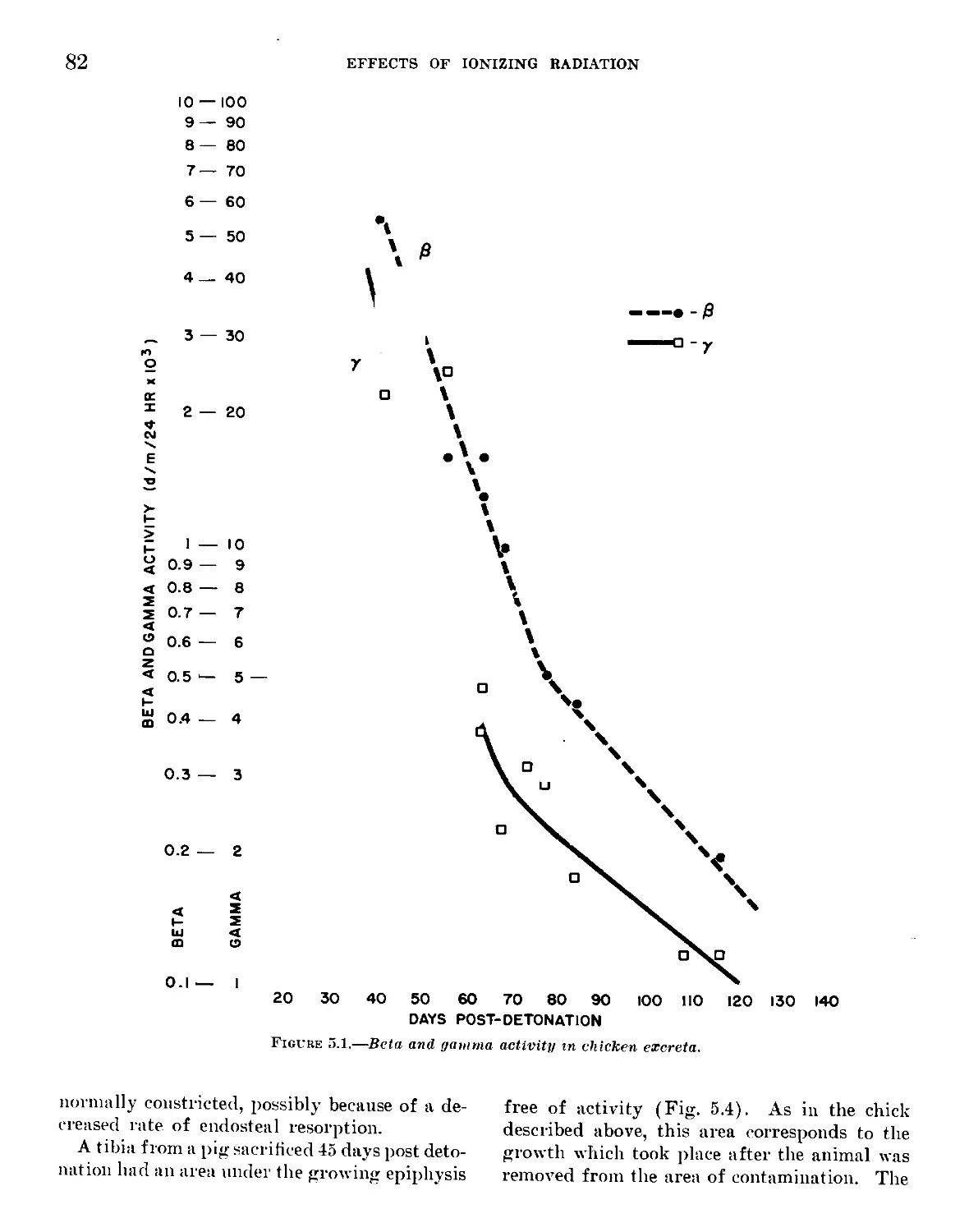
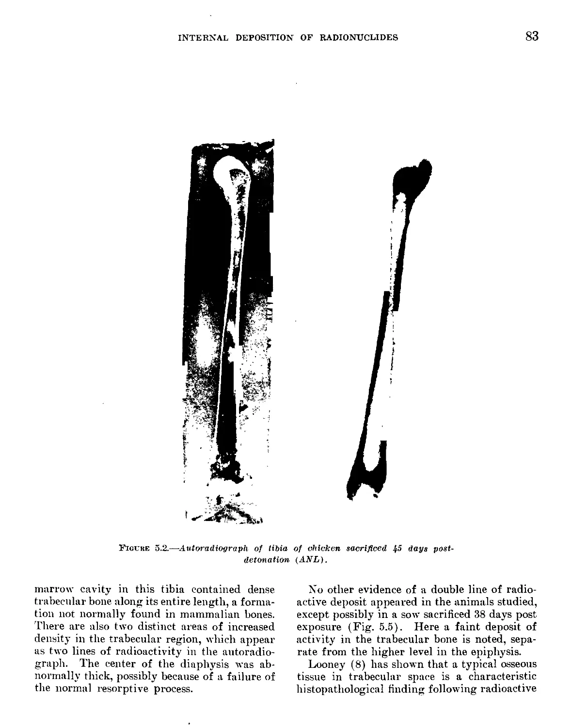
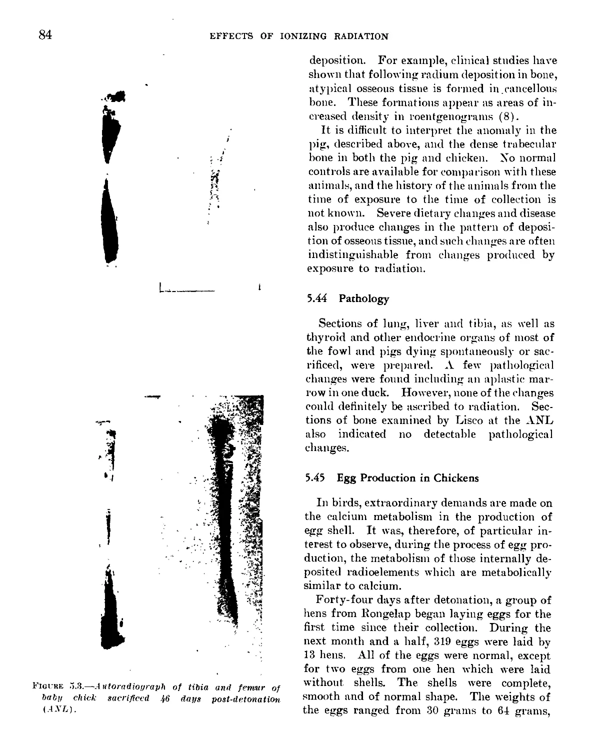


Text
SOME EFFECTS OF
Ionizing
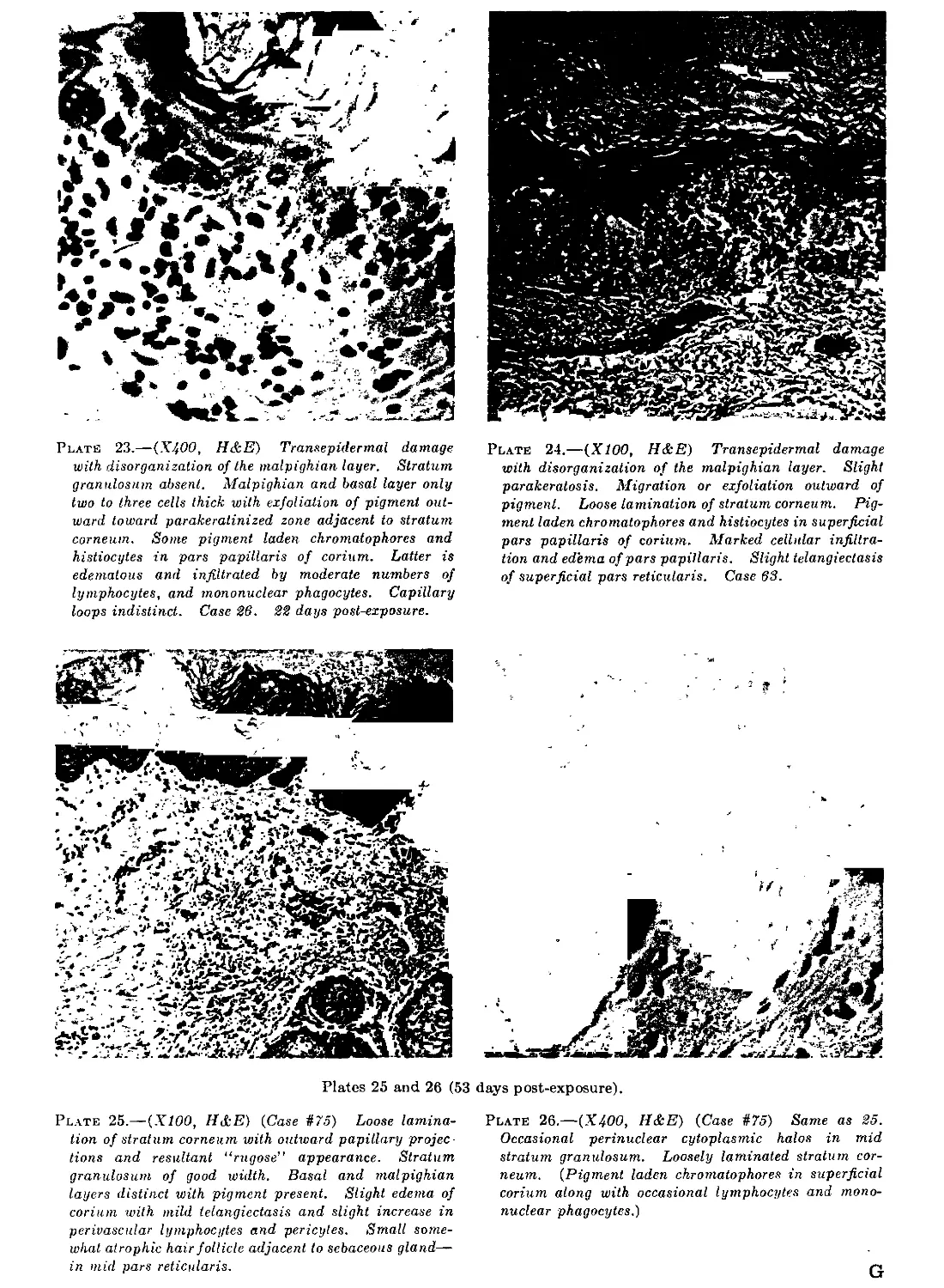
Raciation
ON HUMAN BEINGS
from the
Naval Medical Research Institute
Bethesda 14, Maryland
U. S. Naval Radiological Defense
Laboratory
San Francisco, California
and
Medical Department
Brookhaven National Laboratory
Upton, New York
Edited by
E. P. Cronkite
V. P. Bond
and C. L. Dunham
REPOSITORY Sa/z- RECORDS
COLLECTION MARSHALL. JScAA/QS
The Medical Research Center
Brookhaven National Laboratory
Upton, L. I., New York 401022
A Report on the
Marshallese and Americans
Accidentally Exposed to Radiation
from Fallout and a Discussion of
Radiation Injury in the
Human Being
UNITED STATES
ATOMIC ENERGY COMMISSION
JULY 1956
BOXNo. Helical DEPT. Pveu.cATiorts
FOLDER * HO “ 1^2.
TID 5358
For sale by the Superintendent of Documents, U. S. Government Printing Office,
Washington 25, D. C.
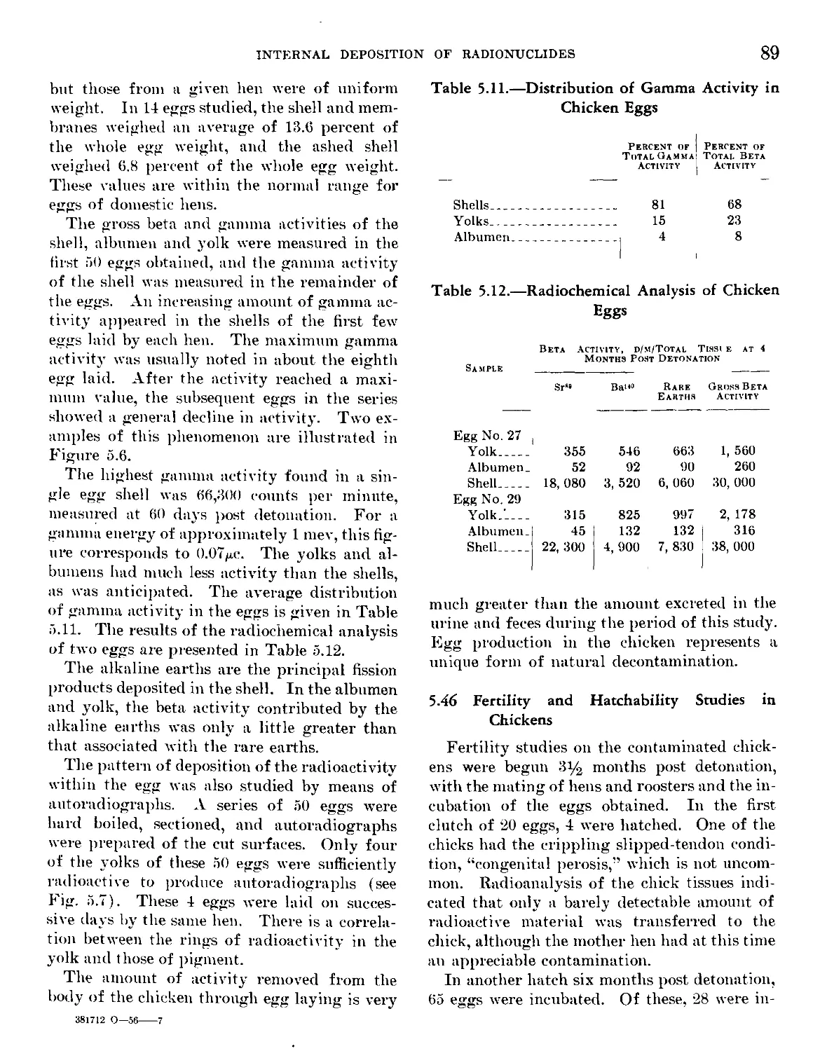
Introduction
On March 1, 1954, an experimental thermo-
nuclear device was exploded at the U. S. Atomic
Energy Commission’s Eniwetok Proving
Grounds in the Marshall Islands. Following
the detonation, unexpected changes in the wind
structure deposited radioactive materials on
inhabited atolls and on ships of Joint Task
Force #7, which was conducting the tests.
Radiation surveys of the areas revealed injuri-
ous radiation levels; therefore, evacuation was
ordered, and was carried out as quickly as pos-
sible with the facilities available to the Task
Force.
Although the calculated accumulated doses to
the exposed human beings were believed to be
well below levels that would produce serious
injury or any mortality, the Commander of the
Task Force requested the Department of De-
fense and the U. S. Atomic Energy Commis-
sion to organize a medical team to provide the
best possible care of the exposed persons and to
make a medical study of the exposures.
Responsibility for organization of the medi-
cal team was shared by the Armed Forces Spe-
cial Weapons Project, Department of Defense,
and the Division of Biology and Medicine, U. S.
Atomic Energy Commission. Experienced
professional and technical personnel were im-
mediately available from the Naval Medical Re-
search Institute and the U. S. Naval Radiologi-
cal Defense Laboratory. Since speed was es-
sential in the organization and transport of the
medical team to the mid-Pacific area, the as-
sistance of the Medical Department of the Navy
was requested, and was promptly received from
the Surgeon General.
A team was organized from personnel of the
two Navy laboratories and representatives of the
AEC Division of Biology and Medicine and the
Armed Forces Special Weapons Project. The
team was air lifted to the Marshall Islands,
arriving on the eighth day after the explosion.
Interim care and study had been capably
handled by the small medical department of the
U. S. Naval Station, Kwaj alein, Marshall Is-
lands The commander of the naval station had
arranged living facilities for the exposed Mar-
shallese, and installed laboratory and clinical
facilities as requested immediately upon arrival
of the medical team.
Full cooperation and support from all agen-
cies in the field enabled the medical team to
operate at maximum efficiency, so that the de-
gree of radiation injury could be assessed
quickly, and appropriate care and study of the
injured could be instituted without delay. All
of the exposed individuals have recovered from
the immediate effects without serious sequelae.
Nevertheless it is planned to evaluate the
medical and genetic status of the group at ap-
propriate intervals with a view to learning
what if any of the known late effects of radia-
tion exposure may be observed. Obviously and
indeed fortunately the number of persons re-
ceiving 75 roentgens exposure and greater is
too small to make it possible to determine with
any degree of accuracy the effect on life span.
In addition to providing medical care for
these persons, the team accumulated a large
body of scientific observations on radiation in-
jury in human beings. The initial data have
been supplemented by field resiirveys 6, and 24
months after the original investigation.
The results of this work are summarized in
the present volume. The data which were ob-
tained substantially increase the fundamental
knowledge of radiation injury and the medical
capability of caring for persons exposed to
large doses of radiation.
Charles L. Dunham, M. D., Director,
Division of Biology and Medicine,
U. S. Atomic Energy Commission.
in
Preface and Acknowledgments
The Undertaking of the care and study of the
human beings accidentally exposed to fallout
radiation following the March 1, 1954, nuclear
test detonation in the Pacific represented the
first instance in which study of a large group
of irradiated human beings was possible soon
after exposure. Although the physical esti-
mates of dose received by the individuals ex-
posed to fallout radiation were thought to be
sublethal. precise knowledge of the relative
sensitivity of human beings to penetrating ion-
izing radiation was lacking. Accordingly, in
addition to the initial medical team, provisions
were made for a second echelon of specialized
personnel in case they were needed. A pre-
ventative medicine unit of the Commander-in-
Chief, Pacific fleet, was alerted for possible bac-
teriological studies; blood bank personnel, and
additional clinicians and nurses were notified
in case conditions justified their services in the
Kwajalein area. Rear Admiral Bartholomew
Hogan, MC, USN, Pacific Fleet Medical Offi-
cer,* promised full support of all the medical
facilities of the Pacific Fleet were they deemed
necessary. With the preceeding planning it
was felt that any medical problem, regardless
of the severity, could be promptly and ade-
quately handled in the field.
The personnel for the team were obtained
within the continental limits of the United
States from the Naval Medical Research In-
stitute and the United States Naval Radiologi-
cal Defense Laboratory. From the former, four
medical officers, E. P. Cronkite, R. A. Conard,
N. R. Shulman, and R. S. Farr were obtained.
Two Medical Service Corps officers, W. H.
Chapman and Robert Sharp, were also ob-
tained from the same institution. In addition,
six enlisted men, C. R. Sipe, HMC. USN; P.
K. Schork, HMC, USN; С. P. A. Strome, HMC,
USN; W. C. Clutter, HM, 1/C; R. E. Hansell,
•Now Surgeon General, U. S. Navy.
IV
HM 1/C; and J. S. Hamby, HM, 2/C were
provided. From the United States Naval Ra-
diological Defense Laboratory, one civilian
physician, Doctor V. P. Bond; one medical
service corps officer, Lt. Com. L. J. Smith; and
four enlisted men, W. H. Gibbs, HMC, USN;
J. C. Hendrie, HM, 1/C; W. S. Argonza, HM,
2/C; and J. Flannagan, HM, were supplied.
The Division of Biology and Medicine, Atomic
Energy Commission, sent two civilian physi-
cians, Dr. C. L. Dunham then Chief of the
Medical Branch and Dr. G. V. LeRoy, Con-
sultant and Special Representative of the Di-
rector of the Division. The Armed Forces Spe-
cial Weapons Project supplied one Army medi-
cal officer, Lt. Col. L. E. Browning, MC, USA.
All personnel were experienced in the study
of radiation injury.
The preliminary studies performed by the
Medical Department of the Naval Station at
Kwajalein were under the direction of Com-
mander W. S. Hall, MC, USN, the station
medical officer and his small staff who are to
be commended for an excellent job.
Upon arrival of the medical team, it became
quite evident that, because of the large numbers
of radiation casualties and the huge amount of
work involved in collecting data, that primary
responsibilities for various phases of the study
would have to be delegated in order to obtain
the necessary information for biological assay
of the degree of injury. In the initial phase,
hematological surveys and establishment of
clinical records on each individual were empha-
sized. Dr. V. P. Bond organized and ana-
lyzed the results of the daily blood studies. Lt.
N. R. Shulman, MC, USN, with the capable
assistance of Mr. John Tobin, anthropologist of
the Trust Territory, and Kathleen Emil, Mar-
shallese nurse, as interpreters, undertook the
establishment of medical histories and initial
physical examinations. As the clinical picture
PREFACE AND ACKNOWLEDGMENTS
V
unfolded, daily sick call and care of the radia-
tion lesions were carried out by Doctor Shul-
man along lines decided in general conference
of the entire group. When epilation and skin
lesions appeared, Commander R. A. Conard,
MC, USN. was assigned primary responsibility
for documentation of the onset, incidence, and
detailed description of the skin lesions. During
the field phase, Lt. Robert Sharp, MSC, USN,
was given the responsibility for decontamina-
tion and collection of data from all sources on
the radiation intensities of the contaminated
atolls and the calculation of probable doses of
radiation received. Paul K. Schork. HMC.
USX, was in charge of the Hematology Labora-
tory. The services of Doctor S. H. Cohn were
requested, and made available by USNRDL to
undertake a field study of the degree of internal
contamination, in addition to the studies that
were to be performed on urine samples returned
to the Los Alamos Scientific Laboratory, New
York Operations Office of the Atomic Energy
Commission, and the USNRDL.
The authors wish to express their gratitude
and indebtedness in particular to Doctor John
C. Bugher, then Director of the Division of
Biology and Medicine, Atomic Energy Com-
mission, who came to the forward area and was
always available for counsel. In addition Cap-
tain Van Tipton, MC, USN, Director of Atomic
Defense Division of the Bureau of Medicine
and Surgery, Department of the Navy; Com-
mander Harry Etter, MC, USN; Captain W. E.
Kellum, MC, USN; and Captain T. L. Willmon,
Commanding and Executive Officers respec-
tively of the Naval Medical Research Insti-
tute: Captain R. A. Hinners, USN, Director
USNRDL. and Captain A. R. Behnke, MC,
USN, Associate Director NRDL; gave unlim-
ited support and reduced administrative pro-
cedures to a bare minimum, thus making it pos-
sible for the unit to be assembled and underway
in a matter of hours.
Upon arrival at Kwajalein, Rear Admiral
R. S. Clarke, USN, Commanding Officer United
States Naval Station, Kwajalein, supported the
project with all of the facilities at his disposal.
As a result, a laboratory and clinic was estab-
lished and operating within 24 hours after ar-
rival of the medical team.
In addition, we wish to acknowledge the out-
standing contributions of Col. C. S. Maupin,
MC, USA, Field Command Armed Forces Spe-
cial "Weapons Project; Captain H. H. Haight,
MC, USN, Division of Military Application,
Atomic Energy Commission; Dr. Gordon Dun-
ning, Division of Biology and Medicine. Atomic
Energy Commission; and Dr. H. Scoville of
Armed Forces Special Weapons Project who in
addition to their primary duties, collected ex-
tensive data in the field on the radiation intensi-
ties of the atolls and kindly furnished this ma-
terial to the project personnel. Drs. T. L. Ship-
man, Thomas White,* and Payne Harris of the
Los Alamos Scientific Laboratory kindly fur-
nished very valuable data on urinary excretion
of radionuclides. The early studies of the Los
Alamos group in particular contributed sig-
nificantly to the information on the degree and
nature of internal deposition of short lived
radionuclides. Dr. G. V. LeRoy, Associate
Dean, School of Biological Sciences, University
of Chicago, participated in the early phase of
the study as a consultant to the Medical Group.
The authors of Chapter I are particularly in-
debted to Dr. C. S. Cook and the Nuclear Radia-
tion Branch at the Navy Radiological Defense
Laboratory for information on energy distri-
bution of the gamma radiation. Data on radio-
chemical and radioactive decay rates were sup-
plied by Dr. C. F. Miller and the Chemical
Technology Division of USNRDL and Dr. R.
W. Spense of Los Alamos Scientific Laboratory.
In collecting data on the skin lesions, the
help of Billiet Edmond, Marshallese school
teacher for the Rongelap group in interpreta-
tion was invaluable. Miss Patricia Roan of
USNRDL prepared the histologic prepara-
tions of the skin biopsies and Mr. William Mur-
ray and George Needum of USNRDL and
С. P. A. Strome, HMC, USN, Naval Medical
Research Institute performed the excellent
color photography.
In preparation of the material and writing of
Chapter V, the authors are indebted to Miss C.
* Deceased.
VI
PREFACE AND ACKNOWLEDGMENTS
Jones of USNRDL, who prepared the autora-
diographs of the tissues. In addition, Dr. W. P.
Norris of Argonne National Laboratory made
autographs of specific tissues. Dr. Rachael
Reed of USNRDL performed the microscopic
pathological studies of the tissues from the ani-
mals in whom radioisotopes were deposited in-
ternally. Lt. Col. R. J. Veenstra, VC, U. S.
Army, was in charge of the care of all the ex-
perimental animals collected in the field and re-
turned to the United States Naval Radiological
Defense Laboratory. Dr. E. R. Thompkins
made the facilities of the chemical technology
division of the USNRDL available and pro-
vided technical advice on the radiochemical
aspects of the project.
The continuous help and cooperation of the
Trust Territory representatives in particular,
Mr. Maynard Neass, District Administrator of
Majuro Atoll and their aid in obtaining the
necessary control data on Marshallese inhabi-
tants was indispensable to the success of this
study. Particular help was obtained from Mr.
John Tobin, the district anthropologist, whose
knowledge of the Marshallese language and
habits, in addition to services as an interpreter,
were invaluable.
The initial measurements on skin and cloth-
ing contamination were made by Lt. J. S.
Thompson, MC, USN, of V. P. 29 Squadron.
We are indebted to him for furnishing his rec-
ords on the contaminated individuals and the
initial decontamination that was performed by
his group.
The care and the study of these human beings
would not have been successful unless the
Marshallese had accepted the importance of
their being under careful medical observation
and of gathering medical data. At all times
these people were most pleasant, cooperative
and actively participated in the project. In
particular the project officer wishes to express
thanks to the Magistrates of the groups, to the
Marshallese health aids, school teachers, and
nurses.
It is quite impossible to acknowledge the
assistance of the numerous individuals in vari-
ous agencies who assisted in collection of data
and editing of the various chapters. The Pro-
ject Officer wishes to commend all of the pro-
fessional and technical members of the group
for their excellent motivation, initiative, and
voluntary long hours of extra work that were
essential for the accomplishment of the clinical
and research objectives and the rapid collection
of the preliminary data in the field. It is
quite evident that the entire study of the ex-
posed individuals was a cooperative endeavor
involving numerous activities, and that it would
have been impossible except for the splendid
spirit of unselfish cooperation by all concerned.
The fine team work of the group itself made
it possible for realistic daily reports on all of
the above phases to be forwarded daily to re-
sponsible agencies and thus keep authorities
informed of the course and severity of events
following this untoward and unavoidable acci-
dent.
Upon completion of the initial phase of the
study, primary responsibility for writing re-
ports on the variouse phases was delegated as
follows: C. A. Sondhaus, dosimetry; N. R.
Shulman, clinical course and care; R. A. Con-
ard, skin lesions; V. P. Bond, hematology; S.
II. Cohn, internal deposition.
The final publication of this monograph on
human radiation injury represents the comple-
tion of the finest in cooperation and team work
of a diverse group who willingly sacrificed
personal ambitions and desires for the good
of the project at large. It was a distinct privi-
lege to be chosen to direct the medical team,
a real pleasure to edit and integrate the sepa-
rate reports and finally realize their fruition
as a homogeneous monograph.
E. P. Cronkite, M. D.,
Medical Department,
Brookhaven National Laboratory,
Upton, New York.
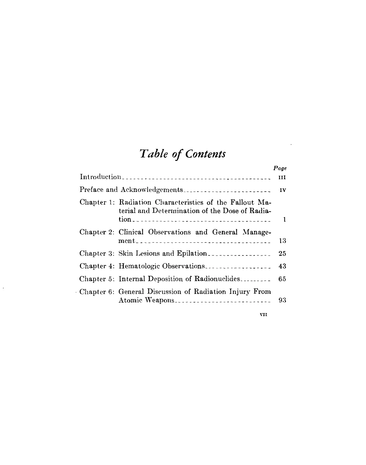
Table of Contents
Page
Introduction___________________________________________ in
Preface and Acknowledgements___________________________ iv
Chapter 1: Radiation Characteristics of the Fallout Ma-
terial and Determination of the Dose of Radia-
tion _________________________________________________ 1
Chapter 2: Clinical Observations and General Manage-
ment___________________________________________________ 13
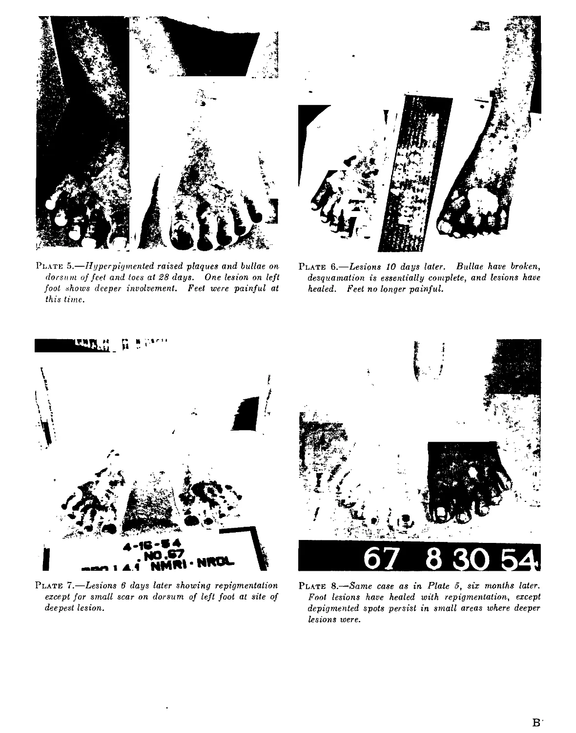
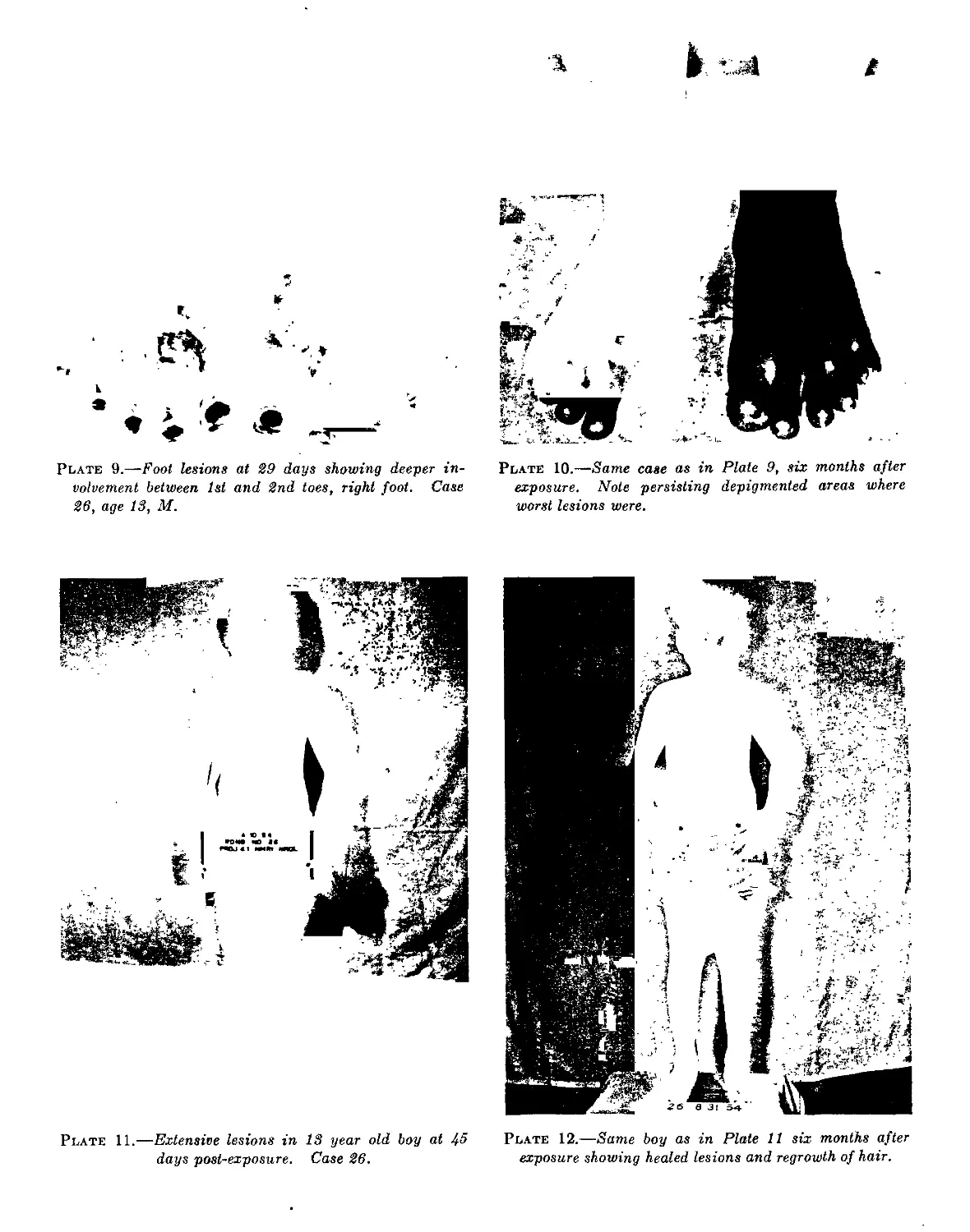
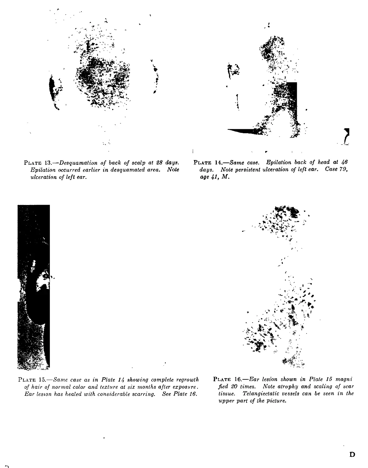
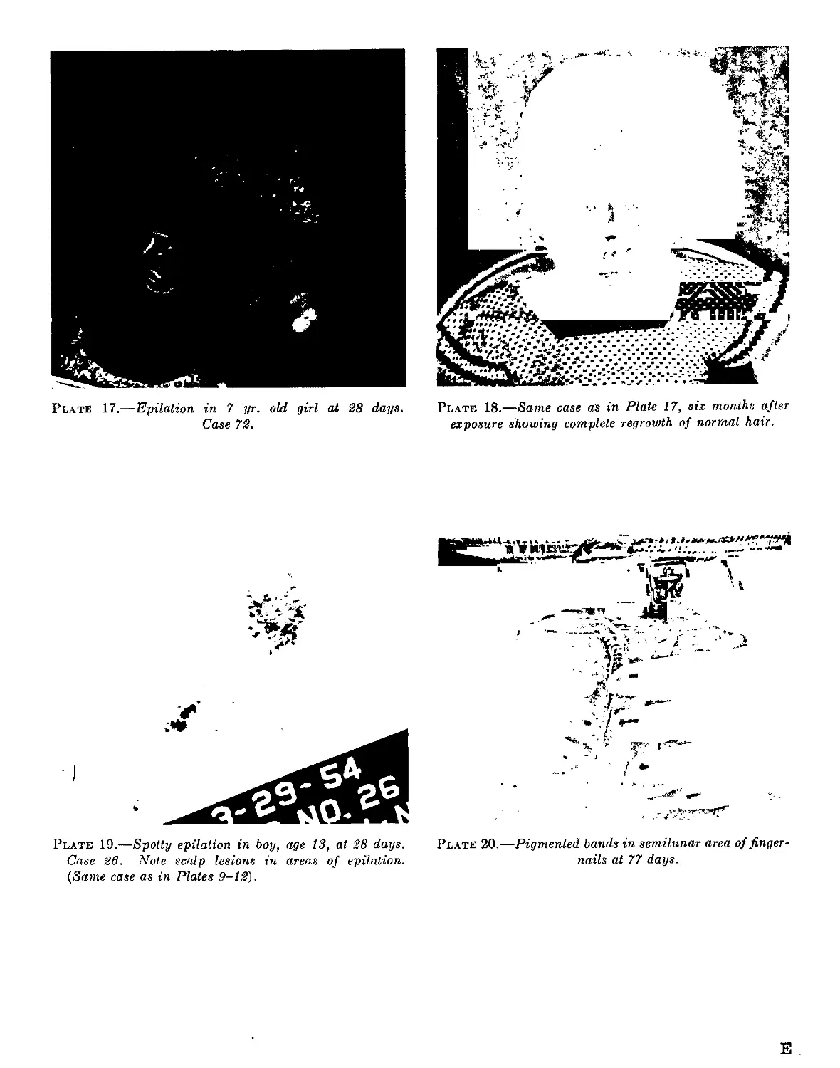
Chapter 3: Skin Lesions and Epilation_________________ 25
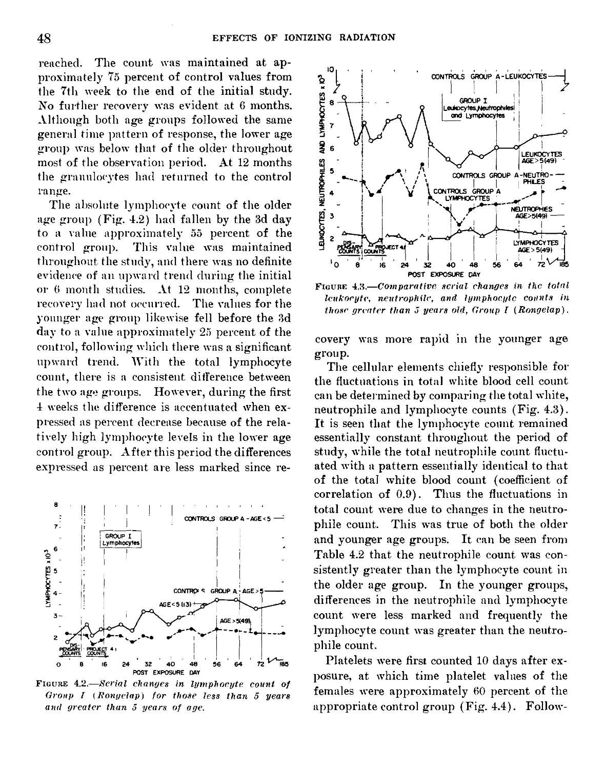
Chapter 4: Hematologic Observations____________________ 43
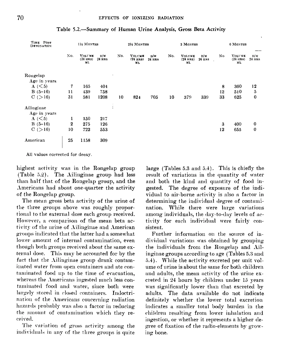
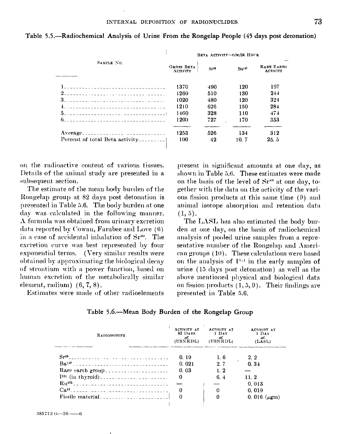
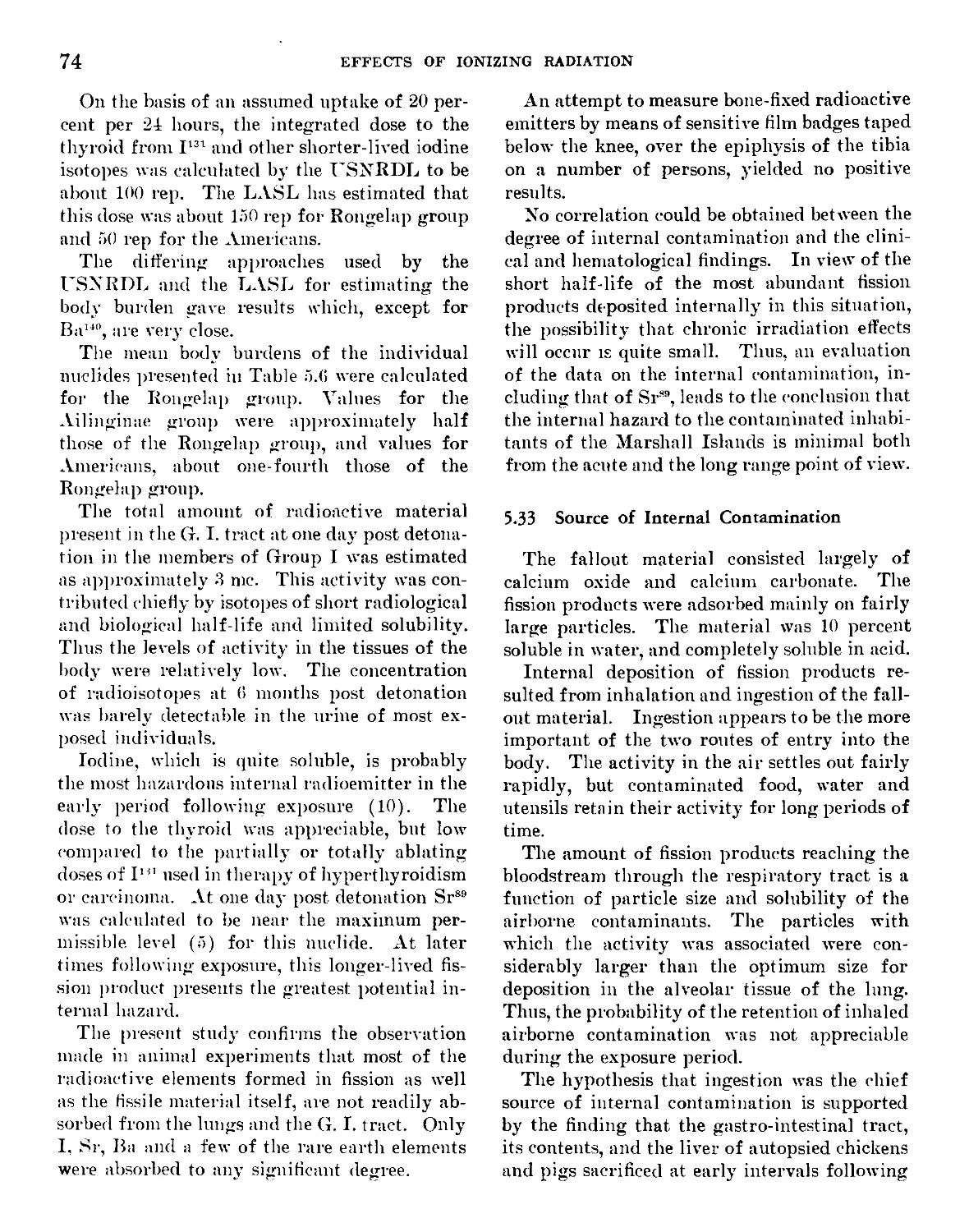
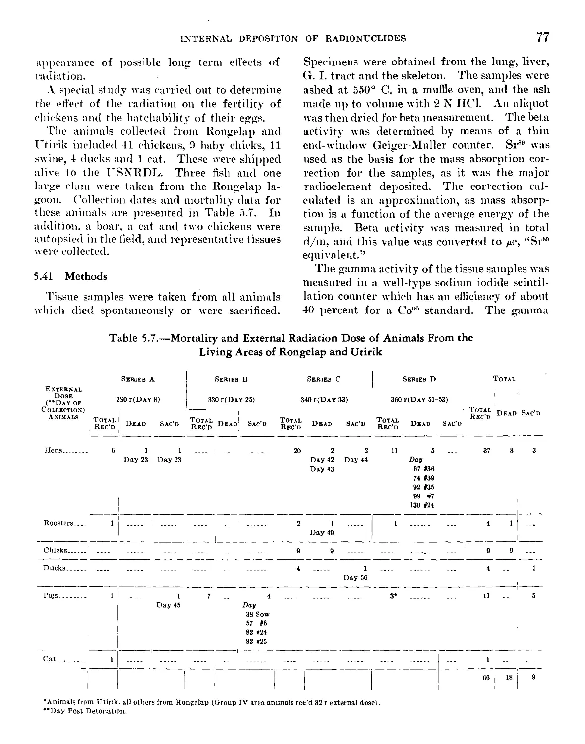
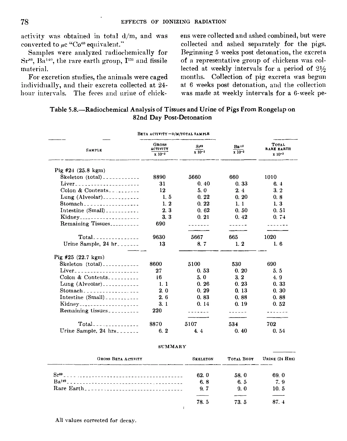
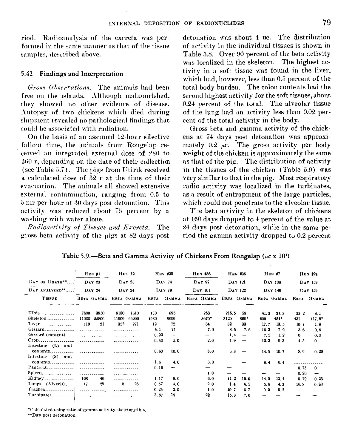
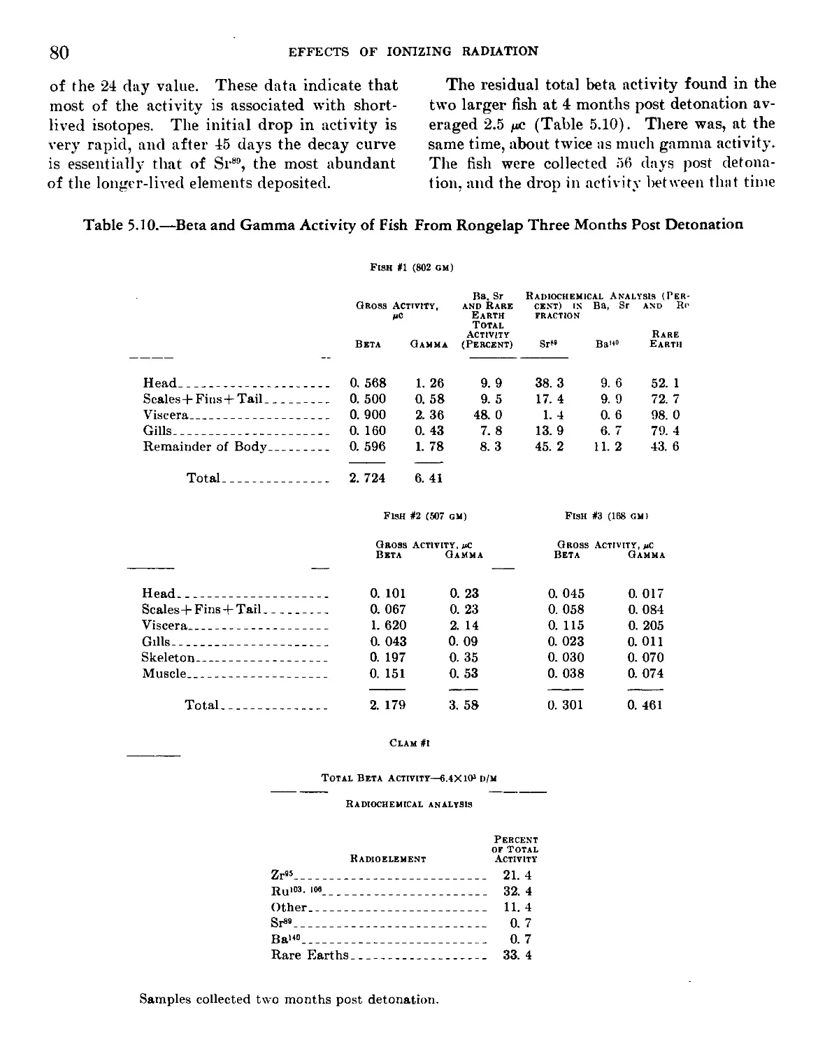
Chapter 5: Internal Deposition of Radionuclides_______ 65
Chapter 6: General Discussion of Radiation Injury From
Atomic Weapons_________________________________________ 93
VII
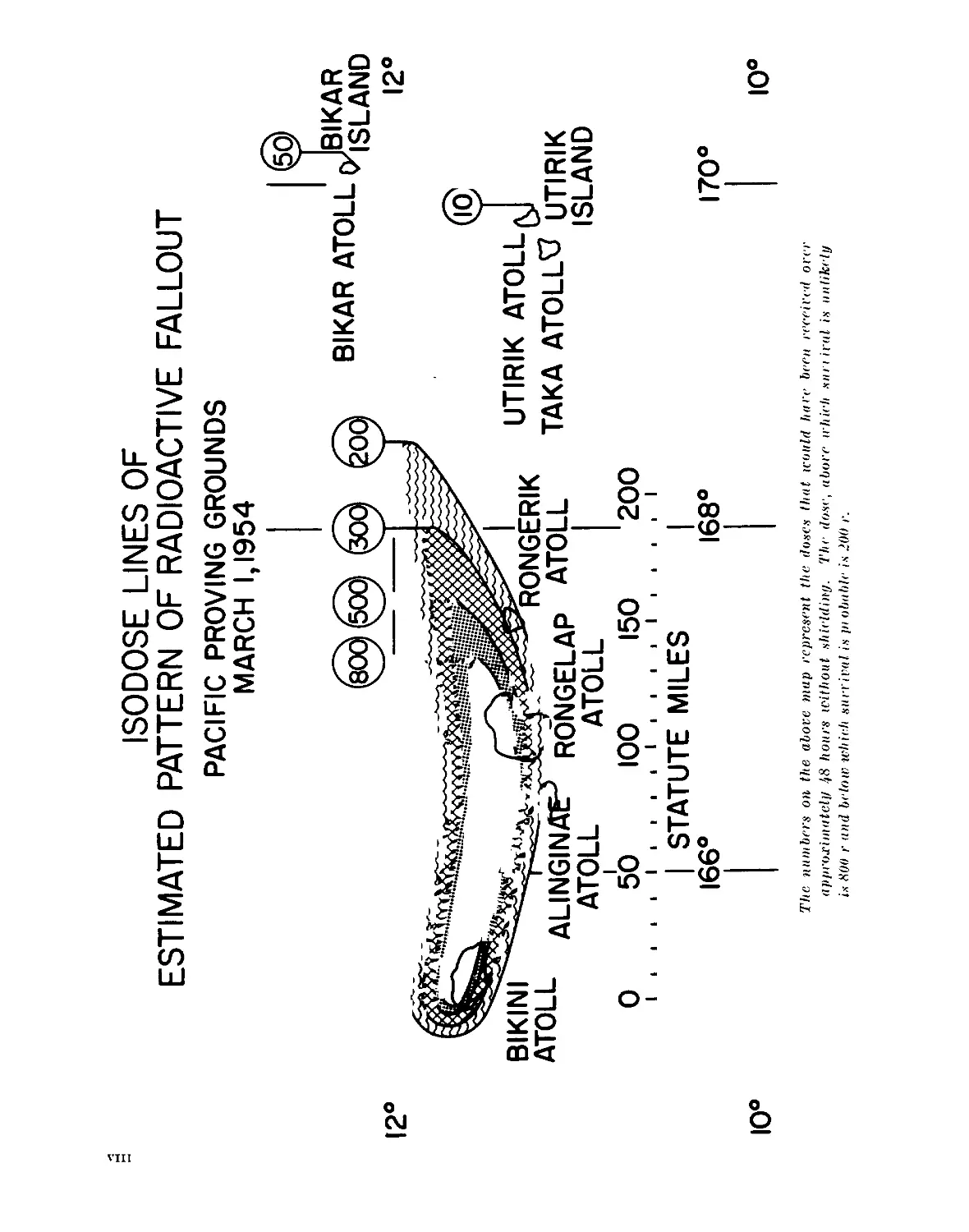
ISODOSE LINES OF
ESTIMATED PATTERN OF RADIOACTIVE FALLOUT
PACIFIC PROVING GROUNDS
12°
MARCH 1,1954
О 50 100 150 200
I ! ! ! I I I t I I 1 I I I i I I 1 l i I
| STATUTE MILES |
166° 168° 170°
The numbers on the above map represent the doses that would hare been received over
approximately !)8 hours il'ithout shielding. The dose, abort’ irhich sari iral is unlikely
is 800 r and below which survival is pi obuble is >00 r.
Chapter I
Radiation Characteristics of the Fallout Material and the
Determination of the Dose of Radiation
C. A. SoNDHAUS
Robert Sharp, Lt. (jg) MSC USN
V. P. Bond, M. D., Ph. D.
E. P. Cronkite, Cdr. (MC) USN
Outline
1.1 Nature of the Event and Description of the
Exposed Groups.
1.2 Whole-Body Gamma Doses.
1.21 Characteristics of the Radiation.
1.22 Duration of the Exposures.
1.23 Geometry of the Exposures.
1.3 Estimation of the Doses From Beta and
Soft Gamma Radiation.
1.4 Summary.
1.1 Nature of the Event and Description of the Exposed Groups
Following the Detonation of a nuclear de-
vice at the Pacific Proving ground in the Spring
of 1951, significant amounts of radioactive ma-
terial fell on neighboring populated atolls.
The Marshallese inhabitants of Rongelap atoll
(designated as Group I) received the highest
calculated dose of radiation. Some of the
Rongelap people were located temporarily on
Ailinginae atoll from the time of the fallout
until they were evacuated (Group II). Their
calculated dose was smaller than that of the
other members of the parent group. The
American service men (Group III) were lo-
cated on Rongerik atoll. The largest group of
Marshallese (Group IV) were located onUtirik
atoll and received the smallest dose. The Mar-
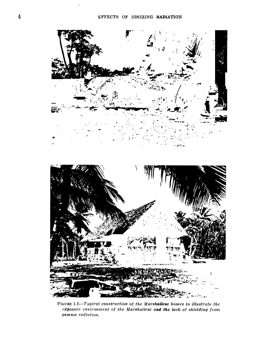
shallese were living under relatively primitive
conditions in lightly constructed palm houses
(Fig. 1.1).
The American military personnel had the
second highest exposure. They were more
aware of the significance of the fallout than
were the Marshallese, and promptly put on ad-
ditional clothing to protect their skin. As far
as duties would permit, they remained inside of
aluminum buildings. In contrast, most of the
Marshallese remained out-of-doors and thus
were more heavily contaminated by the ma-
terial falling on the atolls. Some of the
Marshallese, however, went swimming during
the fallout and many of the children waded in
the water, thus washing a considerable amount
of the material from their skin.
The exposed personnel were evacuated to
Kwajalein by air and surface transportation.
Since a survey of all individuals showed that
there was significant contamination of skin, hair
and clothes, prompt decontamination was in-
stituted. Clothes were removed and laundered
and repeated washings of the skin and hair
with fresh water and soap were carried out. In
many of the Marshallese, it was difficult to wash
the radioactive material from the hair because
of the heavy coconut-oil hair dressing.
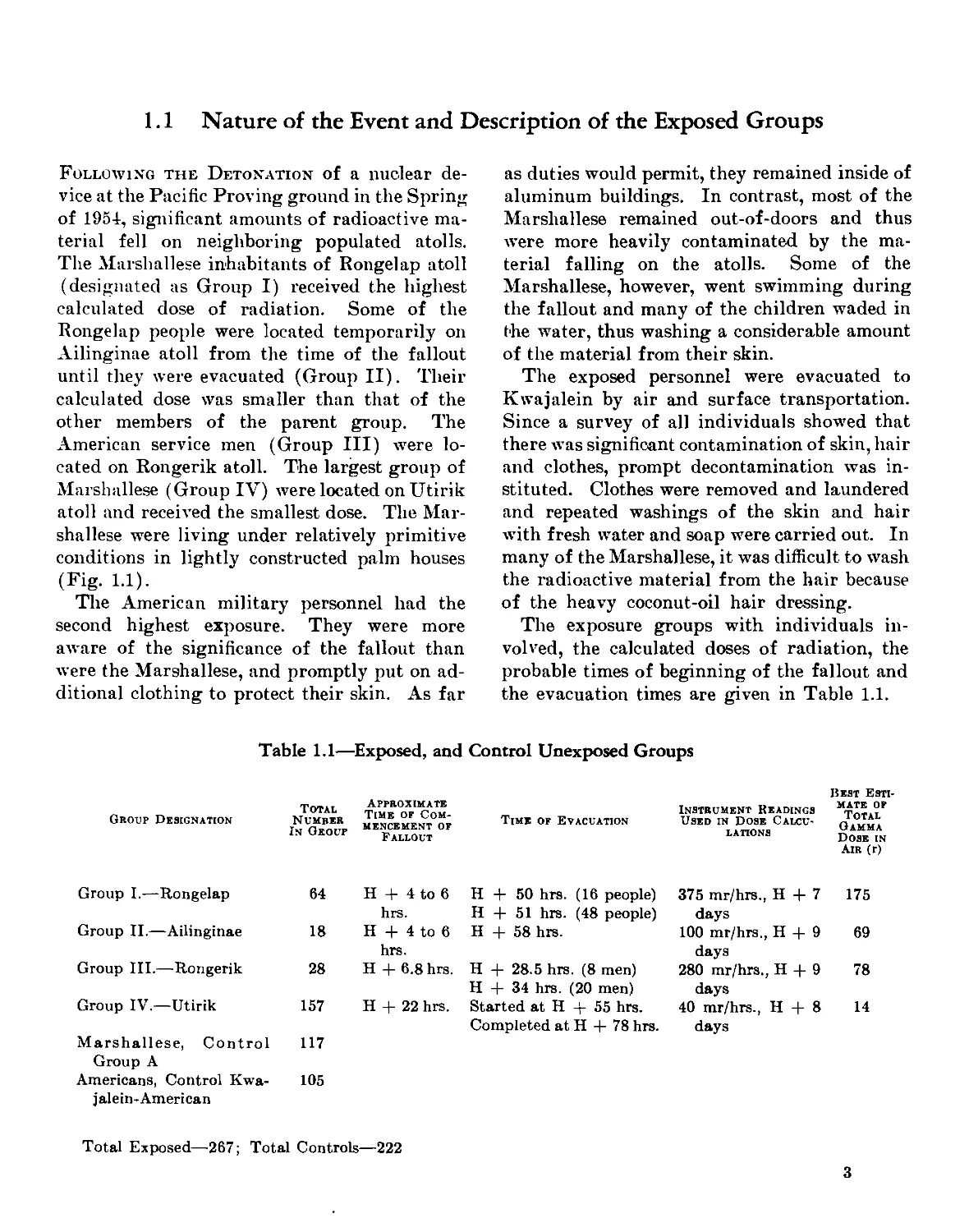
The exposure groups with individuals in-
volved, the calculated doses of radiation, the
probable times of beginning of the fallout and
the evacuation times are given in Table 1.1.
Table 1.1—Exposed, and Control Unexposed Groups
Group Designation
Total
Number
In Group
Approximate
Time of Com-
mencement of
Fallout
Time of Evacuation
Instrument Readings
Used in Dose Calcu-
lations
Best Esti-
mate of
Total
Gamma
Dose in
Air (r)
Group I.—Rongelap 64 H + 4 to 6 H + 50 hrs. (16 people) 375 mr/hrs., H + 7 175
hrs. H + 51 hrs. (48 people) days
Group II.—Ailinginae 18 H + 4 to 6 H + 58 hrs. 100 mr/hrs., H + 9 69
hrs. days
Group III.—Rongerik 28 H + 6.8 hrs. H + 28.5 hrs. (8 men) 280 mr/hrs., H + 9 78
H + 34 hrs. (20 men) days
Group IV.—Utirik 157 H + 22 hrs. Started at H + 55 hrs. 40 mr/hrs., H + 8 14
Completed at H + 78 hrs. days
Marshallese, Control 117
Group A
Americans, Control Kwa- 105
jalein- American
Total Exposed—267; Total Controls—222
3
4
EFFECTS OF IONIZING RADIATION
Figure 1.1—Typical construction of the Marshallese homes to illustrate the
exposure environment of the Marshallese and the lack of shielding from
gamma radiation.
EVENT AND DESCRIPTION OF EXPOSED GROUPS
5
1.2 Whole Body Gamma Doses
The Estimated Values of external dose given
in Table 1.1 were calculated from readings of
radiation field survey instruments.* Averages
of a number of dose rate measurements on each
island at a given time were used. The read-
ings were taken in air, approximately three
feet above ground, several days after the inhab-
carried out, nor was its operating condition
known to be satisfactory under the emergency
condition prevailing at the time of use. For
these reasons the later readings, which were
higher than the early survey by an average of
50 percent when corrected to the same times,
were used in computing the doses listed. The
instruments used for the later measurements
ivere calibrated just prior to the surveys.
30
ORIGINAL SOURCE SPECTRUM
INFINITE PLANE, 3' IN AIR
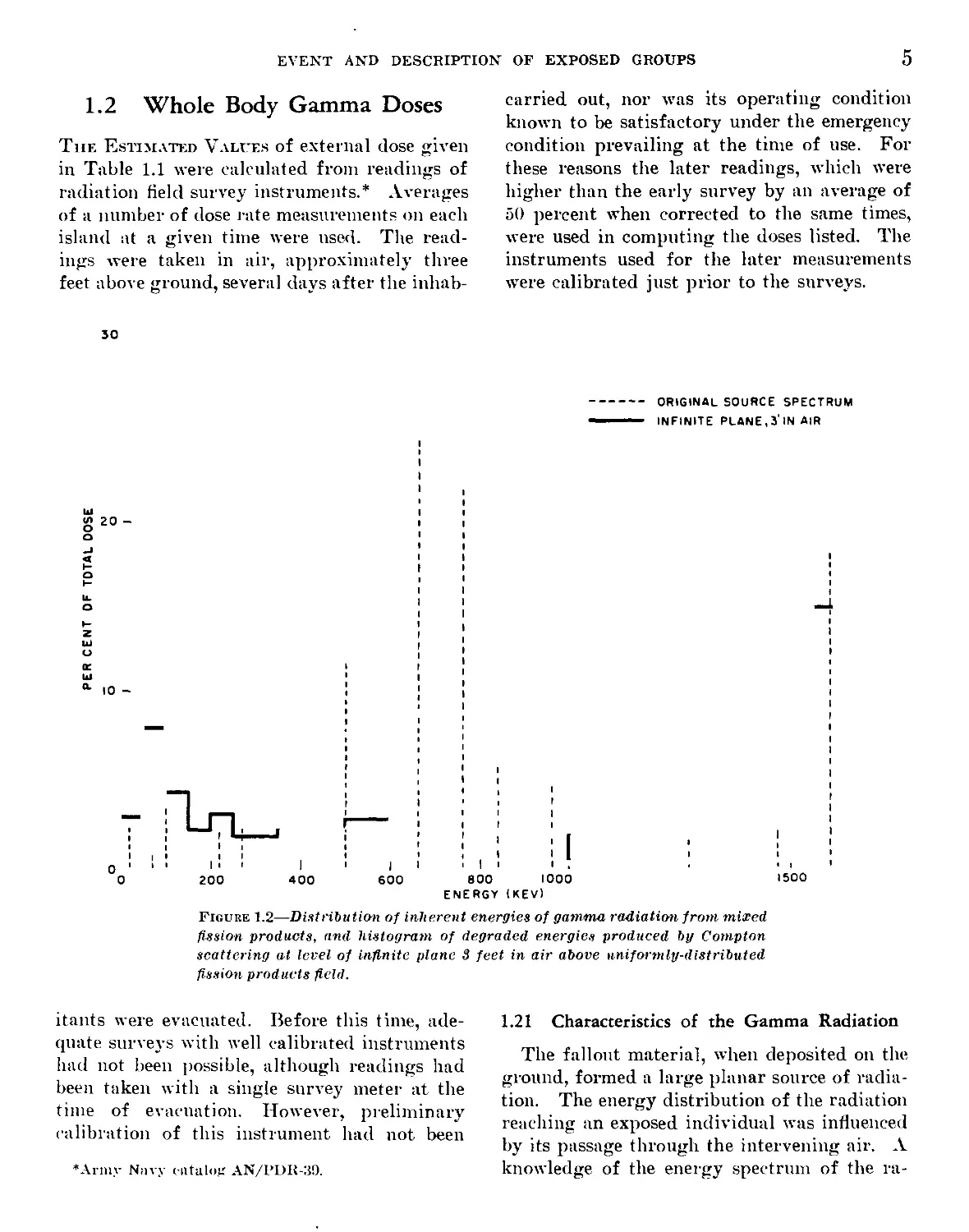
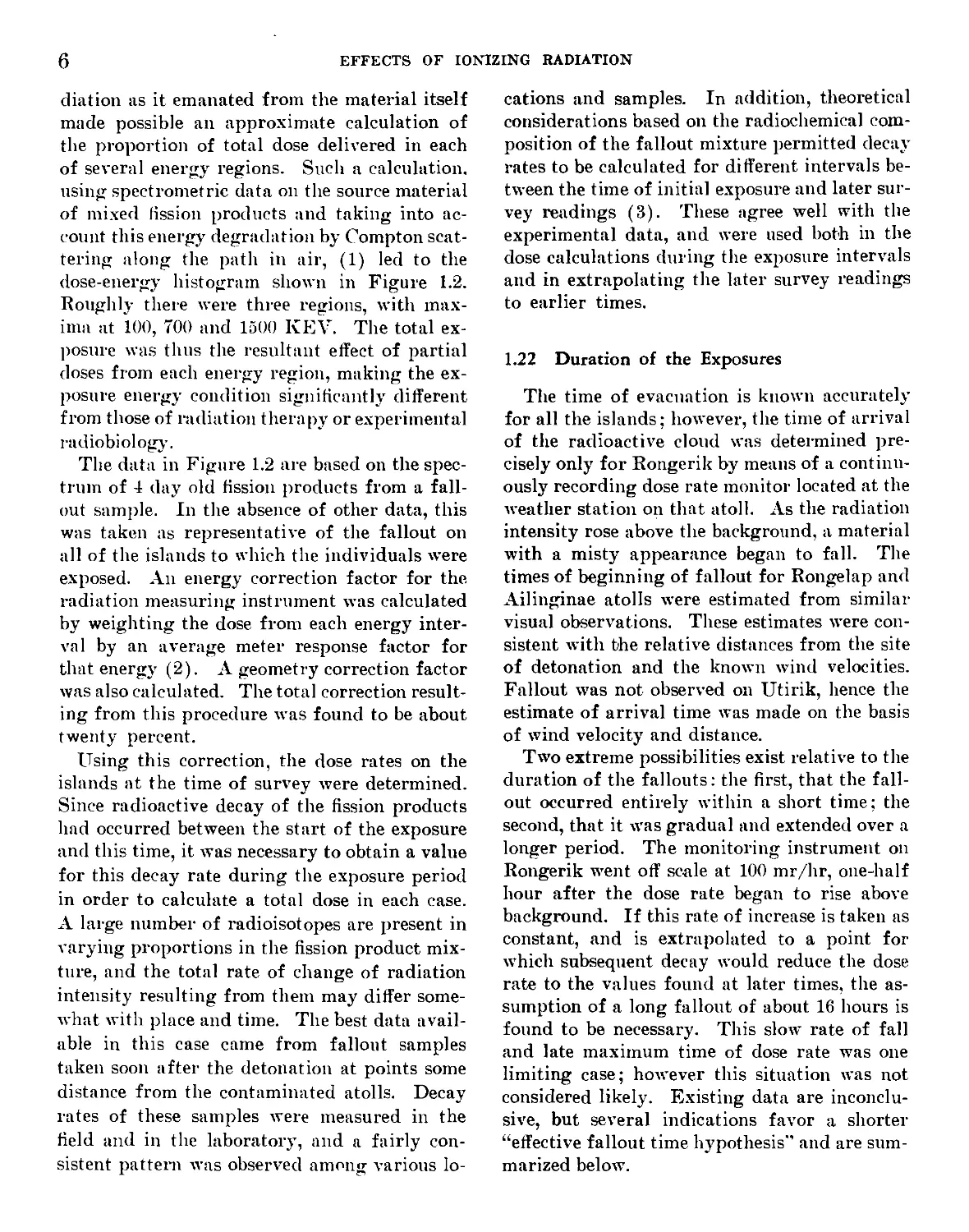
Figure 1.2—Distribution of inherent energies of gamma radiation from mixed
fission products, and histogram of degraded energies produced by Compton
scattering at level of infinite plane 3 feet in air above uniformly-distributed
fission products field.
I
i
1500
itants were evacuated. Before this time, ade-
quate surveys with well calibrated instruments
had not been possible, although readings had
been taken with a single survey meter at the
time of evacuation. However, preliminary
calibration of this instrument had not been
♦Army Navy catalog AN/I’DR-39.
1.21 Characteristics of the Gamma Radiation
The fallout material, when deposited on the
ground, formed a large planar source of radia-
tion. The energy distribution of the radiation
reaching an exposed individual was influenced
by its passage through the intervening air. A
knowledge of the energy spectrum of the ra-
6
EFFECTS OF IONIZING RADIATION
diation as it emanated from the material itself
made possible an approximate calculation of
the proportion of total dose delivered in each
of several energy regions. Such a calculation,
using spectrometric data on the source material
of mixed fission products and taking into ac-
count this energy degradation by Compton scat-
tering along the path in air, (1) led to the
dose-energy histogram shown in Figure 1.2.
Roughly there were three regions, with max-
ima at 100, 700 and 1500 KEV. The total ex-
posure was thus the resultant effect of partial
doses from each energy region, making the ex-
posure energy condition significantly different
from those of radiation therapy or experimental
radiobiology.
The data in Figure 1.2 are based on the spec-
trum of 4 day old fission products from a fall-
out sample. In the absence of other data, this
was taken as representative of the fallout on
all of the islands to which the individuals were
exposed. An energy correction factor for the
radiation measuring instrument was calculated
by weighting the dose from each energy inter-
val by an average meter response factor for
that energy (2). A geometry correction factor
was also calculated. The total correction result-
ing from this procedure was found to be about
twenty percent.
Using this correction, the dose rates on the
islands at the time of survey were determined.
Since radioactive decay of the fission products
had occurred between the start of the exposure
and this time, it was necessary to obtain a value
for this decay rate during the exposure period
in order to calculate a total dose in each case.
A large number of radioisotopes are present in
varying proportions in the fission product mix-
ture, and the total rate of change of radiation
intensity resulting from them may differ some-
what with place and time. The best data avail-
able in this case came from fallout samples
taken soon after the detonation at points some
distance from the contaminated atolls. Decay
rates of these samples were measured in the
field and in the laboratory, and a fairly con-
sistent pattern was observed among various lo-
cations and samples. In addition, theoretical
considerations based on the radiochemical com-
position of the fallout mixture permitted decay
rates to be calculated for different intervals be-
tween the time of initial exposure and later sur-
vey readings (3). These agree well with the
experimental data, and were used both in the
dose calculations during the exposure intervals
and in extrapolating the later survey readings
to earlier times.
1.22 Duration of the Exposures
The time of evacuation is known accurately
for all the islands; however, the time of arrival
of the radioactive cloud was determined pre-
cisely only for Rongerik by means of a continu-
ously recording dose rate monitor located at the
weather station on that atoll. As the radiation
intensity rose above the background, a material
with a misty appearance began to fall. The
times of beginning of fallout for Rongelap and
Ailinginae atolls were estimated from similar
visual observations. These estimates were con-
sistent with bhe relative distances from the site
of detonation and the known wind velocities.
Fallout was not observed on Utirik, hence the
estimate of arrival time was made on the basis
of wind velocity and distance.
Two extreme possibilities exist relative to the
duration of the fallouts: the first, that the fall-
out occurred entirely within a short time; the
second, that it was gradual and extended over a
longer period. The monitoring instrument on
Rongerik went off scale at 100 mr/hr, one-half
hour after the dose rate began to rise above
background. If this rate of increase is taken as
constant, and is extrapolated to a point for
which subsequent decay would reduce the dose
rate to the values found at later times, the as-
sumption of a long fallout of about 16 hours is
found to be necessary. This slow rate of fall
and late maximum time of dose rate was one
limiting case; however this situation was not
considered likely. Existing data are inconclu-
sive, but several indications favor a shorter
“effective fallout time hypothesis” and are sum-
marized below.
EVENT AND DESCRIPTION OF EXPOSED GROUPS
7
a. The estimated durations of fallout which
result from the above extrapolation of initial
fallout rate for Group I and III appear too
long to have occurred at the distances of
these people from the shot island, since the
wind velocity in the area was high enough
to move the cloud over the islands in a
considerably shorter time, as little as one-
half of the above indicated time.
b. The.accounts of the visibility of the fallouts,
although conflicting, do not indicate such
late cessation.
c. Doses calculated on a long fallout constant
rate of increase hypothesis are lower than
those due to a short fallout, since a short
fallout quickly deposits a large amount of
activity. For both a 16 hour and 8 hour
fallout assumption, a dose value was esti-
mated. The ranges are then as follows:
Table 1.2
Location Dose in г Fallout Time
16 hr 8 hr
Rongelap (Group I) 159 r 209 r
Ailinginae (Group II) 72 r 92 r
Rongerik (Group III) 70 r 106 r
Utirik (Group IV) 12 r 15 r
On Rongerik (Group III) a set of film badge
readings were obtained which constitute the
only direct evidence of total dose. Several
badges worn both outdoors and inside lightly
constructed buildings .on the island read
about 50 to 65 r, and one badge which re-
mained outdoors over the 28.5 hour period
read 98 r. Another group of badges, kept
indoors inside a steel refrigerator, read 38 r.
These dose values represent a variety of
conditions, but, considering the shielding
and attenuation factors, are consistent with
the assumption that the dose outside during
the first 28.5 hours after the beginning of
the fallout corresponded to about 12 hours
of constant fallout.
d. For Utirik atoll Group IV, only a fallout
time of about 12 hours or less is consistent
with the later dose rates observed, provided
the fallout actually began as late as was
estimated from wind and distance factors.
e. A long fallout probably would not be uni-
formly heavy throughout, the first portion
being the most intense and the balance de-
creasing with time. The total phenomenon
would thus tend toward the effect of a
shorter fallout. This is supported by moni-
tor data from other nuclear events, where
initially heavy fallout is reported to produce
a peak of air-borne radioactivity soon after
arrival, with the airborne activity level then
decreasing. The latter part of the fallout,
though still detectable as dust, may then
produce only a small fraction of the total
dose from material on the ground. Hence
the total dose may be estimated fairly ac-
curately by assuming a constant fallout to
have been complete in a much shorter
“effective” time.
The dose values given in Table 1.1, based on
film badge, meter and monitor data, are con-
sistent with a constant fallout hypothesis of
about 12 hours effective time.* One exception
is made; the dose values for Group III are about
75 percent of the 12 hour fallout value, averaged
for 28.5 and 34 hour exposures. This was felt
to express most accurately the average air dose
received by personnel who spent roughly half
their time inside structures where the dose rate
was later found to be roughly half that out-
doors. On the other islands such shielding was
not available.
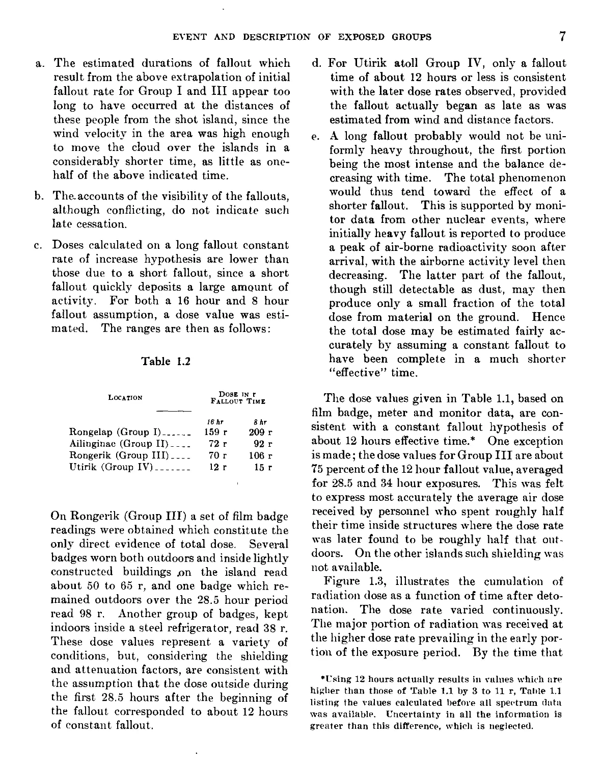
Figure 1.3, illustrates the cumulation of
radiation dose as a function of time after deto-
nation. The dose rate varied continuously.
The major portion of radiation was received at
the higher dose rate prevailing in the early por-
tion of the exposure period. By the time that
•Using 12 hours actually results in values which are
higher than those of Table 1.1 by 3 to 11 r, Table 1.1
listing the values calculated before all spectrum data
was available. Uncertainty in all the information is
greater than this difference, which is neglected.
8
EFFECTS OF IONIZING RADIATION
90 percent of the dose had been received, the
dose rate had fallen to less than 40 percent of
its initial value. Thus the dose rate also dif-
fered from the usual constant rate in the
laboratory.
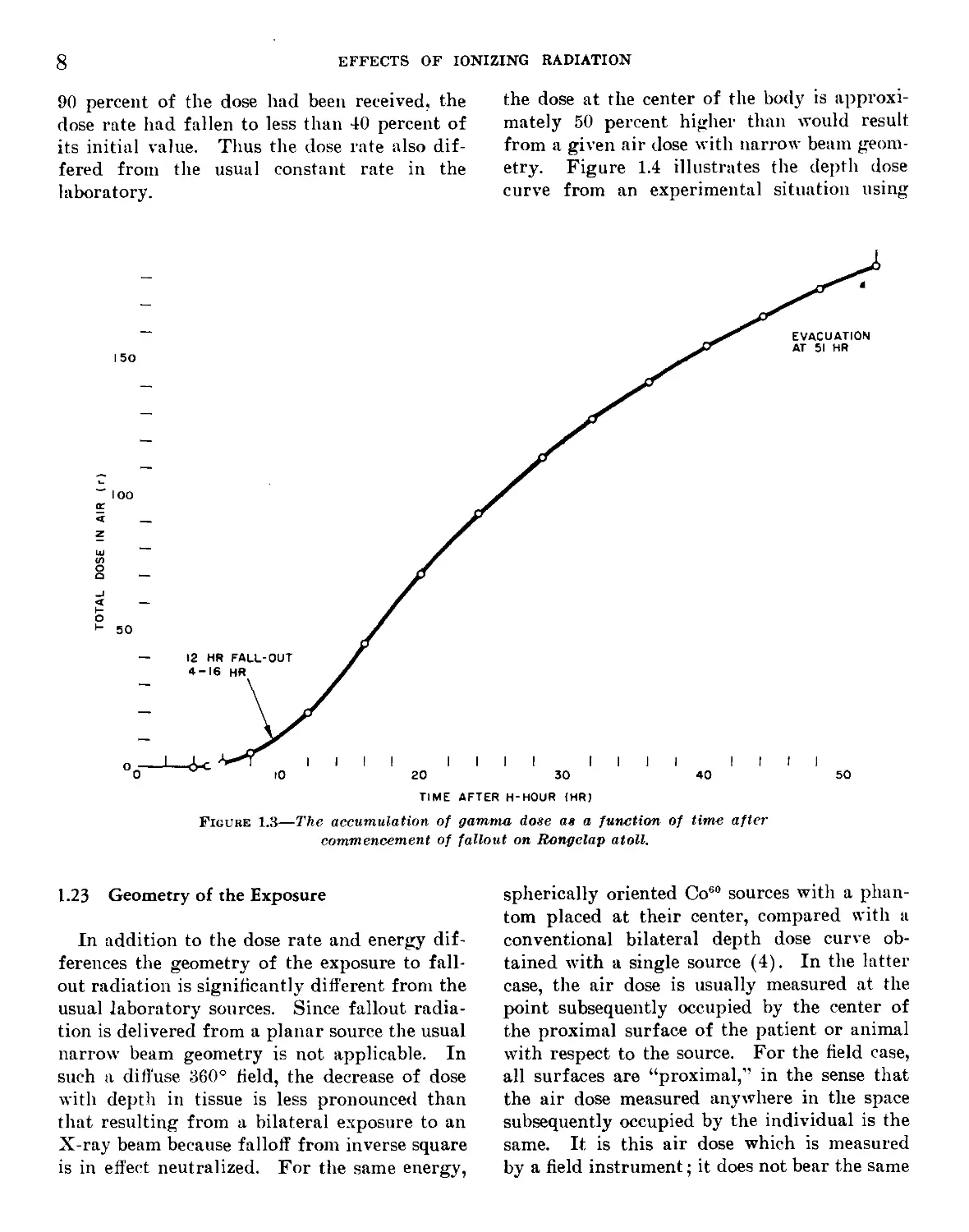
the dose at the center of the body is approxi-
mately 50 percent higher than would result
from a given air dose with narrow beam geom-
etry. Figure 1.4 illustrates the depth dose
curve from an experimental situation using
Figure 1.3—The accumulation of gamma dose as a function of time after
commencement of fallout on Rongelap atoll.
1.23 Geometry of the Exposure
In addition to the dose rate and energy dif-
ferences the geometry of the exposure to fall-
out radiation is significantly different from the
usual laboratory sources. Since fallout radia-
tion is delivered from a planar source the usual
narrow beam geometry is not applicable. In
such a diffuse 360° field, the decrease of dose
with depth in tissue is less pronounced than
that resulting from a bilateral exposure to an
X-ray beam because falloff from inverse square
is in effect neutralized. For the same energy,
spherically oriented Co60 sources with a phan-
tom placed at their center, compared with a
conventional bilateral depth dose curve ob-
tained with a single source (4). In the latter
case, the air dose is usually measured at the
point subsequently occupied by the center of
the proximal surface of the patient or animal
with respect to the source. For the field case,
all surfaces are “proximal,’’ in the sense that
the air dose measured anywhere in the space
subsequently occupied by the individual is the
same. It is this air dose which is measured
by a field instrument; it does not bear the same
EVENT AND DESCRIPTION OF EXPOSED GROUPS
9
relationship to the surface dose and depth dose
as does the air dose measured in a “point source”
beam in the clinic or laboratory. It would
appear under these circumstances and in most
experimental conditions that the midline dose,
rather than dose measured in air, would be the
source” beam air doses with comparable bio-
logic effect are obtained:
Rongelap, Group I___________ 260 r
Ailinginae, Group II-------- 100 r
Rongerik, Group III--------- 120 r
Utirik, Group IV---------------- 20 r
100
CM MASONITE
DEPTH DOSE DISTRIBUTION IN CYLINDRICAL PHANTOM, CO60FACILITY,(NMRI)
Figure 1.4—Comparison of depth dose curves in masonite phantoms from
bilateral exposure to a single point source, and simultaneous exposure to
multiple sources with a spherical distribution around the phantom.
better common parameter in terms of which to
predict biological effect. On this assumption,
the air dose values stated in Table 1.1 should be
multiplied by approximately 1.5 in order to
compare their effects to those of a given air
dose from a “point source" beam geometry de-
livered bilaterally. If this is done, assuming
a fallout of 12 hours, the following “point
The geometry of radiation from a fallout field
is not identical either to the geometry of bi-
lateral point sources or spherically distributed
sources since the plane source delivers the radia-
tion largely at a grazing angle. However, the
total field situation is better approximated by
solid than by plane geometry. Exposure geom-
etry in a radioactive cloud would be spherical.
381712 О—56-----2
10
EFFECTS OF IONIZING RADIATION
1.3 Superficial Doses of Radiation
From Beta and Soft Gamma
Radiation
There Can Be no doubt that the doses of radia-
tion to the surface and the first few millimeters
of the body were substantially higher than the
mid-line dose of gamma radiation as a result of
physical considerations of gamma energy and
depth dose. In addition, the clinical observa-
tions of the skin lesions (see Chap. Ill) force-
fully demonstrated that the dose to the skin
varied considerably between individuals and
over the surface of any given individual. As
will become evident in the following discussions
of surface dose, it is obvious that any numbers
presented are at best only estimates and repre-
sent an approximation of some minimal value.
In areas where lesions were severe the doses
must have been significantly higher than in non-
damaged areas.
To arrive at some physical estimate of the
skin dose, an attempt must be made to add up
the contributions of the high energy gamma,
the very soft gamma, and the higher energy beta
radiation from the large planar source in which
the individuals were of necessity existing.
However, as alluded to above and emphasized
in Chapter III, the largest component of skin
irradiation resulted from the spotty local de-
posits of fallout material on exposed surfaces
of the body. The dose from deposited material
is impossible to estimate; however, that from
the large planar source may be roughly esti-
mated as follows:
The beta dose rate in air 3 feet above the
surface of an infinite plane contaminated with
mixed 24 hour old fission products is estimated
to be about three times the total air gamma dose.
The mid-line gamma dose is approximately 60
percent of the air dose remaining after exclud-
ing that portion of the dose below 80 KV.
This portion in turn is estimated to be 40 per-
cent of the gamma dose measured in air by the
instrument. Thus the dose at the surface of a
phantom exposed to mixed fission product
radiation from an external plane source might
be expected to be 3/(0.6) (0.6) or about 8 times
the mid-line dose, if both are taken at 3 feet off
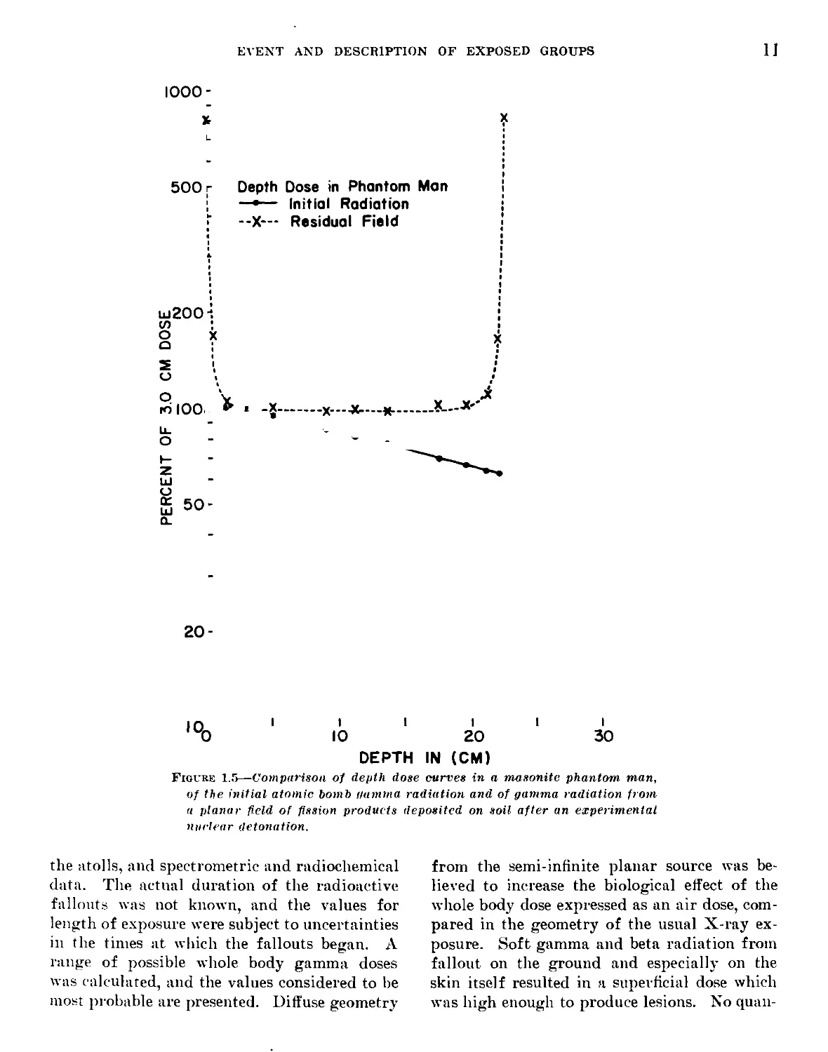
the ground. Such a depth dose measurement
has in fact been made experimentally at a previ-
ous test, using a phantom man exposed to both
the initial and residual radiation (5). The
depth doses for each situation are shown in
Figure 1.5, with all data as percent of the 3 centi-
meter dose. With the diverging initial radia-
tion from the point of explosion, the exit dose
was seen to be 63 percent of the 3 cm. dose, but
with the diffuse residual field of fission products
providing a semi-infinite planar source, a sur-
face dose some 8 times greater than the 3 cm. and
deeper dose from the harder gamma components
was observed. This is seen to be of the same
order of magnitude as that estimated above.
At heights above and below the 3 foot level this
surface dose would become lower and higher
respectively, but since it is due to soft radiation
of short range, it probably would not exceed 50
times the 3 foot air gamma dose or 80 times the
midline dose, even in contact with the ground.
An estimate of skin dose due to ground contami-
nation for the Rongelap case would result, for
example, in a figure of about 2,000 rep at the
level of the dorsum of the foot, 600 rep at the hip
level and 300 rep at the head if continuous ex-
posure with no shielding occurred. Unknown
variation in dose undoubtedly resulted from
shielding and movement. It thus seems prob-
able that the external beta dose from local direct
skin contamination far outweighed that from
the ground in importance, since the latter was
not high enough to produce the observed lesions.
Clothing probably reduced the beta dose from
the ground by 10 to 20 percent.
1.4 Summary
Radiation Doses from gamma rays originating
externally were calculated for the 267 individu-
als who were accidentally exposed to fallout
following the nuclear detonation at the Pacific
Proving Ground in the Spring of 1954. The
dose estimations were made using information
resulting from radiological safety surveys on
EVENT AND DESCRIPTION OF EXPOSED GROUPS
11
IOOO-
X
500 г
Depth Dose in Phantom Man
—•— Initial Radiation
--X—- Residual Field
ш200
3
к
rd lOOl * -X-------X--------*—*'*
о
Ш
о
й 50-
го-
,оо
I I I
20 30
DEPTH IN (CM)
Figure 1.5—Comparison of depth dose curves in a masonite phantom man,
of the initial atomic bomb gamma radiation and of gamma radiation from
a planar field of fission products deposited on soil after an experimental
nuclear detonation.
the atolls, and spectrometric and radiochemical
data. The actual duration of the radioactive
fallouts was not known, and the values for
length of exposure were subject to uncertainties
in the times at which the fallouts began. A
range of possible whole body gamma doses
was calculated, and the values considered to be
most probable are presented. Diffuse geometry
from the semi-infinite planar source was be-
lieved to increase the biological effect of the
whole body dose expressed as an air dose, com-
pared in the geometry of the usual X-ray ex-
posure. Soft gamma and beta radiation from
fallout on the ground and especially on the
skin itself resulted in a superficial dose which
was high enough to produce lesions. No quan-
12
EFFECTS OF IONIZING RADIATION
titative data were available on the beta radia-
tion intensity from either the skin contamina-
tion or from the ground, but a rough estimate
of superficial dose from the latter was made.
References
1. Gates, L. G., and Eisenhauer, C.; Spectral Distri-
bution of Gamma Rays Propagated in Air ; Armed
Forces Special Weapons Project Tech Analysis
Report 502A (Jun. 1954), Washington, D. C.
2. Turke, J. K.. and Reardon, D. A.: NRDL Instru-
ment Evaluation Report NE 051555 (5 October
1951) ; Gamma Survey Meter. V. S. Naval Ra-
diological Defense Laboratory, San Francisco,
Calif.
3. Hunter, H. F., and Ballou, N. E.; Fission Product
Decay Rates; Nucleonics !), 5, pCl-C7 (1951). Also
unpublished data, N. E. Ballou.
4. Unpublished data, C. A. Sondhaus.
5. Personal communication from F. W. Chambers. Jr.,
Naval Medical Research Institute, Bethesda, 14.
Md.
Chapter II
Clinical Observations and Treatment
N. R. Shulman, Lt. (MC) USN.
E. P. Cronkite, Cdr. (MC) USN.
V. P. Bond, M. D., Ph. D.
C. L. Dunham, M.D.
R. A. Conard, Cdr. (MC) USN.
Outline
2.1 Introduction
2.2 Symptoms and Signs Related to Radiation Injury
2.3 Clinical Observations and Therapy With Respect to Hema-
tological Findings
2.31 Clinical Observations and the Leukocyte Count
2.32 Clinical Observations and Platelet Counts
2.33 Hematocrit Changes
2.4 An Epidemic of Upper Respiratory Infection Occurring
During the 4th and 5th Post-Exposure Weeks.
2.5 Comparison of Diseases Seen in Groups I and II With
Those Seen in Group IV
2.6 Changes in Weight as in Indication of a Disturbance in the
General Metabolism
2.7 The Effects on Pregnancy
2.8 Special Examination of the Eyes
2.9 Summary and Conclusions
2.1 Introduction
When The Exposed groups were first seen at
Kwajalein after evacuation from their native
atolls, the amount of radiation they had re-
ceived was not known with certainty. It was
known, however, from instrument readings
taken at the sites of the fallout and from moni-
toring all individuals, that a significant amount
of penetrating irradiation to the entire body
had been received and that extensive contamina-
tion of the skin and possible internal deposi-
tion of radioactive materials had occurred. The
nature of the irradiating material and the cir-
cumstances of exposure prevented a precise
evaluation of dosage (see introduction). Even
if the precise dose had been known it would
not have been possible to predict the biological
effects since the quantitative response of man
is not known. Accordingly, a complete medical
history and physical examination was obtained
on each individual and numerous follow-up
examinations were carried out. In addition,
routine sick-call was held twice daily and in-
spection of the skin of all individuals was made
at frequent intervals. Medical care was avail-
able at all times. Hospital facilities were avail-
able at the Kwajalein Naval Dispensary, and
support by the more extensive medical facilities
of the U. S. Pacific Fleet had been promised
if needed.
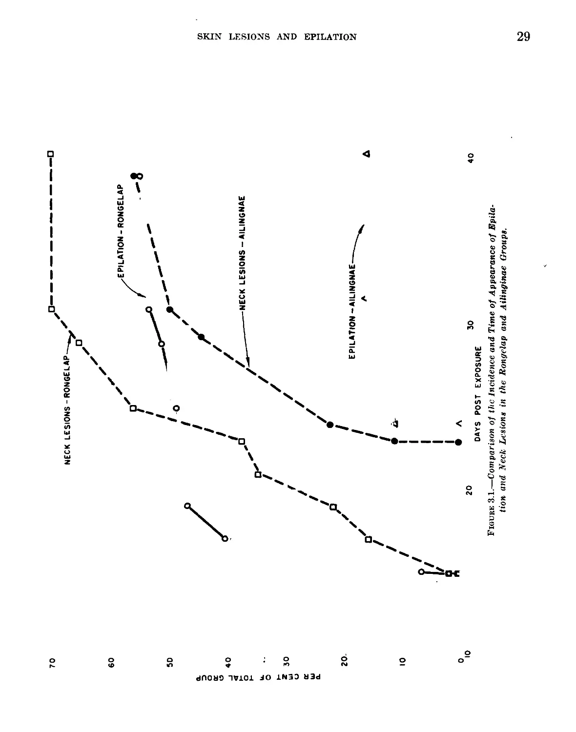
From descriptions of the amount of fallout
material and from radioactivity measurements,
it was apparent that Group I (Rongelap) had
received the highest doses of radiation, Group
II (Ailinginae) and Group III (Americans)
an intermediate amount and Group IV (Utirik)
the least. From physical dosimetry it was later
estimated that Group I had received approxi-
mately 175 r of gamma radiation; Group II,
69 r; Group III, 78 r: and Group IV, 14 r.
The most serious clinical and laboratory mani-
festations of irradiation appeared in Group I
and II. The only abnormalities that could be
attributed with certainty to irradiation were
skin lesions, epilation, granulocytopenia and
thrombocytopenia. The skin lesions were first
observed between the 12th and 14th post-expo-
sure days. These lesions were most prevalent
in Groups I and II but were present to a slight
extent in Group III. Details of the skin symp-
toms and lesions and their treatment are re-
ported in Chapter 3. Details of hematologic
studies are presented in Chapter IV. Granu-
locytopenia and thrombocytopenia of marked
degree developed in many individuals of Groups
I and II and was of sufficient severity to war-
rant serious consideration of prophylactic and
therapeutic measures for potential sequelae of
these cellular deficiencies.
In view of the conflicting opinions about the
value of prophylactic and therapeutic measures
such as antibiotics and whole blood transfusions
in the treatment of radiation disease (1-5), it
was decided that therapy would be instituted
only as indicated clinically for specific condi-
tions as they arose. In order to determine the
effect of the internal deposition of radioactive
material on the course of the externally induced
radiation injury, it was necessary to determine
the degree of internal radioactive contamina-
tion. Details of the measurement of internal
deposition of radionuclides are considered in
Chapter V. It is sufficient to state here that the
contribution from the internally deposited
radionuclides to the total acute dose was
insignificant.
2.2 Symptoms and Signs Related to
Radiation Injury
Several Symptoms That developed during the
first two days could be attributed to radiation.
These symptoms were associated with the skin
and the gastrointestinal tract.
Itching and burning of the skin occurred in
28 percent of Group I (Rongelap), 20 percent
15
16
EFFECTS OF IONIZING RADIATION
of Group II (Ailinginae), 5 percent of Group
III (Americans), and none of Group IV
(Utirik). Three people in Group I and one
in Group II complained of itching and burning
of the eyes and lacrimation. These initial skin
and eye symptoms were most likely due to ir-
radiation since all individuals who experienced
the initial symptoms later developed unques-
tioned radiation induced skin lesions (epilation
and conjunctivitis). (See Chapter III.)
Furthermore the initial symptomatology in
these people was similar to that reported in in-
stances of accidental laboratory overexposure
to radiation, described in Chapter III. It is
possible, however, that chemical irritation by
the fallout material, which was predominantly
highly alkaline calcium oxide, may have ac-
centuated the initial symptoms.
About two-thirds of Group I were nauseated
during the first 2 days and one-tenth vomited
and had diarrhea. One individual in Group
II was nauseated. In Groups III and IV there
were no gastrointestinal (GI) symptoms. The
information concerning symptoms was obtained
by questioning through an interpreter by sev-
eral individuals. Despite the repeated interro-
gations and the inevitable suggestions of the
interrogators, the stories remained consistent.
All GI symptoms subsided by the third day
without therapy and there was no recurrence.
The presence, severity, and duration of nau-
sea, vomiting, and diarrhea are known to bear
a direct relationship to degree of exposure and
probability of the recovery (1, 2, 6), and it is
of note that the incidence of these symptoms
was correlated with the dose received and that
there were no gastrointestinal symptoms in
Group IV, the largest group, which received
only 14 r. GI symptomatology may have been
due to direct injury of the GI tract as observed
in animals after whole body irradiation (7, 8)
or may have been non-specific as is observed
following therapeutic radiation.
Various other clinical conditions, which were
encountered during the course of observation
of the exposed groups were not the results of
radiation exposure. The incidence and type of
disease seen, discussed below, were similar in
all exposure groups and in nonexposed indi-
viduals.
2.3 Clinical Observation and Therapy
With Respect to Hematological
Findings
2.31 Clinical Observations and Leukocyte Counts
Between The 33rd and 43rd post-exposure
days, 10 percent of the individuals in Group I
had an absolute granulocyte level of 1000 per
cubic millimeter or below. The lowest count ob-
served during this period was 700 granulocytes/
mm.3 During this interval the advisability of
giving prophylactic antibiotic therapy to
granulocytopenic individuals was carefully con-
sidered. However, prophylactic antibiotic
therapy was not instituted for the following
reasons:
(1) All individuals were under continuous
medical observation so that infection would be
discovered in its earliest stages.
(2) Premature administration of antibiotics
might have obscured medical indications for
treatment, and might also have lead to the de-
velopment of drug resistant organisms in in-
dividuals with a lowered resistance to infec-
tion.
(3) There was no accurate knowledge of the
number of granulocytes required by man to pre-
vent infection with this type of granulocyto-
penia.
The observed situation was not strictly com-
parable to agranulocytosis with an aplastic
marrow as seen following known lethal doses of
radiation. In the latter instance, granulocytes
fall rapidly with practically none in circula-
tion and no evidence of granulocyte regenera-
tion when infection occurs (6). In the pres-
ent group of individuals exposed to radiation,
most counts reached approximately one-fourth
the normal value, but the fall to that level was
gradual and the presence of immature granu-
locytes in the peripheral blood during the pe-
CLINICAL OBSERVATIONS AND TREATMENT
17
riod of granulocytopenia was indicative of some
grauulocyte regeneration.
White counts were repeated at 3 to 4 day in-
tervals on all of the exposed individuals and
more frequently on those with the lowest counts.
Individuals with symptoms or elevated tem-
peratures were treated only after an attempt to
establish a diagnosis was made, even if a pe-
riod of observation was necessary. During the
observation per’od. the patients were examined
at frequent intervals and the temperatures
checked every few hours.
Twenty-seven individuals had total leukocyte
counts of 4000 or below or absolute neutrophile
counts of 2500 or less at some time during the
period of observation. Of these 27, 13 de-
veloped symptoms of disease that required
evaluation for possible antibiotic therapy. The
13 instances in which it was necessary to con-
sider the use of antibiotic therapy in neutro-
penic individuals are summarized below:
Eight neutropenic individuals had symtoms
of upper respiratory infection (I'RI) char-
acterized by malaise, sore throat, nasal dis-
charge. and temperatures between 99 and 101.4°
F. The temperatures returned to normal
within 24 hours. Since the response of this
group to I’RI appeared identical with that of
other individuals with I’RI without neutro-
penia, no special therapy was given.
Two individuals developed symptoms of
marked malaise, headache, abdominal pain,
nausea and diarrhea. Both were children, one
age 7, the other age 2. In both instances, the
symptoms were out of proportion to the physi-
cal findings, which were negative except for
evidence of head colds and pharyngeal injec-
tion. The 7-year old child had an oral tempera-
ture of 102.6° F. when first seen and 4 hours
later, it was 104° F. The two-year old child
had an initial axillary temperature of 101.8° F.
which rose to 103.5° F. in 4 hours. Both were
given 300,000 units of procaine penicillin intra-
muscularly when the sharp rise in temperature
occurred, and both were afebrile the following
day. A second injection of penicillin was given
at this time, and therapy was discontinued. In
spite of the fact that the neutrophiles remained
depressed in both cases long after the fever had
passed, both individuals recovered and had no
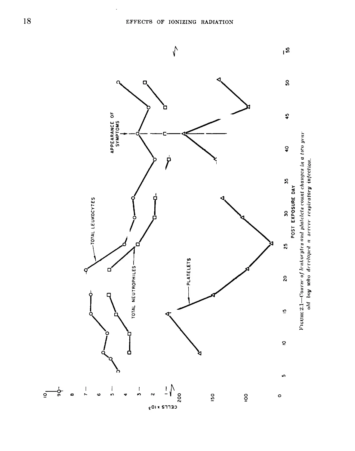
further illness. In Figure 2.1 the leukocyte and
platelet counts of the 2-year old patient and the
time of the occurrence of the febrile illness are
illustrated.
A one-year-old boy had had symptoms of
mild upper respiratory infection for several
days and was brought to the clinic when he
developed a hacking cough. When he was seen,
his axillary temperature was 100.8° F. He
had signs of URI, there was pharyngeal injec-
tion, and numerous coarse rhonchi were heard
throughout the chest. A diagnosis of upper
respiratory infection with associated bronchi-
tis was made and the child was given a single
intramuscular injection of 200.000 units pro-
caine penicillin. On the following day his tem-
perature was 99° F., no rales or rhonchi were
heard, and he recovered without further
treatment.
A 50-year-old man came to the clinic com-
plaining of weakness, nervousness, mild ab-
dominal pain and shooting pain in the upper
anterior chest bilaterally of several hours dura-
tion. He appeared moderately ill, his temper-
ature was 99.6° F., and the only positive physi-
cal finding was moderate tenderness in the right
upper quadrant of the abdomen. Within a
10-hour period the temperature rose to 101.6°
F., following which it fell gradually to nor-
mal. The abdominal tenderness continued for
24 hours and then gradually disappeared dur-
ing the subsequent 2 days. A tentative diag-
nosis of cholecystitis was made. No specific
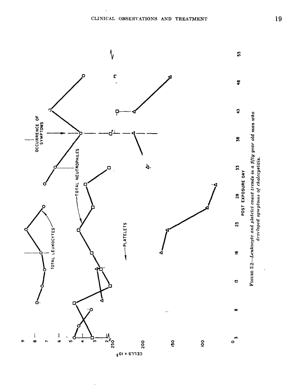
therapy was given. In Figure 2.2 his white
blood cell and platelet counts in relation to the
appearance of symptoms are shown.
A female, age 38, developed generalized urti-
caria. fever, and headache. No cause for the
urticaria was found and the symptoms subsided
within 8 hours without any therapy.
All individuals in Groups I and II that re-
ceived antibiotics are listed in Table 2.1. Of
the individuals treated with antibiotics, only
the first three received it at a time when their
neutrophile count was low. These cases are
00
CELLS « IO5
10
9i
8
25 30 35 40
POST EXPOSURE DAY
45 50
EFFECTS OF IONIZING RADIATION
0
55
Figuke 2.1—Course of leukocytes ond platelets count changes in a two year
old boy who developed a severe respiratory infection.
CELLS x 10
CLINICAL OBSERVATIONS AND TREATMENT
53
0
3 8 13 18 23 28 33 38 43 48
POST EXPOSURE OAY
Figure 2.2—Leukocyte and platelet count trends in a fifty year old man who
developed symptoms of cholecystitis.
20
EFFECTS OF IONIZING RADIATION
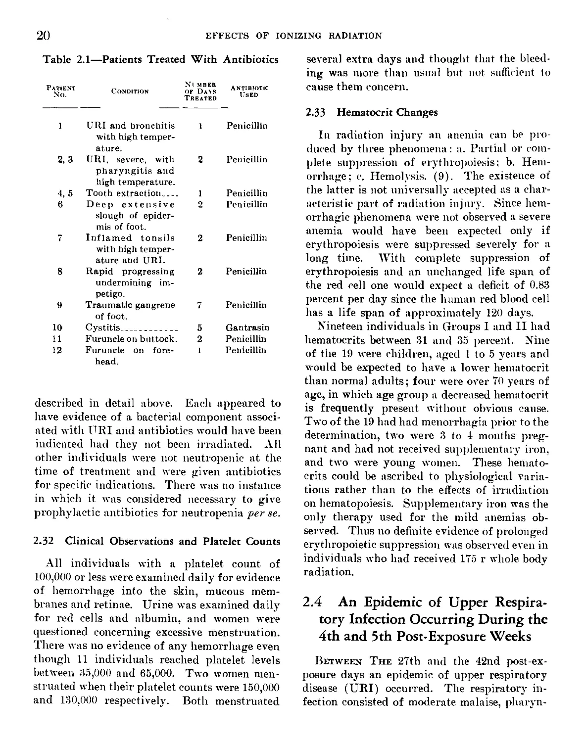
Table 2.1—Patients Treated With Antibiotics
Patient No. Condition Nl MBER of Dais Treated Antibiotic Used
1 URI and bronchitis with high temper- ature. 1 Penicillin
2, 3 URI, severe, with pharyngitis and high temperature. 2 Penicillin
4, 5 Tooth extraction 1 Penicillin
6 Deep extensive slough of epider- mis of foot. 2 Penicillin
7 Inflamed tonsils with high temper- ature and URI. 2 Penicillin
8 Rapid progressing undermining im- petigo. 2 Penicillin
9 Traumatic gangrene of foot. 7 Penicillin
10 Cystitis 5 Gantrasin
11 Furuncle on buttock. 2 Penicillin
12 Furuncle on fore- head. 1 Penicillin
described in detail above. Each appeared to
have evidence of a bacterial component associ-
ated with URI and antibiotics would have been
indicated had they not been irradiated. All
other individuals were not neutropenic at the
time of treatment and were given antibiotics
for specific indications. There was no instance
in which it was considered necessary to give
prophylactic antibiotics for neutropenia per ,se.
2.32 Clinical Observations and Platelet Counts
All individuals with a platelet count of
100,000 or less were examined daily for evidence
of hemorrhage into the skin, mucous mem-
branes and retinae. Urine was examined daily
for red cells and albumin, and women were
questioned concerning excessive menstruation.
There was no evidence of any hemorrhage even
though 11 individuals reached platelet levels
between 35,000 and 65,000. Two women men-
struated when their platelet counts were 150,000
and 130,000 respectively. Both menstruated
several extra days and thought that the bleed-
ing was more than usual but not sufficient to
cause them concern.
2.33 Hematocrit Changes
In radiation injury an anemia can be pro-
duced by three phenomena: a. Partial or com-
plete suppression of erythropoiesis; b. Hem-
orrhage; c. Hemolysis. (9). The existence of
the latter is not universally accepted as a char-
acteristic part of radiation injury. Since hem-
orrhagic phenomena were not observed a severe
anemia would have been expected only if
erythropoiesis were suppressed severely for a
long time. With complete suppression of
erythropoiesis and an unchanged life span of
the red cell one would expect a deficit of 0.83
percent per day since the human red blood cell
has a life span of approximately 120 days.
Nineteen individuals in Groups 1 and II had
hematocrits between 31 and 35 percent. Nine
of the 19 were children, aged 1 to 5 years and
would be expected to have a lower hematocrit
than normal adults; four were over 70 years of
age, in which age group a decreased hematocrit
is frequently present without obvious cause.
Two of the 19 had had menorrhagia prior to the
determination, two were 3 to 1 months preg-
nant and had not received supplementary iron,
and two were young women. These hemato-
crits could be ascribed to physiological varia-
tions rather than to the effects of irradiation
on hematopoiesis. Supplementary iron was the
only therapy used for the mild anemias ob-
served. Thus no definite evidence of prolonged
erythropoietic suppression was observed even in
individuals who had received 175 r whole body
radiation.
2.4 An Epidemic of Upper Respira-
tory Infection Occurring During the
4th and 5th Post-Exposure Weeks
Between The 27th and the 42nd post-ex-
posure days an epidemic of upper respiratory
disease (URI) occurred. The respiratory in-
fection consisted of moderate malaise, pharyn-
CLINICAL OBSERVATIONS AND TREATMENT
21
gitis with prominent lymphoid follicles, fever
of 99-100° F. during the first day, and a puru-
lent nasal and tracheal discharge for about 10
days. It was of interest to determine whether
the appearance of URI could be correlated with
the dose of radiation received or with changes
in the leukocyte count.
Fifty-eight percent of the individuals in
Group I and 56 percent of the individuals in
Group II developed URI. Seventy percent of
the affected individuals developed symptoms
between the 27th and 32nd post-exposure days,
and the others developed symptoms in the sub-
sequent 2 weeks. Fifty-seven percent of the
affected individuals were observed to have an
upward trend in their leukocyte counts, the in-
crease being due primarily to granulocytes.
Since an increase in the mean granulocyte
count of the entire population occurred about
the 29th postexposure day, it seemed pertinent
to determine whether in individual instances
the increase was related to the presence of
respiratory infection.
The relationship between the observed leuko-
cyte increase and the presence or absence of
upper respiratory symptoms in Groups I and
II is shown in Table 2.2. Seven of the 27 indi-
viduals that developed both URI and a leuko-
cyte increase developed the leukocyte increase
3 or more days before symptoms of URI ap-
peared. It is also of interest that the medical
personnel involved in the care and study of the
radiated individuals had an equal incidence and
Table 2.2—URI and Changes in Granulocytes in
Groups I and II
Number
of Indi-
viduals
URI; rise in granulocytes______________ 27
URI; no rise in granulocytes___________ 20
No URI; rise in granulocytes___________ 16
No URI; no rise in granulocytes________ 19
severity of respiratory infections. The inci-
dence and severity of respiratory infection in
Group IV, which had received only slight radia-
tion, was the same as that in Group I and II.
The appearance of URI, therefore, did not ap-
pear to be related to the dose of radiation or
to changes in leukocyte level.
2.5 Comparison of Diseases Seen in
Groups I and II With Those in Group
IV
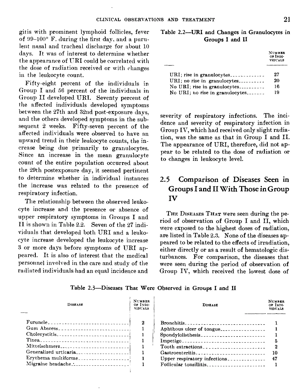
The Diseases That were seen during the pe-
riod of observation of Group I and II, which
were exposed to the highest doses of radiation,
are listed in Table 2.3. None of the diseases ap-
peared to be related to the effects of irradiation,
either directly or as a result of hematologic dis-
turbances. For comparison, the diseases that
were seen during the period of observation of
Group IV, which received the lowest dose of
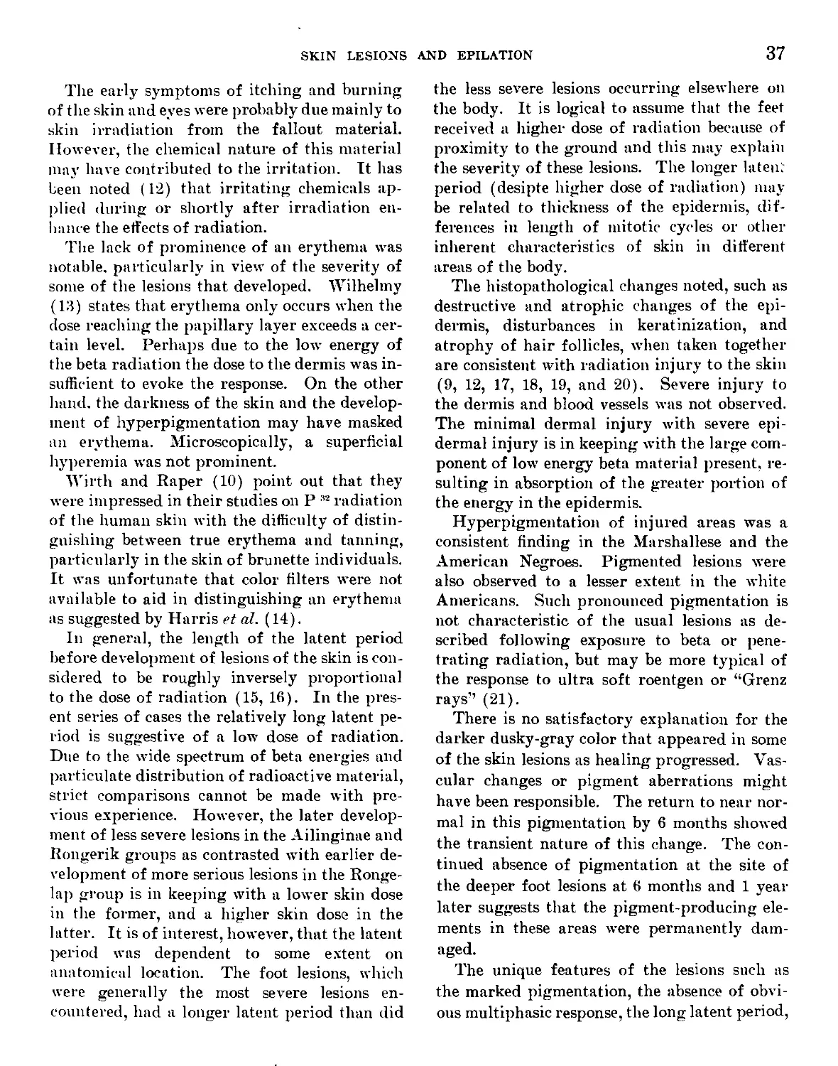
Table 2.3—Diseases That Were
Observed in Groups I and II
Disease
Number
of Indi-
viduals
Disease
Number
of Indi-
viduals
Furuncle_________________________________
Gum Abscess______________________________
Cholecystitis____________________________
Tinea____________________________________
Mittelschmerz____________________________
Generalized urticaria____________________
Erythema multiforme______________________
Migraine headache.'______________________
2 Bronchitis----------------------------------------
1 , Aphthous ulcer of tongue_____________________
1 Spondylolisthesis____________________________
1 Impetigo_____________________________________
1 Tooth extractions_________________________________
1 Gastroenteritis______________________________
1 Upper respiratory infections______________________
1 Follicular tonsillitis____________________________
1
1
1
5
2
10
47
1
22
EFFECTS OF IONIZING RADIATION
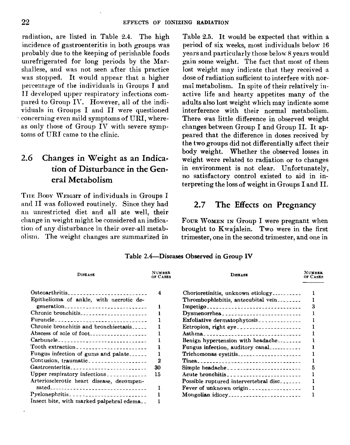
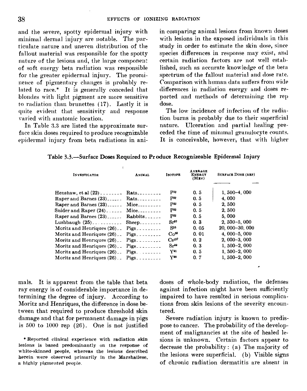
radiation, are listed in Table 2.4. The high
incidence of gastroenteritis in both groups was
probably due to the keeping of perishable foods
unrefrigerated for long periods by the Mar-
shallese, and was not seen after this practice
was stopped. It would appear that a higher
percentage of the individuals in Groups I and
II developed upper respiratory infections com-
pared to Group IV. However, all of the indi-
viduals in Groups I and II were questioned
concerning even mild symptoms of URI, where-
as only those of Group IV with severe symp-
toms of URI came to the clinic.
2.6 Changes in Weight as an Indica-
tion of Disturbance in the Gen-
eral Metabolism
The Body Weight of individuals in Groups I
and II was followed routinely. Since they had
an unrestricted diet and all ate well, their
change in weight might be considered an indica-
tion of any disturbance in their over-all metab-
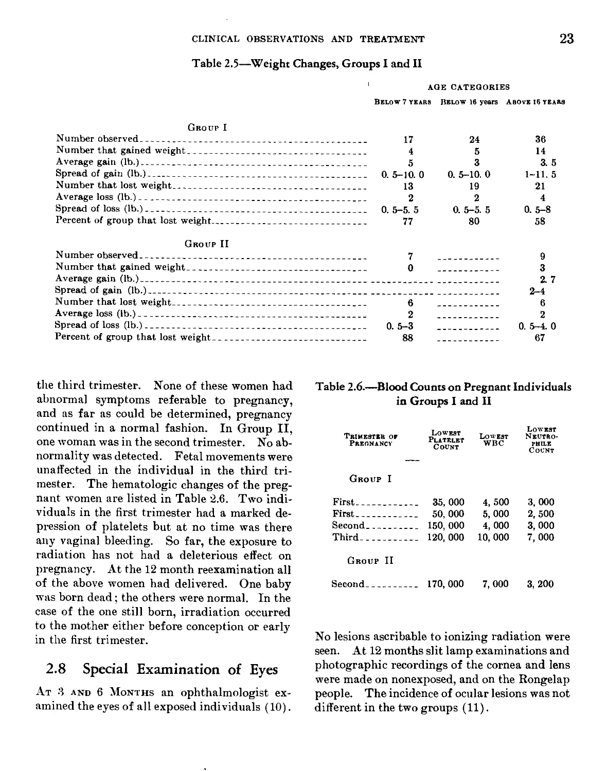
olism. The weight changes are summarized in
Table 2.5. It would be expected that within a
period of six weeks, most individuals below 16
years and particularly those below 8 years would
gain some weight. The fact that most of them
lost weight may indicate that they received a
dose of radiation sufficient to interfere with nor-
mal metabolism. In spite of their relatively in-
active life and hearty appetites many of the
adults also lost weight which may indicate some
interference with their normal metabolism.
There was little difference in observed weight
changes between Group I and Group II. It ap-
peared that the difference in doses received by
the two groups did not differentially affect their
body weight. Whether the observed losses in
weight were related to radiation or to changes
in environment is not clear. Unfortunately,
no satisfactory control existed to aid in in-
terpreting the loss of weight in Groups I and II.
2.7 The Effects on Pregnancy
Four Women in Group I were pregnant when
brought to Kwaj alein. Two were in the first
trimester, one in the second trimester, and one in
Table 2.4—Diseases Observed in Group IV
Disease
Number
or Cases
Disease
Number
or Cases
Osteoarthritis_____________________________ 4
Epithelioma of ankle, with necrotic de-
generation_________________________________ 1
Chronic bronchitis___________________________ 1
Furuncle_____________________________________ 1
Chronic bronchitis and bronchiectasis______ 1
Abscess of sole of foot______________________ 1
Carbuncle____________________________________ 1
Tooth extraction_____________________________ 1
Fungus infection of gums and palate________ 1
Contusion, traumatic_________________________ 2
Gastroenteritis_____________________________ 30
Upper respiratory infections________________ 15
Arteriosclerotic heart disease, decompen-
sated________________________________________ 1
Pyelonephritis_______________________________ 1
Insect bite, with marked palpebral edema.. 1
Chorioretinitis, unknown etiology---------- 1
Thrombophlebitis, antecubital vein--------- 1
Impetigo___________________________________ 3
Dysmenorrhea_______________________________ 1
Exfoliative dermatophytosis________________ 1
Ectropion, right eye_______________________ 1
Asthma_____________________________________ 1
Benign hypertension with headache---------- 1
Fungus infection, auditory canal----------- 1
Trichomonas cystitis_______________________ 1
Tinea______________________________________ 1
Simple headache____________________________ 5
Acute bronchitis___________________________ 1
Possible ruptured intervertebral disc---- 1
Fever of unknown origin____________________ 1
Mongolian idiocy___________________________ 1
CLINICAL OBSERVATIONS AND TREATMENT
23
Table 2.5—Weight Changes, Groups I and II
AGE CATEGORIES
Below 7 years Below 16 years Above 16 years
Group I
Number observed__________________________________________
Number that gained weight________________________________
Average gain (lb.)_______________________________________
Spread of gain (lb.)_____________________________________
Number that lost weight__________________________________
Average loss (lb.)_______________________________________
Spread of loss (lb.)_____________________________________
Percent of group that lost weight________________________
17
4
5
0. 5-10. 0
13
2
0. 5-5. 5
77
24
5
3
0. 5-10. 0
19
2
0. 5-5. 5
80
36
14
3. 5
1-11. 5
21
4
0. 5-8
58
Group II
Number observed__________________________________________ 7 ___________
Number that gained weight________________________________ 0 ___________
Average gain (lb.)_______________________________________________________________
Spread of gain (lb.)_____________________________________________________________
Number that lost weight________________________________________ 6 ___________
Average loss (lb.)_____________________________________________ 2 ___________
Spread of loss (lb.)_____________________________________ 0. 5-3 ___________
Percent of group that lost weight_____________________________ 88 ___________
9
3
2. 7
2-4
6
2
0. 5-4. 0
67
the third trimester. None of these women had
abnormal symptoms referable to pregnancy,
and as far as could be determined, pregnancy
continued in a normal fashion. In Group II,
one woman was in the second trimester. No ab-
normality was detected. Fetal movements were
unaffected in the individual in the third tri-
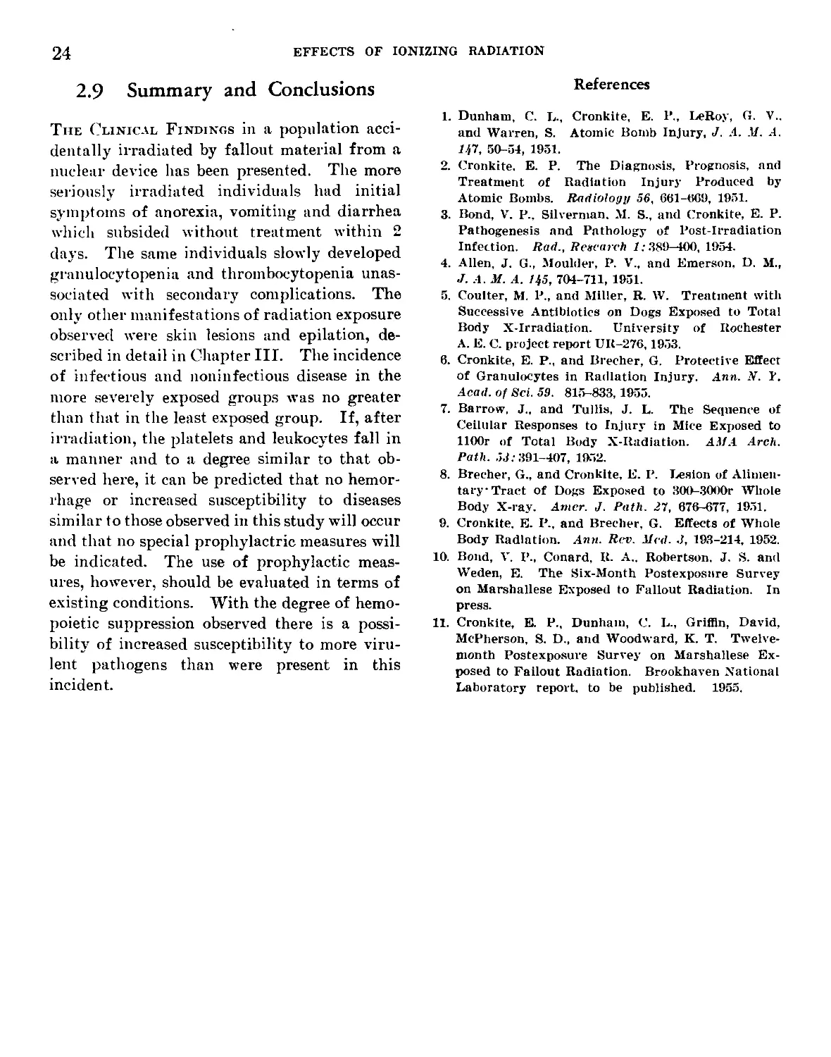
mester. The hematologic changes of the preg-
nant women are listed in Table 2.6. Two indi-
viduals in the first trimester had a marked de-
pression of platelets but at no time was there
any vaginal bleeding. So far, the exposure to
radiation has not had a deleterious effect on
pregnancy. At the 12 month reexamination all
of the above women had delivered. One baby
was born dead; the others were normal. In the
case of the one still born, irradiation occurred
to the mother either before conception or early
in the first trimester.
2.8 Special Examination of Eyes
At 3 and 6 Months an ophthalmologist ex-
amined the eyes of all exposed individuals (10).
Table 2.6.—Blood Counts on Pregnant Individuals
in Groups I and II
Trimester or Pregnancy Lowest Platelet Count Lowest WBC Lowest Neutro- pbile Count
Group I
First ... 35,000 4, 500 3, 000
First ... 50,000 5, 000 2, 500
Second ___ 150,000 4, 000 3, 000
Third ... 120,000 10, 000 7, 000
Group II
Second ... 170,000 7, 000 3, 200
No lesions ascribable to ionizing radiation were
seen. At 12 months slit lamp examinations and
photographic recordings of the cornea and lens
were made on nonexposed, and on the Rongelap
people. The incidence of ocular lesions was not
different in the two groups (11).
24
EFFECTS OF IONIZING RADIATION
2.9 Summary and Conclusions
The Clinical Findings in a population acci-
dentally irradiated by fallout material from a
nuclear device has been presented. The more
seriously irradiated individuals had initial
symptoms of anorexia, vomiting and diarrhea
which subsided without treatment within 2
days. The same individuals slowly developed
granulocytopenia and thrombocytopenia unas-
sociated with secondary complications. The
only other manifestations of radiation exposure
observed were skin lesions and epilation, de-
scribed in detail in Chapter III. The incidence
of infectious and noninfectious disease in the
more severely exposed groups was no greater
than that in the least exposed group. If, after
irradiation, the platelets and leukocytes fall in
a manner and to a degree similar to that ob-
served here, it can be predicted that no hemor-
rhage or increased susceptibility to diseases
similar to those observed in this study will occur
and that no special prophylactric measures will
be indicated. The use of prophylactic meas-
ures, however, should be evaluated in terms of
existing conditions. With the degree of hemo-
poietic suppression observed there is a possi-
bility of increased susceptibility to more viru-
lent pathogens than were present in this
incident.
References
1. Dunham, C. L., Cronkite, E. P., LeRoy, G. V..
and Warren, S. Atomic Bomb Injury, J. .4. V/. .4.
Ц7, 50-54, 1951.
2. Cronkite. E. P. The Diagnosis, Prognosis, and
Treatment of Radiation Injurj- Produced by
Atomic Bombs. Radiology 56, 661-669, 1951.
3. Bond, V. P., Silverman. M. S., and Cronkite, E. P.
Pathogenesis and Pathology of Post-Irradiation
Infection. Ra<t., Reaearch 1:389—400, 1954.
4. Allen, J. G., Moulder, P. V., and Emerson, D. M.,
J. .1. M. А. Ц5, 704-711, 1951.
5. Coulter, M. P„ and Miller, R. W. Treatment with
Successive Antibiotics on Dogs Exposed to Total
Body X-Irradiation. University of Rochester
A. E. C. project report UR-276, 1953.
6. Cronkite, E. P., and Brecher, G. Protective Effect
of Granulocytes in Radiation Injury. Ann. N. 1.
Acad, of Sci. 59. 815-833, 1955.
7. Barrow, J., and Tullis, J. L. The Sequence of
Ceilular Responses to Injury in Mice Exposed to
1100г of Total Body X-Radiation. AMA Arch.
Path. 53: 391-407, 1952.
8. Brecher, G., and Cronkite, E. P. Lesion of Alimen-
tary-Tract of Dogs Exposed to 300-3000r Whole
Body X-ray. Amer. J. Path. 27, 676-677, 1951.
9. Cronkite. E. P., and Brecher, G. Effects of Whole
Body Radiation. Ann. Rev. Med. 3, 193-214, 1952.
10. Bond, V. I’., Conard, R. A.. Robertson, J. S. and
Weden, E. The Six-Month Postexposure Survey
on Marshallese Exposed to Fallout Radiation. In
press.
11. Cronkite, E. P., Dunham, C. L., Griffin, David,
McPherson, S. D., and Woodward, К. T. Twelve-
month Postexposure Survey on Marshallese Ex-
posed to Failout Radiation. Brookhaven National
Laboratory report, to be published. 1955.
Chapter III
Skin Lesions and Epilation
Robert A. Conard, Capt. (MC) USN
Nahum R. Shulman, Lt. (MC) USN
David A. Wood, M. D.
Charles L. Dunham, M. D.
Edward L. Alpen, Ph. D.
L. Eugene Browning, Lt. Col. (MC) USN
V. P. Bond, M. D., Ph. D.
E. P. Cronkite, Cdr. (MC) USN
381712 0—56-3
Outline
3.1 Introduction
3.2 Signs and Symptoms
3.3 Description of the Skin Lesions
3.31 Gross Appearance
3.32 Microscopic Appearance
3.33 Epilation and Nail Pigmentation
3.4 Therapy
3.5 Factors Influencing Severity of the Lesions
3.51 Character of the Fallout Material
3.52 Dose to the Skin
3.53 Protective Factors
3.54 Factors Favoring the Development of Lesions
3.6 Lack of Correlation With Hematological Findings
3.7 Discussion
3.8 Summary
3.1 Introduction
Fallout of Radioactive material commenced
approximately 4—6 hours after detonation of
the thermonuclear device. On the most heavily
contaminated island, Rongelap, the fallout was
described as a powdery material, “snowlike,”
which fell over a period of several hours and
whitened the. hair and adhered to the skin.
I^ess striking fallout described as “mist-like”
was observed on Ailinginae and Rongerik.
Fallout was not visible on Utirik, which was
contaminated to only a mild degree. The se-
verity of the skin manifestations was roughly
proportional to the amount of fallout observed.
The population of the four island groups and
incidence of the skin lesions were as follows:
3.2 Signs and Symptoms
During Tin; First 24—48 hours after exposure,
about 25 percent of the Marshallese in the two
higher exposure groups experienced itching and
a burning sensation of the skin. A few also
complained of burning of the eyes with lachry-
mation. These symptoms were present to a
lesser extent in the Americans on Rongerik
Atoll who were aware of the danger, took shel-
ter in aluminum buildings, bathed and changed
clothes. These precautions greatly reduced
the subsequent development of skin lesions in
this group. The people on Utirik, the farth-
est from the detonation, had no early skin symp-
Orovf
Composition
Fallout Observed
Extensiveness or Skin Lesions and
Epilation
Rongelap__________ 64 Marshallese____________ Heavy (snowlike)__________ Extensive.
Ailinginae________ 18 Marshallese____________ Moderate (mistlike)___ Less extensive.
Rongerik__________ 23 White Americans________ Moderate (mistlike)___ Slight.
5 Negro Americans.
Utirik____________ 157 Marshallese___________ None______________________ No skin lesions or epilation.
Evacuation of exposed personnel to Kwaja-
lein, where medical facilities were available,
was accomplished one to two days after the
event. Decontamination of the skin was com-
menced aboard ship, and completed after ar-
rival at Kwajalein.
Skin examinations were carried out almost
daily during the first 11 weeks and then again at
6 months, 1 and 2 years after the accident. Ex-
aminations of unexposed Americans and native
personnel were also carried out for comparative
purposes. Color photographs and biopsies of
lesions in various stages of development were
taken.
toms. All skin symptoms subsided within 1
to 2 days. On arrival of the medical team on
the ninth post-exposure day, the exposed per-
sonnel appeared to be in good health. The
skin appeared normal. However, evidence of
cutaneous radiation injury appeared about 2
weeks after exposure when epilation and skin
lesions commenced. Erythema of the skin was
not observed either during the early examina-
tions when a primary erythema might be ex-
pected, or later when a secondary erythema
might be expected.
After subsidence of the initial skin symp-
toms, further symptoms referrable to the skin
27
28
EFFECTS OF IONIZING RADIATION
were absent until the visible lesions developed.
During the early stages of development of the
lesions, itching, burning and slight pain were
experienced with the more superficial lesions.
With deeper lesions pain was more severe. The
deeper foot lesions were the most painful and
caused some of the people to walk on their
heels for several days during the acute stages.
Some of the more severe lesions of the neck
and axilla were painful when turning the head
or raising the arms. The lesions did not
produce any constitutional symptoms.
3.3 Description of Skin Lesions*
3.31 Gross Appearance
The time of appearance and the severity of
the lesions varied with the degree of skin con-
tamination in the different groups. The Ronge-
lap group, which showed greatest radioactive
contamination of the skin (according to instru-
ment readings) were the first to develop lesions
and epilation at about 12 to 14 days after the
accident. They also had the most severe lesions.
Skin lesions in the lesser exposed Ailinginae
and Rongerik groups developed approximately
one week after fhose in the Rongelap group, and
were less severe and extensive. The Utirik
group did not develop any lesions which could
be attributed to irradiation of the skin. The
incidence of ulcerating lesions in the different
groups reflected the relative severity of the skin
injury. Twenty percent of the Rongelap people
developed ulcerative lesions while only five per-
cent of the Ailinginae and none of the Rongerik
people developed ulcerative lesions. Ninety
percent of the Rongelap and Ailinginae groups
developed lesions, compared to only forty per-
cent of the Rongerik group. There were more
lesions per individual in the Rongelap group
than in the Ailinginae or Rongerik groups. A
comparison of the incidence and time of appear-
ance of epilation and neck lesions in the two
groups is illustrated graphically in Figure 3.1.
* The description of lesions refers to the Marshallese
unless otherwise indicated.
Nearly all of the lesions were spotty and de-
veloped on exposed parts of the body not cov-
ered by clothing during the fallout. The ma-
jority of individuals developed multiple lesions
(particularly the Rongelap group), most of
which were superficial. There was a difference
of several days in the latent period before de-
velopment of lesions on various skin areas. The
order of appearance was roughly as follows:
scalp (with epilation), neck, axillary region,
antecubital fossae, feet, arms, legs, and trunk.
Lesions on the flexor surfaces in general pre-
ceded those on the extensor surfaces. Tables
3.1 and 3.2 show incidence according to age and
time of appearance of lesions in the various
groups.
In the early stages all lesions were character-
ized by hyperpigmented macules, papules, or
raised plaques. (Plate 1.) These frequently
were small, 1-2 mm. areas at first, but tended to
coalesce in a few days into larger lesions, with
a dry, leathery texture.
The pigmented stage of the superficial lesions
within several days was followed by dry, scaly
desquamation which proceeded from the cen-
ter part of the lesion outward, leaving a pink
to white thinned epithelium. As the desquama-
tion proceeded outward, a characteristic ap-
pearance of a central depigmented area fringed
with an irregular hyperpigmented zone was
seen (Plates 2 and 3). Repigmentation began in
the central area and spread outward over the
next few weeks leaving skin of relatively nor-
mal appearance. Plates 3, 4, 11, and 12 show
superficial lesions as they appeared initially and
six months later. The mildest manifestation of
skin injury was the development of a blotchy
increased pigmentation of the skin with barely
perceptible desquamation. Such lesions were
most often noted on the face and trunk.
Epilation was usually accompanied by scalp
lesions (Plates 13, 17 and 19). Some indi-
viduals developed new scalp lesions over a
period of about a month. Neck lesions usually
had a “necklace” distribution, beginning anteri-
orly and spreading posteriorly. These were
more severe in women in whom thick hair
70
60
5D
NECK LESIONS-RONGELAP
PER CENT OF TOTAL GROUP
40
30
20-
10
0
10
20 30
DAYS POST EXPOSURE
Figure 3.1.—Comparison of the Incidence and Time of Appearance of Epila-
tion and Neck Lesions in the Rongelap and Ailinginae Groups.
SKIN LESIONS AND EPILATION
40
№
CD
30
EFFECTS OF IONIZING RADIATION
Table 3.1.—Lesions in Rongelap Group
Percent or Total in Age Group Having Indicated Lesion Median Time
Type of Lesion Age 0-5 (13 People) Age 6-15 (13 People) Age 16& Over Total Group of First Observation of Lesions*
(38 People) (64 People)
Epilation
1 plus 7. 6 38. 4 13. 8 17. 2 17
2 plus 38. 6 30. 7 5. 5 17. 2 17
3 plus 53. 8 23. 0 8. 3 22. 0 16
Total 100. 0 92. 1 27. 6 56. 2 16
Skin lesions
Anus-groin 38. 4 0. 0 0. 0 7. 8 17
Scalp 100. 0 100. 0 37. 0 62. 5 18
Neck 69. 2 76. 9 68. 0 70. 3 21
Axilla 61. 5 7. 6 15. 7 23. 4 21
Antecubital Fossae 30. 7 38. 4 34. 2 34. 4 28
Hands-wrists 30. 7 23. 0 18. 4 21. 8 33
Feet 23. 0 53. 8 53. 0 45. 3 28
Arms 15. 3 15. 3 10. 3 12. 5 31
Legs 7. 6 23. 0 4. 3 7. 8 33
Trunk 15. 3 23. 0 4. 3 9. 4 33
Nail pigmentation 61. 5 100. 0 95. 0 89. 0 38
*Post-exposure days.
Table 3.2.—Lesions
in Ailinginae and Rongerik Groups
Ailinginae Group (18 people) Rongerik Group (Americans) (28 PEOPLE)
Type of Lesion % or Total With Lesions Mean Time of Appear- ance* % or Total With Lesions op .
Epilation 16. 7 27 3. 5** 42
Lesions of:
Scalp and face 38. 9 26 10. 7 32
Neck and shoulders 61. 0 27 14. 3 30
Back 0. 0 7. 1 28
Axilla 22. 2 24 3. 5 23
Antecubital fossae 11. 1 28 25. 0 29
Hand, wrist 5. 6 38 3. 5 47
Feet 16. 7 33 3. 5 43
Legs Nail discoloration 5. 6 44 0. 0
77. 7 38 17. 9 40
(All Negroes)
♦Days post-exposure.
**One case claimed slight epilation.
SKIN LESIONS AND EPILATION
31
touched the nape of the neck. Neck lesions are
illustrated in Plate 1-4. Axillary lesions (Plate
11) usually consisted of coalescing papules.
Antecubital fossa lesions were characterized by
formation of thickened plaques. Several
babies and one woman developed lesions in the
anal region which, though not deep, were pain-
ful due to excoriation of the epidermis. These
healed rapidly.
Deeper lesions were seen on the scalp, neck,
feet, and in one case on the ear. They were char-
acterized by transepidermal necrosis with wet
desquamation leaving weeping, crusting ulcera-
tions. Vesiculation was not observed except
with foot lesions which developed bullae, fre-
quently several centimeters in diameter, beneath
thickened pigmented plaques. These foot
lesions occurred on the dorsum of the feet and
between the toes, (Only one case showed des-
quamation on the soles of the feet.) After
several days the bullae ruptured and desqua-
mated leaving raw ulcers. Some of these
lesions, particularly of the feet, became second-
arily infected requiring antibiotics. However,
most of the lesions healed rapidly and new
epithelium covered the ulcerated areas within
a week to 10 days. Foot lesions are illustrated
in Plates 5-10. One ear lesion (Plates 13-16)
took several months to heal.
The repigmentation of some deeper lesions
presented abnormalities. Neck lesions often
developed a dusky, grayish brown pigmentation
associated with a thickened “orange peel” ap-
pearance. Histological appearance of epi-
dermal rugosity was also noted in these lesions
(see section on histopathology). In addition,
the deeper lesions of the feet failed to repig-
ment, remaining pink or white. At examina-
tion 6 months and 1 year after the exposure, the
skin appeared normal with no residual changes
in the vast majority of cases. However, some of
the deeper lesions continued to show evidence of
residual damage. Foremost among these was
the ear lesion which had healed with consider-
able scarring, atrophy, scaling of the epidermis
and gross telangiectasis. By 6 months the hy-
perpigmentation and thickening of the skin of
the neck lesions had greatly subsided and by 1
year pigmentation changes were mild. Foot
lesions had not repigmented at sites of deepest
involvement and some atrophy of the skin in
these areas was apparent.
3.32 Microscopic Appearance
Biopsies were taken of seven neck, and one
axillary lesion in the Rongelap group during
the third to fourth week after exposure. At
the time of biopsy these lesions were in the hy-
perpigmented stage with little or no desquama-
tion. Most of the biopsies were taken from in-
dividuals with lesions of average severity. A
second series of biopsies (repeats in three in-
dividuals) were taken from this group, 4 at
the seventh week and 5 at the eighth week post-
exposure. These were taken from the neck and
antecubital fossae. All of these lesions had
desquamated and the depigmented skin had re-
pigmented to a dusky, gray color with some
thickening of the skin (“orange-peel” appear-
ance), plates 25 and 27. Biopsies were not
taken from ulcerative lesions or from the feet
because of the danger of infection. A third
series of 11 biopsies were taken from the Ronge-
lap group at 6 months along with several con-
trol biopsies from unexposed natives. Material
was obtained in many cases adjacent to sites of
previous biopsies.
All biopsy wounds healed rapidly within a
week to 10 days with no secondary complica-
tions.
The microscopic findings are summarized as
follows:
First series—3rd to 4th, week. Epidermis.
Transepidermal damage was noted with a few
intervening arcades showing less damage
(Plates 21 and 22). The epidermis in the most
extensively involved areas showed considerable
atrophy with flattening of the rete pegs and in
places the epidermis was reduced to a thickness
of 2 to 3 cells (Plates 21, 23, and 24). The cells
of the malpighian layer showed pleomorphic
nuclei, pyknosis and cytoplasmic halos, giant
cells and in a few instances multinucleated cells.
32
EFFECTS OF IONIZING RADIATION
Pyknosis of cells of the basal layer was com-
monly seen. Focal disorganization of the
malpighian and basal layers was usually pres-
ent in the more extensively damaged arcades
(Plate 23). Cells laden with pigment were
frequently present throughout the epidermis
and intercellular pigment was noted in some
sections. The stratum granulosum was usually
atrophic or even absent. Imperfect keratiniza-
tion with parakeratosis was visible in all sec-
tions. The stratum corneum was loosely fibril-
lated and hyperkeratotic.
The arcades of minimal damage were usually
found in areas where sweat ducts approached
the epidermis (Plate 22). There was an ap-
parent increase in the number of cells and mi-
totic figures along the neck of the ducts and
the adjoining areas where regeneration was
underway. In these areas the stratum granu-
losum appeared almost normal in width. In
contrast to the more severely damaged areas
where pigment was increased, these areas of
minimal damage showed an actual decrease,
being almost free of pigment.
Dermis. Changes in the dermis were con-
fined largely to the pars papillaris and super-
ficial pars reticularis (Plates 21-24). Mild
edema in some cases were noted. Capillary
loops were often indistinct and when discern-
ible they frequently were associated with an
increased number of pericytes. The endo-
thelial cells showed swelling and were polygo-
nal in shape. Telangiectatic changes were
noted in these areas where the overlying epi-
dermis showed greatest damage which were
associated with perivascular lymphocytic in-
filtration. Chromatophores, filled with mela-
nin were prominent in the superficial dermis.
The fine elastic fibrils running into the pars pap-
illaris were often altered or absent.
Little if any damage was seen below the
superficial pars reticularis. The hair follicles
were narrow and in most instances devoid of
shafts in this region. There was some telangi-
ectasis of the capillaries and slight mononu-
clear cell infiltration. Some of the large elas-
tic fibers in this region showed slight swelling
in some cases. No damage to fibrocytes or col-
lagen fibers was noted.
Second series—7th and 8th weeks post-ex-
posure. E pid-ermis. In general, reparative
processes of the epidermis had proceeded, except
for a few persistent areas of atrophy with nar-
rowing of the epidermis' and finger-like down-
growths of the stratum malpighii (Plate 27).
These changes occurred in areas of the greatest
narrowing of the stratum granulosum. In such
areas the basal cells often showed increased pig-
ment. There were many outward epidermal
excrescences covered by thickened stratum cor-
neum, still loosely laminated (Plate 25), which
probably accounted for the “orange-peel” ap-
pearance of the skin noted grossly. In almost
all instances the basal layer was intact with lit-
tle or no disorganization. There were a few
scattered areas in which occasional epithelial
cells with pyknotic nuclei and perinuclear cyto-
plasmic halos occurred in the malpighian layers
(Plate 26). There were occasional arcades in
which the epidermis and particularly the
stratum granulosum appeared to be widened.
These occurred primarily in relation to con-
tiguous sweat gland ducts where the latter pene-
trated the epidermis. A narrow zone of para-
keratosis and amorphous debris was still present
between the stratum granulosum and the loosely
laminated stratum corneum. The stratum
lucidum was not apparent.
Dermis. The capillary loops in the dermal
papillae were not uniformly distinct. Peri-
cytes remained in increased number but fewer
lymphocytes were present. Generally, there
was a slight telangiectasis of the capillaries in
the pars papillaris and the superficial pars re-
ticularis (Plate 27). There was some edema
of the pars papillaris (Plate 25). Scattered
pigment-laden chromatophores were irregularly
distributed in the papillary layer (Plate 26).
In some cases hair shafts in the superficial pars
reticularis were narrow or absent; in others the
hair shafts appeared normal. Small hair fol-
licles (Plate 25) and sweat ducts in some cases
showed mild atrophy.
Plate 1,—Early hyperpigmented maculopapular neck
lesions at 15 days. Case 39, age 15, F.
Plate 2.—Neck lesions at 28 days. Wet desquamation.
White color is calamine lotion. Case 78, age 87, F.
3-23*54
RO NG NO. 63
Ig-j.f 4Л NMM-ar.-W
Plate 3.—-Neck lesions 28 days post-exposure. Note pig-
mented and desquamated, depigmented areas. Case 63,
Plate 4.—Same case as in Plate 3, six months after
exposure. Neck has healed completely.
Plate 5.—Hyper pigmented raised plaques and bullae on
dorsum of feet and toes at 28 days. One lesion on left
foot shows deeper involvement. Feet were painful at
this time.
Plate 6.—Lesions 10 days later. Bullae have broken,
desquamation is essentially complete, and lesions have
healed. Feet no longer painful.
Plate 7.—Lesions 6 days later showing repigmentation
except for small scar on dorsum of left foot at site of
deepest lesion.
Plate 8.—Same case as in Plate 5, six months later.
Foot lesions have healed with repigmentation, except
depigmented spots persist in small areas where deeper
lesions were.
В
Plate 9.—Foot lesions at 29 days showing deeper in-
volvement between 1st and 2nd toes, right foot. Case
26, age 13, M.
Plate 10.—Same case as in Plate 9, six months after
exposure. Note persisting depigmented areas where
worst lesions were.
Plate 11.—Extensive lesions in 13 year old boy at 45
days post-exposure. Case 26.
Plate 12.—Same boy as in Plate 11 six months after
exposure showing healed lesions and regrowth of hair.
Plate 13.—Desquamation of back of scalp at 28 days.
Epilation occurred earlier in desquamated area. Note
ulceration of left ear.
Plate 14.—Same case. Epilation back of head at 46
days. Note persistent ulceration of left ear. Case 79,
age 41, M.
Plate 15.—Same case as in Plate 14 showing complete regrowth
of hair of normal color and texture at six months after exposure.
Ear lesion has healed with considerable scarring. See Plate 16.
Plate 16.—Ear lesion shown in Plate 15 magni
fied 20 times. Note atrophy and scaling of scar
tissue. Telangiectatic vessels can be seen in the
upper part of the picture.
D
Plate 17.—Epilation in 7 yr. old girl at 28 days.
Case 72.
Plate 18.—Same case as in Plate 17, six months after
exposure showing complete regrowth of normal hair.
Plate 19.—Spotty epilation in boy, age 13, at 28 days.
Case 26. Note scalp lesions in areas of epilation.
(Same case as in Plates 9-12).
Plate 20.—Pigmented bands in semilunar area of finger-
nails at 77 days.
Plate 21.—(.V/00, H&E) Epidermis: Extensive trans
epidermal damage {with slightly less involved zones on
either side). Loose lamination of stratum corneum,
absence of stratum granulosum. Parakeratinization
with exfoliation of pigment containing cells. Disor-
ganization of the malpighian layer. Dermis: Mild
edema of pars papillaris with indistinct capillary loops.
Perivascular cellular infiltrate {lymphocytes and mono-
nuclear phagocytes), in superficial corium with telan-
giectasis. Case 26.
Plate 22.—{X100, H&E) Epidermis: Arcades of mini-
mal damage occur in relation to excretory ducts of sweat
glands. Stratum granulosum of good width and shows
scant alteration. Underlying stratum malpighii shows
decrease in pigment. In the deeper portion of the over-
lying, loosely laminated stratum corneum moderate
amounts of pigment, however, are present. One narrow
arcade of more severe transepidermal damage al the left
of the photomicrograph shows alteration of the stratum
granulosum with intercellular edema, pyknosis, swollen
nuclei, and pigment scattered throughout. The latter
is especially dense in the contiguous parakeratotic
material. Dermis: A moderate cellular infiltrate,
chiefly perivascular, is most pronounced in the super-
ficial pars reticularis where there is a mild telangiectasis.
Case 26.
F.
Plate 23,—(X4OO, H&E) Transepidermal damage
with disorganization of the malpighian layer. Stratum
granulosum absent. Malpighian and basal layer only
two to three cells thick with exfoliation of pigment out-
ward toward parakeratinized zone adjacent to stratum
corneum. Some pigment laden chromatophores and
histiocytes in pars papillaris of corium. Latter is
edematous and infiltrated by moderate numbers of
lymphocytes, and mononuclear phagocytes. Capillary
loops indistinct. Case 26. 22 days post-exposure.
Plate 24.—(X100, H&E) Transepidermal damage
with disorganization of the malpighian layer. Slight
parakeratosis. Migration or exfoliation outward of
pigment. Loose lamination of stratum corneum. Pig-
ment laden chromatophores and histiocytes in superficial
pars papillaris of corium. Marked cellular infiltra-
tion and edema of pars papillaris. Slight telangiectasis
of superficial pars reticularis. Case 63.
Plates 25 and 26 (53 days post-exposure).
Plate 25.—(X100, H&E) (.Case #73) Loose lamina-
tion of stratum corneum with outward papillary projec
tions and resultant ‘‘rugose" appearance. Stratum
granulosum of good width. Basal and malpighian
layers distinct with pigment present. Slight edema of
corium with mild telangiectasis and slight increase in
perivascular lymphocytes and pericytes. Small some-
what atrophic hair follicle adjacent to sebaceous gland—
in mid pars reticularis.
Plate 26.—(X400, H&E) (Case #75) Same as 25.
Occasional perinuclear cytoplasmic halos in mid
stratum granulosum. Loosely laminated stratum cor-
neum. (Pigment laden chromatophores in superficial
corium along with occasional lymphocytes and mono-
nuclear phagocytes.)
G
(46 days post-exposure)
Plate 27.—(X100, H&E) (Case #39) Narrow rugose
epidermis with papillary extensions downward of
stratum malpighii. Latter are heavily laden with
melanotic pigment. Slight telangiectasis of pars
papillaris and pars reticularis of dermis. Occasional
pigment laden chromatophores in superficial dermis.
Plate 28.— (X100, H&E) (Case #39) Six months
post-exposure. Note the marked diffuse atrophy of the
stratum granulosum accompanied by narrow downward
prolongations of the basal papillae. Moderate dis-
turbance of keratinization and moderate telangiectasis
are also seen.
Plate 29.— (X100, H&E) (Case #24) Six months
post-exposure. Moderate focal atrophy of stratum
corneum. Paranuclear halos are present and areas of
depigmentation are prominent. In the dermis a mod-
erate uniformly distributed telangiectasis is seen.
There is also a perivascular distribution of cellular
infiltrate.
H
SKIN LESIONS AND EPILATION
33
Biopsies of three pigmented lesions were
taken from 2 of the white Americans of the
Rongerik group. Only 1 of 3 showed evidence
of damage, which was slight and confined to the
epidermis.
Third scries—6th month post-exposure. Sec-
tions of skin at this time revealed some changes
persisting in the epidermis and to a lesser ex-
tent in the dermis (Plates 28 and 29).
E pidermis. The following changes were
found to varying degrees: focal atrophy of the
stratum granulosum; slight focal pigmentary
disturbances in cells of the basal layer; slight to
moderate hyperkeratinization; and slight dis-
turbances in polarity of epithelial cells in the
still persistent basal papillary projections.
Dennis. In the dermis, telangiectasis super-
ficially persisted from a slight to moderate de-
gree in most of the sections, and contributed the
only abnormality noted.
3-33 Epilation and Nail Pigmentation
Epilation. The incidence and time of appear-
ance of epilation in the various groups is illus-
trated in Tables 3.1 and 3.2, and Figure 3.1.
Epilation was first observed of the fourteenth
post-exposure day in the Rongelap group, and
somewhat later in the other groups. It was of
a spotty nature and was confined almost entirely
to the head region. Epilation was divided
arbitrarily into 3 degrees of severity. “1 + ”
indicated loss of hair without obvious thinning;
“2+" indicated loss of hair sufficient to cause
thin spots; and “3 + ” indicated an extensive
epilation with bald spots. Table 1 illustrates
that there was a greater degree of epilation in
the children (0 to 15 years), with over 90 per-
cent developing epilation to some degree as
compared to only 28 percent in the older age
group. The preponderance of scalp lesions in
the areas of epilation indicated that radiation
from the fallout material on the skin was pri-
marily responsible for the epilation. Only three
cases of mild epilation developed in the
Ailinginae children, and questionable epilation
occurred in one of the Americans, characterized
by loose hair upon combing but without areas
of alopecia.
Regrowth of hair in all individuals com-
menced some time during the third month after
exposure. At the 6 months' examination com-
plete regrowth of hair, normal in color, texture,
and abundance had taken place. Plates 13-15,
17, 18, and 19 show epilation and regrowth of
hair.
Nail Pigmentation. An unusual observation
was the appearance of a bluish-brown pigmenta-
tion of the fingernails which was first well docu-
mented on the 23rd post-exposure day. The dis-
coloration began in the semilunar area of the
fingernails (to a lesser extent in the toenails),
and spread outward sometimes in streaks. As
the discolored area grew distally the semilunar
area usually became clear. Plate 20 shows pig-
mented bands in the nails at 77 days. At six
months, pigmentation had grown out with
the nails, and was no longer evident except in
three cases which still showed pigment at the
distal end of the nail. The pigment was on
the under side of the nail plate. Discoloration
of the nails was seen in a large proportion of
the two higher exposure groups (Tables 3.1 and
3.2). The phenomenon appeared to be a radia-
tion response peculiar to the dark-skinned races
since it was seen in all of the exposed American
Negroes and none of the white Americans sup-
posedly receiving the same exposure. This
lesion was not observed in the Utirik people or
in unexposed Marshallese. Since the nail pig-
mentation occurred in individuals without skin
lesions, it appeared to be the result of a more
penetrating gamma component of radiation.
3.4 Therapy
The Treatment Of the skin lesions was
largely non-specific. Most of the superficial
lesions were treated with calamine lotion with
one percent phenol, which in most cases re-
lieved the itching and burning. A few of the
hyperpigmented lesions not relieved by cala-
mine with phenol were treated with pontocaine
34
EFFECTS OF IONIZING RADIATION
ointment, with apparent success. When the
epithelium was desquamating, all lesions were
treated by daily washing with soap and water
followed by the application of a water soluble
vanishing type ointment which kept the in-
jured skin soft and pliable. Raw areas, which
became secondarily infected, were cleansed
with soap and aureomycin ointment was ap-
plied. Bullous lesions of the feet were left in-
tact as long as no symptoms were present. If
painful, the fluid was aspirated with sterile
technique and a pressure dressing applied. A
single aspiration was adequate since the bullae
did not refill. In one instance, an extensive,
raw, weeping ulcer developed for which peni-
cillin was given for two days. During this
time the lesion developed healthy granulation
tissue. Some of the lesions of the skin of the
foot remained thickened and less pliable after
desquamation. This w-as relieved by the use
of vaseline or cocoa butter to soften the tis-
sues. The one persistent ear lesion did not
heal after desquamation. This was treated
daily with warm boric acid compresses and
washing with surgical soap to remove the
eschar. Slowly, regenerating epithelium grew7
in from the edges of the ulcer. Upon reexam-
ination, 6 months after exposure, healing was
complete with a depigmented scar remaining
as evidence of the previous ulceration.
3.5 Factors Influencing Severity of
the Lesions
3.51 Character of the Fallout Material
This material was composed mainly of cal-
cium oxide from the incinerated coral, with
adherent fission products. Fifty to eighty per-
cent of the beta rays emanating from this ma-
terial during the exposure period had an aver-
age energy of about 100 kev. Since 80 mi-
crons of tissue produces 50 percent attenuation
of such radiation (1), a greater portion of
energy was dissipated in the epidermis which
is roughly 40 to 70 microns in thickness. The
remaining 20 to 50 percent of the beta rays had
an average energy of approximately 600 kev.
The latter would penetrate well into the dermis
since it takes 800 microns of tissue to produce
50 percent attenuation of this energy radiation
(1,2). In addition, a wide spectrum of
gamma energies irradiated the skin. The
gamma contribution to the skin was small
compared to the beta dose and is discussed in
Chapter I.
3.52 Dose to the Skin
The skin lesions observed resulted primarily
from beta radiation from fallout material de-
posited on the skin. The gamma dose to the
skin wras small compared to the beta dose, and
thus relatively unimportant in producing the
lesions. The summation of gamma and beta
contributions to the skin is considered in Sec-
tion 1.3. In general it is evident that skin in-
jury was largely produced by material in con-
tact with the skin. The total surface dose can-
not be calculated with accuracy but minimal
and maximal values at various depths in the
skin can be estimated biologically. Hair fol-
licles in the areas in which epilation occurred
must have received a dose in excess of the
known minimal epilating dose of about 400 r
for 200 kvp X-ray. Since regrowth of hair oc-
curred, the upper limit of dose at the depth of
the hair follicle must not have exceeded the per-
manent epilating dose of around 700 r of 200
kvp X-ray (3). From this a rough idea
of surface dose may be made. A dose to the
hair follicles comparable to 400-700 r of X-
radiation must have been due almost entirely
to the more penetrating beta component (aver-
age energy, 600 kev). Therefore, the minimal
surface dose in rep from this component alone
w'as probably four to five times the dose at the
hair follicle, i. e., roughly 1,600-3,500 rep.
The soft component (average energy, 100 kev)
contributed a considerably larger share to the
surface dose but with only slight penetration.
3.53 Protective Factors
The following factors provided some protec-
tion :
SKIN LESIONS AND EPILATION
35
a. Shelter. Those individuals who remained
indoors or under the trees during the fallout
period developed less severe lesions.
b. Bathing. Small children who went wad-
ing in the ocean developed fewer foot lesions.
Most of the Americans, who were more aware
of the danger of the fallout, took shelter in
aluminum buildings, bathed and changed
clothes and consequently developed only very
mild beta lesions.
e. Clothing. A single layer of cotton material
offered almost complete protection, as was
demonstrated by the fact that lesions developed
almost entirely on the exposed parts of the body.
3.‘54 Factors Favoring the Development of
Lesions
a. Areas of more profuse perspiration.
Lesions were more numerous in areas where
perspiration is abundant such as the folds of the
neck, axillae, and antecubital fossae.
b. Delay in decontamination. There was a
delay of 1 or 2 days before satisfactory decon-
tamination was possible. The prolonged con-
tact of radioactive materials on the skin during
this period increased the dose to the skin.
However, the dose rate fell off rapidly and de-
contamination would have had to be prompt in
order to have been most effective.
c. Difficulties in decontamination. The thick
hair, anointed with a heavy coconut-oil dress-
ing, resulted in heavy contamination. Decon-
tamination of the head was slower than for the
other parts of the body and may have enhanced
the development of epilation and scalp lesions.
3.6 Lack of Correlation With Hema-
tological Findings
Attempts Were Made to correlate the severity
and extensiveness of skin lesions with hemato-
logic findings for individuals in the Rongelap
group. No positive correlation was found with
depression of any element. Thus, the contami-
nation of the skin apparently did not sig-
nificantly contribute to the total-body dose of
radiation.
3.7 Discussion
There Has Been little previous experience
with radiation dermatitis resulting from ex-
posure to fallout material from nuclear detona-
tions, and the general consensus, until this event,
has been that the hazard from fallout material
was negligible. From the present experience
it is evident that following detonation of a large
scale device close to the ground, serious ex-
posure of personnel with resulting radiation
lesions of the skin may occur from fallout
material, even at considerable distances from
the site of detonation. This incident is the first
example of large numbers of radiation burns of
human beings produced by exposure to fall-
out material. With the Hiroshima and Naga-
saki detonations fallout was not a problem since
the bombs were detonated high in the air. The
flash burns of the Japanese were due to ther-
mal radiation only.
Following the Alamogordo atomic detona-
tion, a number of cattle grazing near the point
of detonation developed lesions on their backs
due to the deposit of fallout material (4). Also,
following a detonation at the Nevada Test Site,
sixteen horses near the Test Site developed
lesions resulting from fallout deposit on their
backs (5).
Knowlton et al. (6) described burns of the
hands of four individuals who were handling
fission product material following detonation
of a nuclear device. These burns were due
largely to beta radiation. The gross lesions
of the hands occurred from an exposure of
about 1 hour, resulting in doses between 3,000
and 16,000 rep of beta radiation (maximum en-
ergy about 1 Mev) with a small gamma com-
ponent considered to be insignificant. The
lesions were described as developing in four
phases: (1) An initial phase which began al-
most immediately after exposure and consisted
of an erythema with tingling and burning of
the hands, reaching a peak in 48 hours and sub-
siding rapidly so that by 3 to 5 days there was
a relative absence of signs and symptoms; (2)
A second phase which occurred from about the
36
EFFECTS OF IONIZING RADIATION
third to the sixth or eighth day, and was char-
acterized by a more severe erythema; (3) The
third phase at 8 to 12 days, was characterized
by vesicle and bullae formation. The erythema
spread to new areas during the following 2
weeks, and the active process subsided by 24 to
32 days. The bullae dried up, and desquama-
tion and epithelization took place in less
severely damaged areas; (4) The fourth phase
or chronic stage was characterized by further
breakdown of skin with necrosis in areas which
were damaged sufficiently to compromise the
blood supply. Atrophy of the epidermis and
loss of epithelial structures took place, which
necessitated skin grafting in some cases.
Robbins et al. (7) reported six cases accident-
ally exposed over much of their bodies to scat-
tered cathode rays from a 1200 kv primary
beam with exposure time of about 2 minutes
and a rough estimation of dose to the skin of
between 1000 and 2000 rep. The lesions de-
scribed were similar to those reported by
Knowlton et al. with a primary erythema de-
veloping within 36 hours; secondary erythema
with vesiculation and bullae formation ap-
pearing about 12 to 14 days later; and, in the
more severely affected, a tertiary phase char-
acterized by further breakdown of the skin.
In comparison with severe roentgen ray reac-
tions these investigators stressed the unique
periodicity of cathode ray burns, relative ab-
sence of deep damage to the skin, less pain,
greater rapidity of healing, and absence of
pigmentation. These points would apply to
the Marshallese lesions except for the multi-
phasic reactions and absence of pigmentation.
Crawford (8) reports a case of cathode ray
burns of the hands which were similar to those
described by Robbins et al.
Experimental beta radiation burns in hu-
man beings have been reported by Low-Beer
(9) and Wirth and Raper (10). Both inves-
tigators used P32 discs applied to the flexor
surface of the arms, forearms, or thighs for
varying lengths of time. Low-Beer reported
“monophasic’’ skin reactions. He found that a
calculated dose of 143 rep to the first milli-
meter of skin, ignoring self-absorption, pro-
duced a threshold erythema. Dry, scaly, des-
quamation was produced by 7200 rep in the
first millimeter and bullous, wet desquamation
was produced by 17,000 rep to the first milli-
meter. Erythema developed in 3 to 4 days,
followed later by pigmentation and desquama-
tion with higher doses. Recovery was ob-
served with doses of 17,000 rep. The lesions
later showed depigmented centers with hyper-
pigmented edges (also seen in the present
cases).
Wirth and Raper (10) produced primary
erythema within 6 hours after exposure to a
dose of 635 to 1180 rep of P32 radiation. Mi-
nute vesicles with dry, spotty desquamation
were noted with 1180 rep at about the fifth to
sixth weeks post-exposure.
Twenty-three Japaneses fishermen were ex-
posed to the same fallout material which in-
volved the Marshallese and Americans. There
were many similarities in appearance of skin
lesions that developed. Pigmentation was also
common in the Japanese and some degree of
erythema was reported (11) which was not seen
in the Marshallese. Distribution of lesions was
not the same due to different parts of the body
being protected by clothing. For example, in
the Japanese scalp lesions and epilation were
more common on the crown of the head since
handkerchiefs were usually worn around the
head leaving the crown exposed. Shoes pro-
tected the feet of the Japanese, but lesions of
the hands between thumb and index finger were
common, apparently due to handling contami-
nated fishing lines. Lesions with belt line dis-
tribution occurred in the Japanese fishermen
but not the Marshallese. Similar mild lesions
were observed on several American sailors who
were on ships of the task force exposed to fall-
out. From available information, the severity
and course of the lesions in the Japanese fisher-
men appeared to be similar to those seen in the
Rongelap Marshallese group.
The lesions in this report did not follow
precisely the same course as those beta radiation
lesions described by Knowlton, Robbins, and
others (6-10) and they presented certain unique
features which merit further discussion.
SKIN LESIONS AND EPILATION
37
The early symptoms of itching and burning
of the skin and eyes were probably due mainly to
skin irradiation from the fallout material.
However, the chemical nature of this material
may have contributed to the irritation. Tt has
been noted (12) that irritating chemicals ap-
plied during or shortly after irradiation en-
hance the etfects of radiation.
The lack of prominence of an erythema was
notable, particularly in view of the severity of
some of the lesions that developed. Wilhelmy
(13) states that erythema only occurs when the
dose reaching the papillary layer exceeds a cer-
tain level. Perhaps due to the low energy of
the beta radiation the dose to the dermis was in-
sufficient to evoke the response. On the other
hand, the darkness of the skin and the develop-
ment of hyperpigmentation may have masked
an erythema. Microscopically, a superficial
hyperemia was not prominent.
Wirth and Raper (10) point out that they
were impressed in their studies on P 32 radiation
of the human skin with the difficulty of distin-
guishing between true erythema and tanning,
particularly in the skin of brunette individuals.
It was unfortunate that color filters were not
available to aid in distinguishing an erythema
as suggested by Harris et al. (14).
In general, the length of the latent period
before development of lesions of the skin is con-
sidered to be roughly inversely proportional
to the dose of radiation (15, 16). In the pres-
ent series of cases the relatively long latent pe-
riod is suggestive of a low dose of radiation.
Due to the wide spectrum of beta energies and
particulate distribution of radioactive material,
strict comparisons cannot be made with pre-
vious experience. However, the later develop-
ment of less severe lesions in the Ailinginae and
Rongerik groups as contrasted wTith earlier de-
velopment of more serious lesions in the Ronge-
lap group is in keeping with a lower skin dose
in the former, and a higher skin dose in the
hitter. It is of interest, however, that the latent
period was dependent to some extent on
anatomical location. The foot lesions, which
were generally the most severe lesions en-
countered, had a longer latent period than did
the less severe lesions occurring elsewhere on
the body. It is logical to assume that the feet
received a higher dose of radiation because of
proximity to the ground and this may explain
the severity of these lesions. The longer latent
period (desipte higher dose of radiation) may
be related to thickness of the epidermis, dif-
ferences in length of mitotic cycles or other
inherent characteristics of skin in different
areas of the body.
The histopathological changes noted, such as
destructive and atrophic changes of the epi-
dermis, disturbances in keratinization, and
atrophy of hair follicles, when taken together
are consistent with radiation injury to the skin
(9, 12, 17, 18, 19, and 20). Severe injury to
the dermis and blood vessels was not observed.
The minimal dermal injury with severe epi-
dermal injury is in keeping with the large com-
ponent of low energy beta material present, re-
sulting in absorption of the greater portion of
the energy in the epidermis.
Hyperpigmentation of injured areas was a
consistent finding in the Marshallese and the
American Negroes. Pigmented lesions were
also observed to a lesser extent in the white
Americans. Such pronounced pigmentation is
not characteristic of the usual lesions as de-
scribed following exposure to beta or pene-
trating radiation, but may be more typical of
the response to ultra soft roentgen or “Grenz
rays” (21).
There is no satisfactory explanation for the
darker dusky-gray color that appeared in some
of the skin lesions as healing progressed. Vas-
cular changes or pigment aberrations might
have been responsible. The return to near nor-
mal in this pigmentation by 6 months showed
the transient nature of this change. The con-
tinued absence of pigmentation at the site of
the deeper foot lesions at 6 months and 1 year
later suggests that the pigment-producing ele-
ments in these areas were permanently dam-
aged.
The unique features of the lesions such as
the marked pigmentation, the absence of obvi-
ous multiphasic response, the long latent period,
38
EFFECTS OF IONIZING RADIATION
and the severe, spotty epidermal injury with
minimal dermal injury are notable. The par-
ticulate nature and uneven distribution of the
fallout material was responsible for the spotty
nature of the lesions and, the large component
of soft energy beta radiation was responsible
for the greater epidermal injury. The promi-
nence of pigmentary changes is probably re-
lated to race.* It is generally conceded that
blondes with light pigment are more sensitive
to radiation than brunettes (17). Lastly it is
quite evident that sensitivity and response
varied with anatomic location.
In Table 3.3 are listed the approximate sur-
face skin doses required to produce recognizable
epidermal injury from beta radiations in ani-
in comparing animal lesions from known doses
with lesions in the exposed individuals in this
study in order to estimate the skin dose, since
species differences in response may exist, and
certain radiation factors are not well estab-
lished, such as accurate knowledge of the beta
spectrum of the fallout material and dose rate.
Comparison with human data suffers from wide
differences in radiation energy and doses re-
ported and methods of determining the rep
dose.
The low incidence of infection of the radia-
tion burns is probably due to their superficial
nature. Ulceration and partial healing pre-
ceded the time of minimal granulocyte counts.
It is conceivable, however, that with higher
Table 3.3.—Surface Doses Required to Pr oduce Recognizeable Epidermal Injury
Investigator Animal Isotope Average Energy (Mev) Surface Dose (rep)
Henshaw, et al (22) Rats p32 0. 5 1, 500-4, 000
Raperand Barnes (23) Rats p32 0. 5 4, 000
Raper and Barnes (23) Mice p32 0. 5 2, 500
Snider and Raper (24) Mice p32 0. 5 2, 500
Raper and Barnes (23) Rabbits p32 0. 5 5, 000
Lushbaugh (25) Sheep . Sr'» 0. 3 2, 500-5, 000
Moritz and Henriques (26) - _ Pigs S’5 0. 05 20, 000-30, 000
Moritz and Henriques (26) _ _ Pigs _ Co“> 0. 01 4, 000-5, 000
Moritz and Henriques (26) __ Pigs . Cs137 0. 2 2, 000-3, 000
Moritz and Henriques (26) _ Pigs -_ Sr" 0. 3 1, 500-2, 000
Moritz and Henriques (26) _ _ Pigs yoi 0. 5 1, 500-2, 000
Moritz and Henriques (26) _ Pigs ум 0. 7 1, 500-2, 000
mals. It is apparent from the table that beta
ray energy is of considerable importance in de-
termining the degree of injury. According to
Moritz and Henriques, the difference in dose be-
tween that required to produce threshold skin
damage and that for permanent damage in pigs
is 500 to 1000 rep (26). One is not justified
* Reported elinieal experience with radiation skin
lesions is based predominantly on the response of
white-skinned people, whereas the lesions described
herein were observed primarily in the Marshallese,
a highly pigmented people.
doses of whole-body radiation, the defenses
against infection might have been sufficiently
impaired to have resulted in serious complica-
tions from skin lesions of the severity encoun-
tered.
Severe radiation injury is known to predis-
pose to cancer. The probability of the develop-
ment of malignancies at the site of healed le-
sions is unknown. Certain factors appear to
decrease the probability: (a) The majority of
the lesions were superficial, (b) Visible signs
of chronic radiation dermatitis are absent in
SKIN LESIONS AND EPILATION
39
the vast majority of cases. Such changes have
been generally observed prior to the develop-
ment of radiation cancer, (c) The lack of any
marked histological damage 6 months after ex-
posure implies good repair, (d) Since low en-
ergy radiation was chiefly responsible for the
skin lesions, the prognosis appears better be-
cause none of 1,100 individuals exposed to low
voltage X-ray for dermatological conditions
developed epidermoid carcinoma 5 to 23 years
after treatment (27). (e) Furthermore epi-
theliomata rarely develop after a single dose
of radiation to the skin (12). (f) Lastly the
incidence of skin cancer in Negroes is one-sixth
to one-ninth the incidence in Caucasians (28)
in the United States.
Other factors make the outlook less favor-
able: (a) Deeper lesions of the feet and neck
continued to show pigment aberrations and
slight atrophy at 1 year, and one severe ear
lesion showed marked atrophy and scarring
at this time, (b) It is not known whether or
not radiation of the epidermis per -че can predis-
pose to malignant change. Since the epidermis
was heavily irradiated in these cases, compared
to the dermis, this becomes an important con-
sideration. (c) Since many children and
young adults were involved, the life expectancy
of a large number of the individuals will exceed
the long induction period for the development
of radiation cancer observed in radiologists,
(d) Exposure to tropical sunlight, potentially
carcinogenic in itself, may increase the proba-
bility of neoplastic change, (e) The influence
of the sublethal whole-body exposure received
by these people on induction of skin cancer is
not known.
The occurrence of epilation 2 to 3 weeks after
exposure corresponds roughly to the time of
appearance of epilation in the Japanese exposed
to gamma radiation at Hiroshima and Nagasaki
(29, 30). Since the greater amount of epila-
tion occurred over a period of a week to 10 days
there was apparently no phasic response de-
pendent on the growth cycle of the follicles (in-
active, or telogen and active, or anagen follicles)
as has been reported (31, 32).
The regrowth of hair, beginning about 9
weeks after exposure in the Marshallese, was
at about the same time as noted in the Japanese
fisherman (11), and slightly later than the time
of regrowth (6 to 8 weeks) noted in the Japa-
nese bomb casualties. In contrast to the
marked pigmentation changes noted in the
irradiated skin of the Marshallese, there were
no pigment aberrations in the new hair, which
was observed to be of normal texture and abun-
dance at 6 months. Increased graying has been
reported in animals (33-36) but has not been
seen in human beings. Neither was there any
appearance of dark hair in aged individuals
who already had gray hair as has been reported
in human beings (32, 37, and 38). In the Japa-
nese bomb casualties (30) and the Japanese
fishermen (11) the new hair was also normal in
color, texture, and abundance.
The nature of the bluish-brown transverse
bands of pigmentation that developed beneath
the nails is not known. Since it occurred in
the majority of the more heavily exposed Mar-
shallese groups and in all 5 of the American
Negroes, but none of the white Americans, it
appeared to be a response peculiar to dark-
skinned races. The phenomenon was appar-
ently produced by gamma radiation with a dos-
age as low as 75 r since this was the estimated
dose that the American Negroes received in the
absence of significant contamination of the
hands. Sutton (39) has reported a case of simi-
lar fingernail pigmentation which developed in
a negress, following 150 r of soft X-irradiation
to the hands.
3.8 Summary
Following the Detonation of a thermonuclear
device significant amounts of visible radioactive
material were deposited on inhabited atolls pro-
ducing skin lesions, whole-body radiation in-
jury and some internal deposition of radionu-
clides. The skin lesions in the more heavily
contaminated groups were characterized by
itching and burning of the skin for 24—48 hours.
Epilation and skin lesions were observed, be-
40
EFFECTS OF IONIZING RADIATION
ginning approximately 2 to 3 weeks after ex-
posure, on skin areas contaminated with fall-
out. Bluish-brown pigmentation of the finger-
nails was also a common finding. No primary
or secondary erythema was observed and con-
sistently the first evidence of skin damage was
increased pigmentation in the form of dark
brown to black macules, papules, and raised
plaques. The lesions developed largely on the
exposed parts of the body not protected by
clothing, and occurred usually in the following
order: scalp (with epilation), neck, axillae,
antecubital fossae, feet, limbs, and trunk. Epi-
lation and lesions of the scalp, neck, and foot
(dorsal surface) were the most common. The
majority of lesions were superficial without
vesicle formation, and after simple dry desqua-
mation healed and repigmented. Approxi-
mately 20 percent of the people in the highest
exposure group developed deeper lesions, usu-
ally occurring on the feet or neck and charac-
terized by wet desquamation with ulceration.
Mild burning, itching, and pain accompanied
the lesions. The majority healed rapidly with
non-specific therapy. Residual pigment aber-
rations consisting of hyperpigmentation and
lack of repigmentation and mild atrophic
changes were noted in some deeper healed lesions
at six months and one year. Regrowth of hair,
normal in color and texture, began about 9 weeks
post-exposure and was complete at 6 months.
Biopsies of typical lesions at 3 to 6 weeks showed
changes consistent with radiation damage with
marked epidermal damage and much less severe
dermal damage. Biopsies at 6 months showed
only a few residual changes. The nail discolora-
tion had “grown out” completely at 6 months in
all but a few individuals.
Bibliography
1. Henriques, F. W., Jr. Effect of beta rays on the
skin as a function of the energy, intensity, and
duration of radiation, ./. Lab. Investigation 1:
153, 1952.
2. Glendenin. L. E. Determination of the energy of
beta-particles and photons by absorption, Nucle-
onics 2 (1): 12-32, 1948.
3. Warren, Shields. Effects of radiation on normal
tissues. Arch. Path. .15 : 304-353, 1943.
4. Paysinger, J., Plumleo. M. P.. Sikes. D.. West. J. L.,
Comar, C. L., Hansard. S. L„ Hobbs. ('. S.. and
Hood, S. L. Fission product retention and pa-
thology of Alamogordo cattle, ЛЕС Report UT-
AEC-1, 1953.
5. -Major Activities in Atomic Energy Programs, July-
December 1953. United States Atomic Energy
Commission, loth Semiannual Report, Jan. 1954.
p. 50.
fi. Knowlton, N. P., Leifer E., Hogness. J. R., Hempel-
inann, Louis H., Blaney, Loren F., Gill, Dan C.,
Oakes, William R., and Shnfer, Charles L. Beta
ray burns of human skin, J. .1. M. .4. I-JI; 239-246,
1949.
7. Robbins, Laurence L., Aub. Joseph C., Cope, Oliver,
. Cogan, David G., Langohr, John L., Cloud, R. IV.,
and Merrill, Oliver E.: Superficial “burns” of skin
and eyes from scattered cathode rays, Radiology
46: 1-23, 1946.
8. Crawford, Stanley. Leonard or cathode “ray”
dermatitis, Arch, of Dermat. and Syph. 21: 579-
583, 1933.
9. Low-Beer, В. V. A. External therapeutic use of
radio phosphorous, 1. Erythema studies,
Radiology 41; 213, 1946.
10. Wirth, J. E., and Raper, J. R. Biological effects of
external beta radiation, Chap. 12, McGraw-Hill
Book Co., Inc., First Edition, New York, 1951.
11. Koyania, Y., et al., Iryo (published by Iryo Doko-
kai, Medical Affairs Bureau, Ministry of Health
and Welfare, Tokyo, Japan). 9: No. 1, 5—45, Jan.
1955.
12. MacKee, G. M., Cipollaro, A. C., and Montgomery,
H. M. X-ray and radium treatment of diseases of
the skin, Fourth edition, Lea and Febiger, Phila-
delphia, 1947.
13. Wilhelmy, Ernst. Ueber die Reaction der Haut
auf Langwellige Roentgenstrahlen und Kathoden-
strahlen, Strahleniherapie 55 : 498-523, 1936.
14. Harris, M., Leddy, E. T., and Sheard, C. Spectro-
photometric analysis of color of skin following ir-
radiation by roentgen rays, Radiology 19: 233-256,
1932.
15. Halkin, H. Ueber der Einfluss der Becquerelstrah-
len auf die Haut, Arch. Dermat. u. Syph. 65: 201-
220, 1903.
16. Goldberg, S. V. Cited in Das Radium in der Bio-
logie und Medizin by London, J. S., Leipzig, 1911,
p. 56.
17. Bloom, W., and Bloom, M. A. Radiation Biology,
Vol. I, Part II. p. 1119, McGraw-Hill Book Co., Inc.,
New York, 1954.
SKIN LESIONS AND EPILATION
41
18. Warren, Shields. The histopathology of radiation
lesions. Physio. Rev. 24 : 225-237, 1944.
19. Nodi, F. I'ber Gewebsveranderungen der men-
schlichen Hunt nach Einwirkung von schnellen
Elektronen. Strolllentherapie 92: 576, 1953.
20. Wnlbach. S. B. The pathologic histology of
chronic X-ray dermatitis and early X-ray carci-
noma, J. of Med. Research 1в: 415. 1909.
21. Ellinger, F.: The Biologic Fundamentals of Radi-
ation Therapy, Elsevier Publishing Co.. Inc., Chap.
XXII, New York, 1941.
22. Henshaw, P. S., Snider, R. S., and Riley, E. F.:
Aberrant tissue developments in rats excised to
beta rays. Radiology 52: 401—415, 1949.
23. Raper, J. R., and Barnes, К. K.: Biological effects
of external beta radiation, Chap. 4, McGraw-Hill
Book Co.. Inc.. First edition, New York, 1951.
24. Snider. R. S., and Raper. J. R.: Biological effects of
external beta radiation. Chap. 9, McGraw-Hill
Book Co., Inc. First edition. New York, 1951.
25. Lushbaugh, С. E., Spalding, J. F., and Hale, D. B.:
Report on sheep losses adjacent to the Nevada
Proving Grounds, AEC Report Jan. 6, 1954.
26. Moritz, A. R., and Henriques, F. W.: Effect of
beta rays on the skin as a function of the energy,
intensity, and duration of radiation, J. Lab. In-
vestigation 1: 167, 1952.
27. Sulzberger, M. B., Baer, R. L., and Borota, A.: Skin
changes induced by low voltage roentgen radiation,
AEC Report AECU-1616, n. d.
28. Dorn, H. F.: Illness from cancer in the United
States, Publie Health Report, Reprint No. 2537,
Govt, Printing Office, Jan. 1944.
29. Liebow, A. A., Warren, S., and De Coursey, E.:
Pathology of atomic bomb casualties, t»i. J. Path.
25: 853, 1949.
30. Liebow, A. A.: Effects produced on hair by atomic
bombs, Awn. -V. Y. Acad. Sei. 53: 688, 1951.
31. Argyris. T. S.: The relationship between the hair
growth cycle and the response of Mouse skin
X-irradiation, Am J. of Anat. .’H;439,1954.
32. Ellinger, F.: Effects of ionizing radiation on growth
and replacement of hair, Ann. N. Y. Acad. Sei. 53:
682, 1951.
33. Hance, R. T., and Murphy, J. B.: Studies on X-ray
effects xv. The prevention of pigment formation
in the hair follicles of colored mice with high
voltage X-ray, J. Exp. Med. 44: 339, 1926.
34. Danneel, R., and Lubenow, E.: Zur physiologie der
Kalteschwarzung beim Russenkaninchen. II.
Mitt., Biol. Ztbl. 56: 572,1936.
35. Cameron, J. A.: Hair color change in mice as in-
dicators of the speed of X-ray effects, Genetics 23:
143, 1938.
36. Chase, H. B.: Greying of hair. I. Effects pro-
duced by single doses of X-rays on mice, J. Morph.
84: 57, 1949.
37. Palumbo V.: Ueber einige durch radioaktive
Bestrahlungen hervorgerufane biologische Reak-
tionen, Strahtentherapie 56: 318, 1936.
38. Weiss, K.: Zur Frage der Hyperpigmentation des
Haares nach Temperaver Epilation, Strahlenthera-
pie 69: 340, 1941.
39. Sutton, R. L., Jr.; Transverse band pigmentation
of fingernails after X-ray therapy, J. A. .1/. A. 150:
210, 1952.
381712 О—56-
4
Chapter IV
Hematologic Observations
V. P. Bond, M. D., Ph. D.
E. P. Cronkite, Cdr. (MC) USN
R. S. Farr, Lt. (MC) USN
H. H. Hechter
Outline
4.0 Introduction
4.1 Methods
4.2 Methods of Treating Data, Control Groups
4.3 Hematological Findings, General
4.31 Hematological Findings, Group I Rongelap
4.32 Hematological Findings, Group II Ailinginae
4.33 Hematological Findings, Group IV Utirik
4.34 Hematological Findings, Group III Rongerik
(Americans)
4.35 Monocytes and Eosinophiles, All Groups
4.36 Hematocrit, All Groups
4.37 Morphology of Peripheral Blood
4.38 Comparison of Hematological Findings in
Children and Adults, Group I Rongelap
4.4 Discussion
4.41 General
4.42 Comparison With Japanese Hiroshima and
Nagasaki Data
4.43 Comparison With Data From Laboratory Acci-
dents
4.44 Comparison With Animal Data
4.45 Approximation of Minimal Lethal Dose for
Man
4.46 Peripheral Counts as an Index of Severity of
Exposure
4.5 Conclusions
4.0 Introduction
Following the Detonation of a nuclear device
at the Pacific Proving Ground in the Spring of
1954, 28 Americans and 239 Marshallese were
exposed to fallout radiations. Sixty-four of
the Marshallese on Rongelap atoll (Group I)
received an estimated 175 r. of gamma radiation
as measured in air; 18 Marshallese on Ailing-
inae atoll (Group II) received 69 r.; 28 Ameri-
cans on Rongerik atoll (Group III) received 78
r.: and 157 Marshallese on Utirik atoll (Group
IV) received 14 r. Detailed history of the
event, as well as clinical and internal contamina-
tion findings are reported respectively in Chap-
ters I, II and V. This chapter presents the
hematological findings in the exposed individ-
uals during the first 11 weeks, at 6 months, and
at 12 months after exposure.
Since it is generally agreed that the degree
of change in the formed elements of the blood
is the most useful clinical index of the severity
of radiation damage, peripheral blood changes
were relied upon as a major aid in evaluating
the degree of radiation injury in each exposed
individual. In addition, changes in the mean
blood counts of the exposed groups were fol-
lowed closely to aid in evaluating the changing
status and probable prognosis of the exposed
groups. Therefore, emphasis was placed on
standardized systematic serial determinations
in order that individual and group trends could
be evaluated adequately. Since it was neces-
sary to observe the large number of exposed
individuals at frequent intervals, the number
of different procedures that could be done was
necessarily limited. Determinations employed
were chosen on the basis of known clinical value,
and ease and rapidity with which they could be
done reliably under field laboratory conditions.
Accordingly coagulation and biochemical
studies were omitted.
An extensive literature exists on the hema-
tologic effects of radiation. These data, and the
difficulties attendant on comparing them with
the present results are discussed later in this
report.
4.1 Methods
Hematological Examinations Included total
leukocyte, neutrophile, lymphocyte and plate-
let counts, and hematocrit determinations.
Whenever possible, an entire exposure group
was studied in a single day with 2 days oc-
casionally required to complete the larger
groups.
Capillary blood, usually obtained from the
finger and rarely from the heel or ear was used.
Two pipettes were filled for both the leukocyte
and platelet counts. From each pipette a
single hemocytometer chamber was filled. All
pipettes were rotated for 10 minutes, and the
cells were allowed to settle for 10 minutes in the
hemocytometer chamber before counting. A 3
percent acetic acid diluting fluid was used for
total leukocyte counts. The blood was diluted
with 1 percent ammonium oxalate for platelet
counts and counted in flat bottom hemocytom-
eters using a dark phase contrast microscope
(1). Two blood smears were made using a
beveled end glass slide for spreading. One
blood smear was fixed in methyl alcohol. The
other was stained by Wright’s method, from
which a 100 cell differential count was made.
Hematocrits were performed using heparinized
capillary tubes. One end of the capillary tube
was heat sealed and the tube was centrifuged in
a capillary centrifuge at 12,500 rpm for 5
minutes.
Every effort was made to maintain uniform
procedures in every phase of the laboratory
work. The number of personnel changes for a
given procedure was held to a minimum; per-
sonnel drawing blood from a single puncture
45
46
EFFECTS OF IONIZING RADIATION
were sufficient in number to allow all samples
to be taken in rapid succession, and time inter-
vals were rigidly controlled.
4.2 Methods of Treating Data,
Control Groups
Pre-Exposure Blood counts were not available
on the exposed Marshallese or Americans;
hence the individuals could not be used as their
own controls. In order to estimate the severity
of the hematologic response it was necessary to
establish control groups as comparable as pos-
sible with respect to age, race, sex, background
and habits. A control group of 115 Marshal-
lese from Majuro atoll (Control Group A),
comparable with respect to age and sex to ex-
posure Group I was obtained during the initial
observation period.* For comparison with the
exposed Americans, blood counts were done on
approximately 85 American men on duty at
Kwajalein. All who had not been on duty in
the tropics for more than 2 months were ex-
cluded, since the exposed Americans had been
in the area for that period of time before ex-
posure. In addition, several who were recently
associated with radioactive materials were ex-
cluded. The resulting smaller group of 67 was
used as the Kwaj-American control group.
Data from the control group A were ex-
amined to determine the age and sex dependency
of the several hematological determinations.
To obtain valid comparisons within and among
the various exposure groups, the age and sex
dependencies noted for the control groups were
taken into account. Although each individual
in all groups was studied hematologically, those
Marshallese with serious long-standing diseases
were omitted from the analysis. A total of two
*A second control group of 82 Marshallese from
Majuro atoll (control Group B) were obtained during
the 6 month medical resurvey. While data from these
individuals are given in this report, they are not used
for comparisons because of a measles epidemic during
the resurvey.
from Group A and two from control Group В
were omitted on this basis.
In the following descriptions and compari-
sons of the data, findings in the exposed groups
are frequently expressed in terms of percent of
the appropriate age and sex control group. It
should be noted, however, that in observational
studies of this kind, unknown factors could
possiblу account for part of the differences noted
between the control and exposure groups even
though all possible measures were taken to se-
lect comparable control groups. In addition, it
was not possible to obtain more than a single
blood sample on each control individual. For
these reasons, statistical tests of significance
were applied mainly to time changes within an
exposure group, and not to differences between
control and exposure groups. For the purpose
of detecting significant changes in the hemato-
logical pattern, nonparametric tests (i. e., statis-
tical tests for which it is not necessary to specify
the functional distribution of the variate under
study) were used (2-7). The advantages of
nonparametric methods have been summarized
by Moses (8).
4.3 Hematological Findings, General
In Table 4.1 are shown for control group A, by
age and sex, the mean values for the total white,
neutrophile, lymphocyte and platelet counts, as
well as for the hematocrit. The age and sex
breakdown used for comparisons among ex-
posure groups is shown in Table 4.2. In this
breakdown the age and sex dependencies noted
for the Marshallese control groups were taken
into account insofar as was practicable. It
should be noted that the Group В control values
(Table 4.1) agreed closely with the Group A
control data. To allow additional comparison
between effects on children and adults, the nen-
trophile counts were arbitrarily separated into
the age groups used for the lymphocyte counts.
Monocytes and eosinophiles were broken down
also into the same age groups. The age and
HEMATOLOGIC OBSERVATIONS
47
Table 4.1.—Hematological Results, Marshallese Control Groups
j No. of Individuals WBC j Neutrophile 'Group A Group В Lymphocyte Group a Group В Platelet Hematocrit
Age V Group В Group A Group В Group a M F Grout В M F Group A M F Group В M F
i M F ! м F M F M F M F M F M F M F
<5 1 10 14 ! 9 7 13,9 12.8 12 2 8.8 4.3 4.8 6.4 4.0 8.4 6.6 5.0 4.3 42.2 35.9 35.0 31.7 38. 5 37. 4 35. fl 37. 8
fr-10 6 4 j 4 6 II. 8 9 6 12. 8 9.3 5.5 3.9 6.6 5.2 5.1 5. 1 4.3 3.9 39. 7 38. 7 35.0 36. 2 41. 2 39. 2 38. 5 37. 7
11-15 i I 3 2 4 ... .... ... ... ... ... 28.0 38. 3 24. 5 33. 7 42.0 42.3 38. 0 39. 2
11-20 1 10.8 8.9 9.6 9.3 5. 1 4.6 5. 6 5.2 4.7 3.8 3.4 3. 4 — — —
16-20 ; 5 3 3 5 — — 27. 6 43.3 37.0 32. 6 48. 4 38. 7 42.3 38.2
21-30 ’ 17 10 5 7 8.9 10 6 7 5 9.8 4 3 5.8 4.3 5.6 3.8 4.2 3.3 3. 7 23. 6 34. 2 25. 4 29. 3 46. 9 38. 9 46. 2 40.1
31-40 4 9 2 4 7.9 9.3 14.4 12.9 3.8 4.7 7.6 7. fl 3.3 4 I 6. 2 4.4 25. 0 39. 2 26. 5 30. 0 47.2 41.2 46.0 42.5
41-50 3 12 8 2 7. 5 9. 4 8.9 7.3 4.5 4.7 4.6 3.0 3.6 4.0 3.7 3.6 21.3 35.4 27. 4 23. 5 42.3 41.8 44.1 42.0
>50 10 4 7 8 9. I 10 2 8. 1 9.3 4.9 5.1 4.9 4.6 3. 5 4.2 2.6 3.9 30. 2 32. 2 25. 3 27. 6 43.7 41.7 40.6 41.0
4.31 Hematological Findings, Group I. Ron-
sex dependency of these endpoints are com-
parable to that in published data (9.10), with
the exception of the platelets, on which previous
comparable data were not available.
Total leukocyte, neutrophile, lymphocyte,
monocyte, platelet and eosinophile counts for
the several exposure groups are given by day,
by sex and age in Tables 4.2 to 4.5. The total
white count, neutrophile, lymphoctyte and
platelet counts at the times of maximum depres-
sion (averaged over the time during which
counts were consistently the lowest) are show’n
in Tables 4.6 and 4.7 for each individual in
Groups I and II respectively. Hematocrits for
all exposure groups are shown in Table 4.8.
Hematological findings as a function of time
and age are show’ll also in Figures 4.1 to 4.8.
The cumulative distribution curves for the
various exposure groups, using the average of
counts obtained over the period of maximum
depression (days 39 to 51 for leukocytes; days
26 to 30 for platelets) are shown in Figures 4.9
to 4.12.* In the figures emphasis is placed on
the separate blood elements rather than on the
total leukocyte count, since the component ele-
ments have distinct and different time trends
after irradiation.
*In Group IV the cumulative distribution curve for
platelet counts only is presented since hematological
determinations in this group were not made during the
39 to 51 day period, used for leukocyte comparisons
among the other groups.
gelap
The absolute neutrophile count of both the
younger and older age groups fell during the
second week to a value approximately 70 to 80
percent of that of the controls (see Fig. 4.1).
POST EXPOSURE DAY
Figure 4.1.—Serial changes in neutrophile counts of
Group I (Rongelap) for those less than 5 years and
greater than 5 years of age.
Following the depression of the total neutro-
phile count during the 2nd week, the values
were unstable until the 5th week. At this time
the beginning of a second drop (p<0.01) was
noted for both age groups, and a low value of
approximately 50 percent of controls was
48
EFFECTS OF IONIZING RADIATION
reached. The count was maintained at ap-
proximately 75 percent of control values from
the 7th week to the end of the initial study.
No further recovery was evident at 6 months.
Although both age groups followed the same
general time pattern of response, the lower age
group was below that of the older throughout
most of the observation period. At 12 months
the granulocytes had returned to the control
range.
The absolute lymphocyte count of the older
age group (Fig. 4.2) had fallen by the 3d day
to a value approximately 55 percent of the
control group. This value was maintained
throughout the study, and there was no definite
evidence of an upward trend during the initial
or 6 month studies. At 12 months, complete
recovery had not occurred. The values for the
younger age group likewise fell before the 3d
day to a value approximately 25 percent of the
control, following which there was a significant
upward trend. With the total lymphocyte
count, there is a consistent difference between
the two age groups. However, during the first
4 weeks the difference is accentuated when ex-
pressed as percent decrease because of the rela-
tively high lymphocyte levels in the lower age
control group. After this period the differences
expressed as percent are less marked since re-
Figure 4.2.—Serial changes in lymphocyte count of
Group I (Rongelap) for those less than 5 years
and greater than 5 years of age.
POST EXPOSURE DAY
Figure 4.3.—Comparative, serial changes in the total
leukocyte, neutrophile, and lymphocyte counts in
those greater than .7 years old, Group I (Rongelap).
covery was more rapid in the younger age
group.
The cellular elements chiefly responsible for
the fluctuations in total white blood cell count
can be determined by comparing the total white,
neutrophile and lymphocyte counts (Fig. 4.3).
It is seen that the lymphocyte count remained
essentially constant throughout the period of
study, while the total neutrophile count fluctu-
ated with a pattern essentially identical to that
of the total white blood count (coefficient of
correlation of 0.9). Thus the fluctuations in
total count were due to changes in the neutro-
phile count. This was true of both the older
and younger age groups. It can be seen from
Table 4.2 that the neutrophile count was con-
sistently greater than the lymphocyte count in
the older age group. In the younger groups,
differences in the neutrophile and lymphocyte
count were less marked and frequently the
lymphocyte count was greater than the neutro-
phile count.
Platelets were first counted 10 days after ex-
posure, at which time platelet values of the
females were approximately 60 percent of the
appropriate control group (Fig. 4.4). Follow-
HEMATOLOGIC OBSERVATIONS
49
Table 4.2.—Group I Rongelap Mean Blood Counts by Day and by Age
P. E. Day w. в c. (x 1(P) Xeutrophiles (X 1(P) Lymphocytes (x 103) Platelets (Х1Ф) Monocytes (x LQ!) Eosinophiles (x 10*)
<5 >5 <5 >5 <5 >5 <10 >10 AU Ages <5 >5 <5 >5
3 9. 0 8. 2 6. 4 4. 7 1. 8 2. 2 (M) (M) (F) 0. 8 0. 3 0. 1 0. 7
7 4. 9 6. 2 —
10 6. 6 7. 1 3. 5 4. 5 2. 6 2. 1 28. 2 22. 7 22. 1 2. 9 1. 7 1. 6 1. 6
12 5. 9 6. 3 3. 5 3. 9 2. 1 1. 7 4. 2 5. 4 1. 9 1. 9
15 5. 9 6. 5 3. 2 4. 1 2. 4 1. 9 27. 1 21. 3 21. 7 3. 0 2. 3 1. 1 1. 3
18 6. 7 7. 2 3. 4 4. 7 2. 4 2. 1 21. 8 19. 1 21. 8 2. 7 1. 7 3. 5 1. 6
22 7. 0 7. 4 4. 3 5. 0 2. 6 2. 1 16. 8 14. 6 15. 2 1. 9 2. 0 2. 3 1. 8
26 5. 7 6. 1 3. 0 3. 9 2. 3 1. 8 13. 2 12. 9 10. 9 1. 9 1. 6 1. 8 1. 3
30 7. 6 7. 8 4. 0 5. 3 3. 2 2. 1 14. 1 12. 3 11. 8 1. 5 0. 9 3. 4 2. 2
33 6. 5 6. 2 3. 1 3. 8 3. 2 2. 0 17. 9 16. 6 15. 1 1. 7 1. 6 2. 6 2. 2
39 5. 7 5. 5 3. 0 3. 3 2. 6 2. 0 25. 5 22. 0 22. 4 0. 9 0. 9 0. 5 1. 0
43 5. 2 5. 2 2. 0 2. 6 2. 9 2. 3 26. 8 20. 9 23. 2 1. 1 1. 1 1. 4 0. 8
47 5. 9 5. 8 2. 6 3. 3 3. 1 2. 4 24. 6 20. 6 23. 9 1. 0 1. 0 1. 1 0. 5
51 6. 7 5. 6 2. 6 3. 5 3. 4 2. 1 22. 1 17. 5 21. 2 2. 5 1. 6 0. 8 0. 7
56 7. 0 6. 0 3. 5 3. 5 3. 7 2. 4 1. 7 1. 2
63 7. 7 6. 0 3. 9 3. 6 3. 7 2. 3 23. 1 18. 2 20. 2 0. 5 0. 9 0. 3 0. 6
70 7. 6 6. 5 3. 8 4. 0 3. 3 2. 2 3. 4 1. 9
74 26. 2 21. 7 24. 7
185 8. 5 6. 6 4. 6 4. 2 3. 6 2. 2 24. 4 20. 3 23. 2 1. 4 1.1 2. 5 1. 6
400 10. 1 8. 1 4. 7 4. 8 4. 6 2. 8 26. 6 19. 5 27. 6 0. 7 1. 3 6. 7 2. 8
Controls Group A__ 13. 2 9. 7 4. 8 4. 8 7. 4 4. 1 41. 2 25. 8 36. 5 2. 0 2. 0 9. 5 4. 7
ing this, the platelet count fell reaching a low
of approximately 30 percent of control value
during the 4th week. The platelet count rose
during the 5th and 6th weeks and reached the
value noted for the initial counts on the 10th
day. A second decrease in the platelet count
(p<0.01) developed during the 7th and 8th
weeks, and values remained at approximately
70 percent of the control groups during the re-
mainder of the initial observation period. No
additional recovery had occurred by the 6th
month. At 12 months the counts were higher
but still below the control range. The pattern
of platelet counts in the male groups was re-
markably similar to that noted for the females.
Counts of the lower age group, males, were
consistently higher than those of the adult
group in absolute counts; but consistently lower
as percent of control.
400
,00o
I I 1 I I
CONTROLS GROUP A, MALE <10(16)
Figure 4.4—Serial platelet changes in those less than
10 years and greater than 10 years of age of Group
I (Rongelap).
50
EFFECTS OF IONIZING RADIATION
4.32 Hematological Findings, Group IL
Ailinginae
The pattern of change of all elements in
Group II was essentially identical to that of
Group I: however, the degree of change was not
as marked (Table 4.3). As with Group I, recov-
ery of all elements was incomplete at 6 months.
At 12 months granulocytes were in the control
range but platelets and lymphocytes remained
depressed but higher than at 6 months.
4.33 Hematological Findings, Group IV. Utirik
Since it was known that Group IV had re-
ceived a very small dose of radiation compared
to the other exposure groups, less frequent de-
Table 4.3.—Group II Ailinginae Mean Blood Count by Day and by Age
P. E. Day W. B. c. (Х10») Neutrophiles (X10*) Lymphocytes (X10>) Platelets (X10*) Monocytes (X103) Eosinophiles (XIO3)
<5 >5 <5 >5 <10 (M) >10 (M) All Ages (F) <5 >5
<5 >5
<5 >5
3 6. 0 7. 0 3. 0 5. 0 2. 8 2. 2 — — — 0. 8 1. 6 0. 5 0. 4
7 5. 5 6. 8 — —
10 6. 3 7. 3 4. 2 4. 2 1. 9 2. 2 22. 5 22. 6 20. 9 3. 8 2. 1 2. 6 1.6
12 6. 3 7. 6 1. 8 4. 7 3. 1 2. 2 3. 4 5. 8 4. 4 2. 6
15 7. 1 7. 0 2. 3 4. 5 4. 2 2. 2 29. 0 20. 2 24. 6 3. 7 2. 6 2. 3 1. 4
18 6. 8 7. 8 2. 9 5. 0 3. 5 2. 4 27. 5 21. 7 24. 9 2. 3 1. 5 । 3. 2 2. 3
22 8. 9 8. 7 5. 3 5. 4 2. 7 2. 9 23. 5 17. 0 22. 9 1. 5 2. 4 5. 8 2. 4
26 8. 4 7. 0 4. 8 4. 4 3. 2 2. 2 20. 0 13. 8 17. 4 2. 3 2. 4 0. 6 1. 6
30 9. 6 8. 6 5. 3 6. 2 3. 7 2. 0 19. 5 12. 8 18. 2 1. 9 1. 9 4. 1 2. 0
33 7. 7 7. 8 3. 3 5. 2 3. 5 2. 2 24. 0 15. 8 22. 7 2. 8 2. 2 6. 0 1. 9
39 7. 5 6. 2 2. 9 4. 2 4. 7 1. 9 26. 5 20. 8 27. 0 1. 1 1. 7 2. 7 1. 6
43 6. 9 6. 5 2. 7 3. 6 3. 9 2. 7 28. 0 19. 6 25. 3 0. 6 1. 4 2. 8 0. 6
47 7. 3 6. 7 3. 5 3. 8 3. 4 2. 7 27. 0 20. 0 26. 1 2. 2 1. 9 1. 5 0. 7
51 8. 4 6. 3 3. 8 3. 6 4. 0 2. 2 32. 0 18. 2 25. 0 2. 7 2. 8 2. 2 1. 0
54 4. 6 6. 3 2. 8 3. 5 3. 2 2. 5 37. 0 19. 8 23. 8 1. 5 1. 9 1. 8 0. 8
185 7. 7 6. 5 4. 8 3. 9 2. 7 2. 2 25. 2 19. 2 23. 9 1. 1 1. 4 1. 5 2. 2
400 11. 1 7. 8 4. 2 4. 7 6. 5 5. 6 38. 7 21. 4 28. 3 1. 0 1. 1 1. 7 2. 2
Controls Group A 13. 2 9. 7 4. 8 4. 8 7. 4 4. 1 41. 2 25. 8 36. 5 2. 0 2. 0 9. 5 4. 7
Table 4.4.—Group IV Uttirik Mean Blood
Count by Day and by Age
W. В. C. Neutrophiles Lymphocytes
(X103) (X103) (XUP)
Platelets
(XW)
Monocytes
(X103)
Eosinophiles
(X103)
All
P. E. Day <5 >5 <5 >5 <5 >5 <10 (M) >10 (M) Ages (F) <5 >5 <5 >5
4 9. 4 8. 2 4. 7 4. 2 4. 9 3. 2 0. 6 0. 2 2. 0 1. 2
14 10. 0 8. 6 4. 1 3. 2 5. 1 2. 9 4. 9 4. 2 3. 6 2. 7
19 38. 9 28. 1 35. 6
29 10. 1 9. 7 4. 9 5. 8 4. 8 3. 2 34. 5 25. 6 31. 7 2. 2 1. 7 3. 1 2. 0
Controls Group A___ 13. 2 9. 7 4. 8 4. 8 7. 4 4. 1 41. 2 25. 8 36. 5 2. 0 2. 0 9. 5 4. 7
HEMATOLOGIC OBSERVATIONS
51
terminations were carried out on these people.
In the greater than 5 age group the total white
blood cell and neutrophile counts were depressed
slightly below control values during the 1st and
2d weeks (Table 4.4). The lymphocyte counts
were below control levels consistently, and the
total white count equal to the control value ob-
tained on day 29 was due to a neutrophilic
leukocytosis.
Platelet counts on the 29th day were signifi-
cantly lower than on the 19th day and, except
for the older age males, were lower than control
values. The 29th day coincides with the time
of maximum depression for the more heavily
exposed groups.
4.34 Hematological Findings, Group III
(Americans)
The neutrophile count in general reflected the
time course of the total leukocyte count (Fig.
4.5). Neutrophiles accounted almost entirely
for the marked rise in total count on post-
exposure clay one, and the values for absolute
neutrophile count fluctuated near the control
POST EXPOSURE DAY
Figure 4.5—Serial total leukocyte, neutrophile and
lymphocyte count in exposed Americans (Group III).
values thereafter in the course of the study.
The lymphocyte counts fell to below control
levels in the first few days, and remained at a
level approximately 75 percent of the control
value throughout most of the remainder of the
observation period.
In Groups I and II the fluctuations in the
total leukocyte count were accounted for almost
Table 4.5.—Group III Americans Mean Blood Count by Day
P. E. Dav W. B. c. (X 103) Neutro- P1IILES (X 103) LYMPHO’ CYTES (X 103) Platelets (x 109 Monocytes (X 102) Eostxo PH1LES (X 102)
1 __ . 9. 6 6.1 3. 3 0. 1 1. 6
8 ' 6. 6 4. 3 2. 1 1. 9 0. 5
9 6. 2 4. 0 2. 0 2. 0 0. 4
10 6. 3 3. 8 2. 2 2. 2 0. 3
11 __ 6. 2 3. 9 2. 1 1. 8 0. 5
12 6. 0 3. 7 2. 1 1. 5 0. 7
13 6. 1 3. 7 2. 1 1. 7 1. 2
15 6. 1 3. 8 2. 0 1. 7 1. 3
16 8. 1 4. 7 2. 9 22. 0 2. 8 2. 5
19 7. 9 4. 8 2. 7 22. 2 2. 4 2. 1
23 6. 7 4. 2 2. 1 17. 9 1. 6 1. 4
28 7. 2 4. 1 2. 5 14. 4 2. 0 2. 1
33 6. 7 4. 1 2. 2 16. 1 1. 8 2. 2
39 6. 8 3. 8 2. 7 20. 1 1. 4 1. 5
43 7. 6 4. 4 2. 9 21. 8 1. 4 2. 0
47 7. 8 4. 6 3. 1 20. 2 2. 7 1. 5
51 5. 7 3. 2 2. 2 18. 8 2. 4 1. 7
Kwaj-American Controls 7. 8 4. 1 3. 1 23. 8 2. 6 2. 7
52
EFFECTS OF IONIZING RADIATION
Table 4.6.—Group I Rongelap Mean Blood
Counts at Time of Maximum Depression
Table 4.6.—Group I Rongelap Mean Blood
Counts at Time of Peak Depression—Continued
Age Less Than 5
Case No. W. B. c. (Average from Day 39 to 51) Platelets (xl03) (Average from Day 26 TO 30) Neutro- PH1LES (Average from Day 39 to 51) Lympho- cytes (Average from Day- 39 TO 51)
2 7220 110 2870 4050
3 7320 155 2770 4200
5 5620 115 2570 2650
17 6230 105 3350 2650
19 5650 115 3070 2400
21 4750 85 2670 1950
23 7150 195 4100 2800
32 5450 95 2600 2650
33 5600 85 1670 3570
42 5500 80 2520 2920
54 4750 145 2620 1950
65 6050 105 2520 2400
69 4770 115 1420 3170
Age 5 to IE
15 3920 200 1470 2320
20 5020 120 3020 1950
24 5620 195 3450 1970
26 6020 145 3470 2320
35 5100 140 2700 2150
36 4720 130 2520 2470
39 4720 165 2900 1550
47 7220 120 4720 2250
61 5600 105 2500 2970
67 5120 115 2970 1920
72 4100 185 1800 1970
75 4200 1 110 2320 1720
76 5750 | 150 2800 2870
Age greater than 15
4 6420 130 2650 3550
7 5220 195 2520 2520
9 5470 125 2700 2420
10 4550 । 105 2770 1570
11 3120 85 1570 1350
12 4670 150 3270 1270
13 4050 55 2370 1520
14 4570 55 2770 1700
18 6100 45 4320 1650
22 4470 130 2500 1900
25 6250 110 4050 2120
27 6620 110 3600 2850
30 5700 85 3920 1600
34 5900 125 3650 2350
37 5970 130 3120 2570
40 5600 140 2450 2870
46 4620 135 2350 2100
49 6620 180 4050 2220
Age Greater Than 15
Case No. W. B, c. (Average from Day 39 TO 51) Platelets (xl03) (Average from Day 26 TO 30) Neutro- phil es (Average from Day 39 TO 51) Lympho- cytes (Average from Day 39 to 51)
52 5620 160 2970 2450
55 4400 135 1450 2720
56 6170 125 3520 2550
57 5020 55 2020 2700
58 4750 80 2600 1850
60 6970 160 4050 2470
62 8300 110 5170 2820
63 4270 65 2550 1520
64 5600 70 3220 2050
66 6100 145 2820 3120
68 4600 120 2400 2020
71 7950 105 4950 2700
73 3970 60 2630 1260
74 9900 155 7250 2550
78 5400 95 3350 1950
79 7800 70 5120 2500
80 5670 100 2920 2520
82 5250 130 2620 2470
entirely by changes in the total neutrophile
count. Group III differed since the changes in
total leukocyte count were reflected almost
equally in the lymphocyte and neutrophile
count. The significance of this difference in
response in the two groups is not apparent.
Figure 4.6.—Serial platelet counts in exposed Ameri-
cans (Group III).
The platelet count (Fig. 4.6) were not mark-
edly depressed when the initial counts were
taken during the 3d week. At the end of the
3d week, however, the platelet count began to
HEMATOLOGIC OBSERVATIONS
53
fall to reach a low of approximately 60 percent
of control levels at the end of the 4th week.
The value then returned to approximately the
control level at the start of the 7th week, fol-
lowing which a second depression was noted.
The platelet counts were at a level of 80 percent
of the control value at the time of the last ob-
servation during the 8th post-exposure week.
4.35 Monocytes and Eosinophiles, All Groups
From Table 4.2 it is seen that the mean mono-
cyte count for Group I rose abruptly from an
early value below control levels to a well-defined
peak on day 12, following which it fluctuated at
values below the control level for the duration
of the observation period. A similar time trend
was noted in Groups II and III.
Table 4.7.—Group II Ailinginae Mean Blood
Counts at Time of Maximum Depression
Age Less Than 5
Platelets Neutro- Lympho*
Case No. (Average from Day 39 to 51) (x 10 3) (Average philes (Average cytes (Average
from Day from Day from Day
26 to 30) 39 to 51) 39 to 51)
6 9, 750 215 3, 470 5, 600
8 8, 350 185 3, 520 4, 350
44 4, 570 180 2, 350 2, 070
J ige 6 to 15
48 6, 220 210 2, 970 3, 150
53 6, 170 240 3, 700 2, 500
81 4, 700 240 2, 320 2, 150
Age C treater Tt tan 15
1 6, 170 175 3, 570 2, 370
16 4, 670 195 2, 200 2, 270
28 6, 270 115 3, 720 2, 270
29 6, 750 115 4, 100 2, 220
31 5, 650 145 2, 950 2, 450
41 5, 120 110 3, 050 2, 270
43 6, 150 215 3, 700 2, 000
45 5, 650 180 4, 170 1, 470
50 7, 050 95 3, 970 2, 900
51 7, 750 170 4, 620 2, 950
59 12. 400 105 8, 120 3, 670
70 5, 070 185 3, 000 1, 750
The eosinophile count in the older age in-
dividuals, Group I, rose from very low levels
observed on day 3 to values approximating 35
percent of control during the second week,
where it remained from the 3d to the 5th
week (Fig. 4.7). The counts then decreased
I I | I
CONTROLS GROUP A-AGE < 5
I GROUP 1 1
| Eosinophiles I
OtSPEN- PROJECT 4! CONTROLS GROUP A-AGE > 5
SARY COUNTS
4 COUNTS
POST EXPOSURE OAT
Figure 4.7.—Serial eosinophile counts on those less
than and greater than 5 years of age of Group I
(Rongelap).
(p>0.01) and remained at a value approxi-
mately 15 percent of control throughout the
remainder of the study. The time trend of re-
sponse was similar in the younger age individ-
uals, however changes in the younger age group
were relatively greater if considered in terms
of the control values. Similar trends in
eosinophile count were not evident in other ex-
posure groups.
It is possible that the rise in eosinophiles
represents that reported by Minot and Spurling
(11) as occurring “-----two to three weeks after
short wavelength irradiation”.
4.36 Hematocrit, All Groups
The hematocrit values for all exposed groups
are shown in Table 4.8. When hematocrits were
first done on the 22d day, mean values for
Groups I and II were below those of the control
population. A significant trend in values after
this time could not be detected statistically.
54
EFFECTS OF IONIZING RADIATION
Table 4.8.—Hematocrit, All Exposure Groups
GROUP I Group II Group IV Group III
Day <15* (M) >15 (M) All ages (F) <15 CM) >15 (M) all Ages (F) <15 (M) >15 (MI All ages (F) ADULTS (M)
22 37. 5 43. 9 39. 0 37. 5 43. 7 39. 2
23 45. 7
26 36. 3 41. 6 37. 5 36. 5 43. 2 36. 8 — — — 44. 5
29 — — — 39. 9 45. 1 39. 4
30 37. 9 42. 2 37. 1 36. 0 44. 6 36. 7
33 37. 4 42. 2 36. 8 35. 5 43. 8 37. 3 45. 4
39 37. 8 42. 4 37. 4 36. 0 45. 2 36. 8 46. 7
43 37. 3 41. 8 37. 6 46. 5 40. 2 44. 0
47 39. 0 43. 4 38. 3
185 38. 0 41. 7 38. 2 37. 5 40. 1 37. 3 — — — —
Controls . _ . 39. 6 46. 0 39. 9 39. 6 46. 0 39. 9 39. 6 46. 0 39. 9 44. 9
*Age in years.
(M) = Male.
(F) = Female.
4.37 Morphology of Peripheral Blood
Significant morphological cellular changes,
with the exception of abnormal mononuclear
cells* seen in several individuals during the pe-
riod of most severe neutropenia, were not ob-
served. Apparently similar cells have been ob-
served previously by Minot and Spurling (11).
Complete evaluation of these changes would
necessitate an exhaustive serial study of the
hematology slides. Similar differences of opin-
ion are reported in the literature.
4.38 Comparison of Hematological Findings in
Children and Adults, Group I-
depression of cellular elements were present
between children and adults. In Table 4.9, the
mean values of the neutrophile, lymphocyte and
platelet counts at time of peak depression for
each element are given in terms of absolute
count and percent of appropriate control value
(mean platelet counts were calculated for the
less than 5 and greater than 5 age groups for
this comparison).
Table 4.9.—Comparison by Age of Mean Neutro-
phile, Lymphocyte and Platelet Counts in
Group I (Rongelap) at the time of Peak
Depression
It is seen from Table 4.2 to 4.4 and Figures
4.1, 4.2 and 4.4 that differences in the degree of
‘There was considerable difference in opinion with
respect to classification of these cells. They were
classified as atypical monocytes:, degenerating lym-
phocytes. atypical myelocytes, monocytoid lympho-
cytes and lymphocytes in transition to myelocytes. At
the time of this report there was no unanimity of
opinion with respect to classification and significance
of these cells.
Absolute count x IO3
Type of Cell _________
Age<5
Neutrophile 2. 7
Lymphocyte 2. 9
Platelets
(females
only) । 96
Percent of
CONTROL
AGE>5 Age<5 AGE>5
3. 1 56 64
2. 2 40 54
120 23 34
HEMATOLOGIC OBSERVATIONS
55
cumulative percent
NEUTROPHILES » IO1
Figure 4.8.—Cumulative neutrophile counts for (Iroilp
I (Honi/elap) at the tihic of maximum depressions
and at в months after exposure.
It is seen that in terms of absolute counts, the
children showed a greater depression of the
lymphocyte count. Expressed as percent of
control, all elements were affected more mark-
edly in the younger age group. These results
would indicate that children are more sensitive
to radiation, or that other biological or physi-
cal factors resulted in a relatively greater effect.
One physical consideration that may have ac-
counted in part for the apparently greater dose
received by the children involves a considera-
tion of dose distribution in the body. Because
of their relatively small diameter, the dose re-
ceived at the center of the body of a child would
be greater than for an adult exposed to the
same dose as measured free in air.
4.4 Discussion
4.41 General
An estimation of the severity of radiation
damage incurred can be attempted by com-
paring the present results with previous hema-
tological data on total body exposure. The
present data represent the only large series in
which systematic serial counts on the same in-
dividuals have been possible, and thus they com-
prise the most complete data available on human
beings exposed in the high sublethal range. It
is also of importance, therefore, to examine the
present results in conjunction with past experi-
ence in an effort to gain a better understanding
of the hematological response of human beings
exposed to penetrating radiation in the sub-
lethal range.
In the following discussion it will be gener-
ally assumed that the hematological effects
noted were due primarily to the penetrating
gamma radiation received. The beta radiation
injury of the skin may have contributed to
fluctuations in the white count during the pe-
riod of active lesions during the third, fourth
and fifth week, but is considered not to have
contributed significantly to depression of any
peripheral.elements (Chapter 3). The degree
of internal contamination with fission products
(Chapter 5) was probably too small to con-
tribute significantly to the early hematological
effects observed. Although it is not possible
to say with certainty that these added factors
did not materially affect the hematological pat-
tern seen, it will become evident in the discus-
sion that the changes observed are not incon-
sistent with those to be expected from exposure
to penetrating radiation alone. Thus, the
hematological changes noted are considered to
be the result of a single exposure to penetrating
gamma radiation, delivered at a rapidly de-
creasing dose rate over a period of approxi-
mately 2 days. Unless otherwise stated all dis-
cussion will be limited to the older-age subdi-
vision of Group I.
The principal sources of previous data avail-
able for comparison, and the characteristics and
limitations of each are summarized in Table
4.10. Perusal of the table will make apparent
the difficulties involved in attempting strict
comparisons; however, some statements can be
made despite the obvious limitations. For
easy reference, “normal” values for peripheral
blood counts, from the present data and from
the literature are presented in Table 4.11.
56
EFFECTS OF IONIZING RADIATION
Table 4.10.—Characteristics of Available Data on the Hematological Effects of Penetrating Radiation
Characteristics
Japanese Bombings Cun;1„c^“Jdi°- LaB0RAJ°RYACC1’ EXf0SEiDr^ARS1IAI- Labge Anwals
THERAPY DENTS LEsE
Numbers in groups... large small small large large
Adequacy of Con- fair poor poor good very good
trols Serial counts no yes yes yes yes
Counting tech- fair fair good good good
niques* Chance of bias due large large large small small
to sampling tech- niques
“Normal” individ- yes no yes yes yes
uals
Internal contami- none none none minimal none
nation Additional trauma yes no yes yes no
(burns, etc.) Species extrapola- no no no no yes
tion necessary Type of radiation gamma, some hard X-rays, gamma, neu- gamma, beta hard X-rays,
neutrons gamma trons, X- rays, betas to skin gamma
Dosage estimation poor good poor fair good
Single exposure yes usually no yes yes yes
Dose rate Instantaneous ~5r/min. Instantaneous Varying ~5r/ ~10r/min.
hr.
Body region Total body Usually par- Total and Total body; Total body
tial body partial body beta to skin
Dosage range. Sublethal and Sublethal Sublethal and Sublethal Sublethal and
lethal lethal lethal
Geometry Narrow beam Narrow beam Narrow beam 360° field Narrow beam
Depth dose curve Moderate fall Variable Rapid fall off Essentially Variable;
off flat rapid fall off to flat
*Same technicians for all counts; rigidly standardized techniques throughout, etc.
4.42 Comparison With the Japanese Hiroshima
and Nagasaki Data
The limitations stated in Table 4.10 apply to
the Japanese low dose groups* E to H in par-
ticular, in which values given (Oughtersen et al.
(12) and Le Roy (13)) are pooled and include
individuals located at the time of the bombing
*The Japanese casualties were divided into groups
A to H on the basis of degree of exposure as deter-
mined roughly by distance from the hypo-center and
approximate degree of shielding. In groups E to H
essentially no mortality ascribable to radiation
exposure occurred in the first 3 or 4 months.
such that they may not have received signifi-
cant exposure. Hence, while the pattern of
change tvith respect to time is of value, absolute
counts probably are high. The time course of
hematological change in the people of Group I
correspond most closely with these low exposure
Japanese groups in which definite signs of
severe radiation exposure were present in some
individuals but in which essentially no mor-
tality occurred (initial hematological studies
on the Japanese terminated at 15 weeks). The
early period up to approximately 6 weeks was
characterized by considerable variation in total
white count in both the Group I and Japanese
HEMATOLOGIC OBSERVATIONS
57
Table 4.11.—Mean Peripheral Blood Count Values for Several Control Populations (xlO3)
Source of Data Determination
I
Japanese. Kure Americans* (3) AMERICANS (2) KWAJ- Ambrican CONTROLS Control Group A
1947-1948 (18) 1948-1949 (19)
Total White count 9. 9 9. 5 7. 4 7. 0 7. 8 9. 7
Neutrophile 5. 5 5. 0 4. 4 4. 3 4. 1 4. 8
Lymphocytes 2. 9 2. 8 2. 5 2. 1 3. 1 4. 1
Monocvtes 0. 6 0. 6 0. 3 0. 4 0. 3 0. 2
Eosinophiles 0. 9 1. 0 0. 2 0. 2 0. 3 0. 5
Basophiles — 0. 1 0. 0 0. 0 0. 0 0. 1
Platelets — — — ♦♦250 238 308
♦Age 21 years.
♦♦The mean value for 50 normal young American men, using the technique employed in the
present study, was 257,000.
casualties. This fluctuation may be associated
with the presence of thermal or other injuries
in the Japanese or the active skin lesions in the
Marshallese, or may correspond to the “abortive
rise" noted for animals following exposure
(14, 15). From the 6th week until the termi-
nation of the acute studies on the Marshallese
during the 10th week, the Japanese and
Marshallese counts remained at similar levels.
The neutrophile count in both the Japanese
and Marshallese in general paralleled the total
white count. The lymphocyte count in both
groups was depressed early and remained de-
pressed at values of approximately 2000 until
week 10. The high value of 2692 reported for
the Japanese for weeks 12 to 15 must be sus-
pected of being high for the reasons given
earlier.
Various characteristics of the Japanese
hematological trends should be pointed out: a)
while high dose exposure groups with signifi-
cant mortality showed an early depression with
a definite low point at 4 weeks, the lower dose
groups showed no definite minimum at 4 weeks
but. rather a continued depression until the 8th
or 9th weeks, b) While mean leukocyte counts
of the heavily exposed groups had recovered in
part and were approaching normal ranges,
381712 0—56--5
these means, 15 weeks after exposure were still
below means for control populations listed in
Table 4.11. In fact, data of Kikuchi and
Wakisaka (22, 23) indicate that hematologic
recovery was not complete 2 years after ex-
posure. The studies of these authors, per-
formed independently of the Joint Commission
and Atomic Bomb Casualty Commission, sug-
gest the early blood response and prolonged
recovery of the Japanese was similar to that
reported here for the Marshallese.
The present findings in the Marshallese are
in accord with these characteristics, namely
a) total white cell and neutrophile counts
showed no definite minimum at 4 weeks as evi-
denced in Japanese groups A to D, but rather
fluctuated during the first weeks with minimum
mean counts occurring in the 6th week or later,
b) neutrophile counts were unstable over the
first 5 weeks, and recovery to control levels was
not complete by the 6th month, c) lymphocyte
counts remained depressed throughout the pe-
riod of observation.
Platelet data in the Japanese are not suffi-
cient to allow more than rough qualitative
comparisons. This is unfortunate since changes
in platelet counts in the present studies ap-
58
EFFECTS OF IONIZING RADIATION
peared to show a more consistent pattern than
did the leukocyte counts. Platelet counts on
one individual, considered as a typical response
in a noii-fatal Japanese (13) indicated an ap-
parent low approximately on day 30. This time
trend agrees with that seen in the Marshallese
and Americans exposed to fallout radiation.
It is worthy of note that the period of peak
incidence of purpura in the Japanese victims
occurred between the 25th and 30th day, which
corresponds to the time of maximum platelet
depression in the exposed Marshallese.
4.43 Comparison With Data From Laboratory
Accidents
Although in the Los.Alamos (18) and Ar-
gonne accidents (19) the type of radiation
and the conditions of exposure were markedly
different from either the Japanese or the Group
I situations, a large component of penetrating
gamma and neutron radiation was received and
thus attempts at comparison may be of value.
Some findings in the hematological responses
are pointed out: a) a uniform early rise in
white and neutrophile counts over the first few
days, similar to that seen early in the American
group was observed uniformly.* b) of three
high-exposure but non-lethal cases, the total
white and leukocyte counts continued to show
some degree of depression into the 7th week or
beyond, c) the lymphocyte counts in individ-
uals exposed to as little as 50 rem showed an
initial marked depression. In most cases the
lymphocyte counts remained at low levels
throughout the period of observation, d) plate-
let counts were done by a different method, and
absolute counts are therefore not comparable.
However, of the three high dose survivors,
times of maximum depression were not incon-
sistent with the value of 30 days obtained in the
present studies. In higher dose non-survivors,
however, the platelet counts had reached mini-
mum values as early as the 8th day.
•No counts were taken on Groups I and II during
the first 72 hours.
The Argonne Laboratory accident (19) in-
volved four individuals who were estimated to
have received 136, 127, 60 and 9 rep, respec-
tively. The findings in the two highest exposed
individuals in general were consistent with
those in the present study. An initial neutro-
philic leukocytosis was followed by fluctuations
in total count, with low values continuing into
the 7th week. Recovery was not complete by
the 20th week. The lymphocyte depression was
rapid and marked, recovery was not evident by
the 20th week. Minimum values for the plate-
let counts were obtained between the 25th and
31st day.
Comparison with Animal Data. The time
trends and severity of peripheral blood count
change following total body radiation in ani-
mals has been examined critically recently (15),
and the following general conclusions are
presented.
a) An initial rise in total white count (re-
flected in the neutrophile count) may occur.
Thereafter the magnitude of depression of the
total white and neutrophile counts, and within
limits their duration are a function of radia-
tion dose. A secondary or abortive rise in the
total white count (reflected in the neutrophile
or lymphocyte count) may occur, followed by a
second decrease. There is little species differ-
ence in the rate of depression of the total white
or neutrophile count at comparable doses; how-
ever, the rate of recovery and time for complete
recovery is quite different in various species.
Small animals (mouse, rat, hamster) show rela-
tively complete recovery to control levels, even
at doses in the lethal range, by the end of the
5th week or earlier. Data on dogs are inade-
quate to indicate when recovery is complete;
however, return to control levels at high dose
levels has not occurred by the 5th week. Swine
require 9 to 15 or more weeks for complete
recovery.
b) The response of lymphocytes is essentially
identical in all animal species. Depression can
be detected within a few hours, and recovery
from the minimum values (achieved in 36 to 48
hours) requires longer than does neutrophile
recovery. Lymphocytes fall to very low levels
HEMATOLOGIC OBSERVATIONS
59
at doses well below the lethal range, and increas-
ing dose results in no or minimal further de-
crease in count. Lymphocyte depression ap-
pears to have no causal relationship with acute
radiation deaths.
c) Platelet counts have been studied most
extensively in dogs (20). As with neutrophiles,
the rapidity and magnitude of depression is a
function of dose below the lethal range. Maxi-
mum depression occurs by the 9th or 10th day
with doses in the high lethal range, by the 10th
to 15th day at sublethal levels. Recovery be-
gins during the 3d week, but is not complete by
the 30th day when most studies have been termi-
nated. Insufficient data are available to indi-
cate the time required for complete recovery.
Considerable evidence including studies in
the mouse using splenic homogenates, induced
bacterial infections and spontaneous infections
have indicated that critical neutrophile levels
exist, below which survival is correlated with
the absolute neutrophile count following whole-
body irradiation (15). From data on dogs, it
appears that survival is likely unless neutro-
phile counts remain below 1,200 cells for a
period of time.
Platelet data on dogs indicate that animals
with external purpura have platelet counts of
50,000 or below.
Sufficient data on large animals are not as
yet available to quantify the extent of maxi-
mum depression of either the neutrophile or
platelet counts as a function of dose in the sub-
lethal range. The response of the platelet count
in the present study was much less subject to
fluctuation than were the neutrophile or lym-
phocyte counts. For the preceding reasons,
systematic investigation of the platelet and
leukocyte counts in large animals as a function
of close in the sublethal range are indicated.
It is not possible to say at present whether
severity of exposure, or of radiation damage
correlates better with absolute levels of periph-
eral blood count, or with degree of change
from control or pre-exposure levels. Some
evidence on this point can be gained by com-
paring the degree of depression of the neutro-
phile counts in Groups II and III, both of
which had essentially the same calculated ex-
posure but for which control hematological
values were considerably different (the lympho-
cyte count is not suitable for comparison since
degree of depression was essentially the same
in these groups and the higher-dose Rongelap
group). At the time of peak depression for
each element, the neutrophile counts were
essentially identical in terms of absolute counts,
but considerably different in terms.of the re-
spective control values. Thus, some evidence
is afforded that absolute counts, rather than
counts relative to control values, may be the
more reliable index of exposure in this dose
range.
4.45 Approximation of Minimal Lethal Dose for
Man
Some indication of severity of exposure can
be gleaned from a comparison of minimum in-
dividual counts in Japanese groups in which fa-
talities occurred. In general, a significant
number of deaths was encountered only in in-
dividuals whose neutrophile count fell below
1000. In Group I, 42 or approximately 50 per-
cent had neutrophile counts below 2000 at some
time during the observation period, and 10
percent had counts below 1000. By this cri-
terion, then, the effective dose received by the
Rongelap people approached the lethal range.
In the dog (Cronkite and Bond, unpublished
data), approximately an additional 50 to 100 r
are required to lower the neutrophile count by
1000 cells/mm3 in the high sublethal dose range.
If these data can be applied to man, an addi-
tional 50 to 100 r would have placed the dose
well in the lethal range. On the other hand,
however, it is clear from the present data and
from clinical experience with therapeutic radia-
tion that neutrophile counts between 1000 and
2000 in human beings are in general well toler-
ated. Human beings with these levels of neu-
trophiles show no clinical evidence of illness,
are physically active, and generally do not need
prophylactic antibiotic therapy.
The people of Group I are estimated to have
received 175 r as calculated from dose rate read-
60
EFFECTS OF IONIZING RADIATION
ings measured in air from the planar fission
product field. From the preceding paragraph
it is seen that an additional 50 to 100 r of lab-
oratory radiation on an average of 75 r, prob-
ably would have resulted in some mortality.
Correcting this average value for geometry,*
it follows that the minimal lethal dose for man
exposed in a fission product field is approxi-
mately 225 r measured in air.
It is possible also to estimate the added in-
crement of dose that would have resulted in
some mortality among the Group I people from
consideration of the minimum platelet counts
observed, the platelet levels in dogs exposed in
the high sublethal range (20), and the esti-
mated rate of decrease of platelet level with in-
creasing dose in this dosage range. Such an
analysis leads to the same conclusions as those
derived from neutrophile data.
4.46 Peripheral Counts as an Index of Severity
of Exposure
The relative value of the several hematologi-
cal determinations in estimating the degree of
exposure, as well as the approximate dose
ranges over which maximum sensitivity for
each determination exists, can be estimated by
comparing the degree of hematological change
among the several exposure groups. The rel-
ative degree of change in neutrophiles, lympho-
cytes and platelets can be seen in Tables 4.2 to
4.5 and Figures 4.9 to 4.12. Lymphocyte counts
were depressed appreciably even in the low-
exposure Group IV. In the higher dose groups,
however, with widely different physical esti-
mates of exposure the lymphocyte counts
"From geometric ami depth dose considerations set
forth in Section I. 1 roentgen measured in air in a
fission product field would lie expected to be equivalent
in its effect on man to approximately 1.5 roentgens of
penetrating x- or gamma radiation under geometric
conditions usually used for large animals in the lab-
oratory. Thus, the minimal lethal dose for man ex-
posed to penetrating radiation under the usual labora-
tory conditions would be approximately 335 r. The
degree to which energy differences between the two
radiations may alter this ratio of effects cannot be
evaluated at present.
showed essentially identical degrees of depres-
sion. The lymphocyte counts of Groups I and
II were constantly depressed at a level of ap-
proximately 2000 cells. Thus, while sensitive at
very low doses, this endpoint may be a poor
index of the degree of exposure at higher doses.
neutrophiles ,10s
Figuke 4.9.—Cumulative neutrophile counts for Group*
I (Ronpelap) anti II (.1 Hingiiifie) <m<! control Group
A at the time of ma-iimum depression.
Figuhe 4.10.—Cumulative Ipmphocpte count* for
Groups I (Rongelap) anti II (Ailinginae) and con-
trol Group .4 at the time of maj-imnni depression.
The total neutrophile count of Group I was
consistently more depressed than was that of
Group II and the difference was of the order of
500 to 1000 cells. However, day to day wide
fluctuations in the neutrophile counts occurred.
Accordingly, this endpoint appeared to be of
limited usefulness as an index of relative expo-
HEMATOLOGIC OBSERVATIONS
61
sure severity except when counts on groups to be
compared are performed at the same time.
The platelet count showed a more systematic
trend than did the neutrophile count. Dif-
ferences between the low-dose Group IV and
controls at the time of maximum depression for
all groups with the exception of adult males
Figure 4.11.—Cumulative platelet counts for Groups
I ( Ronpelap), If ( Ailinpinae), anti IV (Utinik) and
control Group .1 at the time of marimum depression.
Figure 4.12.—Cumulative platelet counts for Group I
( Ronpclap) at the time of niariinuni depression and
li months after exposure.
could be noted, and detectable differences existed
between the means for the Marshallese higher
exposure groups. Platelet counting is as easy
as, and more reproducible than leukocyte counts
(1. 21). Thus, the platelet count may prove to
be a useful index of degree of exposure through-
out a large part of the sublethal range.
The above considerations are in accord with
previous findings on human beings and animals.
4.5 Conclusions
1. Consideration Of The degree of depres-
sion of peripheral cellular elements indicates
that exposure of Group I was moderately
severe, probably within 50 to 100 r of the level
where some fatalities would have resulted.
2. The degree of effect evidenced in Group I
people is not inconsistent with the physical
estimates of gamma dose received, when the
geometry of exposure and other factors are
considered. Beta lesions of the skin, and the
low levels of internal radioactive contamina-
tion observed are considered not to have con-
tributed significantly to the hematological
changes seen.
3. The extensive serial hematological data
obtained, considered in connection with pre-
vious data, allow reasonably accurate character-
ization of the hematological response of human
beings exposed to single doses of penetrating
radiation in the high sublethal range. The
pattern of change of some elements may be
different for higher dose levels; (see earlier
discussion).
The time course of events is different from
that observed in large animals and may be de-
scribed as follows:
a) The total white count increases during
the first 2 or more days and then decreases below
normal levels. The total count then fluctuates
over the next 5 or (5 weeks, with no definite
minimum and with some values above normal
(the presence of thermal or beta lesions, or
other acute processes during this time may ac-
count in part for these fluctuations). The
count becomes stabilized during the 7th or 8th
weeks at low levels, and minimum counts prob-
ably occur at this time. A definite trend up-
ward is apparent in the 9th or 10th weeks;
however complete, recovery may require several
months or more.
b) The neutrophile count parallels the total
white blood cell count. Complete return to
normal values does not occur for several months
62
EFFECTS OF IONIZING RADIATION
or more. The initial rise in total white count
is due to a neutrophilic leukocytosis.
c) The drop in lymphocytes is early and pro-
found. Little or no evidence of recovery may
be apparent several months after exposure, and
return to normal levels may not occur for
months or years.
d) The platelet count, unlike the fluctuating
total leukocyte count, falls in a regular fashion
and reaches a low on the 30th day. Some re-
covery is evident early; however, as with the
other elements, recovery may not be complete
several months after exposure.
4. As an index of severity of exposure, par-
ticularly in the sublethal range, the total white
or neutrophile counts are of limited usefulness
because of wide fluctuations and because sev-
eral weeks may be required for maximum de-
pression to become evident. The lymphocyte
count is of more value in this regard, particu-
larly in the low dose range, since depression
occurs within hours of exposure. However,
since a marked depression of lymphocyte counts
occurs with low doses and, since further in-
crease in dose produces little more depression,
this index is of little value at the higher doses.
5. Platelet counts showed a regular pattern
of change in the present studies, with the same
time of maximum depression in all exposure
groups and with the degree of depression
roughly proportional to the calculated doses.
It appears, therefore, that the platelet count
has considerable promise in the sublethal range
as a convenient and relatively easy direct
method of determining the degree of exposure.
References
1. Blecher, G., and Cronkite, E. P.: Morphology and
Enumeration of Human Platelets, J. of Applied
Physiology. 3 : 365, 1950.
2. Walsh, J. E.: Applications of Some Significance
Tests for the Median Which Are Valid Under Very
General Conditions, J. Amer. Stat. Assoc., 44:342-
355, 1949.
3. Rosenbaum, S.: Table for a Nonparametric Test of
Location, Annals of .Vath. Stat., 25: 146-150, 1954.
4. Rosenbaum, S.: Tables for a Nonparanietric Test of
Dispersion, Annals of Math. Stat., 24:663-669,
1953.
5. Dixon. W. J.: The Statistical Sign Test, J. Amer.
Stat. Assoc., 41: 557-566, 1946.
6. Quenouille, M. H.: Associated Measurement, Chap-
ter 3, Ordering Test, Academic Press Inc.. Publish-
ers, New York, 1952.
7. White, C.: The use of ranks in a test of signifi-
cance for comparing two treatments, Biometries
8: 33, 1952.
8. Moses, L. E.: Nonparanietric statistics for psycho-
logical research, Psych. Bull. //0:122. 1952.
9. Wintrobe, M. M.: Clinical Hematology, Lea and
Febiger, Phil., 1951.
10. Standard values in blood, Air Force Technical Re-
port No. 6039, 1951. Published by National Acad-
emy of Science.
11. Minot, G. R„ and Spurting, R. G.: The effect on
the blood of irradiation, especially short-wave
length Roentgen ray therapy, .Du. •/. .lied. Sci. 168:
215, 1924.
12. Oughterson, A. W., and Warren, S.: Medical effects
of the atomic bomb in Japan. McGraw-Hill Book,
Inc., New York, 1956.
13. LeRoy, G. V.: Hematology of atomic bomb casual-
ties, Arch. Int. Med. 86: 691, 1950.
14. Jacobson, L. O., Marks, E. R., and Lorenz, N.:
Hematological Effects of ionizing radiations. Ra-
diology 52: 371, 1949.
15. Cronkite, E. P., and Brecher, A.: The protective
effect of granulocytes in radiation injury, Ana.
N. У. .lead. Sci. 59: 815-833, 1955.
16. Snell, F. M., and Neel, J. V.: Hematologic studies
in Hiroshima and a control city two years after the
atomic bombing, Arch. Int. Med. 8J/: 569, 1949.
17. Yamasoiva, Y.: Hematologic studies of irradiated
survivors in Hiroshima, Japan, Arch. Int. Med. 91:
310, 1953.
18. Hempelmann, L. H., Lisco. H., and Hoffman, J. G.:
The acute radiation syndrome : A study of nine
cases and a review of the problem, .4а». Int. Med.
36: 279, 1952.
19. Hasterlik, R. J.: Clinical report of four individuals
accidentally exposed to gamma radiation and neu-
trons, Argonne National Laboratory, Jan. 1953.
20. Cronkite, E. P., and Brecher, G.: Defects in hemo-
stasis produced by wholebody irradiation, Josiah
Macy, Jr. Foundation, Transactions of the fifth
conference on blood coagulation, New York, 1952.
HEMATOLOGIC OBSERVATIONS
63
21. Brecher. G., Schneiderman, B. S., and Cronkite,
E. 1*.: The reproducibility and constancy of the
platelet count. .Im. Cliu. PotZi. 23: 15, 1953.
22. Kikuchi. T. and Wakisaka. G.: Hematological in-
vestigation of the atomic bomb suffers in Hiro-
shima and Nagasaki city, Acta Scholne Medieinulis
Universitatis in Kyoto 30, Ease. 2, Pages 1 to 33,
1952.
23. Kikuchi, T. et al. Studies on the atomic bomb in-
juries in Hiroshima city. Report to the special
research committee on the atomic bomb disasters,
February 13, 1950.
Chapter V
Internal Deposition of Radionuclides
In Human Beings and Animals
Stanton H. Cohn, Ph. D.
Robert W. Rinehart, Ph. D.
Joseph K. Gong
James S. Robertson, M. D., Ph. D.
Walter L; Milne
V. P. Bond, M. D., Ph. D.
E. P. Cronkite, Cdr. (MC) USN
Outline
5.1 Introduction
5.2 General Nature of Internal Radiation Toxicity
5.3 Internal Contamination in Human Beings
5.31 Methods
5.32 Findings and Interpretation
Beta Activity of the Urine
Radiochemical Analysis of the Urine: Estimate
of Body Burden
5.33 Source of Internal Contamination
5.34 Internal Radioactive Decontamination Therapy
5.35 Summary
5.4 Internal Contamination in Animals
5.41 Methods
5.42 Findings and Interpretation
Gross Observations
Radioactivity of Tissues and Excreta
Radiochemical Analysis of Tissues and Excreta
5.43 Autoradiographs
5.44 Pathology
5.45 Egg Production in Chickens
5.46 Fertility and Hatchability in Chickens
5.47 Internal Radioactive Decontamination Studies in
Chickens
5.48 Summary
5.1 Introduction
Following A Nuclear detonation in the spring
of 1954, a large group of people were contami-
nated with fission products. In addition to a
sublethal external gamma radiation exposure
and beta irradiation of the skin, delectable
amounts of radionuclides were deposited in-
ternally. It has been assumed that in all situ-
ations resulting from a contaminating event, the
ratio of external to internal dose would be ex-
ceedingly high. However, a detailed'study of
the internal contamination in the exposed
human population and in animals was made to
determine the kind and degree of internal
deposition. Three general problems were in-
vestigated: (1) The determination of the con-
tribution of the internal contamination to the
acute radiation syndrome observed; (2) The
possibility of long term effects, and (3) The
qualitative and quantitative nature of the in-
ternal contamination produced by exposure of
individuals to mixed fission products. There
was no previous situation in which human be-
ings were exposed to an environment contami-
nated with mixed fission products. Concur-
rent studies were undertaken by the Japanese,
however, on radioactive materials to which a
small group of Japanese fishermen, near Ronge-
lap at the time of the detonation, were exposed.
The report of the extensive investigations
undertaken on the ashes by the Japanese have
been published (4).
Evaluation of the internal contamination of
the human beings was made by a study of the
radioelements excreted. As very little infor-
mation is presently available concerning the
ratio of excreted radioelements to the amount
deposited in the body, it was necessary to base
the evaluation on data obtained from animals
which had been contaminated in the same
event. Detailed studies of animal tissues and
animal excreta then provided data on which
estimates of the human body burden were based.
5.2 General Nature of Internal
Radiation Toxicity
The Nature Of the radiation hazard from in-
ternally deposited fission products can best be
understood in terms of the biophysical behavior
of the radionuclides.
Fission products entering the body through
inhalation or ingestion concentrate in various
tissues and act as sources of internal radiation.
The ability of a radionuclide to enter the blood
stream is determined by its solubility, chemical
properties and physical state. The radioele-
ments formed in fission are predominantly ox-
ides which have a limited solubility in body
fluids. On this basis, only a few of the radio-
elements can become available to the body.
However, the amount which can produce in-
jurious effects when deposited within the body
is minute because of the close proximity of the
isotope to the tissues it irradiates, and because
the isotope continues to irradiate these tissues
until it is removed by biological turnover or is
rendered harmless by radioactive decay. The
effects of radiation from internally deposited
emitters are the same as those from external
radiation. The distinguishing feature of in-
ternal radiation, however, is its long continuing
nature.
Radioactive isotopes follow the same me-
tabolic processes in the body as the naturally
occurring inactive isotopes of the same element
and of chemically similar elements. Thus
strontium and barium, which are analogous
chemically to calcium, are deposited in the cal-
cifying tissue of the bone. Although nearly two
hundred radioisotopes are produced in the fis-
sion process, only a few are potential chronic
internal radiation hazards. These fission prod-
ucts, which are listed in Table 5.1, constitute a
high percentage of the fission yield, and localize
chiefly in bone. The “bone-seekers"’ have, in
67
68
EFFECTS OF IONIZING RADIATION
Table 5.1.—Biologically Hazardous Internally Deposited Fission Products
Half-Life
Radio-Element Type > of Fis8ION 1 Radiation A|™dea^e гексен Radiol.1 Biol.
Days Days
Fraction Reaching Critical
Organ 2S
By Ingestion
By Inhala-
tion
Sr8’ 0 4. 6 53 3. 9 х 10’ . 25 0. 22
Y»i . 0 5. 9 57 >500 2. 8 х 10~4 0. 14
Zr” 0, У 6. 4 65 >100 . 35
Ru103 0, У 3. 7 42 20 . 04
Hu1M 0 0. 5 365 20 . 04
JI3J 0' У 2. 8 8 180 0. 2 0. 15
Ba140 0, У 6. 0 12. 8 ~200 . 07 . 20
La1*» 0, У 6. 0 1. 7 35 1. 2 х 10-’ 0. 1
Ce1*1 0, У 5. 7 28 >100 . 25
Pr1*3 0 5. 4 13. 8 50 1. 3 х 10-’ . 063
Ce1*’ 0, У 5. 3 275 500 2 х 10~* 0. 10
From: 1 Seaborg and Perlman, Rev. Mod. Physics, 20: 585, 1948.
2 Hamilton, J. G. Rev. Mod. Physics, 20:718, 1948.
3 Handbook 52, U. S. Dept, of Commerce, National Bureau of Standards.
general, long radiological and biological half-
lives and produce high-energy beta particles.
Thus, they cause greater damage to bone and to
the radiosensitive bone marrow than to other
tissues. The damage to the blood forming tissue
results in a reduction of blood cells, and thus
affects the entire body.
Information on the biological effects of in-
ternally deposited isotopes is derived from the
limited studies of accidental radioisotopic pois-
oning in humans, or from animal experimen-
tation. The best documented data on the effects
of small amounts of internally deposited emit-
ters in human beings are obtained from studies
of radium poisoning. As a result of radium
deposition, terminal anemia, bone necrosis and
osteogenic sarcoma appeared after a number of
years. The residual activity in the body as-
sociated with these effects is 1 to 2 micrograms
of radium. Radium is a particularly hazard-
ous element when deposited internally because
of its long biological and radiological half-life.
У ery few data are available on the long term
biological effects in human beings of the shorter
lived isotopes such as Si-89, I131, P32 and Na24.
The metabolism, excretion and biological effects
of a number of fission products have been
studied in animals by Hamilton (1), Abrams
(2), Bloom (3). However, most of these studies
do not cover the problem of the long term effects
in animals produced by small amounts of in-
ternally deposited isotopes.
Few data are available concerning the effects
of internal contamination with mixed fission
products from nuclear detonations. Contami-
nation is not produced by every detonation of
a nuclear device. For example, no internal
contamination was detected in individuals ex-
posed to the air burst at Nagasaki and Hiro-
shima.
In field tests of the contaminating type of
atomic detonation, animals that inhaled fission
products during short periods of exposure were
found to have insignificant amounts of internal
contamination.
The long term effects (primarily malignant
changes) resulting from radium deposition have
been used to set the limits for maximum per-
missible body concentrations of a few bone seek-
ing radioisotopes in the body (5). Maximum
permissible body content of other radioisotopes
are estimated on quantities resulting in a dose
of 0.3 rem per week to the tissue of highest
concentration.
INTERNAL DEPOSITION OF RADIONUCLIDES
69
5.3 Internal Contamination in
Human Beings
The Internal Contamination study was be-
gun 15 days post-detonation with the collection
of pooled 21 hour urine samples from the
Marshallese and American groups. Maximum
activity in the urine occurs during the first few
days after internal contamination. By 1 week
an approximate equilibrium state is reached in
which the contaminants remaining in the body
are firmly fixed, chiefly in the skeletal tissues.
The activity in the urine then derives from
radioelements which have been replaced in the
natural process of biological turnover. Thus,
the study made is essentially that of an equi-
librium condition.
The urine samples were sent to laboratories
in the United States for analysis, since the high
background encountered in the field masked the
relatively low levels of activity in the aliquot
samples used. A field laboratory is most de-
sirable for a rapid survey, and was shown to be
feasible, if adequate facilities are provided for
the counting of the samples.
The first urine samples, mentioned above,
were collected for the Los Alamos Scientific
Laboratory (LASL). Similar samples col-
lected days post detonation were also sent
there. On the 23rd, 24th and 47th days post
detonation, 24-hour urine collections from each
individual from Rongelap and Ailinginae were
sent to the New York Operations Office, Atomic
Energy Commission (NYOO-AEC) for an-
alysis. In addition, samples from representa-
tive individuals in these groups were collected
2y2, 3 and 6 months post detonation and sent
to NYOO-AEC.
The USNRDL collected samples from each
member of the exposed groups at 43 and 46 days
post detonation. Samples from representa-
tives of these groups were also collected at 2^,
3 and 6 months by the USNRDL. In addition,
samples from a representative group of 6 Ameri-
cans and 20 Marshallese were collected for 6
consecutive days beginning 33 days post
detonation.
5.31 Methods
As a complete radiochemical analysis of all
the urine samples was not feasible, samples were
analyzed for only Sr89, Ba140, the rare earth
group and fissile material. These analyses are
the most useful for evaluating the concentra-
tion and identity of all the potentially hazard-
ous internally deposited radioactive isotopes.
Measurement was also made of the gross beta
activity of all the samples.
To facilitate the processing of the large num-
ber of urine samples sent from the field, a scan-
ning method for beta measurement consisting
of a basic oxalate precipitation with a lantha-
num carrier was employed on an aliquot of the
24-hour urine samples. This method rapidly
concentrates the radioactive elements into a
small volume and eliminates the normal K40
background. A carbonate precipitation of the
entire 24-hour sample increased the sensitivity
of measurement sufficiently for analysis of
samples collected later than 2^> months post
detonation.
The beta activity was counted with a thin end
window Geiger-Muller counter. The counter
was calibrated with a U2O3 standard, and an
appropriate correction for self-absorption was
made using a Sr89 standard.
5.32 Findings and Interpretations
1. Beta Activity of the Urine. Internal dep-
osition of radioactive elements was evidenced
by the presence of significant amounts of beta
activity in the urine. This activity decreased
rapidly as a function of time, as it was derived
chiefly from short-lived radioisotopes. For
example, at 3 months post detonation, the mean
activity of the urine of adults from Rongelap
was 28 percent of the value measured 45 days
post detonation, and at 6 months, the activity
in the urine was’barely detectable in most of the
individuals.
Comparison of the means of the urine sam-
ples for the adults from Rongelap and
Ailinginae and from Americans from Rongerik
indicated that at 45 days post detonation the
70
EFFECTS OF IONIZING RADIATION
Table 5.2.—Summary of Human Urine Analysis, Gross Beta Activity
Time Post
Detonation
1И Months
2И Months
3 Months
6 Months
No. Volume d/m
(24 hrs) 24 HRS
No. Volume d/m
(24 hrs) 24 hrs
ML
No. Volume d/m
(24 hrs) 24 HRS
No Volume d/m
(24 hrs) 24 hrs
ML
Rongelap Age in vears A (<5) В (5-16) C (>16) 7 11 31 165 439 581 404 758 1208 10 824 705 10 379 339 8 12 33 360 510 625 12 5 0
Ailinginae Age in years A (<5) В (5-16) C (>16) 1 2 10 150 275 722 217 126 553 3 12 400 655 0 0
American 25 1158 309
All values corrected for decay.
highest activity was in the Rongelap group
(Table 5.2). The Ailinginae group had less
than half that of the Rongelap group, and the
Americans had about one-quarter the activity
of the Rongelap group.
The mean gross beta activity of the urine of
the three groups above was roughly propor-
tional to the external dose each group received.
However, a comparison of the mean beta ac-
tivity of the urine of Ailinginae and American
groups indicated that the latter had a somewhat
lower amount of internal contamination, even
though both groups received about the same ex-
ternal dose. This may be accounted for by the
fact that the Ailinginae group drank contam-
inated water from open containers and ate con-
taminated food up to the time of evacuation,
whereas the Americans ingested much less con-
taminated food and water, since both were
largely stored in closed containers. Indoctri-
nation of the Americans concerning radiation
hazards probably was also a factor in reducing
the amount of contamination which they re-
ceived.
The variation of gross activity among the
individuals in any of the three groups is quite
large (Tables 5.3 and 5.4). This is chiefly the
result of variations in the quantity of water
and both the kind and quantity of food in-
gested. The degree of exposure of the indi-
vidual to air-borne activity is also a factor in
determining the individual degree of contami-
nation. While there were large variations
among individuals, the day-to-day levels of ac-
tivity for each individual were fairly con-
sistent.
Further information on the source of in-
dividual variations was obtained by grouping
the individuals from the Rongelap and Ail-
inginae groups according to age (Tables 5.3 and
5.4). While the activity excreted per unit vol-
ume of urine is about the same for both children
and adults, the mean activity of the urine ex-
creted in 24 hours by children under 15 years
was significantly lower than that excreted by
adults. The data available do not indicate
definitely whether the lower total excretion
indicates a smaller total body burden in the
children resulting from lower inhalation and
ingestion, or whether it represents a higher de-
gree of fixation of the radio-elements by grow-
ing bone.
INTERNAL DEPOSITION OF RADIONUCLIDES
71
Table 5.3.—Gross Beta Activity in Urine of Rongelap People on 46th Day Post Detonation
Case No. ! Total Volume | 24 HR3 (ML) Beta Activity d/m/24 hrs Case No. Total Volume 24 HRS (ML) Beta activity d/m/24 hrs
Age < 1 ; 5 yrs I Age > 16 yrs
2 1 120 712 4 455 634
3 150 894 7 810 1, 700
5 155 313 9 355 201
23 40 223 10 980 549
33 260 0 11 450 1, 583
54 80 385 13 340 1, 677
69 455 301 14 780 2, 460
— 18 455 1, 670
Mean 165 404 22 47 77
30 960 438
34 750 570
37 480 792
40 550 1, 450
— — — 46 330 495
49 425 0
52 780 0
55 320 1,080
56 700 3, 220
Age 6-15 yrs 57 550 1, 095
58 750 2, 170
20 265 1, 900 60 810 580
24 550 0 62 980 1, 985
26 650 1, 032 63 635 2, 260
35 255 0 66 855 1, 715
36 190 236 68 300 2, 010
39 280 1, 100 71 290 1, 450
47 650 1, 705 73 230 0
67 450 674 78 965 52
72 110 507 79 465 2, 038
75 440 0 80 540 1, 353
76 980 1, 180 82 670 2, 140
Mean 439 758 Mean 581 1, 208
Values corrected for decay.
No correlation was found between body
weight of the people from Rongelap and the
total activity per 24 hours excreted in their
urine.
Gross beta activity measurements were also
made on the samples sent to NYOO, AEC.*
Their results essentially corroborate the fmd-
•I’ersonal eoiuumnicntion from Dr. .1. Harley, NYOO,
AEC.
ings by the USNRDL, particularly the ratio
of the activities among the three groups studied.
The absolute values of the activity determined
by NYOO-AEC, however, were lower than
the USNRDL values by a constant factor.
2. Radiochemical Analysis of the Urine:
Estimate of Body Burden. Radiochemical
analysis of the Rongelap urine samples indi-
cated that the alkaline earth and rare earth
groups together contributed 75 percent of the
72
EFFECTS OF IONIZING RADIATION
Table 5.4.—Gross Beta Activity in Urine of People From Ailinginae and the Americans
AILINGINAE Americans
Day 46 Post Detonation Day 44 Post Detonation
Case No. Total Volume Beta Activity Case No. Total Volume Beti Activity
24 HRS (ML) D/M/24 HRS 24 HRS (ML) d/m/24 hrs
Age<5 yr 401 1, 970 0
6 2 650 0
8 3 1, 224 820
44 150 217 4 440 78
5 735 0
Mean 150 217 6 900 248
7 1, 340 0
8 1, 410 1, 260
Age 6-15 yrs 9
164 10
48 180
53 11 1, 580 385
81 370 88 12 1, 460 0
13 1, 810 965
Mean 275 126 14 720 438
15 1, 380 830
16 1, 930 0
17 945
Age>io yrs 1 ’ 900 765 18 1, 520 0
16 880 827 19 1, 300 466
28 680 1202 20 1, 070 0
29 780 260 0 846 21 22 550 353
31
41 920 62 23 1, 180 0
43 610 754 24 I, 160 750
45 850 680 25 1, 380 187
51 410 400 26 510 323
70 440 27 565
0
28 1, 220 0
Mean 722 553 — 309
Mean 1,158
Values corrected for decay.
beta activity at 45 days post detonation (Table
5.5). The predominant radionuclide is Sr89,
which contributes 42 percent of the total beta
activity at this time.
Assays of fissile material made on pooled
samples of urine were all negative within ex-
perimental limits.
The early urine samples analyzed by the
LASL (collected 15 days post detonation) con-
tained fair amounts of radioiodine in addition
to the alkaline and rare earths.
On the basis of the radiochemical analysis
of the urine, the body burden (the radioisotopic
deposition in the tissues) was estimated. The
ratio between the activity of the urine and the
amount of isotope fixed in the body is required
for this calculation. However, few ratios are
available for the deposition of the various ra-
dioelements in humans, so that it was necessary
to utilize ratios obtained from animal studies.
Of the animals collected on Rongelap, the pig
was selected as the closest to the human in size
and metabolism. A detailed study was there-
fore made on the excretion of these animals and
INTERNAL DEPOSITION OE RADIONUCLIDES
73
Table 5.5.—Radiochemical Analysis of Urine From the Rongelap People (45 days post detonation)
Beta activity—d/m/24 Hour
Sample No. Gross Beta | Activity i Sr88 BaH0 Rare Earth ACTIVITY
1 1370 490 120 197
2 — 1260 510 130 244
3 — 1020 480 120 324
4 - 1210 626 150 284
5 — ; 1460 328 110 474
6 — 1200 727 170 353
Average _ _ 1253 526 134 312
Percent of total Beta activity I 100 42 10. 7 25. 5
on the radioactive content of various tissues.
Details of the animal study are presented in a
subsequent section.
The estimate of the mean body burden of the
Rougelap group at 82 days post detonation is
presented in Table a.6. The body burden at one
day was calculated in the following manner.
A formula was obtained from urinary excretion
data reported by Cowan, Farabee and Love (6)
in a case of accidental inhalation of Sr11". The
excretion curve was best represented by four
exponential terms. (Very similar results were
obtained by approximating the biological decay
of strontium with a power function, based on
human excretion of the metabolically similar
element, radium) (6,7,8).
Estimates were made of other radioelements
present in significant amounts at one day, as
shown in Table 5.6. These estimates were made
on the basis of the level of Sr*'1 at one day, to-
gether with the data on the activity of the vari-
ous fission products at this same time (!)) and
animal isotope absorption and retention data
(1,5).
The LASL has also estimated the body bur-
den at one day, on the basis of radiochemical
analysis of pooled urine samples from a repre-
sentative number of the Rongelap and Ameri-
can groups (10). These calculations were based
on the analysis of D ’ in the early samples of
urine (15 days post detonation) as well as the
above mentioned physical and biological data
on fission products (1, 5, 9). Their findings are
presented in Table 5.6.
Table 5.6.—Mean Body Burden of the Rongelap Group
Radioisotope
Activity at
82 Days
mC
(USNRDL)
Activity at
I Day
mc
(USNRDL)
ACTIVITY AT
1 Day
mC
(LASL)
Sr89 0. 19 1. 6 2. 2
Ba140 0. 021 2. 7 0. 34
Rare earth group 0. 03 1. 2 —
I131 (in thyroid) 0 6. 4 11. 2
Ru103 — — 0. 013
Ca13 0 0 0. 019
Fissile materia] 1 0 0 0. 016 Gugm)
381712 0—56-----6
74
EFFECTS OF IONIZING RADIATION
On the basis of an assnmed uptake of 20 per-
cent per 21 hours, the integrated dose to the
thyroid from I131 and other shorter-lived iodine
isotopes was calculated by the USNRDL to be
about 100 rep. The LASL has estimated that
this dose was about 150 rep for Rongelap group
and 50 rep for the Americans.
The differing approaches used by the
USNRDL and the LASL for estimating the
body burden gave results which, except for
Ba140, are very close.
The mean body burdens of the individual
nuclides presented in Table 5.(> were calculated
for the Rongelap group. Values for the
Ailinginae group were approximately half
those of the Rongelap group, and values for
Americans, about one-fourth those of the
Rongelap group.
The total amount of radioactive material
present in the G. I. tract at one day post detona-
tion in the members of Group I was estimated
as approximately 3 me. This activity was con-
tributed chiefly by isotopes of short radiological
and biological half-life and limited solubility.
Thus the levels of activity in the tissues of the
body were relatively low. The concentration
of radioisotopes at (> months post detonation
was barely detectable in the urine of most ex-
posed individuals.
Iodine, which is quite soluble, is probably
the most hazardous internal radioemitter in the
early period following exposure (10). The
dose to the thyroid was appreciable, but low
compared to the partially or totally ablating
doses of I1!1 used in therapy of hyperthyroidism
or carcinoma. At one day post detonation Sr89
was calculated to be near the maximum per-
missible level (5) for this nuclide. At later
times following exposure, this longer-lived fis-
sion product presents the greatest potential in-
ternal hazard.
The present study confirms the observation
made in animal experiments that most of the
radioactive elements formed in fission as well
as the fissile material itself, are not readily ab-
sorbed from the lungs and the G. I. tract. Only
I, Sr, Ba and a few of the rare earth elements
were absorbed to any significant degree.
An attempt to measure bone-fixed radioactive
emitters by means of sensitive film badges taped
below the knee, over the epiphysis of the tibia
on a number of persons, yielded no positive
results.
No correlation could be obtained between the
degree of internal contamination and the clini-
cal and hematological findings. In view of the
short half-life of the most abundant fission
products deposited internally in this situation,
the possibility that chronic irradiation effects
will occur is quite small. Thus, an evaluation
of the data on the internal contamination, in-
cluding that of Sr89, leads to the conclusion that
the internal hazard to the contaminated inhabi-
tants of the Marshall Islands is minimal both
from the acute and the long range point of view.
5.33 Source of Internal Contamination
The fallout material consisted largely of
calcium oxide and calcium carbonate. The
fission products were adsorbed mainly on fairly
large particles. The material was 10 percent
soluble in water, and completely soluble in acid.
Internal deposition of fission products re-
sulted from inhalation and ingestion of the fall-
out material. Ingestion appears to be the more
important of the two routes of entry into the
body. The activity in the air settles out fairly
rapidly, but contaminated food, water and
utensils retain their activity for long periods of
time.
The amount of fission products reaching the
bloodstream through the respiratory tract is a
function of particle size and solubility of the
airborne contaminants. The particles with
which the activity was associated were con-
siderably larger than the optimum size for
deposition in the alveolar tissue of the lung.
Thus, the probability of the retention of inhaled
airborne contamination was not appreciable
during the exposure period.
The hypothesis that ingestion was the chief
source of internal contamination is supported
by the finding that the gastro-intestinal tract,
its contents, and the liver of autopsied chickens
and pigs sacrificed at early intervals following
INTERNAL DEPOSITION OF RADIONUCLIDES
75
detonation were more active than the alveolar
tissue.
The importance of ingestion as a continuing
source of contamination is evidenced by the
level of internal contamination of the pigs from
Rongelap. These animals had about ten times
the body burden of the human population in
the same locality. As the air-borne activity
had already dropped to a low value at the time
of evacuation of the humans, the contamination
of the pigs during their prolonged stay on the
island necessarily derived from ingestion of
radioactive food and water.
Radioanalysis of water and soil samples from
Rongelap indicated high levels of contamina-
tion from the fallout at early times following
detonation.
It appears that during the first month a
limited amount of fission products was avail-
able to plants growing on the contaminated
soil. Significant amounts of beta activity as
well as small amounts of alpha activity were
present on the external surface of plants at 42
days post detonation. Only very small amounts
of beta activity and no alpha activity were de-
tected in the edible portions of fruits such as
pandanus, papayas and coconuts. However,
high levels of activity were found in the coconut
tree sap, and the isotopic concentration was
very similar to that of water.
High levels of activity were found in fish
taken from Rongelap lagoon. It appears that
the ingestion of contaminated water and fish
were the principal sources of internal contami-
nation of human beings. Of the individual
radionuclides, Sr89, because of its high solu-
bility and relatively long radioactive half-life
was probably the isotope of greatest potential
hazard in the environment.
Internal Radioactive Decontamination Ther-
apy. Since there is no method of counteract-
ing the effects of radiation from internally de-
posited emitters, treatment consists of remov-
ing the nuclides from the body as rapidly as
possible. The ability of ethylene-diamine-
tetra-acetic acid (EDTA) to mobilize certain
of the fission products from the skeleton and
to increase the rate of their excretion has pre-
viously been demonstrated (11-13). It is most
effective with the rare earth group, but has
no effect on strontium (13). These studies have
shown that most of the biologically hazardous
material remaining in the body is firmly fixed
in bone within a short time, so that effective
systemic decontamination by chemical agents
can occur only in a short period following ex-
posure. Nevertheless, an attempt to effect in-
ternal decontamination was made 7 weeks post
detonation, since it would mobilize and make
detection of isotopes easier, even though it was
realized that the procedure would have limited
value at this time.
A representative group of seven individuals
from Rongelap were selected for this study.
During a control period of 5 days, 24-hour urine
samples were collected daily for radioanalysis
in order to establish a basal excretion rate.
During the next 3 days, calcium EDTA was ad-
ministered orally, 1 gm per 25 lbs of body
weight daily instead of the preferable intra-
venous drip because parenteral therapy was not
practical under the circumstances.
Twenty-four hour urine samples were col-
lected daily during the treatment period and
for 5 days following treatment to determine the
effectiveness of EDTA in accelerating the ex-
cretion rate of the radioelements.
No side effects from the use of EDTA were
observed. Blood counts and blood pressure re-
mained unchanged throughout the treatment.
The mean activity of the urine during the
EDTA treatment period was 2.5 times the pre-
treatment activity. The probability that the
differences observed are due to chance is less
than 0.01. Thus the oral administration of
EDTA for a period of 3 days beginning 52 days
post detonation increased the excretion rate of
internally deposited fission products, but the
over-all effect on decreasing the body burden
was slight, as the excretion rates were very low
at this time.
Summary. The first instance of internal de-
position of mixed fission products in humans
occurred as a result of fallout following a ther-
76
EFFECTS OF IONIZING RADIATION
monuclear explosion. This internal contami-
nation resulted from both inhalation and inges-
tion of fallout material.
High levels of activity were found in water
and on the external surfaces of plants. The
contamination of the internal portions of fruits
and vegetables was small. Of the individual
radionuclides. Si-89, because of its high solubil-
ity and relatively long radioactive half-life was
probably the isotope of greatest potential haz-
ard in the environment.
Few of the fission products present in the
environment were readily absorbed from the
lungs and the G. I. tract. Radiochemical anal-
ysis of the urine samples from the Rongelap
people indicates that Sr. Ba and the rare earth
group together constituted 75 percent of the
total beta activity of the urine at 45 days post
detonation. Sr89 was the predominant radio-
nuclide at this time, contributing 42 percent of
the total beta activity. Assays for fissile ma-
terial in the pooled urine samples were negative.
The human body burden of individual radio-
nuclides was estimated from radiochemical
analysis of the human urine and of the tissues
and urine of animals from Rongelap. The
mean body burdens of the radionuclides in the
Ailinginae group were approximately one-half
those of Rongelap, and the mean body bur-
dens of the Americans about one-fourth of the
Rongelap group. While the activity excreted
per unit volume of urine was the same for adults
and children from Rongelap, the total activity
excreted in the urine in 24 hours by children
under 15 years of age was significantly lower
than that excreted by the adults.
The total amount of radioactive material in
the G. I. tract at one clay post detonation was
estimated to be 3 me in people from Rongelap.
This activity was contributed chiefly by isotopes
of short radiological and biological lialf-life
and limited solubility, and thus the levels of ac-
tivity in the tissues of the body were relatively
low. The concentration of radioisotopes at 6
months post detonation was barely detectable in
the urine of most of the exposed individuals.
The estimated dose to the thyroid from I131
and other short-lived iodine isotopes was 100
to 150 rep for Rongelap. Iodine is probably
the most hazardous internal radioemitter at
early times after exposure. The dose to the
thyroid, although greater than tolerance, was
low compared to the partially or totally ab-
lating doses of I’31 used in the treatment of
hyperthyroidism or carcinoma.
At one day post detonation, the concentra-
tion of Sr89 was calculated to be near the maxi-
mum permissible level for this nuclide. At
later times following exposure, this longer-lived
fission product presents the greatest potential
internal hazard.
Oral administration of calcium EDTA be-
ginning 7 weeks post detonation to a represent-
ative group of individuals from Rongelap in-
creased the rate of excretion of activity 2.5
times. However, the decrease of the body bur-
den was slight, as the excretion rate was very
low at this time.
Analysis of the internal contamination in-
dicates that the dose to the tissue of the body
was near, but, with exception of the dose to the
thyroid, did not exceed the maximum permis-
sible dose levels. The activity fixed in the body
decreased rapidly as a function of time. The
contribution of the effects of internal contami-
nation to the total radiation response observed
appears to be small on the basis of the estimated
body burden of the radioelements. In view of
the short half-life of the most abundant fission
products in the situation, the possibility that
chronic irradiation effects will occur is small.
5.4 Internal Contamination of
Animals
The Internal Contamination of a number of
animals collected on Rongelap was studied. The
activity in their urine was studied, and radio-
chemical analyses were made of various tissues.
These data provided the basis for estimating
the body burden of the radioisotopes in human
beings. In addition, hematological and patho-
logical studies were made, and autoradiographs
of selected tissues were prepared. A number
of the animals are also being studied for the
INTERNAL DEPOSITION OF RADIONUCLIDES
77
appearance of possible long term effects of
radiation.
A special study was carried out to determine
the effect of the radiation on the fertility of
chickens and the hatchability of their eggs.
The animals collected from Rongelap and
Utirik included 41 chickens, 9 baby chicks, 11
swine, 4 ducks and 1 cat. These were shipped
alive to the USNRDL. Three fish and one
large clam were taken from the Rongelap la-
goon. Collection dates and mortality data for
these animals are presented in Table 5.7. In
addition, a boar, a cat and two chickens were
antopsied in the field, and representative tissues
were collected.
5.41 Methods
Tissue samples were taken from all animals
which died spontaneously or were sacrificed.
Specimens were obtained from the lung, liver,
G. I. tract and the skeleton. The samples were
ashed at 550° C. in a muffle oven, and the ash
made up to volume with 2 N HC1. An aliquot
was then dried for beta measurement. The beta
activity was determined by means of a thin
end-window Geiger-Muller counter. Sr89 was
used as the basis for the mass absorption cor-
rection for the samples, as it was the major
radioelement deposited. The correction cal-
culated is an approximation, as mass absorp-
tion is a function of the average energy of the
sample. Beta activity was measured in total
d/m, and this value was converted to gc, “Sr89
equivalent.”
The gamma activity of the tissue samples was
measured in a well-type sodium iodide scintil-
lation counter which has an efficiency of about
40 percent for a Co00 standard. The gamma
Table 5.7.—Mortality and External Radiation Dose of Animals From the
Living Areas of Rongelap and Utirik
Series A Series В Series C Series D Total
External >
2S0t(Day 8) 330 r(DAY 25) 340 f(Day 33) 360 r(DAY 51-53)
Collection) Animals Total Rec’d Dead Sac’d Rec’d °™ Sac’d Total Rec’d Dead Sac’d Total Rec’d Dead • Total Rec’d Sac’d Dead Sac’d
Hens 6 1 Day 23 1 Day 23 — 20 2 Day 42 Day 43 2 Day 44 11 5 Day 67 #36 74 #39 92 #35 99 #7 130 #24 37 8 3
Roosters.... 1 — 1 — 2 1 Day 49 — 1 — 4 1
Chicks_____1 .... ............. .... .. _____ 9 9 ____ .... _____ ... 9 9
Ducks .... 4 1 Day 56 4 1
Pigs 1 1 Day 45 7 4 Day 38 Sow 57 #6 82 #24 — — 3* ... 11 5
82 #25
66 | 18 I 9
•Animals from Utirik. all others from Rongelap (Group IV area animals rec’d 32 r external dose).
••Day Post Detonation.
78
EFFECTS OF IONIZING RADIATION
activity was obtained in total d/m, and was
converted to /xc “Co"" equivalent."
Samples were analyzed radiochemically for
Sr89, Ba140, the rare earth group, I’31 and fissile
material.
For excretion studies, the animals were caged
individually, and their excreta collected at 24-
hour intervals. The feces and urine of chick-
ens were collected and ashed combined, but were
collected and ashed separately for the pigs.
Beginning 5 weeks post detonation, the excreta
of a representative group of chickens was col-
lected at weekly intervals for a period of 2^
months. Collection of pig excreta was begun
at 6 weeks post detonation, and the collection
was made at weekly intervals for a 6-week pe-
Table 5.8.—Radiochemical Analysis of Tissues and Urine of Pigs From Rongelap on
82nd Day Post-Detonation
Beta activity—d/m/total sample
Sample Gross activity x 10-3 Sr™ x 10-’ Ba"° x 10-’ Total rare earth x io-’
Pig #24 (25.8 kgm)
Skeleton (total) 8890 5660 660 1010
Liver 31 0. 40 0. 33 6. 4
Colon & Contents 12 5. 0 2. 4 3. 2
Lung (Alveolar) 1. 5 0. 22 0. 20 0. 8
Stomach 1. 2 0. 22 1. 1 1. 3
Intestine (Small) 2. 3 0. 62 0. 50 0. 51
Kidney 3. 3 0. 21 0. 42 0. 74
Remaining Tissues 690 — — —
Total 9630 5667 665 1020
Urine Sample, 24 hr 13 8. 7 1. 2 1. 6
Pig #25 (22.7 kgm)
Skeleton (total) 8600 5100 530 690
Liver 27 0. 53 0. 20 5. 5
Colon & Contents 16 5. 0 3. 2 4. 9
Lung (Alveolar) 1. 1 0. 26 0. 23 0. 33
Stomach 2. 0 0. 29 0. 13 0. 30
Intestine (Small) 2. 6 0. 83 0. 88 0. 88
Kidney 3. 1 0. 14 0. 19 0. 52
Remaining tissues 220 — — —
Total 8870 5107 534 702
Urine Sample, 24 hrs 6. 2 4. 4 SUMMARY 0. 40 0. 54
Gross Beta activity Skeleton Total Body Urine (24 Hrs)
Sr8» 62. 0 58. 0 69. 0
Ba140 6. 8 6. 5 7. 9
Rare Earth 9. 7 78. 5 9. 0 73. 5 10. 5 87. 4
All values corrected for decay.
INTERNAL DEPOSITION OF RADIONUCLIDES
79
riod. Radioanalysis of the excreta was per-
formed in the same manner as that of the tissue
samples, described above.
5.42 Findings and Interpretation
Gross Observations. The animals had been
free on the islands. Although malnourished,
they showed no other evidence of disease.
Autopsy of two chickens which died during
shipment revealed no pathological findings that
could be associated with radiation.
On the basis of an assumed 12-honr effective
fallout time, the animals from Rongelap re-
ceived an integrated external dose of 280 to
360 r, depending on the date of their collection
(see Table 5.7). The pigs from Utirik received
a calculated dose of 32 r at the time of their
evacuation. The animals all showed extensive
external contamination, ranging from 0.5 to
5 mr per hour at 30 days post detonation. This
activity was reduced about 75 percent by a
washing with water alone.
Radioactivity of Tissues and Excreta. The
gross beta activity of the pigs at 82 days post
detonation was about 4 uc. The distribution
of activity in the individual tissues is shown in
Table 5.8. Over 90 percent of the beta activity
was localized in the skeleton. The highest ac-
tivity in a soft tissue was found in the liver,
which had, however, less than 0.5 percent of the
total body burden. The colon contents had the
second highest activity for the soft tissues, about
0.24 percent of the total. The alveolar tissue
of the lung had an activity less than 0.02 per-
cent of the total activity in the body.
Gross beta and gamma activity of the chick-
ens at 74 days post detonation was approxi-
mately 0.2 fic. The gross activity per body
weight of the chicken is approximately the same
as that of the pig. The distribution of activity
in the tissues of the chicken (Table 5.9) was
very similar to that in the pig. Most respiratory
radio activity was localized in the turbinates,
as a result of entrapment of the large particles,
which could not penetrate to the alveolar tissue.
The beta activity in the skeleton of chickens
at 160 days dropped to 4 percent of the value at
24 days post detonation, while in the same pe-
riod the gamma activity dropped to 0.2 percent
Table 5.9.—Beta and Gamma Activity of Chickens From Rongelap (p.c x 10’)
Hen #1 Hex #2 Hen #39 Hen #36 Hen #35 Hen #7 Hen #24
Day of Death**.... Day 23 Day 23 Day 74 Day 97 Day 121 Day 138 Day 159
Day analyzed** Day 24 Day 24 Day 79 Day 107 Day 122 Day 140 Day 159
Tissue Beta Gamma Beta Gamma Beta Gamma Beta Gamma Beta Gamma Beta Gamma Beta Gamma
Tibia 7600 3850 8180 4610 133 695 253 215.5 59 41.3 31.3 33 2 8.1
Skeleton 11030 55800 11900 66900 1930 8600 3670* 3120 860* 600 454* 437 117.5*
Liver 119 21 352 271 12 72 34 32 33 17.7 13. 5 10. 7 1.8
Gizzard 4. 1 17 7.0 8.5 7.6 10.3 7.9 3.6 0,6
Gizzard (content).... 0. 93 — — 1.4 — 7. 5 1.2 0 0.3
Crop. Intestine (L) and — — 0, 43 5.0 2,0 7.9 — 12.2 9,3 4.5 0
contents Intestine (S) and — 0,63 10.0 3.0 6.3 — 14.0 10.7 8.9 0.29
contents 1.6 4,0 3.0 — 8.4 6.4
Pancreas 0. 16 — — — — — — 0, 75 0
Spleen — — 1.0 — — — — 0. 26 —
Kidney 198 46 1. 17 9.0 9.0 14. 2 10.0 14.9 12.4 0. 79 0.23
Lungs (Alveoli).... Trachea.... 17 28 0 26 0 57 0. 24 4.0 2.0 2.0 1.0 1.4 10. 7 4.5 3.7 7.6 5.6 0.9 4.3 0.2 16.8 0.83
Turbinates — — 3, 87 19 22 15.3 — —
•Calculated using ratio of gamma activity skeleton/tibia.
••Day post detonation.
80
EFFECTS OF IONIZING RADIATION
of the 24 day value. These data indicate that
most of the activity is associated with short-
lived isotopes. The initial drop in activity is
very rapid, and after 45 days the decay curve
is essentially that of Sr89, the most abundant
of the longer-lived elements deposited.
The residual total beta activity found in the
two larger fish at 4 months post detonation av-
eraged 2.5 pc (Table 5.10). There was, at the
same time, about twice as much gamma activity.
The fish were collected 56 days post detona-
tion, and the drop in activity between that time
Table 5.10.—Beta and Gamma Activity of Fish From Rongelap Three Months Post Detonation
Fish #1 (802 gm)
Gross Beta Activity, яс Gamma Ba, Sr and Rare Earth Total Activity (Percent) Radiochemical Analysis (Per-
CENT) IN FRACTION Sr*9 Ba, Sr AND Re Rare Earth
Head ... 0. 568 1. 26 9. 9 38. 3 9. 6 52. 1
Scales + Fins + Tail ... 0. 500 0. 58 9. 5 17. 4 9. 9 72. 7
Viscera ... 0. 900 2. 36 48. 0 1. 4 0. 6 98. 0
Gills ... 0. 160 0. 43 7. 8 13. 9 6. 7 79. 4
Remainder of Body ... 0. 596 1. 78 8. 3 45. 2 11. 2 43. 6
Total ... 2.724 6.41 FISH #2 (507 GM) Fish #3 (168 GM)
Gross activity, uc Gross Activity, uc
Beta Gamma Beta Gamma
Head 0. 101 0. 23 0. 045 0. 017
Scales+ Fins + Tail 0. 067 0. 23 0. 058 0. 084
Viscera 1. 620 2. 14 0. 115 0. 205
0. 043 0. 09 0. 023 0. 011
Skeleton 0. 197 0. 35 0. 030 0. 070
Muscle 0. 151 0. 53 0. 038 0. 074
Total 2. 179 3. 58 0. 301 0. 461
Clam #1
Total Beta Activity—6.4X1O3 d/m
Radiochemical analysis
Radioelement
Percent
of Total
Activity
Zr’5_________________________________ 21. 4
Ru103- 109___________________________ 32. 4
Other________________________________ 11. 4
Sr8»__________________________________ 0. 7
Ba140_________________________________ 0. 7
Rare Earths__________________________ 33. 4
Samples collected two months post detonation.
INTERNAL DEPOSITION OF RADIONUCLIDES
81
and the analysis at 4 months represents only
radiological decay. Thus, the results are not
directly comparable to those obtained from ani-
mals which were returned alive, and in which
biological turnover as well as radiological decay
were operating.
The largest fraction of the gross beta activity
in the fish was contributed by the concentration
of radioactive material in the viscera. In two
of the fish in which bones and muscle were sep-
arated and analysed, equal amounts of activity
were found in each fraction. However, the
storage of these fish in formaldehyde for 3
months may have permitted the diffusion of
the radioelements from bone to muscle to take
place. Further studies on fresh fish will clarify
this point.
The contamination of the fish in the lagoon
was considerably greater than that of the land
animals studied. As fish form a large staple
item in the diet of the Marshallese, the high
level of contamination is important.
At the end of a 21/2-month experimental
period, the excretion by the chickens of both
beta and gamma activity per 24 hours was 5
percent of the value measured at the start at 37
days post detonation (Fig. 5.1).
Analysis of pig excreta indicated a similar
decrease of activity with time. In a 6-week
period, the gamma activity excreted per 24
hours decreased to about 2.5 percent of the ac-
tivity excreted at 44 days post detonation.
The excreta of the pigs from Utirik contained
less than 10 percent of the gross beta activity
found in the excreta of the pigs from Rongelap
at the same time. This ratio of 10 was approx-
imately the same ratio found between the ac-
tivity of the food, water and soil samples of the
two locations.
Radiochemical Analysis of Tissues and Ex-
creta. Radiochemical analysis of pig tissues
indicated that 62 percent of the skeletal beta
activity was derived from Sr89, 7 percent from
Ba140, and 10 percent from the rare earth group
at 82 days post detonation (Table 5.8). The
radioisotopic composition of the urine at this
time was similar to that of the skeleton. The
distribution of activity in the body of the pig
may represent the distribution in human beings.
The absolute amount of internal contamination
in the Rongelap people was, however, only a
tenth of that found in the animals.
At 4 months post detonation, the alkaline
earths comprised less than 2 percent of the total
activity in the clam (Table 5.10). The rare
earth group constituted 33 percent of the total
beta activity. The balance of the activity was
contributed chiefly by Zr95 (21 percent) and
Ruio3.ioe (32 percent). About 50 percent of the
material found in the viscera of the fish was
of the rare earth group. Very small amounts
of strontium and barium were found. In the
tissues of the fish, strontium, barium and the
rare earths contributed only about 10 percent of
the total activity.
5.43 Autoradiographs
A number of autoradiographs of the tibiae
and femurs of 1 chick, 4 pigs. 1 rooster and 2
chickens were prepared both at the USNRDL
and at the Argonne National Laboratory
(ANL) to determine the pattern of deposition
of fission products. Contact printing on X-ray
no-screen film was found to be the most satis-
factory method of preparing the autoradio-
graphs. The discussion and conclusions pre-
sented below summarize the findings reported
by Norris (15).
The autoradiograph of a tibia from a chicken
sacrificed at 45 days post detonation (Fig. 5.2)
indicated a relatively uniform distribution of
the activity throughout most of the bone, with
the highest concentration of activity in the area
adjacent to the epiphysis. This area of high
activity corresponds to an area of dense trabe-
cular bone.
The tibia and femur of a baby chick, which
died spontaneously 47 days post detonation,
showed the heaviest concentration of radioac-
tive material in the diaphysis (Fig. 5.3). The
end regions of the bone, which were laid down
after the animals were removed from the con-
taminated environment, were relatively lacking
in activity. The region of greatest activity was
in the diaphysis, which appeared to be ab-
82
EFFECTS OF IONIZING RADIATION
IO — IOO
9—90
8—80
7—70
6—60
5—50
4—40
3—30
2—20
4 0.8 — 8
2
2 0.7 — 7
4
° 0.6 — 6
Q
Z
<0.5^ 5 —
0.3 — 3
0.2 — 2
4
£ 1
Ш 4
m <9
0.1 — 1
20
30 40 50 60 70 80 90 100 110 120 130
DAYS POST-DETONATION
Figure 5.1.—Beta and gamma activity tn chicken excreta.
140
normally constricted, possibly because of a de-
creased rate of endosteal resorption.
A tibia from a pig sacrificed 45 days post deto-
nation had an area under the growing epiphysis
free of activity (Fig. 5.4). As in the chick
described above, this area corresponds to the
growth which took place after the animal was
removed from the area of contamination. The
INTERNAL DEPOSITION OF RADIONUCLIDES
83
Figure 5.2.—Autoradiograph of tibia of chicken sacrificed 45 days post-
detonation (ANL).
marrow cavity in this tibia contained dense
trabecular bone along its entire length, a forma-
tion not normally found in mammalian bones.
There are also two distinct areas of increased
density in the trabecular region, which appear
as two lines of radioactivity in the autoradio-
graph. The center of the diaphysis was ab-
normally thick, possibly because of a failure of
the normal resorptive process.
No other evidence of a double line of radio-
active deposit appeared in the animals studied,
except possibly in a sow sacrificed 38 days post
exposure (Fig. 5.5). Here a faint deposit of
activity in the trabecular bone is noted, sepa-
rate from the higher level in the epiphysis.
Looney (8) has shown that a typical osseous
tissue in trabecular space is a characteristic
histopathological finding following radioactive
84
EFFECTS OF IONIZING RADIATION
I
Figure 5.3.—Autoradiograph of tibia anil femur of
baby chick sacrificed J,6 days post-detcmation
U-VL).
deposition. For example, clinical studies have
shown that following radium deposition in bone,
atypical osseous tissue is formed in.cancellous
bone. These formations appear as areas of in-
creased density in roentgenograms (8).
It is difficult to interpret the anomaly in the
pig, described above, and the dense trabecular
bone in both the pig and chicken. Xo normal
controls are available for comparison with these
animals, and the history of the animals from the
time of exposure to the time of collection is
not known. Severe dietary changes and disease
also produce changes in the pattern of deposi-
tion of osseous tissue, and such changes are often
indistinguishable from changes produced by
exposure to radiation.
5.44 Pathology
Sections of lung, liver and tibia, as well as
thyroid and other endocrine organs of most of
the fowl and pigs dying spontaneously or sac-
rificed, were prepared. A few pathological
changes were found including an aplastic mar-
row in one duck. However, none of the changes
could definitely be ascribed to radiation. Sec-
tions of bone examined by Lisco at the ANL
also indicated no detectable pathological
changes.
5.45 Egg Production in Chickens
In birds, extraordinary demands are made on
the calcium metabolism in the production of
egg shell. It was, therefore, of particular in-
terest to observe, during the process of egg pro-
duction, the metabolism of those internally de-
posited radioelements which are metabolically
similar to calcium.
Forty-four days after detonation, a group of
hens from Rongelap began laying eggs for the
first time since their collection. During the
next month and a half, 319 eggs were laid by
13 hens. All of the eggs were normal, except
for two eggs from one hen which were laid
without shells. The shells were complete,
smooth and of normal shape. The weights of
the eggs ranged from 30 grams to 64 grams,
INTERNAL DEPOSITION OF RADIONUCLIDES
85
Figure 5.4.—Autoradiograph of tibia of pig sacrificed days post-
detooation (AXL).
86
EFFECTS OF IONIZING RADIATION
Figure 5.3.—Autoradioyrapli of tibia of adult sow sacrificed 38 days post-
detonation.
GAMMA ACTIVITY (C'M)
70.000
0 07
13,000
Figure 5.6.—Gamma activity in egg xhcll ax a function of time (maximum}
gamma activity wax noted in Sth egg laid ax typically illuxtiatcd in hen 15.
The gamma activity in hen (> reprexentx the highext concentration of
activity xecn in any of the shellx}.
INTERNAL DEPOSITION OF RADIONUCLIDES
00
EFFECTS OF IONIZING RADIATION
Figure 5.7.—.4utoradioyraph of chicken eyys shoiciny pattern of deposition of fission products in yolk.
INTERNAL DEPOSITION OF RADIONUCLIDES
89
but those from a given hen were of uniform
weight. In 14 eggs studied, the shell and mem-
branes weighed an average of 13.6 percent of
the whole egg weight, and the ashed shell
weighed 6,8 percent of the whole egg weight.
These values are within the normal range for
eggs of domestic hens.
The gross beta and gamma activities of the
shell, albumen and yolk were measured in the
first 50 eggs obtained, and the gamma activity
of the shell was measured in the remainder of
the eggs. An increasing amount of gamma ac-
tivity appeared in the shells of the first few
eggs laid by each hen. The maximum gamma
activity was usually noted in about the eighth
egg laid. After the activity reached a maxi-
mum value, the subsequent eggs in the series
showed a general decline in activity. Two ex-
amples of this phenomenon are illustrated in
Figure 5.6.
The highest gamma activity found in a sin-
gle egg shell was 66,300 counts per minute,
measured at 60 days post detonation. For a
gamma energy of approximately 1 mev, this fig-
ure corresponds to 0.07/w. The yolks and al-
bumens had much less activity than the shells,
as was anticipated. The average distribution
of gamma activity in the eggs is given in Table
5.11. The results of the radiochemical analysis
of two eggs are presented in Table 5.12.
The alkaline earths are the principal fission
products deposited in the shell. In the albumen
and yolk, the beta activity contributed by the
alkaline earths was only a little greater than
that associated with the rare earths.
The pattern of deposition of the radioactivity
within the egg was also studied by means of
autoradiographs. A series of 50 eggs were
hard boiled, sectioned, and autoradiographs
were prepared of the cut surfaces. Only four
of yolks of these 50 eggs were sufficiently
radioactive to produce autoradiographs (see
I ig. 5.7). These 4 eggs were laid on succes-
sive days by the same hen. There is a correla-
tion between the rings of radioactivity in the
yolk and those of pigment.
The amount of activity removed from the
body of the chicken through egg laying is very
381712 0—56-7
Table 5.11.—Distribution of Gamma Activity in
Chicken Eggs
Percent of Percent of
Total Gamma Total Beta
Activity Activity
Shells_________________ 81 68
Yolks__________________ 15 23
Albumen________________, 4 8
Table 5.12.—Radiochemical Analysis of Chicken
Eggs
Beta Activity, d/m/Total Tissi e at 4
Months Post Detonation
Sample_______________________ ______________________
Sr*’ Bai*0 Rare Gross Beta
Earths Activity
Egg No. 27
Yolk 355 546 663 1, 560
Albumen. 52 92 90 260
Shell 18, 080 3, 520 6, 060 30, 000
Egg No. 29
Yolk.'.... 315 825 997 2, 178
Albumen. 45 132 132 316
Shell 22, 300 4, 900 7, 830 38, 000
much greater than the amount excreted in the
urine and feces during the period of this study.
Egg production in the chicken represents a
unique form of natural decontamination.
5.46 Fertility and Hatchability Studies in
Chickens
Fertility studies on the contaminated chick-
ens were begun 3y2 months post detonation,
with the mating of hens and roosters and the in-
cubation of the eggs obtained. In the first
clutch of 20 eggs, 4 were hatched. One of the
chicks had the crippling slipped-tendon condi-
tion, “congenital perosis,” which is not uncom-
mon. Radioanalysis of the chick tissues indi-
cated that only a barely detectable amount of
radioactive material was transferred to the
chick, although the mother hen had at this time
an appreciable contamination.
In another hatch six months post detonation,
65 eggs were incubated. Of these, 28 were in-
90
EFFECTS OF IONIZING RADIATION
fertile, 3 fertile ones were opened prematurely,
11 developed complete embryos but failed to
hatch, and 23 live «'hicks were hatched, one of
which had congenital perosis. The latter chick
and six normal ones were sacrificed and their
tissues radioanalyzed. Again, only barely de-
tectable amounts of internally deposited ac-
tivity were found. The remaining baby chicks
are being raised and observed for possible long
term effects. At the present time all the chicks
are growing normally and are in good health.
Comparison of the fertility and hatchability
data of Rongelap hens with those from domestic
hens does not demonstrate any effect of radia-
tion on these phenomena.
5.47 Internal Radioactive Decontamination
Studies in Chickens
A study was undertaken to determine the
ability of both sodium EDTA and zirconium
citrate (15) to increase the excretion rate of
internally deposited fission products in the con-
taminated chickens. On the basis of previous
experience, it was not expected that any appre-
ciable decontamination could be effected at the
time of this experiment (4 months following
internal radioactive deposition).
The excretion rates of 8 chickens with large
body burdens of internal contaminants were
determined for a period of 4 days as the base
line for the study. Following this, two chickens
were injected daily I. P. with 75 mg. sodium
EDTA for four days; two received injections
of 70 mg. of zirconium citrate (15), and two
were injected with both zirconium citrate and
sodium EDTA. Two chickens were kept as
controls. The mean beta and gamma activity
excreted by these chickens was determined in-
dividually for each of the treatment days and
for 1 day following cessation of treatment.
Neither the zirconium citrate nor the sodium
EDTA alone was effective in increasing the
excretion rate as reflected by the beta activity
measurements made. The combined adminis-
tration of zirconium citrate and sodium EDTA,
however, doubled the excretion rate of the beta
activity. No detectable change in the rate of
excretion of gamma activity was noted. The
excretion rate of fission products at this long
period post contamination was less than 0.1 per-
cent per 24 hours. Thus, the enhancement of
the excretion rate by the combination of zir-
conium citrate and sodium EDTA did not sig-
nificantly decrease the total body burden.
5.48 Summary
Studies of animals provided data on the
nature and distribution of the radioisotopes in
the tissues and the excreta. Over 90 percent of
the activity in the body of animals was localized
in the skeleton. The pattern of deposition of
the fission products in the skeleton seen in auto-
radiographs resembles that of the alkaline
earths. Morphological changes which were ob-
served in some of the bones may be the result of
the exposure of the animal to external radiation,
although the effects of severe dietary changes
and other disease cannot be ruled out.
The alkaline earths Sr89 and Ba140 and the
rare earth group together constituted 75 percent
of the gross beta activity in the pig at 82 days
post detonation. The fish and clam had a much
lower concentration of the alkaline and rare
earths, and a body burden considerably higher
than that of the land animals.
The internal distribution of fission products
in the pig is probably representative of the dis-
tribution in human beings. An estimate of the
human body burden was derived from the data
on pigs.
Studies made on egg production of contami-
nated hens gave no evidence of any effect of
radiation. The rate of production and the eggs
produced were both normal. The extraordi-
nary ability of fowl to mobilize calcium in shell
formation resulted in the presence of very high
activity in the shells of the first few eggs. The
activity was associated with the fission products
of the alkaline earth group. A significant
amount of activity was found in the yolk, and
lesser amounts in the albumen. The removal
of activity from the body of chickens by egg
production provides an effective natural de-
contamination process.
INTERNAL DEPOSITION OE RADIONUCLIDES
Fertility of the hens and hatchability of the
eggs produced by the mating of contaminated
roosters and hens showed no effect of radiation.
The baby chicks hatched from these eggs are
growing normally, and the amount of radio-
activity in their tissues is barely detectable.
While the administration of the combination
of zirconium citrate and sodium EDTA to
chickens doubled the excretion rate of fission
products, the rate at this long time after ex-
posure was so low that the body burden was
little affected.
In the 6 month period post detonation neither
significant gross changes nor pathological
changes which could be definitely ascribed to
radiation were detected in any of the animals.
Gross beta activity of urine and tissue samples
indicated that all the animals had significant
internal contamination. The level of inter-
nally deposited radioisotopes in the pigs from
Rongelap was ten times the amount in human
beings from this area. The difference in the
amount of internal contamination of the animals
and the human beings was the result of the pro-
longed stay of the animals in the contaminated
area. The chickens were found to have the
same concentration of radioisotopic material
per unit of body weight as the pigs.
All of the animals remaining will be observed
throughout their lifetime for the possible ap-
pearance of any long term biological effects re-
sulting from their exposure to external and in-
ternal radiation.
Biblography
1. Hamilton, J. G.: The metabolic properties of the
fission products and Actinide elements, Rev. Med.
Physics 20: 718, 1948.
91
2. Abrams, R., et al.; Metabolism of inhaled fission
product aerosols, PPR, 22G, 5.16 (CH-3485:
MDDC-248). U. S. Atomic Energy Commission.
3. Bloom, W.: Histopathology of irradiation from
external and internal sources, McGraw-Hill Book
Co., New York, 1948.
4. Kikuchi, T. et al.: The radioactive dust from the
nuclear detonation. Bulletin of the Institute for
Chemical Research. Supplementary Issue Kyoto,
November 1954.
5. National Bureau of Standards Handbook 52 (Gov-
ernment Printing Office, Washington 25, D. C.),
1953.
6. Cowan, F. P., Farabee, L. B., and Love, R. A.:
Health physics and medical aspects of a strontium"0
inhalation incident, Am. J. Roent.. Rad. Ther. and
Nuc. Med. 67: 805, 1952.
7. Looney, W. B., Hasterlik, W., Brues, A. M., and
Skirmont, H. Late effects of the early medical
and industrial use of radioactive material. Amer.
J. Roentgenol. LXXII, 1006-1037, 1955.
8. Looney, W. B.: Late effects (25-40 years) of the
early medical and industrial use of radioactive
material. Presented in part at the 35tll annual
session of American College of Surgeons, Chicago.
Ill., April 9, 1954.
9. Hunter, H. F., and Ballou, N. E.: Fission-product
decay rates. Nucleonics 0: 1951.
10. Harris, P.: Personal communication, 1954.
11. Foreman, H., and Hamilton, J. G.: The use of
chelating agents for accelerating excretion of radio-
elements AECD—1357, 1951.
12. Cohn, S. H., Gong, J. K., and Fishier, M. C.: Studies
on EDTA treatment of internal radioactive con-
tamination, Nucleonics 11: 56, 1953.
13. Cohn, S. H., and Gong, J. K.: Effect of chemical
agents on skeletal content and excretion of injected
strontium"", Proc. Soc. Exp. Biol, and Med. 83:
550, 1953.
14. Norris, W. P., Woodruff, L. A., Gustafson, P. F.,
and Brues, A. M.: Report on biological specimens
collected in the Marshall Islands in the spring of
1954 (To be published as ANL-5328).
15. Schubert, J., and White, M. R.: The effect of dif-
ferent dose levels of zirconium citrate on the ex-
cretion and distribution of plutonium and yttrium,
J. Biol. Chem. 184: 191, 1950.
381712 0—56------8
Chapter VI
Human Radiation Injury Resulting From the Use of
Nuclear Devices
E. P. Cronkite, M. D.
and
V. P. Bond, M.D.,Ph. D.
Outline
6.1 Introduction
6.11 Significance of the 1 March Shot
6.12 Extrapolation of the Present Findings to More
General Situations
6.2 The Effects of Kiloton Weapons
6.21 Blast and Thermal Effects
6.22 Immediate Gamma and Neutron Radiations
6.23 Dependence of Effects on Circumstances of Weapon
Detonation
6.3 Added Effects of Megaton Weapons
6.31 Immediate Blast, Heat and Radiation Effects
6.32 Phenomenology of Fallout
6.33 The Effects of Gamma Radiation From Fallout
6.34 The Effects of Beta Radiation From Fallout
6.35 The Effects of Internal Emitters From Fallout
6.36 Evasive Action; Protection From Fallout
6.4 Estimation of the Severity of Exposure to Gamma Radia-
tion
6.41 Prediction From Physical Estimates of Dose
6.42 Influence of the Geometry of Exposure on the Effec-
tive Dose; the LDSo for man.
6.5 Radiation Syndromes as a Function of Type of Exposure,
Dose and Time After Exposure
6.51 Effects of Superficial, Penetrating and Internal
Radiations
6.52 The Syndromes from Total Body Penetrating
Radiations
6.53 Probability of Survival as Related to Symptoms
6.6 Relative Hazard of Beta and Gamma Radiations from
Fallout
6.7 Therapy of Acute Radiation Injury
6.8 Potential Long-Term Effects
6.9 Summary and Conclusions
6.1 Introduction
6.11 Significance of the 1 March Shot
The events following the first shot detonated
at the Pacific proving grounds in 1954, de-
scribed in this report, served to emphasize new
problems resulting from the use of atomic
weapons. These different effects, the impor-
tance of which was only vaguely appreciated
before, were brought into sharp focus by the
present episode. In this chapter the medical
problems associated with the use of atomic
weapons or nuclear reactor accidents will be
discussed. In particular, the problems asso-
ciated with large scale fallout, as they were
brought out in the present experience and as
they may pertain to the thinking and planning
of civil defense, the military and industries em-
ploying nuclear power will be discussed. Hu-
man radiation injury resulting from exposure
to fallout and other nuclear radiations will be
described, as well as current thought on the
diagnosis and treatment of the disease states
resulting from exposure to these radiations.
6.12 Extrapolation of the Present Findings to
More General Situations
It must be emphasized that the large experi-
mental nuclear device, the detonation of which
led to the exposure of human beings to fallout
radiations, was exploded close to the ground on
a tropical coral atoll under geologic and geo-
graphic conditions that are significantly dif-
ferent from most populated areas of thtj world.
Each of these conditions, i. e., size of weapon,
height of burst, type of terrain, weather con-
ditions, presence or absence of water under or
near the burst will obviously influence markedly
the rate and extent of contamination by fallout,
and the particle size and chemical nature of the
fallout material. These factors have been dis-
cussed in official releases (1-4). It follows,
therefore, that the events observed in the acci-
dent reported here are not necessarily typical
of potential fallout situations in the future. It
is clear, however, that the cardinal effects to be
expected from fallout radiations, as exemplified
by the events described in this report, are clear-
cut and can be predicted with a reasonable de-
gree of assurance.
In particular, this accident has emphasized
the particulate nature of the fallout material
that rendered it visible in many areas. It should
not be inferred that serious fallout will neces-
sarily be visible under other conditions of deto-
nation. Also, the chemical nature of the ma-
terial (calcium oxide) will be encountered in
only limited areas of the world. Although,
as stated in Chapter III, the chemical action
of the fallout material was considered to have
contributed little or none to the effects seen, the
degree of adhesiveness of the material to skin
and hair might be quite different with different
fallout material, and in a colder climate where
sweating would be minimal.
6.2 The Effects of Kiloton Weapons
6.21 Blast and Thermal Effects
Before the problems of fallout associated with
megaton weapons are discussed in detail, the
medical effects of kiloton weapons will be re-
viewed briefly for contrast. The effects of such
weapons have been considered chiefly in the
context of the nominal or 20 KT weapon deto-
nate'd high in the air. (5). The blast and heat
effects have been treated thoroughly by Ough-
tersen et al. (6) and little additional comment
is required here. Blast and heat accounted for
the vast majority of serious casualties in the
Hiroshima and Nagasaki incidents. It should
be pointed out, however, that in cities with more
substantial dwellings than were present in
95
96
EFFECTS OF IONIZING RADIATION
Japan, or if partial shelters are employed, the
percentage of casualties from these sources
would decrease and the percentage with radia-
tion damage would increase.
6.22 Immediate Gamma and Neutron Radiations
The radiation hazard is due essentially en-
tirely to the immediate neutron and gamma
radiation from the weapon, and exposure to
these radiations is only a matter of seconds in
duration. Fallout is relatively of no signifi-
cance.* Thus, there is no significant contami-
nation of the skin and, therefore, no beta lesions
of the skin. Likewise, there is no significant
danger of ingestion or inhalation of radioactive
materia], and hence, no “internal emitter” prob-
lem.
Both the immediate gamma and neutron
radiations are highly penetrating and will pro-
duce acute total body radiation injury in man.
The ratio of neutron to gamma ray contribu-
tion to the total effective dose at distances of
biological significance varies with weapon type.
With most common shielding materials (earth,
concrete), the relative neutron contribution to
the total dose decreases with passage through
the materials.
6.23 Dependence of Effects on Circumstances of
Weapon Detonation
The effects previously described were for a
high air burst only. With surface, under-
ground and underwater bursts of kiloton
weapons, in addition to blast, heat, and imme-
diate ionizing radiations, serious contamina-
tion from fallout can occur. Its extent would
of course be less than with the “megaton”
weapon; however, its potential seriousness can-
not be ignored.
’’Significant levels of neutron-induced radioactivity
may be present for a short time near ground zero.
6.3 Added Effects of Megaton
Weapons
6.31 Immediate Blast, Heat and Radiation Effects
With The Megaton Bomb, the same prob-
lems encountered with earlier atomic weap-
ons are also encountered, only magnified many
times. The area of total destruction, instead of
one or two miles in diameter, may extend sev-
eral times that far, depending upon the size of
the weapon. There are blast, heat and radia-
tion casualties as before, and the same problems
of handling mass casualties on an unprece-
dented scale with minimal or no facilities per-
tain. In addition, the problem of extensive
fallout is likely to enter.
6.32 Phenomenology of Fallout
Significant Fallout Results only when the
fire ball of the bomb comes in contact with the
surface of the earth. With the high air burst,
radioactivity condenses only on solid particles
from the bomb components itself, and on dust
in the air. The particles are small, are
drawn high into the atmosphere and do not
settle to the earth for periods of days or even
months. By the time they reach the earth'?
surface, the major part of their radioactivity
has been dissipated harmlessly in the atmos-
phere and no significant hazard results. If,
however, the weapon is detonated on the surface
or close enough so that the fire ball touches the
surface, then large amounts of material are
drawn up into the bomb cloud. Many of the
particles thus formed are heavy enough to de-
scend rapidly while still intensely radioactive.
The result is a comparatively localized area of
extreme radioactive contamination and a much
larger area of some hazard.
The fallout area consists, in effect, of a large
contaminated plane (except as modified by
buildings or other structures), emitting alpha,
beta and penetrating gamma rays. It is ap-
HUMAN RADIATION INJURY
97
parent that most of this fallout area is beyond
the range of destruction by blast or heat, and
thus one is dealing with essentially a “pure’’
radiological situation.
The extent and potential seriousness of fall-
out was clearly indicated in official releases of
the Atomic Energy Commission (1-4). From
these statements, the bomb’s cloud could drop
radioactive ashes in a cigar-shaped zone about
220 miles long and 20 to 40 miles wide. There
could be sufficient radioactivity in a downwind
belt about 140 miles in length and of varying
width up to 20 miles to seriously threaten the
lives of nearly all persons remaining in the area
for 36 hours and who did not take protective
measures. The zones thus outlined for potential
morbidity and lethality depend obviously on
weapon size, wind and other weather condition,
etc. Strauss(l) emphasized that possible cas-
ualty figures given are for the worst possible sit-
uation. Casualties might be reduced greatly in
number because many in the area would take
shelter or evacuate the area. Also, the pattern
of fallout might be spotty in nature, and thus,
many would escape exposure. Nevertheless, the
area where potentially serious casualties may
result may exceed by orders of magnitude the
relatively small areas for conventional weapons.
6.33 The Effects of Gamma Radiation From
Fallout
The gamma radiations are penetrating and,
as seen in the Marshallese, produce the same
type of injury produced by the initial radiation
from the conventional weapon. In the one case
radiation is delivered from a distant source; in
the other from essentially a plane field. In
both situations, penetrating radiation of the
entire body results. Qualitatively, the results
are identical. Quantitatively (e. g., dose-effect
relationships), there may be differences due to
incompletely known and understood differences
in the energy of radiation and in dose rate, and
in the geometry of exposure (see sec. 6.42). For
these reasons, and for additional reasons to be
advanced later, instrument readings of roentgen
dose measured in air and published dose-effect
tables for man should be used only as a rough
guide in casualty estimation.
For order of magnitude of doses that may be
encountered in the fall out area, the following
figures for total dose for the first 36 hour period,
are quoted from chairman Strauss’ release (1).
Ten miles downwind from the large device fired
at the Bikini Atoll on March 1,1954, within the
test site, a total dose of 5,000 roentgens was de-
livered over a period of 36 hours. The largest
total dose delivered outside the test site was
2,300 r for the same period at the north-west
end of Rongelap Atoll about 100 miles from
Bikini. Two other areas in Rongelap 110 and
115 miles from Bikini received 2,000 and 150 r
respectively. Another area, 125 miles from
Bikini received 1,000 r over the 36 hour period.
Effects that may be expected for given doses
of penetrating radiation given over a few min-
utes or hours are indicated in Table 6.1 (7). It
is emphasized that such tables are derived
chiefly from animal data and thus, should
be taken as approximations only. These
values vary considerably from the British
estimates (8).
Table 6.1.—Effects of Acute Total Body Irradia
tion on Human Beings
50 r
100 г
150 r
200 г
300 r
450 r
Over 650 r
No casualties. No reduction in
effectiveness.
Two percent may be casualties
(nausea and/or vomiting) for
short period of time. No evacu-
ation contemplated. No signifi-
cant reduction in effectiveness.
Twenty-five percent casualties in
a few hours. First definite re-
duction in effectiveness. Fifty
percent of the casualties in this
group will have to be evacuated.
All must be evacuated as soon as
possible. Fifty percent will be
noneffective.
Approximately 20 percent deaths.
All need evacuation immedi-
ately. All are noneffectives.
Fifty percent deaths.
Lethal dose, but not necessarily
for all so exposed.
98
EFFECTS OF IONIZING RADIATION
With regard to the problem of dose rate, there
is essentially no difference in effect of a given
dose delivered over a few seconds, a few minutes
or a few hours. However, a dose delivered over
several days or weeks will be much less effective
for some effects, than will the same dose de-
livered over a few minutes. Some data indicate
that the effect of a given total dose decreases
roughly as the fourth root of the number of days
over which the dose is given; thus, a dose de-
livered over 16 days would be one-half as effec-
tive as the same dose delivered over one day.
These relationships were worked out on animals,
using the so-called “rectangular" dose schedules,
e. g., doses delivered at a constant rate. There
are no data available to aid in evaluating ade-
quately the effect of a constantly changing dose
rate as encountered in a fission product field.
Also, the relationships were worked out using
acute effects, such as 30-day mortality and it is
not at all certain how closely they apply to
longer-range effects such as cancer production,
shortening of life span, etc. Genetic effects
apparently are dependent on total dose and
show little or no dependence on dose rate.
6.34 The Effects of Beta Radiation From Fallout
Extensive beta lesions from fallout in human
beings had not been encountered previously.
As described in Chapter III, the lesions, both
clinically and histologically, were consistent
with previous data on experimental human and
animal beta ray burns.
Several points should be made regarding the
beta lesions from fallout radiations. Beta le-
sions of the skin and depilation can occur in
the absence of lethal doses of gamma rays and
can be serious. Thus steps should be taken to
prevent them. And it would appear that, with
reasonable precautions they can be prevented, or
at least markedly reduced in severity. Contact
of the fallout with the skin can be prevented by
remaining within suitable shelter or by wear-
ing ordinary clothing. If exposure cannot be
prevented, early and complete decontamination
of the skin and hair would prevent or lessen the
severity of the lesions. Particular attention
should be given to the hair because of the like-
lihood of activity being trapped there. If the
hair is contaminated, and it cannot be cleansed
promptly by washing, clipping or shaving
should not be delayed.
6.35 The Effects of Internal Emitters From
Fallout
The fallout material can be inhaled or in-
gested and it will, of course, contaminate ex-
posed food or water supplies. Thus, as with
beta burns the possibility of a hazard from this
source is possible. As with the beta burns,
however, the problem may not be too serious
and relatively simple measures will aid in min-
imizing exposure. The particle sizes of the
fallout material probably will exceed the opti-
mal size for a major inhalation hazard. From
data on the Marshallese exposed to fallout, it is
seen that the degree of internal hazard in the
exposed persons was small. This is encourag-
ing, since these people lived in a relatively prim-
itive state where maximum probability of con-
tamination of food and water supplies existed.
If the hazard was minimum under those con-
ditions, it should be even less under conditions
of modern American living. With all of the
testing of nuclear devices in Nevada and else-
where, the level of strontium, the most impor-
tant fission product as far as internal hazard
is concerned, is still only about 1/1000 of the
permissible body burden as recommended by the
National Committee on Radiation Protection in
National Bureau of Standards Handbook 52,
for industrial workers (9).
The problem should not be neglected, how-
ever. The effects of internally deposited radio-
active materials may not become apparent for
many years and, thus, the problem in the Mar-
shallese will not be fully evaluated for years.
Every possible precaution against inhaling ra-
dioactive material, or of ingesting contaminated
food and water should be taken. Gas masks
that efficiently remove fission product particles
from the air are available and even a wet doth
over the face is of considerable value for this
purpose. Sprinkling of an area is effective in
reducing the amount of dust in the air. Plain
water, or soap and water will remove a large
HUMAN RADIATION INJURY
99
proportion of contaminant from most surfaces.
That remaining is firmly fixed and is not likely
to become airborne easily. If a personnel decon-
tamination center is established, it should be
relatively mobile and isolated from more per-
manent buildings where definitive care is given.
This stems from the fact that contamination
can only be transferred, not destroyed, and the
decontamination area is likely to become quite
“hot" in a relatively short time. Tinned goods
can be eaten with complete safety and it is
highly unlikely that city water systems outside
the area of blast damage will be contaminated
soon after a burst. One thing appears to be
certain—any effects from internal radiation will
be long range and will be of no concern in the
acute period. Total body radiation from
gamma rays, and skin irradiation from beta
emitters will be the chief radiological concern
at early times following an explosion.
6.36 Evasive Action; Protection From Fallout
Some warning of possible fallout will be
available and the falling radio-active material
may actually be visible. As stated, the pattern
of fallout will depend on wind velocities and
other weather conditions, and the pattern is thus
difficult to predict under the best of circum-
stances. However, it will be apparent that in
closer-in areas, fallout may not occur for sev-
eral minutes after the blast and this period may
extend to several hours at greater distances and
with slower wind velocities. Thus, there is some
time for evasive action. Consideration might
be given to evacuating the area if possible fall-
out patterns have been investigated and are be-
lieved to be predictable. Or it may be possible
to take shelter. Sufficient time probably would
be available to allow relatively complete prepa-
ration for an extended stay in adequate shelters
with storing of sufficient food and water to
allow some advantage to be taken of the decay
of fission product radiation with safer evacua-
tion of an area a few days after the fallout.
Facilities may, for the most part, be essentially
intact, such as water, power, fire-fighting equip-
ment, etc. In this sense, at least, one is im-
measurably better off than within the area of
blast and thermal damage.
With regard to effectiveness of shelters in the
fallout area, the following estimates have been
released. A frame house would reduce the total
dose received by one-half, and a brick or con-
crete structure would be more effective. A
basement would reduce the total exposure to
one-tenth of its value. In a shelter of thickness
equivalent to three feet of earth, the dose would
be reduced to one five-thousandth of its value,
affording complete protection in the most heav-
ily contaminated areas.
It should also be noted, on the other hand,
that while the decay of fission product radia-
tions is extremely rapid over the first few min-
utes after detonation, the rate of decay becomes
considerably less rapid in the succeeding hours
(2). Thus, with fallout occurring some hours
after the blast, if adequate shelter is not avail-
able, earlier evacuation may be better than re-
lying on partial shelter and on rapid decay of
the radiation field. Starting at 1 hour after
the blast, a given dose rate will fall to about
44 percent of its value by 1 hour later. How-
ever, at 10 hours after the blast, a given dose
rate will fall by only 11 percent of its value in
a period of 1 hour, e. g., the dose rate at 11 hours
will be 89 percent of what it was at 10 hours.
Such statements as “more than 80 percent of
the radiation dose from atomic debris will be
delivered within 10 hours of the explosion time”
are true only if fallout occurs immediately after
the detonation. If the maximum fallout and
thus maximum exposure rates in a fallout area
have not occurred for several hours, the rate
of fallout in the area obviously will not be as
rapid as it would be for earlier fallout material.
6.4 Estimation of the Severity of Ex-
posure to Gamma Radiation
6.41 Predictions From Physical Estimates of Dose
If the absolute sensitivity of man to radia-
tion were known, and if it were feasible to de-
termine the dose to groups under catastrophe
100
EFFECTS OF IONIZING RADIATION
conditions a realistic statistical prognosis could
be made. However, the problems involved with
estimation of dose received by the individual
present real practical difficulties. It is prob-
able that does estimates will be available from
dosimetry devices or from dose contour lines
and the position of the individual during ex-
posure. Some of the difficulties of relying
heavily on dose estimates are obvious. The ex-
act position of the individual and the degree of
shielding will not be known precisely. The
dosimetry device records the dose or a dose rate
which may not reflect accurately because of
shielding, energy dependence of the device, etc.,
the deposition of energy within the individuals
at the site of interest, namely bone marrow and
gastrointestinal tract. More important, be-
cause of individual differences in sensitivity,
individuals exposed to the same measured dose
may differ widely in their responses. Thus,
estimates, of dose calculated from dose rates or
derived from an integrating dosimeter or from
position of an individual during exposure can-
not be accepted as the best index of the probable
fate of an individual, or as the final index to
therapy, triage or prognosis. Since the syn-
dromes of radiation injury have varying symp-
toms and are dose dependent, the symptomology
is in sense, a personal indicator of one’s fate.
Experience with human radiation injury at
Hiroshima, Nagasaki, with reactor and critical
assembly accidents and the fallout accident de-
scribed herein strongly suggest that the best
method for estimating the seriousness of ex-
posure at the individual level is the sympto-
matic approach. As with any disease, an ac-
curate appraisal of the patient’s condition re-
sults only from a thorough evaluation of the
history, physical and laboratory examination
(see Section 6.53 below).
6.42 Influence of Geometry of Exposure on the
Effective Dose; LD50 for Man
The influence of the geometry of exposure on
the effective dose is discussed in Chapter I, and
the minimal lethal dose for man in Chapter IV.
Dose rates from which the total dose received by
the Marshallese was calculated were measured
free in air in a plane 3 feet above the ground sur-
face. Because of the planar geometry of ex-
posure and the energy of the beam, for this
measured dose rate, the dose rate at the center
of the body would be greater than for the same
dose rate from a high energy X-ray source,
measured in air at the proximal skin surface.
The effects of fallout gamma radiation would
thus be expected to be greater, for the same dose
measured in air, than woidd laboratory radia-
tions.
The high initial incidence of nausea, vomiting
and diarrhea in the high-exposure Marshallese
group, and the profound neutrophile and plate-
let count depression indicated a greater effect
than might have been expected from 175 r in
the laboratory, in keeping with the above. As
indicated in Chapter IV, from this value for
the dose received, and from the degree of leuko-
cyte depression it is possible to estimate the dose
at which a small incidence of mortality would
have resulted without treatment. These con-
siderations would place the threshold for mor-
tality at approximately 225 r, and the LI)-)(I at
approximately 350 r for fallout gamma radia-
tion. It is also clear from the above consider-
ations, that a figure for an LD50 for man,
independent of the condition of exposure is
essentially meaningless.
The LD50 figure of 350 r is below the value of
400 or 450 r commonly quoted (7). A recent
re-evaluation of the Japanese Nagasaki and
Hiroshima bombing data has resulted in a fig-
ure well above the 400 or 450 r value for the
immediate radiation from the bomb. The error
in this figure, as well as that obtained from the
Marshallese data, is very great. However, the
profound hematological effects seen in the
Marshallese would argue strongly for lowering,
or at least not raising, the current LD50 esti-
mates for civil defense and other planning, this
particularly under circumstances where fallout
radiations may be expected to be the chief
radiological hazard.
HUMAN RADIATION INJURY
101
6.5 Radiation Syndromes as a Func-
tion of Type of Exposure, Dose
and Time After Exposure
6.51 Effects of Superficial, Penetrating and In-
ternal Radiations
Radiation injuries can be divided into three
general classes:
a. The syndromes of whole body radiation
injury which are produced by penetrating ion-
izing radiation, and which are dose dependent.
b. Superficial radiation burns produced by
soft radiations (beta and low energy X- or
gamma radiations).
c. Radiation injury produced by the deposi-
tion of radionuclides within the body. The
clinical picture varies with the site and amount
of deposition.
Each of the above is associated with an early
phase in which acute symptoms and signs may
be observed, and a late phase in which chronic
changes or manifestations such as cancer may
be observed. Also, the degree of injury is pro-
portioned to dose. Particularly in Class a,
total-body irradiation, the disease entity seen
is highly dependent on dose.
6.52 The Syndromes From Total Body Pene-
trating Radiations
The dose-dependent syndromes resulting
from total-body exposure in the mammal have
been described in detail (10-13) and need only
be summarized here. After large doses (ap-
proximately 6,000 r or more*) the central nerv-
ous system syndrome (CNS) is produced
(10). Death may occur under the beam after
some hours, and is preceded by hyperexcita-
bility, ataxia, respiratory distress, and inter-
mittent stupor. Doses capable of producing
this syndrome are always uniformly fatal. If
an occasional animal survives this CNS he has
yet to experience the gastrointestinal syndrome
(GIS), (10, 12) which when produced by doses
♦Species variation.
in excess of 1500 r is always fatal within 3-9
days.** The GIS is so named because of the
marked nausea, vomiting, diarrhea, and denu-
dation of the small bowel mucosa. The GIS is
a uniformly fatal syndrome in most laboratory
animals. If the short duration GIS of a few
hours does not produce the 3-4 day death, the
survivors of this syndrome have yet to experi-
ence the sequelae of bone marrow depression
which has been termed the hemopoietic syn-
drome (HS). The HS is not necessarily fatal.
It is the clinical picture that is seen in the
lethal range for all mammals and in general
the LD50 values reported represent the LD50 for
the sequela of hemopoietic depression—granu-
locytopenia and depressed defenses against in-
fection, thrombopenia, and anemia with the pos-
sible resulting infections, diffuse purpura, and
hypoxia due to anemia, any of which may be
fatal. More detailed descriptions of the path-
ogenesis of these phenomena have been pub-
lished (10-16).
The above picture of radiation syndromes is
based on animal experimentation; however,
human experience (6, 17-22) has indicated that
man probably corresponds quite closely to the
general mammalian response outlined above
with the exception of some differences in time
of occurrence. The CNS apparently was not
observed by the Japanese at Hiroshima and
Nagasaki (21, 22) nor would one expect it to
be observed since doses to produce this syn-
drome were well within the area of total de-
struction. The GIS with deaths in the 1st
week are well documented clinically and patho-
logically as are deaths from the HS (6, 18,
21, 22). However, in the case of man, deaths
from infection were most prevalent in the 2d
to 4th weeks (maximum incidence during 3d
week) and from hemorrhagic phenomena in the
3d to 6th weeks (maximum incidence in 4th
week). In the Japanese, after the bombing of
Hiroshima and Nagasaki, deaths from radia-
tion injury were occurring as late as the 7th
♦♦There are species and strain variations. The 3-4
day deaths are most prevalent in dogs, rats and mice,
but deaths on 5-6th days are seen. Guinea pigs and
hamsters survive 6-8 days.
102
EFFECTS OF IONIZING RADIATION
week. This is in contrast to other mammals
where deaths from the acute phase are uncom-
mon after the 30th day.
6.53 Probability of Survival as Related to
Symptoms
Hence, individuals exposed in the lethal
range (where some, but not all, will die in the
first several weeks following exposure) can be
divided according to symptoms and signs, into
groups having a different prognosis. Thus
they may be divided into three groups in which
survival is, respectively, improbable, possible,
and probable. It will be apparent that there is
no sharp line of demarcation among the groups.
Group 1,—Survival improbable:
If vomiting occurs promptly or within a
few hours and continues and is followed in
rapid succession by prostration, diarrhea,
anorexia, fever, the prognosis is grave;
death will almost definitely occur in 100
percent of the individuals within the 1st
week. There is no known therapy for these
people; accordingly, in a catastrophe, at-
tention should be devoted principally to
others for whom there is some hope.
Group 2.—Survival possible:
Vomiting may occur early but will be of
relatively short duration followed by a
period of well-being. In this period of
well-being marked changes are taking place
in the hemopoietic tissues. Lymphocytes
are profoundly depressed within hours and
remain so for months. The neutrophile
count is depressed to low levels, the degree
and time of maximum depression depend-
ing upon the dose. Signs of infection may
be seen when the total neutrophile count
has reached virtually zero (7-9 days). The
platelet count may reach very low levels
after 2 weeks. External evidence of bleed-
ing may occur within 2 or 4 weeks. This
group represents the lethal dose range in
the classical pharmacologic sense. In the
higher exposure groups of this category
the latent period lasts from 1 to 3 weeks
with little clinical evidence of injuries other
than slight fatigue. At the termination of
the latent period, the patient may develop
purpura, epilation, oral and cutaneous
lesions, infections of wounds or burns,
diarrhea, and melena. The mortality will
be significant. With therapy the survival
time can be expected to be prolonged and
if sufficient time is provided for bone mar-
row regeneration the survival rate will be
increased.
In groups 1 and 2 the blood picture is not as
well documented as in group 3. There are good
clinical reasons to believe that in the lethal
range the granulocyte depressions will be
marked and below 1,000 per mm3 during the
2d week. Good observations in Japan (21,
22) confirm this contention. However, in the
sublethal range it takes much longer for the
granulocyte count and platelet count of man
to reach minimal values, as compared to other
mammals (see Chapter IV and reference 10).
Despite the chaotic conditions that existed in
Hiroshima, the data of Kikuchi and Wakisaka
(22) shows that there was a more rapid and
marked decrease in Groups 1 and 2 than in
Group 3.
Group 3.—Survival probable:
This group consists of individuals who may
or may not have had fleeting nausea and
vomiting on the day of exposure. In this
group there is no further evidence of ef-
fects of the exposure except the hema-
tologic changes that can be detected by
serial studies of the blood with particular
reference to lymphocytes and platelets.
The lymphocytes reach low levels early,
within 48 hours, and may show little evi-
dence of recovery for many months after
exposure. The granulocytes may show
some depression during the second and
third week. However, considerable varia-
tion is encountered. A late fall in the
granulocytes during the 6th or 7th week
may occur and should be watched for.
Platelet counts reach the lowest on approx-
imately the 30th day at the time when
HUMAN RADIATION INJURY
103
maximum bleeding was observed in Jap-
anese who were exposed at Hiroshima and
Nagasaki. This time trend in the platelet
count and the development of hemorrhage
is in marked contrast to that seen in lab-
oratory animals when platelets reach their
lowest level around the 10th to 15th days
and hemorrhage occurs shortly thereafter.
In this group, individuals with neutro-
phile counts below 1,000/innr may be com-
pletely asymptomatic. Likewise, patients
with platelet counts of 75,000/mm3 or less
may show no external signs of bleeding.
It is well known that all defenses against
infection are lowered even by sublethal
doses of radiation and thus, patients with
severe hematological depression should be
kept under close observation and admin-
istered appropriate therapy as indicated.
6.6 Relative Hazards of Beta and
Gamma Radiation From Fallout
Combined Beta Burns to the skin and whole
body gamma radiation injury can be sustained,
as in the present experience. However, situa-
tions may occur following fallout in which
prompt evacuation from the area would limit
the whole body dose to minimal levels, but in
which delay in decontamination of the skin
would permit severe radiation burns. The re-
verse situation is not only conceivable but oc-
curred to a limited extent in the Marshallese
and Americans. Those, who were inside, and
or completely clothed, received practically no
skin burns but received apparently the same
degree of whole body radiation. One might
also be exposed in the open, decontaminated
promptly and then enter a shelter because of
delay in exacuation. Under these circum-
stances, one would receive predominantly
whole body radiation injury.
In the course of the present accident the pres-
ence of some open skin burns did not seem to
exert a deleterious influence on the spontaneous
course of the hematologic depression. How-
ever, with more severe degrees of hematologic
depression open wounds of any type would pre-
sent additional potential portals of entry for
bacteria. Certainly in the case of thermal
burns (23, 24), the chances of recovery are
diminished as a result of the combined injury.
6.7 Therapy of Radiation Injury
The Treatment Of acute radiation injury
has been discussed (25). It is essentially that
which sound clinical judgment would dictate.
Supplies and medications are those indicated
for any mass casualty situation, and emphasis
should lie chiefly on the magnitude of the sup-
ply problem. Antibiotics will be required in
large amounts to combat the infection that
plays a large role in morbidity and mortality
among irradiated individuals, and blood,
plasma and other intravenous fluids will be re-
quired to correct the shock, anemia and fluid
imbalance. These agents should be used, as in
all clinical conditions, when clinical and labo-
ratory findings (if laboratory work is possible)
indicate their need. Any marked prophylactic
value of these agents has not been demonstrated,
and considerations of probable short supply in
the face of overwhelming demand would mili-
tate against their use in the absence of clear
clinical indications. There are no drugs spe-
cific for radiation injury in man. Considerable
progress has been made in developing agents
effective in animals if given prior to irradiation.
Of great experimental interest in post ex-
posure therapy has been the development of
effective therapy by injection of splenic and
bone marrow preparations. However, the ex-
treme lability and genetic specificity of these
preparations indicates that these agents may
never be of practical value. In addition sub-
stitution therapy by transfusion of separated
platelets and neutrophiles to combat hemor-
rhage and infection is of experimental interest
but at present techniques are not sufficiently
developed to warrant consideration of stock-
piling.
There are no specific drugs for the treatment
of beta lesions of the skin. Careful cleanli-
104
EFFECTS OF IONIZING RADIATION
ness should be observed and bland, watersoluble
lotions may be applied. Infections should be
treated with antibiotics as may be indicated.
A similar situation pertains with regard to
the internal radiation hazard. Certain chelat-
ing agents and chemical compounds such as
EDTA* and zirconium citrate have shown con-
siderable promise in animals both in preventing
deposition of certain of the fission products in
the bones and in accelerating their removal
following deposition. The earlier these com-
pounds are given following exposure, the more
effective they are. However, as indicated above,
it is doubtful that the need for such agents in
the acute period following an attack would be
great.
The following additional suggestions regard-
ing the care of bomb casualties are submitted
for consideration. Although civil defense or-
ganizations in general have made great strides,
it is apparent that even with a well-integrated
plan some degree of chaos will be present and
early aid to many victims will not be forth-
coming. Hence, the importance of self-aid and
mutual-aid in effecting survival must be
stressed. Doctors and medical facilities of any
kind will be in critical short supply; thus, train-
ing of lay individuals in more definitive treat-
ment, rather than only first aid, deserves care-
ful consideration. Since accurate prediction of
where a bomb will fall is impossible, central
civil defense organization in critical target
areas should be augmented by a “cellular” plan,
a plan of geographical units within the area
that are essentially self-sufficient in terms of
supplies and communications, and which can
render aid to other cells damaged by the bomb.
Thinking in terms of damage within a target
area adequately handled by the facilities of the
region must be replaced with consideration of
possible complete immobilization of facilities,
with resultant dependence on adjacent non-
affected regions for aid.
♦di Na salt of ethylene diamine tetraacetic acid.
6.8 Potential Long Term Effects
The Long Term effects of radiation on man
have been the subject of an exhaustive survey
by panels convened by the National Academy
of Sciences (26, 27). Accordingly there is no
need to review the subject in detail. The effects
are dose dependent. The quantitative rela-
tionship of dose to effect in man is not well
known. The following qualitative long term
effects have been observed in animals:
1) Shortening of life span
2) Increased incidence of mutations
3) Increased incidence of leukemia and other
benign and malignant tumors
4) Cataracts
5) Cardiovascular renal diseases
6) Sterility or lowered fertility
7) Impaired growth rate
In some of the survivors from the atomic
bombs at Hiroshima and Nagasaki the follow-
ing have been definitely observed :
1) Cataracts
2) Leukemia
3) Impaired growth patterns in some children
Intensive study of the exposed population at
Hiroshima and Nagasaki is a continuing activ-
ity of the Atomic Bomb Casualty Commission
as is the study of the Marshallese who were ex-
posed to fallout. In the absence of quantitative
dose response data for man, it is impossible to
prognosticate, with certainty, what, if anything,
will develop in the exposed Marshallese. All
of the phenomena enumerated above that have
been observed in animals are being searched for
by the medical team, that has undertaken the
continuing care and study of the Marshallese
on behalf of the Atomic Energy Commission.
Annual studies are being performed and will be
reported upon at regular intervals.
6.9 Summary and Conclusions
Although The Findings in human beings ex-
posed to fallout radiations in the spring, 1954
Pacific field tests cannot be carried over exactly
HUMAN RADIATION INJURY
105
to nuclear devices detonated under different
conditions, it is possible to predict from the
findings, with reasonable assurance, the chief
problems that will result from fallout. These
are as follows:
1) The medical problems in the immediate
vicinity of a kiloton atomic weapon or a
megaton bomb will be essentially similar.
With the larger weapon, of course, the areas
of damage are much larger and, thus, the
numbers of casualties with mechanical,
thermal, or radiation injury are greatly in-
creased.
2) In addition, with large weapons, an area of
fallout can extend for thousands of square
miles beyond the range of thermal and blast
injury, resulting in gamma irradiation, beta
irradiation of the skin and a potential in-
ternal hazard in the absence of blast or
thermal injury. Serious fallout can occur
several hours after detonation and at con-
siderable distances. At this late time, the
early, very steep fall in dose rate has already
occurred and the dose rate falls off at a much
slower rate. There may be adequate time
for countermeasures and early evacuation
or other effective evasive action will reduce
by a large amount the total dose received.
3) The gamma radiation is by far the most
serious hazard in the fallout area. It is
penetrating, and exposure can result in the
same acute radiation injury observed in the
Japanese at Hiroshima and Nagasaki. The
quantitative dose-effect relationships may be
altered because of dose rate and other differ-
ences between the two types of exposure.
4) Beta radiation of the skin from fallout
definitely can be a problem in the absence of
lethal doses of associated gamma radiation.
Although late in appearing, the skin lesions
may be sufficiently serious to result in a
‘‘casualty". Of equal importance, however,
is the consideration of the effectiveness of
rather simple countermeasures in preventing
the lesions. The lesions apparently result
mainly from material deposited directly on
the skin, although beta radiation from the
ground, building, or even clothes may con-
tribute to a small degree. Thus, shelter
within a building, covering exposed skin
areas with clothing and early skin and hair
decontamination would go far toward pre-
venting this hazard.
5) Some degree of internal contamination will
occur in persons exposed to fallout. The
amounts deposited in the body, however, will
be relatively small. It appears certain that
no contribution to the acute medical picture
seen will result from this cause. It appears
also, although data are incomplete, that
little or no long-term hazard is likely to
result from this cause, particularly if
reasonable precautions are taken to avoid
excessive inhalation or ingestion of the ma-
terial. The acute medical problems in the
fallout area will be concerned principally
with total-body gamma exposure; some with
beta irradiation of the skin.
References
1. Strauss, L. L., A Statement by Chairman and a
Report by the U. S. Atomic Energy Commission on
“The Effects of High Yield Nuclear Explosions”,
February 15, 1955.
2. Libby, W. F. Address on radioactive fallout to 4th
Annual Conference of U. S. Civil Defense Council.
Boston. Mass. 29 Sept. 1955.
3. Libby, W. F. Address to Washington Conference
of Mayors. Dec. 1954.
4. Libby, W. F. Address in Chicago, June 3, 1955.
5. The effects of atomic weapons, Superintendent of
Documents, U. S. Government Printing Office, 1950.
6. Oughterson, A. W., and Warren, S.: Medical effects
of the atomic bomb in Japan. McGraw-Hill Book,
Inc., New York, 1956.
7. Handbook of Atomic Weapons for Medical Officers.
Prepared by the Armed Forces Medical Policy
Council for the Army, Navy and Air Force. June,
1951.
8. Taylor. D. Radiac instrumentation. J. Sci.
Instru. 29, 315-322, 1952.
106
EFFECTS OF IONIZING RADIATION
9. Maximum. Permissible Amounts of Radiosotopes in
the Human Body and Maximum Permissible Con-
centrations in Air and Water. National Bureau of
Standards Handbook 52, 1953.
10. Cronkite, E. P. and Brecher, G. Protective effect
of granulocytes in radiation injury. Ann. N. Y.
Acad. Sci. 59, 815-833, 1955.
11. Langham, W. H., Rothermel, S. M., Woodward, K.
T.. Lushbough, C. C„ Storer. S. B. and Harris, P.
S. Los Alamos Scientific' Laboratory LA-1643,
1953.
12. Bond, V. P., Silverman, M. S., and Cronkite, E. P.
Pathogenesis and pathology of radiation infection.
Radiation Research 1, 389—400. 1954.
13. Cronkite, E. P. and Brecher, G. Defects in hemo-
stasis produced by whole body irradiation in the
dog. 5th Ann. Conf, on Blood Coagulation. Josiah
Macy Foundation. New York. 1952.
14. Miller. С. P., Hammond, C. W., and Tompkins, M.
The role of infection in radiation injury. J. lab.
clin. med. 38 331, 1951.
15. Jacobson, L. O. Modification of radiation injury
in experimental animals. Amer. J. Roentgenology
A Rad. Then. 72, 543-555, 1954.
16. Hale, W. M. and Stoner, R. D. Effect of ionizing
radiation on immunity. Radiation Research 1,
459-470, 1954.
17. Hemiielman, L. H., Lisco, H. and Hoffman, J. G.
The Acute Radiation Syndrome: A Study of Nine
Cases and a review of tbe problem. Ann. Int. Med.
36: 279. 1952.
18. LeRoy, G. V. Hematology of Atomic Bomb Casual-
ties. Arch. Int. Med. 86: 691, 1950.
19. Hasterlik, R. J. Clinical Report of Four Individ-
uals Accidentally Exposed In Gamma Radiation
and Neutrons. Argonne National Laboratory. Jan.
1953.
20. Guskova, A. K. and Baisogolov, G. D. Two Cases
of Acute Radiation Disease in Man. Presented at
the International Conference on the Peaceful Uses
of Atomic Energy. Geneva, July, 1955.
21. Kikuchi, T. et. al., Studies on the atomic bomb in-
juries in Hiroshima City. Report to the Special
Research Committee on the Atomic Bomb Disasters.
Japan, 13 February 1950.
22. Kikuchi, T. and G. Wakisaka. Hematological in-
vestigations of the atomic bomb sufferers in Hiro-
shima and Nagasaki City. Acta Scholae Medi-
cinalis. Universitatis in Kioto 30: 1-33, 1952.
23. Baxter, H., Drummond, J. A., Stephens-Newsham,
L. G., and Randall, R. G. Reduction of Mortality
in Swine from Combined Total-body Radiation and
Thermal Burns by Streptomycin. Ana. Surg. 137:
450, 1953.
24. Bucks, J. W., Evans, E. I., Ham, W. T., and Reid,
J. D. The Influence of External Body Radiation
on Mortality from Thermal Burns. Ann. Surg:
136, 533, 1952.
25. Dunham, C. L., Cronkite, E. P., LeRoy, G. V. and
Warren, S. Atomic bomb injury: radiation. J.
A. Jf. A. 747, 5Q-54, 1951.
26. Weaver, W., chairman, Report on the Genetic Ef-
fects of Radiation. National Academy of Sciences,
Washington, D. C., June, 1956.
27. Warren, S., chairman, Report on the Acute and
Long Term Effects of Radiation on Man. National
Academy of Sciences, Washington, D. C., June 1956.
U. S. GOVERNMENT PRINTING OFFICE . 1956 О—381712