/


Author: Delavier F.
Tags: sport physical training body health physical culture strength sports
ISBN: 0-7360-6368-4

Text
Frederic Delavier
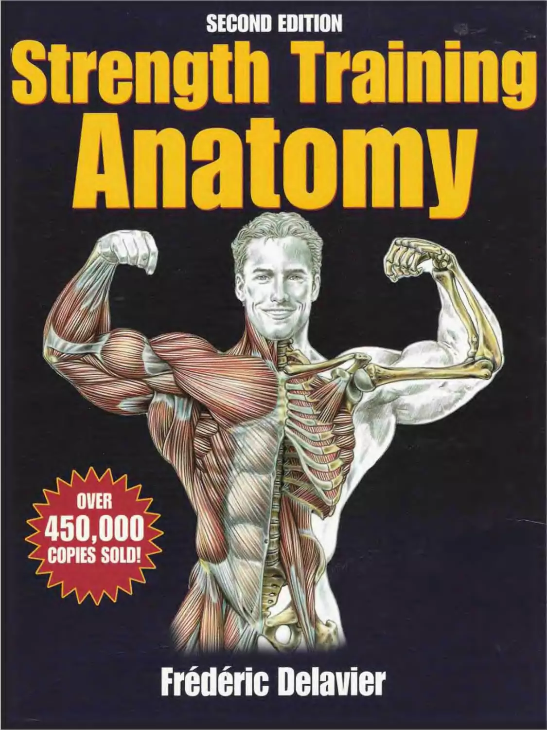
SECOND EDITION
Strength Training
Anatomy
A OVER
450,000
COPIES SOLD! x
Sternocleidomastoid
Semispinalis Capitis
Sternum
Pectoralis major,
abdominal part
Splenius capitis
Levator scapula
Scalene»
Omohyoid
Trapezius
Acromion
Clavicle
Deltoid
Pectoralis major, sternocostalis part
Triceps brachii, long head
Triceps brachii, medial head
Triceps brachii, lateral head
Biceps brachii
Brachialis
Brachioradialis (supinator)
Latissimus dorsi
Anconeus
Serratus anterior
Palmaris longus
Flexor digitorum superficialis
Extensor digitorum
Gluteus medius (under gluteal fascial
Gluteus maximus
Greater trochanter
Tensor fascia lata
Fascia lata, iliotibial band
Biceps femoris, long head
Vastus lateralis
Vastus medialis
Biceps femoris, short head
Vastus intermedius
Plantaris
Femur, lateral condyle
Meniscus
Head of fibula
Gastrocnemius, lateral head
Soleus
Fibularis longus
Extensor digitorum longus
Tibialis anterior
Fibularis brevis
Peroneus
Extensor hallucis longus
Lateral malleolus
Extensor digitorum brevis
Alxluctor digiti minimi pedis
External oblique
Rectus abdominis i under aponeurosis
Anterior superior iliac spine
Pronator teres
Brachioradiali'
Extensor carpi radialis longu'
Extensor carpi radialis brevis
Flexor carpi radialis
Palmaris longus
Flexor digitorum
sublimis
Thenar
Metacarpal bone II
Proximal phalanx
Distal phalanx
Rictus femoris
Adductor longus
Sartorius
Gracilis
Vastus medialis
Patella
Femur, medial condyle
Meniscus
Gastrocnemius, medial head
Tibia medial surface
Tibialis anterior
Soleus
Flexor digitorum longus
Flexor hallucis longus
Tendo calcaneus
i Achilles!
Medial malleolus
Talus
Navicular bone of foot
Medial cuneiform bone
Metatarsal Ixine I
Abductor hallucis
I inea alba
Iliopsoas r
r LU
(remaskw
Pyramida^
(under Hxneurdsis
Semispinalis capitis
Splenius capitis
Levator scapula
Sternocleidomastoid
Ztti cervici vertebra, spinous process
Trapezius
Spine of scapula
Acromion
Deltoid
Infraspinatus
Teres minor
Teres major
Rhomboid major
Thoracic vertebra, spi¬
nous process
Latissimus dorsl
Thoracolumbar fascia
External oblique
Lumbar trigone
Iliac crest
Sacrum, dorsal surface
Gluteus medius, beneath
the gluteal fascia
Tensor fascia lata
Greater trochanter
Coracobrachialis
Triceps brachii, Jong head
Biceps brachii
Triceps brachii, medial head
Triceps brachii, lateral head
Triceps brachii, tendon
Brachialis
Biceps brachii, tendon
Pronator teres
Medial epicondyle
Olecranon
Brachioradialis
Flexor carpi radialis
Palmaris longus
Flexor carpi ulnaris
Extensor carpi ulnaris
Aexor digitorum superficialis
Abductor pollicis longus
Aexor pollicis longus
Pisiform
Abductor pollicis brevis
Palmar aponeurosis
Distal phalanges
Extensor digitorum tendon
Adductor magnus
Iliotibial tract (fascia lata)
Vastus lateralis
Biceps femoris, long head
Vastus intermedius
Biceps femoris, short head
Femur, popliteal surface
Plantaris
Gastrocnemius, lateral head
Gastrocnemius, medial head
Peroneus longus
Soleus
Gastrocnemius tendon
Peroneus brevis
Flexor hallucis longus
Flexor digitorum longus
Lateral malleolus
Trochlea of the latus
Extensor digitorum brevis
Abductor digiti minimi
Gluteus maximus
Gracilis
Semitendinosus
Semimembranosus
Medial malleolus
Tendo calcaneus (Achilles)
Calcaneal tuberosity
Rectus abdominis
Lateral epicondyle
Olecranon
Anconeus
Umbilicus
External oblique
Gluteus medius
Tensor fascia lata
Fascia lata, iliotibial tract
Peroneus longus
Extensor digitorum longus
Peroneus brevis
i
Sternocleidomastoid
Scalenas
Trapezius
Sternohyoid
Sternum
Biceps brachii, long head
Serratus anterior
Latissimus dots!
Omonyoic. nfedoroeiiy
Clavicle
Acromion
Pectoralis major
Deltoid
Pectoralis major, abdominal portion
Biceps brachii
Brachialis
Triceps brachii, medial head
Pronator teres
Brachioradialis
Extensor carpi radialis longus
Flexor carpi radialis
Extensor carpi radialis brevis
Extensor digitorum
Abducta pollicis longus
Extensor pollicis brevis
Internal oblique
Pyramidalis
Anterior superior Iliac spine
Iliopsoas
Pectineus
Sartorius
Adductor longus
Gracilis
Rectus femoris
Vastus lateralis
Vastus medialis
Patella
Lateral condyle
Meniscus
Patellar ligamen!
Tibial tuberosity
Iliotibial tract
— Head of fibula
Gastrocnemius
Gastrocnemius
Tibialis anterior
Soleus
Medial surface of tibia
Flexor digitorum longus
Extensor hallucis longus
Peroneus tertius
Medial malleolus
Extensor hallucis brevis
Extensor digitorum brevis
Abductor hallucis
. Radius
i-'
STRENGTH
TRAINING
ANATOMY
HUMAN KINETICS
J _
L
1
L
1 ARMS 4
2 SHOULDERS 27
3 CHEST 49
4 BACK 6G
5 LEGS 93
G BUTTOCKS 115
7 ABDOMEN 128
1 CURLS 6
2 CONCENTRATION CURLS 7
3 HAMMER CURLS 8
4 LOW-PULLEY CURLS 9
5 HIGH-PULLEY CURLS 10
6 BARBELL CURLS 11
0 ELBOW STRUCTURE AND ITS EFFECT ON TRAINING 12
7 MACHINE CURLS 13
8 PREACHER CURLS 14
9 REVERSE CURLS 15
10 REVERSE WRIST CURLS 18
11 WRIST CURLS 17
12 PUSH-DOWNS 18
13 REVERSE PUSH-DOWNS 19
14 ONE-ARM REVERSE PUSH-DOWNS 20
15 TRICEPS EXTENSIONS 21
16 DUMBBELL TRICEPS EXTENSIONS 22
17 ONE-ARM DUMBBELL TRICEPS EXTENSIONS 23
18 SEATED DUMBBELL TRICEPS EXTENSIONS 24
19 SEATED E-Z BAR TRICEPS EXTENSIONS 24
20 TRICEPS KICKBACKS 25
21 TRICEPS DIPS 26
4
ARMS
Extensor pollicis longus, tendon
Extensor retinaculum
Teres major
Latissimus dorsi
Subscapularis
Pectoralis major
Serratus anterior
Anterior deltoid
Flexor digitorum profundus, tendon
Adductor pollicis
Abductor pollicis brevis
Flexor pollicis longus
Flexor digitorum superficialis
Flexor carpi radialis
Palmaris longus
Pronator teres
Biceps brachii, aponeurosis
Brachialis
Triceps brachii, medial head
Tnceps brachii, long head
Coracobrachialis
Extensor pollicis brevis
Abductor pollicis longus
Extensor carpi radialis brevis
Extensor carpi radialis longus
Brachioradialis
Biceps brachii, tendon
Extensor carpi radialis longus, tendon
carpi radialis brevis, tendon
1st dorsal interosseous muscle
digitorum, tendon
Flexor digitorum superficialis, tendon
Biceps brachii, long head
Biceps brachii, tendon
Flexor pollicis longus
Rexor digitorum superficialis
Flexor digitorum profundus
Flexor digitorum superficialis, tendon
Lumbrical
Brachialis, tendon
Brachialis
5
Middle deltoid
Biceps brachii
Opponens pollicis
Biceps brachii
Brachialis
Triceps brachii, long head
Triceps brachii, medial head
Coracobrachialis
Capitate
Trapezoid
Lunate
Metacarpal
Proximal phalanx
Capitulum
Head of radius
Bicipital groove
Lesser tubercle
Greater tubercle
Middle phalanx
Acromion
Clavicle,
Coracoid process,
Distal phalanx
Trapezium
Radial tuberosity
Scaphoid
Radius
Humerus
Trochlea
Ulna
Deltoid tuberosity
ARMS
1j CURLS
Flexor carpi ulnaris
Pectoralis major
(clavicular head)
Anterior deltoid
Middle deltoid
Flexor carpi radialis
Palmaris longus
Pronator teres
Triceps brachii, medial head
Brachialis
Triceps brachii, long head
Posterior deltoid
Triceps brachii, lateral head
Brachialis
Brachioradialis
Extensor carpi radialis longus
Anconeus
Extensor carpi radialis brevis
Extensor digitorum
Extensor carpi ulnaris
Extensor digiti minimi
Sit holding a dumbbell in each hand with arms hanging down and the palms of the hands
facing the body:
• Inhale and bend the the elbow, rotating the palm up before the forearm reaches horizontal.
• Continue by raising the elbows at the end of the movement.
This exercise primarily uses the brachioradialis (long supinator), brachialis, biceps brachii, and
anterior deltoid and, to a lesser extent, the coracobrachialis and clavicular head of the
pectoralis major.
COMMENT: This exercise takes the biceps through its complete
range of motion, which includes flexion, protraction, and
supination.
THREE WAYS TO EXECUTE CURLS
T] EMPHASIZE BICEPS
2 WORK BRACHIORADIALIS INTENSELY
S WORK MAINLY BICEPS AND BRACHIALIS
6
Biceps brachii
E SUPINATION
E PRONATION
ARMS
CONCENTRATION CURLS
Trapezius
Coracobrachialis
Triceps brachii, long head
Brachialis
Triceps brachii, medial head
Pronator teres
Flexor carpi radialis
Palmaris longus
Rexor carpi ulnaris
Humerus
Scapula
Pectoralis major
Anterior deltoid
Middle deltoid
Biceps brachii
Triceps brachii, lateral head
Brachioradialis
Extensor carpi radialis longus
Extensor carpi radialis brevis
Clavicle
Clavicle
Sternum
Coracobrachialis
Rib
Radius
Carpal
Brachialis
Proximal phalanx
Biceps brachii, tendon
Distal phalanx
Biceps brachii,
long head
Costal
cartilages
Biceps brachii,
short head
Coracoid
| process
Biceps brachii, aponeurosis
BRACHIALIS MUSCLE
Acromion
Humerus
Brachialis
Ulna
Radius
Melacarpai
Middle phalanx
Acromion
Sit holding a dumbbell with the
palm facing forward and the elbow
positioned against the inner thigh:
• Inhale and lift the forearm by
bending the elbow.
• Exhale at the end of the effort.
This isolation exercise allows you to
control the range of motion, speed,
and form of the movement.
It mainly works the biceps brachii
and brachialis.
7
Biceps brachii, tendon
FINAL POSITION
Ulna
ARMS
HAMMER CURLS
Trapezius
Infraspinatis
Teres minor
Teres major
Latissimus dorsi
Lateral head
Extensor carpi radialis longus
Extensor carpi radialis brevis
Anconeus
Extensor carpi ulnaris
Flexor carpi ulnaris
Long head
Medial head
BRACHIORADIALIS MUSCLE
Posterior deltoid
Middle deltoid
Anterior deltoid
Pectoralis major
Biceps brachii
Brachialis
Brachioradialis
Deltoid
Metacarpal
THE MOVEMENT
Extensor digitorum
Extensor digiti minimi
Clavicle
Costal cartilage
Sternum
Brachioradialis
Middle phalanx
Triceps bractiii
Radius
Ulna
Scapula
Humerus
Proximal phalanx
Stand or sit gripping a dumbbell in each hand with
the palms facing each other:
• Inhale and raise the forearms together or
alternately.
• Exhale at the end of the movement
This is the best exercise for developing the
brachioradialis.
It also develops the biceps brachii, brachialis, and, to
a lesser degree, the extensor carpi radialis brevis
and longus.
8
Caipal
Distal ptoanx
ARMS
LOW-PULLEY CURLS
4
Spterfus capitis
Sternocleidomastoid
Levator scapula
Trapezius
Infraspinatus
Teres minor
Teres major
Triceps brachii
Latissimus dorsi
Brachialis
Extensor carpi
radialis longus
Anconeus
Scalenes
Deltoids
Pectoralis major
Biceps brachii
Stand facing the machine, grasping the handles with an underhand grip (thumbs facing
away from each other):
• Inhale and bend the elbows to raise the forearms.
• Exhale at the end of the movement.
This exercise focuses the effort on the biceps brachii and works the muscle intensely.
Extensor pollicis brevis
Flexor carpi radialis
Abductor pollicis longus
Extensor digitorum
VARIATION
Two-handed lovz-pulley curb
9
Extensor carpi
radialis brevis
Brachioradialis
THE MOVEMENT
ARMS
HIGH-PULLEY CURLS
Flexor carpi radialis
Clavicle
Scapula
Sternum
Rib
Pectoralis major
Long head
Short head
Biceps brachii
Flexor carpi ulnaris
Palmaris longus
Aponeurotic
expansion of
biceps brachii
Triceps brachii,
medial head
Brachialis
Triceps brachii, long head
Coracobrachialis
Teres major
Radius
Humerus
Brachialis
Stand between the pulleys with the arms outstretched in a "cross" and grasp the handles of the high pulleys with an underhand grip:
• Inhale and bend the elbows to bring the hands toward the body. Exhale at the end of the movement.
This exercise, which is most often performed as a cool-down at the end of an arm session, focuses the work on the short head
of the biceps brachii, which has been stretched and put under tension in the "cross" start-up position.
This exercise also contracts the monoarticular brachialis elbow flexor.
Perform this exercise with light weights so that you can concentrate and feel the contraction at the inside of the biceps brachii.
Sets of high reps provide the best results.
VARIATION
One-handed execution
Extensor carpi ulnaris
Flexor carpi
radialis
Palmaris
longus
Flexor carpi
radialis
Aponeurosis ol
biceps brachii
Pronator teres
Triceps brachii,
medial head
Brachialis
Brachioradialis
Coracobrachialis
Latissimus dorsi
Serratus anterior
External oblique
Teres major
Subscapularis
triceps brachii, long head
When the hand is pronated, the distal
tendon of the biceps brachii muscle is
partially wrapped around the radius.
When the biceps brachii contracts, the
force placed on its distal tendon causes
the radius to pivot on its axis, bringing
the hand into supination.
Comment: In addition to its role as a forearm flexor, the
biceps brachii is the most powerful supinator.
10
Flexor digitorum superfaalis
Sternocleidomastoid
Pronator teres
■ Ulna
Latissimus dors!
Serratus anterior
Flexor digitorum superficialis
Phalanx
Radius
Ulna
Metacarpal
Carpal
Humerus
Phalanx
Metacarpal
Carpal.
Humerus
Radius
Deltoid
Ulna
Biceps brachii
Biceps brachii tendon,
partially wrapped around
the radius
Biceps brachii tendon,
partially wrapped around
the radius
ARMS
BARBELL CURLS f 6 I
Biceps brachii
Long head
Short head
Pectoralis major
Deltoid
Sternocleidomastoid
Flexor digitorum superficialis ■
Long head
Short head
Sternum
Os coxa
Floating ribs
Lumbar vertebra
Costal cartilages
Biceps brachii
Brachialis
Biceps brachii, tendon
Radius
Omohyoid
Sternohyoid
1st rib
Clavicle
Acromion
Coracoid process
Scapula
Triceps brachii,
Brachialis
Pronator teres.
Biceps brachii, aponeurosis
Flexor carpi radialis
Flexor carpi ulnaris
Palmaris longus-
Extensor carpi radialis longus
Extensor carpi radialis brevis
Flexor pollicis longus
Biceps brachii
BRACHIALIS MUSCLE
Stand with the back straight, grasping the barbell with an underhand grip and hands slightly wider than shoulder-width apart:
• Inhale and raise the barbell by bending the elbows, taking care to stabilize the torso and spine by isometrically contracting
the gluteal muscles, abdominal muscles, and spinal muscles.
• Exhale at the end of the movement.
This exercise mainly contracts the biceps brachii, brachialis, and,
to a lesser degree, the brachioradialis, pronator teres, and the
wrist flexor group.
Variations: Vary the width of the grip to work different parts of the
muscle more intensely:
• Placing the hands farther apart isolates the short head of the
biceps brachii.
• Placing the hands closer together isolates the long head of the
biceps brachii.
Raising both elbows after they are flexed increases the
contraction of the biceps brachii and contracts the anterior deltoid.
To make the exercise more difficult perform the movement with
the back against a wall so that the shoulder blades don't move.
You can lift more weight and gain strength by leaning the torso back
while lifting the bar; however, to prevent injury, this requires good
technique and well-developed abdominal and lumbar muscles.
BARBELL CURLS
Q] Narrow grip
Mainly worts the long heart of the biceps brachii.
[H Wide grip
Mainly works the short head of lhe biceps brachii
THE MOVEMENT
Scalenes
Trapezius
Brachioradialis.
Ulna
Clavicle
Coracrxd process
Lesser
tubercle
taomlon
Head ni humerus
ftrr^fpr tutarnlp
Rib
Bicipital
groove
Ulna
Brachialis,
tendon
Brachialis
Radius
ARMS
ELBOW STRUCTURE ANO ITS EFFECT ON TRAINING
Clavicle
Lunate
Styfcd process
Scaphoid
Trapezium
Trapezoid
Capitate
Glenoid
cavity
Acromion
Metacarpal
Proximal phalanx
Middle phalanx
Distal phalanx
Coronoid fossa
Medial epicondyle
Trochlea
Coronoid
process
Ulnar tuberosity
Ulna
Head of ulna
Styloid process
Pisiform
Triquetral
Hamate
Coracoid
Head of humerus
Greater tuberosity
Lesser tuberosity
Bicipital groove
Crest of lesser tubercle
Crest of greater tubercle
Deltoid tuberosity
Radial fossa
Lateral epicondyle
Capitulum
Head of radius
Radial tuberosity
Radius
Biceps training with an E-Z bar eases excessive wrist tension.
E GO
E Upper extremity with a small angle
E Upper extremity with a significant valgus angle
(more common in women)
When training the biceps brachii using a barbell,
take into account variations in each person’s
physical structure.
In the anatomical position (arms hanging alongside
the body, palms facing forward, and thumbs pointing
laterally), the angle at the elbow between the upper
arm and the forearm varies from person to person.
Someone whose forearm hangs distinctly away from
the body in a valgus position must break excessively
at the wrist when performing a curl with a straight
bar, which is painful. Therefore, these people should
work with an E-Z bar to spare their wrists.
Comment: Valgus of the elbow is usually more pronounced in women.
12
Scapula
ARMS
Trapezius
Triceps brachii, lateral head
Omohyoid
Scaienes
Deltoid
Sternocleidomastoid
MACHINE CURLS
Pectoralis major
Biceps brachii
Brachialis
INITIAL POSITION
Sit at the machine and grasp the bar with an underhand grip, arms extended, and resting on the support:
• Inhale and raise the forearms.
• Exhale at the end of the movement.
This is one of the best exercises for working the biceps brachii. Fixing the arms against the support makes it
impossible to “cheat."
At the beginning, the muscle tension is intense, so be sure to warm up properly using light weights. To avoid the
risk of tendonitis, do not completely extend the arm.
This movement also works the brachialis and. to a lesser extent, the brachioradialis and pronator teres.
Performing the cud with an Atlas pulley
is a great way to pump up the muscle
13
ARMS
PREACHER CURLS
Sternocleidomastoid
Trapezius
Deltoid
Pectoralis major
Biceps brachii
Brachialis
Brachioradialis
Pronator teres
Flexor carpi radialis
Lateral head
Long head
Medial head
Triceps
brachii
Biceps brachii,
aponeurotic expansion
Palmaris longus
Sit or stand with the arms resting on the support pad and grasp the bar with an underhand grip:
• Inhale and raise the forearms by bending the elbows. Exhale at the end of the effort.
This is one of the best exercises for isolating the biceps.
Attention: The angle of the support pad places significant tension on the forearms when the arm is
completely extended. Therefore, warm up the muscles properly and begin with lighter weights.
Brachioradialis
Deltoid
Brachialis
Triceps brachii, medial Head
Biceps brachii
flexor
digitorum
Extensor carpi
radialis longus
Extensor pollcis
brevis
Extensor pollicis
Pectorals ma|«
Flexor carpi ulnaris
Serratos
anterior
Triceps brachii,
long head
Teres major
Latissimus dorsi
Abductor polHcis longus
Palmaris longus
Extensor carpi radialis brevis
Flexor carpi radials
THE MOVEMENT
Pronator teres
14
ARMS
REVERSE CURLS
M
Triceps brachii
Extensor carpi radialis longus
Olecranon
Splenius capitis
Sternocleidomastoid
Sealeries
Infraspinatus
Teres minor
Teres major
Long head
Lateral head
Brachioradialis
Anconeus
Extensor retinaculum
Thyrohyoid
Levator scapula
Flexor carpi ulnaris
Head of ulna
Extensor carpi radialis brevis
Extensor digitorum
Extensor digiti minimi
Extensor carpi ulnaris
Extensor pollicis
brevis
Triceps brachii,
lateral bead
Triceps brachii,
long head
Triceps brachii,
tendon
Lateral epicondyle
Anconeus
Olecranon
Extensor
digitorum
Extensor
carpi ulnaris
Abductor
pollicis
longus
Brachialis Braps brachii
Extensor
digiti minini
Brachioradialis
Extensor carpi radialis longus
Extensor carpi radialis brevis
Abductor pollicis longus
Extensor
podicis
brevis
MUSCLES OF THE FOREARM (LATERAL VIEW)
Omohyoid
Sternohyoid
Trapezius
Deltoids
Pectoralis major
Brachialis
Biceps brachii
THE MOVEMENT
Stand with the legs slightly apart and arms extended and grasp the bar with an overhand grip (with the thumbs facing each other):
• Inhale and raise the forearms by bending the elbows.
• Exhale at the end of the movement.
This exercise works the extensor muscles of the wrist: extensor carpi radialis longus, extensor carpi radialis brevis, extensor
digitorum, extensor digiti minimi, and extensor carpi ulnaris.
It also acts on the brachioradialis, brachialis, and, to a lesser degree, the biceps brachii.
Comment: This is an excellent exercise for strengthening the wrist, which is often weak because of an imbalance caused by using the wrist flexors rather
than the wrist extensors. For this reason, many boxers include it in their training. Many bench press champions use it to keep their wrists from trembling
under extreme weights.
15
Una
Flexor carpi
ulnaris
Radius
Extensor pollicis
longus, tendon
ARMS
10
REVERSE WRIST CURLS
Ulna
Radius
Extensor carpi radialis longus
Extensor carpi radialis brevis
Extensor digitorum
Extensor digiti minimi
Flexor carpi ulnaris
Palmaris longus
Extensor digitorum
— Radius
FINAL POSITION
Carpal
Extensor
indicis
Comment: This exercise strengthens the wrists, which are
often vulnerable because of weak wrist extensors.
Extensor carpi radialis
brevis
Extensor digit minimi
Humerus
Extensor carpi radialis
longus
Brachioradialis
Flexor carpi radialis
Extensor carpi
radialis longus
Extensor carpi
radialis brevis
Extensor digitorum
Abductor pollicis longus
Extensor pollicis brevis
Flexor pollicis longus
Flexor digitorum
superficialis
Extensor pollicis
Extensor indicis
1st dorsal interosseous
I Metacarpal
Humerus
Extensor carpi ulnaris
Medial
epicondyle
Olecranon
WRIST EXTENSORS
Ulna —!
Head of ulna
Metacarpal
Middle phalanx -
Distal phalanx
Proximal phalanx
Extensor carpi y \ j
ulnaris
■'x Sit with the forearms resting on the thighs or on a bench and grasp
the bar with an overhand grip and keep the wrists relaxed:
• Raise the hands by extending at the wrists.
This exercise contracts the extensor carpi radialis longus and
brevis, extensor digitorum, extensor digiti minimi, as well as the
extensor carpi ulnaris.
16
Extensor indicis
Phalanx
ARMS
WRIST CURLS
11
Pectoralis major
Deltoid
Biceps brachii
Triceps brachii, long head
Brachialis
Humerus
Brachioradialis
Radial tuberosity
Radius
Pisiform
Flexor carpi ulnaris
Flexor pollicis longus
irapezlum
Metacarpal
WRIST FLEXORS
Pronator teres
Palmaris longus
Flexor
digitorum
superficialis
Flexor
digitorum
profundus
Flexor pofllcis
longus
Flexor carpi
radialis
Flexor digitorum superficialis
covering flexor digitorum
profundus
Flexor carpi
ulnaris
Sit with the forearms resting on the thighs or on
a bench and grasp the bar with an underhand
grip with wrists relaxed:
• Inhale and raise the hands by flexing at the
wrists.
This exercise contracts the flexor carpi radialis,
palmaris longus, flexor carpi ulnaris, and the
flexors digitorum superficialis and profundus.
The latter two muscles, although located deep in
the wrist, make up most of the muscle mass of
the wrist flexors.
Flexor carpi radialis
Palmaris longus ——
Flexor digitorum
superficialis and
profundus
Pisfforrrr
Triceps brachii, medial head
Pronator teres
Superficial layer
Middle layer
Deep layer
THE MOVEMENT
17
Ulna
[T| Begin
[2] End
ARMS
12
PUSH-DOWNS
Splenius
Sternocleidomastoid
Levator scapula
Sealeries
Trapezius
Spine of scapula
Deltoid
Infraspinatis
Teres minor
Teres major
Triceps brachii
Lateral head
Long head
Medial head
Olecranon
External oblique
Anconeus
Flexor carpi ulnaris
Biceps brachii
Brachialis
Brachioradialis
Triceps
brachii
Medial head
Lateral head
Long head
Pectoralis major
Extensor carpi radialis longus
Extensor carpi radialis brevis
Extensor digitorum
Extensor digiti minimi
Extensor carpi ulnaris
Head of ulna
Extensor retinaculum
Stand with the back to the machine and grasp the handle with an
overhand grip, keeping the elbows tucked into the body;
• Inhale and extend the forearms, keeping the elbows tucked
into the body.
• Exhale at the end of the movement.
Comment: This exercise isolates the triceps and the
anconeus.
The variation using a rope rather than a handle engages the
lateral head of the triceps more intensely.
Performing the movement with an underhand grip requires
more contribution from the medial head of triceps.
Hold an isometric contraction for one or two seconds at the
end of the movement to feel the effort more intensely.
When using heavy weights, lean forward with the torso.
Beginners can use this exercise to develop enough strength
to move on to more difficult exercises.
VARIATION WITH BACK TO THE MACHINE
To isolate ol the tong head ol the triceps.
VARIATION WITH A ROPE
To isolate die lateral head of the triceps
18
THE MOVEMENT
ARMS
REVERSE PUSH-DOWNS
Trapezius
Omohyoid
Deltoid
Infraspinatus
Teres minor
Teres major
Triceps brachii, long head
Latissimus dorsi
Triceps brachii, lateral head
Biceps brachii
Brachialis
Brachioradialis
Triceps brachii, medial head
Lateral epicondyle
Olecranon
Splenius capiti
Sternocleidomastoid
Levator scapula
Scalenes
1st dorsal interosseous
Extensor pollicis longus
Flexor digitorum superficialis
Flexor carpi radialis
Extensor carpi radialis brevis
Extensor digitorum
Flexor carpi ulnaris
Extensor carpi ulnaris
Extensor carpi radialis longus
Anconeus
Stand facing the machine with the arms next the body and elbows bent and grasp the handle with an
underhand grip:
• Inhale and extend the forearms by straightening the elbows, keeping them tucked into the body.
• Exhale at the end of the movement.
The underhand grip isolates the medial head of the triceps brachii and precludes working with heavy
weights.
When extending the forearms, the anconeus and wrist extensors also contract
The extensor carpi ulnaris, extensor digitorum, extensor digiti minimi, and extensors carpi radialis longus
and brevis keep the wrist straight with isometric contraction during the exercise.
Acromion
Triceps brachii,
long head
Triceps brachii,
lateral head
Ulna
Triceps brachii,
medial head
Styloid
process
epicondyle
Olecranon
Anconeus
Carpal
Metacarpal
Proximal phalanx
Middle phalanx
Distal phalanx
Clavicle
Coracoid
process
Spine of
scapula
Scapula
Vertebra
Rii
Triceps
brachii,
tendon
19
Head of humerus
■ Pectoralis major
Radius
Medial
ARMS
14
ONE-ARM REVERSE PUSH-DOWNS
Infraspinatus
Trapezius
Teres minor
Deltoid
Pectoralis major
Biceps brachii
Brachialis
INITIAL POSITION
Brachioradialis
Flexor carpi ulnaris
Anconeus
Extensor carpi ulnaris
Extensor carpi radialis longus
Extensor digiti minimi
Extensor carpi radialis brevis
Extensor digitorum
Triceps brachii,
medial head
Triceps brachii,
lateral head
Triceps brachii,
long head
Triceps
brachii,
tendon
Teres major
Latissimus dorsi
Stand facing the machine and grasp the handle with an underhand grip:
• Inhale and extend the forearm,
• Exhale at the end of the movement.
This exercise mainly works the lateral head of the triceps.
INSERTIONS OF THE ARM
Supraspinatus
Infraspinatus
Tnceps brachii, long heap
Triceps brachii, lateral head
Deltoid
Brachialis ■
Triceps brachii, medial head
Medial epicondyle musdes
Triceps brachii, lentiori
Anceneus
Brachioradialis
Supraspinatus
Coracobrachialis
Infraspinatus
Latissimus dorsi
Teres major
Coracobrachialis
Brachialis
Lateral epicondyle muscles
Extensor carpi
radlaBs longus
Brachialis
Medial epicondyle muscles
20
Deltoid
Biceps brachii
Pectoralis major
Posterior view
Anterior view
ARMS
TRICEPS EXTENSIONS 15
Olecranon
Pectoralis major
Serratos anterior
Subscapularis
Teres major
Posterior deltoid
Flexor carpi ulnaris
Palmaris longus
Flexor carpi radialis
Flexor digitorum superficialis
Flexor pollicis longus
Abductor pollicis longus
Extensor pollicis brevis
Biceps brachii,
aponeurotic expansion
Pronator teres
Brachialis
Medial head
Lateral head
Long head
Triceps
brachii
Biceps brachii
Coracobrachialis
jtisstmus dorsl
|T| Lowering the bar to the forehead
Focuses the work on the medial and lateral heads of the triceps txachli.
[2] Lowering the bar behind the head
focuses the work on rhe tong head of the triceps brachii.
Lie on a horizontal bench and grasp the barbell with an overhand grip and the arms vertical:
• Inhale and lower the barbell to the forehead or behind the head by bending the elbows.
• Return to the initial position.
• Exhale at the end of the effort.
Comments: Because of individual variations In shoulder width, valgus angle at the elbows, and wrist flexibility, the hands
can be closer or farther apart on the bar and the elbow angle more or less open during the exercise.
Using an E-Z bar helps prevent excessive strain at the wrists.
VARIATION ON A MACHINE
Performing this exercise al an Atlas triceps
pulley simulates the movement with a
barbell, but enables you to isolate the long
head of the triceps brachii.
21
ARMS
16 DUMBBELL TRICEPS EXTENSIONS
Abductor pollicis longus
Extensor pollicis brevis
Extensor
pollicis
longus
Flexor carpi ulnaris
Extensor carpi ulnaris
Extensor digiti minimi
Extensor digitorum
Anconeus
Extensor carpi radialis brevis
Extensor carpi radialis longus
Brachioradialis
Brachialis
Serratus anterior
Latissimus dorsi
Anterior deltoid
Middle deltoid
Triceps
brachii
Lateral head
Long head
Teres minor Teres major
Infraspinatus
Lie on a flat bench and grasp a dumbbell in each hand with the arms vertical:
• Inhale and lower the forearms by bending the elbow with a controlled
movement.
• Return to the initial position.
• Exhale at the end of the effort.
This exercise works all three heads of the triceps brachii equally.
22
Medial head
Biceps brachii
Posterior deltoid
THE MOVEMENT
Clavicle
Coracoid
process
Spine of scapula
Scapula
Vertebra
Rib
Acromxn
Head of humerus
Triceps brachii, long head
Triceps brachii, lateral head
Triceps brachii, medial head
Triceps brachii, tendon.
Radius ■
Ulna -
Styloid process
Medial epicondyle
Carpal
Metacarpal
Proximal phalanx
Middle phalanx
Distal phalanx
Olecranon
Anconeus
ARMS
ONE-ARM DUMBBELL TRICEPS EXTENSIONS 17
Phalanx
Metacarpal
Carpal
Ulna
Radius
Sternocleidomastoid
Splenius
Levator scapula
Trapezius
Spine ot scapula
Deltoid
Infraspinatus
Teres minor
Anconeus
Olecranon
Tendon
Lateral head
Long head
Triceps brachii
Triceps brachii
Lateral head
Long head
Medial head
Biceps brachii
Brachialis
Olecranon
Pronator teres
Biceps brachii, aponeurosis
Brachioradialis
Flexor carpi radialis
Palmaris longus
Flexor digitorum
Flexor carpi ulnaris
Humerus
Acromion
Clavicle
Scapula
Rhomboid major
Teres major
Rib
Latissimus dorsi
Vertebra
Thoracolumbar fascia
External oblique
Sit or stand and grip a dumbbell in one hand with the arm vertical:
• Inhale and bend the elbow to lower the dumbbell behind the head to the neck.
• Return to the initial position.
• Exhale at the end of the movement,
The vertical position of the arm stretches the long head of the triceps brachii, emphasizing its contraction while working.
Comment: Contract the abdominal core to prevent arching the low back. If possible use a bench with support for the
low back.
THE MOVEMENT
23
ARMS
fl 8 SEATED DUMBBELL TRICEPS EXTENSIONS
Triceps brachii,
medial head
Pectoralis major
Teres minor
Palmaris longus
Flexor carpi uinaris-
Extensor carpi ulnaris
Extensor digiti minimi
Extensor digitorum
Extensor carpi radialis brevis
Extensor carpi radialis longus
Brachioradialis
Lateral head
Long head
Deltoid
Infraspinatus
Latissimus dorsi
Serratus anterior
Triceps brachii
Sit and grasp a dumbbell, holding
it behind the neck:
• Inhale, and extend the forearm.
• Exhale at the end of the
movement
The vertical position of the arm
strongly stretches the long head
of the triceps brachii, emphasizing
its contraction while working.
Contract the abdominal core to
prevent arching the low back. If
possible use a bench with
support for the low back.
19 SEATED E-Z BAR TRICEPS EXTENSIONS
Flexor carpi
ulnaris'
Medial head
Lateral head
Long head
Head of humerus
Clavicle
THE MOVEMENT
Triceps
brachii
Scapula
Rib
Radius
Ulna
Triceps brachii, tendon
Sit or stand and grasp an E-Z bar with an overhand
grip and arms vertical:
• Inhale and bend the elbows to lower the bar
behind the head.
• Return to the initial position.
• Exhale at the end of the extension.
The vertical position of the arms strongly stretches
the long head of the triceps brachii, emphasizing
its contraction while working.
An overhand grip isolates the lateral head of the
triceps brachii.
Contract the abdominal muscles and avoid arching
the low back. If possible use a bench with support
for the low back.
Palmaris longus
Flexor carpi radiate
Brachioradialis
Biceps brachii,
aponeurotic expansion
Pronator teres
Brachiate
Triceps brachii,
medial head
Biceps brachii
Triceps brachii,
long head
Deltoid'
Teres major'
Coracobrachialis
Latissimus dorsi'
24
Anconeus
ARMS
TRICEPS KICKBACKS 20
Flexor carpi ulnaris
Extensor digitorum
Extensor digiti minimi
Extensor carpi ulnaris
Extensor
pollicis brevis
Teres major
INITIAL POSITION
Deltoid
Biceps brachii
Pectoralis major
Brachialis
Extensor carpi radialis longus
Extensor carpi radialis brevis
Stand with the knees slightly bent and lean forward at the waist, maintaining a straight back.
Bend the elbow and hold the upper arm horizontally alongside the body:
• Inhale and extend the forearm.
• Exhale at the end of the movement.
This is an excellent exercise for pumping the triceps group.
Perform this exercise until you feel a burn for best results.
25
Abductor pollicis longus
Triceps brachii
Lateral head
Long head
Brachioradialis
Anconeus
ARMS
21 TRICEPS DIPS
Deltoid
Pectoralis major
Lateral head
Long head
Triceps brachii
Extensor digitorum
Extensor digiti minimi
Trapezius
Teres minor
Infraspinatus
Teres major
Rhomboid
Latissimus dorsi
Flexor carpi radialis
Palmaris longus
Extensor carpi ulnaris
Flexor carpi ulnaris
TRICEPS BRACHII MUSCLE
Scapula
Clavicle
Triceps brachii,
long head (sectioned)
Triceps brachii, lateral head
Acromion
Spine of scapula
.Head of humerus
Glenoid cavity
Triceps brachii, long head
Triceps brachii,
long head (selection)
Vertebra
Triceps brachii, tendon
Triceps brachii, medial head
Anconeus.
Os coxa
Sacrum
Lateral
epicondyle
Olecranon
Radius
Medial epicondyle
Ulna
Suspend the body between two benches by placing the hands on the edge of one bench and the feet on the edge of the other bench:
• Inhale, then dip by bending the elbows and rise by extending the forearms.
• Exhale at the end of the movement.
This exercise works the triceps and pectorals as well as the anterior deltoid.
Resting weights on top of the thighs increases the difficulty and intensity of the dip.
26
Medial head
Anconeus
INITIAL POSITION
Rib
Latissimus dorsi
BACK
Clavicle
Humerus
Spine el
scapula
Scapula
Vertebra
External
oblique
Semispinalis capitis
Splenius
Sternocleidomastoid
Anterior
deltoid
Jriceps brachii,
long head
Triceps brachii,
lateral head
Teres major
Teres minor
Infraspinatus
1 BACK PRESSES 28
2 SEATED FRONT PRESSES 29
l+l SHOULDER INJURIES / LYING DUMBBELL PRESSES 30
3 SEATED DUMBBELL PRESSES 33
4 FRONT DUMBBELL PRESSES 34
5 BENT-OVER LATERAL RAISES 35
6 LATERAL DUMBBELL RAISES 36
7 ALTERNATE FRONT ARM RAISES 30
8 SIDE-LYING LATERAL RAISES 39
9 LOW-PULLEY LATERAL RAISES 40
10 LOW-PULLEY FRONT RAISES 41
11 HIGH-PULLEY LATERAL EXTENSIONS 42
12 LOW-PULLEY BENT-OVER LATERAL RAISES 43
13 ONE-DUMBBELL FRONT RAISES 44
14 BARBELL FRONT RAISES 45
15 UPRIGHT ROWS 4G
IB NAUTILUS LATERAL RAISES 47
17 PEC DECK REAR-DELT LATERALS 48
27
FRONT
Scalenes,
1st rib
Clavicle
Humerus
Deltoid
Trapezius
trapezius
Biceps brachii
Middle deltoid
Anterior deltoid
Brachialis
Triceps brachii,
medial head
Triceps brachii, long head-
Coracobrachialis
Teres major
Latissimus dorsi'
Subscapularis'
Pectoralis major'
Serratus anterior
Scapula
Sternum
Rectus abdominis,
under the aponeurosis
Umbilicus
Anterior superior
iliac spine
Pyramidalis
Dither- ct/mnhudie
Rib
Posterior
deltoid
SHOULDERS
Deltoid
BACK PRESSES
Epicranius, occipital belly
Brachioradialis
Extensor
digitorum
Extensor carpi
radialis brevis
Extensor carpi
ulnaris
Extensor carpi
radialis longus
Anconeus
Triceps
brachii
Anterior deltoid
Middle deltoid
Posterior deltoid
Lateral head
Medial head
Long head
Brachialis
Biceps
brachii
/Teres minor
' Teres major
Infraspinatus
Rhomboid major
Latissimus dorsi
External oblique
Semispinalis
capitis
Splenius
capitis
Sternocleido¬
mastoid
Trapezius,
Cranium
Mastoid process
Cervical vertebra
Supraspinatus
Spine of sea
Radius
Ulna
Humerus
Scapula
9th nb
Thoracic vertebra
Lumbar vertebra
Thoracolumbar fascia
Sit with the back straight, holding the bar across the back of the neck with an overhand grip:
• Inhale and extend the bar straight up, keeping the tow back as straight as possible.
• Exhale at the end of the effort.
This exercise uses the deltoid, mainly the middle and posterior fibers, as well as the trapezius, triceps brachii, and
serratus anterior. Although not worked as Intensely, the rhomboids, infraspinatus, teres minor, and, deeper in, the
supraspinatus also contract. You can also perform this exercise while standing at a frame that guides the barbell.
Various specific machines can help with the performance of this exercise.
To prevent injury to the shoulder joint, which is vulnerable, lower the bar only as far as your
unique shoulder structure and flexibility allow you to do comfortably.
28
THE MOVEMENT
Clavicle
Acromion
SHOULDERS
SEATED FRONT PRESSES
2
Deltoid
Splenius
Sternocleidomastoid
Trapezius
Anterior deltoid
Posterior deltoid
Middle deltoid
Spine of scapula
Acromion
Biceps brachii
Brachialis
Brachioradialis
Extensor carpi
raoialis longus
Anconeus
Triceps brachii
Lateral head
Medial head
Long head
Rhomboid major
Latissimus dorsi
FINAL POSITION
Teres minor
Teres major
Infraspinatus
Pectoralis major
clavicular head
Anterior deltoid
Triceps
FRONT VIEW
Q] Narrow grip with the elbows forward
Isolates the anterior deltoid and the clavicular head of the pec¬
toralis major.
|?J Wide grip with the elbows out to the side
Isolates the anterior and middle deltoids.
Sit with the back straight and hold the bar with an
overhand grip, resting it across the upper chest:
• Inhale and extend the bar vertically.
• Exhale at the end of the movement.
This fundamental exercise mainly uses the anterior
and lateral deltoids, clavicular head of the pectoralis
major, triceps brachii, serratus anterior, trapezius and.
deeper in, the supraspinatus.
You can also perform this exercise standing, as tong as you keep the back straight, avoiding excessive curvature of the lumbar spine. Extending the barbell with the
elbows forward isolates the anterior deltoid.
Extending the bar with the elbows spread apart isolates the middle deltoid.
You can use various machines for this exercise.
29
SHOULDERS
SHOULDER INJURIES
HIP JOINT
SHOULDER JOINT
Shoulder injuries occur frequently in weightlifting and especial¬
ly in bodybuilding where developing the entire deltoid group
requires the athlete to perform a significant number of repeti¬
tions and variations in exercises, which multiplies the risk of
chii anteriorly. Raising the arm becomes extremely painful and
eventually can cause irreversible deterioration of the supraspi¬
natus tendon through calcification and even tearing; however,
this usually happens to people 40 years of age or older.
Compared to the relatively
stable coxofemoral joint, the
shoulder joint is less enca¬
sed and is more mobile,
which makes it more vulne¬
rable to injury.
injury.
Compared to the stability of the hip joint, where the head of the
femur sits deep in the glenoid cavity of the pelvis, the shoulder
joint, which is very mobile and allows the arm to move through
a wide range of motion, Is in fact much less contained and pro¬
tected.
The shoulder is defined as a ball-and-socket joint because the
head of the humerus is mainly held within the glenoid cavity of
the scapula by a complex musculotendinous group,
Most weightlifting injuries occur when training the deltoids, and
they rarely result in muscle pulls or tears. They are usually cau¬
sed by poor technique or overuse of the tendons reinforcing the
articular capsule.
In contrast to contact sports, such as football, where sudden
arm movements can create serious injuries involving dislocation
or even tom tendons, the most senous injury in weightlifting
involves entrapment.
When some people perform exercises in which they raise the
arms, such as extensions from the neck or lateral raises, the
supraspinatus tendon is rubbed and compressed between the
head of the humerus and the osteoligamentous ceiling created
by the inferior surface of the acromion and the coracoacromial
ligament.
Inflammation follows. This generally begins with the serous
bursa, which normally protects the supraspinatus from excessi¬
ve friction, and extends to the supraspinatus tendon itself,
which, without treatment, ends up affecting the adjacent infras¬
pinatus tendon posteriorly and the long head of the biceps bra-
The space between the humerus and the osteoligamentous
acromiocoracoid ceiling varies from person to person. Some
athletes cannot raise their arms laterally without excessive fric¬
tion. These people should avoid all extensions from the neck,
lateral raises that go too high, and back presses.
All barbell extensions for the shoulders must be performed to
the front with the elbows slightly forward. When doing lateral
dumbbell raises, you’ll need to determine the proper height to
raise the arms to. The correct movement is the one you can per¬
form without causing pain.
Not everyone responds the same way to the same shoulder inju¬
ry. Some people may perform all sorts of arm raises that com¬
press the tendon, sometimes even causing tendon degenera¬
tion, without initiating a painful inflammatory process. This is
how a tom supraspinatus tendon can be discovered during
assessment without that person ever having complained of
pain.
Another cause of shoulder pain may an imbalance in muscle
tension around the articular capsule. Remember that the head
of the humerus is solidly fixed against the glenoid fossa of the
scapula by a group of muscle tendons adhering to or crossing
over the articular capsule: In front, this is the subscapularis; a
little more anterior is the long head of the biceps; superioriy, is
the supraspinatus; and finally posteriorly, the infraspinatus and
teres minor. Spasm, hypertonicity, or hypotonicity in one or more
of these muscles can pull the shoulder joint into an incorrect
position. This position can cause friction during arm movements,
resulting in inflammation.
30
. Os coxa
■ Femur
Humerus
ROTATOR CUFF MUSCLES FROM THE FRONT
Coracoclavicular ligaments
Supraspinatus
Clavicle
Acromioclavicular joint
Acromion
Coracoacromial ligament
Coracoid process '
Supraspinatus tendon
Humerus lesser tubercle
Biceps brachii,
tong head, tendon
Coracobrachialis
Biceps brachii
Short head
Long head
Scapula, medial border
Subscapularis
SHOULDERS
ROTATOR CUFF MUSCLES, SEEN FROM BEHIND
Supraspinatus
Spine of scapula
Acromion
Supraspinatus tendon
Humerus, greater tubercle
Infraspinatus
Teres minor
Scapula, medial border
Long head
Lateral head
Medial head
Triceps
brachii
Example: Shortening or spasm of the teres minor and the infras¬
pinatus will pull the head of the humerus in external rotation,
which will cause rubbing at the anterior shoulder joint during
arm movement. Over time, this will injure the long head of tire
biceps brachii.
Balance the training of the shoulder muscles and avoid exer¬
cises that feel awkward or painful.
CORONAL SECTION OF THE SHOULDER JOINT
DISPLAYING THE SEROUS BURSA
LATERAL VIEW OF SCAPULA
HIGHLIGHTING THE CORACOACROMIAL
LIGAMENTOUS CEILING
Supraspinatus
Acromion
Capsular ligament
Supraspinatus tendon
Subacromial serous bursa
Head of humerus
Epiphyseal line
Deltoid
Recess
Humerus
Hyaline cartilage
of glenoid fossa
Hyaline cartilage
of humeral head
Clavicle
Comment
Massage, either manually
or even better with an
electric massager, and
electrical stimulation are
effective for decreasing or
eliminating spasms and
shortening of the teres
minor and infraspinatus.
31
Acromion
Coracoid
process
Spine of scapula
Coracoacromial
ligament
Glenoid lossa
Lateral border
Scapula
SHOULDERS
LYING DUMBBELL PRESSES
This is one of the rare exercises that may be performed by people suffering
from the all-too-common entrapment syndrome.
Performing arm extensions with dumbbells while lying on a bench and kee¬
ping the elbows next to the body works the anterior deltoid and, to a lesser
degree, the middle deltoid intensely while preventing excessive rubbing at
the anterior shoulder.
When performed regularly, this maintains size and tone of deltoids despite the
existence of injury. You can also use this exercise to reeducate the pectoralis
major following tearing. Extending while keeping the elbows against the body
reduces its stretch, thus reducing the risk of tearing the scarred area.
Performing the exercise:
Lie on a bench with the chest expanded, back slightly arched, feet flat on the
ground, and the elbows bent next to the body, holding a dumbbell in each
hand.
• Inhale and extend the arms vertically.
• Exhale at the end of the movement.
• Return to the initial position with a controlled movement.
32
SHOULDERS
SEATED DUMBBELL PRESSES 3
Splenius capitis
Sternocleidomastoid
Levator scapula
Trapezius t
Semispinalis capitis
Phalanx
Extensor pollicis
longus
Extensor digiti
minimi
Biceps
Brachioradialis
Extensor
digitorum
Abductor
pollicis longus
Biceps Jk
brachii MTa
Acromion
Humerus
Metacarpal
Carpal
Radius
Flexor carpi ulnaris
Extensor carpi
ulnaris
Extensor carpi
radialis brevis
Anconeus
Spine ol
scapula
Extensor carpi
radialis longus
Teres minor
Triceps
brachii
Medial head
Lateral head
Long head
Teres major
Infraspinatus
Latissimus dorsi
Clavicle
Scapula
9th rib
Middle
deltoid
Posterior
deltoid
Anterior
deltoid
Deltoid
Lumbar vertebra
Sit on a bench, keeping the back straight, and hold dumb¬
bells at shoulder level with an overhand grip (thumbs poin¬
ting Inward):
• Inhale and extend the arms vertically.
• Exhale at the end of the movement.
This exercise contracts the deltoid, mainly the middle del¬
toid. as well as trapezius, serratus anterior, and triceps
brachii.
This movement may also be performed standing or alter¬
nating the arms. A backrest helps prevent an excessive
arch in the back.
VARIATION
PALMS FACING EACH OTHER
FINAL POSITION
33
.Ulna
SHOULDERS
I 4 front dumbbell presses
Anterior deltoid
Pectoralis major,
clavicular head
Posterior
deltoid
VARIATION WITH ALTERNATING EXTENSIONS
Sit on a bench, keeping the back
straight. With elbows bent and
pointing forward, hold the dumb¬
bells at shoulder level with an
underhand grip (thumbs pointing
away from each other):
• Inhale and extend the arms
vertically while rotating 180
degrees at the wrists, bringing
them into an overhand grip
(thumbs pointing toward each
other).
• Exhale at the end of the move¬
ment.
This exercise solicits the deltoid,
mainly the anterior deltoid, as well
as the clavicular head of the pec¬
toralis major, triceps brachii, trape¬
zius, and serratus anterior.
Variations:
This exercise may be performed
seated against a backrest to help
prevent an excessive arch in the
back, standing, and alternating
arms.
Scalenes
Triceps brachii
Lateral head
Long head
Medial head
Extensor digitorum
Biceps brachii
Trapezius
Spine of scapula
Pectoralis major,
clavicular head
Brachioradialis
Extensor carpi radialis brevis
Brachialis
Extensor carpi radialis longus
Anconeus
Sternocleidomastoid
Splenius
Levator scapula
Deltoid
FINAL POSITION
Middle deltoid
Posterior deltoid
Infraspinatus
minor
Teres major
Latissimus dorsi
Serratos anterior
External oblique
Comment Working with the elbows pointing forward prevents excessive friction, which triggers inflammation in the
shoulder that can eventually develop into a more serious Injury.
This movement is recommended for people with weak shoulders and is meant to replace more intense exercises, such
as classic dumbbell extensions with the elbows pointing to the sides or extensions from behind the neck.
34
Anterior deltoid
Middle
deltoid
SHOULDERS
Clavicle
Deltoid
Infraspinatus
Latissimus dorsi
External oblique
Teres minor
Teres major
Acromion
Anterior deltoid ""
Posterior deltoid
Middle deltoid
Triceps brachii
Brachialis
Biceps brachii
Brachioradialis
Extensor carpi radialis longus
Anconeus
Flexor carpi ulnaris
Extensor carpi radialis brevis
Extensor carpi ulnaris
EExtensor digiti minimi
DELTOID INSERTIONS
Coracoid
process
BENT-OVER LATERAL RAISES
s
Trapezius
Sternocleidomastoid
Pectoralis major
Biceps brachii
Brachioradialis
Pronator teres
Flexor carpi radialis
Palmaris longus
Flexor digitorum superficialis
Extensor digitorum
Scapula
Spine of
scapula
taomicn
Clavicle
Humetus
Deltoid
Stand with legs slightly apart and knees slightly bent and lean
forward at the waist while keeping the back straight With
arms hanging down, grasp the dumbbells with the elbows
slightly bent:
• Inhale and raise the arms to horizontal.
• Exhale at the end of the effort.
This exercise works the shoulder group, accenting the work of
the posterior deltoid. Squeeze the shoulder blades together at
the end of the movement to contract the middle and lower
portions of the trapezius, rhomboids, teres minor, and infras¬
pinatus.
Variation: The exercise may be performed facedown on an
incline bench.
FINAL POSITION
35
SHOULDERS
6 LATERAL DUMBBELL RAISES
SUPRASPINATUS MUSCLE ACTION
The supraspinatus helps the deltiod raise the arm laterally
and helps keep the head ol the humerus in the glenoid cavity.
[T] Dumbbells
to the side
[2] Dumbbells behind [5] Dumbbells in front
INITIAL POSITIONS: VARIATIONS
Stand with a straight back, with legs
slightly apart, arms hanging next to the
body, holding a barbell in each hand:
• Raise the arms to horizontal with the
elbows slightly bent.
• Return to the initial position.
This exercise mainly uses the middle del¬
toid.
The three divisions of the deltoids create a
multipennate muscle whose different fiber
directions converge on the humerus. Their
function is to support relatively heavy
weight and to move the arm through its full
range of motion with precision. Therefore, it is important to adapt training to the specifics of this muscle by
varying the initial position of the movement (hands behind, to the side, or in front). This thoroughly works all
the fibers of the middle deltoid. Because everyone’s physical structure is different (length of the clavicle, shape
of the acromion, and height of the insertion at the humerus), you must find the angle of the initial position that
is best for you. Lateral raises contract the supraspinatus, although you can’t see this because it is located deep
in the supraspinatus fossa of the scapula (shoulder blade), where it attaches to the lesser tubercle of the
humerus.
Raising the arm above horizontal contracts the upper part of the trapezius; however, many bodybuilders don't
work above horizontal so that they isolate the the lateral deltoid. This exercise should not be performed with
heavy weights, but instead in sets of 10 to 25 reps, while varying the working angle without much recuperation
time until you feel a burn. To increase the intensity, maintain an isometric contraction for a few seconds with
the arm at horizontal between each repetition.
36
Sternocleidomastoid
Pectoralis major
Trapezius
1st rib
Clavicle
Sternohyoid
Deltoid
Posterior deltoid
Anterior deltoid
Middle deltoid
Deltoid
Coracobrachialis
Biceps brachii
Brachialis \
Radius
Brachioradialis
Medial head
Long head
Pronator teres
Triceps brachii
Humerus
'Scapula
•Sternum
•Rib
Ulna
Teres major
Latissimus dorsi
Serratos anterior
External oblique
Rectus abdominis
Gluteus medius
Iliopsoas
Pectineus
Tensor fascia lata
Adductor longus
Sartorius
-Pubic symphysis
■Os coxa
Sacrum
Pubic symphysis
■Femur
■Gracilis
Acromion
Greater
tubercle
Head of humerus
Glenoid cavity
Supraspinatus
Spine ol
scapula
Scapula
Humerus
SHOULDERS
Anterior deltoid
Splenius Sternocleidomastoid
Extensor carpi radialis brevis
Levator scapula
Deltoid
Middle deltoid
Extensor digitorum
Posterior deltoid
Anconeus
Extensor carpi ulnaris
Brachialis
Biceps brachii
Flexor carpi ulnaris
Extensor carpi
radialis longus
Brachioradialis
Upper portion
Teres major
Teres minor
Infraspinatus
Rhomboids
Triceps brachii
Trapezius
Middle portion
Serratus anterior
Lower portion
Latissimus dorsi
External oblique
CONVERGENT
MUSCLE
PENNATE
MUSCLE
CONVERGENT
MUSCLE
PENNATE
MUSCLE
A pennate muscle displaces greater weight
than a convergent muscle but over a shorter
distance.
During lateral raises, because the pennate
fibers of the middle deltoid produce great force
but weak contraction, they work synergistically
with the anterior and posterior deltords to bring
the arm to horizontal.
The sum of the actin' and myosin' filaments of
a fusiform muscle Is equal to its transverse
section A.
The sum of the actin and myosin filaments of a
pennate muscle is equal to the sum A of its
oblique sections At and A2.
The motor elements of a muscle whose maxi¬
mum force of contraction is approximately 5 kg
per cm2 per second
FINAL POSITION: VARIATIONS
Q] Arms raised to horizontal works the deltoid.
[2] Arms raised above horizontal isolates
the upper and middte portions of the trapezius.
37
SHOULDERS
7 ALTERNATE FRONT ARM RAISES
Omohyoid
Sternocleidomastoid
Deltoid
Trapezius
Pectoralis major
clavicular head
Anterior
deltoid
Middle
deltoid \ .
Sternohyoid
Medial head
Long head
Palmaris longus
Flexor carpi
radialis
Pronator teres
Triceps brachii
Biceps brachii
Brachialis
Triceps brachii
Coracobrachialis
Brachioradialis
Extensor carpi
radialis longus
Anconeus
Extensor
digitorum
Extensor carpi
radialis brevis
Teres major
Latissimus dorsi
Pectoralis major
Serratus anterior
Extensor
digiti minimi
Abductor
pollicis longus
Extensor
carpi ulnaris
Extensor
pollicis brevis
THE MOVEMENT
Stand with the feet slightly apart, holding the barbells with an
overhand grip as they rest against the front of the thighs or slightly
to the side:
• Inhale and alternate raising the arms to the front to eye level.
• Exhale at the end of the effort.
This exercise uses mainly the anterior deltoid, the clavicular head of
the pectoralis major, and. to a lesser degree, the remaining deltoids.
All movements that raise the arms contract the muscles that anchor
the scapula to the rib cage, such as the serratus anterior and rhomboids,
which create a stable support for the humerus to move from.
VARIATION
lying facedown on an incline bench.
VARIATION
Raising to the front using both hands
38
SHOULDERS
SIDE-LYING LATERAL RAISES 8
Lie on one side on the floor or on a bench holding a dumbbell with an overhand
grip:
• Inhale and raise the arm to vertical.
• Exhale at the end of the movement.
Comment: This movement contracts the
supraspinatus, the muscle mainly responsible
for initiating abduction. Varying the initial
position (dumbbell in front of or behind the
thigh) allows you to work all the deltoid fibers.
Unlike standing raises, which progressively work the muscle to maximum
intensity at the end of the movement (when the arm reaches horizontal), this
exercise works the deltoid differently by focusing the effort at the beginning of
the raise, Sets of 10 to 12 repetitions work best.
Extensor carpi radialis brevis
Extensor digitorum
Extensor digiti minimi
Extensor carpi
radialis longus
Triceps brachii
Brachioradial s
Deltoid
Trapezius
Infraspinatus
Rhomboids
Anconeus
flexor carpi
ulnaris
Extensor carpi ulnaris
Flexor carpi ulnaris
Palmaris longus
Teres minor Teres major Latissimus dorsi
Clavicle
Deltoid,
multipennate
middle deltoid
Clavicle
Scapula
Anterior
deltoid
Posterior
deltoid
Rib
FRONT
Humerus
Rib
BACK
SHOULDERS
LOW-PULLEY LATERAL RAISES
Deltoid
Triceps
long head
Coracobrachialis
Triceps brachii,
medial head
Teres major
Triceps brachii
Extensor digiti minimi
Abductor pollicis longus
Extensor pollicis brevis
Sternocleidomastoid
Scatenes
Trapezius
Omohyoid
Anterior deltoid
Middle deltoid
Posterior
deltoid
Biceps brachii
Brachialis
Extensor carpi radialis longus
Flexor carpi ulnaris
Extensor carpi brevis
Extensor digitorum
Pectoralis majoi
Rectus abdominis,
under toe aponeurosis
External oblique
FINAL POSITION
Brachioradialis
Longhead
Lateral
Medial
Anconeus
Extensor carpi ulnaris
Clavicle
Head of
Humerus
Posterior
deltoid
Middle
deltoid
Anterior deltoid
Sternum
Glenoid cavity
Humerus
Scapula Rib
Grasp the handle with the arm next to the body:
• Inhale and raise the arm to horizontal.
• Exhale out at the end of the movement.
This exercise mainly develops the middle del¬
toid. Because the muscle is multipennate, com¬
posed of many fibers in the shape of a feather,
it is best to vary the working angles in order to
work all the fibers.
40
Brachialis
Middle
deltoid
Anterior
deltoid
Latissimus dors'
DELTOID
ACTION
SHOULDERS
LOW-PULLEY FRONT RAISES 10
Trapezius
Deltoid
Brachialis
Middle deltoidx
Posterior deltoid
Brachioradialis
Extensor carpi
radialis longus
Extensor carpi
radialis brevis
Extensor digitorum
Extensor
carpi ulnaris
Teres minor
Infraspinatus
Teres major—4-
Pectoralis major
Latissimus dorsi
Flexor carpi ulnaris
Serratus anterior
Anconeus
iriceps brachii, medial head
Triceps brachii, lateral head
Tnceps brachii, long head
Stand with the feet slightly apart, arms next to the body. Grasp the handle with an overhand
grip with one hand:
• Inhale and raise the arm up to eye level.
• Exhale at the end of the movement.
This exercise contracts the delto d (mainly the anterior deltoid) as well as the clavicular head
of the pectoralis major and, to a lesser degree, tie short head of the biceps brachii.
SHOULDERS (LATERAL VIEW)
41
Acromion
Trapezius .
Spine of .
scapula
Infraspinatus .
Posterior ’
deltoid
Teres major
Teres minor
Triceps brachii
Pectoralis major
SHOULDERS
11
HIGH-PULLEY LATERAL EHTENSIONS
Trapezius
Deltoid
Anterior deltoid
Middle deltoid
Posterior deltoid
Brachialis
Triceps brachii
Brachioradialis.
Teres minor
Infraspinatus
Rhomboid
Teres major
Latissimus dors
External oblique
Gluteus medius
Gluteus maximus
Cranium
6th cervical vertebra
Rhomboid minor
Clavicle
Spine of scapula
Acromion
Head of humerus
Rhomboid major
4th thoracic vertebra
Scapula
Deltoid
tuberosity
Humerus
Rib
1st lumbar vertebra
Acromion
Triceps brachii
\ Extensor carpi radialis longus
Anconeus /
Extensor carpi ulnar
Extensor digiton
i
Extensor digili
ipinimi
Flexor parpi ulnaris
~x Stand facing the pulleys with the arms extended to the
front, gripping lite right handle with the left hand and the
left handle with die right hand:
• Inhale and extend arms to the side and back.
• Exhale at the end of the movement.
Return to the initial position with a controlled movement
and begin again.
Tliis exercise mainly contracts the posterior deltoid,
infraspinatus, teres minor, and, at the end oi the movement
as the shoulder blades come together, Ihe trapezius and
deeper In, the rhomboids
Comment: People who carry their shoulders tor-
ward because of cliest muscle development can
perform this exercise in addition to posterior shoul¬
der work at a machine to help rebalance their pos¬
ture.
Extensor carpi
radialis brevis
INITIAL POSITION
RHOMBOID MAJOR AND MINOR MUSCLES
Located deep under the trapezius, the rhomboids pull lhe shoulder blades together toward
die spine and press them against lhe rib cage.
In some people, the major and minor rhomboids are fused, creating one muscle
To realign shoulders where they belong, work with
moderate weights, and at the end of the movement
squeeze the shoulders back.
42
SHOULDERS
LOW-PULLEY BENT-OVER LATERAL RAISES 12
Stand with the feet apart, legs slightly bent, and lean forward from the waist, keeping a flat back. Grip a handle
In each hand with the cables crossed:
• Inhale and rase the arms to the side to horizontal.
• Exhale at the end of the effort.
This exercise mainly wortrs the posterior deltoid. At the end of the movement, as the shoulder blades squeeze
together, the trapezius (middle and lower portions) and the rhomboids contract
43
Posterior deltoid
Infraspinatus
Middle deltoid
Teres minor
Triceps brachii, lateral head
Teres major
Biceps brachii
Brachialis
Trapezius
Anterior deltoid
Pectoralis major
Trapezius
Teres minor
Deltoid
Inlraspmatus
Teres major
Latissimus dorsi,
upper fibers
Rhomboid
PARTS WORKED
SHOULDERS
[13 ONE-DUMBBELL FRONT RAISES
Sternocleidomastoid
Splenius
Levator scapula
Scalenes
Omohyoid
Trapezius
Pectoralis major,
clavicular head
Deltoid
Middle deltoid
Posterior deltoid
Anterior deltoid
Triceps brachii, long head
Triceps brachii, lateral head
Brachioradialis
Anconeus
Extensor digitorum
Extensor carpi radialis brevis
Flexor carpi ulnaris
Extensor carpi ulnaris
Extensor digiti minimi
Pectoralis major —
Extensor carpi radialis longus
Biceps brachii
Brachialis
Stand with the tegs slightly apart, a straight back, and tire abdominal muscles contracted. With arms
extended, grasp a dumbbell in both hands with fingers crossed over each other as it rests against
the thighs:
• Inhale and raise the dumbbell to eye level.
• Lower gently, avoiding abrupt movement.
• Exhale at the eno of the movement.
This exercise mainly contracts the anterior deltoid, the clavicular heac ol the pectoralis major, and
the short head of the biceps
Note that all the fixators ol the scapula are used during the isometric contraction, which allows the
humerus to move from a stable position.
CLAVICULAR HEAD OF THE PECTORALIS MAJOR ACTIVELY
PARTICIPATES IN RAISING THE ARM ANTERIORLY
44
Rib
Sternal manubrium
Clavicle
Acromion
Pectoralis major,
clavicular head
Coracoid
process
Body of
sternum
Costal
cartilages
Humerus
Xiphoid
process
SHOULDERS
Deltoid
VARIATION
Front raise at a low pulley.
BARBELL FRONT RAISES
14
Sternocleidomastoid
Pectoralis major
Scalenes
Omohyoid
Trapezius
Pectoralis major, clavicular head
Anterior deltoid
Middle deltoid
Posterior deltoid
Teres major
Latissimus dorsi
Tnceps brachii
Brachialis
Anconeus
Extensor digitorum
Extensor digiti minimi
Extensor carpi ulnaris
Flexor carpi ulnaris
flexor carpi radialis
THE MOVEMENT
Biceps brachii Extensor carpi
radialis longus
Stand with the legs slightly apart and the back straight,
contracting the abdominal muscles. Hold the barbell with an
overhand grip as it rests against the thighs
• Inhale and raise the barbell with extended arms to eye
level.
• Exhale at the end of the movement.
This exercise contracts the anterior deltoid, the clavicular
head of the pectoralis major, the infraspinatus, and, to a
lesser degree, the trapezius, serratus anterior, and short head
of biceps.
If you continue raising the arms, the posterior deltoid
contracts, reinforcing the work of the other muscles and
allowing you to rase the arms to vertical.
The exercise may also be performed with your back to a low
pulley and the cable passing between the legs,
E Begin
Pronator teres
Extensor carpi
radialis brevis
E End
Comment: The biceps brachii participates to a lesser degree in all anterior arm raises
45
Brachioradialis
SHOULDERS
15
UPRIGHT ROWS
m
Stand with the tegs slightly apart and back straight. Grasp the barbell with an overhand grip slightly wider than shoulder
width as it rests against the thighs:
• Inhale and putt the barbell up along the body to the chin keeping the elbows as high as possible
• Lower the bar in a controlled manner without abrupt movements
• Exhale at the end ol the effort.
This exercise mainly uses the deltoid, trapeaus, and biceps, and Io a lesser degree, the muscles of the forearms the
gluteal muscles, the lumbosacralis group, and the abdominal muscles
This is a fundamental exercise that is comprehensive and helps develop a "Hercules’ physique.
46
The deltoid raises the arm to horizontal. The trapezius takes
over to rotate the scapula (shoulder blade), allowing the arm
to continue its upward course.
Superior portion
Middle portion
Inferior portion
Trapezius
Splenius
Sternocleidomastoid
Middle
deltoid
Anterior
deltoid
Medial head
Lateral head
Long head
Triceps
Brachialis
Teres minor
Infraspinatus
Latissimus dorsi
External oblique
Posterior
deltoid
Teres major
Rhomboids
THE MOVEMENT
SHOULDERS
Triceps brachii
Sternohyoid
Pectoralis major, clavicular head
Pectoralis major
Clavicle
Sternum
Deltoid
\ Biceps brachii
Long head.
Medial head
Brachialis
Pronator teres
Brachioradialis
NAUTILUS LATERAL RAISES
IB
— Sternocleidomastoid
Levator scapula
Omohyoid
Trapezius
Scalenes
Middle deltoid
Flexor
digitorum
superficialis
Flexor carpi
ulnaris
/ / Palmaris longus
Extensor
carpi radialis
longus
Flexor carpi
radialis
Anterior deltoid
Triceps brachii, lateral head
Deltoid
Sit at a machine, grasping the handles:
• Inhale and raise the elbows to horizontal
• Exhale at the end of the movement.
This exercise rises the deltoid (focusing most of the effort on the middle deltoid) and the supraspinatus
located under the deltoid. If raising the arm above horizontal, the upper portion ol the trapezius also
becomes involved.
[2] End
Comment: Tins is an excellent beginning exercise because you don't have to worry about
your form or positioning, and it allows long sets of repetitions.
FT] Begin
THE MOVEMENT
47
SHOULDERS
17 PEC DECK REAR-DELT LATERALS
Trapezius,
upper portion
Posterior deltoid
Anterior deltoid
liddle deltoid
'■ Triceps brachii, lateral head
/ । Brachialis
Trapezius,
middle
portion
Brachioradialis i _
* Extensor carpi radialis longus
s
Extensor carpi radialis brevis
Anconeus
Infraspinatus
Teres minor
Trapezius, lower portion
Rhomboid
Teres major
Trceps brachii,
long head
Latissimus dorsi
Serratus anterior
External oblique
Triceps brachii, medial head
Teres minor
Greater tubercle
Scapula
Sulcus for radial nerve
Splenius
Sternocleidomastoid
Levator scapula
Trapezius
Infraspinatus
Deltoid tuberosity
Humerus
Olecranon fossa
Capitulum, humerus
Trochlea, humerus
Medial epicondyle
Supraspinatus fossa
Spine of scapula
Infraspinatus
Acromion
Gluteus maximus
Posterior deltoid
Middle deltoid
Teres minor
Rhomboids
Teres major
Long bead
Lateral head
Medial head
Lattissimus dorsi
External oblique
Thoracolumbar fascia
Gluteus medius
Triceps
INFRASPINATUS AND TERES MINOR
Both of these muscles arise from the posterior surface ot the scapula (shoulder Mattel, pass onto the scapulohumeral articulation,
adhering to Ils articular capsule, and Insert at the greater tubercle of the humerus.
They play an important role in external rotation of the arm and reinforce the action of the shoulder ligaments
by actively rerntorcmg the attachment of the arm to the chest.
| Comment: In some people, the teres minor and infraspinatus are fused, forming one musde.
THE MOVEMENT
Sit feeing the machine, with the torso agairst the back pad,
arms forward, gripping the handles:
• Inhale and separate the arms, squeezing the shoulder
blades together at the end ot the movement.
• Exhale.
This exercise mainly engages the posterior deltoid, infraspi¬
natus, and teres minor and, at the end of the movement,
when the shoulder blades squeeze together, the trapezius
and rhomboids.
48
Deltoid
wln®u
1 INCLINE PRESSES 50
+ PECTORALIS MAJOR TEAR 51
2 BENCH PRESSES 52
3 CLOSE-GRIP BENCH PRESSES 54
+ BENCH PRESS AND ELBOW PAIN 54
4 DECLINE PRESSES 55
5 PUSH-UPS 5G
6 PARALLEL BAR DIPS 57
7 DUMBBELL PRESSES 58
8 DUMBBELL FLYS 59
9 INCLINE DUMBBELL PRESSES GO
10 INCLINE DUMBBELL FLYS 61
11 PEC DECK FLYS 62
12 CABLE CROSSOVER FLYS 63
13 DUMBBELL PULLOVERS 64
14 BARBELL PULLOVERS 65
49
CHEST
INCLINE PRESSES
Flexor digitorum superficialis
Anterior deltoid
Biceps brachii
Coracobrachialis
Flexor carpi radialis
Palmaris longus
Flexor carpi ulnaris
Extensor carpi ulnaris
Anconeus
Triceps brachii, long head
Brachialis
Triceps brachii, medial head
Comment Contrary to popular lore, the incline
press does not tone the breasts and in no way
prevents their sagging. Breasts are composed
of adipose tissue containing the mammary
glands, all of which is contained in a net of
connective tissue that rests on top of the
pectoralis major.
Sit on an incline bench angled at 45 to
60 degrees, grasp the barbell with an
overhand grip wider than shoulder
width:
• Inhale and lower the barbell to the
sternal notch.
• Extend the arms.
• Exhale at the end of the movement.
This exercise mainly solicits the
clavicular head of the pectoralis
major, anterior deltoid, triceps brachii,
serratus anterior, and pectoralis
minor. This exercise may be done at a
frame that guides the bar.
50
PART OF THE PECTORALS
MAINLY USED
Pectoralis major
Brachioradialis
Rectus
abdominis, _
under the
aponeurosis
Linea alba'
Teres major
External oblique
Latissimus dorsi
Serratus anterior
Subscapularis
Clavicle
Trapezius
Deltoid
Pectoralis major
Infraspinatus
Body ol the breast
Teres minor
Mammary papilla
Teres major
Serralus anterior
Latissimus dors
Tnceps brachii
Brachialis
Biceps brachii
CHEST
PECTORALIS MAJOR TEAR f
7th cervical vertebra
1st rib
Pectoralis major,
clavicular head
Clavicle
Acromion
Tear in the tendon
of the clavicular head
of the pectoralis major
Sternum
Humerus
5th rib
Costal cartilages
Middle deltoid
Biceps brachii
Triceps brachii
Latissimus dorsi
Serratus anterior
Brachialis
Clavicular head
Sternocostal head
Abdominal read
Anterior deltoid
Omohyoid
Sternocleidomastoid
Sternohyoid
Trapezius
Rectus abdominis
External oblique
Pronator teres
Pectoralis major
The pectoralis major originates at the anterior surface
of the rib cage and inserts at the anterior surface of
the upper end of the humerus.
It is a powerful muscle whose main function is to
bring the arms together in front of the rib cage. (It is
the hugging muscle.)
Unlike most sports, where pectoralis major injuries
are rare, weightlifting, especially the bench press, can
lead to small tears and even partial rupture of its
tendon.
This ultimate injury is seen only in relatively powerful
athletes using abnormally rapid force before the
tendon has had time to strengthen. Sometimes it is
associated with a low-calorie diet aimed at increased
muscle definition. (These diets tend to weaken the
muscles, tendons, and joints.)
The injury, which always occurs during heavy bench¬
pressing, generally affects only the tendon of the
clavicular head of the pectoralis major.
A tom tendon is extremely painful, and the athlete
may faint. Swelling and bruising often appear on the
anterior surface of the arm, and retraction of the
clavicular head leads to a hollow that is medial to the
anterior deltoid.
The problem with this injury is that doctors often
misdiagnose it. This mistake is unfortunately common
but is understandable because during the
posttraumatic examination the injured party is able to
perform all the movements that indicate full motor
function of the pectoralis major. Therefore, the injury
appears to be a simple muscle tear rather than the
more serious tear of the tendinous insertion.
Head of the humerus
Lesser tubercle
Greater tubercle
Anatomical neck
Blcipilal groove
Section of the muscle
corresponding to
the clavicular head
(susceptible to tearing)
Section of the musete
corresponding to the
sternal portion
Section of the musete
corresponding to the
chondro abdominal portion
INSERTION OF THE PECTORALIS
MAJOR MUSCLE ON THE HUMERUS
DISPLAYING HOW THE TENDON
TWISTS ON ITSELF CREATING
A U-SHAPE
During bench presses or flys, the most
lateral part of the pectoralis major
tendon, which corresponds to the
clavicular head, is put under the most
stress.
Therefore, when lifting heavy weights,
this is the tendon that tears or pulls
away from its insertion.
For example, despite a tear of the clavicular head of
the pectoralis major, anterior elevation of the arm.
which is part of its function, is compensated for by the
anterior deltoid. And abduction is performed by the
sternal and abdominal heads of the pectoralis major.
If the tendon of the clavicular head of the pectoralis
major is torn, it must be surgically reinserted onto the
humerus as soon as possible. If this is not done
promptly, retraction and fibrosis of the muscle occurs,
and the operation will no longer be possible.
Although you can move your arm through its full
range of motion without the superior head of the
pectoralis major, you will never recover your initial
strength and will be at a serious disadvantage if you
want to continue heavy weight training.
51
insertion of the
pectoralis major
CHEST
BENCH PRESSES
m
Lie faceup on a horizontal bench, with buttocks on the bench and feet flat on the ground;
• Grasp the barbell with an overhand grip wider than shoulder width.
• Inhale and lower the bar to the chest with a controlled movement.
• Extend the arms and exhale at the end of the effort,
This exercise engages the complete pectoralis major muscle, pectoralis minor, anterior deltoid, serratus anterior, and coracobrachialis.
Variations
1. This movement may be performed while arching the back power-lifter style. This position brings the more
powerful lower part of the pectoral muscle into play, allowing you to lift heavier weights.
2. Executing the extension with the elbows next to the body concentrates the work onto the anterior deltoid.
3. Varying the width of the hands isolates different parts of the muscle:
• Hands closer together isolates the central part of the pectorals.
• Hands wider apart isolates the lateral part of the pectorals.
4. Varying the angle of the barbell isolates different parts of the muscle:
• Lowering the bar to the chondrocostal border of the rib cage isolates the lower part of the pectorals.
• Lowering the barbell onto the middle part of the pectorals isolates the midline fibers.
• Lowering the bar onto the sternal notch isolates the clavicular head of the muscle.
5. If you have back problems or want to isolate the pectorals, perform the extension with the legs raised.
6. Perform the extension at a frame that guides the bar.
52
THE MOVEMENT
PART OF THE PECTORALS
MAINLY USED
Brachioradialis
Subscaoularis
Pronator teres
Serratus anterior
Anterior deltoid
Biceps brachii
Pectoralis major
Coracobrachialis
Teres major
Latissimus dorsi
Flexor
digitorum
superficialis
Palmaris longus
Aexor carpi
' ulnaris
Flexor
' carpi radialis
Extensor
carpi ulnaris
Anconeus
Medial head
Long head
Triceps
brachii
CHEST
ARCHED-BACK VARIATION
Executing the bench press witli an arched back, power-lifter
style, limits the range of the movement and allows you to lift
significantly heavier weights because it uses mainly lhe lower
part of the pectorals, which are the strongest hi competition, the
feet and the head should not move, and lhe buttocks should
remain ut contact with lhe bench. People with back problems
should not perform Ibis variation
RAISEO-LEG VARIATION
Performing the movement with raised legs helps prevent
excessive arching, which can cause low back pain
This variation diminishes the effort of tire lower pectorals by
working the midd’e and superior fibers more.
ATTENTION
□ For maximum safety, lock onto the bar with
a grip in which the thumb and fingers oppose
each other.
[3 If the grip on the bar is not locked on in
opposition, the bar could slip out of your
hands and fall on the jaw or, worse, the neck
and cause a serious injury.
POSITIONING FOR A POWER BENCH PRESS
53
CLASSIC POSITION
Hands gapping the bar
lightly to stabilize the wrists
Chest raised to limit the
towering of the bar
Chin pulled In and head In
contact with Hie bench
Back arched to limit the
towering of the barbell and
nb cage positioned to mobilize
the tourer pan of the pectorals,
which are by tar the most powerful
Buttocks in contact with the bench
Feet fixed and heels
on the ground to ensure
stabilization during the
exerase
CHEST
3 CLOSE-GRIP BENCH PRESSES
m
Flexor carpi ulnaris
Anconeus
Biceps brachii
Pectoralis major
Medial head
Lateral head
Long head
Teres major
Posterior deltoid
Serratus anterior
Latissimus dorsi
Subscapularis
Palmaris longus
Brachioradialis
Flexor carpi radialis
Pronator teres -
Brachialis—
PART OF THE PECTORAL MUSCLES
THAT ARE MAINLY USED
EXECUTION WITH ELBOWS
OPEN TO THE SIDES TO BETTER
ISOLATE THE TRICEPS BRACHII
Lie on a horizontal bench with the buttocks on the bench and the feet on the ground, gripping the barbell with an overhand grip and wrists 4 to 15 inches apart, depending
on the flexibility of the wrists: Inhale and lower the bar with a controlled movement to the chest, with the elbows out to the side. Extend and exhale at the end of the effort
This exercise develops the pectoral muscles at the sternal notch and the triceps brachii. (With this in mind, it may be included in a program for the arms.) By extending and
keeping the elbows next to the body, a greater part of the work is performed by the anterior deltoid. This movement may be performed at a frame that guides the bar.
Attention: Depending on your physical structure, the narrow grip may cause wrist pain. In this
case, use a wider grip.
SECTION THROUGH THE ELBOW JOINT
Bench Presses and Elbow Pain
Elbow pain most often develops after bench pressing. This overuse injury is generally
related to excessive training with long sets. In bench pressing, locking the extended arms
at the end of the movement subjects the elbow to rubbing and microtrauma, which over
time may lead to inflammation.
Comment: Occasionally, this condition can lead to intra-articular calcifications, which are particularly
crippling. In this case, surgery is often the only solution for regaining complete arm extension.
At the first sign of elbow pain, avoid for several days exercises that involve arm extension in order to
prevent serious injury.
When you resume exercises that include arm extension, avoid completely extending the forearms at
the end of the movement until the pain has completely disappeared.
Diaphysis of the humerus
Adipose mass
Adipose mass
Main zone of friction prone
to inflammation injuries
Olecranon fossa
Olecranon
Articular cavity
Trochlea, cartilage
Coronoid process
Ulna
With repeated extension of the forearm, the olecranon butts up
against the olecranon fossa of the humerus. The articulation then
suffers from microtrauma, which over time may generate painful
inflammation at the dorsal surface of the elbow.
54
Flexor digitorum superficialis
Triceps brachii
CHEST
DECLINE PRESSES
S
Flexor carpi ulnaris
Flexor carpi
radialis
Pectoralis major
Brachioradialis
Rectus abdominis
Serratus anterior
Brachialis
Subscapularis
External oblique
Lie on a decline bench (between 20 and 40 degrees), with the head angled down and feet fixed to
prevent sliding and grasp the barbell with an overhand grip shoulder width or more:
• Inhale and lower the barbell to the lower pectorals with a controlled movement.
• Extend the arms and exhale at the end of the movement.
This exercise contracts the pectoralis major (mainly its inferior fibers), triceps brachii, and the anterior
deltoid.
This exercise is useful for outlining the inferior groove of the pectorals. Using light weights and lowering
the bar to the neck stretches the pectoralis major correctly. The decline press may be performed at a
frame that guides the bar.
THE MOVEMENT
PECTORALIS MAJOR
Clavicle
Bicipital
groove
Pectoralis
major tendon
Humerus
Pectoralis major,
abdominal head
Pectorafe major;
clavicular head
Pectoralis major,
sternocostal head
Chondrocostal border
of rib cage
Rib
PECTORALIS MAJOR
MUSCLE INSERTIONS
PART OF PECTORALIS MUSCLES
MAINLY USED
55
Palmans longus
Superior finger
flexors
Long head
Short head
Triceps brachii
Latissimus dorsi
Pronator teres
Teres major
Acromion.
Sternum
Clavicle-
Sternum
Scapula
Humerus
Rib
CHEST
PUSH-UPS
Support yourself facedown on the ground, with arms
extended, hands shoulder-width (or more) apart, and
feet touching or slightly apart:
• Inhale and bend the elbows to bring the rib cage
close to the ground without arching the low back
excessively.
• Push back up to complete arm extension.
• Exhale at the end of the movement.
This movement is excellent for the pectoralis major
and the triceps brachii.
Varying the tilt of the chest focuses the work on different parts of the pectorals:
• Feet higher isolates the the clavicular head of the pectoralis major.
• Chest higher isolates the inferior part of the pectoralis major.
Varying the width of the hands focuses the work on different parts of the pectorals:
• Hands wider isolates the lateral part of the pectoralis major.
• Hands closer together isolates the sternal head of the pectoralis major.
While performing push-ups the serratus anterior contracts to maintain
the scapula against the rib cage, locking the arms onto the torso.
56
INITIAL POSITION
PART OF THE PECTORAL
MUSCLES MAINLY USED
Sternocleidomastoid
Sealeries
Trapezius
Anterior deltoid
Middle deltoid,
pennate fibers
Triceps brachii
Pectoralis major,
clavicular head
Posterior deltoid
Pectoralis major
Biceps brachii-
Brachioradialis
Pronator teres
Extensor carpi radialis longus
Flexor
carpi radialis
Abductor
pollicis longus
Extensor
pollicis brevis
Anconeus
Extensor carpi radialis brevis
Extensor digitorum
Extensor digiti minimi
Extensor carpi ulnaris
CHEST
PARALLEL BAR DIPS
6J
Triceps
brachii
Long head--
Lateral head
Medial head
Middle deltoid Trapezius
Posterior deltoid
Anterior deltoid
Brachialis
Olecranon
Anconeus
Extensor carpi radialis longus
Flexor carpi ulnaris
Extensor carpi ulnaris
Extensor digitorum
Hang from the parallel bars with arms extended and legs
suspended:
• Inhale and bend the elbows to bring the chest level with
the bars.
• Return to the extended arm position.
• Exhale at the end of the effort.
The more the chest is angled forward during the exercise, the
more the inferior fibers of the pectorals are used. Conversely,
the more vertical the chest, the more the triceps brachii will
be used.
This exercise is excellent for stretching the pectoralis major
and for working on the flexibility of the shoulder girdle.
However, it is not recommended for beginners because it
requires a certain amount of initial strength.
If you are a beginner, use a dips machine to familiarize
yourself with the movement.
Sets of 10 to 20 repetitions provide the best results.
For developing more strength and also more size, athletes
used to this movement may use a weight belt, or hang a v
weight from their legs.
Comment: Execute the dips with caution to prevent shoulder trauma.
Biceps brachii
Pectoralis major
Serratus anterior
Brachioradialis
Extensor carpi
radialis brevis
EXECUTING DIPS AT A MACHINE
[T] Initial position
[T| Final position
PART OF THE PECTORAL
MUSCLES MAINLY USED
THE MOVEMENT
57
CHEST
DUMBBELL PRESSES
•••
Pectoralis major
Extensor digiti minimi
Extensor digitorum
Biceps brachii
Flexor carpi ulnaris
Extensor carpi ulnaris
Extensor carpi
radiate brevis
Anterior
deltoid
Brachioradialis
Anconeus
Middle deltoid
Brachialis
Extensor carpi /
radialis longus
Triceps brachii
Trapezius
Lie faceup on a horizontal bench, with feet flat on the ground for
stability and elbows bent, holding dumbbells with an overhand grip at
the chest level:
• Inhale and extend the arms vertically while rotating the forearms
so that the palms face each other.
• Once the hands face each other, perform an isometric contraction
to focus the effort on the sternal head of the pectoralis major.
• Exhale at the end of the movement.
This exercise is similar to the bench press, but with its greater range
of motion, it stretches the pectoralis muscles.
Although not contracted as intensely, the triceps brachii and anterior
deltoid are also used.
PART OF THE PECTORAL
MUSCLES MAINLY USED
58
VARIATION
Executing the exercise without rotating the forearms
CHEST
DUMBBELL FLYS
(Jj
Coracobrachialis
Pectoralis major, sternocostal head
\
Pectoralis major, clavicular head
Anterior deltoid
Subscapularis
Serratus anterior
Latissimus dorsi
Sternum
Scalenes,
Sternocleidomastoid
Trapezius
Flexor digitorum
superficialis
Palmaris longus
Teres major
Flexor carpi
radialis
Pronator teres
Biceps brachii
Brachialis
Tnceps brachii,
lateral head
Middle deltoid -
Abductor
pollicis longus
1 Extensor
pollicis brevis!
Extensor
carpi ulnaris
Flexor carpi
ulnaris
Brachioradialis i
Extensor carpi
radialis longus .
Anconeus
; Extensor
digiti minimi
Extensor digitorum
Extensor carpi radiate brevis
Lie on a narrow bench that won't interfere with the shoulder
movement and hold a dumbbell in each hand with arms
extended or slightly bent to relieve stress on the joint:
• Inhale and open the arms to horizontal.
• Raise the arms to vertical while exhaling.
• Perform a small isometric contraction at the end of the
movement to emphasize the work on the sternal head of the
pectoralis major.
This exercise is never performed with heavy weights.
This exercise focuses the work on the pectoralis major. It serves
as a basic exercise to increase thoracic expansion, which
contributes to increased pulmonary capacity. It also develops
muscle flexibility.
PART OF THE PECTORAL
MUSCLES MAINLY USED
Z'X Comment: To avoid the risk of tearing the pectoral muscles,
perform the exercise with extreme caution when using
heavier weights.
THE MOVEMENT
59
CHEST
9 INCLINE DUMBBELL PRESSES
Pectoralis major,
clavicular head
Anterior deltoid
Deltoid
|_ Middle deltoid
Pectoralis major
Biceps brachii
Subscapularis
Teres major
Brachialis
PART OF THE PECTORAL
MUSCLES MAINLY USED
Triceps brachii, medial head
Triceps brachii, long head
Serratus anterior
Latissimus dorsi
Sit on a bench with an angle of no more than 60 degrees (to prevent too much work with the deltoid), with elbows bent and
grasping the dumbbells with an overhand grip:
• inhale and extend the arms vertically, bringing the dumbbells together.
• Exhale at the end of the movement.
This exercise, which is midway between an incline press and incline dumbbell fly, works the pectorals (mainly the clavicular
head) and increases their flexibility. It also contracts the anterior deltoid, the serratus anterior, and the pectoralis minor (these
last two muscles are fixators of the scapula, which stabilize the arm at the torso). It also uses the triceps brachii, but not as
intensely as the barbell press does.
Variation: Beginning the press with the hands in an underhand grip and rotating the wrists halfway to an overhand grip so that
the dumbbells face each other focuses the effort on the sternal head of the pectoralis major.
FINAL POSITION
60
CHEST
Brach alis
Tnceps brachii
Medial head
Long head '
INCLINE DUMBBELL FLYS
10
Deltoid
Coracobrachialis
Biceps brachii
flexor pollicis longus
Extensor carpi radialis longus-■
Brachioradialis--^
Flexor digitorum
superficialis
Palmaris longus
Flexor carpi ulnaris
Flexor carpi radialis
Biceps brachii,
aponeurotic expansion
Medial epicondyle
Teres major
Subscapularis
Pronator teres
Latissimus dorsi
Serratus anterior
Pectoralis major
Sit on a bench angled between 45 and 60 degrees, dumbbells in hand and arms extended vertically or slightly
bent to ease stress when bringing the arms together:
• Inhale and extend the arms to horizontal.
• Raise the arms to vertical while exhaling.
This movement should not be performed with heavy weights. It focuses the effort mainly on the clavicular head
of the pectoralis major. Along with the pullover, it is a fundamental exercise for developing thoracic expansion.
PART OF THE PECTORAL
MUSCLES MAINLY USED
FINAL POSITION
61
CHEST
11
PEC DECK FLYS
Biceps brachii
Deltoid,
anterior
division
Triceps brachii, medial head
Humerus
Brachialis
Triceps brachii, long bead
Pectoralis major
Rectus abdominis
Scapula
External oblique
Intercostal muscle
Sit at the machine with the arms open and horizontal, bent at the elbows. Rest the
forearms on the pads, with the forearms and wrists relaxed:
• Inhale and squeeze the arms together. Exhale at the end of the movement.
This exercise works the pectoralis major by stretching it. As the elbows come
together, focus the effort onto the sternal head of the pectoralis major,
This exercise also develops the coracobrachialis and the short head of the biceps
brachii. Long sets allow you to pump the muscles intensely.
This exercise helps beginners develop enough strength to move onto more complex
movements.
Biceps
brachii Merior deltoid
Pectoralis major,
Pectoralis major clavicular head
FINAL POSITION
Triceps brachii,
long bead
Coracobrachialis
VARIATION
Al a machine applying force with the hands.
Coracobrachialis
Teres major
Latissimus dorsi
Sternum Serratus anterior Subscapularis
62
CHEST
CABLE CROSSOVER FLYS
12
PART OF THE PECTORAL
MUSCLES MAINLY USED
Triceps, lateral head-
Brachialis-
Clavide
Acromion \
Trapezius
Deltoid
Clavicle
1st rib /
Acromion
Coracoid process
Humerus
Radius
Biceps brachii
Latissimus dora
Pectoralis minor'
Manubrium sterni
Sternocleidomastoid
Serratus anterior
) Pectoralis major
External oblique
Rectus abdominis, under the aponeurosis
Linea alba
Cartilage
Body ol sternum
Intercostal muscles
Vertebre lumbalis
Sacrum
Os coxa
Stand with the legs slightly apart and lean the torso forward a bit, with the arms spread apart and elbows slightly
bent:
• Inhale and squeeze the arms together until the wrists touch. Exhale at the end of the contraction.
This is an excellent exercise for working the pectoralis major muscles. Sets with a lot of reps allow you to pump
the muscle well. You can work all the fibers of the pectoralis major by varying the angle of the chest and the
working angle of the arms (squeezing the arms at various heights)
Comment: Cable crossover flys also contract the pectoralis minor, which is located deeper than the pectoralis
major. Besides stabilizing the scapula (shoulder blade), this muscle also pulls it forward.
THE MOVEMENT
63
|T| Crossed arm.
Increases the focus onto the sternal Head ot the pectoralis major.
[2] Classic execution
Flexor carpi ulnaris
Flexor digitorum superficialis
Palmaris longus
Flexor carpi radialis
Biceps brachii
Brachioradialis
Pronata
teres
Deltoid
Sternocleidomastoid
Brachialis
Trapezius
Triceps brachii,
medial head
Pectoralis major
Triceps, long head
lalissimus dorsi
Coracobrachialis
Subscapularis
Teres major
Serratus anterior
CHEST
13 DUMBBELL PULLOVERS
Anconeus
Extensor
carpi radialis
longus
. Medial head
/, Lateral head
Long head
Triceps brachii
Brachioradialis
Latissimus dorsi
Serratus anterior
Pectoralis major
External
oblique
Thoracolumbar
fascia
Extensor
digitorum
Extensor
carpi
ulnaris
Brachialis
Flexor carpi
ulnaris
Biceps
brachii
\ \ Infraspinatus
Deltoid
Trapezius
Teres major
Abductor
pollicis
longus
Teres minor
Extensor
digiti
minimi
Extensor
carpi
radialis
brevis
Extensor
pollicis
brevis
Lie on a bench with the feet flat on the ground and hold a dumbbell in the
palms of both hands, with the thumbs surrounding the handle and arms
extended:
• Inhale and lower the dumbbell behind the head, bending slightly at the
elbows.
THE MOVEMENT
• Exhale and return to the initial position.
This exercise develops the bulk of the pectoralis major, long head of triceps brachii, teres major, latissimus dorsi, serratus anterior, rhomboids, and pectoralis minor.
The last three muscles stabilize the scapula so that the humerus can move from a stable base.
C STABILIZERS OF THE SHOULDER BLADES
BACK
FRONT
If you use this exercise to open the rib cage, you must work with light weights and avoid bending too much at
the elbows. If possible, use a convex bench or place yourself across a horizontal bench and position the pelvis
lower than the shoulder girdle. Take in a deep breath at the beginning of the movement and breathe out only
at the end of the execution.
PERFORMING THE MOVEMENT
AT A MACHINE
64
VARIATION POSITION
ACROSS A BENCH
Placing yourself transversely across
a bench opens the rib cage.
Cranium
Levator
scapula
Vertebra
Clavicle
Levator
scapula
Acromion
Spine ol
scapula
Pectoralis
minor
Sternum
Serratus
anterior
Rib
Costal
cartilage
Trapezius
Rhomboid
minor
Rhomboid
major
Serratus
anterior
CHEST
BARBELL PULLBVERS
14
With arms extended, hold the barbell with an overhand grip and hands
shoulder-width apart:
• Inhale and expand the chest as much as possible, lowering the barbell
behind the head bending slightly at lhe elbows.
• Exhale while returning to the Initial position.
This exercise develops the pectoralis major, long head of the triceps
brachii, teres major, latissimus dorsi, serratus anterior, rhomboids, and
pectoralis minor.
This is an excellent movement for developing the flexibility and
expansion of the rib cage. It should be performed with light weights
using proper form and breathing.
65
Palmans longus
Pronator teres
Flexor carpi radialis
THE MOVEMENT
Brachialis
Triceps brachii,
long head
Biceps brachii
Brachioradialis
Pectoralis major
Extensor
carpi ulnaris
Flexor
carpi ulnaris
Triceps brachii, lateral head
Triceps brachii, medial head
Anconeus
Posterior deltoid
Teres minor'
Infraspinatus
Ribs
Sternum
Serratus anterior
Humerus
INSERTIONS OF THE SERRATUS ANTERIOR
Vertebra Scapula
SERRATUS ANTERIOR MUSCLE
Scapula
Acromion
Coracoid process-
Glenoid cavity
Lateral Border ■
Vertebra
Rib
Costal
cartilages
Intercostal
muscles
Serratus anterior
Spinous process,
thoracic vertebra
Parietal
Occipital
Occipitalis
« K
Abductor pollicis longus
Extensor carpi
radialis brevis'
Flexor
carpi ulnaris''
Extensor¬
carpi ulnaris"
Extensor digiti minimi
Extensor digitorum’
Olecranon—'
Anconeus'
Extensor carpi radialis longus
Biceps brachii, tendon
Sternocleidomastoid
Semispinalis capitis
Splenius cervicis
Rhomboid minor
Mastoid
Splenius capitis
Triceps brachii
„ .* .. .. Trapezius
Brachioradialis >
o ..Biceps brachii
Brachialis
Rhomboid major
Radius
Medial head ।
lateral HeadL^d
Long head ■
Teres major;
Teres minor
Infraspinatus
Rhomboid major'
Latissimus dorsi'
External oblique
Thoracolumbar fascia
Gluteus medius
’ Infraspinatus
’ Teres minor
" Teres major
Spine of scapula
Supraspinatus
Iliocostalis thoracis
Spinalis thoracis
Latissimus dots!
Floating rib
Internal oblique
Iliac crest
Os coxa
Humerus
Acromion
Clavicle
Levator scapula
1 REVERSE CHIN-UPS 67
2 CHIN-UPS 68
3 LAT PULL-DOWNS 70
4 BACK LAT PULL-DOWNS 71
+ TRICEPS BRACHII TEARS 72
5 CLOSE-GRIP LAT PULL-DOWNS 73
6 STRAIGHT-ARM LAT PULL-DDWNS 74
7 SEATED ROWS 75
8 ONE-ARM DUMBBELL ROWS 76
9 BENT ROWS 77
10 FREESTANDING T-BAR ROWS 78
11 T-BAR ROWS WITH ABDOMINAL SUPPORT 79
12 STIFF-LEGGED DEADLIFTS 80
13 SUMO DEADLIFTS 81
14 DEADLIFTS 82
+ BICEPS BRACHII TENDON TEAR 84
+ LOW BACK PAIN + SHOULD YOU ARCH YOUR BACK? 86
15 BACK EXTENSIONS 87
16 TORSO EXTENSIONS AT A MACHINE 88
17 UPRIGHT ROWS 89
18 BARBELL SHRUGS 90
19 DUMBBELL SHRUGS 91
20 MACHINE SHRUGS 92
66
Ulna
BACK
REVERSE CHIN-UPS
Triceps brachii, lateral head
Teres major
Deltoid
Infraspinatus
Taes tninoi
Teres major
Subscapularis
latissimus dorsi
Senatus anterior
Palmaris longus
Flexor carpi radials
Brachioradialis
Pronator teres
Triceps brachn, medial bead
Brachialis
Tnceps brachii, long head
Flexor carpi ulnaris
Flexor digitorum
Biceps brachii
Coracobrachialis
Extensor carpi ulnaris
Extensor digit/ minimi
Extenso- digitorum
Anconeus
Hang from a bar with an underhand grip, hands shoulder-width apart:
• Inhale and push out the chest as you raise the chin to the bar.
• Exhale at the end of the movement.
This movement develops the latissimus dorsi and teres major and is
associated with the intense work of the biceps brachii and brachialis.
Therefore, it could be included in an arm workout program.
This exercise also contracts the middle and lower portions of the
trapezius, the rhomboids, and the pectorals.
Performing this exercise takes a certain amount of strength: use a high
pulley to make it easier.
MUSCLE INSERTIONS AT THE SHOULDER BLADE
POSTERIOR VIEW
ANTERIOR VIEW
67
Infraspinatus
Trapezius
Levator
scapula
Supraspinatus
Infraspinatus
Serralus anterior
Teres minor
Biceps brachii
Coracobrachialis
■ Omohyoid
- Subscapularis
- Serratus anterior
Deltoid
Triceps txaciii
Supraspinatus
Triceps brachii
Deltoid
Teres major
BACK
2 CHIN-UPS
Teres minor Sternocleidomastoid
Splenius capitis
Trapezius
Clavicle
Deltoid
Scapula
Humerus
Teres major
Extensor digitorum
Extensor
carpi ulnaris
Extensor catpL
radialis brevis
Flexor carpi ulnaris
Anconeus'
Triceps brachii'
Teres major ,
Infraspinatus'' Rllomboid
Trapezius, lower portion
Latissimus dorsi
External oblique'
Gluteus medius
Greater trochanter—
Gluteus maximus—
Tensor fascia lata—
Adductoi magnus—
Iliotibial band"
Quadriceps vastus lateralis
Biceps femoris, long head
Semitendinosus"
Biceps femoris short head''
Semimembranosus'
Biceps brachii
Brachioradialis
Extensor carpi
radialis longus
Abductor pollicis longus.
Extensor pollicis brevis
Extensor
digiti minimi
Cranium Mastoid process
7 th cervical vertebra
Radius
Brachioradialis
Rhomboid minor
Rhomboid major
Latissimus dorsi
Thoracolumbar fascia
VARIATIONS
The chin-up may Ik performed
tty bringing the back of the neck
almost level with the nor.
1 Iliac crest
Os coxa
Posterior superior iliac spine
Greater trochanter
ATTENTION
To avoir! terxlinilis ot tire
distal teuton of the biceps
brachii tlhe biceps tendon
that inserts al lhe radius),
keep the elbows slightly
bent.
Coccyx
Fomur
Hang from a fixed bar with a very wide overhand grip:
• Inhale and pull the chest up to the level of the bar.
• Exhale at the end of the movement.
Return to the initial position with a controlled descent and begin again.
This exercise takes a certain amount of strength and is excellent for
developing die latissimus dorsi and teres major and, when the shoulder
blades come together at the top of the chin-up, the rhomboids and middle and
lower portions of the trapezius. It also works the biceps brachii, brachialis, and
brachioradialis.
Variations: By sticking out the chest you can raise your chest to chin level. To increase the intensity, wear a weight belt. Keeping the elbows in next to the body during
the movement contracts mainly the external fibers of the latissimus dorsi and develops the width of the back.
Bringing the elbows back and the chest out as you raise the chin to the bar mainly solicits the upper and central fibers of the latissimus dorsi and those of the teres major.
This exercise develops the bulk of the back when the shoulder blades come together and the rhomboids and the upper and lower portion of the trapezius are used equally.
Comment: Although not as strongly contracted, the pectoralis major works with the latissimus dorsi and teres major to create the angle between the arm
and the trunk.
68
Brachialis
Ulna
BACK
BRINGING THE BAR TO THE NECK WITH ARMS ALONGSIDE THE BODY
Bringing the elbows alongside the body
manly solicits the lateral fibers of fits latissimus rtorsf
and develops lhe width of the back.
BRINGING THE BAR TO THE CHEST WITH ELBOWS BEHIND
Pulling the eiixiws back to raise the chin to the bar
mainly solicits the tipper and central fibers of lhe latissimus dorsi
This variation is excellent for developing lhe bulk of the back.
Originally, the teres major and latissimus dorsi muscles of our far-off ancestors played a role in their
getting around on all fours by helping them to push off with the front paws.
As our ancestors became tree climbers, these muscles became powerful, specializing In vertical
displacement. Returning to the ground, our more recent ancestors adopted bipedal displacement
without losing the possibility of climbing. For this reason we possess powerful back muscles capable
of pulling our bodies up. still allowing us to climb trees.
Comment: The main difference between our locomotor mechanism and that of our close
simian relatives is the development of lower extremities that allow us to walk on two legs.
Our chest and upper extremities have not developed differently and have the same structure
and proportions. Contrary to popular opinion, apes do not have relatively big arms: we just
have big legs!
In both monkeys and humans, the latissimus ttasi
and teres major are particularly well developed
69
Teres major
. Latissimus ttorsi
BACK
LAT PULL-DOWNS
12th cervical vertebra
Clavicle
Acromion
\ Spine of
scapula
Teres major
12th vertebra.
Latissimus dorsi
Trapezius-
Floatlng tib,
iliac crest--
Sacrum-
Neck of lemur-
Greater Irochanter-
Head of femur/
Ischial tuberosity'
Coccyx'
Trapezius
I Infraspinatus
I / Deltoid
Taes minor
Teres major
Rhomboid major
Triceps brachii,
long head
taps brachii,
lateral head
Thoracolumbar fascia
Femur Gracilis Semitendinosus
External oblique
Lumbar triangle
Gluteus medius
- Gluteus maximus
Greater trodianter
Tensor fascia lata
Adductor magnus
Biceps femoris,
long head
Sit facing the machine with the legs positioned under the pads, gripping the bar in with a wide
overhand grip: Inhale and pull the bar down to the sternal notch while puffing out the chest and
pulling the elbows back. Exhale at the end of the movement.
This exercise develops the bulk of the back. It mainly works the upper and central fibers of the
latissimus dorsi. The middle and lower portions of the trapezius, the rhomboids, the biceps brachii,
the brachialis, and, to a lesser extent, the pectorals also contract.
VARIATION WITH A WIDE BAR
AND PALMS FACING EACH OTHER
70
Biceps brachii
Brachialis
Sternodeido-
mastoid
Splenius
Trapezius
Deltoid
Infraspinatus
Teres minor
Extensor carpi
radialis brevis
flexor
carpi ulnaris1
Extensor digitorum i
Extensor carpi ulnaris'
Anconeus'
Extensor carpi.
Radialis longus
Brachioradialis
Teres major
Latissimus dorsi
Triceps brachii
Aponeurosis for insertion of the latissimus dorsi
External oblique
Ulna
Humerus
Radius
Spine of scapula
Acromion
The teres major
gets its name
from its circular
transverse
section.
Scapula
Teres major
BACK
Trapezius
Trapezius, lower portion
Brachioradialis Triceps brachii
Olecranon
Teres minor
Teres major
Infraspinatus
Latissimus dorsi
External oblique
Os coxa
ACTION OF TERES MAJOR AND LATISSIMUS DORSI
Supraspinatus
Clavicle
Infraspinatus
Teres minor
Acromion
Humerus
Teres major
Sacrum
Coccyx
Pubic symphysis
Spine of
scapula
Zlh thoracic
vertebra
Latissimus dorsi
iliac crest
Vertebral
aponeurosis
BACK LAT PULL-DOWNS
Splenius
Sternocleidomastoid
Spine of scapula
Biceps brachii
Deltoid
Brachialis
Extensor carpi
radialis brevis
Extensor
carpi ulnaris
Flexor
carpi ulnaris
Extensor digitorum
Anconeus
Rhomboid
Extensor carpi radialis longus
Aponeurosis of insertion
of latissimus dorsi
Sit facing the machine with the thighs positioned under the
pads, grasping the bar with a wide overhand grip:
• Inhale and pull the bar down to the back of the neck,
bringing the elbows alongside the body.
• Exhale at the end of the movement.
This exercise develops the width of the back. It works the
latissimus dorsi (mainly the lateral and lower fibers), the
teres major, the forearm flexors (biceps brachii, brachialis,
and brachioradialis), the rhomboids, and the lower portion
of the trapezius. The latter two muscles come into play
when the shoulder blades are pulled together. Back lat
pull-downs help beginners develop enough strength to
move on to chin-ups.
THE MOVEMENT
VARIATION AT A MACHINE
WITH A FIXED AXIS
71
BACK
TRICEPS BRACHII TEARS
Heavy training of the back and injury to the long head of the triceps brachii
Although it is not the most-used muscle when working the back, the long head of the triceps
brachii is the most freguently injured muscle during back lat pull-downs with heavy weights or
during chin-ups with added weight.
The latissimus dorsi is a powerful, fan-shaped muscle that attaches the arm to the rib cage,
and whose distal tendon is strongly attached to the humerus.
This is the main climbing muscle.
The long head of the triceps brachii, on the other hand, is a smaller muscle whose main
function is to extend the forearm and secondarily to bring the arm toward the rib cage. In this
way it complements the action of the latissimus dorsi.
Tearing of the long head of the triceps occurs when the muscle is fatigued, most frequently
after an improper warm-up.
It only takes a sudden relaxation of the latissimus dorsi during chin-ups with added weight to
immediately shift the tension to the long head of the triceps.
This tendon may partially tear, most often close to its insertion on the scapula. (Fortunately
complete tears are infrequent.)
Unlike incapacitating shoulder injuries, which may completely halt upper-body training, a tear
in the long head of the biceps is less devastating.
You can still perform back exercises such as seated rows or T-bar rows and movements for the
triceps such as forearm extensions at a high pulley with the elbows next to the body despite
the injury as long as you begin with lighter weights.
However, a brief rest period is recommended before beginning upper-body training.
Extensor carpi
rarlialis brevis
Extensor carpi
radialis longus'
Anconeus
Ulna
Radius
Humerus
Deltoid
Trapezius
Teres minor
Teres major
Infraspinatus
Rhomboid major
Latissimus dorsi
Lumbar vertebra
Rib
Thoracic vertebra
Medial head
Lateral head
Long head
Triceps
brachii
TRICEPS STRETCH
Comment: Tearing the long head of the triceps may also occur during bench presses.
To prevent this triceps tear, warm up with stretching exercises
Stand or s.; with your back straight and one am, raised vertically beside your
head, Bend the arm at tire elbow and touch the top of the back with your hand
With the otner hand grasp the etl»v and slowly try to pull II behind your head
This stretches the teres major, the long head ol the triceps brachii, and. to a
lesser extent, the latissimus dorsi.
Variation: Pull the hand rather than the etbow. For greater Intensity, place the
raised arm against a wall.
72
Brachioradialis
Biceps brachii
Triceps brachii
Brachialis
■Acromion
■ Clavicle
Scapula
Scapula
BracliioradiaJis
Extensor carpi radialis longus
Extensor carpi radialis brevis
i Extensor digitorum longus
\ । Extensor carpi ulnaris
Tear of long
head of triceps
brachii
Biceps brachii
Brachialis,
Deltoid
Metacarpal
Humerus
Carpal
Radius-
Lateral head •
Long nead-
Medial head
Triceps brachii
Anconeus
Lateral head
Long head
' Medial head
Triceos
bracni:
Teres mlnor\
Teres major
Infraspinatus
Trapezius
Latissimus dorsi
BACK
CLOSE-GRIP LAT PULL-DOWNS
Flexor digitorum
Palmaris longus
Flexor carpi radialis
Brachioradialis
Pronator teres
Brachialis
Triceps, medial head
Biceps brachii
Flexor carpi ulnaris
Extensor carpi ulnaris
Anconeus
Triceps brachii, lateral head
Triceps bracliii, long head
Coracobrachialis
Teres minor
Infraspinatus
Teres major
Latissimus dorsi
Subscapularis
Serratus anterior
Pectoralis major
FINAL POSITION
Sit and face the machine with knees positioned under the pads
• Inhale and bring the handle to tire sternum while expanding the chest and leaning slightly back with the torso.
• Exhale at the end ol the movement.
This exercise develops the latissimus dorsi and teres major.
When the shoulder blades come together, the trapezius and the posterior deltoid contract.
As with every pulling exercise, the biceps brachii and brachialis contract and when the palms face each other, the brachioradialis comes into play.
73
BACK
6 STRAIGHT-ARM LAT PULL-DOWNS
Middle deltoid
Triceps brachii, lateral head
Brachialis I
Sternocleidomastoid
Splerius
Trapezius
Brachioradialis
Extensor carpi
radialis brevis
Extensor digitorum,
Extensor digiti minimi
Posterior
deltoid
, Teres minor
, Infraspinatus
, Rhomboid
Anconeus
Flexor carpi ulnaris
Triceps
brachii,
long head
Teres major
Latissimus dorsi
External oblique
Aponeurosis foi latissimus dorsi
Extensor
carpi ulnaris
Triceps brachii,
media) head
Stand and face the machine with feet slightly apart; grip
the bar with an overhand grip, arms extended and
shoulder-width apart; fix the back and contract the
abdominal core;
• Inhale and bring the bar to the thighs, keeping the arms
extended (elbows can be slightly bent).
• Exhale at the end of the movement.
This exercise, which works the latissimus dorsi,
strengthens the teres major and the long head of the
triceps, which stabilizes the arm-trunk hinge.
Comment: Many swim coaches use this exercise
to develop a powerful crawl stroke.
THE MOVEMENT
74
Extensor carpi
radialis longus
MUSCLES USED
Scapula.
Glenoid cavity
Teres major
Latissimus dorsi
\ Triceps brachii,
lateral t'.earl
.Triceps brachii,
long head
Head of humerus
-10th. 11th. 12th ribs
Iliac crest
Sacrum
Os coxa
, Pubis
BACK
SEATED ROWS
Sternocleidomastoid
Splenius capitis
Levator scapula
Spine of scapula
Trapezius
Rhomboid major s
Teres minor..
Teres major
Pectoralis major
Latissimus dorsi j
Serratus ar terior-
Infraspinatus
Posterior deltoid
Medial deltoid
Long head
.Lateral bead
' Medial head
Triceps brachii
External oblique
Erector
spinae,
under the
thoracolumbar
fascia
Brachioradialis
. Extensor carpi radialis longus
Extensor caror radialis brevis
Extensor digitorum
Abductor pollicis longus
Extensor pollicis brevis
Extensor carpi ulna
Extensoi digili minimi
Anconeus
Flexor carpi ulnaris
Sit facing the machine, feet resting on the foot pad and the torso bent forward. Inhale and bring the handle to the base of
the sternum by straightening the back and pulling the elbows back as far as possible. Exhale at the end of the movement
and return smoothly to the initial position.
This exercise works the bulk of the back; it focuses the effort on the latissimus dorsi, teres major, posterior deltoid, biceps
brachii, and brachioradialis, and at the end of the movement, when the shoulder blades come together, the trapezius and
rhomboids.
While raising the chest, the spinal muscles (erector spinae) also contribute.
Allowing the weight to pull you on the return helps develop back flexibility.
Attention: To prevent back injury, never round the back when performing seated rows with heavy weights.
THE MOVEMENT
Variations with a wide ban
[T| An underhand grip isolates the inferior
portion of the trapezius
(21 An overhand gnp isolates the posterior
deltoid and the middle portion of the
trapezius.
75
BACK
8 ONE-ARM DUMBBELL ROWS
Infraspinatus
Latissimus dorsi
Thoracolumbar _
fascia
Teres major —
Teres minor —
Rhomboid
major
Cervical vertebra.
Levator scapula
Trapezius
Scalene
Splenius,
Sternocleidomastoid
External oblique
Spine ol scapula
Serratus anterior
Pectoralis major
Anterior celtoid
Middle deltoid
Posterior deltoid
Deltoid
Triceps brachii
Longhead—1
Lateral head -
Medial head ‘
Biceps brachii
Brachialis
Brachioradialis
Extensor carpi
radialis longus
Anconeus
Extensor digitorum
Extensor
carpi ulnaris
Flexor
carpi ulnaris
Extensor carpi
radialis brevis
Extensor digiti minimi
Extensor
pollicis brevis
Extensor
pollicis
longus
Abductor
pollicis
longus
Grasp a barbell with the palm facing in; use the opposite hand and knee on the bench
to support the back:
• Inhale and lift the upper arm and elbow as high as possible next to the body with the
elbow bent.
• Exhale at the end of the movement.
To maximize the contraction, rotate the torso slightly toward the working side at the end
of the row.
This exercise mainly works the latissimus dorsi, teres major, and posterior deltoid, and,
at the end of the contraction, the trapezius and rhomboids. The forearm flexors (biceps
brachii, brachialis, and brachioradialis) are also used.
FINAL POSITION
76
BACK
BENT ROWS 9
Sealeries
Trapezius
Posterior deltoid
Levator scapula
Sternocleidomastoid
Deltoid
Long head
Short head
Semimembranosus
Gastrocnemius medial head
Gastrocnemius, lateral Ireac
Soleus
Peroneus longus
Pemneus brevis
Middle deltoid
Anterior deltoid
Brachialis
Biceps brachii
Brachioradialis
Pectoralis major
Extensor carpi radialis longus
Extensor carpi radialis brevis
Extensor digitorum
Extensor carpi ulnaris
Rexoi carpi ulnaris
Palmare longus
Teres minor
Hnomboid major
Teres major
Triceps brachii
Latissimus dorsi
Anconeus Erector spinae, under the
thoracolumbar fascia
■ External oblique
Iliac crest
Gluteus medius
Tensor fascia lata
Gluteus maximus
Greater trochanter
Rectus femoris
Vastus lateralis
Quadriceps
Adductor magnus
t-ascia lata, iliotibial hand
Semitendinosus
Biceps femoris
THE MOVEMENT
[T] Hands in pronation
(overhand grip)
Emphasizes the rhomboids
and lower portion of tire trapezius.
Stand with legs slightly bent, grasping the bar with overhand grip and the hands wider than shoulder-width apart. With the back
straight, lean forward at the waist 45 degrees, so that the bar is at knee level:
• Inhale and hold the breath during an isometric contraction of the core and pull the barbell up to the chest.
• Return to the initial position and exhale.
This exercise contracts the latissimus dorsi, teres major, posterior deltoid, and the forearm flexors (biceps brachii, brachialis,
brachioradialis). When the shoulder blades come together, the rhomboids and trapezius contract.
The forward lean of the torso uses the spinal muscles in an isometric contraction.
Varying the hand position in width and grip (overhand or underhand) along with changing the angle of the torso allows you to work
the back from a variety of angles.
[2] Hands in supination
(underhand grip)
Emphasizes the upper portion
ol the trapezius and the bleeps brachii.
Attention: To prevent injury, never round the back during this exercise.
4
77
Infraspinatus
BACK
10
FREESTANDING T-BAR ROWS
Sternocleidomastoid
Splenius
Levator scapula
Trapezius
Infraspinatus
Rhomboid,
Latissimus dorsi
Serratus anterior
Erector spinae, under
the thoracolumbar
fascia
Spine of scapula
Posterior deltoid
Acromion
Teres minor
Teres major
Middle deltoid
Pectoralis major
Exter nal oblique
Triceps brachii
- Brachialis
Brachioradialis
Gluteus maximus
Gluteus medius'
Iliac crest
Greater trochanter
Tensor fascia lata
Biceps femoris, long her
Fascia lata, iliotibial band
Rectus femoris'
Extensor carpi radialis longus
Anconeus
“ Extensor digitorum
Extensor carpi radialis brevis
Biceps femoris,
tong head
Gastrocnemius
Peroneus longus
Quadriceps
Vastus lateralis
Vastus intermedius'
Vastus medialis
Never round your back when
performing freestanding T-bar rows.
Extensor digitorum longus
Soleus
Peroneus brevis
Brachialis
Brachioradialis I
Biceps brachii
Straddle the bar with the legs slightly bent, leaning forward with a flat back about 45 degrees.
Grasp the bar with an overhand grip:
• Inhale and raise the bar to the chest
• Exhale at the end of the movement.
This exercise is similar to bent rows and allows you to concentrate on working your back
because you do not have to focus too much effort on positioning.
This exercise uses mainly the latissimus dorsi, teres major, infraspinatus, rhomboids, trapezius
(mainly the middle portion), and the flexors of the forearm.
The forward lean Isolates the abdominal and spinal muscles in isometric contraction.
Using a supinated (underhand) grip transfers some of the effort to the triceps brachii and the
upper portion of the trapezius at the end of the pull.
Some machines are equipped with parallel handles that allow a grip between pronated and
supinated, which contracts the brachioradialis more intensely.
T-bar rows also work the forearm flexors.
78
BACK
T-BAR ROWS WITH ABDOMINAL SOPPORT
11
Anconeus
Pectoralis minor
Pectoralis major
• Inhale and bring the bar to the chest with an overhand grip.
• Exhale at the end of the movement.
VARIATION AT A MACHINE
REPRODUCING THE MOVEMENT
AS PERFORMED AT A T-BAR
This exercise is similar to bent rows and allows you to concentrate on working your back because you do not
have to focus too much effort on positioning.
It mainly uses the latissimus dorsi, teres major, posterior deltoid, arm flexors, trapezius, and rhomboids.
Some machines are equipped with an abdominal support, which eliminates the work of the abdominal and spinal
muscles. However, when using heavy weights, the rib cage is compressed against the abdominal-support pad,
which interferes with breathing and makes the exercise painful to perform.
Comment: A pronated (overhand) hold shifts some of the effort to the biceps brachii and the upper
portion of the trapezius at the end of the pull.
79
Sotenius
Sternocleidomastoid
Rhomboids
Posterior deltoid
Middle deltoid
Deltoid
Trapezius
Brachialis
Biceps brachii
Infraspinatus
Latissimus dorsi
Triceps brachii
Brachioradialis
Extensor carpi radialis longus
Serratus anterior
External oblique
Aponeurotic insertion
of latissimus dorsi
BACK
12
STIFF-LEGGED DEADLIFTS
Erector spinae, Lac crest Latissimus dorsi Rhorrboids Trapezius
under the
aponeurosis
Infraspinatus
Teres minor
Teres major
Gluteus medius^
Gluteus maximus _
Deltoid
Greater trochanter „
Acmmion
Tensor fascia lata
Serratus anterior
Triceps brachii
Medial head
Vastus lateralis—
Quadriceps, vastus intermedius
ACTION OF THE
HAMSTRINGS AND
GLUTEUS MAXIMUS
WHEN SHIFTING THE
PELVIS TO VERTICAL
Fascia lata_
Iliotibial band
Biceps femoris,
short head
Biceps femoris,
long head
Adductor magnus
Semitendinosus
- Long head
" Lateral head
Triceps surae
Peroneus brevis
DEADLIFT WITH
STRAIGHT LEGS
ACTION
OF THE
GLUTEUS
MAXIMUS
Semimembranosus "
Sartorius
Gastrocnemius
lateral head
Gastrocnemius
medial head
Soleus
Stand with the feet slightly apart in front of the bar as it rests on the ground:
• Inhale and bend forward at the waist with the chest forward, back arched, and legs as straight as
possible.
• Grasp the bar with an overhand grip. Keeping the arms relaxed, stand up straight by rotating the hips.
Keep the abdominal muscles tight and a slight arch in the back for support.
• Exhale out at the end of the movement.
• Bend forward and return to the initial position, but without returning the bar to the floor.
To avoid injury, keep the back straight.
This exercise contracts the deep spinal muscles on either side of the spinal column that straighten the spine.
Straightening the torso by tilting the pelvis from front to back contracts the gluteus maximus and
hamstrings (except the short head of the biceps femoris).
Deadlifting from the ground with extended knees stretches the back of the thighs.
To increase the intensity, stand on a box so that the feet are higher than the bar on the ground.
Comment: To stretch the hamstrings, perform the stiff-legged deadlift with very light weights.
The greater the weight, the more the gluteal muscles take over from the hamstrings to
straighten the pelvis to vertical.
— External oblique
Patella
.—- Head of fibula
Extensor
digitorum longus
— Peroneus
longus
Soleus
HAMSTRING ACTIONS
Floating ribs
Patella
Meniscus
Tibial tuberosity
Head of fibula
The hamstrings, except lor the short head ol the biceps femoris,
actively participate m titling Ute pelvis back.
.Os coxa
Head of femur
Ischial tuberosity
Neck of femur
Greater trochanter
Biceps femoris,
long head
Semitendinosus
Femur
Semimembranosus
Biceps femoris,
short head
80
ACTIONS
Rib
Lumbar vertebra Sacrum
Costal cartilage
Tibia
Fibula
BACK
SUMO DEADLIFTS 13
THE MOVEMENT
Sternocleidomastoid
Scalenes
Vastus medialis -
Quadriceps
Vastus lateralis
Sternohyoid
Deltoid
External oblique
Rectus abdominis under the aponeurosis
Rectus femoris
— Omohyoid
Pectoralis major
Biceps brachii
Brachialis
Triceps brachii
Tensor fascia lata
Iliopsoas
Pectineus
Adductor longus
Gracilis
Semimembranosus
Adductor magnus
Gluteus maximus
Adductor
muscles
INITIAL POSITION
FINAL POSITION
Trapezius
Patella ;
Sartorius'!
Tibialis anterior
Gastrocnemius, medial head
Soleus
DEEP MUSCLES OF THE BACK
USED DURING THE SUMO DEADLIFT
Longissimus cervicis
Iliocostalis
thoracis
Spinalis
thoracis
Iliocostalis lumborum
Quadratus lumborum
Serratus
posterior
superior
Serratus posterior
Interior
Os coxa
Sacrum
Femut
Longissimos
Semispinalis capitis
Splenius capitis
Splenius cervicis
Semitendinosus
Biceps femoris
Stand facing the bar, with legs wider than shoulder-width apart and toes pointing out in line with the
knees:
• Inhale and bend the legs until the thighs are horizontal to the ground; grasp the bar with an
overhand grip about shoulder-width apart. If you are lifting very heavy weights, use a reverse grip
(grasping the bar with one overhand and one underhand grip) to keep the bar from rolling.
• Hold your breath and contract the core, slightly round the back, and extend the legs, bringing the
torso vertical and pulling the shoulders back.
• Exhale at the end of the movement.
• Return the bar to the ground holding your breath and never round your back.
The difference between this and the classic deadlift is that this exercise works the quadriceps and
adductor muscles intensely. Because the pelvis is not as tilted, it works the back less.
Comment: When beginning the movement, slide the bar along the shins. High reps (10
maximum) with light weights strengthen the lumbar region and work the thighs and the
gluteal muscles.
When using heavy weights, perform this exercise with great caution to prevent injuries to
the hip joints, adductor group of the thighs, and the lumbosacral junction. The sumo
deadlift is one of the three power-lifting movements.
81
.Coccyx
Mastoid process^
Vertebra—.
Iliocostalis cervicis
Rib
insertion of aponeurosis
BACK
DEADLIFTS
Spinal mt)
Spinous
process
Biceps brachii
Pectoralis major
Serratus anterior
Sternum.
Sternocleidomastoid
Splenius capitis
Levator scapula
Trapezius
Scalenes
Vertebral,
canal
Articular
process
Brachialis
Deltoid
External oblique
Rectus abdominis,
under the aponeurosis
Linea alba
Iliopsoas
Palmaris longus
Flexor carpi radialis
Flexor digitorum
Triceps brachii
Trapezius
Brachioradialis
Extensor carpi radialis longus
Extensor carpi radialis brevis
Anconeus
Exrensor digitorum
Addu
s' Sartorius’
Patella
Triceps surae
Gastrocnemius, medial head
Gastrocnemius, lateral head-
Soleus ■
Tibialis anterior
Peroneus longus
Peroneus brevis
Flexor digitorum longus
Extensor digitorum longus
Nixtleus pulposus,
Annulus fibrosus
Vertebral body
With renewal flexion, me disc is compressed
in front and expanded in back. The fluid of Hie
nucleus pulposos migrates to the back and
may compress on the neural elements
(which causes sciatic pain),
Transverse
process
intervertebral
Vertebral
Articular
process
Spinous
[hoc ess
Vertebral foramen
(Hole through which a spinal cord nerve runs)
Extensor digiti minimi
Gluteus maximus
Flexor carpi ulnaris
Extensor carpi ulnaris
Fascia lata, iliotibial band
Rectus femoris
Vastus lateralis
Vastus medialis
Biceps fempris
Quadriceps
COMMENT
By limiting me rolling of the bar. lbs reverse grip
allows you io lift much heavier weights.
Reverse power grip
Classic grip
Stand facing the barbell, legs slightly apart with the abdominal muscles contracted and the back slightly arched. Bend
the knees until the thighs are horizontal with the floor. This position will vary depending on the flexibility at the ankles
and your physical structure. (The thighs will be horizontal for someone with short femurs and arms, Ttie thighs will be
above than horizontal for someone with long femurs and arms.) Grasp the barbell with extended arms in an overhand
grip a little wider than shoulder-width apart (reversing the grip of one hand—one overhand and one underhand—
keeps the bar from rolling, which allows you to use much heavier weights):
• Inhale, hold the breath, and contract the abdominal muscles and low back and raise the bar by straightening the
legs and allowing the bar to slide up the shins. When the bar reaches the knees, straighten the torso while straightening the legs.
• Exhale at the end of the effort.
Throughout the exercise, never straighten your back.
This exercise works nearly every muscle in the body and is effective for developing the lumbosacral and trapezius muscles. It also works the gluteal muscles and
quadriceps intensely.
The deadlift, along with the bench press and the squat, make up the exercises in power-lifting competitions.
82
BACK
Peroneus longus
Spine of scapula
Trapezius
Rhomboid
major
Biceps femoris,
long head
Vastus lateralis
Biceps femoris,
short head
Extensor digitorum longus
Peroneus brevis
Semi¬
membranosus
Sartorius
Plantaris
Vastus medialis
Gastrocnemius,
lateral head
Gastrocnemius,
media1 head
Soleus
Flexor digitorum
longus
Gastrocnemius
tendon
Latissimus
dorsi
Gluteus
maximus
Adductor magnus
Splenius capitis
Levator scapula
Sternocleidomastod
Scalene
Deltod
Infraspinatus
Teres minor
Teres major
Triceps brachii, lateral head
Triceps brachii, long head
Triceps brachii, medial head
External oblique
Gluteus medlus
Greater trochanter
Tensor fascia lata
Rectus femoris
Fascia lata,
iliotibial band
No matter what the exercise, as soon as heavy weights are involved, it
is essential to create a "block."
7. Expanding the chest and holding a deep breath fills the lungs, which
supports the rib cage and prevents the chest from collapsing forward.
2. Contracting the abdominal muscle group supports the core and
increases the intra-abdominal pressure, which prevents the torso
from collapsing forward.
3. Finally, arching the low back by contracting the lumbar muscles
positions the spinal column in extension.
These three actions together are referred to as blocking, which keeps
you from rounding the back (vertebral flexion). A rounded back when
lifting heavy weights can cause a herniated disc.
THE MOVEMENT
Splenius capitis
Serratus posterior superior
Levator
Supraspinatus
Infraspinatus
Teres minor
Teres major
Sternocleidomastoid
Rhomboid minor
Semitendinosus
Biceps femoris,
long head
Vastus lateralis
Rhomboid
major
Deltoid
Teres minor
Teres major
Latissimus dorsi
External
oblique
Gluteus medius
MUSCLES USED DURING THE DEADLIFT (HIGHLIGHTED)
Spinalis thoracis
Iliocostalis
Longissimus thoracis
Serratus anterior
Serratus posterior
inferior
Quadratus lumborum
Piriformis
Superior
gemellus
Obturator
externus
Gluteus
maximus
83
To prevent injury, never round Ute back
during the exercise.
GracilisX
Semi- \
tendinosus
.Infraspinatus
Inferior gemellus
Quadratus femoris
Biceps femoris,
long head
Semitendinosus
BACK
BICEPS BRACHII TENDON TEAR
1st rib
Lesser tubercle.
Greater tubercle.
Coracoid process
Head of humerus
Acromion
Xiphoid process
Biceps brachii
Costal cartiiage
Brachiate
Linea alba
Biceps brachii, distal
tendon, sectioned
Pectoralis major
Senatus anterior
Sternocleidomastoid
Trapezius
Omohyoid
Deltoid
Biceps brachii
Quadriceps
Vastus lateralis
Rectus femoris
Vastus medialis
Adductor longus
Zygomaticus minor
Zygomaticus major
Masseter
Orbicularis oris
Depressor labii inferioris
Short tread
Long head-
Triceps brachii
Latissimus dorsi
Biceps brachii
External oblique
Brachialis
Medial epicondyle
Biceps brachii, tendon
Pronator teres
Extensor carpi radiaiis longus
Biceps brachii,
aponeurotic expansion
Brachioradialis
Rexor carpi radiate
Palmaris longus
Rexor carpi ulnaris
Flexor digitorum
superficialis
Os coxa
Anterior superior
Iliac spine
Ulna
Radius
5th lumbar vertebra
Sacrum
Neck of femur
Greater trochanter
Carpal
2nd metacarpal
Proximal phalanx
Middle phalanx
Distal phalanx
Pubic symphysis
Diaphysis of femur
.Rectus abdominis,
under the aponeurosis
Iliopsoas
Tensor fascia ata
Pyramidate
Sartorius
Tearing the long head of the biceps brachii is by far the most common serious
sport-related biceps injury.
Generally, it occurs in a muscle, already weakened by tendinitis, after a sudden
backward movement of the arm, e.g., during a throw. This movement is relatively
common in baseball, tennis, and any sport involving a throwing action, but it also
occurs in the snatch in weightlifting. During this motion, tension is suddenly
placed on the long head of the biceps brachii, most often where its tendon passes
through the bicipital groove of the humerus.
Weightlifting, specifically the deadlift, with heavy weights can cause another
characteristic biceps brachii injury.
A common practice when using heavy weights in the deadlift that prevents the bar
from rolling in the hands is to use a reverse grip (one overhand grip and one
underhand grip).
This technique, although usually safe, can in rare instances cause the tearing or
the pulling away of the inferior tendon of the biceps brachii where the muscle
inserts onto the humerus.
84
Clavicle.
.Manubrium of sternum
Sternohyoid
Rib
Pectineus
Gracilis
BACK
During the positive phase of the deadlift, the effort is mainly exerted by the
muscles of the legs and gluteal muscles, the back, and the abdominal muscles.
The arms hang down, completely extended and relaxed.
Unfortunately, the slight shortening caused by contracting either head of the
biceps brings the hand into supination (the biceps being the strongest supinator),
which with extra heavy weights may cause complete rupture of the tendon at the
radius.
This injury occurs at the distal attachment because as the arms hang next to the
body, the proximal tension is divided between the short and long heads of the
biceps brachii whereas, distally, only one tendinous insertion supports the tension.
Compared to other tendon tears such as the pectoralis major or the adductors of
the thigh in which the pain is unbearable and stops the athlete from continuing,
the pain of a biceps tendon tear is relatively mild despite the seriousness of the
actual injury.
In competitive power lifting, athletes have continued their lift despite the biceps
tendon tear incurred during that lift.
After the accident the diagnosis is obvious: swelling caused by hemorrhaging
appears in the forearm. But what is most striking is the appearance of the biceps
brachii, which becomes ball shaped at the upper arm close to pectoralis major and
the deltoid, revealing the brachialis muscle lower down.
Despite the tear, the brachialis, brachioradialis, extensor carpi radialis longus and
brevis, and pronator teres muscles can still flex the arm, just not as strongly.
Supination of the forearm becomes much more of a problem because the end
range of this movement relies only on the supinator muscle.
If this injury is not immediately treated with surgery to reattach the biceps tendon
onto the radius, irreversible retraction of the muscle will occur with fibrous change.
And although moving the arm will still be possible, there will be permanent loss of
strength in flexion and supination. It is possible to prevent this injury by regularly
working the biceps, not to develop the muscle, but to strengthen its tendon. For this
reason add forearm flexion isolations using a bar in a series of "cheats" by leaning
the chest back to give the bar a boost. If practiced regularly, this technique
reinforces the distal tendon of the biceps by the tension it places on it. Nevertheless
it must be performed carefully without rounding the back to avoid injury.
Pectoralis major
Deltoid
Biceps brachii,
sectioned and retracted
Tticeps brachii
Brachialis
BICEPS BRACHII MUSCLE
RETRACTED WITH TEARING
OF ITS DISTAL TENDON
TYPICAL APPEARANCE OF AN UNTREATED
DISTAL BICEPS TENDON TEAR
BICEPS TENDON ON THE ARM
OF THE SUPINATED HAND
CAN TEAR DURING A HEAVY DEADLIFT
If. after teanng the distal tendon of the biceps brachii,
surgery to reattach it to the radius is not performed quickly,
permanent retraction and atrophy of the muscle wilt occur.
85
NORMAL BICEPS BRACHII MUSCLE
BACK
LOW BACK PAIN
Back pain is the most common problem of the lumbar
spine region.
Generally, it is not serious and is most often caused by
the shortening of the small, deep vertebral muscles
that attach to the transverse processes.
If, during a poorly executed rotation or extension of the
spine, one of these muscles is overstretched or is torn,
it will automatically shorten along with its neighboring
muscles and the superficial erector spinae. The back
muscles cramp in pain; however, this cramping limits
movement that otherwise might tear or increase the
tearing of the small deep muscle
This general shortening of a portion of the back
muscles often disappears when the small deep
muscle heals. But sometimes the back pain becomes
entrenched, and even after the muscles heal, the local
shortening can last several weeks and in some people
for years.
Levator
costarum longus
Interspinalis
Intertransversarii
lumborum medialis
Intertransversarii
lumborum lateralis
Levator
costarum brevis
Rotatores
thoracis
Lumbar
costoid process
SMALL DEEP MUSCLES OF THE BACK
Thoracic vertebra,
spinous process
12th rib
Multifidus
Lumbar vertebra,
superior articular process
Iliac
Lumbar vertebra,
inferior articulat process
Sacrum
Comment: Although not serious in and of itself, lumbago, which is a painful contracture of the back J
muscles, can be part of more serious vertebral injuries such as herniated discs, tears in the j
paravertebral muscles and ligaments, and fractures.
I | SHOULD YOU ARCH YOUR BACK?
SHOULD YOU ARCH YOUR BACK?
For people without vertebral problems, arching the back during an exercise is not risky. In fact, with movements such as the squat (page 46) or the deadlift (page
132), where the back tends to round, arching the back can prevent injury. However, for some people arching the back during an exercise can be very dangerous.
* For people suffering from congenital spondylolysis (incomplete fusing of the vertebral arch), putting the lumbar spine in extension can cause the vertebra to
slide (spondylolisthesis), which may cause serious nerve compression and lead to sciatica.
* For people who are not fully grown or people experiencing osteoporosis, extending the lumbar spine may lead to spondylolysis because of fractures in the
vertebral arch. This fracture in the posterior anchoring system of the vertebra may allow die vertebra to slide forward and seriously compress the neural
elements (which leads to sciatica).
LUMBAR VERTEBRA FROM ABOVE
86
Mamillary process
Spinous process
Spondylolysis
llracwe ot Dre vertebral arch)
Intervertebral disc
Lumbar vertebra
Sacrum
Lamina of the vertebral arch
Accessory process
Superior articular process
Cestoid process
Pedicle ol the vertebral arch
Vertebral foramen
Vertebral body
intervertebral surface
With spondylolysis (fracture ot
the vertebral arch) the vertebra may
slide forward (spondylolisthesis) and
compress the neural elements,
causing sciatica.
BACK
Gluteus medius
BACK EXTENSIONS 15
Gastrocnemius
Iliocostalis
lumborum
Quadratus
lumborum
Gluteus maximus
Fascia lata, iliotibial band
Latissimus dorsi
Rhomboid
major
Teres major
Infraspinatus
Trapezius
Biceps femoris, short head
Semitendinosus
Soleus
Peroneus longus
THE MOVEMENT
VARIATION
EXTENDING AT AN
INCLINE BENCH
Iliocostalis thoracis
Scapula
Quadriceps,
vastus lateralis
Biceps femoris,
long head
lli<: crest
Spinalis tho •
racis
Longissimus thor;
acis
. Extensor
\ digitorum longus
Tibialis anterior
External IntercoBtals
Humerus
Lie facedown on a Roman chair and place the ankles under the roller pads. Because the axis of flexion passes through the
coxofemoral joints, the pubic bone should not rest on the support pad:
• With the torso bent forward, extend the back to horizontal. Raise the head and continue into hyperextension by arching the
lumbar spine. This must be performed carefully to protect your low back.
This exercise mainly develops the group of paraspinal erectors of the spine (iliocostales, longissimus thoracis, spinalis thoracis,
splenius, and semispinalis capitis) and quadratus lumborum and. to a lesser degree, the gluteus maximus and the hamstrings
except for the short head of the biceps femoris. Complete flexion of the torso develops the flexibility of the lumbosacral mass.
Supporting the pelvis on the bench, so that the axis is displaced to the back of the body, focuses the movement completely at
the lumbosacral level but less intensely given the range of motion and the greater power of the lever arm.
To increase the intensity, sustain the horizontal position of the torso at the end of the extension for a few seconds. Using an incline
bench makes this exercise easier for beginners to execute.
VARIATION WITH A BAR
ACROSS THE SHOULDERS
Variations
• Performing the torso extension with a bar on the shoulders stabilizes the upper back, which focuses the effort on the lower
part of the erector spinae muscles.
• The back extension machine allows you to focus on the lumbosacral mass of the spinal muscles (see page 88. Torso
Extensions at a Machine).
• To increase the intensity, perform the exercise white holding a weight to the chest or behind the neck.
87
Semimembranosus
BACK
16) TORSO EXTENSIONS AT A MACHINE
Thin
Spinalis thoracis
Thick
Longissimus thoracis
External intercostais
Radius
Iliocostalis lumborum
Quadratus lumborum
Iliac crest
Femur
3s coxa
Neck ol femur
Hbula
.um»;
BEGIN
END
ERECTOR MUSCLE
OF THE SPINE
Lumbosacral,
under the aponeurosis
Scapula
Humerus
Sit at the machine, leaning the torso forward and the pad of the machine at shoulder-blade (scapula) level:
• Inhale and press back, straightening the torso as much as possible.
• Return slowly to the Initial position while exhaling and begin again.
This exercise works the erector muscles of the spine, focusing the effort on the low back, specifically the lumbosacral mass of
the spinal muscles.
This exercise is excellent for beginners. Done in sets of 10 to 12 repetitions, it develops the strength to progress to more
technically demanding exercises for the back,
To perform this exercise with heavier weights, reduce the number of repetitions in the set.
Because the machine regulates the range of motion and the weight, the number of repetitions may vary during the same session.
THE MOVEMENT
Example: Two series of 15 repetitions with moderate weights and complete range of performance followed by two series of 7
repetitions with more weights and reduced range.
88
Rib
Ulna.
Tibia
BACK
UPRIGHT ROWS
17
Stand with the legs slightly apart, keeping the back straight and grasping the barbell with an overhand grip. The grip should be
hand width or slightly wider:
• Inhale and pull the barbell up along the front of the body to the chin, raising the elbows as high as possible.
• Exhale and lower the barbell with a controlled movement.
This exercise mainly uses the superior portion ot the trapezius as well as the deltoid, levator scapula, biceps brachii, brachialis,
muscles of the forearm, abdominal muscles, gluteal muscles, and lumbosacral group.
A wider grip uses the deltoid more than the trapezius.
THE MOVEMENT
89
Sternocleidomastoid
Brachialis
Brachioradialis
Extensor carpi
radialis longus.
Semispinalis
capitis
Levator scapula
Splenius
Lateral head.
Long head -
Medial liead.
Triceps brachii
Teres minor
Teres major
Infraspinatus
Rhomboid major
Serratus anterior
Latissimus dorsi
External oblique
Aponeurosis insertion of latissimus dorsi
Upper portion
Middle portion
Lower portion
Trapezius
Middle deltoid
Posterior deltoid
Anterior deltoid
Deltoid
BACK
18
BARBELL SHRUGS
Cranium
Spine ot
scapula
Trapezius
TRAPEZIUS
Splenius
7th cervical vertebra
trapezius, middle portion
Trapezius, lower portion
Infraspinatus
Rhomboid
Teres major
Latissimus cio<si
External oblique
Superior
nuchal line
Clavicle
Acromion
Scapula
R.'b
Thoracic
vertebra
Sternocleidomastoid
Levator scapula
Trapezius, upper portion
Middle deltoid
Spine of scapula
Posterior deltoid
Teres minor
Long head
Lateral head
Medial tread
Triceps brachii
FINAL POSITION
TRAPEZIUS CONTRACTED
Stand with the legs slightly apart and face the bar that is either on the ground or on a stand:
• Grasp the bar with an overhand grip, with the hands slightly wider than shoulder-width apart, or use a
reverse grip if the weight is heavy.
• Shrug the shoulders keeping the arms relaxed, back straight, and abdominal muscles contracted.
This exercise develops the upper portion of the trapezius, mainly its occipital-clavicular fibers, and the levator
scapula.
Comment: Use a reverse grip when working with heavy weights. To balance the work on the
trapezius, change the hands with each set. For example, perform one set with the right hand
overhand and the left hand underhand. Switch on the following set.
BACK
DUMBBELL SHRUGS 13
Spleniis
Sternocleidomastoid
Levator scapula
Trapezius
Clavicle
Spine of scapula
Deltoid
Acromion
Infraspinatus
Spine of scapula
Teres minor
Rhomboid
Teres major
Scapula
Triceps brachii
Humerus
Rhomboid
Vertebra
Latissimus dorsi
INITIAL POSITION
TRAPEZIUS ACTION
Middle portion
Upper portion
Lower portion
EXECUTING THE ROTATION AT THE END OF THE MOVEMENT
Stand with the legs slightly apart,
head upright or slightly flexed
forward, and arms relaxed at the
sides gripping a dumbbell in each
hand:
• Shrug the shoulders with an
anterior to posterior rotation.
• Return to the initial position.
This exercise contracts the superior,
or clavicular, portion of the trapezius,
the levator scapula, and the middle
portion of the trapezius and the
rhomboids when squeezing the
shoulder blades together and
rotating the shoulders to the back.
Comment: It is impossible to rotate the shoulders when using heavy weights.
91
Skul1
Rib
BACK
20 MACHINE SHRUGS
Splenius
Sternocleidomastoid
Levator scapula
Scalene
Trapezius, upper portion
Spine of scapula
Deltoid
Infraspinatus
Teres minor
Trapezius, lower portion
Triceps brachii
Teres major
Latissimus dorsi
Anconeus
Brachioradialis
Extensor carpi
radial is longus
Extensor digitorum
Extensor carpi
radial s brevis
Stand facing the machine and hold the handles with an overhand grip slightly wider
than shoulder width, or if the machine allows, with the palms facing each other:
• Shrug the shoulders, keeping the head and back straight.
This exercise allows you to perform long sets. It is excellent for developing the
upper portion of the trapezius and the levator scapula.
FINAL POSITION
TRAPEZIUS CONTRACTED
92
Gluteus minimus
Iliopsoas
Pectineus
Adductor longus
Adductor magnus
Tibialis anterior
Extensor hallucis longus
Gluteus medius
Sartorius
Tensor fascia lata
Adductor longus
Gracilis
Rectus femoris
Vastus medialis
Vastus lateralis
Vastus intermedius
Quadriceps
Gastrocnemius, medial head
Gastrocnemius, lateral head
Peroneus longus
Tibialis anterior
Extensor digitorum longus
Soleus
Peroneus brevis
Extensor hallucis longus
Gluteus minimus
Piriformis
Superior gemellus
Obturator internus
Inferior gemellus
Quadratus femoris
Biceps femoris, long head
Semimembranosus
Biceps femoris, short head
Semimembranosus
Popliteus
Peroneus longus
Flexor digitorum longus
Tibialis posterior
Flexor hallucis longus
Peroneus brevis
Gluteus medius
Gluteus maximus
Greater trochanter
Tensor fascia lata
Adductor magnus
Fascia lata, iliotibial oand
Gracilis
Semitendinosus
Biceps femons, long head
Biceps femoris, short head
Semimembranosus
Sartorius
Plantaris
Gastrocnemius, lateral head
Gastrocnemius, medial head
Soleus
Peroneus longus
Peroneus brevis
1 DUMBBELL SQUATS
94
2 FRONT SQUATS 95
3 SQUATS 96
4 POWER SQUATS 98
+ DISC HERNIATION 99
5 ANGLED LEG PRESSES 100
6 HACK SQUATS 101
7 LEG EXTENSIONS 102
8 LYING LEG CURLS 103
9 STANDING LEG CURLS 104
10 SEATED LEG CURLS 105
+ HAMSTRING MUSCLE TEARS 106
11 GOOD MORNINGS 107
12 CABLE ADDUCTIONS 108
13 MACHINE ADDUCTIONS 109
14 STANDING CALF RAISES 110
15 ONE-LEG TOE RAISES Ill
16 DONKEY CALF RAISES 112
17 SEATED CALF RAISES 113
18 SEATED BARBELL CALF RAISES 114
93
IU®®
LEGS
DUMBBELL SDUATS
Latissimus dorsi
External oblique
Iliac crest
Tensor fascia lata
Gluteus medius
Greater trodianter
Rectus femoris
Gluteus maximus
Quadriceps
Vastus lateralis
Vastus intermedius
Patella
Fascia lata
Biceps femoris, long head
Peroneus longus
Extensor digitorum long us
Biceps femoris, short head
Stand with the feet slightly apart, a dumbbell in each hand and the arms relaxed, looking straight ahead:
• Inhale, slightly arch the back, and bend the knees.
• When the thighs reach horizontal, extend the legs to return to the initial position.
• Exhale at the end of the effort
This exercise mainly works the quadriceps and the gluteal muscles.
Comment: There is no point in working with heavy weights. Working with moderate weights in sets of 10 to 15
repetitions provides the best results.
INITIAL POSITION
94
LEGS
FRONT SQUATS
Flexor digitorum longus
Head of fibula
Tibialis antenor
Extensor digitorum
longus
Peroneus longus
Rectus abdominis, under
the aponeurosis
Pubic symphysis
Iliopsoas
Pectineus
Adductor longus
Gracilis
Sartorius
Patella
Patellar ligament
Gastrocnemius,
medial head
Tibia, medial surface
Soleus
Latissimus dorsi
Gluteus medius
Tensor fascia ata
Gluteus maximus
Greater trochanter
Adductor magnus
Fascia lata
Rectus femoris
Vastus lateralis
Quadriceps
External
oblique
Anterior supenor
iliac spine
Vastus medialis
Vastus intermedius
THE MOVEMENT
Lowering lhe thighs by bending tbe knees
while the legs are held by equipment
looses a maior pad of the work on the
quadriceps muscles.
Stand with the legs about shoulder-width apart, holding the bar with an overhand grip as
it rests on the upper pectoral muscles and the anterior deltoid:
• Inhale deeply to maintain intrathoracic pressure, which prevents the torso from
collapsing forward, slightly arch the low back, contract the abdominal core, and bend
the knees to lower the thighs horizontal to the floor.
• Return to the initial position and breathe out at the end of the movement.
Stick out the chest and to raise the elbows as high as possible to prevent the barbell from
sliding forward.
Even though the barbell is in front, keep the back upright and don’t lean the torso
forward. To make the exercise easier, place blocks under the heels.
This type of squat focuses a greater part of the effort onto the quadriceps and is
performed with lighter weights than the classic squat. This exercise contracts the gluteal
muscles, hamstring, abdominal core, and the erector spinae. This is a movement
frequently used in weight training because it corresponds perfectly with the work the
thighs do at the end of a snatch.
95
CORRECT POSITION
INCORRECT POSITION
LEGS
3 SQUATS
The squat is the number one bodybuilding movement: It uses nearly the entire
muscular system, and it also works tile cardiovascular system. It helps develop
thoracic expansion, and therefore, respiratory capacity:
• With the barbell resting on a stand, slide under the bar and place it on the
trapezius a bit higher than the posterior deltoid. Grasp the bar firmly with the
hands at a comfortable width and the elbows back.
• Inhale deeply (to maintain the intrathoracic pressure, which will prevent the
torso from collapsing forward), slightly arch the back by rotating the pelvis
forward, contract the abdominal core, look straight ahead, and remove the
barbell from the stand.
• Step back one or two steps and stop with both feet parallel to each other (or
toes pointing slightly outward) and about shoulder-width apart. Bend forward
from the hips (the axis of flexion should pass through the coxofemoral joints)
and avoid rounding the back in order to prevent injury.
• When the thighs are horizontal to the floor, straighten the legs and lift the torso
to return to the initial position.
• Exhale at the end of the movement.
The squat mainly works the quadriceps, gluteal muscles, adductor group, erector
spinae, abdominal muscles, and the hamstrings.
Comment: The squat is one of the best exercises for developing the
shape of the buttocks.
Quadriceps
Vastus lateralis^
Rectus femoris ?
Vastus intermedius
Vastus medialis -.^
External oblique
Iliac crest
Gluteus medius
Tensor fascia lata
Greater trochanter
Gluteus maximus
Fascia lata
Short head
Long head
Biceps femoris
Gastrocnemius, lateral head
Soleus
Peroneus longus
Peroneus brevis
Extensor digitorum longus
Tibialis anterior
Sartorius
Patella
Patellar tendon
Gaslrocnemius, medial head
Tibia
Soleus
TWO WAYS TO HOLD THE BARBELL
[T| On the trapezius
U] On the deltoid and the trapezius.
power-lifter style.
LEGS
Variations:
• People with rigid ankles or long femurs can place a block
under their heels to keep from tilting the torso too much. This
variation isolates the quadriceps.
• Lowering the bar onto the posterior deltoid increases the
leverage of the back, which helps you lift heavier weights. This
technique is essential for power lifters.
• The squat can be performed at a frame, which keeps you from
tilting the torso and lets you focus on the quadriceps.
FOOT PLACEMENT IN THE SQUAT
When executing the classic squat, that Is, with the feet approximately
shoulder-width apart, you must place the feet properly. They should be
parallel or slightly pointed to the outside. However, you must take into
consideration your unique physical structure and make adjustments as
necessary to ensure that the feet are in line with the knees.
For example: If you naturally walk with the feet pointed out. perform the
squat with the teet pointed out
(T] CORRECT POSITIONS
When executing squats, hold the back straight.
Given the variations in each person's physical structure (different leg
lengths and ankle flexibility) and the variations in technique (width of
stance, use of heel blocks, barbell higher or lower), the tilt of lhe torso
will vary; however, the lean should start at the hips.
E INCORRECT POSITION
Never round the back when executing squats.
This mistake is responsible for most lumbar spine injuries, especially
herniated discs.
To feel the working of the gluteal muscles,
lower the thighs to horizontal.
11-2-3 | NEGATIVE PHASE
0] COMPLETE SQUAT
To feel the working of the gluteal muscles even more, lower the thighs
past horizontal. However, this technique can only be performed by
people with flexible ankles or short femurs. Furthermore, you must
perform the complete squat carefully and avoid the tendency to round
the low back, which can lead to serious injury.
No matter what the exercise, as soon as heavy weights are involved, it is essential to create a "block.* 1 2 3'
1. Expanding the chest and holding a deep breath fills the lungs, which supports the rib cage and prevents the chest from collapsing forward.
2. Contracting the abdominal muscle group supports the core and increases the intra-abdominal pressure, which prevents the torso from collapsing
forward.
3. Finally, arching the low back by contracting the lumbar muscles positions the spinal column in extension.
These three actions together are referred to as blocking, which keeps you from rounding the back (vertebral flexion). A rounded back when lifting heavy
weights can cause a herniated disc.
97
TORSO ANGLE BASED ON PHYSICAL DIFFERENCES
Short femur
Long temur
1 Short legs,
long torso
Less tilting,
less cantilever
Long legs,
short torso
Moro tiSmg.
more cantilever
TRADITIONAL HORIZONTAL SQUAT
COMPLETE SQUAT
LEGS
POWER SQUATS
Rectus abdominis,
under the aponeurosis
Internal oblioue,
unoer the aponeurosis
Iliopsoas
Pubic symphysis
Pectineus
Quadriceps,
rectus femoris
Sartorius
Adductor longus
Quadriceps, vastus medialis 1
Meniscus
Gracilis
Gastrocnemius, medial head
Tibia, medial surface
Soleus
This movement is performed the same as the classic squat, except that the
legs are farther apart and the toes point out, which works the inner thigh
intensely.
The working muscles are as follows:
• quadriceps
• adductor muscle group (adductor magnus, adductor longus, adductor
brevis, adductor pectineus, and gracilis)
• gluteal muscles
• hamstrings
• abdominal muscles
• lumbosacral muscle group
External oblique
Gluteus medius
Anterior superior iliac spine
Tensor fascia lata
Greater trochanter
Gluteus maximus
Pyramidalis, under the aponeurosis
Fascia lata, iliotibial band
Quadriceps, vastus lateralis
Quadriceps, vastus intermedius
Head of fibula
Patella
Patellar ligament
Peroneus longus
Tibialis anterior
Extensor digitorum longus
Peroneus brevis
98
Adductor magnus
Semimembranosus
‘Semitendinosus
THREE FEET POSITIONS FOR SQUATS
Muscles used extensively
Muscles used
LEGS
Perianal branches
Adductor magnus.
Semitendinosus
Semimembranosus
Gastrocnemius.
Soleus-
Tibial nerve,
Plantar nerves.
Medial sural
cutaneous nerve
Medial
calcaneal branches
Greater sciatic notch
Articular branch
Tibial nerve
Biceps femons,
short head, cut
lateral sural
cutaneous nerve
Posterior cutaneous nerve
of lhe thigh. S1-S2-S3
Common peroneal
segment ol sciatic nerve
Peroneal communicating
branch
Biceps lemons,
long head, cut
•Adductor magnus
. Tibial segment
of sciatic nerve
Biceps femons,
long head, cut
Common peroneal nerve
Sciatic nerve,
LA-L5-S1-S2-S3
Sural nerve—
jjl l/n! Lateral calcaneal branch
Lateral dorsal cutaneous nerve
DISC HERNIATION
Disc herniation is a relatively frequent injury in weightlifting, most often caused by incorrect back position
during the squat, deadlift, or bent row.
When executing these exercises, the main thing to avoid is rounding the back (vertebral flexion), which
expands the back of the disc and pinches the front of it.
If the intervertebral disc is cracked or aging, the gelatinous liquid of the nucleus pulposus migrates backward
and can compress on the spinal cord or the roots of the spinal nerves. Symptoms depend on the type of lesion,
the amount of nucleus pulposus pushed out. and the surface that is compressed. The nucleus pulposus can
bulge or, worse, explode through the annulus fibrosus, which surrounds it, and sometimes tear the posterior
ligament that connects the vertebrae to each other. Compression of the neural elements caused by the tearing
of the annulus fibrosus is particularly painful and incapacitating.
In weight training, herniations usually occur at the lumbar level and most frequently between the third and
fourth or between the fourth and fifth lumbar vertebrae. The pain is dull and deep, sometimes accompanied by
swelling and tingling. The pain is located in the middle of the back or more often to one side, radiating to the
gluteal muscles, pelvis, and pubis and down the leg following the path of the sciatic nerve (hence the name
sciatica to define this type of pain). Generally, disc herniations are spontaneously reabsorbed, and the pain
eventually disappears. But in some cases, the bulge in the disc does not disappear and continues to press
painfully against the nerves, or a detached piece of intervertebral cartilage compresses the neural elements.
In both these cases, a surgeon can remove the part that is pressing against the nerves.
To prevent disc herniation, use proper form when performing risky exercises such as the squat, deadlift, good
morning, and bent row.
No matter what the exercise, as soon as heavy weights are involved, it is essential to create a "block."
1. Expanding the chest and holding a deep breath fills the lungs, which supports the rib cage and prevents the
chest from collapsing forward.
2. Contracting the abdominal muscle group supports the core and increases the intra-abdominal pressure,
which prevents the torso from collapsing forward.
3. Finally, arching the low back by contracting the lumbar muscles positions the spinal column in extension.
These three actions together are referred to as blocking, which keeps you from rounding the back (vertebral
flexion). A rounded back when lifting heavy weights can cause a herniated disc.
SCIATIC NERVE
AND POSTERIOR CUTANEOUS NERVE OF THIGH
Comment: After a heavy workout, stretch the back by hanging from a chinning bar and focusing on
relaxing the body. This allows the muscles to relax and rebalance the pressures inside the
intervertebral discs.
EFFECT OF AGE ON DISC HERNIATION LUMBAR VERTEBRAL SEGMENT (CUT)
Ul Older vertebral segment
With age, the annulus fibrosus begins to develop fissures and the
viscous gel of lhe nucleus pulposus begins to dehydrate the
intervertebral disc collapses and the vertebral segments lose their
mobility.
From the age of 30, the intervertebral discs degenerate, and the annulus fibrosus can crack as the
nucleus pulposus begins to dehydrate. The discs of older athletes are more rigid and less elastic, and the
mobility of the spine is limited. On the other hand, as the viscous gel of the nucleus pulposus gradually
dehydrates, it becomes less likely that it will be displaced and compress against the nerve.
In comparison, disc herniation in a young person involves the movement of a greater amount of the
gelatinous fluid of the nucleus pulposus, causing more compression, pain, and incapacity of the neural
elements. Disc herniation therefore occurs more frequently with young athletes.
99
To prerent Injury at the lumbar level, never round the back
vrlten executing a deadlift or squat.
Foramen
Annulus fibrosus
Cartilaginous covering
Vertebral body
Inferior
articular process
Spinous process
Pedicle of the
vertebral arch
Superior articular process
(Tj Young vertebral segment
The intervertebral disc is still healthy.
DISC HERNIATION
Spinal cord
Intervertebral
disc
-Annulus fibrosus
Nucleus pulposus
Nerve
root
Vertebral flexion using weights
can cause a herniated disc,
usually in the lumbar vertebrae.
These herniations occur most
frequently with the squat and
deadlift, and most often are the
result of incorrect back position
because of bad technique.
Articular
process
Spinous
process
Cestoid
process
LEGS
5 ANGLED LEG PRESSES
Soleus
Tibialis anterior
Extensor digitorum longus
Peroneus Iona us
Patella
ATTENTION
For some people, using the press
with heavy loads can provoke a
movement of the sacroiliac hinge.
This can induce very painful
muscle spasms.
Vastus medialis
Vastus intermedius
Vastus lateralis
Quadriceps
Rectus femoris
Fascia lata
Gluteus maximus Greater trochanter Tensor fascia lata
External oblique
Position the back properly against the backrest on a machine, with the feet slightly apart:
• Inhale and release the safety bars then bend the knees completely so that the thighs touch or nearly touch the torso.
• Return to the initial position, exhaling at the end of the movement.
Placing the feet low against the foot plate isolates the quadriceps; placing the feet higher on the foot plate calls on the
gluteal muscles and the hamstrings. Placing the feet wider apart focuses effort on the adductors.
Comment: People with back pain who are unable to perform squats can do this exercise. However, they must
never lift the back off the back pad.
Feet high on the foot plate
Feet low on the foot plate
Feet apart
Feet close together
Strong use of the gluteal muscles
and the hamstrings
Strong use of
the quadriceps
Strong use of
the adductors
Strong use of
the quadriceps
100
v&iceps femoris, short heaX
\
\BKBks femoris, long head ’
INITIAL POSITION
LEGS
HACK SQUATS 6
Rib
Vertebra
Os coxa
Sacrum
Femur
Adductor longus
Sartorius
Quadriceps
Biceps femoris
Gastrocnemius, medial head
Patella
Tibialis anterior
Patellar ’igament
Soleus
Tibia
Extensor digitorum longus
Fibula
Peroneus longus
Soleus
Peroneus brevis
Vastus lateralis
—' Rectus femoris
Vastus medialis
External oblique
Gluteus medius
Iliopsoas
Tensor fascia lata
Pectineus
Stand with the legs straight and feet slightly
apart, back against the back pad, and shoulders
positioned under the shoulder pads. (Hack refers
to a "yoke." The pads are reminiscent of the
collar placed around the neck of draft animals.):
• Inhale and release the safety catch. Bend the
knees, then return to the initial position.
• Exhale at the end of the exercise.
This movement focuses the effort on the
quadriceps. The more the feet are forward, the
more the gluteal muscles will be used. To protect
the back, contract the abdominal core, which
eliminates lateral movement of the pelvis or
cervical spine.
BIPEDAL ADAPTATION
In the chimpanzee, our closest relative, the well-
developed torso is paired with an underdeveloped
gluteus maximus, which makes raising the trunk and
standing erect difficult and causes an awkward two-
footed gait.
The human is the only primate who has completely
adapted to walking upright on two legs. Besides the
well-developed gluteus maximus, the entire human
structure has adapted to walking on two legs. For
example, the torso is relatively small, which makes
holding it erect easier, and, unlike the gorilla or
chimpanzee, humans can lock the knee joint when it is
extended, which makes standing much less tiring.
101
HUMAN
CHIMPANZEE
LEGS
LEG EXTENSIONS
[2] End
|T| Begin
THE MOVEMENT
Pectus abdominis,
under the aponeurosis
Anterior superior iliac
spine
Iliopsoas
External oblique
.Pectineus
Gluteus medius
Adductor longus
Tensor fascia lata
Sartorius
Rectus femoris
Fascia lata
Gluteus maximus
Biceps femoris
Long head
Short head
Head of fibula
Tibia, medial surface
Tibialis anterior
Gastrocnemius,
lateral head
Vastus lateralis
Vastus medialis
Vastus intermedius
Quadriceps
Patella
Patellar ligament
Peroneus longus
Extensor
digitorum longus
Soleus
Anterior
superior
ilrac spine
— Os coxa
Sit at the machine and grasp the handles or the seat to
hold the torso immobile. Bend the knees and place the
ankles under the ankle pads.
• Inhale and raise the legs to horizontal.
• Exhale at the end of the exercise.
This is the best exercise for isolating the quadriceps. The
greater the angle of the backrest, the farther toward the
back the pelvis rotates. This exercise stretches the rectus
femoris, which is the midline biarticular portion of the
quadriceps, which makes the work on it more intense
while extending the legs.
This movement is recommended for beginners so that
Neck of femur
Sacium
Coccyx
Anterior view
Posterior view
Greater /
tr «planter
Vastus
lateralis
Vastos medialis
Vastus
intermedius
Vastus medialis
- Vastus lateralis
Vastus
intermedius
INSERTIONS OF THE QUADRICEPS FEMORIS MUSCLE
Vastus lateralis
Vastus
intermedius
Meniscus
Fibula
Rectus
femoris
Vastus
medialis
Patella
Patellar
ligament
Tibial
tuberosity
they can develop enough strength to move on to more
technically demanding exercises.
ONTO THE FEMUR
QUADRICEPS FEMORIS MUSCLE
102
LEGS
LYING LEG CURLS
8
Lie facedown on the machine, holding the handles, legs extended, and ankles positioned under the
ankle pads:
• Inhale and bend both legs at the same time, trying to touch the gluteal muscles with the heels.
• Exhale at the end of the effort.
• Return to the initial position with a controlled movement.
This exercise works the hamstring group and gastrocnemius and deeper, the popliteus muscle. In
theory, during flexion, it is possible to target the semitendinosus and semimembranosus by internally
rotating the feet, or to target the long and short heads of the biceps femoris by externally rotating the
feet. But in practice, this proves to be difficult, and only emphasis on the hamstrings and the
gastrocnemius can be easily achieved:
• Point the toes (plantar flexion) to feel the effort in the hamstrings.
• Flex the feet (dorisflexion) to feel the effort in the gastrocnemius.
HAMSTRING MUSCLES
Variation: This exercise may be performed by alternating the legs.
103
VARIATION WITH A DUMBBELL
HELD BETWEEN THE ANKLES
Semimembranosus
Biceps femoris, short head
Biceps femoris, long head
Semitendinosus
Fascia lata, iliotibial band
, Soleus
Gluteus maximus,
Peroneus brevis
Greater trochanter.
Gluteus medius,
Tensor fascia lata
Latissimus dora
External oblique
: Pefeneus longus
Gastrocnemius
Quadriceps,
rectus femoris
Patella
E tensor ।
d gitorum lorigus
Vastus medialis/
Vastus lateralis7
Vastus intermedius
Tibialis anterior
Quadriceps
Sacrum
Coccyx
PuWc
symphysis
Ischial
tuberosity
Os coxa
Head of lemur
Neck of femur
Greater trochanter
Semimembranosus
Lesser trochanter
Biceps femoris,
long head
Biceps femoris,
short head
Head of fibula
Semitendinosus
Intercondylar
fossa'
Tibia
Begin
THE MOVEMENT
End
LEGS
9 STANDING LEG CURLS
Iliac crest
Gluteus medius
Sacrum
Greater trochanter
Gluteus maximus
Tensor fascia lata
Fascia lata, iliotibial band
Semitendinosus
Biceps femoris, long head
Semimembranosus
Gastrocnemius, medial head
Gastrocnemius, lateral head
Head of fibula
Biceps femoris, short head
Plantaris
Adductor magnus
Gracilis
THE MOVEMENT
[Ti End
(Tj Begin
Soleus
Peroneus longus
Latissimus dorsi
.External oblique
, Erector spinae, under the aponeurosis
Stand with the torso resting against the pad, thigh positioned against the
knee pad. the knee straight, and the back of the ankle resting against the
ankie roll:
• Inhale and bend at the knee.
• Exhale at the end of the movement.
This exercise uses the hamstring group (semitendinosus and
semimembranosus and the long and short heads of the biceps femoris)
and, to a lesser extent, the gastrocnemius. To engage the gastrocnemius
more, simply bend at the ankle when bending at the knee. To decrease its
participation, which is often the goal, simply point the toes.
Meniscus
Fibula
Contracting the hamstrings and lhe
lateral and medial heads of Ute
gastrocnemius flexes die knee joint.
Hamstrings
SHORT HEAD OF THE BICEPS FEMORIS
Of all the itemr mirsdos.
only lhe short head of lhe biceps femoris
worts across just one joint: It bends the knee.
Femur
Head of fibula
Greater
trochanter
Biceps femoris,
short head
Pubic
tubercle
Biceps femoris,
long head
Patella
coxa
Head of femur
Patella
Tibia
104
Tibia
Gastrocnemius
. Hamstrings
Femur
LEGS
SEATED LEG CURLS
10
Sit at the machine, with the legs extended, ankles resting on the ankle
pad, thighs positioned between the thigh pad and the seat, holding the
handles:
• Inhale and bend the knees.
• Exhale at the end of the movement.
This exercise uses the hamstring muscles and, deeper, the popliteus. To a
lesser extent, it works the gastrocnemius.
Variations
• Perform the exercise with the feet in dorsiflexion (feet flexed) to
increase the work of the gastrocnemius muscles.
• Perform the exercise with the feet in plantar flexion (toes pointed) to
focus the effort on the hamstrings.
END OF MOVEMENT
Biceps temoris,
long head
Semitendinosus
Semimembranosus
Biceps femoris,
short head
Gastrocnemius,
medial head
Gastrocnemius,
lateral head
MUSCLES USED
105
Quadriceps, vastus intermedius
Patella
. Quadriceps,
rectus femoris
External
oblique
Gluteus medius
. Tensor fascia lata
Fascia lata, iliotibial band
Greater trochanter
Gluteus maximus
Quadriceps, vastus lateralis
Biceps femoris,
long head
Tibialis anterior
Extensor
digitorum longus
Peroneus
longus'
Peroneus tertius
Peroneus brevis
Soleus
Gastrocnemius Semimembranosus Semitendinosus
Biceps femoris,
short head
pnpi ITFIIS MUSCI F
Femur
Condyles
Popliteus
Fhula
Tibia
The popliteus, located on the posterior side of the leg at the knee joint,
works with the hamstrings and gastrocnemius to bend the leg
LEGS
j HAMSTRING MUSCLE TEARS
(T| The hamstrings contract to straignten the pelvis.
While performing the squat, the hamstring muscles contract to straighten ’he
pelvrs. which at the same time prevents the torso from tilting too far forward (as
long as pelvis is aligned with lire torso by contracting the abdominal and lumbar
muscles).
HAMSTRING MUSCLES
External oblique
Iliac crest
Os coxa
Gluteus medius.
Sacrum
Coccyx
Gluteus maximus,
Pubic symphysis
Tensw
fascia lata'
Neck of femur
Greater trochanter
Muscle tearing of the hamstrings
In bodybuilding, hamstring tears occur frequently. This injury occurs most often
during the squat when the torso is too far forward. The hamstring muscle group,
with the exception of the short head of biceps femoris, is in an extremely stretched
position and contracts forcefully to straighten the pelvis. This can lead to tearing,
most often at the high or middle portion of the muscle group.
Hamstring tears can also occur when working at a leg curl machine using heavy
weights. This most often occurs at the beginning of the movement when the legs
are extended and the muscles are stretched.
Although in general, the tears in hamstring muscle fibers are not extensive and not
serious (it is rare to see a significant tear in the muscle or its tendinous insertion),
they are always painful and prone to complications.
In fact, fibrous scarring frequently occurs after a tear in this muscle group, which
creates friction that is especially painful and incapacitating during sport activity.
Furthermore, this inelastic scar tissue is liable to tear during intense effort.
Preventing hamstring tearing
To prevent muscle tears, perform either a specific stretching workout or incorporate
hamstring stretches during a lifting workout between sets of squats and deadlifts
and exercises for the back of the thigh.
Certain weightlifting exercises, such as good mornings or stiff-legged deadlifts, can
be considered the best hamstring protectors because of their combined action of
muscle strengthening and stretching.
After hamstring tearing
To prevent the formation of fibrous scar tissue in the hamstrings, it is essential to
reeducate the muscles as soon as possible. A week after a tear, you must perform
gentle stretches for the back of the thighs. The goal is to stretch the injured
muscles and especially to soften the scar so that it doesn’t tear when you resume
training.
Greater trochanter
Ischial tuberosity
Adductor magnus.
Lesser trochanter
Fascia lata
Biceps femoris,
’long head, sectioned
RETRACTION OF THE HAMSTRINGS
Gracilis
Ouadnceps. j fi
vaslus lateralis
Semitendinosus
Bicep:
femoris
Short head
Semimembranosus
Plantaris-
Gastrocnemius,
lateral head
Gastrocnemius,
medial head
Semitendinosus,
sectioned
Femur
Short head
Long head,
sectioned
Biceps
femoris
Lossol
lumbar curve’
Pelvis in,
posterior tilt
Hamstring _
muscles"
Semimembranosus
Condyles of femur
Meniscus
Head of fibula
Soleal line
Retraction of the
hamstrings causes the
pelvis to tip back
(posterior rotation),
which reduces the
lumbar curve and sets
the stage for vertebra!
injuries.
In today's modern world, sitting for long
periods during the day can lead to retraction
of the hamstring muscles in certain people,
This retraction of the muscles on the back of
the thigh tips the pelvis back and reduces
the normal curvature of the spine.
This causes the person to adopt poor
posture with the pelvis tucked under and the
back rounded, which over time can lead to
vertebral Injuries. To limit this relatively
frequently occurring retraction of the
hamstrings, stretching movements such as
an easy good morning with straight legs and
the stiff-legged deadlift are recommended.
Hamstnng stretches after a hamstring
workout are also recommended.
Comment: A massage therapist can also treat fibrous scars by using massage or mechanical techniques aimed at softening the lesion.
106
ACTION OF THE HAMSTRING MUSCLES DURING THE SQUAT
[2] Straightening the pelvis erects the torso.
Linea aspaa
Long head
LEGS
GOOD MORNINGS
11
Spinalis thoracis
Teres minor
Longissimus thoracis
Iliocostalis
Quadratus lumborum
Sacrum
Neck ol femur
Tensor fascia lata
Greater trochanter
Greater trochanter
Femur
Biceps femoris
Tibia
Gracilis
External oblique
Semimembranosus
Plantaris
Gastrocnemius, lateral head
Semitendinosus
Gastrocnemius, medial head
Gastrocnemius, medial head
Gastrocnemius, lateral head
Tensor fascia lata
Greater trochanter.
Triceps brachii
Latissimus dorsi
Head of
fibula
Long head
Short head
Biceps femons,
short head
Bending the knees while tilting forward
allows the hamstrings to relax, which
facilitates hip flexion
Coccyx
ischial tuberosity
Straightening the legs white tilling foiward
lengthens the hamstrings, helping to better
feel the contraction while straightening the
trunk.
Stand with the feet slightly apart and the bar resting on the trapezius or a little
lower on the posterior deltoid:
• Inhale and bend the torso forward, keeping the back straight. The axis of
Teres major'
Biceps brachii
Brachialis
Biceps femons,
long head
Semimembranosus
Ouadnceps. vastus lateralis
Patella
Tibialts anterior -
Latissimus dorsi Erector spinae, under
.. \ the aponeurosis
Quadriceps, vastus intermedius
Semimembranosus
Gluteus maximus
Fascia lata. Iliotibial band
Adductor magnus-
Quadriceps, vastus lateral is -
Erector spinae, underthe
thoracolumbar fascia
External oblique
Iliac crest
Gluteus medius
Extensor digitorum longus
Peroneus longus
Soleus
Peroneus brevis
Quadriceps, rectus femoris ■
Fascia lata
Semitendinosus
TWO WAYS TO PERFORM GOOD MORNINGS
□ Bent knees [H Straight knees
Gluteus medius
Gluteus maximus
Trapezius
Spine of scapula '
Infraspinatus \
. Rhomboid \
rotation should pass through the coxofemoral joints.
• Return to the initial position and exhale.
To make the exercise easier, bend slightly at the knees.
This movement, which works the gluteus maximus and the spinal group, is
especially noteworthy for the action on the hamstrings (except the short head of
the biceps femoris, which only flexes the knee). Besides knee flexion, the main
function of the hamstrings is to tip the pelvis back (posterior rotation) and
straighten the torso when the pelvis is locked to the torso through isometric
contraction of the abdominal core and the lumbosacral muscle group.
To better feel the work of the hamstrings, don't work with heavy weights. In the
negative phase, the good morning is excellent for stretching the back of the thighs.
Worked regularly, it helps prevent injury when executing a heavy squat.
107
Rib
Deltoid
Iliac
LEGS
12
CABLE ADDUCTIONS
THE MOVEMENT
Gluteus medius
Tensor fascia lata
Rectus abdominis,
under the aponeurosis
Iliopsoas
Pectineus
Adductor longus
Adductor magnus
Fascia lata, iliotibial band
External oblique
Anterior superior
iliac spine
Pyramidalis,
under the aponeurosis
Head of fibula
Patella
Patellar ligament
Tibialis anterior
Gracilis
Pubic symphysis
Sartorius
Extensor digitorum longus
Peroneus longus
Rectus femoris
Vastus lateralis
Tibia, medial surface
Vastus medialis
Vastus intermedius
Gastrocnemius
Soleus
Flexor digitorum
longus
Stand on one leg, with the other leg in the ankle cuff and the hand of the supporting leg holding onto the
machine:
• Pull the cable across the support leg.
This exercise works the adductor group (pectineus; adductors brevis, longus, and magnus; and gracilis).
To develop definition of the inside of the thighs, perform sets of high repetitions.
Quadriceps
ADDUCTOR MUSCLES
OF THE THIGHS
Os coxa
Sacrum
Obturator internus
Adductor longus
Femur
Patella
Fibula
Pubis
Pectineus
Adductor
brevis
Gracilis
Common
insertion
Tibia
108
LEGS
MACHINE ADDUCTIONS
13
Anterior supenor iliac spine
Anterior inferior iliac spine
Pubic symphysis
Pectineus
Adductor brevis
Adductor longus
Adductor magnus
Femur
.Patella
Adductor tubercle
Tibial tuberosity
Medial meniscus
J- Sacrum
Tibia, subcutaneous medial surface
Metatarsals
Proximal phalanx
Distal phalanx
Head ot femur
Ischial tut
Calcaneus
Sustentaculum tali
Cuneiform
Navicular
Talus
ADDUCTORS MINIMUS AND MAGNUS (POSTERIOR VIEW)
lilac crest
Sit at the machine with the legs spread apart:
• Contract the thighs to bring the legs together.
• Return to the initial position with a controlled movement.
This exercise works the adductor muscle group (pectineus; adductors minimus, magnus,
brevis and longus; and gracilis) and allows you to use heavier weights than you can with the
cable adductions but with a decreased range of motion.
Best results are achieved in long sets until you feel a burn.
Anterior superior
iliac spine
Os coxa
Sacrum
Coccyx-
Symphysis pubis
Neck of femur
Greater trochanter
Comment Perform this exercise
to strengthen the adductors. This
muscle group is often injured
during intense exertion.
Therefore, increase the weights
gradually and perform adductor
muscle stretches at the end of
the workout.
Lesser trochanter
Ischial tuberosity
Adductor minimus
[■.
BEGIN
END
THE MOVEMENT
Adductor magnus
Popliteal surface
Adductor tubercle
Linea aspera
Medial condyle
Lateral cwirfyle
109
LEGS
(141
STANDING CALF RAISES
GASTROCNEMIUS MUSCLE
MEDIAL HEAD
Fibula, lateral malleolus
Calcaneal tuberosity—
THE MOVEMENT
GASTROCNEMIUS MUSCLE
LATERAL HEAD
Rib
Lumbar vertebra
Greater trochanter
Lesser trochanter
Diaphysis of femur
Tibia, medal malleolus
ischial
tuberosity
Iliac crest
Os coxa.
Sacrum
Neck of femur
Gastrocnemius,
lateral head
Triceps surae Gastrocnemius,
medial head
Soleus
Latissimus dorsi
Gluteus maximus
Greater trochanter
Tensor fascia lata
'y External oblique
Gluteus medius
Adductor magnus
Fascia lata, Iliotibial band
Semitendinosus
Quadriceps, vastus lateralis
- Biceps lemons, long head
Gracilis
Semimembranosus
Quadriceps, vastus intermedius
Biceps femoris, short head
Plantaris
Gastrocnemius, lateral head
Gastrocnemius, medial head
Soleus
Peroneus longus
Peroneus brevis
XT Flexor hallucis longus
.Flexor digitorum longus
^'Achilles tendon
Variation: Performing the exercise
at an Incline machine works the
calves without overloading the back.
HI Begin [2] End
THE MOVEMENT
Stand at the machine with a straight back, shoulders under the pads, and the balls of the feet on the foot plate, with
the calves relaxed and the heels hanging down:
• Rise up by extending (plantar flex) the feet, keeping the knees straight,
This exercise uses the triceps surae (made up of the soleus and the lateral and medial heads of the gastrocnemius).
Move the feet through the complete range of flexion with each repetition in order to stretch the muscles properly. In
theory, it is possible to isolate the medial gastrocnemius by pointing the toes out and to isolate the lateral
gastrocnemius by pointing the toes in. But in practice, this is difficult to achieve. Only separating the work of the
soleus and gastrocnemius is easy to achieve. This is done by flexing the knees to relax the gastrocnemius and to
put more effort on the soleus.
Variations: Perform the exercise at a frame with a wedge under the feet or with a free bar without the wedge for
more balance; however, this reduces the amplitude of movement.
Calcaneus Cuboid Metatarsal
Comment: The triceps surae is an extremely powerful, tough muscle group that alone raises the entire
weight of the body thousands of times in a day when we walk. Don’t hesitate to work it with heavy weights.
TRICEPS SURAE ACTION
110
Femur
Femur
Tibia
Patella
Gastrocnemius
Soleus
- Tibia
Fibula
- fibula
Talus
Cuneiform
Achilles
tendon
LEGS
INITIAL POSITION
Biceps femoris,
long head
Semitendinosus
Biceps femoris, short head
Semimembranosus
Fascia lata
Quadriceps,
vastus externus
Quadriceps,
vastus
intermedius
Pateila
ONE-LEG TOE RAISES
TWO TYPES OF CALVES
Achilles tendon
Calcaneus.
Femur
—Plantaris
Gastrocnemius.
T lateral head
Gastrocnemius,
medial head
Soleus
LEI Long caff:
Gastrocnemius and
soleus are low
Gastrocnemius,
medial head
Triceps
surae Gastrocnemius,
lateral head
Soleus
Achilles tendon
Calcaneus
15
Triceps surae
[2] Short calf.
Gastrocnemius
and soleus are high
with a long tendon.
Comment: Some people have an unusual triceps surae that
does not grow larger with training. These people can develop
strength only. Long gastrocnemius and soleus muscles develop
easily. Conversely, short calves resist developing bulk.
Peroneus longus
Extensor
digitorum longus
Tibialis anterior
Peroneus brevis
Extensor
hallucis longus
Peroneus tertius
Stand with the toes of one foot on the foot plate and hold a dumbbell in one hand and use the other hand for
support and balance:
• Rise up on the toes (plantar flexion), keeping the knee joint straight or slightly flexed.
• Return to the initial position.
This exercise contracts the triceps surae. Completely flex the foot with each repetition in order to stretch the
triceps surae property. Optimal results are obtained through long sets until you feel a burn.
TRICEPS SURAE MUSCLE GROUP
- Vele'lxa
Os coxa
Sacrum
Fenur
Tibia
Fibula -z
Soleus
Calcaneus —
Gastrocnemius,
lateral head
Gastrocnemius,
medial bead
Achilles tendon
111
LEGS
IB
DONKEY CALF RAISES
Quadriceps
Triceps
surae
Gastrocnemius, lateral head
Gastrocnemius, medial head
Soleus
Peroneus longus-
Extensor digitorum longus-
Tibialis anterior-
Ftexor digitorum longus
Extensor hallucis longus
Extensor retinaculum
Lateral malleolus
TRICEPS SURAE
INSERTION
Plantaris, inconstant
Soleus-
Gastrocnemius,
medial head
Gastrocnemius,,
lateral head
Achilles
tendon
Fascia lata, iliotibial band
Vastus ateralis
Vastus medialis
Biceps femoris, short head.
Patella.
Head of fibuia ■
Triceps surae
Tibia, medial face
Medial malleolus
Peroneal retinaculum
Femur
— Tibia
Fibula
Cubokl
Calcaneus
Metatarsal
Achilles
tendon
Gastrocnemius,
medial head
Soleus
ACTION OF TRICEPS SURAE
Gastrocnemius
. Stand with the legs relaxed, toes on the foot plate and heels
hanging down, legs extended, torso leaning forward, forearms
resting on the front support, and the padded plate of the machine
resting on the back of the hips:
• Rise up by extending the ankles and pointing the toes (plantar
flexion).
This exercise focuses on the triceps surae, especially the
gastrocnemius.
Variation: If there’s no machine for this exercise, put a block under
the feet, bend forward, and rest the forearms on a support, and
perform this exercise while someone straddles your hips.
I— Soleus
— Tibia
Fibula
.Talus
'Navicular
Cuneiform
112
Femur
Patella
LEGS
SEATED CALF RAISES 17
Fibula
Flexor
digitorum
longus’
" Peroneus brevis'
Pateila
Peroneus
longus'
- Vctobra
Sacrum
Os coxa
Femur—
Ota
Quad'iceps
TRICEPS SURAE MUSCLE
Achilles tendon
Tibialis
posterior'
Flexor hallucis
longus*
Tensor fascia lata
Fascia lata, iliotibial band
Gluteus maximus
Patella
Patellar ligament
Vetebra
Triceps surae
Sacrum
Soleus
Lateral malleolus
Extensor retinaculum
Extensor digitorum brevis
Gastrocnemius,
'lateral head
Fascia lata,
Iliotibial band
Gastrocnemius,
medial head
Achilles
tendon
Head of fibula
Tibialis anterior
Extensor digitorum longus
Peroneus longus
Gastrocnemius
Peroneus brevis
Extensoi hallucis longus
Peroneus tertius
Interior peroneal
retinaculum
Calcaneal tuberosity
’Although it is not their main function
these muscles participate in fool extension
Rectus femoris
Vastus lateralis—
Vastus intermedius
Os coxa
Feme
Tibia
Fibula
Soleus
Calcaneus
Sit at a machine, with the knees positioned under the pads, the toes on the foot bar, and the ankles relaxed:
• Extend the feet and point the toes.
This exercise isolates the soleus,
whose name is derived from its
resemblance to the flat fish, the
sole. (This muscle inserts at the top at the tibia and fibula under the knee joint and
attaches at the bottom to the calcaneus by the Achilles tendon. Its purpose is to
extend the feet at the ankles.)
Bending at the knees relaxes the gastrocnemius, which attaches at the top above
the knee joint and at the bottom onto the Achilles tendon, and reduces its
contribution to ankle extension.
Variation: You can also perform this exercise by sitting on a bench with a wedge
under the feet and a barbell resting on the thighs. Wrap the bar for comfort.
[T] Begin
[2] End
VARIATION WITH A BARBELL RESTING ON THE KNEES
113
Biceps remori
Long head
Short head
Semimembranosus
LEGS
18
SEATED BARBELL CALF RAISES
Quadriceps, vastus lateralis.
Fascia lata, iliotibial band
Short head
Long head
INITIAL POSITION
Pectineus
- Patella
Adductor longus
Biceps femoris
Gracilis
Semimembranosus
Semitendinosus
Gastrocnemius, lateral head
Gastrocnemius, medial head
Tibialis anterior"
Soleus
Soleus
Tibia
Extensor digitorum longus
Flexor digitorum longus
Peroneus longus
Peroneus brevis
Fibula -
Soleus
Calcaneus
Talus Cuboid
Achilles tendon
Navicular
! Cuneiform
Sit on a bench with a block under the toes and the barbell resting
on the thighs:
• Extend (plantar flex) the ankle.
Gastrocnemius,
relaxed \
Attention: Cushion the bar on the thighs with a rubber pad or a
folded towel to reduce pain.
This exercise mainly uses the soleus. This muscle, which is part of
the triceps surae group, inserts at the top below the knee joint on
the tibia and fibula. At the bottom, it attaches to the calcaneus via
the Achilles tendon. Its function is to extend the feet at the ankles.
Unlike the calf raises, which allow you to work with heavy weights,
this exercise does not allow heavy weights because of the
awkwardness of the bar position. For best results, work in sets of
15 to 20 repetitions.
Variation: You can also perform this exercise without additional
weights while sitting on a chair or bench. In this case, work in very
long sets until you feel a bum.
Sartorius
Vastus medialis
Metatarsal
Hi
When the knees ate bent, the gastrocnemius muscle,
which attaches above the knee, is relaxed In this
position, it weakly assists ankle extension because most
ol lhe work is done by the soleus.
Conversely, when tlie knee is straight, the
gastrocnemius is stretched. In this position, il actively
participates In ankle extension and completes the
action of the soleus,
114
- Femur
Paiella
Tibia
Gastrocnemius,
stretched
Fibula
Soleus
Achilles
tendon
Navicular
Metatarsal
Cuneiform
Calcaneus
Talus
Cuboid
-eq n n TFT? nri ~^i cb
dj^uj j J Qj LbLaaqO
Gluteal muscles, a human characteristic
Although some of the larger primates occasionally walk,
humans are the only primates and one of the few mammals
that has completely adapted to two-legged locomotion. One of
the structural features directly related to this way of getting
around is the significant development of the gluteus maximus
muscle, which has become the biggest and most powerful
muscle in the human body.
The development of the gluteal muscles is truly a human charac¬
teristic. In comparison, the gluteal muscles in quadrupeds are
proportionately underdeveloped, and the hindquarters of the
horse, which some consider as typical for animals, is in fact
made up of the hamstrings (the back of the thigh in humans).
In humans, the gluteus maximus, which extends the hip, does
not play an important role in walking. Instead, the hamstrings
play the major role in straightening the pelvis (hip extension)
with each stride. Just put your hand on the buttocks while
walking, and you can feel that they do not contract much.
However, as soon as the effort becomes significant, such as
when walking uphill, walking quickly, or running, the gluteal
action is called into play to extend the hip and erect the torso.
These biomechanical points help explain why in exercises for
the gluteal muscles and the hamstrings, such as good
mornings (see page 107) and leg raises, either the gluteal
muscles or the hamstrings are isolated depending on the
amount of weight involved.
1 LUNGES 116
2 DUMBBELL LUNGES 117
+ KNEE INSTABILITY 118
3 CABLE BACK KICKS 119
4 MACHINE HIP EXTENSIONS 120
5 FLOOR HIP EXTENSIONS 121
6 BRIDGING 122
7 CABLE HIP ABDUCTIONS 123
0 INDIVIDUAL VARIATIONS IN HIP MOBILITY 124
8 STANDING MACHINE HIP ABDUCTIONS 125
9 FLOOR HIP ABDUCTIONS 126
10 SEATED MACHINE HIP ABDUCTIONS 127
115
Gluteus maximus
Biceps of—
the crura
Semi¬
tendinosus
Hamstring
muscles
Gluteus maximus
Gluteus maximus
In quadrupeds, such ’
as the horse, the
gluteus maximus
muscle is proportionally
less developed than in ,
the human being. j
HUMAN
CHIMPANZEE
HORSE
BUTTOCKS
1 LUNGES
Quadriceps
Soleus
Extensor
digitorum longus
Fascia lata,
iliotibial band
Vastus medialis
External oblique
Tensor fascia lata
Rectus
Vastus
Vastus
Vastus
Patella
Biceps femoris,
short head
Peroneus longus
Stand with the legs slightly apart and the bar behind the neck resting on
the trapezius muscles:
• Inhale and take a big step forward, keeping the trunk as straight as
possible.
• Lunge until the front thigh is horizontal to the floor or slightly less.
• Exhale and return to the initial position.
This exercise, which works the gluteus maximus intensely, can be
performed two different ways: either by taking a small step (which
isolates the quadriceps) or taking a big step (which isolates the
hamstrings and gluteus maximus and stretches the rectus femoris and
iliopsoas of the back leg).
V\ Comment: Because the front leg must support almost all the
weight in the lunge position and the exercise demands a good
sense of balance, begin with very light weights.
Gluteus medius
Greater trochanter
Gluteus maximus
Adductor magnus
Semitendinosus
Semimembranosus
Gracilis
Gastrocnemius, lateral head
H EXECUTION WITH A SMALL STEP:
PREDOMINANTLY WORKS
THE QUADRICEPS
g] EXECUTION WITH A BIG STEP:
PREDOMINANTLY WORKS
THE GLUTEUS MAXIMUS
116
Biceps femoris,
long head
'Sartorius
Tibialis anteror'
BUTTOCKS
DUMBBELL LUNGES
®
Gluteus medius
Gluteus maximus
Adductor magnus
Semitendinosus
Patella
Semimembranosus
Gracilis
Sartorius
Gastrocnemius
Quadriceps
Biceps femoris
External oblique
Tensor tascia
Rectus femoris
Vastus lateralis
Vastus medialis
Vastus
intermedius
INITIAL POSITION
Extensor
digitorum longus
Tibialis anterior
Soleus
Peroneus brevis
Stand with the legs slightly apart and hold a dumbbell in each hand:
• Inhale and take a big step forward, keeping the torso as straight as
possible.
• When the forward thigh reaches horizontal or slightly below, use tonic
extension to return to the initial position.
• Exhale at the end of the movement.
This exercise mainly works the gluteus maximus and quadriceps.
Variations: The bigger the step, lhe more the gluteus maximus of the
forward leg is used and the iliopsoas and rectus femoris of the back leg
is stretched.
A smaller step isolates the quadriceps of the forward leg.
You can perform a complete set on one side and then the other or work
the legs alternately during the same set.
v Comment: Because all of the weight is supported by the front
leg in the lunge position and the exercise requires a good sense
of balance, work with light weights to protect the knee.
ANATOMY OF THE BUTTOCKS
117
Short head.
Long head.
Semtendinosus
Gastrocnemius
Soleus
Peroneus longus-
Fascia lata,
iliotibial band
Quadriceps, vastus medialis
latissimus dorsi
External obiitiue
Iliac crest
Gluteus medius
Erector spinae,
under the
thoracolumbar fascia
Gluteus maximus
Tensor tascia lata
Coccyx
Greater trochanter
Fascia lata,
iliotibial band
Semitendinosus
Atlduclor magnus
Biceps femoris,
long head
Gracilis
Semimembranosus
Quadriceps,
vastus lateralis
BUTTOCKS
KNEE INSTABILITY
When the knee is extended, the medial and lateral collateral ligaments are stretched and prevent rotation of the joint. When you are standing, the knee locks
in extension, and there is no need for muscle tension to stabilize the joint
When the knee is bent, the medial and lateral collateral ligaments are relaxed. In this position muscle tension provides the stability.
When the knee flexes and rotates, the meniscus travels forward. Then, if extension is not controlled, the meniscus may not return to its normal position fast
enough and becomes pinched between the condyles, which can tear the meniscus. If a piece of the meniscus is severed when it is pinched, surgery may be
necessary to remove it.
With asymmetrical exercises such as the lunge (see page 116), control the speed and the form of the movement to protect the knee.
MENISCI
Wrth meniscis
No meniscus
One of the main functions of the meniscus is to disperse pressure in the knee joint by
increasing the supporting surface area for tine femur on fee tibia. avoiding premature
wear on fee articular surfaces.
118
KNEE IN EXTENSION
KNEE IN FLEXION
Femur
Patella
- Meniscus
Medial collateral
ligament
Tibia
fibula
When the knee is flexed, the lateral ligaments are relaxed.
Rotation of the Joint is possible.
MENISCI AND KNEE LIGAMENTS
Femur
Anterior cruciate ligament
Posterior cruciate ligament
Medial collateral ligament
Lateral collateral ligament
Lateral meniscus
- Medial meniscus
Tibia
Head of fibula
BUTTOCKS
CABLE BACK KICKS
3
The deep gluteus maximus
attaches to the linea aspera ot the lemur
Coccyx,
Ischial tuberosity
Linea aspera of
femur
. Greater
trochanter
Ischial
ramus
Diaphysis
lemur
Hip extension is limited
by the iliofemoral (Berlin's) ligament
.Iliac crest
.Os coxa
Anterior
superior
Iliac spine
Iliofemoral
(Berlin's)
ligament
Stand on one leg facing the machine, the other leg attached to the ankle strap of the low pulley, and the pelvis tilted forward. Grasp the handle:
• Extend the hip and pull the leg back.
• Hip extension is limited by the tension of die iliofemoral (Berlin’s) ligament.
This exercise mainly works the gluteus maximus and, to a lesser extent, the hamstrings (except the short head of biceps femoris).
It helps develop the profile of the hips while firming the gluteal region.
119
Posterior superior
Iliac spin®
Greater trochanter
Superficial
gluteus maximus
^Lumbar vertebra
lilac crest
Os coxa
Anterior
. superior
lilac spine
Head ot femur
Ischial
ramus
Deep gluteus
maximus
Diaphysis
' ot femur
Tibia
Patella
Linea aspera ot femur
Head of libula
Posterior superior
iliac spine'
lumbar
'vertebra
Sacrum
Head ol frbula
Patella
Tibia
External oblique ■
Gluteus medius.
Gluteus maximus
Greater trochanter
Tensor fasca lata
Fascia lata, iliotibial band
Vastus lateralis, quadriceps
Biceps femoris, short head
Extensor digitorum longus
Tibialis anterior
Peroneus brevis
Semitendinosus
Biceps femoris, long head
Gastrocnemius, lateral head
Semimembranosus
Peroneus longus
Soleus
BUTTOCKS
4 MACHINE HIP EXTENSIONS
Sacrum
Os coxa
Iliofemoral ligament
Head of femur
Pubis
Greater trochanter
Latissimus dorsi
External oblique
Gluteus medius
Gluteus maximus
Coccyx
Gracilis.
Adductor magnus
B eeps femoris, long tier
Semitendinosus
Gastrocnemius, medial head
Semimembranosus
Soleus
Gluteus medius
luteus maximus
Greater trochanter
Adductor magnus.
Gracilis.
.Fascia lata
Semitendinosus.
.Vastus lateralis
External oblique
Latissimus dorsi
Gluteus medius
Greater trochanter
Rectus femoris
Quadriceps
Vastus lateralis
Adductor magnus
Semitendinosus
Gluteus
maximus
Biceps femoris, short head
Gastrocnemius, lateral head
Biceps femoris,
longhead
Biceps lemons,
longhead
Quadriceps
vastus lateralis
Tensor fascia lata
Fascia lata
This exercise mainly works the gluteus maximus and, to a lesser extent, the semi
tendinosus. semimembranosus, and the long head of the biceps femoris.
Inhale and push the thigh back until the hip is hyperextended.
Maintain the position with an isometric contraction for a couple of seconds.
Return to the initial position.
Exhale at the end of the extension.
Stand on one leg with the other slightly forward and position the roll against the calf
halfway between the knee and ankle. Lean the torso forward slightly and grasp the
handles:
Hip extension is limited by the tension of tire iliofemoral
ligament, also referred to as Berlin's ligament,
which is a thickening of ttie articular capsule.
120
. Ischial
tuberosity
BUTTOCKS
FLOOR HIP EXTENSIONS
Kneel on one leg and bring the other knee to the chest while leaning on the elbows or on the hands with the
arms extended:
• Extend the bent leg back with complete hip extension.
With the leg extended, this exercise uses the hamstrings and gluteus maximus. With the knee bent, only the
gluteus maximus is used and less intensely.
This exercise can be performed with higher or lower amplitude during the last part of the extension. You can
maintain an isometric contraction for a couple of seconds at the end of the movement.
To increase the intensity, use ankle weights.
Its ease of execution and its effectiveness has made this exercise popular, and it is frequently used in group
classes.
121
Soleus
Gastrocnemius, lateral head
Biceps femoris short head
Peroneus longus
, Semimembranosus
Biceps femoris, long head
Semitendinosus
Gluteus maximus
Tibialis anterior
Extensor digitorum longus
Gluteus medius
Vastus lateralis
Rectus femoris
Fascia lata
Quadriceps
Tensor tasoa lata
Greater trochanter
External oblique
THE MOVEMENT
BEGIN
END
VARIATION ON A BENCH
VARIATION WITH BENT KNEE
BUTTOCKS
6 BRIDGING
Lie on the back, with hands flat on the ground, arms alongside the body, and knees bent:
• Inhale and lift the buttocks off the ground, pushing down through the feet.
• Maintain the position for a couple of seconds and lower the pelvis without touching the buttocks on the ground.
• Exhale and begin again.
This exercise mainly works the hamstrings and gluteus maximus.
Perform this exercise in long sets, making sure to contract the muscles at the top of the lift, when the pelvis is
off the ground.
Comment: Because it is easy and effective, bridging has become part of most group exercise classes.
Variation 1:
To perform bridging with the feet raised, lie on the
back, with hands fiat at the sides, arms alongside the
body, thighs vertical, and feel resting on a bench:
• Inhale and raise the pelvis off the ground; maintain
the position for two seconds and lower without
touching the buttocks to the ground.
• Exhale and begin again.
This exercise works the gluteus maximus and espe¬
cially the hamstrings. The hamstrings are used more
in this exercise than when bridging from the ground.
Execute this exercise slowly, and focus on the muscle
contraction.
Sets of 10 to 15 repetitions provide the best results.
Another variation is to perform bridging with the
calves resting on the bench. This isolates the ham¬
strings even more intensely and also requires strong
work from the gastrocnemius.
Variation 2:
Limit the range of the movement by not lowering the
pelvis as far and create a bum.
Comment: Bridging is actually extending the
hips.
122
THE MOVEMENT
BEGIN
END
Quadriceps
Vastus intermedius -
Vastus medialis
Vastus lateralis
Patella
Rectus femoris
Short head
Long head
Biceps femoris
Fascia lata, iliotibial band
Tensor fascia lata
Greater trochanter
. Gastrocnemius,
lateral head
' Peroneus longus
Soleus
Peroneus brevis
Iliac crest
Gluteus medius
Gluteus maximus
External oblique -
VARIATION AT A BENCH
Begin
End
INITIAL POSITION
BUTTOCKS
CABLE HIP ABDUCTIONS
GLUTEAL INSERTIONS
AT THE OS COXA
Gluteus
medius
Gluteus,
maximus
Gluteus
minimus
Latissimus dorsi
External oblique
Gluteus medius
Gluteus maximus
Tensor fascia lata
Greater trochanter
Coccyx
Fascia '-ata
Adductor magnus
Quadriceps, vastus lateralis
Biceps femoris,
long head
Biceps femoris,
short head
Gastrocnemius, lateral head
Gastrocnemius, medial head
Soleus
Gracilis
Semitendinosus
Semimembranosis
Sartorius
Plantaris
Stand on one leg with a pulley
attached to the other ankle:
• Raise the leg laterally as high
as possible.
This exercise mainly works
gluteus medius and the deeper
gluteus minimus.
Long sets until you feel a burn
are most effective.
Gluteal
"deltoid"
GLUTEAL "DELTOID
Gluteus medius
Tensor
fascia lata
Gluteus maximus
SECTION THROUGH A FEMALE HIP
DISPLAYING THE GLUTEUS MEDIUS
OVERLYING THE GLUTEUS MINIMUS
External oblique
Internal oblique
Transversus abdominis
Iliac crest —
Os coxa, vertical
and transverse cut
Gluteus medius —
Gluteus minimus
Coxolemoral joint
Head of femur
Neck of femur
Greater trochanter
Lesser trochanter —
Femur ;
Quadriceps, vastus lateralis
Subcutaneous
adipose layer II
GLUTEUS MINIMUS
Os coxa
— Gluteus minimus
Greater trochanter
Femur
Patella
Ischial
ramus
Although located
deep, the gluteus
minimus contributes
to the bulk ot the
upper buttock.
123
BUTTOCKS
INDIVIDUAL VARIATIONS IN HIP MOBILITY
Regardless of individual muscle elasticity and ligamentous tension, it is
mainly the shape of the bones of the coxofemoral joint that is responsible
for hip mobility. The configuration of the bone is most important in hip
abduction.
Example
• When the neck of the femur is almost horizontal (coxa vara) and asso¬
ciated with a well-developed superior rim of the acetabulum covering
the head of the femur, abduction movements are limited.
• When the neck of the femur is close to vertical (coxa valga) and asso¬
ciated with an undeveloped superior acetabular rim, abduction
movements are facilitated.
Therefore, it is useless to try to raise the leg high laterally if your hip joint
is not made for it.
ATTENTION
If hip abduction is forced, the neck of the femur will butt up against
the rim of the acetabulum, and the pelvis will tilt onto the head of the
opposite femur to compensate for lateral extension of the leg. When
some people perform sets of forced abductions, over time micro¬
trauma may occur, which develops excessive growth of the superior
rim of the acetabulum, limiting the mobility of the hip and risking
painful inflammation.
[U Abduction of the hip (limited by the neck of the femur butting against the acetabulum)
0 Forced abduction of the hip (tilling the pelvis onto the head of the opposite femur)
VARIATIONS IN OSSEOUS HIP STRUCTURE
Acetabulum
An almost horizontal neck of the femur is
referred to as a coxa vara. It limits abduc¬
tion movements because it bulls up against
the rim ol the acetabulum sooner.
Neck of femur
An almost vertical neck of the femur is
referred to as coxa valga. It allows greater
abduction movements.
ABDUCTION IS LIMITED BY THE NECK OF THE FEMUR
BUTTING UP AGAINST THE RIM OF THE ACETABULUM.
124
.Acton of the gluteus,
'medius and minimus’
Neck of femur
0s coxa
Greater trochanter
Ischial spine
Head of femur
Ischion
Lesser trochanter
BUTTOCKS
BEGIN
THE MOVEMENT
STANDING MACHINE HIP ABDUCTIONS
Gluteus medius
Grand trochanter
Gluteus maximus
Adductor magnus
Semitendinosus
Semimembranosus
Gracilis
Sartorius
Quadriceps, vasus medialis
Biceps femoris, short head
Gastrocnemius, medial head
External
oblique
Soleus
Peroneus longus
Extensor digitorum longus
Biceps femoris,
long head'
Quadriceps,
rectus femoris'
Fascia lata
I I Quadriceps
' astus lateralis J
I Quadriceps,-
vastufc intermedius
Tensor
fascia lata
Tibialis anterior<7'J
i’’ v
Stand on one leg at the machine, placing the other
leg against the roll below the knee:
• Slowly raise the leg as high as possible.
• Return to the initial position.
Abduction is limited by how soon the neck of the
femur butts up against the rim of the acetabulum.
This exercise develops the gluteus medius, it also
develops the deeper gluteus minimus, whose func¬
tion is the same as that of the anterior fibers of the
gluteus medius. For best results, use long sets.
GLUTEUS MEDIUS AND MINIMUS RAISE THE LEG LATERALLY
125
Patella'
Tensor fascia lala
Quadriceps, rectus femoris
Fascia lala. iliotibial band
Quadriceps, vastus lateralis
Quadricaps, vastus intermedius.
Long head
Short head
Biceps femoris
Gluteus medius
Gluteus maximus
Iliac crest
Gluteus minimus
Greater trochanter
Diaphysis of femur
Sacrum
Coccyx
ischial tuberosity
Semitendinosus
. Semimembranosus
. Gastrocnemius, medial head
Gastrocnemius, lateral head
Condyle of femur
Soleus
Peroneus longus
Peroneus brevis
Calcaneal tendon
Peroneus tertius
BUTTOCKS
9 floor hip adductions
Sacrum Os coxa
Lie on the side and support the head with the hand:
• Raise the leg laterally no more than 70 degrees, keeping the knee straight.
This exercise works the gluteus medius and minimus. You can vary how high
you raise the leg. Hold the leg at the height of the movement for a few seconds
with an isometric contraction.
You can raise the leg slightly to the front or the back or raise it vertically.
To increase the intensity, use ankle weights, elastic band, or low pulley.
[T] Leg raised vertically
[2] Leg raised slightly behind
g] Leg raised slightly in front
ISOLATED ZONES
126
Greater trochanter
Femur
Gluteus medius
Coccyx
Lumbar vertebra
THE MOVEMENT
THREE WAYS TO RAISE THE LEG
BUTTOCKS
SEATED MACHINE HIP ABDUCTIONS
10
Sit at the machine:
• Spread the legs as wide as possible.
• Return to the initial position with a controlled movement.
The more angled the backrest, the more the gluteus medius is isolated. The more vertical the backrest, the more the gluteus maximus is worked.
Ideally, lean forward or back to change the angle of the torso during a set.
Example: Perform 10 repetitions with the torso resting against the backrest and 10 repetitions with the torso leaning forward.
This exercise sculpts and firms the top of the hip, which makes the waistline look narrower.
127
END
BEGIN
THE MOVEMENT
External oblique
Rectus abdominis
Gluteus medius
Tensor fascia lata \ \
Greater trochanter
Quadriceps, rectus femoris
Quadriceps, vastus lateralis
Fascia lata, iliotibial band.
Gluteus maximus
GLUTEAL PORTION ISOLATED
WITH THE TORSO AGAINST THE BACKREST
GLUTEAL PORTION ISOLATED
WITH THE TORSO LEANING FORWARD
WllfflM
Deltoid
Xiphoid process
Biceps brachii
Brachialis
Latissimus dorsi
Rectus abdominis, aponeurosis
External oblique
Linea alba
Tensor fascia lata
Iliopsoas
Pectineus
Adductor longus
Gracilis
Pectoralis major
Pectoralis major, abdominal part
Serratus anterior
Intercostal
Rib
Costal cartilage
Rectus abdominis
Internal oblique
Anterior superior iliac spine
Inguinal ligament
Pyramidalis
Cremaster
Suspensory ligament ot the penis
Sartorius
Rectus femoris
— Vastus medialis
Vastus lateralis
Quadriceps
!\ ATTENTION 129
1 CRUNCHES 130
2 SIT-OPS 131
3 GYM LADDER SIT-UPS 132
4 CALUES OVER BENCH SIT-UPS 133
5 INCLINE BENCH SIT-UPS 134
0 SUSPENDED BENCH SIT-UPS 135
7 HIGH-PULLEY CRUNCHES 136
8 MACHINE CRUNCHES 137
9 INCLINE LEG RAISES 138
10 LEG RAISES 139
11 HANGING LEG RAISES 140
12 BROOMSTICK TWISTS 141
13 DUMBBELL SIDE BENDS 142
14 ROMAN CHAIR SIDE BENDS 143
15 MACHINE TRUNK ROTATIONS 144
128
ABDOMEN
ATTENTION!
Costal angle
12th thoracic vertebra
12th rib, floating rib
Lumbar vertebra
Psoas minor
Iliac crest
Psoas major
Anterior superior iliac spine
Sacrum
Head of femur
Pecten pubis
Ischial ramus
Intervertebral disc
Anterior superior Iliac spine
Greater trochanter
Lesser trochanter
ACTION OF PSOAS MAJOR ON THE LUMBAR CURVE
Aside from its role as a powerful hip flexor, the psoas muscle pulls the lumbar spine into lordosis, increasing the curve.
INCORRECT POSITION, BACK ARCHED
Unlike other weightlifting movements, exercises for the abdominal core and
especially those for the rectus abdominis absolutely must be worked with a
rounded back (rolling up the spine).
When performing exercises that roll the spine up off the floor, as in crunches, you
hold the spine differently than when performing squats, deadlifts, or other
standing movements.
If during exercises with additional weights, such as squats, deadlifts, or good
mornings, the vertebral column is not arched at the lumbar spine, vertical
pressure combined with rounding the back pushes the nucleus pulposus of the
intervertebral disc posteriorly, which can compress the nerves and cause sciatica
or a herniated disc.
On the other hand, when performing specific exercises for the abdomen, if the
back is not rounded with intense contraction of the rectus abdominis and the
internal and external obliques, the powerful psoas hip flexors will increase the
lumbar curve, forcing the intervertebral discs forward.
This causes increased pressure at the posterior lumbar vertebral articulations,
which can cause low back pain or, more seriously, articular compression or
shearing.
129
Pubic symphysis
When performing exercises for the abdominal muscles, round lhe back.
As with most movements involving the abdominal core,
leg raises either on the ground or at an incline bench
should never be performed with an arched back.
CORRECT POSITION, ROUNDED BACK
INCORRECT POSITION, ARCHED BACK
ABDOMEN
1 CRUNCHES-
Lie on the back, with hands behind the head, thighs vertical, and knees bent:
• Inhale and raise the shoulders off the ground, bringing the knees and head toward each other by crunching, which means rounding the back and rolling the spine up.
• Exhale at the end of the movement.
This exercise mainly uses the rectus abdominis.
To work the obliques more intensely, bring the right elbow to the left knee, then the left elbow to the right knee alternately with each crunch.
* Perform a crunch by rounding the back and rolling the spine up, bringing the pubis and sternum toward each through voluntary contraction.
130
Tibialis anterior
Extensor digitorum longus
Gastrocnemius, lateral head
Quadriceps, vastus intermedius
Peroneus longus
Patella
Rectus abdominis
External oblique
Internal oblique
Soleus
Biceps femoris, short head
Pectoralis major
Quadriceps, vastus lateralis
Biceps femorts, long head
Fascia lata
Quadriceps,
rectus femoris
Greater trochanter
Gluteus maximus
Gluteus medius
Serratus
anterior
Teres major
Latissimus dorsi
Tensor fascia lata
Begin
End
THE MOVEMENT
VARIATION
SEATED CRUNCH ON A BENCH
ABDOMEN
Pectoralis major
Rectus abdominis
Quadriceps, vastus lateralis
Quadriceps, vastus medialis
Patella
Biceps femoris, short head
Semimembranosus
Tibialis anterior
Extensor digitorum longus
Peroneus longus
Soleus
Gastrocnemius, lateral head
Gluteus maximus
Fascia lata, d:ot»bia band
Biceps femoris, tong head
Semitendinosus
Lie on the back, with knees bent, feet flat on the ground, and hands behind the head:
• Inhale and raise the torso by rounding the back.
• Exhale at the end of the movement.
• Return to the initial position without touching the ground.
Continue until a bum develops in the abdominal muscles.
This exercise works the hip flexors as well as the obliques, but it mainly acts on the rectus abdominis.
Variations:
1, Having a partner hold the feet makes the exercise easier.
2. Extending the arms forward makes the exercise easier for beginners.
3. Working on an incline bench makes the exercise more intense.
SIT-UPS 2
— Latissimus dorsi
Serratus anterior
External oblique
Gluteus medius
fascia lata
Greater trochanter
PERFORMING THE EXERCISE
WITH A PARTNER ANCHORING THE FEET
VARIATION ON AN INCLINE BENCH
The greater the angle, the greater the effort.
Comment: Because, in general, a woman’s
torso is not as bulky proportionate to the legs as
in men, performing sit-ups without lifting the feet
off the ground is easier for women than tor men.
131
Quadriceps,
rectus femoris
[fl The movement,
2} Variation with arms extended to make the movement easier.
ABDOMEN
GYM LADDER SIT-UPS
Lie faceup on the ground and position the feet between two bars in the ladder,
with the thighs vertical, and hands behind the head:
• Inhale and raise the torso as high as possible, rounding the spine.
• Exhale at the end of the movement.
This exercise works the rectus abdominis and. to a lesser degree, the external
oblique.
Position the feet lower on the ladder so that the pelvis can rock more and
better contract the flexor muscles of the hip (iliopsoas, rectus femoris, and
tensor fascia lata) when lowering the torso.
132
Tibialis anterioi
Extensor digitorom brevis
Peroneus brevis
Peroneus longus
Patella
Vastus lateralis,
quadriceps
Soleus
Gastrocnemius, lateral head
Biceps femoris, short head
Quadriceps,
rectus femoris
Pectoralis major
Biceps femoris, long head
Fascia lata, iliotibial band
Teres major
Jarratt ic antnrinr
Latissimus dorsi
Gluteus maximus
External oblique
Gluteus medius
Tensor fascia lata
Rectus abdominis
Pectoralis major
Sternum
Serratus anterior
Unea alba
Umbilicus
External oblique
Anterior superior
Iliac spine
Pyramidalis
Pubic symphysis
Rectus abdominis
Area of cross section
CROSS SECTION
Quadratus
lumborum
Erector spinae . tr. Vertebra
Aponeurosis z
Transversus abdominis
Internal oblique
External oblique Rectus abdominis
Aponeurosis
ABDOMEN
CALVES OVER BENCH SIT-UPS 4
Correct position
PLACEMENT OF THE HANDS AND ELBOWS
To prevent pulling on the neck excessively, place the hands behind the ears rather than behind
the head.
The wider the eibows. the more difficult lhe movement.
Conversely, lhe closer together and more forward the elbows, the easier the execution.
Lie on your back with your calves laying over a flat exercise bench. Place
your hands behind your head:
• Inhale and lift your shoulders off the floor.
• Try to touch your knees with your head.
• Exhale as you complete the movement.
This exercise focuses on the rectus abdominis, particularly above the
navel. By placing your torso farther from the bench you Increase pelvic
mobility, which allows your torso upward by contracting the iliopsoas,
tensor fasciae latae, and rectus femoris in order to flex the hips.
133
THE MOVEMENT
Incorrect position
Extensor digitorum bogus
Quadriceps.
vastus lateralis
Pectoralis major
Rectus abdominis
Quadriceps,
vastus medialis
Tibialis anterior
Patella
Tibia
Quadriceps,
rectus
femoris
Teres major
Serratus anterior
Peroneus brevis
Peroneus longus
Semi¬
tendinosus
Gastrocnemius, lateral head
External oblique
Gluteus medius
Biceps femoris,
tong head
Tensor fascia lata
Greater trochanter
Gluteus maximus
Biceps femoris, short head
ABDOMEN
5 INCLINE BENCH SIT-UPS
ORIENTATION OF THE ABDOMINAL MUSCLES
THAT SUPPORT AND PROTECT THE INTERNAL ORGANS
VARIATION WITH TORSO ROTATION
Patella
Quadriceps, vastus lateralis —
Fascia lata
Tibialis anterior
Pectoralis major
Rectus abdominis
Quadriceps, rectus femoris
Teres major
Latissimus dorsi
Serratus anterior
External oblique
Gluteus medius
Tensor fascia lata
Greater trochanter
Gluteus maximus
4
In quadrupeds, the muscles of
the abdominal core create a
hammock-like structure that
passively supports the internal
organs. These muscles move
little during locomotion.
With the shift to bipedal
locomotion rn humans, the
muscles of the abdominal
core have grown stronger to
align the pelvis with the trunk
in a vertical position and to
prevent the trunk from
swaying too much during
walking or running.
The abdominal core has
developed into powerful
muscles lhat actively contain
the internal organs.
1_ Rectus abdominis
2 External oblique
3 Internal oblique
m Transversus abdominis
Sit on a bench with the feet positioned under the rolls, hands behind the ears:
• Inhale and lower the torso less than 20 degrees.
• Raise the torso while slightly rounding the back to better focus on the rectus abdominis.
• Exhale at the end of the movement.
Perform this exercise in long sets. It works the abdominal core as well as the iliopsoas,
tensor fascia lata, and rectus femoris of the quadriceps. The latter three muscles tilt the
pelvis forward.
Variation: Rotating the torso on the way up focuses some of the effort on the internal and
external obliques.
Example: Rotating to the left works the right external oblique, the left internal oblique, and
the right rectus abdominis more intensely. Rotations can be performed in sets of alternating
sides or sets all on the same side. In either case, concentrate on feeling the muscles
contract. There is no point in angling the bench excessively.
134
^Gastrocnemius,
lateral head
Extensor digitorum longus
Soleus
ABDOMEN
SUSPENDED DENCH SIT-UPS 6
Rectus abdominis
Patella
Quadriceps,
rectus femoris
Soleus
Fascia lata, ilotibial band
Greater trochanter
Tensor fascia lata
Gluteus maximus
FLEXOR MUSCLES OF THE HIP
Iliopsoas
Iliopsoas
Psoas major
Psoas minor
Iliacus
Quadriceps,
Peroneus rastus intermedius
longus
Peroneus
brevis
Gastrocnemius,
lateral head
Extensor
digitorum longus
Quadriceps,
vastus lateralis
External oblique
Gluteus medius
Tensor fascia lata
Rectus femoris
Sartorius
Vastus lateralis
Rectus femoris
Vastus mediate
Position the feet under the pads, with the torso suspended in midair, hands near the ears:
• Inhale and raise the torso, trying to bring the head to the knees while rounding the spine.
• Exhale at the end of the contraction.
This exercise develops the rectus abdominis. It also contracts the obliques, but less
intensely.
Because of the forward tilt of the pelvis, the rectus abdominis, iliopsoas, and tensor fascia
lata contribute strongly.
Quadriceps
Comment: This movement requires a fair amount of strength, which you can
build through other easier exercises.
135
. Tibialis anterior
jT The movement
[2j Variation with arms
extended in front to
make the movement
easier.
ABDOMEN
HIGH-PULLEY CRUNCHES
Pectoralis major
Senatus anterior
Latissimus dorsi
External oblique
Rectus abdominis
Iliac crest
Gluteus medius
Pyramidalis
Iliopsoas
Tensor fascia lata
Pectineus
Quadriceps, rectus femoris
Sartorius
Greater trochanter
Gluteus maximus
Fascia lata, iliotibial band
ACTION OF THE ABDOMINAL MUSCLES
Kneel in front of the machine, holding the handle behind the neck:
• Inhale.
• Exhale and roll the spine as you lower the sternum toward the pubis.
This movement is never performed with heavy weights. Concentrate on feeling the muscles
contract, mainly the rectus abdominis, in order to focus the work on the abdominal core.
136
ABDOMEN
Latissimus dorsi
Serratus anterior
External oblique
Rectus abdominis
Quadriceps, vastus lateralis
Gluteus medius
Tensor fascia lata
Fascia lata, iliotibial band
Biceps femoris, short head
Peroneus longus
Extensor digitorum longus
RECTUS ABDOMINIS MUSCLES
Sternum
5th rib
Costal cartilage
Rectus
abdominis
Linea alba
111b rib
12 th rib
Iliac fossa
Anterior
superior.
Iliac spine
Head of tj
Femur
Greater trochanter
Tendinous
inscription
Umbilicus
Pyramidalis
Inguinal ligament
Neck of
femur
Pubic symphysis
MACHINE CRUNCHES
Pectoralis major
Quadriceps, rectus femoris
Quadriceps, vastus medialis
— Patella
Sartorius
Tibialis anterior
Gastrocnemius, medial head
Soleus
Tibia
Sit at the machine, grasp the handles, and position the feet under the pad:
• Inhale and roll the spine, trying to bring the sternum to the pubis.
• Exhale at the end of the movement.
This exercise allows you to adjust the weight, or workload, to your ability. To
avoid risk, beginners should use light weights. Well-trained athletes can use
heavier weights.
137
ABDOMEN
9 INCLINE LEG RAISES
Soleus
Extensor digitorum longus
Peroneus longus.
Tibialis anter ior
Gastrocnemius, medial head
Biceps femoris, short head
Semimembranosus
Biceps femoris, long head
Quadriceps, vastus lateralis
Semitendinosus
Fascia lata, iliotibial band
Quadriceps, rectus femoris
Tensor fascia lata
Adductor magnus
Gluteus maximus
Greater trochanter
Rectus abdominis
External oblique
Gluteus medius
Latissimus dorsi
Lie on an incline bench and grip the bars or handles:
• Inhale and raise the legs to horizontal, then raise the pelvis, rolling the spine up as if trying to faring the
knees to the head.
This exercise first works the iliopsoas, tensor fascia lata, and rectus femoris of the quadriceps when
raising the legs. Then it works the abdominal core and contracts mainly the infraumbilical portion of the
rectus abdominis when raising the pelvis and rolling up the spine.
VARIATION
Performing leg flutters
Comment: This is an excellent exercise if you have trouble feeling the work on the lower abdominal
muscles. Given the difficulty of the exercise, beginners should start with the bench only slightly inclined.
[T Pelvis in posterior tilt
(T Pelvis in neutral position
m Pelvis In anterior tilt
138
ABDOMEN
Sternum
Serratus anterior
Deltoid,
Clavicle
Scapula
Humerus
LEG RAISES 10
THE MOVEMENT
Costal cartilages
Latissimus dorsi.
Serratus anterior ■
Rectus abdominis,
under the aponeurosis
External oblique
Anterior superior
iliac spine
Gluteus medius
Sacrum
Iliopsoas
Pyramidalis,
under the aponeurosis
Tensor fascia lata
Fascia lata,
iliotibial band
Quadriceps
Patellar ligament
Gastrocnemius
Tibialis anterior
Extensor digitorum longus
Peroneus longus
Soleus
Flexor digitorum longus
Peroneus brevis
Extensor
hallucis longus
12th rib
Vertebra
Psoas minor
Iliacus
Psoas major
Pubic symphysis
Greater trochanter
Pectineus
Lesser trochanter
Adductor longus
Gracilis
Iliopsoas
Rectus
femoris
Vastus medafe'
Vastus lateralis'
Vastos inteanedlus
Sartorius
Femur
Patella
Tibial tuberosity
Fibula
Tibia, medial surface
Talus j
Navtuar
ILIOPSOAS ACTION
Iliopsoas
Psoas
Iliacus
Os
coxa
Prox trial phalanx
Distai|phalanx
Sacrum
Pubic symphysis
Head of femur
Femur
Support the body by resting the elbows on the pads. Position the back against the
back support:
• Inhale and raise the knees to the chest, rounding the back in order to contract the abdominal core.
• Exhale at the end of the movement.
This exercise works the hip flexors, mainly the iliopsoas, and the obliques. It intensely works the lower part of the rectus abdominis.
Variations:
HI To target the lower abdominal muscles, perform small flutters with the legs when rolling up the spine.
HI To make the exercise more intense, extend the legs horizontally. However, this requires flexible hamstrings.
SI Hold the knees to the chest for a few seconds with an isometric contraction.
139
Cirie'orr
... »
wfejrsal
Vertebra
ABDOMEN
11 HANGING LEG RAISES
VARIATION
Alternately raising the legs to the left
and then to the right side uses the internal
and external obhqives more intensely.
Quadriceps, vastus lateralis
Biceps femoris, short head
Patella
Quadriceps, vastus intermedius
Extensor digitorum longus
Peroneus longus
Tibialis anterior
Tibia
Peroneus brevis
Rectus abdominis
External oblique
Quadriceps, rectus femoris
Gluteus medius
Tensor fascia lata
Fascia lata
Greater trochanter
Gluteus maximus
Biceps femoris, long head
Semitendinosus
Semimembranosus
Hang from a chin-up bar:
• Inhale and raise the knees as
high as possible by rolling up
the spine and bunging the pubis
toward the sternum.
• Exhale al the end of the
movement.
This exercise uses the iliopsoas
rectus femoris, and tensor fascia
lata when you raise tire legs and the
rectus abdominis and. to a lesser
degree, the internal and external
obliques when you bring the pubis
toward the sternum.
Small leg flutters without lowering
the knees betow horizontal focus
the effort on the abdominal core.
Gastrocnemius lateral head
Soleus
Hypertonic
spinal erector
muscles
create an
excessive
lumbar curve.
Hypotonic
abdominal
muscles
creaie
abdominal
ptosis.
Kyphosis
(rounding
of the
upper
back)
Hypotonic
spinal
erector
muscles
with loss
of lumbar
curve.
Hyper¬
tonic
abdo¬
minal
muscles.
ABDOMINAL-LUMBAR EQUILIBRIUM
Balance the work between the abdominal muscles and
the erector muscles of the spine.
Hypotonicity or hypertonicity of either of these muscle
groups can lead to poor posture, which over time can
cause injury.
Example
Hypertonicity of the lower part of the erector muscles of
the spine (lumbosacral mass) associated with
hypotonicity of the abdominal muscles leads to
hyperlordosis with abdominal ptosis (sagging).
If addressed in time with exercises to strengthen the
abdominal core, this postural fault can sometimes be
corrected.
Conversely, hypertonic abdominal muscles associated
with slack erector muscles, especially in the upper part
(spinalis thoracis, longissimus thoracis, iliocostalis
thoracis) leads to kyphosis (rounding of the upper back)
with toss of the lumbar curve. Tills postural fault can be
corrected with exercises to strengthen the erector
muscles of the spine
140
ABDOMEN
BROOMSTICK TWISTS 12
Deltoid
Biceps brachii
Pectoralis major
Latissimus dorsi
Serratus anterior
Rectus abdominis
External oblique
Gluteus medius
Tensor fascia lata
Iliopsoas
Pectineus
Fascia lata, iliotibial band
Adductor longus
Quadriceps, rectus femoris
Quadriceps, vastus lateralis
Internal oblique, in deep
Pyramidalis
Sartorius
Gracilis
Adductor magnus
Quadriceps, vastus medialis
Stand with the tegs apart, holding a stick across the trapezius above the posterior deltoid, hands
resting on the stick without pushing:
• Rotate the torso to one side and to the other, keeping the pelvis fixed with isometric
contraction of the gluteal muscles.
When the right shoulder is forward, this exercise works the right external obligue and, deep in,
the left internal oblique and, to a lesser degree, the rectus abdominis, quadratus lumborum, and
the extensor muscles of the spine on the left side.
To increase the intensity, round the back slightly.
You can also perform the movement while seated, which helps to fix the pelvis so that you can
focus the effort on the abdominal core.
Best results are obtained with sets lasting several minutes.
VARIATION
Seated on a bench.
141
ABDOMEN
13
DUMBBELL SIDE BENDS
Sternum
Rib
Xiphoid process
Lumba* vertebra
Os coxa
Sacrum
Pyramidalis
Pubic symphysis
Vertebra
Costal cartilages
Rectus abdominis
External oblique
Rectus abdominis,
under the aponeurosis
Internal oblique,
under the aponeurosis
Femur
VARIATION AT A LOW PULLEY
Stand with the legs slightly apart, one hand behind the head and holding a dumbbell in the
other hand:
• Bend the torso to the side opposite to the dumbbell.
• Return to the initial position or beyond with passive flexion of the torso.
• Alternate sets changing the side of the dumbbell without resting.
This exercise mainly works the obliques on the side the torso bends toward. It works the
rectus abdominis, deep muscles of the back, and quadratus lumborum (back muscle that
inserts on the 12th rib, the transverse processes of the lumbar vertebrae, and the iliac crest)
less intensely
Os coxa
Quadratus
lumborum
Intercostal
muscles
Coccyx
Sacrum
QUADRATUS LUMBORUM MUSCLE
142
Rib
ABDOMEN
ROMAN CHAIR SIDE BENDS
14
This exercise is performed on a bench originally designed for lumbar extensions.
Lie on your side with the hip on the bench, torso in the air, hands near the ears or on the chest, and feet positioned under the rolls:
• Raise the side of the body toward the ceiling.
This exercise mainly works the obliques and rectus abdominis on the side that is bending, but the opposite obliques and rectus
abdominis are also used in isometric contraction to prevent the torso from lowering below horizontal.
Comment: The quadratus lumborum muscle is always used when bending the torso toward the side.
EXTERNAL OBLIQUE MUSCLE OF THE ABDOMEN
INTERNAL OBLIQUE MUSCLE OF THE ABDOMEN
143
Pectoralis major
Serratus anterior
Rectus abdominis
External oblique
Internal oblique, under the aponeurosis
Tensor fascia lata,
Quadriceps rectus femoris
Adductor longus
Quadriceps, vastus medialis.
Patella
Latissimus dorsi
Pyramidalis
Quadriceps, vastus lateralis
Gluteus medius
Iliopsoas
Pubic symphysis
Sartorius
Pectineus
External oblique
Anterior superior
iliac spine
■ Inguinal ligament
Pubic tubercle
12th rib-
lumbar vertebra -
Iliac creat¬
es coxa
Sacrum -
Acetabulum -
5th rib
■ Rectus abdominis
Vertebra,
spinous process
Erector spinae,
under the aponeurosis
Sternum
f®
Rectus abdominis
Costal cartilage
Rectus abdominis,
under the aponeurosis
Internal oblique
Anterior superior
liiac spine
Inguinal ligament
Pubic tubercle
fee crest
Os coxa
Sacrum
Ischial tuberosity
ABDOMEN
15
MACHINE TRUNK ROTATIONS
VARIATION SEATED AT A MACHINE
Gluteus medius
Tensor fasca lata
Pyramidalis
Pubic symphysis
Quadriceps, rectus femoris
Fascia lata, Iliotibial band
Rectus abdominis
Anterior superior
iliac spine
Internal oblique,
under the aponeurosis
External oblique
iliopsoas
Pectineus
Sartorius
Adductor longus
Gracilis
Quadriceps, vastus medialis
Quadriceps,
vastus lateralis
Stand on the swivel plate and grasp the handle:
• Rotate the pelvis to one side then to the other, keeping the shoulders fixed. The
knees should be slightly bent. Control the rotations.
This exercise mainly works the external and internal obliques and, to a lesser
degree, the rectus abdominis.
To feel the effort more intensely on the external and internal obliques, round the
back slightly.
Best results are obtained with very long sets.
144
- Sternum
- 5#> rib
Rectus abdominis
- Costal cartilage
. Lmeaalba
- Transversus fascia of die abdomen
Internal oblique
Transversus abdominis
.Umbilicus
Rectus abdominis, section
Anterior superior
"iliac spine
- Inguinal ligament
- Neck of femur
Head of femur
Pubic symphysis
Femur
Greater trochanter
Pubic tubercle
DEEP MUSCLES OF THE ABDOMEN
MAIN MUSCLE GROUPS
Abdominals
Arm extensors
Adductors
Wrist extensors
Dorsal muscles
Gluteals
Shoulders
Arm flexors
Quadriceps
Trapezii
Wrist flexors
Scapulae fixers
Thigh flexors
Lumbar muscles
Pectorals
Surae
Parietal bone
Superior nuchal line
Inferior nuchal line
Mastoid notch •
Atlantoocclpitai membrane
Sagittal suture
Parietal foramen
Pisiform'
Interosseous membrane -
Posterior tibiofibular ligament.
Lesser trochanter
Gluteal tuberosity
' Ischial tuberosity
Diaphysis of femur
Inferior pubic ramus
Medial malleolus
. Lateral malleolus
- Navicular
Calcaneus
Cuboid bone
Metatarsal
— Proximal phalanx
Deltoid ligament _
Lateral talocalcaneal ligament
Medial talocalcaneal ligament
Calcaneof ibular ligament
Calcaneal tendon
3rd lumbar vertebra
Spinous process
Sacrum
Head of femur
r- Ischial spine
Superior transverse scapular ligament
Acromioclavicular ligament
Coracohumeral ligament
Articular capsule
Supraspinous ligament
External intercostal muscle
Intertransverse ligament
Articular capsule
Lateral intermuscular septum
Medial intermuscular septum _
Radial collateral ligament
Ulnar collateral ligament ]
Radial annular ligament
Interosseous membrane
Ulnar collateral ligament
Dorsal radiocarpal ligament
Radial collateral ligament
Dorsal intercarpaf ligament-
Dorsal metacarpal ligament
Collateral ligament
Iliofemoral
ligament
Ischiofemoral
ligament
Greater trochanter
Neck of femur
Collateral ligament
Articular capsule
Deep transverse
metacarpal ligament
Posterior sacroiliac ligament
Sacrotuberous ligament
Sacrospinous ligament'
Sacrococcygeal ligament
Coccyx' ,
Pubic symphysis
Adductor magnus tendon
Medial head of gastrocnemius -
Articular capsule
Gastrocnemius lateral head
Oblique popliteal ligament
Fibular collateral ligament
Arcuate popliteal ligament'
Popliteus muscle
Posterior ligament of head of fibula
Semimembranosus tendon
Tibial collateral ligament
Occipital bone
Temporal bone
. Lateral occipital protuberance
.Mastoid process
_ Mandible
Atlas. 1st cervical vertebra
Axis, 2nd cervical vertebra
Clavicle Acromion
Spine of scapula
Head of humerus
Greater tuberosity
Medial border “I
Lateral borderj
Diaphysis of humerus
Deltoid tuberosity
Groove of radial nerve
Transverse process
10th thoracic vertebra
12th rib (floating rib)
Olecranon fossa
Lateral epicondyle
Medial epicondyle
Olecranon
_ Head of radius
Costoid process
Iliac crest
Anterior gluteal line
r—~ Posterior superior Iliac spine
Radius
Anterior superior iliac spine
Ulna
— Styloid process
Scaphoid
Lunate
Trapezium
Triquetral
Linea aspera
Medial supracondylar line
Lateral supracondylar line
— Popliteal surface
Adductor tubercle
- Lateral epicondyle
Lateral condyle
Medial condyle
Intercondylar fossa
Meniscus
' Medial condyle
Head of fibula
Soleat line
Body of tibia
Body of fibula
Trapezoid
Capitate
Hamate
Metacarpal
Proximal phalanx
Middle phalanx
Distal phalanx
Head of ulna, styloid process
Frontal bone
Supraorbital foramen
Parietal bone \
Sphenoid bone
Infraorbital foramen \
Mandible
Mental foramen,
Interclavicular ligament s'''
Anterior sternoclavicular ligament \
Costoclavicular ligament \
Conoid ligamenl\
Trapezoid ligament
Acromioclavicular ligament,
Coracoacromial ligament „
Supraspinatus tendon —
Coracohumeral ligament —
Subscapularis muscle —
Tooth
Nasal bone
Superior orbital fissure
Lacrimal bone
Inferior orbital fissure
Temporal bone
Zygomatic bone
Mastoid process
Mandible
Biceps brachii tendon, long head
Sternocostal ligament •
Interchondral ligament -
internal Intercostal muscle —
External intercostal muscle —
Medial intermuscular septum —.
Lateral intermuscular septum
Radiate ligament of costal head
Anterior longitudinal ligament
Articular capsule
Ulnar collateral ligament —"
Annular ligament of radius
Radial collateral ligament
Oblique cord
Intertransverse ligameni
interosseous membrane
Lumbar vertebra 1
, Intervertebral disc
Sacral pro-
.montory
1 Iliac ata—
Palmar radioulnar ligament
Palmar radiocarpal ligameni
Palmar ulnocarpal ligament
W/ / Costoid pro-,
/ / cess
Xlioiumbar ligament
Anterior <(1
sacroiliac ligament I
Sacrum
Iliofemoral liga¬
ment
Radial collateral ligament'/
Flexor retinaculum'/
Deep transverse metacarpal ligament
Ulnar collateral ligament
Pisohamate ligament
Palmar ligament
Inguinal liga-
rW ment
ff Pubofemoral ligament
Sacrospinous ligament
Sacrotuberous ligament
Obturator membrane'
Superior pubic ligament
Adductor magnus tendon
Quadriceps tendon
Suprapatellar bursa
Quadriceps, vastus medialis
Quadriceps, vastus lateralis
Lateral patellar retinaculum
Fibular collateral ligament
Medial patellar retinaculum
Tibial collateral ligament
Interosseous membrane
Anterior tibiofibular ligament
Articular capsule
Deltoid ligament-
Anterior talofibular ligament -
Deep transverse metatarsal ligament
Coccyx
Arcuate pubic
ligament PuBlc
symphysis
Clavicle
Acromion
Coracoid process
/ Head dl humerus
/ Greater tuberosity
^2— Lesser tuberosity
— Bicipital groove
Glenoid cavity
Scapula
— Sternum
— 5th rib (true rib)
— Deltoid tuberosity
— 12th rib (false nb) costal cartilage
Xiphoid process
12th rib (floating rib)
' , Medial epicondyle
Trochlea of humerus
Capitulum of humerus
Head of radius
Coronuid process
■ Ulnar tuberosity
■ Radial tuberosity
- Iliac crest
_ Anterior superior iliac spine
- Anterior inferior iliac spine
-Ulna
Radius
Head of ulna
Lunate
—— Scaphoid
~— Trapezium
— Capitate
' Trapezoid
Triquetral
Pisiform
Hamate
Metacarpal
Proximal phalanx
Middle phalanx
Distal phalanx
Greater trochantt
Head of femur
Neck of femur
Lesser tn
Superior pubic ramus
Ischial tuberosity
Pubic tubercle
Inferior pubic ramus
Femur
Adductor tubercle
Patella
Medial condyle
Lateral condyle
Meniscus
Head of fibula
Tibial tuberosity
Medial surface of tibia
Tibia
Fibula
Medial malleolus
Talus
Navicular
Lateral malleolus
Cuneiform bone
Cuboid bone
Metatarsal
Proximal phalanx
Middle phalanx
Distal phalanx
Strength Training
Anatomy
Discover for yourself the magic of Strength Training Anatomy,
one of the best-selling strength training books ever published!
Get an inside look at the human form in action with more
than 400 full-color illustrations. This detailed artwork showcases
the muscles used during each exercise and delineates how
these muscles interact with surrounding joints and skeletal
structures. Like having an X-ray for each exercise, the information
gives you a multifaceted view of strength training not seen in
any other resource.
This updated bestseller also contains detailed anatomical
analysis of training injuries and preventive measures to help
you exercise safely. Chapters are devoted to each major muscle
group, with 115 total exercises for arms, shoulders, chest, back,
legs, buttocks, and abdomen.
The former editor in chief of
PowerMag in France, author and
illustrator Frederic Delavier is a
journalist for Le Monde du Muscle
and a contributor to Men's Health
Germany and several other
strength publications.
Human Kinetics