/


Tags: medicine
Year: 1918


Text
For official use
Qour ct*
MEDICAL RESEARCH 6OMMEFFEE
AN ATLAS
O F
GAS POISONING
The Medical Research Committee have made the necessary arrange-
ments for the preparation and reproduction of the drawings shown in
this Atlas. The Atlas is printed for distribution in size uniform with
the series of Reports issued by the Chemical Warfare Medical
Committee with the sanction of the Director-General A.M.S. and of
the Controller of the Chemical Warfare Department, Ministry of
Munitions. The arrangements have been facilitated by the co-opera-
tion of the American Red Cross Society, who have undertaken to
provide part of the present issue of this Atlas for official distribution
in the American Army Medical Service.
The following Reports of the Chemical Warfare Medical Committee
have already been issued :
No. i. Notes on the Pathology and Treatment of the Effects of
Pulmonary Irritant Gases. (March, 1918.)
No. 2. The Histological Effects produced by Gas Poisoning and
their Significance. (April, 1918.)
No. 3. The Symptoms and Treatment of the Late Effects of Gas
Poisoning. (April, 1918.)
No. 4. Polycythaemia after Gas Poisoning and the Effect of Oxygen
Administration in the Treatment of Chronic Cases. (April, 1918.)
No. 5. The Reflex Restriction of Respiration after Gas Poisoning
(April, 1918.) 1
No. 6. Investigations into the Reaction of the Blood after Gas
Poisoning, and the Results of the Administration of Saline and other
Substances. The Effects of Bleeding and of the Injection of Calcium
Chloride. (April, 1918.)
No. 7. Changes observed in the Heart and Circulation and the
general After-Effects of Irritant Gas Poisoning. (April, 1918.)
No. 8. Reports on Fatal Cases of Poisoning, (i.) Ethyl iodo-
acetate (June, 1918).
AN ATLAS
O F
GAS POISONING
These drawings have been reproduced by the per
mission of the Director-General of Medical Services,
В. E. F., and they are presented as a supplement to the
official memoranda on the Nature and Treatment of
Gas Poisoning that have already been issued by
General Headquarters to Medical Officers.
The drawings illustrate only the chief features in the
pathology of the lesions produced by Enemy Gas, and
the primary aim of their distribution is that of general
instruction for Officers who are not already familiar
with the subject by experience in the field.
The copyright of all these drawings is reserved and
the contents of the Atlas must be regarded as con-
fidential and not to be communicated to the press.
В. E. F. France.
August i, 1918.
Digitized for Microsoft Corporation
by the Internet Archive in 2007.
From University of Toronto.
May be used for non-commercial, personal, research,
or educational purposes, or any fair use.
May not be indexed in a commercial service.
GENERAL INTRODUCTION
Out of all the various substances used by the Enemy in Gas
Warfare only two have been chosen for illustration of their
effects in this Atlas. They are Phosgene (COC1J, and Di-chlor-
ethyl-sulphide, (C2H4C1)2S, or ‘ Mustard Gas
Phosgene is the chief of all the many gasses and liquids
that are used for their effects as pulmonary irritants. Chlorine
belongs to this group and was the first Poison Gas used by the
Germans in April 1915, but it has long since been superseded
by more effective chemical substances. The pulmonary irritants
are inhaled as gasses or vapours. They may cause some water-
ing of the eyes, but the chief effect noticed at once is a catching
of the breath or a choking sensation so that the chest feels
gripped and incapable of free respiration. Coughing and
vomiting may follow, and then after a delay of time varying
from a few minutes to several hours an inflammatory reaction
appears in the lungs themselves, with the development of an
acute oedema that may commence insidiously and yet progress
so rapidly as soon to be an immediate menace to life itself.
The alveoli fill with oedema fluid, which then rises into the
bronchial tubes and may appear in a most abundant expectoration
of thin frothy fluid. Aeration of the blood is seriously interfered
with, because the air sacs are either drowned with oedema fluid
or burst by the efforts of coughing. Moreover the actual
circulation through the lungs is embarrassed, both by the
pressure of the oedema fluid on the capillary vessels and by
the local thrombosis that occurs in many places in the smaller
lung vessels. The blood itself is concentrated by the loss of serum
so that the count may rise to even eight or nine million red
corpuscles to the cu. mm. and this change probably adds to the
difficulties of the circulation.
The gassed man can no longer get the oxygen that he wants,
and he either dies in obvious asphyxia with progressive
circulatory failure, or he collapses as the result of some muscular
effort that suddenly makes a greater call for oxygen and so reveals
the deficiency of the supply. Death is the result simply of this
inflammatory oedema of the lung, and it occurs chiefly in the first
and second day after exposure to Phosgene. A few cases may
chance to develop secondary bacterial infections of the lungs
and to succumb to a later broncho-pneumonia, but they are
relatively rare.
The main clinical features of acute Phosgene poisoning may
therefore be summarized as follows:
(i) Catching of the breath, choking, and coughing immediately
on exposure to the gas. ,
(ii) Inability to expand the chest in a full breath after removal
from the poisoned air.
(iii) Vomiting, hurried shallow respiration, and sometimes
coughing with an abundant expectoration, follow. Pain is felt
behind the sternum and across the lower part of the chest. Fine
rales are heard in the axillae and over the back.
(iv) Cyanosis next appears, in association either with a full
venous congestion or with the pallid face of circulatory failure.
The development of these dangerous symptoms may occur after
many hours’ delay, and sometimes with unexpected rapidity in
an apparently slight case as the result of muscular effort.
(v) Death, which may or may not be preceded by mild delirium
or unconsciousness, rarely occurs after the first or second day.
Di-chlor-ethyl-sulphide is spoken of as being a vesicant. It
may exert its irritant action either as a vapour in low concentra-
tion in the air or by direct contact from splashes of the liquid.
The liquid or vapour clings to the clothing of men exposed to
Yellow Cross shells, and thus slowly exerts its continuously
irritant action on their bodies.
No irritant effect at all is felt on first exposure, whatever the
concentration may be, but after a delay of about two to six hours
the skin and mucous membranes begin to react with a progressive
inflammation that may result in local necrosis and desquamation
of these covering membranes. There is intense conjunctivitis ;
the skin turns an angry red, and this erythema is soon followed
by skin blistering here and there over the face and body. The
passage of the vapour down the respiratory tract may cause such
severe injury to the lining mucous membranes of the trachea and
bronchioles that theyare eventually destroyed and sloughedaway.
Bacterial infection then seizes upon these raw surfaces, and the
patient may die from secondary septic broncho-pneumonia.
Death is never the direct result of the action of the poisonous
vapour. From the 2nd day onward through the first and second
week severely affected men may die, but only as the result of
secondary bacterial infection. This poison therefore differs
entirely from the lung irritants such as Phosgene, which kill
directly and speedily by flooding the lungs with oedema fluid.
The main features of poisoning from Mustard Gas may be
resumed as follows:
(i) Delay of the irritant effect for at 'least two to three hours,
and then a comparatively slow development of the various
inflammatory reactions.
(ii) Vomiting, and a sense of burning in the eyes, with
discomfort in the throat, hoarse cough, and some retro-sternal
pain.
(iii) Intense conjunctivitis that temporarily ‘blinds’ the man.
(iv) Burning of the exposed skin surfaces and of the moist areas
in the axillae and groin, followed by blistering, excoriation, and
brown staining.
(v) Inflammatory necrosis of the mucous membrane of the
trachea and bronchi, with the secondary development of infec-
tive bronchitis or septic broncho-pneumonia.
(vi) Death is relatively uncommon : it occurs later than the first
day and only as the result of septic complications.
LIST OF PLATES
No.
I. Microscopic section of human lung from phosgene shell
poisoning. Death at the nineteenth hour after gassing.
II. Blue type of asphyxia from phosgene poisoning, with
intense venous congestion.
III. Pallid type of asphyxia from phosgene poisoning, with
circulatory failure.
IV. Gangrene of foot caused by vascular thrombosis from
chlorine poisoning.
V. Erythema of skin from general exposure to the vapour of
Yellow Cross substance.
VI. Blistering of buttocks by mustard gas.
VII. Burning of scrotum and penis by mustard gas.
VIII. Brown staining from mustard gas.
IX. Ulceration of trachea by mustard gas.
X. Microscopic section of human lung from mustard gas
poisoning, with death at end of second day (40 hours).
XI a. Severely burned eye in the acute stage.
XIb. Slightly later stage of acute burning.
XI Ia. Stage of resolution after severe burning.
XIIb. Late stage of resolution.
XIIIa. Drawing of the cornea in the acute stage of severe burning.
XIIIb. Drawing of cornea in the stage of resolution after severe
burning.
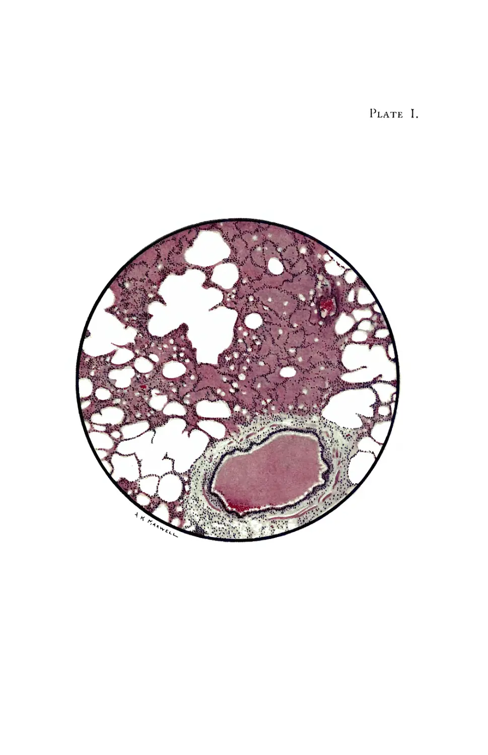
PLATE NO. 1
Microscopic section of human lung from phosgene shell
poisoning. Death at the nineteenth hour after gassing.
The piece of lung shown is almost entirely useless for aeration of
the blood. Most of the pulmonary alveoli are filled with oedema
fluid, and the walls of the air-sacs are burst asunder in many places.
The rounded edges of these torn walls can be recognized both in the
areas of emphysema and in the parts that are flooded with oedema
fluid. The bronchus also is filled with oedema fluid, but it should
be noted that its lining epithelium is intact and pus cells have not
accumulated in the secretion. The blood vessels of the alveolar net-
work are congested ; and intravascular thrombosis is frequently found
in these smaller vessels, though it is not actually shown in the area of
this section.
The main changes in the lung are:
Congestion, and occasional thrombosis, of the network of pul-
monary blood vessels.
Abundant outpouring of inflammatory oedema fluid both into
the tissues and into the air spaces of the alveoli and bronchi.
Disruptive emphysema of the weakened lung tissue.
The result of these changes is that the blood circulation through the
lungs is impeded, and the respiratory exchange of gasses between the
blood and the air in the lung is seriously diminished. The gassed
man is in danger of death by asphyxia so long as his lung is drowned
in oedema fluid.
From the third day onwards the oedema fluid is reabsorbed or
expectorated, and the lung soon resumes its functions. Broncho-
pneumonic complications may develop from secondary infections, but
they are not very common.
The recovery of the lung, even after severe gassing appears to be
functionally good. In the earlier stages of convalescence there may
still be signs of persisting oxygen want, so that tachycardia with
excessively rapid respiration is the result of even slight physical effort.
Later these disabilities vanish. The microscopic examination of lungs
in these stages of recovery has not been made.
Plate I.
PLATE NO. II
Blue type of asphyxia from phosgene poisoning, with
intense venous congestion.
History of case. Drawing made early on second day after gassing;
when there was copious frothy sputum, frequent cough, and hurried
shallow respiration of 40 to 48 with temperature of ioi° and pulse 100.
The patient was bled 15 ozs. and oxygen added to the air that he
breathed. * He soon made a complete recovery.
Such venous congestion was more frequent with chlorine poisoning
than it now is with phosgene. It is associated with a full strong pulse
at the outset, though later the pulse may fail and the asphyxia change
to the pallid type shown in Plate III. The patient as a rule is fully
conscious and complains chiefly of headache and pains in the chest; he
turns restlessly to and fro in extreme general discomfort, and his
hurried breathing is interrupted from time to time by short bursts of
coughing and of expectoration. The lung is in the oedematous state
shown in Plate I.
Oxygen, when given by an efficient apparatus, will at once change
the blue tint of the face to a full pink colour, showing that it can still
be absorbed by the blood through the lungs. Venesection relieves the
discomfort felt by the patient, and probably lessens the embarrassment
of the circulation.
Plate IL
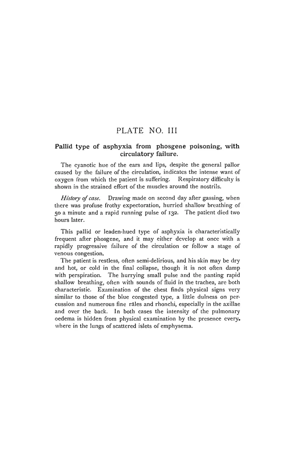
PLATE NO. Ill
Pallid type of asphyxia from phosgene poisoning, with
circulatory failure.
The cyanotic hue of the ears and lips, despite the general pallor
caused by the failure of the circulation, indicates the intense want of
oxygen from which the patient is suffering. Respiratory difficulty is
shown in the strained effort of the muscles around the nostrils.
History of case. Drawing made on second day after gassing, when
there was profuse frothy expectoration, hurried shallow breathing of
50 a minute and a rapid running pulse of 132. The patient died two
hours later.
This pallid or leaden-hued type of asphyxia is characteristically
frequent after phosgene, and it may either develop at once with a
rapidly progressive failure of the circulation or follow a stage of
venous congestion.
The patient is restless, often semi-delirious, and his skin may be dry
and hot, or cold in the final collapse, though it is not often damp
with perspiration. The hurrying small pulse and the panting rapid
shallow breathing, often with sounds of fluid in the trachea, are both
characteristic. Examination of the chest finds physical signs very
similar to those of the blue congested type, a little dulness on per-
cussion and numerous fine rales and rhonchi, especially in the axillae
and over the back. In both cases the intensity of the pulmonary
oedema is hidden from physical examination by the presence every,
where in the lungs of scattered islets of emphysema.
Plate III.
PLATE NO. IV
Gangrene of foot caused by vascular thrombosis from
chlorine poisoning.
History oj case. Gassed by chlorine in 1915 under conditions which
could not have induced frost-bite. Severe dyspnoea from pulmonary
oedema.
Drawing of foot made on fifth day. Both feet were then anaesthetic,
stone cold, and no pulsation could be felt in the dorsalis pedis artery
The right hand also was mottled, cold, and painful. The circulation
was restored in a few days with complete recovery, except that two
toes became black and shrivelled.
Such arterial thrombosis, of slowly progressive onset, is quite
uncommon in the extremities, though it is occasionally seen with
phosgene. The obstruction is very rarely so complete as to cause
gangrene and death of the tissues. But this drawing of a visible
condition is introduced in order to emphasize the fact that an unseen
vascular thrombosis of smaller vessels in deeper organs of the body is
frequently found with phosgene poisoning. Such thrombosis is
^revealed by the microscope in fatal cases in the smaller lung vessels, in
the kidney, in the mucous membrane of the stomach, and in the brain.
Indeed in deaths with prolonged asphyxia from gassing by phosgene
the white matter of the brain is often seen to be thickly sown with
brownish-red petechial spots around each tiny arterial thrombus. The
obstruction to the lung circulation has already been referred to; the
kidney thrombosis does not appear to have any serious results; and,
except where larger haemorrhages have burst in the brain, the scattered
cerebral thrombi do not appear to be of grave clinical import. The
petechial areas within the stomach may occasionally become the seat
of a superficial ulceration. Large thrombi are sometimes found
within the heart, but they also are associated with rather than the
cause of the other changes that lead to death.
PLATE NO. V
Erythema of skin from general exposure to the vapour of
yellow cross substance. Dermatitis of this distribution
and associated with conjunctivitis forms a characteristic
picture of poisoning by this vesicant.
History of case. Exposed to 4 mustard gas’ at Ypres on July 12,
1917, when this substance was first employed by the enemy. Wore
box respirator for only 30 minutes, so that he was exposed without any
protection for nearly four hours. No symptoms were felt until some
hours later, when severe vomiting commenced and conjunctivitis
developed.
Drawing made on the fifth day. The laryngitis and bronchitis were
slight, so that the poisonous vapour must have acted only in low
concentration. But the reddening of the skin was fairly intense
because the man had been sweating freely when exposed to the gas,
and he was not washed afterwards nor was his clothing changed. The
erythema was succeeded by staining in the same areas of the skin.
This reddening, as though the skin had been scorched or deeply
sun-burned, is the first cutaneous reaction to mustard gas, though it
sometimes may not appear until several days after exposure. It is
accompanied by only a slight feeling of warmth and irritation. In
addition to the face and arms which are directly exposed to the vapour
in the air, the moist surfaces of the axillae, the flexures of the elbows,
and the perineum and inner surfaces of the thighs are particularly
affected, that is in the places where the skin is often sodden with
fatty perspiration. This special distribution of the diffuse erythema
characterizes the general dermatitis of mustard gas vapour; but the
reaction may be limited to a smaller area in any part of the body, for
example where the clothing may have chanced to be splashed by the
liquid.
The inflammatory reaction is chiefly superficial, and it is not
accompanied by much oedema of the subcutaneous tissues except in
the eyelids and over the penis and scrotum. Later the dusky red
colour deepens, and patches of cyanotic or whitish oedema may arise
amid it. Blisters then appear, and the cuticle becomes excoriated ; or
the skin may be retained while the erythema fades and a brown staining
slowly darkens the original area of irritation.
Plate V.
PLATE NO. VI
Blistering of buttocks by mustard gas.
History of case. The man sat down on ground that was contaminated
by the poison -and the vapour passed through his clothing, causing
inflammation of the buttocks and of the scrotum. A diffuse reddening
appeared twenty-four hours after exposure, and this was followed by an
outcrop of superficial blisters. On the eighth day the erythema began
to be replaced by a brown staining, and the drawing was made on the
eleventh day during this change of tints. Infection of the raw surface
was avoided, and the healing was complete in three weeks.
The blisters in this case were probably aggravated by pressure, for
the inflamed skin becomes very fragile, so that the surface layer is
readily loosened by pressure or careless rubbing. The blisters may be
very tiny bullae, as on the eyelids, or they may coalesce into areas
many inches across, covering a collection of serous fluid which perhaps
itself contains enough of the irritant substance to injure other skin if it
is allowed to flow over it.
The blisters are usually quite superficial and almost painless in their
development. But the raw surface that is left after the blister has
burst becomes most acutely sensitive to all forms of mechanical
irritation. Deeper destruction of the dermis may be caused by spread-
ing necrosis where the substance attacks the skin locally in high
concentration, or when secondary infections are implanted on the raw
surface. Chronic and painful sores then result, and in this event the
skin does not regenerate completely, so that thinly covered scars for a
long time will mark the site of the burn.
Plate VI.
-
PLATE NO. VII
Burning of scrotum and penis by mustard gas.
History of case. From the same incident as that described under
Plate No. VI. Inflammation commenced at the close of the first day
after exposure. Drawing made on the eleventh day when' the red
erythema had almost faded from the inner aspects of the thighs. The
scrotum is oedematous and the raw surfaces have become the seat of a
mild secondary eczematisation. The injuries were soon and com-
pletely healed.
The perineum is peculiarly liable to be inflamed after exposure to the
vapour of mustard gas, and the penis and scrotum become oedematous
as well as reddened. Balanitis and pain with micturition may be
troublesome. ” When the skin is excoriated, secondary infections of the
raw surface are very likely to develop unless adequate precautions are
taken to prevent sepsis. But with careful cleansing of the skin and
clothes of a casualty after exposure to the vapour, inflammation of the
perineum can be reduced to a comparatively trifling incidence.
Plate VII.
PLATE NO. VIII
Brown staining from mustard gas.
This purplish-brown, or brown, or brownish-black tint usually
appears in areas that were first inflamed and red, but it may arise
without such preceding erythema. Its distribution is in the same
areas as those in which erythema occurs, that is over the exposed skin
surfaces of the neck and hands, or on the sheltered moist flexures of
the body. It may appear at any time from the fifth or sixth day
onwards, and it persists for several weeks, until the stained cuticle
desquamates. There is no deep pigmentation.
The drawing was made from a case on the eighteenth day after
exposure to gas, and the brown tint was present on the sixth day.
Plate VIII
PLATE NO. IX
Ulceration of trachea by mustard gas.
The characteristic feature is the sloughing of the tracheal mucous
membrane. The reddening of the base of the tongue and of the pharynx,
with a sharp delimitation where the oesophagus has refused ingress to
the toxic vapour, is seen also with chlorine and other irritant gasses.
But the pharyngeal inflammation with mustard gas may proceed
further to a local ulceration that will cause dysphagia for many days.
The mucous membrane of the trachea and bronchi is affected by di-
chlor-ethyl-sulphide in much the same way as is the skin. It reacts
with an intense inflammation, and death of the surface layers soon
results. The mass of necrotic tissue, exuded fibrin, and pus cells may
form a yellowish-grey slough in which all manner of organisms
flourish. Subsequently this false membrane comes away in patches
or in entire casts from the raw surface of the bronchial wall.
Meantime the infected debris and secretions tend to accumulate in
the bronchial ramifications at the bases of the lungs, and infection may
spread from them into the lung tissues and alveoli. Septic broncho-
pneumonia, localised abscesses, superficial pleurisy, and even
empyema or pyopneumothorax then develop and cause death.
The drawing is of a trachea at the twelfth day after gassing. The
base of the tongue and the pharynx show characteristic inflammation.
Yellow necrotic sloughs lie on the larynx and at the bifurcation of the
trachea. Between these the trachea is red and glistening, because it
is now completely denuded of both mucous membrane and of slough.
The dotted line points t6 a little group of ulcers on the posterior wall
from which bleeding has occurred. The trachea and bronchi con-
tained an abundance of thin yellow pus.
Plate IX.
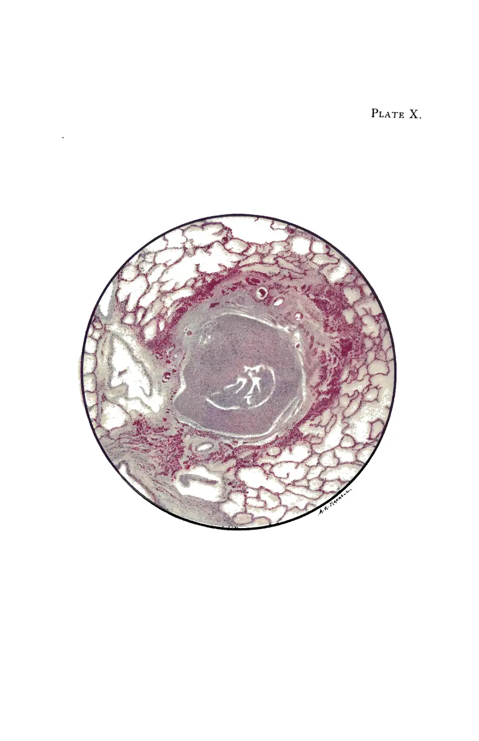
PLATE NO. X
Microscopic section of human lung from mustard gas
poisoning, with death at end of second day (40 hours).
The bronchiole is filled with fibrin and pus cells, and its lining
epithelium has been completely destroyed. The inflammation has
caused a characteristic ring of haemorrhage in the tissues around the
bronchial tube, and infection is beginning to appear in the alveoli
nearest to these inflamed tissues. But there is no generalised
pulmonary oedema and no disruptive emphysema.
Di-chlor-ethyl-sulphide may cause some catarrhal desquamation of
the pulmonary endothelial cells, but it rarely excites an outpouring of
oedema fluid from the pulmonary vessels. The pathological changes
in the bronchioles and in the alveoli are therefore in the sharpest
contrast with those caused by phosgene (see Plate No. I). As
infection spreads into the lung tissues, patches of septic broncho-
pneumonia and small abscesses develop, and these often excite
an inflammatory oedema around them.
If the patient lives, his bronchial mucous membrane is slowly
regenerated; and during this time he is naturally subject to reflex
spasms of coughing or even to a protracted bronchitis.
Plate X.
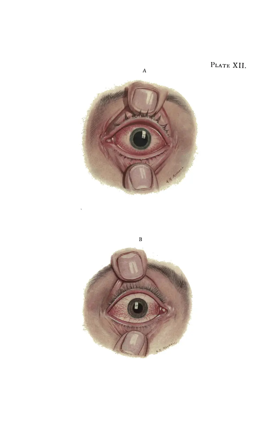
PLATE NO. XIa
Severely burned eye in the acute stage.
Early in the second day after exposure to mustard gas vapour the
eyelids and the external surface of the globe show an intense inflam-
matory reaction. Tears stream from between the closed oedema-
tous eyelids, which may even be blistered, and there is often severe
pain behind the eyes and in the forehead. The conjunctiva is swollen,
oedematous, and bright red from injection of the blood vessels. The
injury of the cornea, even when severe, is not so obvious, and careful
examination is of great importance for its detection. Photophobia and
blepharospasm render examination of the eye very difficult.
The majority of gassed eyes exhibit inflammation of a general
character that is not illustrated in this Atlas. But examples are
continually occurring in which the eye is more severely burned, and
these may be recognized by certain characteristic features that are
depicted in the drawing, Plate No. XIa. Whenever a dead white
band crosses the exposed area of the conjunctiva, while the parts of
this membrane covered by the upper and lower lids are red and
oedematous, serious injury from the burning is likely to have occurred.
In the case illustrated, the caustic effect of the vapour is seen chiefly
in the interpalpebral aperture. On each side of the cornea there is a
dead white band due to coagulative oedema, which compresses the
vessels, impairs the circulation, and thus acts as a menace to the
nutrition of the cornea. The swelling in the region of this white band
is slight, while the protected conjunctiva above and below it is greatly
swollen and injected and may even bulge between the lids.
The exposed portion of the cornea is grey and hazy; it has lost its
lustre, and when viewed with a bright light and a magnifying glass it
shows a blurred ‘ window reflex’ and a typical * orange-skinned’ surface.
The haze gradually fades off above in the region of the protected
part of the cornea where the surface is usually bright and smooth.
The pupil is at first contracted as the result of irritation and congestion.
In this drawing it is shown as artificially dilated by atropine ointment,
which should always be used early in severe cases or where there is
much pain and blepharospasm.
PLATE NO. XIb
Slightly later stage of acute burning.
The swelling in the conjunctiva above and below has subsided, but the
vascular injection remains, and the solid white oedema in the palpebral
aperture is still well marked. The cornea is grey in the exposed
area.
[For History of the case see page facing Plate XIL
Plate XI.
A
в
Plate No. XIb continued.]
History of the case. The casualty was caused by the bursting of a
Yellow Cross shell close to the man when he was riding a restive mule,
and his box respirator was momentarily displaced. A fine spray of
the liquid must have splashed lightly over his right side, for cutaneous
blisters developed on the neck, the cheek, and the forehead on this
side only. The right eye showed serious burning with the central
white band, while the left eye was only in the state of general red
conjunctivitis.
With the lowering of the nutrition of the corneal epithelium,
secondary infection is liable to take place. In this case an infiltrated
corneal ulcer is seen associated with a hypopyon. It is therefore
important when there is conjunctival discharge, which indicates
secondary infection, that in addition to the use of atropine the
conjunctival sac should be cleansed by frequent bland irrigations and
by the instillation of antiseptic drops so as to check infection of any
corneal ulceration which may develop. Otherwise the infective pro-
gress which has led to hypopyon may progress till panophthalmitis
supervenes.
PLATE NO. XIIa
Stage of resolution after severe burning.
The vascular injection is passing off, the solid oedema is becoming
absorbed, and the corneal epithelium has regained its normal lustre.
In this stage the use of atropine should be discontinued.
PLATE NO. XIIb
Late stage of resolution.
The earlier vascular injection above and below the cornea has
practically disappeared; the solid white oedema has been absorbed,
and the conjunctiva in the palpebral aperture now shows definite
injection, often of a bright violet tint. The entire picture has changed,
so that the parts which were red in the acute stage are now white and
the part which was formerly white is now red. This drawing would
illustrate equally well the condition that may follow immediately on a
very slight exposure to the irritant gas, when only a slight central band
of red injectiop develops instead of the bloodless state of white oedema
that is caused by the more severe burns.
At this stage atropine and shades should be abandoned. Astringent
drops should be instilled and photophobia combated with cold douching,
&c., while fresh air and occupation will help to restore the general
health of the individual and mitigate any tendency to neurasthenia.
Plate XII.
A
в
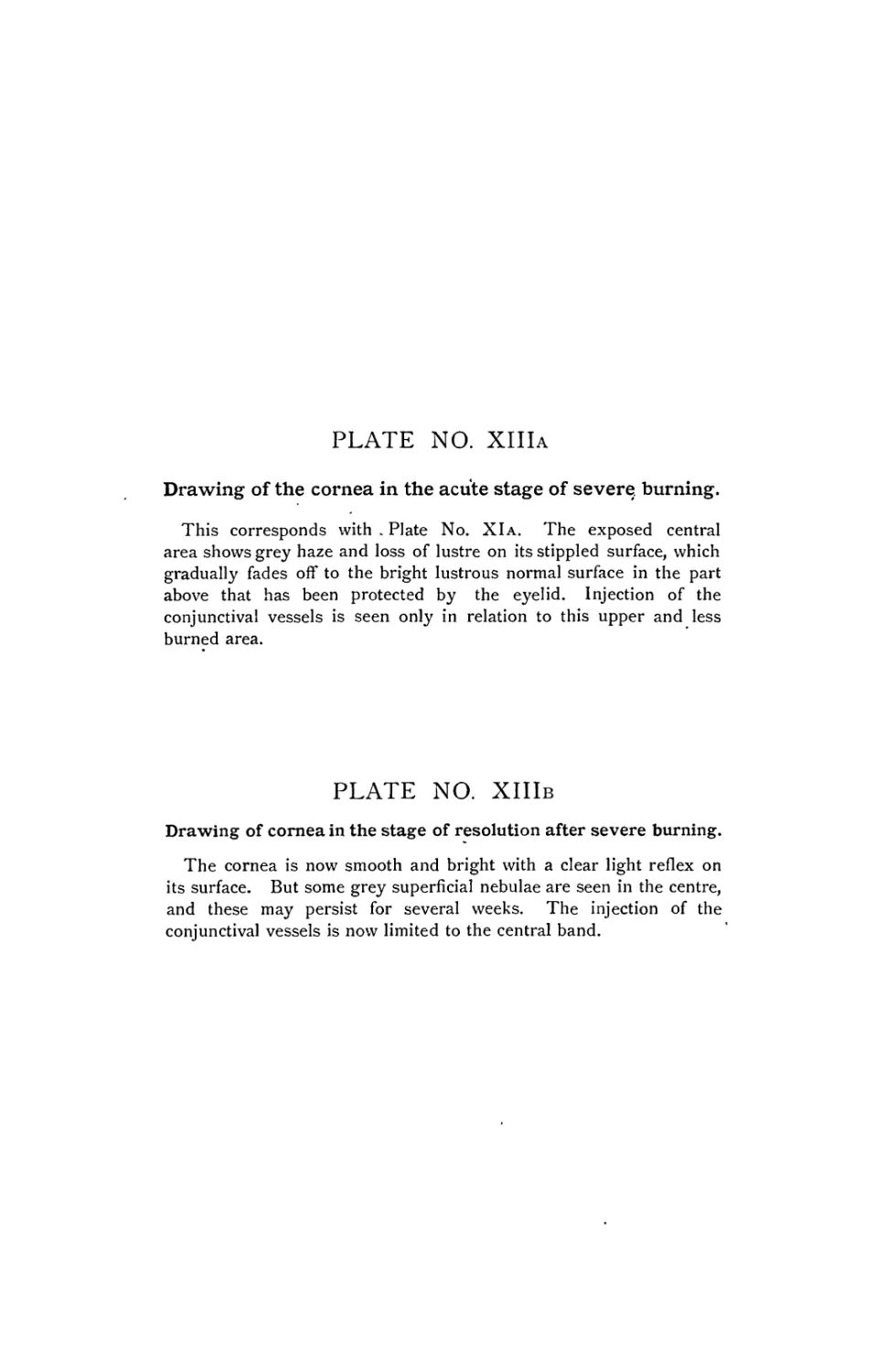
PLATE NO. XIIIa
Drawing of the cornea in the acute stage of severe, burning.
This corresponds with . Plate No. XIa. The exposed central
area shows grey haze and loss of lustre on its stippled surface, which
gradually fades off to the bright lustrous normal surface in the part
above that has been protected by the eyelid. Injection of the
conjunctival vessels is seen only in relation to this upper and less
burned area.
PLATE NO. XIIIb
Drawing of cornea in the stage of resolution after severe burning.
The cornea is now smooth and bright with a clear light reflex on
its surface. But some grey superficial nebulae are seen in the centre,
and these may persist for several weeks. The injection of the
conjunctival vessels is now limited to the central band.
Plate XIII.