


Author: Аносов С.Е.
Tags: articulata (членистые) биология зоология рыбы рыбное хозяйство
ISBN: 978-5-85382-364-6
Year: 2012

Text
ИЛЛЮСТРИРОВАННЫЙ ОПРЕДЕЛИТЕЛЬ
DECAPODA АТЛАНТИЧЕСКОГО СЕКТОРА
АНТАРКТИКИ И ПРИЛЕГАЮЩИХ ВОД
ФЕДЕРАЛЬНОЕ АГЕНТСТВО ПО РЫБОЛОВСТВУ
ФЕДЕРАЛЬНОЕ ГОСУДАРСТВЕННОЕ УНИТАРНОЕ ПРЕДПРИЯТИЕ
«ВСЕРОССИЙСКИЙ НАУЧНО-ИССЛЕДОВАТЕЛЬСКИЙ ИНСТИТУТ
РЫБНОГО ХОЗЯЙСТВА И ОКЕАНОГРАФИИ» (ФГУП «ВНИРО»)
FEDERAL AGENCY FOR FISHERIES
FEDERAL STATE UNITARY ENTERPRISE
«RUSSIAN FEDERAL RESEARCH INSTITUTE OF FISHERIES
AND OCEANOGRAPHY» (FSUE «VNIRO»)
S.E. ANOSOV
ILLUSTRATED GUIDE OF DECAPODA
FOR ATLANTIC SECTOR OF ANTARCTIC
AND SURROUNDING WATERS
MOSCOW
VNIRO PUBLISHING
2012
СЕ. АНОСОВ
ИЛЛЮСТРИРОВАННЫЙ ОПРЕДЕЛИТЕЛЬ
DECAPODA АНТАРКТИЧЕСКОГО СЕКТОРА
АТЛАНТИКИ И ПРИЛЕГАЮЩИХ ВОД
МОСКВА
ИЗДАТЕЛЬСТВО ВНИРО
2012
УДК 595.384.(261.6)
Редакционный совет ФГУП «ВНИРО»:
д-р биол. наук А.Н. Макоедое, д-р биол. наукМ.К. Глубокоеский, д-р техн, наук Л.С. Абрамова,
д-р биол. наук В. А. Бизиков, д-р биол. наук О. А. Булатов, д-р биол. наук Л.If. Глубокое,
канд. биол. наук МЛ/. Куманцое, д-р с/х наук Ю.П. Мамонтов, канд. техн, наук МВ. Сытоеа
Аносов С.Е.
А69 Иллюстрированный определитель Decapoda Атлантического сектора Антарктики и прилегающих
вод.— М.: Изд-во ВНИРО, 2012.— 90 с.
Книга представляет собой первый в мире определитель Decapoda для юго-западной Атлантики. Она содержит краткое
описание внешнего строения отряда Decapoda с оригинальными иллюстрациями, краткие данные о современной систематике
отряда, оригинальные текстовые ключи и две сравнительные фототаблицы для визуального определения видов. По этим таб-
лицам легко определить вид сможет даже не специалист.
Книга содержит 14 оригинальных фототаблиц с цветными фотографиями Decapoda. Девять из которых сделаны на живом
материале и точно передают окраску живых животных. Остальные приложения сделаны на фиксированном материале из кол-
лекции Департамента по Рыболовству Фолклендских островов и дают полное представление о внешнем виде этих животных.
Книга будет полезна в качестве полевого определителя для научных наблюдателей на промысловых судах, специалистам
по фауне Антарктики и Субантаркгики, а также всем, кто интересуется десятиногими раками.
Anosov S.E.
Illustrated guide of Decapoda for Atlantic sector of Antarctic and surrounding waters.— M.: VNIRO Pub-
lishing, 2012.— 90 p.
This book is a first in the world Decapoda key for the south-western Atlantic. It contains a short description of the external struc-
ture of the order Decapoda with original illustrations, a summary of the order of modem taxonomy, the original text keys, and two com-
parative tables for visual identification of species. According to these tables can easily determine the kind not even a specialist.
It includes 14 original plates with color photos of Decapoda. Nine of which were made on living material and accurately convey
the color of live animals. The other five applications were made on material from the collections of the Falkland (Malvinas) Islands Fish-
eries Department and provides a complete appearance of these animals.
The book will be useful as a field guide for scientific observers on fishing vessels, for experts on the fauna of the Antarctic and sub-
Antarctic, and as anyone who interested in Decapod Crustaceans.
Translation by N.R. Popova and V V Anosova
Anosov S.E.
Guia Ilustrada Decapoda para Atlantic sector de la Antartida у aguas circundantes.— Moscu, VNIRO.
2012,— 90 p.
Este libro es el primero en el mundo Decapoda clave para el Atlantico suroccidental. Contiene una breve description de la estruc-
tura externa del orden Decapoda con ilustraciones originales, un resumen de la orden de la taxonomia modema, claves originales del
texto, у dos tablas comparativas para la identification visual de las especies. De acuerdo con estas tablas se puede determinar facilmente
el tipo sin siquiera ser un especialista.
Incluye 14 aplicaciones originales con fotos a color de los decapodos. Nueve de las cuales fueron hechas con material vivo у que
comunican con precision el color de los animates vivos. Las otras cinco aplicaciones fueron hechas con material de las colecciones del
Departamento de Pesquerias de las Islas Falkland (Malvinas) que proporcionan una apariencia completa de estos animates.
El libro sera util сото una guia de campo para que los observadores cientificos a bordo de buques de pesca, para los expertos en la
fauna de la Antartida у subantarticas, у para cualquier persona que este interesada en crustaceos decapodos.
Traduction por Raul Balam Martinez Perez
В работе использованы фотографии: CM. Игнатьева (рис. 2.1) и Н.Н. Кухарева (рис. 2.2).
Photo by Sergey Ignatiev (fig. 2.1), Nikolay Kucharev (fig. 2.2).
Foto de Nikolay Kucharev (fig. 2.1), Sergey Ignatiev (fig. 2.2).
ISBN 978-5-85382-364-6
©Anosov S.E., 2012
© Аносов C.E., 2012
© VNIRO Publishing, 2012
© Издательство ВНИРО, 2012
ВВЕДЕНИЕ
Растущий спрос на морепродукты и истощение запасов традиционных объектов про-
мысла заставляет искать новые объекты промысла. Вместе с этим приходится осваивать но-
вые, как правило, мало изученные районы, перспективные для промысла. Один из таких
районов — море Скотия и прилегающие к нему акватории юго-западной части Атлантиче-
ского океана.
С XIX века там находился один из основных центров китобойного промысла, вплоть
до его запрета. С 60-70-х годов XX века появился интерес к антарктическому крилю (Еир-
hausia superba). В этот период в этом районе весьма активно работали советские суда. Толь-
ко во ВНИРО было организовано более десятка экспедиций. Кроме того было организова-
но множество рейсов научно-исследовательских судов Института океанологии, ИнБЮМ,
АтлантНИРО, промразведок и других организаций. Однако основные исследования были
посвящены пелагиали. Донная фауна моря Скотия была изучена слабо.
Первые материалы по донным десятиногим ракообразным из этого района были по-
лучены наНПС «Академик Книпович» в 1965, 1967 гг, когда в Атлантическом секторе Ан-
тарктики было обнаружено значительное количество крабоидов сем. Lithodidae. Тогда же
был описан новый вид Paralomis spinosissima. Тем не менее, фауна десятиногих ракообраз-
ных этой части океана остается недостаточно изученной. Пока еще не совсем точно уста-
новлен видовой состав обитающих там Decapoda, а из-за значительной схожести некото-
рых видов возникает неверное определение и, как следствие, — путаница.
Определение видовой принадлежности улова и прилова, на промысловых судах и не
только, играет большую роль в изучении биологического разнообразия определенных рай-
онов и помогает более точно установить границы обитания разных видов. Накопленные та-
ким образом данные помогут больше узнать о жизнедеятельности промысловых и связан-
ных с ними видов животных и, как следствие, — рационально организовать промысел.
Поскольку большинство людей, имеющих доступ к бентосным животным в Антарктике
и Субантарктике, не всегда имеют полное представление о систематике Decapoda, то тре-
буется простое и доступное для всеобщего понимания руководство, по которому можно бу-
дет быстро и точно определить попавшееся животное.
Этот труд предназначен для определения представителей отряда Decapoda, встречаю-
щихся в южной части Атлантического океана. Он также может помочь в открытии пока не-
известных науке видов и выявить появление в изучаемом районе уже известных из других
мест новых видов — вселенцев.
Работа базируется на фотографиях, сделанных автором в ходе экспериментально-про-
мыслового рейса СРТМ «Таманго» весной 2010 г. и фотографиях коллекций Decapoda из
юго-западной части Атлантического океана, имеющихся в Департаменте Рыболовства на
Фолклендских островах (Порт Стэнли). А также фундаментальной работе Э. Макферсона
«Revision of the Family Lithodidae Samouelle, 1819 (Crustacea, Decapoda, Anomura) in the At-
lantic Ocean» и разрозненных работах разных авторов, посвященных описанию разных ви-
дов десятиногих раков, встречающихся в Южной части Атлантического океана.
5
КАК ПОЛЬЗОВАТЬСЯ ЭТИМ ОПРЕДЕЛИТЕЛЕМ
Пользоваться нашим определителем очень просто.
Можно использовать классический метод и определить с помощью имеющихся ниже
ключей или воспользоваться методом экспресс определения. Для этого нужно:
I. Взять определяемое животное и открыть Сравнительную таблицу 1 или Сравни-
тельную таблицу 2;
II. Выбрать похожий вид. Для сравнения предлагаются таблицы с крупными изобра-
жениями панциря крабов и крабоидов (Сравнительная таблица 1) или общим изобра-
жением для Macrura или Anomura (Сравнительная таблица 2);
III. Далее перейти по указанной цифре к Фототаблице (I-XIV) или описанию живот-
ного в тексте (см. оглавление). Фототаблицы находятся в ПРИЛОЖЕНИИ.
Если Вы ошиблись, то снова вернитесь к Сравнительной таблице (1 или 2), посмотри-
те более внимательно и повторите операцию, изложенную в и. II;
IV. Если Вы таким образом не смогли определить животное, или пойманный экзем-
пляр не похож ни на одно из прилагаемых изображений или не подходит ни под одно
из имеющихся описаний, то по возможности сделайте восемь фотографий:
1. Общий вид сверху;
2. Общий вид снизу;
3. Карапакс — крупный вид сверху;
4. Карапакс — крупный вид снизу;
5. Карапакс — крупный вид снизу с отогнутым абдоменом, так чтобы были хоро-
шо видны стерниты;
6. Карапакс — крупный вид сбоку;
7. Клешня левая;
8. Клешня правая.
Далее напишите автору по адресу: anosov@vniro.ru или anosov@aquarius-s.ru
В письме опишите сложности, возникшие с определением, вышлите сделанные фото-
графии, координаты места находки, глубину и дату поимки. По возможности сохраните эк-
земпляр, который не удалось определить. Эта информация может оказаться очень ценной и
наверняка поможет при последующей работе над определителями для фауны Decapoda Ан-
тарктики и Субантарктики.
6
КРАТКОЕ ОПИСАНИЕ ЮГО-ЗАПАДНОЙ ЧАСТИ
АТЛАНТИЧЕСКОГО ОКЕАНА
К юго-западной части Атлантического океана относятся Патагонский шельф, Пата-
гонский склон и акватория моря Скотия.
К восточному побережью Южно-Американского континента (рис. 1.1) примыкает ши-
рокая полоса относительного мелководного Патагонского шельфа (рис. 1.12), который с вос-
точной стороны довольно резко, через Патагонский склон, переходит в океаническое ложе.
На широте 50-53° ширина шельфа достигает максимума — около 900 км. В центре мелко-
водья расположен архипелаг Фолклендских (Мальвинских) островов (рис. 1.4).
С юга к Южной Америке (рис. 1.1) прилегает своеобразная область дуги моря Скотия
(Скоша), которая отделяет его от Антарктического континента. Дуга Скотия вытянута на
3 тыс. км в широтном направлении и состоит из трех сегментов: Северного хребта Скотия,
Южно-Сандвичевой вулканической дуги (рис. 1.6) и Южного хребта Скотия.
Северный хребет в основном подводный; выше уровня моря находится только о. Юж-
ная Георгия (рис. 1.5). В пределах Южного хребта Скотия над водой возвышаются Южные
Оркнейские острова (рис. 1.7), сходные по своему геологическому строению с Антаркти-
ческим полуостровом (рис. 1.3) и Южно-Шетландскими островами (рис. 1.8).
Внутри дуги Скотия находится глубоководная впадина моря Скотия (рис. 1.11) шири-
ной более 600 км, которая составляет восточное продолжение котловины Беллинсгаузена
(рис. 1.10), расположенной в юго-восточной части Тихого океана. Они соединяются через
пролив Дрейка (рис. 1.2), но при этом разделены подводным порогом глубиной менее 2 км
(рис. 1.9), протягивающимся в юго-восточном направлении от юго-западной оконечности
Огненной Земли к крайнему восточному острову Южно-Шетландского архипелага. Сред-
няя глубина моря Скотия составляет более 5 км, максимальная глубина — 6022 м. Общая
площадь — 1247 тыс. км2.
Средняя температура воды на поверхности от 6 °C до —1 °C. Солёность около 34 %о.
Море в основном находится в субполярных широтах. Воды умеренных широт присутствуют
лишь в его северо-западной части. В средней части моря Скотия преобладают южные по-
лярные воды Антарктического циркумполярного течения, а в юго-восточную часть с юга по-
ступает еще более холодная вода из моря Уэдделла. Над морем господствуют сильные за-
падные ветры, часто случаются штормы. Встречаются айсберги.
Море было названо в 1932 г. по имени экспедиционного судна «Скотия» шотландской
антарктической экспедиции У. Брюса [http://rusnel.ru/2010/01/02/duga-i-more-skotiya].
7
ОБЩИЕ СВЕДЕНИЯ ОБ ОТРЯДЕ DECAPODA
ВНЕШНЕЕ СТРОЕНИЕ
К отряду Decapoda относятся наиболее высокоорганизованные представители класса
ракообразных, отличительной чертой которых является наличие головогруди (цефалото-
ракса). Он образован восемью сегментами груди соединенными с пятью сегментами голо-
вы и покрыт общим хитиновым панцирем (карапаксом). На переднем конце тела находят-
ся стебельчатые глаза (рис. 6.4; рис. 8.4), которые обычно состоят из двух члеников — ба-
зального и конечного. Конечный членик представляет собой фасеточный глаз, состоящий из
большого числа отдельных глазков — омматидиев, покрытых общей роговицей. У некото-
рых видов, живущих в темноте, наблюдается редукция глаз.
Между глазничными ямками имеется лобный край или рострум (рис. 6.1; рис. 8.1), раз-
мер и форма которого у разных видов может сильно варьировать. На панцире могут быть и
другие шипы, названия которых, как правило, совпадают с их расположением в соответ-
ствующих областях карапакса (рис. 6.7-11). На верхней поверхности карапакса у многих
видов проходят бороздки, оконтуривающие отдельные области, которые соответствуют рас-
положению внутренних органов (рис. 3.7-11). С боков панцирь образует жаберные крыш-
ки (бранхиостегиты), прикрывающие жабры (рис. 3.10а). На нижней стороне головогруди
между конечностями расположены стерниты (рис. 4.5-8; рис. 7.7-11).
За головогрудью находится абдомен (брюшко), состоящий из семи сегментов
(рис. 7.15). Последний сегмент не имеет конечностей и называется тельсоном (рис. 7.13).
Каждый брюшной сегмент покрыт хитиновым кольцом, состоящим из верхней части (тер-
гум) (рис. 8.24), нижней (стернум) (рис. 7.16) и боковой (плевральная пластинка) (рис. 8.25),
прикрывающей основания ног.
Расчлененных конечностей у Decapoda до 19 пар:
• антеннулы, или антенны I (внутренние усики) (рис. 8.2), состоят из 3-членистого стеб-
ля и обычно двух (реже трех) жгутиков. В основном членике стебля антеннул находится
статоцист — орган равновесия.
• антенны, или антенны II (наружные усики) (рис. 8.3), состоят из 5-членистого стеб-
ля и жгутика. На первом членике стебля находится отверстие выделительной антеннальной
железы. Второй членик стебля со скафоцеритом (рис. 7.5).
• мандибулы (верхние челюсти) (рис. 3.6) состоят из широкой жевательной пластинки и
обычно имеют щупик из одного-двух или трех члеников. За мандибулами следуют две пары
нижних челюстей (максиллулы и максиллы) и затем восемь грудных ножек, из которых пер-
вые три именуются максиллипедами (ногочелюстями). Первая и вторая пары максиллипед
состоят из 2-членистого пластинчато расширенного протоподита и обычно расширенных эк-
зоподита и эндоподита. У максиллипед I от первого членика протоподита отходит эпиподит.
• максиллипеды III (наружные ногочелюсти) (рис. 4.4; рис. 7.6) у представителей разных
инфраотрядов устроены различно. У креветок максиллипеды III сходны с переоподами и име-
ют удлиненную форму; у крабов ишиум и мерус максиллипеда III расширены и прикрывают
ротовой аппарат, а три последних членика (карпус, проподус и дактилюс) узкие и короткие,
и имеют вид щупика. У разных видов число члеников эндоподита может быть различным.
У некоторых видов экзоподит отсутствует. У некоторых видов иногда имеется эпиподит.
8
• за максиллипедами расположены пять пар переопод (рис. 5.1-V), что и определило на-
звание отряда.
Переопода представляет собой одноветвистую 7-членистую конечность, состоящую
из коксы (рис. 8.23), базиса (рис. 8.22), ишиума (рис. 8.21), меруса (рис. 8.20), карпуса
(рис. 8.19), проподуса (рис. 8.18) и дактилюса (рис. 8.17). Первая пара переопод, — хели-
педы (клешненосные ноги) (рис. 5.1) у всех Decapoda несет клешни. У многих Масшга
(раки и креветки) вторая и третья пары переопод также несут клешни. Четвертая и пятая
пары оканчиваются дактилюсом и представляют собой ходильные ноги. У представителей
инфраотряда Anomura вторая-четвертая пары переопод являются ходильными ногами.
Пятая пара редуцирована. У Brachyura II-V пары переопод представляют собой ходильные
ноги (рис. 5.I-V).
• плеоподы (плавательные ножки) находятся на нижней части абдомена (рис. 7.12).
У Decapoda имеется пять пар, построенных по типу двуветвистой конечности. Первая или
две первых пары плеопод у самцов преобразованы в копулятивные органы различного типа.
• уроподы (хвостовые ноги) (рис. 7.14) являются видоизмененными конечностями ше-
стого брюшного сегмента [Кобякова, Долгопольская, 1969].
КРАТКИЕ СВЕДЕНИЯ О СОВРЕМЕННОЙ СИСТЕМАТИКЕ DECAPODA
Тип — ARTHROPODA
Подтип — BRANCHIATA
Класс — CRUSTACEA
Подкласс — MALACOSTRACA
Отряд — DECAPODA
По особенностям передвижения существует уже сильно устаревшее на сегодняшний
день деление на 2 подотряда:
NATANTIA — включает некрупных десятиногих раков, обладающих длинным брюш-
ком с хорошо развитыми плеоподами, которые используются для плавания.
REPTANTIA — включает различных по размерам и строению десятиногих раков,
обычно не плавающих.
Однако это деление весьма относительно, поскольку некоторые животные, относя-
щиеся к подотряду Reptantia плавают весьма активно. Например, многие крабы из семей-
ства Portunidae (рис. 2.4).
И далеко не все креветки, относящиеся к подотряду Natantia, могут хорошо плавать.
Например, креветки из семейства Alpheidae.
Более точно различие Decapoda по внешнему строению:
Инфраотряд ANOMURA — мягкохвостые ракообразные. Объединяет мало похожие
друг на друга формы животных: раков-отшельников (рис. 2.3) (раки, использующие
пустую раковину брюхоногого моллюска), галатеид (рис. 2.2) (имеют вид креветки)
и крабоидов (рис. 2.1) (раки, имеющую форму тела, сходную с крабами). Для предста-
вителей инфраотряда характерно наличие редуцированных последних пар переопод:
IV и V пары у раков-отшельников; только V пара у крабоидов (рис. 3.V) и галатеид
(рис. 7.V).
Инфраотряд BRACHYURA — короткохвостые ракообразные. Объединяет всех на-
стоящих крабов (рис. 2.4). Характерно наличие укороченного цефалоторакса и подог-
нутого под грудь абдомена, что и определяет характерную форму крабов.
9
Инфраотряд CARIDEA — креветки (рис. 2.5). Характеризуется сжатым с боков телом.
Цефалоторакс без поперечного шва. Его передняя часть удлинена в рострум. Все пе-
реоподы хорошо развиты и имеют длинную и тонкую форму. Абдомен, как правило,
длиннее цефалоторакса. Плеоподы хорошо развиты. По устаревшей классификации
относятся к Macrura Natantia.
Инфраотряд ASTACIDEA — раки, лангусты, омары (рис. 2.6). Отличаются хорошо
развитым абдоменом, у большинства представителей имеющим примерно такую же
длину, как и цефалоторакс. Абдомен обычно имеет полное число редуцированных
плеопод и оканчивается хорошо развитым хвостовым плавником. По современной
классификации относятся к подотряду Macrura Reptantia.
Количество инфраотрядов, входящих в состав отряда Decapoda, намного больше, чем
упомянуто выше. Здесь указаны только те инфраотряды, представители которых встре-
чаются в юго-западной части Атлантического океана.
ОПРЕДЕЛИТЕЛЬНЫЕ ТАБЛИЦЫ DECAPODA
ЮГО-ЗАПАДНОЙ ЧАСТИ АТЛАНТИЧЕСКОГО ОКЕАНА
Ключи к определению инфраотрядов, входящих в состав отряда DECAPODA
1(2) Тело короткое, абдомен прижат к груди .............................................3
2(1) Тело длинное, абдомен хорошо выражен ..............................................5
3(4) 5 пар хорошо различимых переопод ...........................................Brachyura
4(3) 4 пары хорошо различимых переопод; 2 последние редуцированы
и спрятаны под панцирем ........................................Anomura (fam. Lithodidae)
5(6) Все пары переопод одинаково хорошо развиты .......................................8
6(5) Пятая пара переопод редуцирована..................................Anomura (Galatheidae)
7(8) Плеоподы хорошо развиты...............................................Macrura (Caridea)
8(7) Плеоподы редуцированы ..............................................Macrura (Astacidea)
Инфраотряд ANOMURA MacLeay, 1838
На переднем крае цефалоторакса имеется рострум. Края брахиостегитов срастаются с
грудными сегментами. У всех Anomura абдомен относительно мягкий. По его строению и
последним редуцированным парам переопод различают 3 формы:
Галатеиды — симметричный абдомен вытянут в длину, оканчивается хорошо разви-
тым хвостовым веером (например, сем. Galatheidae).
Крабоиды — абдомен подогнут под укороченную головогрудь, что делает их похожи-
ми на крабов. У самцов абдомен симметричный, у самок - несимметричный (например,
сем. Lithodidae).
Раки-отшельники — абдомен у большинства видов несимметричен и имеет вид спи-
рально закрученного кожистого мешка, который они прячут в пустые раковины брю-
хоногих моллюсков. У этой формы редуцированны IV и V пары переопод. Плеоподы
имеются только на левой стороне: у самок на II-V сегменте, у самцов — III-V. Уропо-
ды больше развиты на левой стороне вместе с тельсоном, на котором имеются изогну-
тые шипы. С их помощью рак-отшельник удерживает свое тело в раковине (например,
сем. Diogenidae).
Инфраотряд Anomura включает в себя 4 надсемейства с 13 семействами, из которых в
юго-западной части Атлантики встречаются представители двух семейств: Lithododae и
Galatheidae.
10
Семейство Lithodidae Samouelle, 1819
Входит в надсемейство Paguroidea, Latreille, 1802.
Относительно твердый панцирь, имеющий укороченную форму, как у настоящих кра-
бов. Подогнутый абдомен прижат к груди. Спинная часть карапакса обычно хорошо раз-
граничена и имеет гранулы и (или) шипы, сильно варьирующие по количеству и размерам.
Исключение составляют виды рода Cryptolithodes, представители которого обладают глад-
ким карапаксом. Рострум сильно варьирует по размерам и строению, однако имеется всег-
да. Глаза пигментированы. Скафоцерит или хорошо выражен, или редуцирован до малень-
кой гранулы.
На первой паре переопод имеются хорошо развитые клешни. Клешни развиты нерав-
номерно. Одна больше (чаще правая), другая меньше. Следующие три пары переопод пред-
ставляют собой более-менее длинные ходильные ноги. Пятая пара переопод редуцирована
и находится под карапаксом.
Широкий, короткий, относительно кальцинированный, абдомен покрыт узелками или
щитками, которые варьируют по количеству и размеру. Первый сегмент практически пол-
ностью закрыт карапаксом. Второй-седьмой сегменты абдомена хорошо видны. У самок
щитки на абдомене с правой стороны более развиты, чем аналогичные щитки с левой сто-
роны. В результате абдомен у самок выглядит несимметричным, хотя это не всегда хорошо
видно у некоторых родов.
У самцов плеоподы отсутствуют. Самки имеют пару небольших плеопод на первом аб-
доминальном сегменте и один простой объединенный плеопод с левой стороны со второго
по пятый сегменты. Уроподы отсутствуют. Абдомен оканчивается тельсоном [Macpherson,
1988].
Семейство Lithodidae объединяет 10 родов со 120 видами и разделено на два подсе-
мейства: Hapalogastrinae Brandt, 1850 и Lithodinae Samouelle, 1819. В юго-западной части Ат-
лантического океана пока обнаружены представители только подсемейства Lithodinae.
Таблица для определения родов семейства Lithodidae
1 (2) На карапаксе имеются хорошо выраженные длинные острые парные шипы;
на первой паре стернитов имеются хорошо выраженные гранулы.......................Neolithodes
2(1) На карапаксе имеются короткие или средние шипы и (или) гранулы; первая пара
стернитов гладкая, или на ней имеются мелкие гранулы ......................................3
3 (4) В гастральной области карапакса имеется хорошо выраженный одиночный шип,
расположенный в центре гастральной области или несколько смещенный
к переднему краю карапакса ........................................................Paralomis
4 (3) На карапаксе расположено множество парных шипов или гранул .........................Lithodes
Род Lithodes Latreille, 1806
Относится к подсемейству Lithodinae, Samouelle, 1819.
Карапакс обычно твердый, относительно выпуклый. Все области карапакса хорошо
выражены. Гастральная область выдается над другими областями. Сердечная область име-
ет треугольную форму. Она отделена от гастральной области глубоким поперечным жело-
бом и неглубокими поперечными швами от бранхиальных областей. Затылочный желоб вы-
ражен плохо.
Карапакс, клешненосные и ходильные ноги вооружены шипами и гранулами, варьи-
рующими по количеству и размерам. Шипы относительно пропорциональны по размерам
у молодых и взрослых особей.
11
Передний выступ рострума сравнительно длинный с одним или двумя парами верхних
шипов, окончание раздвоено. На нижней части рострума имеется большой изогнутый шип.
Жгут антенны короче, чем длина карапакса. Скафоцерит обычно отсутствует, либо име-
ет форму одиночного шипа, иногда с добавочным шипом.
Между третьей парой максиллипед иногда имеется пара шипов в области стернитов.
Третьи максиллипеды с зубчатым краем с 11-13 роговыми зубцами и одним дополнитель-
ным зубом. Клешни примерно равны по длине. Правая клешня, как правило, более разви-
та. Две трети проксимального края режущей кромки пальцев правой клешни имеют не-
сколько хорошо различимых плотных, закругленных зубчиков. Зубчики на левой клешне
мельче, но более многочисленны. Пальцы обоих клешней оканчиваются когтями. Все паль-
цы несут более-менее многочисленные пучки щетинок.
Область стернитов в середине имеет глубокий желобок между первой парой ходиль-
ных ног. Второй абдоминальный сегмент сформирован сросшимися щитками: средним с
двумя боковыми и одной парой краевых щитков. У некоторых видов сращены все щитки.
Средние щитки третьего-пятого сегментов замещены мембраной, покрытой кальциниро-
ванными узелками.
Боковые и краевые щитки не имеют кальцинированных узелков у обоих полов. Боко-
вые щитки у самок развиты больше с левой стороны и сращены с краевыми щитками, что
делает абдомен ассиметричным. Краевые щитки обычно разделены.
Ходильные ноги удлиненные. Вторая пара длиннее, чем первая и третья [Macpher-
son, 1988].
Род Lithodes включает 29 видов, из которых в юго-западной части Атлантики встреча-
ется 3 вида - L. santolla, L. confundens и L. turkayi.
Таблица для определения видов рода Lithodes
1(2) Нижний шип на роструме значительно короче переднего выступа
с раздвоенным окончанием ...............................................................L. turkayi
2(1) Нижний шип на роструме значительно больше остальной части .................................3
3(4) На поверхности карапакса имеется множество крупных острых шипов ....................L. santolla
4(3) Поверхность карапакса покрыта маленькими толстыми острыми шипами и гранулами . ... L. confundens
Lithodes santolla (Molina, 1782)
(ФОТОТАБЛИЦА I)
Синонимы: L. antarcticus Jacquinot, 1853; Lithodes antarctica Jacquinot, 1844; Cancer santolla Molina,
1782; Lithodes santolla Philippi, 1867; Lithodes antarcticus White, 1847
Карапакс имеет форму пятиугольника. Карапакс, клешни и ходильные ноги вооруже-
ны многочисленными шипами среднего размера. По сравнению с другими видами семейства
Lithodidae, шипы обычно одинаковы по размеру. Вся поверхность карапакса почти гладкая,
за исключением редких разбросанных гранул.
Области карапакса выпуклые, выступы областей сходны. Гастральная область в сере-
дине вооружена двумя парами шипов одинакового размера, расположенных в форме ква-
драта. Кардиальная зона с четырьмя шипами одинакового размера, которые также распо-
ложены в виде квадрата. Интерстинальная область на крае карапакса имеет два шипа тако-
го же размера, как и шипы в бранхиальной области. Птеригостомальная область на
переднебоковом крае имеет маленький шип.
Рострум хорошо выражен и образован тремя шипами, нижним, направленным вперед
и немного вверх, и двумя верхними шипами, имеющими несколько меньшую длину, чем
нижний шип. В основании рострума имеется пара шипов почти такого же размера, как и
верхние ростральные шипы.
12
Первая пара переопод несет хорошо развитые клешни, одна из которых (обычно пра-
вая) крупней. Длинные и относительно толстые ходильные ноги имеют примерно одинако-
вую длину.
Цвет всего тела более-менее однородно красный, иногда с фиолетовым оттенком.
Самцы достигают 198 мм в длину и 250 мм в ширину. Самки достигают 142 мм в дли-
ну и 140 мм в ширину.
Встречается в Тихом океане на побережье Чили и в прибрежной зоне Южной Атлан-
тики от Магелланова пролива до Уругвая, а также поблизости от Фолклендских (Мальвин-
ских) о-вов. По литературным данным, L. santolla встречается на разных глубинах, от лито-
рали до глубин более 700 м, однако промысловые скопления образует на глубинах 10-50 м
[Macpherson, 1988]. В ходе рейса на СРТМ «Таманго» этот вид был встречен только один раз
на глубине 108 м.
Один из наиболее изученных видов из юго-западной части Атлантического океана.
Имеет ценное промысловое значение.
Lithodes confundens Macpherson, 1988
(Фототаблица II)
Пятиугольный карапакс вооружен мелкими многочисленными шипиками и заострен-
ными гранулами. Все области карапакса примерно одинаково выпуклые. Гастральная об-
ласть имеет 6 четко различимых маленьких шипов, четыре из которых расположены в цен-
тральной части по форме прямоугольника, а два расположены по бокам гастральной обла-
сти, на границах с бранхиальными областями. По всей гастральной области расположены
многочисленные заостренные гранулы. Кардиальная область имеет четыре маленьких ши-
па, расположенных в форме квадрата и множество заостренных гранул расположенных по
поверхности всей области. Каждая из бранхиальных областей имеет примерно по тридцать
одинаковых по размеру маленьких шипов. На интерстинальной области расположена пара
маленьких шипов. На сгибе птеригостомальной области также имеются мелкие шипы. Боль-
шинство шипов на карапаксе имеют одинаковые размеры.
Короткий рострум немного выступает за орбитальную область. Основной нижний шип
хорошо развит и у взрослых особей не выступает за конец базального членика антеннул.
У основания рострума имеется пара дорсальных шипов. Возле глазничной выемки распо-
лагается пара мелких шипиков. Внешний орбитальный шип хорошо развит и заметно круп-
нее, чем другие шипы на карапаксе. Переднебоковой шип меньше, чем орбитальный.
Глазной стебель имеет гранулы на дорсальной поверхности. Базальный сегмент ан-
тенн имеет маленький шип на переднем углу. Второй сегмент антенн с краевым наружным
шипом.
Второй абдоминальный сегмент имеет пару маленьких шипов в середине и несколько
большее количество мелких шипиков на границах средних и боковых щитков. Имеются
многочисленные гранулы на боковых щитках третьего-пятого сегментов. В середине абдо-
мена имеются кальцинированные узелки.
На мерусе хелипед расположены несколько шипов, развитых в дорсальном и латераль-
ном направлениях. Карпус хелипед имеет 5-6 относительно крупных дорсальных шипов и
несколько шипов, разбросанных по наружной и нижней поверхности. Проподус имеет два
плохо различимых ряда шипов на дорсальной стороне. Подвижный палец имеет несколько
гранул на верхней стороне. Имеются пучки щетинок на обоих пальцах [Macpherson, 1988].
Достигает 96 мм в длину и 101 мм в ширину.
Встречается в юго-западной части Атлантического океана, в Магеллановом проливе, к
югу от Фолклендских (Мальвинских) островов и на подводных горах Северного хребта мо-
ря Скотия.
Этот вид близок к Lithodes santolla.
13
По литературным данным встречается на глубинах от 50 до 119 м [Macpherson, 1988].
Однако, в ходе рейса на СРТМ «Таманго» этот вид встречался с 280 м до 775 м. На мень-
ших глубинах постановок не было, поэтому определить верхнюю границу обитания этого
вида было нельзя. Возможно, встречающиеся в литературе сообщения о поимке L. santolla
на больших глубинах связаны с ошибкой в определении L. confundens, по причине большо-
го внешнего сходства этих двух видов.
О промысловом значении сведений нет.
Lithodes turkayi Macpherson, 1988
(Фототаблица III)
Синонимы: Lithodes murrayi Campodonico and Guzman, 1972.
Карапакс имеет грушевидную форму. Его длина обычно превышает ширину. Области
карапакса четко разграничены. Гастральная область имеет четыре шипа расположенных по
форме квадрата, передняя пара крупнее последующей. Кардиальная область, несет пару
шипов, по размеру меньших, чем шипы гастральной области, и две толстых гранулы меж-
ду гастральными шипами и гастро-кардиальным углублением. Бранхиальные области несут
по четыре шипа. Передние шипы крупнее последующих. Интерстинальная область имеет
два хорошо развитых шипа на краю карапакса. Птеригостомальная область имеет шип на пе-
реднем крае.
Рострум длинный, на конце раздвоен, на верхней части ближе к концу имеется два на-
правленных вверх шипа. На нижней части присутствует крупный одиночный шип, направ-
ленный вперед. Орбитальный шип длинный и достигает конца оболочки глаза. Глазной сте-
бель имеет гранулы на дорсальной стороне.
Второй абдоминальный сегмент имеет 2 шипа в середине, и несколько тупых шипов на
границах. Боковые и крайние щитки на следующих сегментах имеют рассеянные гранулы.
Клешненосные ноги длинные и имеют крупные шипы на внешней стороне. Развиты не-
равномерно. Правая клешня обычно больше, чем левая. Ходильные ноги длинные, имеют
различную длину. Наибольшую длину имеет вторая пара [Macpherson, 1988].
Достигает в длину 141 мм и в ширину 140 мм.
Встречается недалеко от берега Чили [Macpherson, 1988], на юго-восток от Фолклендс-
ких (Мальвинских) о-вов и на подводных горах в море Скотия на глубинах от 300 до 1500 м.
Этот вид близок к индоокеанскому виду L. murrayi.
Несмотря на то, что целенаправленный промысел этого вида пока еще не велся, его
можно рассматривать как потенциально ценный промысловый вид.
Род Neolithodes A. Milne-Edwards & Bouvier, 1894
Относится к подсемейству Lithodinae, Samouelle, 1819.
Не очень твердый карапакс имеет пятиугольную или грушевидную форму. Области ка-
рапакса выпуклые и хорошо различимы. Сердечная область имеет треугольную форму. Она
отделена от желудочной области глубоким поперечным углублением и мелкими углубле-
ниями отделена от бранхиальных областей. Цервикальный желобок четко не выражен. Га-
стральная и бранхиальные области выступают больше, чем кардиальная область. Крупные
шипы на карапаксе, клешнях и ходильных ногах варьируют по размерам и количеству. У мо-
лодых особей они длиннее по отношению к телу. Рострум сформирован основным шипом
и парой крупных дорсальных шипов, направленных вверх и расходящихся в разные сторо-
ны. С увеличением размера животного длина всех шипов, как правило, уменьшается.
Второй абдоминальный сегмент образован пятью щитками: одним средним, двумя бо-
ковыми и двумя краевыми. Третий-пятый сегменты образованы маленькими, более-менее
14
иглообразными узелками. Щитки у самцов плохо выражены. У самок щитки хорошо раз-
виты на левой стороне, что делает абдомен ассиметричным.
Область стернитов между первой парой ходильных ног имеет глубокое продольное уг-
лубление в середине. На тех же стернитах имеются четко выраженные гранулы.
Глаза с пигментированными роговыми оболочками. Скафоцерит обычно отсутствует, ес-
ли есть, то простой. Он сформирован одиночным шипом с маленькими шипиками по краям.
Клешни примерно равны по длине, правая клешня, как правило, более развита. Режу-
щая кромка правой клешни имеет приплюснутые закругленные зубцы, занимающие при-
мерно 2/3 длины пальцев, последняя треть имеет роговидный коготь. Аналогичные зубцы
на левой клешне развиты плохо, и почти не видны. Пальцы обеих клешней немного изо-
гнуты внутрь.
Ходильные ноги длинные. Вторая пара несколько длиннее первой и третьей [Macpher-
son, 1988].
Род Neolithodes включает 12 видов, из которых в юго-западной части Атлантики встре-
чается только один вид.
Neolithodes diomedeae (J.E. Benedict, 1895)
(ФОТОТАБЛИЦА IV)
Синонимы: Lithodes diomedeae Benedict, 1894; Neolithodes diomedeae Bouvier, 1896; Neolithodes
martii Birstein and Vinogradov, 1972.
Карапакс, клешни и ходильные ноги вооружены многочисленными крупными остры-
ми шипами. У молодых особей шипы пропорционально крупнее. С возрастом размер ши-
пов увеличивается гораздо медленнее, чем размеры тела. За счет этого молодые особи ка-
жутся более колючими, чем взрослые. Поверхность между шипами покрыта рассеянными
гранулами, которые у молодых особей более острые. На гастральной области имеются че-
тыре шипа, расположенные в форме прямоугольника. Передние шипы крупнее. В центре
этого прямоугольника есть маленький шип, у взрослых особей обозначенный как бугорок.
Кардиальная область имеет четыре шипа, расположенные в форме прямоугольника. Бран-
хиальная область имеет шесть хорошо развитых шипов. Интерстинальная область имеет
два длинных шипа у края карапакса. Птеригостомальная область имеет маленький шип на
крае переднего угла.
Рострум крупный, образован направленным вверх нижним шипом и двумя верхними
расходящимися шипами, которые по длине равны нижнему шипу.
Второй абдоминальный сегмент имеет шесть-семь крупных шипов на среднем щитке,
три шипа на боковом крае боковых щитков и несколько маленьких шипов на крайних щит-
ках. Щитки и узелки последующих сегментов с маленькими, более-менее острыми шипа-
ми. У самок ассиметричные сегменты имеют более крупные шипы.
Клешни развиты неравномерно. Правая клешня, как правило, крупнее и имеет на ре-
жущей кромке хорошо заметные закругленные зубцы, занимающие примерно две трети дли-
ны пальцев. Крайняя часть кромки представляет собой зазубренный ороговевший коготь. Ре-
жущая кромка левой клешни закругленных зубцов не имеет.
Ходильные ноги длинные. Вторая пара ходильных ног длиннее первой и третьей.
Третья пара короче первой [Macpherson, 1988].
Достигает в длину 145 мм и в ширину 162 мм.
Встречается в южной части Атлантического океана на глубинах от 640 до 2450 м [Macp-
herson, 1988]. Однако, в ходе рейса на СРТМ «Таманго» этот вид на глубинах до 950 м не
попадался.
Целенаправленный промысел этого вида пока еще не велся, но учитывая его размеры
и вкусовые качества, N. diomedeae можно рассматривать как потенциально ценный промы-
словый вид.
15
Род Paralomis White, 1856
Относится к подсемейству Lithodinae, Samouelle, 1819.
Твердый карапакс имеет пятиугольную или грушевидную форму. Области карапакса
выпуклые и хорошо различимы. Гастральная область более выпуклая, чем другие области
и примерно в центре имеет хорошо различимый крупный одиночный шип. Кардиальная об-
ласть, треугольная по форме, отделена от желудочной области глубокой бороздой.
Цервикальный желобок плохо выражен, и между бранхиальными областями и сердеч-
ной областью практически неразличим.
Рострум сформирован основным шипом и одной парой более или менее расходящих-
ся дорсальных шипов направленных вверх.
Второй абдоминальный сегмент образован одним щитком.
Третий-пятый абдоминальные сегменты образованы четко разделенными твердыми
пластинами без узелков или мембранных областей. Срединный щиток, пара боковых пла-
стин и пары краевых пластин имеются на каждом сегменте. Краевые и боковые пластины
на третьем сегменте (очень редко и на четвертом сегменте) иногда сращены.
Область стернитов между первой парой ходильных ног без углубления.
Глаза с пигментированными роговыми оболочками. Скафоцерит имеет относительно
заостренную форму. Третьи максиллипеды имеют зубчатый край с более чем 10 основны-
ми зубчиками и дополнительным зубчиком.
Клешни равны по длине, но правая клешня, как правило, значительно крупнее. Режу-
щий край на правой клешне имеет закругленные, приплюснутые зубцы, занимающие при-
мерно 2/3 от общей длины клешни, последняя треть имеет роговидный коготь. Аналогич-
ное строение имеет левая клешня, но зубцы на ней существенно меньше. Пальцы обеих
клешней немного загнуты внутрь.
Ходильные ноги более-менее удлиненные. Вторая пара ходильных ног длиннее, чем
первая и третья пары [Macpherson, 1988].
Род Paralomis объединяет 66 видов, из которых в юго-западной Атлантике встречают-
ся только три: Р. granulosa, Р. spinosissima и Р. formosa.
Таблица для определения видов рода Paralomis
1(2) Все тело покрыто множеством небольших, но относительно длинных,
загнутых шипов ...............................................................Р. spinosissima
2(1) На карапаксе имеются только основные шипы, остальная поверхность покрыта гранулами .3
3(4) Ходильные ноги превышают длину карапакса больше чем в 2 раза ...............Р. formosa
4(3) Ходильные ноги сравнительно короткие и ненамного превышают длину карапакса .Р. granulosa
Paralomis granulosa (Hombron & Jacquinot, 1846)
(Рис. 2.1)
Карапакс пятиугольный. Ширина всегда превышает длину. Вся поверхность карапак-
са покрыта крупными гранулами. Все области карапакса хорошо различимы. Гастральная
область возвышается над кардиальной и бранхиальными областями.
Основной шип рострума короткий и изогнутый. С вентральной стороны он гладкий. На
дорсальной стороне рострума имеется два несколько расходящихся шипа, немного направ-
ленных вверх. Переднебоковой край карапакса вооружен 12-14 шипами одинакового разме-
ра. Глазные стебельки имеют гранулы на дорсальной поверхности. На основном сегменте
антеннального стебля имеется крупный шип. Скафоцерит имеет длинный центральный шип.
Абдоминальные сегменты покрыты группами гранул. На третьем сегменте крайние и
боковые щитки сращены.
Хелипеды несут шипы и гранулы. На пальцах имеются многочисленные пучки щети-
нок. Ходильные ноги толстые и короткие.
16
Цвет тела красный, на гранулах и шипах более темный.
Достигает 100 мм в длину и 115 мм в ширину.
Встречается в Тихом океане на побережье Чили и юго-западной части Атлантическо-
го океана на глубине до 50 м [Macpherson, 1988].
Один из наиболее изученных видов из юго-западной части Атлантического океана.
Имеет ценное промысловое значение.
Paralomis formosa Henderson, 1888
(Фототаблица V)
Синонимы: Paralomis formosus Henderson, 1888; Paralomis formosa Bouvier, 1896; Paralomis spec-
tabilis Birstein and Vinogradov, 1972.
Карапакс примерно пятиугольный. Длина почти равна ширине. Поверхность панциря
покрыта гранулами. Там же имеются несколько шипов. Выпуклая гастральная область зна-
чительно выше, чем кардиальная и бранхиальные области. В передней части гастральной
области имеется большой, несколько загнутый вперед одиночный шип. В её задней части
расположены два маленьких шипа. По бокам кардиальной области в центральной части
имеются два маленьких бугорка. В кардиальной области имеются четыре шипа, располо-
женные по форме квадрата. В бранхиальных областях имеются крупные шипы на уровне
первой пары сердечных шипов. Имеется шип на крае птеригостомальной области.
Рострум образован основным, немного загнутым толстым шипом, и двумя направлен-
ными вперед и немного загнутыми вверх расходящимися шипами сверху. Орбитальный шип
хорошо развит. Глазные стебельки имеют гранулы на дорсальной поверхности и малень-
кий бугорок на крае.
Абдоминальные сегменты покрыты гранулами. В центре второго сегмента есть два ши-
па. Краевые и боковые щитки на третьем сегменте обычно сращены. Краевые щитки на че-
твертом и пятом сегментах разделены.
На хелипедах имеются крупные шипы. Клешни развиты неравномерно. Как правило,
правая клешня, значительно крупнее, чем левая. На режущей кромке правой клешни име-
ются крупные приплюснутые округлые зубы. На левой клешне таковые отсутствуют. На
пальцах обеих клешней имеются пучки щетинок.
Ходильные ноги длинные, с шипами. Все пары примерно одинаковой длины [Macp-
herson, 1988].
Достигает 98 мм в длину и 106 мм в ширину.
Встречается в Юго-Западной Атлантике на глубинах от 400 до 1600 м [Macpherson,
1988]. Однако, в ходе рейса на СРТМ «Таманго» самцы Р. formosa начинали попадаться с
глубины 790 м. Самки встречались только ниже 1140 м.
Как объект промысла использовался редко. Но если учесть количество этого крабоида
в улове и прилове, а также хорошие вкусовые качества, то этот вид вполне можно считать
перспективным промысловым видом.
Paralomis spinosissima Birstein & Vinogradov, 1972
(ФОТОТАБЛИЦА VI)
Синонимы: Paralomis spinosissimus Birstein and Vinogradov, 1972; Paralomis spinosissima Takeda
andOhta, 1979.
Поверхность карапакса покрыта множеством шипов и заостренных бугорков. Кончи-
ки многих шипов иногда имеют длинную, тонкую щетинку у молодых особей. Гастральная
область более выпуклая, чем остальные. В передней части гастральной области имеется
крупный одиночный шип. Бранхиальные области имеют шип чуть меньшего размера на
уровне гастро-кардиального углубления и несколько крупных шипов по краю. Птеригосто-
мальная область имеет шип на переднем угловом крае.
17
Основной шип рострума прямой, слегка направлен вверх. Пара верхних расходящих-
ся шипов несколько короче основного рострального шипа. Глазные стебельки имеют ма-
ленькие дорсальные шипы. Основной членик антеннального стебля имеет наружный шип
с одним-тремя шипиками у основания. Скафоцерит имеет длинный центральный шип, до-
стигающий кончика антеннального основания и два-три длинных шипа на внешней по-
верхности.
Абдоминальные сегменты покрыты многочисленными шипами. Краевые и боковые
щитки на третьем сегменте сращены.
Все переоподы густо покрыты шипами и щетинками. Клешни развиты неравномерно.
Правая клешня, обычно больше, чем левая. Подвижный и неподвижный пальцы покрыты
пучками щетинок.
Ходильные ноги длинные. Третья пара переопод длиннее первой и второй пары [Macp-
herson, 1988].
Достигает 114 мм в длину и 125 мм в ширину.
Встречается в Юго-Западной Атлантике на глубинах от 132 до 650 м [Macpherson,
1988]. Однако, в ходе рейса на СРТМ «Таманго» несколько экземпляров этого вида было
встречено на глубине 812 м.
Впервые был описан советскими учеными Я.А. Бирштейном и Л.Г. Виноградовым
в 1972 г.
Основной промысловый вид в районе Южной Георгии.
Семейство Munididae Ahyong, Baba, Macpherson, Poore, 2010
Входит в надсемейство Galatheoidea Samouelle, 1819.
Цефалоторакс галатеид сверху приплюснут, абдомен обычно подогнут под него. Пер-
вая пара переопод сильно удлинена и имеет длинные клешни. Пятая пара переопод обыч-
но спрятана внутри жаберной полости под карапаксом. Семейство объединяет 34 рода, в
которые входят более 200 видов. В юго-западной части Атлантического океана встречается
только один вид.
Munida gregaria (Fabricius, 1793)
(Фототаблица VII)
Синонимы: Galathea gregaria Fabricius, 1793; Grimotaea gregaria Leach, 1820; Grimothea gregaria
Milne Edwards, 1837; Munida gregaria White, 1846; Grimothea novae zeelandiae Filhol.
Продолговатый карапакс расширен в средней части. Рострум тонкий, прямой и длин-
ный. Глаза направлены вперед. Надглазничные шипы тонкие и прямые. Абдомен хорошо
развит. Второй, третий и четвертый абдоминальные сегменты имеют пару маленьких ши-
пиков на переднем крае. Абдомен оканчивается хорошо развитым хвостовым веером [Julio
Н. Vinuesa & Martin Varisco, 2007].
Окраска живых особей ярко-красная (рис. 2.2)
Этот вид широко распространен в Южном полушарии.
О промысловом значении сведений нет.
18
Инфраотряд BRACH YURA
Все пять пар переопод хорошо развиты. Первая пара всегда несет хорошо развитые
клешни. Характерной особенностью является абдомен, подогнутый под укороченную го-
ловогрудь. Глазные стебельки могут укладываться в особые глазничные ямки, между кото-
рыми расположен лобный край; передний край бранхиостегитов срастается с надротовой
пластинкой; верхняя сторона карапакса у Brachyura отделена от боковой стороны резким
краем, который не является границей соединения верхней и нижней частей панциря. Эта
граница расположена ниже бокового края. Передние стерниты, расположенные между ан-
те ннулами, срастаются и образуют эпистом. Укороченные антеннулы могут укладываться в
особые углубления на нижней поверхности панциря. Уроподы отсутствуют.
Инфраотряд Brachyura разделен на 2 секции: Dromiacea de Haan, 1833 (объединяет
6 семейств) и Eubrachyura de Saint Laurent, 1980, которая включает 21 надсемейство с
65 семействами, из которых в юго-западной части Атлантического океана встречаются пред-
ставители только надсемейства Majoidea.
Надсемейство Majoidea Samouelle, 1819
Карапакс имеет грушевидную форму, в передней части сильно зауженную, а в задней
части обычно закругленную. Дорсальная поверхность выпуклая, может нести шипы, бугор-
ки, гранулы и щетинки. Лобный край узкий и у многих видов преобразован в раздвоенный ро-
струм. Глазные выемки выражены плохо, или отсутствуют совсем. Переднебоковой край ка-
рапакса часто вооружен хорошо выраженными шипами. Клешни обычно одинакового раз-
мера. У некоторых видов ходильные ноги могут иметь очень большую длину, за счет чего
представителей этого семейства называют «краб-паук». На карапаксе и переоподах у многих
видов имеются щетинки, на которые крабы прикрепляют водоросли, губки, гидроиды и пр.
Крупное надсемейство крабов, объединяющее примерно 700 видов, относящихся к 52 родам.
В юго-западной части Атлантического океана встречаются представители двух
семейств: Epialtidae и Inachidae.
Семейство Epialtidae MacLeay, 1838
Род Libidoclaea Н. Milne Edwards & Lucas, 1842
Libidoclaea granaria H. Milne Edwards & Lucas, 1842
(ФОТОТАБЛИЦА VIII)
Синонимы: Libidoclea coccinea Dana, 1851; Libinia gracilipes Miers, 1886.
Очень твердый карапакс имеет треугольную форму. На поверхности имеются мелкие
гранулы и тупые шипы, без щетинок. Гастральная область отделена неглубокими выемка-
ми и слегка возвышается над карапаксом. Остальные области плохо выражены. Карапакс
сверху имеет розово-красный цвет, бока и нижняя часть грязно-белые. Лобный край пре-
образован в раздвоенный рострум, основание которого покрыто щетинками, а конец голый.
Шипы рострума к окончанию расходятся и слегка направлены вверх.
Посередине первого абдоминального сегмента имеется большой заостренный выступ.
На поверхности первого и второго абдоминальных сегментов имеются гранулы. Поверх-
ность третьего-шестого абдоминальных сегментов гладкая.
19
Клешни длинные, поверхность густо покрыта мелкими гранулами. Режущая кромка
почти вся состоит из хорошо дифференцированных зубчиков практически одинакового раз-
мера. Единственное исключение составляет первый зубец на подвижном пальце. Он круп-
ный, округлый и слегка приплюснутый. Клешни имеют одинаковый размер. Строение обеих
клешней идентично. Ходильные ноги очень длинные, но все имеют разную длину, равно-
мерно уменьшающуюся со второй пары по пятую. Мерус, карпус и проподус имеют утол-
щение к периферийному окончанию. Ходильные ноги и клешни от основания до примерно
середины меруса имеют грязно-белую окраску, плавно переходящую в розово-красную.
Верхняя часть карпуса и проподуса имеет грязно-белую полосу. Дактилюс желтоватого цве-
та [http://atlas.ambiente.gov.ar].
Встречается на Патагонском шельфе на глубине от 30 до 120 м. Видимо, ведет мало-
подвижный образ жизни, т.к. панцирь часто покрыт губками, гидроидными полипами, си-
дячими полихетами и актиниями.
Промыслового значения не имеет.
Семейство Inachidae MacLeay, 1838
Род Eurypodius Gu6rin, 1825
Удлиненный, умеренно выпуклый карапакс имеет треугольную форму. На его поверх-
ности имеются шипы или бугорки. Имеется отдельный посторбитальный шип. Шипы ро-
струма перекрещиваются в последней части их длины.
Клешни самцов хорошо развиты. Примерно одного размера. Ходильные ноги длин-
ные. Проподус расширен и сплющен. Абдомен состоит из семи сегментов.
Eurypodius latreillei Guerin, 1825
(ФОТОТАБЛИЦА IX)
Синонимы: Eurypode tuberculateux Eydoux & Souleyet, 1842; Eurypode tuberculatus Eydoux & Souleyet,
1842; Eurypodius audouinii H. Milne Edwards & Lucas, 1842; Eurypodius brevipes Dana,
1851; Eurypodius cuvieri Audouin, in De Haan, 1838; Eurypodius danae Targioni-Tozzetti,
1877; Eurypodius quiriquinensis Yanez, 1948; Eurypodius septentrionalis Dana, 1851;
Paramithrax pernonii Targioni-Tozzetti, 1872.
Твердый карапакс вытянут в длину. Имеет грушевидную форму. Вся поверхность ка-
рапакса густо покрыта щетинками. Если щетинки удалить, то все области хорошо различи-
мы. Гастральная область более выпуклая, чем другие. Карапакс без щетинок имеет голубо-
вато-синий цвет. Лобный край преобразован в раздвоенный рострум. Части рострума по-
крыты щетинками и в оконечной части перекрещиваются. Под ним имеется характерный
одиночный когтеобразный шип, направленный вниз и вперед.
Посередине первого абдоминального сегмента имеется хорошо заметный выступ. У сам-
цов поверхность абдомена и области стернитов покрыты относительно редкими щетинка-
ми. У самок абдомен закрывает всю область стернитов и густо покрыт щетинками.
Поверхность клешней без каких либо выростов. Режущая кромка почти вся состоит
из одинаковых мелких зубчиков. На средине подвижного пальца имеется крупный, округ-
лый зубец. Клешни развиты неравномерно. Как правило, правая клешня немного крупнее,
чем левая. Строение обеих клешней идентично. Ходильные ноги длинные. Проподус рас-
ширен и уплощен. Дактилюс имеет когтеобразную форму. Ходильные ноги и клешни густо
покрыты щетинками [Garth John S. 1958].
Встречается на Патагонском шельфе от литоральной зоны до глубины 120 м. Видимо,
ведет малоподвижный образ жизни, т.к. панцирь часто покрыт водорослями, губками, ги-
дроидными полипами и кораллами.
Промыслового значения не имеет.
20
Инфраотряд Astacidea
На переднем крае цефалоторакса имеется рострум. Края брахиостегитов никогда не
срастаются с грудными сегментами. Отличаются сильно развитым абдоменом, который у
большинства представителей имеет такую же длину, как и цефалоторакс. Абдомен обычно
имеет полное число плеопод и оканчивается хорошо развитым хвостовым веером.
Семейство Nephropidae Dana, 1852
Омары имеют очень твердый цефалоторакс трубчатой формы. Его поверхность почти
гладкая или покрыта толстыми волосками. Рострум хорошо развит и вооружен шипами или
гранулами. Антенны нитевидные и длинные. Абдомен имеет плеоподы и оканчивается хо-
рошо развитым хвостовым веером, который состоит из уроподов и тельсона. Первые три па-
ры переопод несут клешни. На первой паре клешни крупные, как правило, одинаковые по
размеру. Четвертая и пятая пары переопод оканчиваются дактилюсом.
В юго-западной части Атлантического океана встречается один вид, относящийся к ро-
ду Thymops Holthuis, 1974.
Thymops birsteini (Zarenkov & Semenov, 1972)
(Фототаблица X)
Синонимы: Nephropides birsteini, Zarenkov et Semenov sp.n.
Гранулированный карапакс покрыт многочисленными короткими волосками и имеет
глубокую цервикальную борозду. Также имеется медиальная борозда, переходящая на ро-
струм. Хепатическая область хорошо выражена. Рострум с двумя парами боковых шипов и
мелким дополнительным шипом на левой стороне. Шипики верхней стороны основания
рострума переходят на карапакс и составляют два ряда по восемь-десять шипиков. Га-
стральная область с мелкими шипиками, которые быстро уменьшаются по направлению к
заднему краю карапакса. Имеется крупный надглазничный шип, а также бранхиостегальный
шип. Глаза небольшие, с коричневым пигментом.
Пальцы клешней перекрещиваются. Внутренний край неподвижного пальца с харак-
терной выемкой на проксимальной части. На проподусе наиболее крупные шипы распола-
гаются одним рядом посередине и несколькими по краям. Карпус несет 2 крупных шипа,
прочие шипы на нижней поверхности развиты слабо. Ишиум в задней проксимальной ча-
сти имеет вырезку, прикрытую лопастью, сочлененную с базисом.
Абдоминальные сегменты II-VI с широким тупым медиальным килем. На боковых по-
верхностях этих же сегментов у заднего края имеется по одной поперечной борозде. VI сег-
мент с тремя шипами на заднем крае. Экзоподит уроподов слабо зазубрен по переднему
краю; его дистальная часть обособлена от проксимальной части глубоким швом. Эндоподит
уроподов имеет продольный киль и субдистальный шип [Заренков, Семёнов, 1972]
Глубоководный вид. Встречается на глубине 800-2100 м.
Этот вид был впервые описан советскими учеными Н.А. Заренковым и В.Н. Семено-
вым в 1972 г, и отнесен к роду Nephropides. Позже он был отнесен в новый род Thymops
Holthuis, 1974.
Промыслового значения не имеет.
21
Инфраотряд Caridea
Характеризуется сжатым с боков телом. Цефалоторакс без поперечного шва, передняя
часть которого удлинена в рострум. Края брахиостегитов никогда не срастаются с грудны-
ми сегментами. Базальный членик стебля антеннул со стилоцеритом. Антенны состоят из
очень длинного жгутика. Мандибулы состоят из широкой пластинки, которая почти у всех
креветок разделена на две лопасти и обычно имеет щупик из одного-трех члеников. Все
переоподы хорошо развиты и имеют длинную и тонкую форму. Абдомен, как правило, за-
метно длиннее цефалоторакса. Плеоподы хорошо развиты. Уроподы вместе с тельсоном об-
разуют хвостовой веер.
Таблица для определения семейств инфраотряда Caridea
1(2) Рострум гладкий, без шипов; или отсутствует................................................Alpheidae
2(1) Рострум имеет шипы................................................................Campylonotidae
Семейство Alpheidae Bate, 1888
Рострум ровный, без зубчиков и если имеется, то он неподвижно прикреплен к пере-
днему краю карапакса. Глаза короткие, частично или полностью скрыты краем карапакса.
Антеннулы со спиннобоковым жгутом, обычно более-менее раздвоенным. Второй максил-
липед с крайним сегментом присоединен как полоска к среднему краю согнутого предпо-
следнего сегмента. Третьи максиллипеды имеют хорошо развитый экзопод. Переоподы без
отчетливо выраженных экзоподов. Две передние пары имеют клешни. Три последующих па-
ры обычно не длинные, карпус короче, чем проподус. Характерной чертой является нали-
чие ассиметричных клешней, большая из которых может издавать громкий звук.
Большинство видов живет в норах от литорали до 875 м.
Большое, широко распространенное семейство, объединяющее более 600 видов, кото-
рые относятся к 38 или более родам. В юго-западной части Атлантического океана имеет-
ся только один вид, относящийся к роду Betaeus.
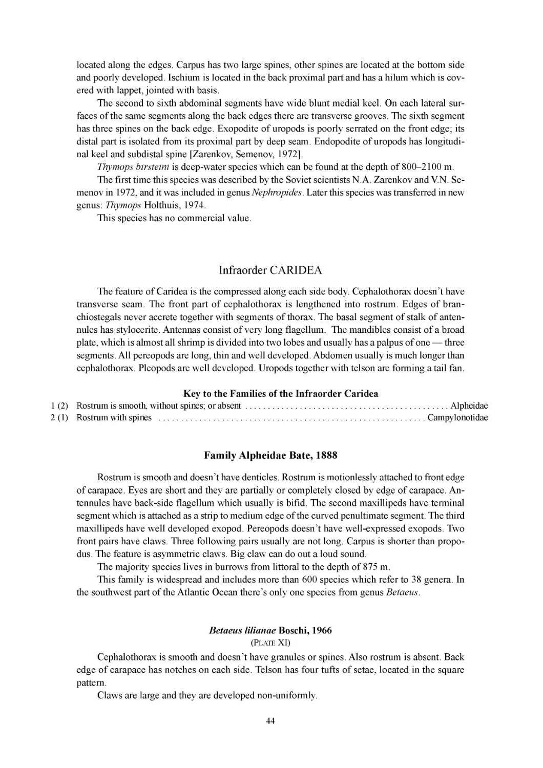
Betaeus lilianae Boschi, 1966
(ФОТОТАБЛИЦА XI)
Цефалоторакс гладкий, без гранул и шипов. Рострум отсутствует. Задний край кара-
пакса с зазубриной на каждом боку. Тельсон имеет четыре пучка щетинок, расположенных
в форме квадрата.
Клешни крупные, развиты неравномерно.
Встречается в юго-западной части Атлантического океана. В основном на Патагонском
шельфе, среди зарослей растений, в камнях и трещинах скал на глубине до 168 м [http://at-
las. ambiente .gov. ar].
Промыслового значения не имеет.
Семейство Campylonotidae Sollaud, 1913
Семейство содержит два рода — Campylonotus и Bathypalaemonella.
В юго-западной части Атлантического океана встречаются только виды рода Campy-
lonotus.
22
Род Campylonotus Bate, 1888
На первой и второй парах переопод имеются клешни с одним подвижным пальцем;
третья-пятая пары переопод оканчивается дактилюсом; переоподы без экзоподов.
Верхний жгутик антенн простой. Базальная часть рострума имеет не более пяти зуб-
чиков, первый из которых находится за срединой карапакса [Sven Thatje, 2003].
К роду Campylonotus относятся разные виды, встречающиеся от сублиторали до боль-
ших глубин в районах Субантарктики [Thatje et al., 2001].
В коллекции Департамента Рыболовства Фолклендских (Мальвинских) о-ов в Порт
Стэнли находились 3 вида, относящиеся к данному роду. Все они были отловлены в разное
время у о-ва Южная Георгия.
Таблица для определения видов рода Campylonotus
1(2) На верхней части рострума 1 зубчик...........................................................3
2(1) На верхней части рострума 2 зубчика ...............................................С. arntzianus
3(4) Рострум широкий и оканчивается большим гладким шипом ..............................С. semistriatus
4(3) Конец длинного, сравнительно тонкого рострума разделен на 3 маленьких шипика...........С. vagans
Campylonotus vagans Bate, 1888
(ФОТОТАБЛИЦА XII)
Цефалоторакс имеет короткие щетинки. Зубцы, расположенные в дорсальной части
образуют киль, переходящий на рострум. Два шипа находятся в гастральной области ка-
рапакса, один в начале рострума на уровне глаз и еще один примерно посередине ро-
струма. Рострум оканчивается тремя маленькими зубчиками, направленными вперед.
Первая и вторая пары переопод имеют клешни.
Абдомен гладкий, третий сегмент выпирает вверх, образуя горб. Оканчивается хорошо
развитыми хвостовым веером. Тельсон на конце заострен, дорсолатеральный край вооружен
тремя маленькими равноотстоящими шипиками. Плеоподы хорошо развиты [http://atlas.am-
biente.gov.ar].
Живые креветки ярко окрашены. Встречаются на небольшой глубине в юго-западной
части Атлантического океана.
Промыслового значения не имеет.
Campylonotus semistriatus Bate, 1888
(ФОТОТАБЛИЦА XIII)
Длина карапакса составляет одну четверть от полной длины креветки. На боковых ча-
стях карапакса имеются два небольших киля, из которых верхний переходит в антенналь-
ный шип, нижний, — в бранхиостегальный шип. По спине имеется четыре направленных
вперед зубчика. Они образуют верхний киль, который переходит на широкий длинный ро-
струм, который немного загнут вверх. Два зубчика расположены в гастральной зоне кара-
пакса, один в основании рострума на уровне глаз и четвертый находится на середине ро-
струма.
Первая и вторая пары переопод имеют клешни.
Абдомен гладкий, закругленный с дорсальной стороны. Третий сегмент выпирает
вверх, образуя горб. Оканчивается хорошо развитым хвостовым веером. Тельсон на конце
заострен. Плеоподы хорошо развиты.
Живые креветки имеют красный цвет [Patricio Arana & Mauricio Ahumada, 2006].
Встречаются на глубине 570-682 м в юго-западной части Атлантического океана.
Промыслового значения не имеет.
23
Campylonotus arntzianus Thatje, 2003
(Фототаблица XIV)
Нижняя часть рострума вооружена четырьмя направленными вперед зубчиками, ро-
стральный наконечник с одним апикальным зубчиком. Из пяти верхних зубцов три нахо-
дятся на роструме.
Глаза черного цвета, не достигают первого сегмента основания антеннул. Карапакс
гладкий, цилиндрический с округленным переднебоковым краем. Имеет птеригостомальный
и бранхиостегальный шипы.
Первая пара переопод имеет клешни. Вторая пара переопод также несет клешни и в
два раза длиннее первой. Третьи-пятые пары переопод оканчиваются коротким дактилу-
сом. На клешнях дактилус немного длиннее, чем половина длины проподуса. Пальцы клеш-
ней опушены. Мерус примерно в два раза длиннее карпуса, базис короткий, с двумя пучка-
ми щетинок.
Плеоподы расширены. Уроподы с эндоподитом, по длине равном хвостовому вееру.
Тельсон с прямыми боковыми краями к концу сужается.
Цвет живых креветок яркий, темно-оранжевый. Встречаются в юго-западной части Ат-
лантического океана [Sven Thatje, 2003].
Этот вид близок к Campylonotus capensis.
Промыслового значения не имеет.
БЛАГОДАРНОСТИ
Автор весьма признателен руководству компании «Sedna Industries Inc.» и экипажу
СРТМ «Таманго» за помощь в организации и проведении научных работ в районе Юго-За-
падной Атлантики, в результате которых стало возможным создание этой работы.
А также коллегам, к.б.н. В.А. Спиридонову и к.б.н. И.Н. Марину за консультацию и
неоценимую помощь в определении некоторых видов, и старшему научному сотруднику
Департамента Рыболовства Фолклендских (Мальвинских) островов к.б.н. А.И. Архипкину
за предоставленный материал.
Кроме того, хотелось бы выразить благодарность коллегам: Н.Н. Кухареву из ЮгНИРО
(г. Керчь) и С.М. Игнатьеву из ИнБЮМ (г. Севастополь) за консультации и предоставленные
фотографии Decapoda из юго-западной части Атлантического океана.
24
СЛОВАРЬ ТЕРМИНОВ
Macrura Natantia (рис. 2.5) — включает некрупных десятиногих раков, обладающих
длинным брюшком с хорошо развитыми плеоподами, которые используются для плавания.
Macrura Reptantia (рис. 2.1-4; рис. 2.6) — включает различных по размерам и строе-
нию десятиногих раков, обычно не плавающих.
Portunidae (рис. 2.4) — семейство крабов имеющих, как правило, уплощенный дак-
тилюс на последней паре переопод и обладающих способностью к передвижению в толще
воды.
Абдомен (брюшко) (рис. 3.12; рис. 4.9; рис. 7.15) — находится за головогрудью и со-
стоит из семи сегментов, последний из которых не имеет конечностей и называется тель-
соном.
Антеннальные железы — парные выделительные железы у ракообразных.
Антеннулы, или антенны I (внутренние усики) (рис. 6.2; рис. 7.4; рис. 8.2) — первая
пара подвижных членистых придатков головы у ракообразных. Служат органами чувств.
Антенны, или антенны II (наружные усики) (рис. 3.2; рис. 4.2; рис. 6.3; рис. 7.3;
рис. 8.3) — вторая пара головных придатков. Состоят из 5-членистого стебля и жгутика.
Служат органами чувств.
Бранхиальные (жаберные) области карапакса (рис. 3.10; рис. 6.10) — располагают-
ся по бокам центральной части карапакса над жабрами.
Бранхиостегиты (жаберные крышки) — боковые части панциря, образующие жабер-
ные полости.
Брюшной сегмент (рис. 8.7-12) — составляющая часть абдомена, состоящая из тер-
гума, стернума и плевральной (эпимеральной) пластинки.
Галате иды (рис. 2.2) — представители инфраотряда ANOMURA, имеющие вид рака
или креветки.
Гастральная (желудочная) область карапакса (рис. 3.7; рис. 6.5) — располагается в
центре передней части карапакса над желудком.
Глаз (рис. 4.3; рис. 6.4; рис. 7.2; рис. 8.4) — орган зрения. У Decapoda обычно состоит
из двух члеников — базального и конечного, несущего роговицу. Глаза фасеточные и со-
стоят из большого числа омматидиев.
Головогрудь (цефалоторакс) (рис. 8.6) - передняя часть тела. Она образована восемью
сегментами груди соединенными с пятью сегментами головы и покрыта общим хитиновым
панцирем.
Грудь (рис. 4.5-8; рис. 7.7-11) — состоит из сегментов (стернитов), каждый из которых
несет членистую конечность.
Дактилюс (рис. 8.17) — подвижный палец, крайний сегмент ходильных ног и под-
вижная часть клешни.
Интерстинальная (кишечная) область карапакса (рис. 3.11; рис. 6.8) — располага-
ется в центре задней части карапакса над кишечником.
Инфраотряд ANOMURA (рис. 2.1-3) — мягкохвостые. Объединяет мало похожие
друг на друга формы животных: раков-отшельников, галатеид и крабоидов.
Инфраотряд BRACHYURA (рис. 2.4) — короткохвостые. Объединяет всех настоя-
щих крабов.
Ишиум (исхиум) (рис. 8.21) — пятый членик переопод.
Карапакс (рис. 8.6) — хитиновый панцирь, покрывающий головогрудь у представи-
телей отряда Decapoda. Имеет разные области.
25
Кардиальная (сердечная) область карапакса (рис. 3.9; рис. 6.7) — располагается в
центральной части карапакса над сердцем.
Карпус (рис. 8.21) — третий членик переопод.
Клешненосные ноги (хелипеды) (рис. 3.1; рис. 4.1; рис. 5.1; рис. 6.1; рис. 7.1) — перед-
ние переоподы от одной из пяти пар, приспособленные для хватания. И несущие на своих
дистальных концах клешни, в связи с чем название клешни часто переносится на хелипе-
ды в целом.
Клешни (рис. 8.15) — хватательные органы, которыми оканчиваются некоторые пе-
реоподы у ракообразных.
Крабоиды (рис. 2.4) — представители инфраотряда ANOMURA, имеющую форму те-
ла, сходную с настоящими крабами.
Лобный край — передняя часть карапакса между глазничными выемками.
Максиллипеды (ногочелюсти) — первые и вторые пары грудных ножек. Состоят из рас-
ширенного, как пластинка, двух членистого протоподита. И, как правило, расширенных эк-
зоподита и эндоподита. У максиллипед I от первого членика протоподита отходит эпиподит.
Максиллипеды III (наружные ногочелюсти) (рис. 3.5; рис. 4.4; рис. 7.6; рис. 8.4) —
третьи грудные ножки, у представителей разных подотрядов устроены различно. У креве-
ток максиллипед III сходен с переоподами и имеет удлиненную форму; у крабов ишиум и
мерус максиллипеда III расширены и прикрывают ротовой аппарат, а узкие и короткие три
последних членика (карпус, проподус и дактилюс) имеют вид щупика. У разных видов чис-
ло члеников эндоподита может быть различным. У некоторых видов экзоподит отсутству-
ет. У некоторых видов иногда имеется эпиподит.
Максиллулы - первая пара нижних челюстей.
Максиллы - вторая пара нижних челюстей.
Мандибулы (верхние челюсти) (рис. 3.6) — состоят из широкой жевательной пла-
стинки и обычно имеют щупик из одного-двух или трех члеников.
Мерус (рис. 8.20) — четвертый членик переопод.
Омматидии — отдельные глазки, в большом количестве составляющие сложный фа-
сеточный глаз.
Переоподы (рис. 3.I-V; рис. 4.I-IV; рис. 5.I-V; рис. 6.I-IV; рис. 7.I-V) — конечности гру-
ди, состоящие из семи члеников. У Decapoda имеется пять пар, что и дало название отряду.
Плевральная (эпимеральная) пластинка (рис. 8.25) — боковая часть брюшного сег-
мента.
Плеоподы (рис. 7.12) — брюшные ножки, в количестве пяти пар, служат для плавания
и построены по типу двуветвистой конечности.
Проподус (рис. 8.18) — второй членик переопод.
Протоподит — первые два членика основной конечности ракообразных. Состоит из
двух члеников: коксоподита и базиподита. На коксоподите обычно имеется жаберный при-
даток — эпиподит, а к базиподиту причленяются экзоподит и эндоподит.
Птеригостомальные (боковые) области карапакса (рис. 6.11) — располагаются по
бокам карапакса.
Рак-отшельник — представители инфраотряда ANOMURA, использующие для своей
жизнедеятельности пустую раковину брюхоногого моллюска (рис. 2.3а). Большинство ви-
дов имеет спирально закрученный абдомен (рис. 2.3b).
Рострум (рис. 3.1; рис. 4.1; рис. 6.1; рис. 7.1; рис. 8.1) — шип, который находится на
переднем крае карапакса между глазничными ямками. У разных видов рострум имеет раз-
личное строение.
26
Ротовой аппарат (рис. 3.5-6) — состоит из шести пар видоизмененных конечностей:
верхних челюстей (мандибулы), двух пар нижних челюстей (максиллулы и максиллы) и
трёх пар ногочелюстей (максиллипеды).
Скафоцерит (рис. 7.5) — пластинчатый, или заостренный придаток (экзоподит), рас-
положенный на втором членике антенн.
Статоцист — орган равновесия, находится в основном членике стебля антеннул.
Стерпит (рис. 4.5-8; рис. 7.7-11) — сегмент груди.
Стернум (рис. 7.16) — нижняя часть брюшного сегмента.
Стилоцерит — округлый или шиповидный отросток на наружной части ножки ан-
теннулы у некоторых десятиногих ракообразных.
Тельсон (рис. 4.10; рис. 7.13; рис. 8.13) — последний сегмент абдомена.
Тергум (рис. 8.24) — верхняя часть брюшного сегмента.
Уроподы (хвостовые ноги) (рис. 7.14; рис. 8.14) — видоизмененные конечности ше-
стого брюшного сегмента, состоящие из протоподита, экзоподита и эндоподита.
Хвостовой веер (рис. 7.13-14; рис. 8.13-14) — расположен на конце абдомена, обра-
зован тельсоном и уроподами.
Хелипеды — клешненосносные ноги. Первая пара переопод, несущая клешни.
Хепатические (печеночные) области карапакса (рис. 3.8; рис. 6.9) — располагаются
по бокам передней части карапакса над печенью.
Ходильные ноги (рис. 3.II-V; рис. 4.II-IV; рис. 5.II-V; рис. 6.II-IV; рис. 7.II-IV, Anomu-
га; рис. 7.IV-V, Astacidea) — переоподы, приспособленные только к хождению. Как прави-
ло, имеют большую длину и оканчиваются дактилюсом.
Экзоподит — наружная (расположенная ближе к внешнему краю тела) ветвь типичной
двуветвистой членистой конечности ракообразных.
Эндоподит — внутренняя (находящаяся ближе к средней линии тела) ветвь типичной
двуветвистой членистой конечности ракообразных.
Эпиподит — добавочный придаток, являющийся выростом основного членика или
протоподита членистой конечности.
Эпистом — надротовая пластинка, образованная передними стернитами, располо-
женными между антеннулами.
27
INTRODUCTION
The growing demand for seafood and depletion of traditional fishery objects’ makes to search
for new objects of fishery. At the same time it is important to survey new areas, which are ex-
plored insufficiently but may be very perspective for fishery. Those are, for example, the Scotia
Sea and bordering water areas of the southwest part of the Atlantic Ocean.
From the XlX-th century until time of prohibition of whaling the was one of the basic whal-
ing center. In 60-70-th years of the XX-th century interest to the Antarctic krill (Euphausia su-
perba) has emerge. During this period Soviet vessels actively worked in this area. For example
VNIRO has organized more than ten expeditions. Moreover there have been arranged a great num-
ber of research surveys carried out by Shirshov Institute of Oceanology, IBSS, AtlantNIRO and
other organizations. But the majority researches have been devoted to pelagic. The fauna of the
Scotia Sea has been studied not enough.
The first materials concerning Decapods Crustaceans from this area were received during
survey conducted on board of research vessel «Academic Knipovich» in 1965 and in 1967. When
in the Atlantic sector of Antarctic significant amount of crab-like Decapods (family Lithodidae)
was discovered. At the same time new species Paralomis spinosissima has been described. None
the less, fauna of Decapoda of this part of the ocean remains explored insufficiently. Till nowadays
species composition of Decapoda which are inhabited there, hasn’t been exactly identified. Since
some species of Order Decapoda are very alike this leads to incorrect definition and after that to
mess.
Definition of species of bioresources of catch and bycatch carried out by fishing-boat and etc.,
has an important role for studying of biodiversity. It can help to define borders of habitation for
different species more exactly. Thus accumulated data will help to learn more about biology of
commercial species and species which are connected with them. And as a result, fishery will be
organized more rationally.
Since the majority of people having an access to benthic animals in Antarctic and Subantarctic
usually don’t have any knowledge about taxonomic classification of Decapoda. That why they
need accessible and simple for understanding manual identification guide which help to identify
collected specimens quickly and correctly.
The aim of this work is to describe the diversity of representatives of Order Decapoda which
can be found in the southern part of the Atlantic Ocean. This book also can help to discover new
species and to reveal new invasive species from the area.
The work is based on photos which were made by the author during exploratory fishery on
board of F/V «Tamango» carried out in spring 2010 and photos of Decapoda collections from the
southwestern part of the Atlantic Ocean available in the Falkland Islands Fisheries Department
(Port Stanley). Also was used fundamental work of Dr. E. Macpherson «Revision of the Family
Lithodidae Samouelle, 1819 (Crustacea, Decapoda, Anomura) in the Atlantic Ocean», and other
scientific papers dedicated to the description of different species of Decapoda from the southern
part of the Atlantic Ocean.
28
HOW TO USE THIS GUIDE
It is very easy.
You can use classical method and to define the animal with the help of traditional keys which
available below or to use express identification method. For this purpose it is necessary:
I. Take a defined specimen and open Comparison plate 1 or Comparison plate 2.
II. Choose similar species. For comparison are offered tables containing large images of
carapace of crabs and crab-like Decapods (Comparison plate 1) or the general view of
Macrura or Anomura (Comparison plate 2).
III. Further it is necessary to pass according to specified figure to Plate (I-XIV) or to the
description of individual in the text. If you were mistaken, you could return again to Com-
parison plate 1 or Comparison plate 2, you should look more attentively and repeat the op-
eration mentioned in item II. Plate is in the Appendix.
IV. If you couldn’t define available specimen with the help of the above mentioned meth-
ods, or the caught example doesn’t look like any applied images or doesn’t correspond with
any available descriptions you should make eight photos if it is possible:
1. General view from the top;
2. General view from below;
3. Carapace — large-scale view from the top;
4. Carapace — large-scale view from below;
5. Carapace — large-scale view from below with deflexed abdomen in order to see
stemites.
6. Carapace — large-scale side view.
7. Left claw.
8. Right claw.
Further please write to the author to the following E-mails: anosov@vniro.ru or
anosov@aquarius-s.ru
In the letter please describe all difficulties you have had with identification, send your pho-
tos, coordinates of place of disclosure, depth and catching date. If it is possible keep the specimen
which you couldn’t define. This information can be very useful and will help during further work.
29
BRIEF DESCRIPTION OF THE SOUTHWESTERN PART
OF THE ATLANTIC OCEAN
Patagonian shelf, Patagonian slope and the Scotia Sea water area are parts of the southwest
of the Atlantic Ocean.
The coast of the Southern-American continent (fig. 1.1) borders with wide zone of rather
shallow Patagonian shelf (fig. 1.12) which from the east side passes into oceanic channel through
Patagonian slope. In latitude 50-53° the width of shelf is maximum, about 900 km. There is ar-
chipelago of Falkland Islands (Malvinas) (fig. 1.4) in shallow water’s center.
From the south (fig. 1.1) South America borders with original area of the Scotia Ridge which
separates it from the Antarctic continent. The Scotia Ridge is extended on 3 thousand km. in lati-
tude and consists of three segments: North Scotia Ridge, South Sandwich Ridge and South Sco-
tia Ridge.
Northern Scotia Ridge is basically underwater. Above the sea level there is only South Geor-
gia Island (fig. 1.5). Southern Scotia Ridge is basically underwater too. Above the sea level there
is only South Orkney Islands (fig. 1.7). They are have similarity in geological structure to Antarc-
tic Peninsula (fig. 1.3) and South Shetland Islands (fig. 1.8).
Inside Scotia Ridge there is located the Scotia Sea deep-water depression (fig. 1.11) the width
of which is more than 600km. Scotia Sea deep-water depression is the eastern continuation of
Bellingshausen Basin (fig. 1.10), which located in the southeast part of the Pacific Ocean. They
are joined through Drake Passage (fig. 1.2), but they are separated by underwater threshold, which
situated on the depth less than 2 km. (fig. 1.9). This threshold extends in the southeast direction
from the southwest border of Tierra del Fuego to the last east island of South Shetland Archipel-
ago. Average depth of the Scotia Sea is more than 5km., the maximum depth is 6022 m. The total
area of the Scotia Sea is 1247 thousand km2.
Average water temperature on the surface varies from 6 °C to -1 °C. Salinity is about 34 %o.
The sea basically locates in subpolar latitudes. Waters of temperate latitudes are located only in its
northwest part. In the central part of the Scotia Sea the south polar waters of Antarctic circumpo-
lar stream dominate. In the southeast part, colder water comes from the Weddell Sea. Strong west-
ern winds blow over the sea, often there are storms. There are icebergs.
The sea was named in 1932 by the name of research vessel «Scotia» of the Scottish Antarc-
tic expedition, which was headed by U. Bruce [http://rusnel.ru/2010/01/02/duga-i-more-skotiya].
30
THE GENERAL INFORMATION ABOUT ORDER DECAPODA
EXTERNAL ANATOMY OF THE ORDER DECAPODA
The Order Decapoda includes higher crustaceans within the Class Crustacea distinguishing
feature is the presence of cephalothorax. It is formed by eight segments of thorax which are con-
nected to five segments of the head and covered with the common chitin carapace. At the front edge
of the body there are stalked eyes (fig. 6.4; fig. 8.4) which usually consist of two segments —
basal and final. Final segment of the compound eye consists of a great number of separated
eyes — ommatidia, are covered with common cornea. Some deep-water and some cave species
may have reduction of eyes.
Between orbital regions there is a front edge or rostrum (fig. 6.1; fig. 8.1). The size and form
of rostrum can be variable by the different species. On carapace can be other spines, which names,
as a rule, coincide with their location on carapace (fig. 6.7-11). On the top surface of carapace by
many species there are grooves, which are contouring the separate regions. These regions are cor-
responding to location of internal organs (fig. 3.7-11). On each side of carapace there are gills’ cov-
ers (branchiostegal) (fig. 3.1 Oa). At the bottom side of cephalothorax between extremities there are
stemites (fig. 4.5-8; fig. 7.7-11).
Abdomen is located behind cephalothorax and consists of seven segments (fig. 7.15). Last
segment haven’t extremities and called telson (fig. 7.13). Each abdominal segment is covered by
chitin ring which consists of the top part (tergum) (fig. 8.24), bottom part (sternum) (fig. 7.16)
and lateral part (pleura) (fig. 8.25), which is covering the bases of feet.
The Decapoda have to 19 pairs of partitioned extremities:
• Antennules or Antennas I (internal feelers) (fig. 8.2) consist of 3-segmented stalk and usu-
ally two (rarely three) flagellums. In the basic stalk segment of antennules there is statocyst which
is an organ of equilibrium.
• Antennas or Antennas II (external feelers) (fig. 8.3) consist of 5-segmented stalk and a fla-
gellum. On the first segment of the stalk there is the opening of the excretory antennal glands. The
second segment of stalk includes scaphocerite (fig. 7.5).
• Mandibles (upper jaws) (fig. 3.6) consist of wide chewing plate and usually have palpus in-
cluding one, two or three segments. Under mandibles there are two pairs of lower jaws (first maxil-
la and second maxilla) and eight thorax legs. Three first legs are called maxillipeds. The first and the
second pairs of maxillipeds consist of 2-segmented laminated protopodite and usually enlarged ex-
opodite and endopodite. Epipodite deviates from the first segment of protopodite of Maxillipeds I.
• Maxillipeds III (external maxillipeds) (fig. 4.4; fig. 7.6) can be various by different subor-
ders. Maxillipeds III of shrimps are similar to pereopods and have elongated form. Ischium and
merus of maxillipeds III of crabs are enlarged and cover the mouthparts. The three last segments
(carpus, propodus and dactylus) are narrow and short, and look like as a palpus. Different species
can have various quantity of endopodite segments. Some species have no exopodite at all. Some
species sometimes have epipodite.
• Behind maxillipeds there are five pairs of pereopods (fig. 5.I-V). This fact has defined the
order name.
The pereopod is uniramous 7-segmented extremity which consists of coxa (fig. 8.23), basis
(fig. 8.22), ischium (fig. 8.21), merus (fig. 8.20), carpus (fig. 8.19), propodus (fig. 8.18) and dac-
31
tilus (fig. 8.17). The first pair of pereopods is chelipeds — legs which have claws (fig. 5.1). The
second and third pairs of pereopods by the majority of Macrura specimens (crawfish and shrimps)
also have claws. The fourth and fifth pairs of pereopods are walking legs, they end with dactylus.
By species of infraorder Anomura walking legs are the second — fourth pairs of pereopods. The
fifth pair is reduced. By species of infraorder Brachyura walking legs are the second — fifth pairs
of pereopods (fig. 5.II-V).
• Pleopods (swimming legs) are at the bottom side of abdomen (fig. 7.12). Decapoda have five
pairs, constructed as a biramous extremity. Males the first or two first pairs of pleopods are trans-
formed into copulative organs of various types.
• Uropods (tail legs) (fig. 7.14) are modified extremities of the sixth abdominal segment [Ko-
byakova Z.I., Dolgopolskaya M.A., 1969].
BRIEF INFORMATION ABOUT MODERN TAXONOMY
OF ORDER DECAPODA
Phylum — ARTHROPODA
Subphylum — BRANCHIATA
Class — CRUSTACEA
Subclass — MALACOSTRACA
Order — DECAPODA
There is out of date division into two suborders as regards to characteristics of movement:
NATANTIA — includes small specimens of Order Decapoda, which have long abdomen
with well-developed pleopods which are usually used for swimming.
REPTANTIA — includes specimens of various sizes and structures of Order Decapoda
which usually don’t swim.
However this division is rather relative because some animals of suborder Reptantia can swim
actively. For example, many crabs of family Portunidae can swim well (fig. 2.4). And some shrimps
of suborder Natantia can’t swim well. For example, shrimps from family Alpheidae.
More exact difference of Order Decapoda concerning external structure:
Infraorder ANOMURA — unites different forms of animals which are little similar to each
other: hermit crabs (fig. 2.3) (using an empty shell of gastropods), squat-lobsters (fig. 2.2)
(look like shrimps) and crab-like Decapods (fig. 2.1) (have body similar to crabs). The last
pairs of pereopods of this crustaceans are reduced. Hermit crabs have the forth and the fifth
pairs of reduced pereopods. Crab-like Decapods (fig. 3.V) and squat-lobsters (fig. 7.V) have
the fifth pair of reduced pereopods.
I nfraorder BRACHYURA — unites all genuine crabs (fig. 2.4). All of them have shorter
cephalothorax and abdomen, which is bent under thorax.
Infraorder CARIDEA — shrimps (fig. 2.5) have well-developed abdomen and the body
compressed on sides. Cephalothorax is without transverse suture. The front part of
cephalothorax is lengthened in rostrum. All pereopods are well-developed and have long and
thin form. Abdomen, as a rule, is longer than cephalothorax. Pleopods are well developed. Ac-
cording to out of date classification infraorder CARIDEA refers to Macrura Natantia.
Infraorder ASTACIDEA — crawfish, spiny lobsters, lobsters (fig. 2.6). have well-devel-
oped abdomen. The majority of specimens have abdomen which is the approximately length
as cephalothorax. Abdomen usually has full quantity of reduced pleopods and ends with well-
developed tail fan. According to modem classification infraorder ASTACIDEA refers to sub-
order Macrura Reptantia.
32
The quantity of infraorders which are parts of Order Decapoda is much more, than is men-
tioned above. This guide describes only those infraorders which species could be observed in the
southwest part of the Atlantic Ocean.
KEY TO THE DECAPODA OF THE SOUTHWEST ATLANTIC
Key to the Infraorders of the Order Decapoda
1 (2) Body is short, abdomen is bent under thorax...........................................................3
2(1) Body is long, abdomen is well expressed ..............................................................5
3(4) 5 pairs of well discernible pereopods ........................................................Brachyura
4(3) 4 pairs of well discernible pereopods, 2 last pairs are reduced
and hidden under carapace.......................................................Anomura (fam. Lithodidae)
5 (6) All pairs of pereopods are equally well developed ....................................................8
6 (5) The fifth pair of pereopods is reduced...............................................Anomura (Galatheidae)
7 (8) Pleopods are well developed ............................................................Macrura (Caridea)
8 (7) Pleopods are reduced.................................................................Macrura (Astacidea)
Infraorder ANOMURA MacLeay, 1838
Rostrum is on the front edge of cephalothorax. Edges of branchiostegal grow together with
thorax segments. All species of Anomura have rather soft abdomen and subdivided on the three
forms in accordance with structure of abdomen and the last reduced pairs of pereopods:
Squat-lobsters — have symmetric abdomen which is lengthened and ends with well-devel-
oped tail fan (for example, family Galatheidae).
Crab-like Decapods — have abdomen which is bent under shortened cephalothorax that
makes them similar to crabs. Males have symmetric abdomen, females have asymmetric abdomen
(for example, family Lithodidae).
Hermit crabs — have abdomen which they hide in empty shells of gastropods. Majority
species have asymmetrical abdomen looks like spirally twisted. The IV and V pairs of pereopods
are reduced. Pleopods are located on the left side: females have pleopods on the II-V segments,
males have pleopods on the III-V segments. Uropods are more developed on the left side togeth-
er with telson. Telson has curve spines. With their help hermit crab keeps the body in the shell (for
example, family Diogenidae)
Infraorder Anomura includes 4 superfamily with 13 families. Only two families can be found
in the southwest part of the Atlantic. They are family Lithododae and family Galatheidae.
Family Lithodidae Samouelle, 1819
Family Lithodidae refers to superfamily Paguroidea, Latreille, 1802.
More or less calcified, crab-like carapace, with the abdomen appressed underneath. Dorsal
carapace regions usually well-defined, bearing granules or spines variable in number and size.
Genus Cryptolithodes is the exception, because its species have smooth carapace. Rostrum vari-
able in size and shape, but always present. Eyes with pigmented, terminal-ventral cornea.
Scaphocerite can be well present or reduced to small granule.
33
The first pair of pereopods bearing are well-developed claws. Claws usually are developed
non-uniformly. One is larger (more often right), another is smaller. The following three pairs of
pereopods are more or less elongate walking legs. The fifth pair of pereopods is reduced and hid-
den under carapace.
Brad, short, more or less calcified abdomen covered with nodules or plates, which are vari-
able in number and size. The first segment is completely covered by carapace. The second-seventh
segments of abdomen are well visible. Lateral plates in females more developed on the left side
than on the right and fused with the marginal plates, making the abdomen asymmetrical.
Pleopods absent in males. Females with a pair of small pleopods on the first abdominal seg-
ment and one single, simple, uniramose pleopod on the left side from the second to the fifth seg-
ments. Uropods absent. Abdomen ends with telson [Macpherson, 1988].
Family Lithodidae comprises 10 genera with 120 species and is divided into two subfamilies:
Hapalogastrinae Brandt, 1850 and Lithodinae Samouelle, 1819. In the southwest part of the At-
lantic Ocean only subfamilies Lithodinae are found.
Key to the genera of the family Lithodidae
1 (2) Big, long, sharp, binate spines on carapace; well present granules on the first pair of stemites .Neolithodes
2(1) Short or medium spines and (or) granules on carapace; the first pair of stemites is smooth,
or with small granules ................................................................................3
3 (4) Well presented single spine in gastric region of carapace; it is located in the center of gastric
region or displaced to the front edge of carapace ...................................................Paralomis
4 (3) A lot of binate spines or granules are on the carapace...........................................Lithodes
Genus Lithodes Latreille, 1806
Genus Lithodes refers to subfamily Lithodinae, Samouelle, 1819.
The carapace is usually well calcified. Regions well-defined, more or less convex. The gas-
tric region is somewhat more prominent than the others. Cardiac region is triangular in shape, sep-
arated from the gastric region by a deep, transverse groove and shallow sutures from the branchial
region. Cervical groove is poorly defined.
Carapace, chelipeds, and walking legs are armed with spines and granules are variable in
size and number. Young and adult specimens have spines which are rather proportional accord-
ing to sizes.
The rostrum is formed by a normally bifid, more or less long anterior projection with one or
two pairs of spines dorsally. There are strong, curved basal spine.
Flagellum is shorter than length of carapace. Scaphocerite is normally absent. When it pres-
ent, it is very simple, formed by a single spine, sometimes with an accessory spine.
The third maxilliped is with a crista dentate, which is with 11-13 corneous denticles and one
accessory denticle. Sometimes there is a pair of spines on the sternal region between the third pair
of maxillipeds. Chelipeds are sub equal in length, the right cheliped is more developed. Proximal
two-thirds of cutting edges of fingers on right cheliped are with several thick, rounded teeth. The
one-third of cutting edges is covered with a corneous unguis. Teeth on left cheliped are smaller but
more numerous. The one-third of cutting edges is covered with a corneous unguis. All fingers are
bearing more or less numerous tufts of setae.
Sternal region is with a deep, medial groove which is between the first pair of walking legs.
Second abdominal segment is formed by a median plate fused with the two lateral plates and one
pair of marginal plates. All plates are fused on some specimens. Median plates are on third to fifth
segments replaced by a membranous region covered with calcified nodules.
Lateral and marginal plates are with clearly calcified nodules. Lateral and marginal plates
are clearly differentiated in both males and females. Lateral plates in females are more developed
34
on the left side than on the right and fused with the marginal plates, making the abdomen asym-
metrical. Marginal plates are usually subdivided.
Walking legs are elongate. The second is longer than the first and third [Macpherson, 1988].
The genus Lithodes comprises 29 species, three of which are found in the southwest part of
the Atlantic Ocean: —L. santolla, L. confundens and A. turkayi.
Key to the species of the genus Lithodes
1 (2) Bottom spine of rostrum is much shorter than front bulge which has binate ending ...........L. turkayi
2(1) Bottom spine of rostrum is larger than the rest part.................................................3
3 (4) Surface of carapace is covered by big, sharp spines.........................................L. santolla
4 (3) Surface of carapace is covered by small thick, sharp spines and granules ................L. confundens
Lithodes santolla (Molina, 1782)
(Plate I)
Synonyms: L. antarcticus Jacquinot, 1853; Lithodes antarctica Jacquinot, 1844; Cancer santolla Molina,
1782; Lithodes santolla Philippi, 1867; Lithodes antarcticus White, 1847.
The carapace is more or less pentagonal. Carapace, chelipeds and walking legs are armed
with numerous spines. In contrast to other species of Lithodidae, spines are mostly similar in size.
Rest of surface is smooth, except for scattered granules.
Carapace regions are convex, prominence of regions similar. Gastric region is armed medi-
ally with two pairs of spines the same size in a pattern describing a square. Cardiac region has four
spines of similar size, also arranged in a square-shaped pattern. Intestinal region is bearing two
spines near the posterior edge of the carapace, similar to the branchial spines in size. Pterygosto-
mian region is with a small spine on the terminal angle.
Rostrum is well defined and consists of three spines. The lower spine is directed forward and
slightly upward. The two top spines are some shorter than the lower spine. At the base of the ros-
trum there is a pair of spines is almost the same size as the upper rostral spines.
The first pair of pereopods has well developed claws, one of which (usually right) is larger.
Long and rather thick walking legs are of the same length.
The colour of the whole body is more or less uniformly red, sometimes with purple.
Males are reached about 198 mm. to length and 250 mm. to width. Females are about
142 mm. to length and 140 mm. to width.
L. santolla can be found in the Pacific Ocean at the coast of Chile and in the coast of Argentina
from Magelan Passage to Uruguay, and also near Falkland (Malvinas) Islands. According to liter-
ary data, L. santolla can be found on different depths, from littoral to depths which are more than
700 m. However fishing of this species are usually located at the depth of 10-50 m. [Macpherson,
1988]. During survey carried out on board of F/V «Tamango» L. santolla has been found only
once at the depth of 108 m.
Lithodes santolla is one of the most examined species of the southwest part of the Atlantic
Ocean, and has a great commercial value.
Lithodes confundens (Macpherson, 1988)
(Plate II)
The carapace is more or less pentagonal, armed with numerous spinelets and spiniform gran-
ules. Regions are convex, prominence of regions similar. Gastric region is bearing six small spines,
four of which are in a pattern describing a square. And two of which are located on each side of
gastric region on the borders with branchial regions. A transverse row of four spiniform granules
is between the anterior pair and the base of the rostrum. The region is covered with numerous
spiniform granules. Cardiac region has four small spines in a square-shaped pattern. And numer-
ous spinelets or spiniform granules are distributed over the region. Each branchial region has about
35
thirty small spines, which are generally similar in size. Intestinal region is bearing a pair of small
spines. On the bend of pterygostomian region there are also small spines. The majority of spines
of the carapace is the same size.
The rostrum is short and slightly stands for the orbital region. The basic bottom spine is well
developed in adults and is not acting beyond the end of basal antennular segment. A pair of dor-
sal spines is on the base, and usually another, smaller pair (or granules in some specimens) is near
the orbits. External orbital spine is well-developed, larger than the other spines on the carapace,
and extending beyond the base of the last antennal segment. Anterolateral spine is smaller than the
external orbital spine.
Ocular peduncle has some granules on the dorsal surface. Basal segment of antennal pedun-
cle has a small spine on the antero-extemal angle. A terminal, outer spine is on the second segment.
Second abdominal segment is bearing a pair of small spines medially. And several more spi-
nes are on the borders on both the median and lateral plates. Numerous non-spiniform granules are
on the lateral plates on the third to fifth segments. Calcareous, non-spiniform nodules are medially.
On the merus of chelipeds is several spines, which are more developed on the dorsal and ter-
minal borders. Carpus is armed with five-six spines dorsaly and several more scattered over the
outer, ventral surface. Propodus is bearing two poorly defined rows of spines dorsally. Movable fin-
ger has some granules dorsally on the base. Tufts of setae are on both fingers [Macpherson, 1988].
L. confundens is about 96 mm. to length and 101 mm. to width.
This species can be found in the southwest part of the Atlantic Ocean, in Magellan Passage
to the south from Falkland (Malvinas) Islands and in underwater mountains of Northern Ridge of
the Scotia Sea.
L. confundens is closely related to Lithodes santolla.
According to literary data this species can be found at the depths of 50 to 119 m. [Macpher-
son, 1988]. However, during survey carried out on board of F/V «Tamango» this species was found
at the depth of 280-775 m. Smaller depths haven’t been discovered, therefore it was impossible
to define the top border of this species’ habitat. Probably, notes about catching of L. santolla at
deeper place can be connected with mistake in definition of/.. confundens, because this species is
very similar to Lithodes santolla .
There is no information concerning commercial value.
Lithodes turkayi (Macpherson, 1988)
(Plate III)
Synonyms: Lithodes murrayi Campodonico and Guzman, 1972.
The carapace is more or less pyriform, slightly longer than wide. Regions are well-defined,
prominence of regions similar. Gastric region is armed with four spines in a pattern describing a
square. The anterior pair is somewhat larger than the posterior pair. Cardiac region is bearing a pair
of spines which are smaller than the gastric spines and two thick granules between the gastric
spines and the gastro-cardiac groove. Branchial region has four spines, the two posterior spines
which are smaller than the anterior pair. Intestinal region has two well-developed spines on the
carapace edge. Pterygostomian region has a small spine on the terminal angle.
The rostrum is long, bifid at the end. In upper part near the end there are two spines which
are directed upwards. At the bottom there is large single spine is directed forward. The orbital
spine is long and reaches the end of an eye cover. Eye stalk has granules on dorsal side.
The second abdominal segment is bearing two spines in the centre of the median plate and
several blunt spines on the borders. Lateral and marginal plates are on subsequent segments, have
some scattered granules.
Chelipeds are long and have large spines on external side. They are developed non-uniform -
ly. The right claw is usually more than the left. Walking legs are long and have different length.
The second pair is the longest [Macpherson, 1988].
36
Lithodes turkayi is about 141 mm. to length and 140 mm. to width.
This species can be found in the southeast from Falkland (Malvinas) Islands, in underwater
mountains in the Scotia Sea and near the coast of Chile at the depths of 300-1500 m.
Lithodes turkayi is similar to species of L. murrayi from the Indian Ocean.
In spite of the fact that purposeful catching of this species hasn’t been carried out yet, it can
be considered as potentially valuable for commercial fishery.
Genus Neolithodes A. Milne-Edwards and Bouvier, 1894
Genus Neolithodes refers to subfamily Lithodinae, Samouelle, 1819.
The carapace is not heavily calcified. Carapace shape is more or less pentagonal or pyriform.
Regions are well-defined, convex. Cardiac region is triangular in shape, separated from the
gastric region by a deep, transverse groove and from the branchial regions by a shallow groove.
Cervical groove isn’t well-defined. Gastric and branchial regions are slightly more prominent than
the cardiac region. Spines on carapace, chelipeds, and walking legs are variable in size and num-
ber, more developed in juveniles. Rostrum is formed by a basal spine slanting slightly upwards and
a pair of divergent, upwardly directed, dorsal spines. Spine’s length is tending to decrease with size.
The second abdominal segment is formed by five plates, one median, two lateral, and two
marginal.
The third to fifth segments are formed by small, more or less needle-shaped nodules. Plates
is not well-defined in males. Females have a well-developed lateral plate on the left side, that is
making the abdomen asymmetrical.
Sternal region is between the first pair of walking legs with a deep longitudinal groove me-
dially. The same stemites have well expressed granules.
Eyes have pigmented corneas. Scaphocerite is normally absent; simple when present, formed
by a single spine with some spinules on the borders.
Chelipeds are sub equal in length, the right cheliped, as a rale, is more developed than the left.
Cutting edges of right cheliped is bearing thick, rounded teeth on the proximal 2/3, terminal third
is bearing a corneous unguis. Fingers of both chelipeds are curved slightly inwards.
Walking legs are long. The second pair is somewhat longer than the first and the third.
[Macpherson, 1988].
Genus Neolithodes includes 12 species. In the southwest part of the Atlantic Ocean only one
species can be found.
Neolithodes diomedeae (J. E. Benedict, 1895)
(Plate IV)
Synonyms: Lithodes diomedeae Benedict, 1894; Neolithodes diomedeae Bouvier, 1896; Neolithodes mar-
tii Birstein and Vinogradov, 1972.
The surfaces of carapace, chelipeds and walking legs are armed with numerous, thick, well-
developed spines and some scattered small spines. Relative size is different between spines on ju-
veniles and adults not so marked as in other species. Rest of surface is smooth, with some scat-
tered granules tending to be more acute in juveniles. Gastric region is bearing four long spines in
a pattern describing a square, the anterior two longer. A small spine is outside the anterior pair. An-
other small spine is in the centre of the square, sometimes turning into a tubercle in larger speci-
mens. A transverse row of four granules, is more acute in juveniles and sometimes not readily ap-
parent in adults, between the anterior pair and the base of the rostrum. Cardiac region also has
four spines in a square-shaped pattern, the anterior pair is somewhat longer than the posterior pair.
Branchial regions are bearing six well-developed spines, with some small spines or more or less
spiniform granules. Intestinal region has two long spines near the posterior edge of the carapace.
Pterygostomian region has a small spine on the anterior terminal angle.
37
Rostrum is large and formed by bottom spine which is directed upwards and two top diverged
spines which are of the same length as the bottom spine.
Second abdominal segment has six-seven thick spines on the median plate. There are three
spines on the terminal border of the lateral plates, and some smaller spines on the marginal plates.
Plates and nodules of remaining segments have small, more or less acute spines. In females the
asymmetrical plates have larger spines.
Claws are developed non-uniformly. The right claw, as a rule, is larger. On the cutting edge
of the right claw there are well expressed, rounded dents, which are occupying approximately two
thirds of fingers’ length. Terminal part of the cutting edge has corneous unguis. There are no round-
ed dents on the cutting edge of the left claw.
Walking legs are long. Second walking leg is somewhat longer than the first and third walk-
ing legs. The third walking legs are shorter than the first [Macpherson, 1988].
N. diomedeae is about 145 mm. to length and 162 mm. to width.
In the southern part of the Atlantic Ocean this species can be found at the depths of 640-
2450 m [Macpherson, 1988]. However, during survey carried out on board of F/V «Tamango»
this species was not found above the depths of 950 m.
The targeted fishing for this species was not still conducted, but given its big size and taste
quality, N. diomedeae can be potentially valuable commercial species.
Genus Paralomis White, 1856
This genus refers to Lithodinae, Samouelle, 1819.
Carapace is well-calcified, more or less pentagonal or pyriform. Regions are well-defined
and convex. Gastric region is more prominent than the others. Cardiac region is triangular in shape,
separated from the gastric region by a deep groove. Cervical groove is poorly marked and even less
apparent between the branchial areas and the cardiac region.
The rostrum is formed by a basal spine and at least one pair of more or less divergent, up-
wardly inclined, dorsal spines.
Second abdominal segment is formed by a single plate.
The third to fifth abdominal segments are exhibiting clearly differentiated, well-calcified
plates without nodules or membranous areas. There are median plate, a pair of lateral plates, and
a pair of marginal plates on each segment. Marginal and lateral plates are on the third segment (and
very rarely on fourth segment) sometimes fused. Marginal plates are on any segment sometimes
subdivided.
Sternal region between the first pair of walking legs is without a longitudinal, medial groove.
Eyes have pigmented corneas. Scaphocerite is more or less spinulated, always present.
The third maxilliped has a crista dentata which is bearing more than ten corneous denticles
and an accessory denticle.
Chelipeds are sub equal in length. The right cheliped is stouter. Cutting edges on right che-
liped are bearing thick, rounded teeth on the proximal two-thirds. The third terminal is covered by
a corneous unguis. Left cheliped is bearing small, rounded teeth on the proximal region and a
more or less denticulate, corneous unguis on the terminal region. Fingers are curved slightly in-
wards, with numerous tufts of setae.
Walking legs are more or less elongate. Second walking legs are somewhat longer than the
first and third. The third walking legs are always longer than the carapace length [Macpherson,
1988].
Genus Paralomis includes 66 species from which only three species: P granulosa, P. spino-
sissima and P. formosa can be found in the southwest part of the Atlantic Ocean.
38
Key to the species of the genus Paralomis
1 (2) All body is covered by small, but relatively long, curved spines .......................P. spinosissima
2(1) On carapace there are only basic spines, the rest surface is covered by granules......................3
3 (4) Walking legs are exceed the length of carapace more than twice..............................P. formosa
4 (3) Walking legs are rather short and slightly exceed the length of carapace...................P. granulosa
Paralomis granulosa (Hombron, Jacquinot, 1846)
(Fig. 2.1)
Carapace is more or less pentagonal. The width of carapace always exceeds its length. All re-
gions are well-defined and covered with clusters of granules. Gastric region is convex, somewhat
more prominent than the cardiac and branchial regions.
Basal spine of rostrum is short, curved, and smooth ventrally, reaching the midpoint of the
last antennal segment. A pair of somewhat divergent, dorsal spines is pointing slightly upwards.
Anterolateral margins of carapace are armed with 12-14 spines, all similar in size. Ocular pe-
duncles have small granules dorsally. There is big, outer spine on the basal segment of the anten-
nal peduncle. Scaphocerite has a long, central spine extending well past the tip of the antennal pe-
duncle.
Abdominal segments are covered with clusters of granules. Marginal and lateral plates are
sometimes fused on the third segment.
The chelipeds are armed with a spines and granules. Numerous tufts of setae are on both fin-
gers. Walking legs are thick and short.
The body is red, granules and spines are darker.
Paralomis granulosa is about 100 mm. to length and 115 mm. to width.
This species can be found in the Pacific Ocean at the coast of Chile and in the southwest part
of the Atlantic Ocean at the depth of 50 m. [Macpherson, 1988].
It is one of the most examined species of the southwest part of the Atlantic Ocean and has
great commercial value.
Paralomis formosa (Henderson, 1888)
(Plate V)
Synonyms: ParalomisformosusHenderson, 1888; ParalomisformosaBouvier, 1896; Paralomisspectabilis
Birstein and Vinogradov, 1972.
The carapace is more or less pentagonal, as long as wide. All surface of carapace is covered
with small granules and a few spines. Gastric region is convex, much more prominent than the car-
diac and branchial regions. On the anterior half of the gastric region is a thick, medial spine slant-
ing slightly upwards. Two small spines are in the centre of the posterior half. One small spine is
on each side of the gastric region, a little behind the large, medial spine. Four small spines are
rather close together in a square-shaped pattern on the cardiac region. A strong, medial spine is on
each branchial region at the level of the first pair of cardiac spines. A spine is on the terminal an-
gle of the pterygostomian region.
Rostrum is slightly pedunculate, with the basal spine which is slightly recurved and bearing
a small tubercle on the ventral border. There are two divergent, upwardly inclined, dorsal spines.
External orbital spine is well-developed.
Ocular peduncles have granules on the dorsal surface along with a small, terminal spine.
Abdominal segments are bearing small granules. There are two spines in the centre of the sec-
ond segment. Marginal and lateral plates are on the third segment usually fused in both males and
females. Marginal plates on the fourth and fifth segments are fused or subdivided.
Chelipeds are bearing several big spines. Claws are developed non-uniformly.
The right claw, as a rule, is larger than the left. On the cutting edge of the right claw there
are big, flattened, rounded dents. But there are no such dents on the left claw. Tufts of setae are
on fingers.
39
Walking legs are elongate and have spines. All pairs are of the same length [Macpher-
son, 1988].
Paralomis formosa is about 98 mm. to length and 106 mm. to width.
This species can be found in the southwest of the Atlantic Ocean at the depths of 400-
1600 m [Macpherson, 1988]. However, during the survey carried out on board of F/V «Tamango»
males of P. formosa were found at the depth of 790 m. Females were found only at the depth of
1140 m. and deeper.
This species was rarely used in fishery. But taking into account its quantity in catch and taste
quality it can be potentially valuable commercial species.
Paralomis spinosissima (Birstein, Vinogradov, 1972)
(Plate VI)
Synonyms: Paralomis spinosissimus Birstein and Vinogradov, 1972; Paralomis spinosissima Takeda
andOhta, 1979.
The carapace is more or less pyriform, covered with more or less developed spines and spini-
form tubercles. Tips of many spines are sometimes bearing long, thin setae in juveniles.
Gastric region is more convex and prominent than the cardiac and branchial regions, has a
medial spine much more robust than the rest. Prominence of cardiac region is the same or slight-
ly more than that of the branchial regions. The branchial regions, like other regions, are entirely
covered by spines evenly distributed over the whole surface, except for a few spines clustered
around the three large spines (one gastric spines and two branchial spines). And some spines are
grouped closer together near the posterior edge. Pterygostomian region is bearing a spine on the
terminal angle.
Rostrum has a straight, slightly upwardly inclined basal spine. A pair of divergent slightly up-
ward-slanting, dorsal spines has a single spine between them which is slightly shorter than the ba-
sic rostral spine.
Ocular peduncles are bearing small dorsal spines. The basal article of antennal peduncle is
bearing a terminal, outer spine with one-three spinules at the base. Scaphocerite is rather com-
pressed, with a long central spine reaching the tip of the antennal peduncle. Two-three long spines
are on the outer surface.
Abdominal segments are covered with numerous spines. Marginal plates and lateral plates
are sometimes fused on third segment. Marginal plates are on the fourth and fifth segments sub-
divided.
All pereopods are densely covered by spines and setas. Claws are developed non-uniformly.
The right claw usually is bigger than the left. Moving finger and pollex are covered with tufts
of setas.
Walking legs are long. The third pair of pereopods is longer than the first pair and the second
pair [Macpherson, 1988].
P spinosissima is about 114 mm. to length and 125 mm. to width.
This species can be found in the southwest part of the Atlantic Ocean at the depths of 132—
650 m. [Macpherson, 1988]. However, during the survey carried out on board of F/V «Tamango»
several specimens of this species were found at the depth of 812 m.
For the first time Paralomis spinosissima was described by the Soviet scientists Y. A.Birstein
and L.G.Vinogradov in 1972.
P. spinosissima is the basic species of commercial fishery in region of South Georgia Island.
40
Family Munididae Ahyong, Baba, Macpherson, Poore, 2010
Family Galatheidae refers to superfamily Galatheoidea Samouelle, 1819.
Cephalothorax of squat-lobsters is flattened at the top side; abdomen is usually bent under it.
The first pair of pereopods is greatly lengthened and has long claws. The fifth pair of pereopods
is usually hidden in branchial chamber under carapace. The family unites 34 genera which in-
cludes more than 200 species. Only one species is in the southwest part of the Atlantic Ocean.
Munida gregaria (Fabricius, 1793)
(Plate VII)
Synonyms: Galathea gregaria Fabricius, 1793; Grimotaea gregaria Leach, 1820; Grimothea gregaria
Milne Edwards, 1837; Munida gregaria White, 1846; Grimothea novae zeelandiae Filhol.
Oblong carapace is widen in the medium part. Rostrum is thin, straight and long. Eyes are for-
ward directed. Supraorbital spines are thin and straight. Abdomen is well-developed. The second,
the third and the fourth abdominal segments have a pair of spinules on the front edge. Abdomen
ends with well-developed tailed fan [Julio H. Vinuesa and Martin Varisco, 2007].
Alive specimens are bright red (fig. 2.2). This species is wide spread in southern hemisphere.
On the commercial value of information there.
Infraorder BRACHYURA
All species have five pairs well-developed pereopods. The first pair always has well-devel-
oped claws. Abdomen is shortened and always bent under cephalothorax. Eyestalk can located in-
to special orbital regions. Between them there is frontal edge. The front edge of gill covers are
grows together with epistome. The top side of carapace is separated from lateral side of sharp
edge. This edge isn’t a border of connection of the top and bottom parts of carapace. This border
is located lower than lateral edge. Front stemites are located between antennules, grow together
and make epistome. Antennules are shortened and can go into special clefts at the bottom surface
of carapace. Uropods are absent.
Infraorder Brachyura is divided into 2 sections: Dromiacea de Haan, 1833 (includes 6 fam-
ilies) and Eubrachyura de Saint Laurent, 1980 (includes 21 superfamily with 65 families).
Only superfamily Majoidea (belong to section Eubrachyura) was found in the southwest part of
Atlantic Ocean.
Superfamily Majoidea Samouelle, 1819
Carapace has a pear-shaped form. The front part of carapace is greatly narrowed and the back
part is usually rounded. Dorsal surface of carapace is convex and can have spines, prominences,
granules and setae. The front edge is narrow. The front edge by many species is transformed to bi-
furcated rostrum. Orbital regions are badly expressed or absent. Front-side edge of carapace often
has well-expressed spines. Claws usually are of the same size. This family is called «crab-spider»
because some species have long walking legs. Many species have setas on carapace and pere-
opods. On these setas crabs attach seaweed, sponges, hydroid’s etc. This superfamily is very large
and includes approximately 700 species, which refers to 52 genera.
Only two family in the southwest part of the Atlantic Ocean: Epialtidae and Inachidae.
41
Family Epialtidae MacLeay, 1838
Genus Libidoclaea H. Milne Edwards & Lucas, 1842
Libidoclaea granaria (H. Milne Edwards & Lucas, 1842)
(Plate VIII)
Synonyms: Libidoclea coccinea Dana, 1851; Libinia gracilipes Miers, 1886
The triangular carapace is well calcified. Carapace is covered with small granules and blunt
spines without setae. Gastric region is above the carapace and separated by shallow grooves. Oth-
er regions are badly expressed. Top side of carapace is rose-red, lateral sides and bottom side are
dirty-white. The front edge is transformed to bifurcated rostrum. The base of rostrum is covered
with setae, the end part is bare. The spines of rostrum diverge to the end and are slightly directed
upwards.
The big pointed bulge is in the middle of the first abdominal segment. The surface of the first
and second abdominal segments have a granules. The surface of the third to sixth abdominal seg-
ments is smooth.
Claws are long. The surface of claws is densely covered with small granules. The cutting
edge is almost all consists of well differentiated denticles of the same size. The exception is the
first dent on the moving finger. This dent is large, rounded and slightly flattened. Claws are of the
same size. Both claws are of the same structure. Walking legs are very long. All they are different
length and evenly decreasing from the second to the fifth pairs. Merus, carpus and propodus have
a thickening at the peripheral end. Walking legs and claws, from the basis to approximately mid-
dle of merus, are dirty-white and smoothly passing into rose-red. The top part of carpus and propo-
dus have dirty-white strip. Dactylus is yellowish [http://atlas.ambiente.gov.ar].
L. granaria can be found at the Patagonian shelf at the depth of 30-120 m. Probably, this
species leads sedentary life, because the carapace is often covered with sponges, hydroid polyps,
sedentary polychaetes and anemones.
This species has no commercial value.
Family Inachidae MacLeay, 1838
Genus Eurypodius Guerin, 1825
Triangular carapace is elongated. The surface of carapace is covered with spines or promi-
nences. Also there is a single postorbital spine. Spines of rostrum are crossed in the last part of their
length.
Males’ claws are well developed. Claws are of the same size. Walking legs are long. Propo-
dus is widened and flattened. Abdomen consists of seven segments.
Eurypodius latreillei (Guerin, 1825)
(Plate IX)
Synonyms: Eurypode tuberculateuxEydoux & Souleyet, 1842; Eurypode tuberculatus Eydoux & Souleyet,
1842; Eurypodius audouinii H. Milne Edwards & Lucas, 1842; Eurypodius brevipes Dana, 1851;
Eurypodius cuvieri Audouin, in De Haan, 1838; Eurypodius danae Targioni-Tozzetti, 1877;
Eurypodius quiriquinensis Yanez, 1948; Eurypodius septentrionalis Dana, 1851; Paramithrax
pernonii Targioni-Tozzetti, 1872.
The carapace is calcified, lengthened and has pear-shaped form. All surface of carapace is
densely covered with setae. If setae are removed, all regions are well discernible. Gastric region
is more prominent, than others. If carapace without setae, it is bluish-dark blue. The front edge is
42
transformed to bifurcated rostrum. The parts of rostrum are covered with setae and crossed at the
end. Under it there is single claw-shaped spine which is directed downwards and forward.
In the middle of the first abdominal segment there is well-expressed bulge. The surface of
males abdomen and regions of stemites are covered with rather rare setae. The female’s abdomen
is densely covered with setae and closes the region of stemites.
Surface of claws doesn’t have any overgrowth. Almost whole cutting edge consists of iden-
tically small denticles. In the middle of moving finger there is a large, rounded dent. Claws are de-
veloped non-uniformly. As a mle, the right claw is larger, than the left. Both claws are of the same
stmcture. Walking legs are long. Propodus is widened and flattened. Dactylus has claw-shaped
form. Walking legs and claws are densely covered with setae [Garth John S. 1958].
Eurypodius latreillei can be found at the Patagonian shelf from littoral zones to the depth of
120m. Probably, this species leads sedentary life, because the carapace is often covered with
sponges, hydroid polyps, sedentary polychaetes and anemones.
This species has no commercial value.
Infraorder ASTACIDEA
The rostmm is on the front edge of cephalothorax. Edges of branchiostegals never accrete to-
gether with segments of thorax . The feature of Astacidea is well developed abdomen which is of
the same length as cephalothorax. Abdomen usually has full number of pleopods and has well-de-
veloped tail fan at the end.
Family Nephropidae Dana, 1852
The cephalothorax of lobster is very well calcified and has tubular form. Its surface is almost
smooth or covered by thick hairs. Rostmm is well developed and has spines or granules. Anten-
nas are long and threadlike. Abdomen has pleopods. On the end of abdomen there is well-devel-
oped tail fan which consists of telson and uropods. The first three pairs of pereopods have claws.
On the first pair of pereopods claws are large and, as a mle, of the same size. The fourth and fifth
pairs of pereopods end with dactylus.
In the southwest part of the Atlantic Ocean only T. birsteini of genus Thymops Holthuis, 1974,
can be found.
Thymops birsteini (Zarenkov and Semenov, 1972)
(Plate X)
Synonyms: Nephropides birsteini, Zarenkov et Semenov sp.n.
The carapace is granulated and covered with numerous short hairs. Carapace has deep cer-
vical groove. Also it has medial groove which is passing into rostmm. Hepatic region is well ex-
pressed. Rostmm has two pairs of lateral spines and a small additional spine which is located on
the left side. Spinules of the top part of rostmm basis pass into carapace and form two lines which
consist of eight-ten spinules. Gastric region has small spinules which decrease in direction to back
edge of carapace. There is large superorbital spine and also branchiostegal spine. Eyes are small
and have brown pigment.
The fingers of claws are crossed. Internal edge of pollex has representative cut on proximal
part. On propodus the largest spines are located in one line in the middle. And several spines are
43
located along the edges. Carpus has two large spines, other spines are located at the bottom side
and poorly developed. Ischium is located in the back proximal part and has a hilum which is cov-
ered with lappet, jointed with basis.
The second to sixth abdominal segments have wide blunt medial keel. On each lateral sur-
faces of the same segments along the back edges there are transverse grooves. The sixth segment
has three spines on the back edge. Exopodite of uropods is poorly serrated on the front edge; its
distal part is isolated from its proximal part by deep seam. Endopodite of uropods has longitudi-
nal keel and subdistal spine [Zarenkov, Semenov, 1972].
Thymops birsteini is deep-water species which can be found at the depth of 800-2100 m.
The first time this species was described by the Soviet scientists N. A. Zarenkov and V.N. Se-
menov in 1972, and it was included in genus Nephropides. Later this species was transferred in new
genus: Thymops Holthuis, 1974.
This species has no commercial value.
Infraorder CARIDEA
The feature of Caridea is the compressed along each side body. Cephalothorax doesn’t have
transverse seam. The front part of cephalothorax is lengthened into rostrum. Edges of bran-
chiostegals never accrete together with segments of thorax. The basal segment of stalk of anten-
nules has stylocerite. Antennas consist of very long flagellum. The mandibles consist of a broad
plate, which is almost all shrimp is divided into two lobes and usually has a palpus of one — three
segments. All pereopods are long, thin and well developed. Abdomen usually is much longer than
cephalothorax. Pleopods are well developed. Uropods together with telson are forming a tail fan.
Key to the Families of the Infraorder Caridea
1 (2) Rostrum is smooth, without spines; or absent............................................Alpheidae
2(1) Rostrum with spines ................................................................Campylonotidae
Family Alpheidae Bate, 1888
Rostrum is smooth and doesn’t have denticles. Rostrum is motionlessly attached to front edge
of carapace. Eyes are short and they are partially or completely closed by edge of carapace. An-
tennules have back-side flagellum which usually is bifid. The second maxillipeds have terminal
segment which is attached as a strip to medium edge of the curved penultimate segment. The third
maxillipeds have well developed exopod. Pereopods doesn’t have well-expressed exopods. Two
front pairs have claws. Three following pairs usually are not long. Carpus is shorter than propo-
dus. The feature is asymmetric claws. Big claw can do out a loud sound.
The majority species lives in burrows from littoral to the depth of 875 m.
This family is widespread and includes more than 600 species which refer to 38 genera. In
the southwest part of the Atlantic Ocean there’s only one species from genus Betaeus.
Betaeus lilianae Boschi, 1966
(Plate XI)
Cephalothorax is smooth and doesn’t have granules or spines. Also rostrum is absent. Back
edge of carapace has notches on each side. Telson has four tufts of setae, located in the square
pattern.
Claws are large and they are developed non-uniformly.
44
This species can be found in the southwest part of the Atlantic Ocean. Basically it can be
found at the Patagonian shelf, among tangle of plants, in stones and cracks of rocks at the depth
of 168 m. [http://atlas.ambiente.gov.ar].
This species has no commercial value.
Family Campylonotidae Sollaud, 1913
The family includes two genera Campylonotus and Bathypalaemonella.
Only species of genus Campylonotus can be found in the southwest Atlantic.
Genus Campylonotus Bate, 1888
On the first and second pairs of pereopods there are claws with one moving finger; the third
to fifth pairs of pereopods end with dactylus. Pereopods don’t have exopods.
Top flagellum of antennas is simple. Basal part of rostrum has no more than five denticles,
first of which is behind the middle of carapace [Sven Thatje, 2003].
Different species of genus Campylonotus can be found from sublittoral to the deep sea in
different regions of Subantarctic [Thatje et al., 2001].
In the collections of Falkland Islands Fisheries Department (Port Stanley) there were
3 species which refer to this genus. These species were caught in different times near South
Georgia Island.
Key to the species of the genus Campylonotus
1 (2) One denticle at the top of rostrum ............................................................................3
2(1) Two denticles at the top of rostmm .................................................................C. arntzianus
3 (4) Rostmm is broad and ends with big smooth spine.....................................................C. semistriatus
4 (3) End of long and rather thin rostmm is divided into 3 small spinules....................................C. vagans
Campylonotus vagans Bate, 1888
(Plate XII)
Cephalothorax has short setae. The teeth are located in the dorsal keel form, which goes to
the rostmm.Two spines are located in gastric region of carapace. One spine is located at the be-
ginning of rostmm at the level of eyes and another is located approximately in the middle of ros-
trum. Rostmm ends with three small denticles which are directed forward.
The first and second pairs of pereopods have claws.
Abdomen is smooth, the third segment of which is directed upwards, forming a hump. Ab-
domen ends with well-developed tail fan. Telson is sharp at the end. Dorsal-lateral edge has three
small equidistant spinules. Pleopods are well developed, [http://atlas.ambiente.gov.ar]
Alive shrimps are brightly colored. These shrimps can be found at small depth in the south-
west part of the Atlantic Ocean.
This species has no commercial value.
Campylonotus semistriatus Bate, 1888
(Plate XIII)
The length of carapace includes one quarter from full length of a shrimp. On lateral parts of
carapace there are two small keels. The top keel passes into antennal spine and the bottom keel
passes into branchiostegal spines. On the back there are four denticles directed forward. They are
45
formed the top keel. This keel passes on long broad rostrum which is a little bit turned up. Two den-
ticles are located in gastric region of carapace. One denticle is located in the basis of rostrum at
eyes’ level and the fourth is in the middle of rostrum.
The first and the second pairs of pereopods have claws.
On dorsal side of abdomen is smooth and rounded. The third segment is directed upwards,
forming a hump. The end of abdomen has well-developed tail fan. Telson is sharp at the end.
Pleopods are well developed.
Alive shrimps are red [Patricio Arana and Mauricio Ahumada, 2006].
This species can be found at the depth of 570-682 m in the southwest part of the Atlantic
Ocean.
This species has no commercial value.
Campylonotus arntzianus Thatje, 2003
(Plate XIV)
The bottom of rostrum has four denticles are directed forward. Rostral tip has one apical den-
ticle. Three from five dorsal dents are located on rostrum.
Eyes are black and don’t reach the first segment of antennules’ basis. Carapace is smooth,
cylindrical and has rounded front-side edge. Carapace also has pterygostomian and branchioste-
gal spines.
The first pair of pereopods has claws. The second pair of pereopods also has claws which are
twice longer than the first. The third to fifth pairs of pereopods end with short dactylus. Dactylus
which is located on claws is a bit longer than half of propodus length. Fingers of claws are pulled
down. Merus is twice longer than carpus. Basis is short and has two tufts of setae.
Pleopods are widened. Uropods have endopodite which is of the same length as tail fan. Tel-
son has straight lateral edges which are narrowed at the end.
Alive shrimps are bright, dark-orange. This species can be found in the southwest part of the
Atlantic Ocean [Sven Thatje, 2003].
This species is close to Campylonotus capensis.
This species has no commercial value.
ACKNOWLEDGEMENTS
The author is very grateful to the management of «Sedna Industries Inc.» and the crew of the
F/V «Tamango» for assistance in organizing and carrying out scientific work in the South-West At-
lantic, from which it became possible to create this identification guide.
The author is also very grateful to colleagues Dr. V.A. Spiridonov, and Dr. I.N. Marin for ad-
vice and kindly assistance in identifying some species. And the senior fisheries scientist of Fish-
ery Department of Falkland Islands Dr. A.I. Arhipkin for the given materials.
Besides, the author is sincerely grateful to colleagues: N.N. Kuharev from YugNIRO (Kerch)
and Dr. S.M. Ignatyev from IBSS (Sevastopol) for consultations and given photos of Decapoda
from the Southwest part of the Atlantic Ocean.
46
GLOSSARY
Abdomen (fig. 3.12; fig. 4.9; fig. 7.15) — located behind cephalothorax and consists of sev-
en segments. Last segment (telson) has not extremities.
Abdominal segment (fig. 8.7-12) — a part of abdomen, consists from tergum, sternum
and pleura.
Anomura (fig. 2.1-3) — infraorder, which includes different forms of animals: hermit crabs
(fig. 2.3) (using an empty shell of gastropods), squat-lobsters (fig. 2.2) (look like a shrimp) and
crab-like Decapods (fig. 2.1) (have body similar to crabs).
Antennal glands — binate secretory glands of crustaceans.
Antennas, or antennas II (external small feelers) (fig. 3.2; fig. 4.2; fig. 6.3; fig. 7.3; fig. 8.3) —
the second pair of head appendages. Consist of 5-segmented stalk and a flagellum. On the first seg-
ment of the stalk there is the opening of the excretory antennal glands. The second segment of
stalk includes scaphocerite (fig. 7.5).
Antennules , or antennas I (internal small feelers) (fig. 6.2; fig. 7.4; fig. 8.2) — the first pair
of moving segmental appendages of crustaceans’ head. Consist of 3-segmented stalk and usually
two (rarely three) flagellums. In the basic stalk segment of antennules there is statocyst which is
an organ of equilibrium.
Brachyura (fig. 2.4) — infraorder, which includes all genuine crabs. All of them have short-
er cephalothorax and abdomen, which is curved under thorax.
Branchial region of carapace (fig. 3.10; fig. 6.10) — two regions of carapace, are located
on each side of the central top part over gills.
Branchiostegals (gills’ covers) — the lateral parts of carapace forming branchial chamber.
Carapace (fig. 8.6) — composed with chitin and covers cephalothorax of Decapoda crus-
taceans. Carapace has different regions.
Cardiac region of carapace (fig. 3.9; fig. 6.7) — single region of carapace, is located in the
central part over heart.
Carpus (fig. 8.21) — the third segment of pereopods.
Cephalothorax (fig. 8.6) — is formed by eight segments of thorax which are connected to
five segments of the head and covered with the common chitin carapace.
Chelipeds (fig. 3.1; fig. 4.1; fig. 5.1; fig. 6.1; fig. 7.1) — the front pereopods from one of five
pairs, adapted for grasp. Chelipeds have claws at distal ends and due to this fact the name of these
claws is often transferred on the whole chelipeds.
Claws (fig. 8.15) — grasping organs of crustaceans which are ended with pereopods.
Crab-like decapods (fig. 2.4) — specimens of suborder ANOMURA, which have form of
a body similar to geniune crabs.
Dactylus (fig. 8.17) — moving finger; last segment of walking legs or moving finger
of claw.
Endopodite — internal (located closer to medium part of the body) branch of typical bira-
mous segmented extremity of crustaceans.
Epipodite — an additional appendage, which is the outgrowth of the main segment, or joint-
ed protopodite of segmented extremity.
47
Epistome — a plate located over oral apparatus. Epistome is formed by front stemites, which
are located between antennules.
Exopodite — external (located closer to external edge of the body) branch of typical bira-
mous segmented extremity of crustaceans.
Eye (fig. 4.3; fig. 6.4; fig. 7.2; fig. 8.4) — organ of vision. Decapoda’s eyes usually consist
of two segments — basal and final, with cornea. Compound eyes consist of a great number of om-
matidia.
First maxilla- the first pair of the bottom jaws.
Front edge — the front part of carapace which is located between orbital regions.
Gastric region of carapace (fig. 3.7; fig. 6.5) — single region of carapace, is located in the
center of the front part over the stomach.
Hepatic region of carapace (fig. 3.8; fig. 6.9) — is located on each side of front part of cara-
pace above hepar.
Hermit crab — specimens of infraorder ANOMURA, which use empty shell of gastropoda
(fig. 2.3a) and have spirally circinate abdomen (fig. 2.3b).
Intestinal region of carapace (fig. 3.11; fig. 6.8) — single region of top of carapace, locat-
ed in the back part of center over intestinal canal.
Ischium (fig. 8.21) — the fifth segment of pereopods.
Macrura Natantia (fig.2.5) — includes small specimens of Order Decapoda, which have
long abdomen with well-developed pleopods which are usually used for swimming.
Macrura Reptantia (fig. 2.1-4; fig. 2.6) — includes ten-footed crustaceans of different sizes
and constitution which usually don’t swim.
Mandibles (upper jaws) (fig. 3.6) — consist of wide chewing plate and usually have palpus
consisting of one-two or three segments.
Maxillipeds — the first and second pairs of thorax legs. Maxillipeds consist of 2-segment-
ed, enlarged, as a plate protopodite. And usually exopodite and endopodite are widened. From the
first segment of protopodite of maxillipeds I grows epippodite.
Maxillipeds III (external organs) (fig. 3.5; fig. 4.4; fig. 7.6; fig. 8.4) — the third thorax legs.
Specimens of different suborders have different thorax legs. Shrimps’ maxillipeds III is similar to
pereopods and has the lengthened form; crabs’ ischium and merus of maxillipeds III are widened
and cover oral apparatus. The last three segments (carpus, propodus and dactylus) which are nar-
row and short look like palpus. Different species have various quantity of segments of endopodite.
Some species don’t have exopodite at all. Some species sometimes have epipodite.
Merus (fig. 8.20) — the fourth segment of pereopods.
Ommatidia — separate eyes, which in quantity compose facet eye.
Oral apparatus (fig. 3.5-6) — consists of six pairs of modified extremities: upper jaws
(mandibles), two pairs of bottom jaws (first maxilla and second maxilla) and three pairs of max-
illipeds.
Pereopods (fig. 3.I-V; fig. 4.I-IV; fig. 5.I-V; fig. 6.I-IV; fig. 7.I-V) — are extremities of tho-
rax which consist of seven segments. Specimens of Order Decapoda have five pairs of pereopods,
which has entitled the order.
Pleopods (fig. 7.12) — five pairs of abdominal legs, which are used for swimming and are
formed as biramous extremity.
Pleura (fig. 8.25) — lateral parts of abdominal segment.
Portunidae (fig. 2.4) — the family of Brachyura. The dactylus on the last pair of pereopods
is flattened. These crabs can swim in pelagic.
Propodus (fig. 8.18) — the second segment of pereopods.
48
Protopodite — the first two segments of the basic extremity of crustaceans. Protopodite con-
sists of two segments: coxapodite and basipodite. On coxapodite usually there is a branchial ap-
pendage — epipodite. Basipodite is connected with exopodite and endopodite.
Pterygotomian region of carapace (fig. 6.11) — are located on each lateral side of carapace.
Rostrum (fig. 3.1; fig. 4.1; fig. 6.1; fig. 7.1; fig. 8.1) — a spine which is located on the front
edge of carapace between orbital regions. Different species have rostrum of various constitution.
Scaphocerite (fig. 7.5) — laminated, or sharp appendage (exopodite), located on the second
segment of antennas.
Second maxilla — the second pair of the bottom jaws.
Squat-lobster (fig. 2.2) — specimens of infraorder ANOMURA. Look like as crawfish or
shrimp.
Statocyst — organ of equilibrium, is located in the basic segment of antennas’ stalk.
Sternite (fig. 4.5-8; fig. 7.7-11) — thorax segment.
Sternum (fig. 7.16) — the bottom part of abdominal segment.
Telson (fig. 4.10; fig. 7.13; fig. 8.13) — last segment of abdomen.
Tail fan (fig. 7.13-14; fig. 8.13-14) — is located at the end of abdomen and is formed by tel-
son and uropods.
Tergum (fig. 8.24) — the top part of abdominal segments.
Thorax (thoracic sternum) (fig. 4.5-8; fig. 7.7-11) — consists of segments (stemites), each
of which has segmented extremity.
Uropods (tail legs) (fig. 7.14; fig. 8.14) — modified extremities of the sixth abdominal seg-
ment which are consisting of protopodite, exopodite and endopodite.
Walking legs (fig. 3.II-V; fig. 4.II-IV; fig. 5.II-V; fig. 6.II-IV; fig. 7.II-IV, Anomura; fig.
7.IV-V, Astacidea) — pereopods, adapted only for walking. Usually they are very long and end
with dactylus.
49
Рис. 1. Район моря Скотия
и прилегающие районы юго-западной
части Атлантического океана:
1 — Южная Америка;
2 — Пролив Дрейка;
3 — Антарктический полуостров;
4 — Фолклендские (Мальвинские) о-ва;
5 — О-в Южная Георгия;
6 — Южные Сандвичевы о-ва;
7 — Южные Оркнейские о-ва;
8 — Южные Шетландские о-ва;
9 — Подводный порог;
10 — Котловина Беллинсгаузена;
11 — Впадина моря Скотия;
12 — Патагонский шельф.
Fig. 1. The region of the Scotia Sea and
surrounding areas of the South-West Atlantic:
1 — South America;
2 — Drake Passage;
3 — Antarctic Peninsula;
4 — Falkland Islands;
5 — South Georgia;
6 — South Sandwich Islands;
7 — South Orkney Islands;
8 — South Shetland Islands;
9 — Underwater threshold in the Drake Passage;
10 — Bellinghausen Basin;
11 — Scotia Sea;
12 — Patagonian shelf.
Fig.l. La region del Mar de Escocia
у los areas circundantes del Atlantico
Sudoccidental
1 — America del Sur;
2 — Paso del Drake;
3 — Peninsula Antartica;
4 — Islas Malvinas;
5 — Isla Georgias del Sur;
6 — Islas Sandwich del Sur;
7 — Islas Orcadas del Sur;
8 — Islas Shetland del Sur;
9 — Umbral bajo el agua en el Pasaje de Drake;
10 — Bellinghausen Cuenca;
11 — Mar de Scotia;
12 — Plataforma patagonica
Puc. 2. Представители отряда Decapoda:
Инфраотряд Anomura
1 — Крабоид Paralomis granulosa,
2 — T^a^aQ^^Munida gregaria',
3a — Рак-отшельник Diogenes pugilator
в раковине;
3b — Рак-отшельник Diogenes pugilator
без раковины;
Инфраотряд Brachyura;
4 — Краб Callinectes sapidus',
Инфраотряд Caridea;
5 — Palaemon adspersus',
Инфраотряд Astacidea;
6 — Омар Tymops birsteini.
Fig. 2. Species of the order Decapoda:
Infraorder Anomura
1 — Crab-like Decapod Paralomis granulosa,
2 — Squat lobster Muni da gregaria',
3a — Hermit crab Diogenes pugilator in the
shell;
3b — Hermit crab Diogenes pugilator without
shell;
Infraorder Brachyura
4 — Crab Callinectes sapidus',
Infraorder Caridea
5 — Shrimp Palaemon adspersus',
Infraorder Astacidea
6 — Lobster Tymops birsteini.
Fig. 2. Especimen de la orden Decapoda:
Infraorder Anomura
1 — Como de Cangrejo Paralomis granulosa',
2 — Langostino Munida gregaria',
3a — Cangrejo ermitano Diogenes pugilator en
la concha;
3b — Cangrejo ermitano Diogenes pugilator sin
concha;
Infraorder Brachyura
4 — Centolla Callinectes sapidus',
Infraorder Caridea
5 — Camaron Palaemon adspersus',
Infraorder Astacidea
6 — Langosta Tymops birsteini
Рис. 3. Строение крабоидов
Paralomis formosa, дорсальная сторона:
I—IV — Наружные переоподы;
V — Пятая пара переопод, находящаяся под
панцирем;
1 — Рострум;
2 — Антенны;
3 — Глазничная выемка;
4 — Глазничный шип;
5 — Максиллипеды;
6 — Мандибулы;
7 — Гастральная область карапакса;
8 — Хепатическая область карапакса;
8а — Печень;
9 — Кардиальная область карапакса;
10 — Бранхиальная область карапакса;
10а — Жабры;
11 — Интерстинальная область карапакса;
12 — Абдомен.
Fig. 3. The structure of crab-like Decapods
(Paralomis formosa}, dorsal side:
I—IV — external pereopods;
V — fifths pair of pereopod, under carapace;
1 — rostrum;
2 — antennas;
3 — orbital region;
4 — orbital spine;
5 — maxillipeds;
6 — mandibles;
7 — gastric region;
8 — hepatic region;
8a — liver;
9 — cardiac region;
10 — branchial region;
10a — gills;
11 — interstinal region ;
12 — abdomen.
Fig. 3. La estructura del сото de cangrejo
(Paralomis formosa}, parte dorsal:
I—IV — exterior pereiopodos;
V — quinto par pereiopodos, bajo caparazon;
1 — rostro;
2 — antenas;
3 — region orbitaria;
4 — espina orbital;
5 — maxilipedos;
6 — mandibulas;
7 — region gastrica;
8 — region hepatica;
8a — higado;
9 — region cardiaca;
10 — region branquial;
10a — las branquias;
11 — region interstinal;
12 — abdomen
Puc. 4. Строение крабоидов
Lithodes turkayi, вентральная сторона:
I—IV — Переоподы;
1 — Рострум;
2 — Антенна;
3 — Глаз;
4 — Максиллипеды;
5 — Стерниты первой пары переопод;
6 — Стерниты второй пары переопод;
7 — Стерниты третьей пары переопод;
8 — Стерниты четвертой пары переопод;
9 — Абдомен;
10 — Тельсон;
11 — Центральная мембранная область;
12 — Краевые щитки;
13 — Боковые щитки;
14 — Средний щиток;
Fig. 4. The structure of crab-like Decapods
(Lithodes turkayi}, ventral side:
I—IV — pereopods;
1 — rostrum;
2 — antennas ;
3 — eye;
4 — maxillipeds;
5 — stemites of first pair pereopods;
6 — stemites of second pair pereopods;
7 —stemites of third pair pereopods;
8 — stemites of fourth pair pereopods;
9 — abdomen;
10 — telson;
11 — central nodules;
12 — marginal plate;
13 — lateral plate;
14 — median plate.
Fig. 4. La estructura del сото de cangrejo
(Lithodes turkayi}, parte ventral:
I—IV — pereiopodos;
1 — rostro;
2 — antenas;
3 — ojo;
4 — maxilipedos;
5 — Estemitos primer par de pereiopodo;
6 — Estemitos del segundo par pereiopodo;
7 — Estemitos del tercer par pereiopodo;
8 — Estemitos cuarto par de pereiopodo;
9 — abdomen;
10 — telson;
11 —nodulos centrales;
12 — placa marginal;
13 — placa lateral;
14 — placa mediana
Рис. 5. Различие в строении краба
и крабоида. (справа Eurypodius latreillei,
слева Paralomis birsteini):
I— Первая пара переопод (хелипеды)
II — Вторая пара переопод
III — Третья пара переопод
IV — Четвертая пара переопод
V — Пятая пара переопод
Fig. 5. The differences in structure of the crab
and crab-like Decapoda (right Eurypodius
latreillei, left Paralomis birsteini):
I— First pair of pereopod (chelipeds)
II — Second pair of pereopod
III—Third pair of pereopod
IV — Fourth pair of pereopod
V — Fifth pair of pereopod
Fig. 5. La diferencia en la estructura del
cangrejo у la сото de cangrejo. (Derecho
Eurypodius latreillei, izquierda Paralomis
birsteini):
I — El primer par de pereiopodo (quelipdos)
II — El segundo par de pereiopodo
III — El tercer par de pereiopodo
IV — El cuarto par de pereiopodo
V — Quinto par de pereiopodo
Puc. 6. Строение панциря крабоидов
Anomura (Neolithodes diomedeae):
I—IV — Переоподы
1 — Po струм
2 — Антеннула
3 — Антенна
4 — Глаз
5 — Гастральная область
6 — Поперечный желобок (гастрально-кар-
диальное углубление)
7 — Кардиальная область
8 — Интерстинальная область
9 — Хепатическая область
10 — Бранхиальная область
11 — Птеригостомальая область
Fig. 6. The structure of carapace of the
crab-like Decapoda of Anomura
(Neolithodes diomedeae):
I—IV — pereopods
1 — rostrum
2 — antennule
3 — antenna
4 — eye
5 — gastric region
6 — transverse groove (cervical groove)
7 — cardiac region
8 — interstinal region
9 — hepatic region
10 — branchial region
11 — pterygostomian region
Fig. 6. La estructura la caparazon сото
de cangrejo del Anomura (Neolithodes
diomedeae):
I—IV — pereiopodos
1 — rostro
2 — antennula
3 — antena
4 — ojo
5 — region gastrica
6 — cervical surco
7 — region cardiaca
8 — region interstinal
9 — region hepatica
10 — region branquial
11 — region pterigostomiana
Puc. 7. Строение Astacidea и галатеид
(сверху Munida gregaria, снизу
Astacus leptodactylus):
I—IV — Переоподы
1 — Po струм
2 — Глаз
3 — Антенна
4 — Антеннулы
5 — Скафоцерит
6 — Максиллипеды
7 — Стерниты первой пары переопод
8 — Стерниты второй пары переопод
9 — Стерниты третьей пары переопод
10 — Стерниты четвертой пары переопод
11 — Стерниты пятой пары переопод
12 — Плеоподы
13 — Тельсон
14 — Уроподы
15 — Абдомен
16 — Стер ну м
Fig. 7. The structure of squat-lobster
and Astacidea (top Munida gregaria,
bottom Astacus leptodactylus):
I—IV — pereopods
1 — rostrum
2 — eye
3 — antenna
4 — antennule
5 — scaphocerite
6 — Максиллипеды
7 — sternites of first pair pereopods
8 — sternites of second pair pereopods
9 — sternites of third pair pereopods
10 — sternites of fourth pair pereopods
11 — sternites of fifth pair pereopods
12 — pleopods
13 — telson
14 — uropods
15 — abdomen.
16 — sternum
Fig. 7. La estructura de cuclillas-langosta
у Astacidea (arribaA/wnda gregaria,
inferior Astacus leptodactylus):
I—IV — pereiopodos
1 — rostro
2 —ojo
3 — antena
4 — antennula
5 — escafocerito
6 — maxilipedos
7 — Esternitos primer par de pereiopodo
8 — Esternitos del segundo par pereiopodo
9 — Esternitos del tercer par pereiopodo
10 — Esternitos cuarto par de pereiopodo
11 — Esternitos quinto par de pereiopodo
12 — pleopodos
13 — telson
14 — uropodos
15 — abdomen
16 — estemon
Puc. 8. Строение Astacidea
(Thymops birsteini):
1 — Рострум
2 — Антеннула
3 — Антенна
4 — Глаз
5 — Надглазничный шип
6 — Карапакс
7 — Первый абдоминальный сегмент
8 — Второй абдоминальный сегмент
9 — Третий абдоминальный сегмент
10 — Четвертый абдоминальный сегмент
11 — Пятый абдоминальный сегмент
12 — Шестой абдоминальный сегмент
13 — Тельсон
14 — Уроподы
15 — Клешня
16 — Неподвижный палец
17 — Дактилюс (подвижный палец)
18 — Проподус
19 — Карпу с
20 — Мерус
21 — Ишиум
22 — Базис
23 — Кокса
24 — Тергум
25 — Плевральная пластинка
Fig. 8. The structure of Astacidea
Thymops birsteini):
1 — rostrum
2 — antennule
3 — antenna
4 — eye
5 — supraorbital spine
6 — carapace
7 — frst abdominal segment
8 — second abdominal segment
9 — third abdominal segment
10 — fourth abdominal segment
11 — fifth abdominal segment
12 — sixth abdominal segment
13 — telson
14 — uropods
15 — claw (or chela, pincer)
16 — pollex
17 — dactilus
18 — propodus
19 — carpus
20 — merus
21 — ischium
22 — basis
23 — coxa
24 — tergum
25 — pleura
Fig. 8. La estructura de Astacidea
(Thymops birsteini):
1 — rostro
2 — antennula
3 — antena
4 — ojo
5 — espina supraorbital
6 — caparazon
7 — primer segmento abdominal
8 — segundo segmento abdominal
9 — tercer segmento abdominal
10 — cuarto segmento abdominal
11 — quinto segmento abdominal
12 — sexto segmento abdominal
13 — telson
14 — uropodos
15 — quela (pinza)
16 — dedo fijo
17 — dactilo (dedo movil)
18 — propodio
19 — carp о
20 — meros
21 — isquio
22 — basis
23 — coxa
24 — tergo
25 — pleura
Сравнительная таблица 1 для визуального определения Anomura и Brachyura по панцирю.
Comparison plate 1 for visual definition for Anomura and Brachyura by carapace.
Comparacion tabla 1 para la definition visual de Anomura у Brachyura por caparazon:
LATINUS РУССКИЙ ENGLISH ESPANOL
I — Lithodes santollcr, I — Сантолла, Сентолья, Патагонский каменный крабоид; I— Southern king crab; I — Centolla, centolla patagonica, centolla del sur;
II — Lithodes confundens', II — Lithodes confundens', II — Lithodes confundens', II — Lithodes confundens',
III — Lithodes turkayi', III — Субантарктический каменный крабоид; III— Subantarctic stone crab; III — Centolla subantartica;
IV—Neolithodes diomedeae', IV— Антарктический королевский краб; IV—Antarctic king crab; IV— Centolla patache;
V— Paralomis formoscr, V— Шаровидный каменный крабоид; V— Globose king crab; V— Centolla redonda;
VI — Paralomis spinosissima', VI — Антарктический каменный крабоид; VI — Antarctic stone crab; VI — Centolla antartica;
VIII — Libidoclaea granaria', VIII — Краб-паук; VIII— Spider crab; VIII — Arana de mar;
IX— Eurypodius latreillei. IX— Краб-паук. IX— Spider crab. IX— Arana de mar
ANOMURA & ASTACIDEA & CARIDEA
Сравнительная таблица 2 для визуального определения Anomura, Astacidea и Caridea;
Comparison plate 2 for visual definition for Anomura, Astacidea and Caridea;
Comparacion tabla 2 para la definition visual de Anomura, Astacidea у Caridea.
LATINUS
VII — Munida gregaria',
X— Thymops birsteini',
XI — Betaeus lilianae',
XII— Campylonotus vagans',
XIII— Campylonotus semistriatus',
XIV— Campylonotus arntzianus.
РУССКИЙ
VII — Карликовая лангустина;
X— Южный глубоководный
омар;
XI— Betaeus lilianae',
XII — Раскрашенная креветка;
XIII — Бритвенная креветка;
XIV— Campylonotus arntzianus.
ENGLISH
VII — Swarming squat lobster,
Lobster krill;
X— Southern lobsterette;
XI — Betaeus lilianae',
XII— Paint shrimp;
XIII — Razor shrimp;
XIV— Campylonotus arntzianus.
ESPANOL
VII — Langostino enano;
X— Cigala del Sur;
XI — Betaeus lilianae',
XII— Campylonotus vagans',
XIII — Camaron navaja;
XIV— Campylonotus arntzianus
ЛИТЕРАТУРА
REFERENCES
Бирштейн Я. А., Заренков НА. 1988. Отряд десятиногие ракообразные (Decapoda). Жизнь животных.—
М.: Просвещение.— Т.2.— С.388-412.
Birstein Y.A., Zarenkov N.A. 1988. The order of decapod crustaceans (Decapoda). Life of Animal.— M.:
Prosveschenie. V2. P. 388—412.
Бирштейн Я.А., Виноградов Л.Г. 1972. Крабоиды (Decapoda, Anomura, Lithodidae) Атлантического сек-
тора Антарктики, Южной Америки и Южной Африки // Зоол. жури.— Т.51.— Вып.З.— С.351-363.
Birstein Y.A., l inorgadov T.G. 1972. Crab-like Decapods (Decapoda, Anomura, Lithodidae) of Atlantic sec-
tor of Antarctic, South America and South Africa // Russian Journal of Zoology (Zoologicheskii Zhumal). V. 51. Is-
sue З.Р. 351-363.
Заренков H.A., Семёнов B.H 1972. Новый вид рода Nephropides (Decapoda, Macrura) из Юго-Западной
Атлантики // Зоол. жури.— Т.51.— Вып.4.— С.599-601.
Zarenkov N. A, Semenov V.N. 1972. New species of genus Nephropides (Decapoda, Macrura) from south-west
Atlantic //Russian Journal of Zoology (Zoologicheskii Zhumal). V51. Issue 4. P.599-601.
Заренков H.A. 1983. Членистоногие (общие сведения). Ракообразные. Большой практикум по зоологии
беспозвоночных.— М.: Изд-во МГУ.— 4.2.— С.96-143.
Zarenkov N.A. 1983. Arthropoda (general informations). Crustaceans. Large workshop on invertebrate zoolo-
gy.—M.: MSU Press. Part 2. P. 96-143.
Кобякова З.И., Долгопольская M.A. 1969. Отряд десятиногие — Decapoda. Определитель фауны Черно-
го и Азовского морей // Киев: Наукова думка. Т.2. С.270-362.
Kobyakova Z.I., Dolgopolskaya M.A. 1969. The order Decapoda. In series: M.I. Kisseleva, F.D. Mordukhai-
Boltovskoi& VV Murina(eds.), Opred. Fauny Chem. Azov. Morei [Identification keys of the Black Sea and Azov Sea
basis fauna], 2: 270-362 (Naukova dumka, Kiev).
Arana Patricio, Ahumada Mauricio. 2006. Razor shrimp (Campylonotus semistriatus), deep-water crustacean off
central coast of Chile (Crustacea, Decapoda, Campylonotidae). Invest. Mar, Valparaiso, 34( 1): 3—14.
Arana Patricio M. 1999. Fishing with pots in deep-waters in the Antarctic region (Statistical Subareas 48.1,48,2
and 88.3). Ser. Cient. INACH 49:09-22.
Boschi Enrique E, Gavio Maria A. 2005. On the distribution of Decapod crustaceans from the Magellan Bio-
geographic Province and the Antarctic region. SCI. MAR., 69 (Suppl.2): 195-200.
Calcagnoa J.A., Lovrich GA, Thatje S., Nettelmannd U., Anger K. 2005. First year growth in the lithodids
Lithodes santolla and Paralomis granulosa reared at different temperatures. Journal of Sea Research 54 221-230.
Garth John S. 1958. Brachyura of the Pacific coast of America. OXYRHYNCHA//Allan Hancock, Pacific ex-
peditions. V21. The University of Southern California press. LA, California. P.39M6. 289-292.
Macpherson E. 1988. Revision of the Family Lithodidae Samouelle, 1819 (Crustacea, Decapoda, Anomura) in
the Atlantic Ocean.— Monografias de Zoologia Marina. 2:9-153.
Thatje Sven. 2003. Campylonotus amtzianus, a new species of the Campylonotidae (Crustacea: Decapoda:
Caridea) from the Scotia Sea (Antarctica). Polar Biol (2003) 26: 242-248.
Thatje Sven, Anger Klaus, Calcagno Javier A, Lorvich Gustavo A., Portner Hans-Otto, tint- Wolf E. 2005.
Challenging the cold: crabs reconquer the Antarctic. Ecology, 86(3), P.619-625.
Thatje Sven, tint- Wolf E. 2004. Antarctic reptant decapods: more than a myth? Polar Biol 27: 195-201.
l inuesa Julio H, l arisco Martin. 2007. Trophic ecology of the lobster krill Munida gregaria in San Jorge Gulf,
Argentina. Invest. Mar, Valparaiso, 35(2): 25-34.
Watts Jamie, Thatje Sven, Clarke Sarah, Belchier Mark 2006. A description of larval and early juvenile de-
velopment in Paralomis spinosissima (Decapoda: Anomura: Paguroidea: Lithodidae) from South Georgia waters (South-
ern Ocean). Polar Biol29: 1028-1038.
Atlas de Sensibilidad Ambiental de la Costa у el Mar Argentine, 2011. Accessible via: http://atlas.ambiente.gov.ar
Беляев И. 2010. Дуга и море Скотия. Доступно через: http://rasnel.ru/2010/01/02/duga-i-more-skotiya/
Belyaev I. 2010. Arc and Sea of Scotia. Accessible via: http://rasnel.ru/2010/01/02/duga-i-more-skotiya/
WoRMS. 2012. World Register of Marine Species. Accessible via: http://www.marinespecies.org
58
ПРИЛОЖЕНИЕ
APPENDIX
APPENDICE
ФОТОТАБЛИЦЫ: I-XIV
PLATES: I-XIV
FIGURAS: I-XIV
Фототаблица I
Сантолла, Сентолья, Патагонский
каменный крабоид:
1 — Самец. Общий вид сверху;
2 — Самец. Общий вид снизу;
3 — Самка. Общий вид сверху;
4 — Самка. Общий вид снизу;
5 — Вид карапакса сбоку;
6 — Правая клешня;
7 — Левая клешня;
8 — Карапакс;
9 — Икра. 1 стадия;
10 — Икра. 1 стадия;
11 — Икра. 1 стадия.
Plate I
Southern king crab:
/ — Male. General view from the top;
2 — Male. General view from below;
3 — Female. General view from the top;
4 — Female. General view from below;
5 — View from the side of the carapace;
6 — Right claw;
7 — Left claw;
8 — Carapace;
9 — Caviar. Stage 1;
10 — Caviar. Stage 1;
11 — Caviar. Stage 1.
FiguraI
Centolla, centolla patagonica, centolla del sur:
/ — Macho. Vista general desde la parte superior;
2 — Macho. Vista general desde abajo;
3 — Hembra. Vista general desde la parte suprior;
4 — Hembra. Vista general desde abajo;
5 — Vista desde el lado del caparazon;
6 — Pinza derecha;
7 — Pinza izquierda;
8 — Caparazon;
9 — Huevas. Etapa 1;
10 — Huevas. Etapa 1;
11 — Huevas. Etapa 1
Plate II
Figura II
Фототаблица II
Lithodes confundens:
1 — Самец. Общий вид сверху;
2 — Самец. Общий вид снизу;
3 — Самка. Общий вид сверху;
4 — Самка. Общий вид снизу;
5 — Вид карапакса сбоку;
6 — Правая клешня;
7 — Левая клешня;
8 — Различия в окраске;
9 — Икра. 1 стадия;
10 — Икра. 1 стадия;
11 — Икра. 2 стадия.
Lithodes confundens:
1 — Male. General view from the top;
2 — Male. General view from below;
3 — Female. General view from the top;
4 — Female. General view from below;
5 — View from the side of the carapace;
6 — Right claw;
7 — Left claw;
8 — Differences in color;
9 — Caviar. Stage 1
10 — Caviar. Stage 1
11 — Caviar. Stage 2
Lithodes confundens:
1 — Macho. Vista general desde la parte superior.
2 — Macho. Vista general desde abajo.
3 — Hembra. Vista general desde la parte suprior.
4 — Hembra. Vista general desde abajo.
5 — Vista desde el lado del caparazon.
6 — Pinza derecha.
7 — Pinza izquierda.
8 — Las diferencias de color.
9 — Huevas. Etapa 1
10 — Huevas. Etapa 1
11 — Huevas. Etapa 2
Фототаблица III
Plate III
Figura III
Субантарктический каменный крабоид: Subantarctic stone crab:
1 — Самец. Общий вид сверху; 1 — Male. General view from the top; 1
2 — Самец. Общий вид снизу; 2 — Male. General view from below; 2
3 — Самка. Общий вид сверху; 3 — Female. General view from the top; 3
4 — Самка. Общий вид снизу; 4 — Female. General view from below; 4
5 — Вид карапакса сбоку; 5 — View from the side of the carapace; 5
6 — Правая клешня; 6 — Right claw; 6
7 — Левая клешня; 7 — Left claw; 7
8 — Различия в окраске; 8 — Differences in color; 8
9 — Икра. 1 стадия; 9 — Caviar. Stage 1; 9
lO- Икра. 1 стадия; lO- - Caviar. Stage 1; 10
ll — Икра. 2 стадия; ll— Caviar. Stage 2; 11
12 — Икра. 2 стадия. 12 — - Caviar. Stage 2. 12
Centolla subantartica:
Macho. Vista general desde la parte superior;
Macho. Vista general desde abajo;
Hembra. Vista general desde la parte suprior;
Hembra. Vista general desde abajo;
Vista desde el lado del caparazon;
Pinza derecha;
Pinza izquierda;
Las diferencias de color;
Huevas. Etapa 1;
Huevas. Etapa 1;
Huevas. Etapa 2;
Huevas. Etapa 2
IV Decapoda/Апотига/fam. Lithodidae /Neolithodes diomedeae x . л 5
Plate IV
Figura III
Фототаблица IV
Антарктический королевский краб:
1 — Самец. Общий вид сверху;
2 — Самец. Общий вид снизу;
3 — Самка. Общий вид сверху;
4 — Самка. Общий вид снизу;
5 — Вид карапакса сбоку;
б — Правая клешня;
7 — Левая клешня;
8 — Различия в окраске;
9 — Икра. 1 стадия;
10 — Икра. 1 стадия;
11 — Икра. 2 стадия.
Antarctic king crab:
/ — Male. General view from the top;
2 — Male. General view from below;
3 — Female. General view from the top;
4 — Female. General view from below;
5 — View from the side of the carapace;
6 — Right claw;
7 — Left claw;
8 — Differences in color;
9 — Caviar. Stage 1;
10 — Caviar. Stage 1;
11 — Caviar. Stage 2.
Centolla patache:
/ — Macho. Vista general desde la parte superior;
2 — Macho. Vista general desde abajo;
3 — Hembra. Vista general desde la parte suprior;
4 — Hembra. Vista general desde abajo;
5 — Vista desde el lado del caparazon;
6 — Pinza derecha;
7 — Pinza izquierda;
8 — Las diferencias de color;
9 — Huevas. Etapa 1;
10 — Huevas. Etapa 1;
11 — Huevas. Etapa 2
Фототаблица V
Шаровидный каменный крабоид:
1 — Самец. Общий вид сверху;
2 — Самец. Общий вид снизу;
3 — Самка. Общий вид сверху;
4 — Самка. Общий вид снизу;
5 — Вид карапакса сбоку;
6 — Правая клешня;
7 — Левая клешня;
8 — Различия в окраске;
g 9 — Икра. 1 стадия;
10 — Икра. 1 стадия;
11 — Икра. 2 стадия.
Plate V
Globose king crab:
/ — Male. General view from the top;
2 — Male. General view from below;
3 — Female. General view from the top;
4 — Female. General view from below;
5 — View from the side of the carapace;
6 — Right claw;
7 — Left claw;
8 — Differences in color;
9 — Caviar. Stage 1;
10 — Caviar. Stage 1;
11 — Caviar. Stage 2.
Figura V
Centolla redonda:
/ — Macho. Vista general desde la parte superior;
2 — Macho. Vista general desde abajo;
3 — Hembra. Vista general desde la parte suprior;
4 — Hembra. Vista general desde abajo;
5 — Vista desde el lado del caparazon;
6 — Pinza derecha;
7 — Pinza izquierda;
8 — Las diferencias de color;
9 — Huevas. Etapa 1;
10 — Huevas. Etapa 1;
11 — Huevas. Etapa 2
VI Decapoda I Anomura I fam. Lithodidae / Paralomis spinosissina
Фототаблица VI Plate VI
Антарктический каменный крабоид: Antarctic stone crab:
1 — Самец. Общий вид сверху; 1 — Male. General view from the top; 1
2 — Самец. Общий вид снизу; 2 — Male. General view from below; 2
3 — Самка. Общий вид сверху; 3 — Female. General view from the top; 3
4 — Самка. Общий вид снизу; 4 — Female. General view from below; 4
5 — Вид карапакса сбоку; 5 — View from the side of the carapace; 5
6 — Правая клешня; 6 — Right claw; 6
7 — Левая клешня; 7 — Left claw; 7
8 — Карапакс; 8 — Carapace; 8
9 — Вид спереди; 9 — Front view; 9
10 — Икра. 1 стадия. 10 — Caviar. Stage 1. 10
Figura VI
Centolla antartica:
Macho. Vista general desde la parte superior;
Macho. Vista general desde abajo;
Hembra. Vista general desde la parte suprior;
Hembra. Vista general desde abajo;
Vista desde el lado del caparazon;
Pinza derecha;
Pinza izquierda;
Caparazon;
Vista frontal;
Huevas. Etapa 1
V11 Decapoda /Anomura / fam. Munididae / Munida gregaria
Фототаблица VII
Карликовая лангустина:
1 — Общий вид сбоку;
2 — Общий вид снизу;
3 — Общий вид сверху;
4 — Карапакс;
5 — Правая клешня;
6 — Левая клешня.
Plate VII
Swarming squat lobster; Lobster krill:
1 — Common side view;
2 — General view from below;
3 — General view from the top;
4 — Carapace;
5 — Right claw;
6 — Left claw.
Figura VII
Langostino enano:
/ — Vista lateral comun;
2 — Vista general desde abajo;
3 — Vista general desde la parte suprior;
4 — Caparazon;
5 — Pinza derecha;
6 — Pinza izquierda
Фототаблица VIII
Краб-паук:
1 — Самец. Общий вид сверху;
2 — Самец. Общий вид снизу;
3 — Самка. Общий вид сверху;
4 — Самка. Общий вид снизу;
5 — Вид карапакса сбоку;
б — Карапакс;
7 — Правая клешня;
8 — Левая клешня;
71 9 — Общий вид;
10 — Икра. 1 стадия.
Plate VIII
Spider crab:
/ — Male. General view from the top;
2 — Male. General view from below;
3 — Female. General view from the top;
4 — Female. General view from below;
5 — View from the side of the carapace;
6 — Carapace;
7 — Right claw;
8 — Left claw;
9 — General view;
10 — Caviar. Stage 1
Figura VIII
Arana de mar:
/ — Macho. Vista general desde la parte superior;
2 — Macho. Vista general desde abajo;
3 — Hembra. Vista general desde la parte suprior;
4 — Hembra. Vista general desde abajo;
5 — Vista desde el lado del caparazon;
6 — Caparazon;
7 — Pinza derecha;
8 — Pinza izquierda;
9 — Vista general;
10 — Huevas. Etapa 1
Фототаблица IX Plate IX
Краб-паук: Spider crab:
1 — Самец. Общий вид сверху (очищен от обрастаний); / — Male. General view from the top (cleaned of fouling); 1
2 — Самец. Общий вид снизу (очищен от обрастаний); 2 — Male. General view from below (cleaned of fouling); 2
3 — Самка. Общий вид сверху; 3 — Female. General view from the top; 3
4 — Самка. Общий вид снизу; 4 — Female. General view from below; 4
5 — Вид карапакса сбоку (очищен от обрастаний); 5 — View from the side of the carapace (cleaned of fouling); 5
6 — Карапакс (очищен от обрастаний); 6 — Carapace (cleaned of fouling); 6
7 — Правая клешня; 7 — Right claw; 7
8 — Левая клешня; 8 — Left claw; 8
9 — Общий вид; 9 — General view; 9
10 — Икра. 1 стадия; 10 — Caviar. Stage 1; 10
11 — Одна икринка. 11 — One berry. 11 -
FiguraIX
Arana de mar:
Macho. Vista general desde la parte
superior (limpieza de la suciedad);
Macho. Vista general desde abajo
(limpieza de la suciedad);
Hembra. Vista general desde la parte suprior;
Hembra. Vista general desde abajo;
Vista desde el lado del caparazon (limpieza
de la suciedad);
Caparazon (limpieza de la suciedad);
Pinza derecha;
Pinza izquierda;
Vista general;
Huevas. Etapa 1;
Uno huevecillo
X Decapoda /Astacidea / fam. Nephropidae / Thymops birsteini
Фототаблица X
Южный глубоководный омар:
1 — Общий вид сбоку;
2 — Общий вид сверху;
3 — Общий вид снизу;
4 — Хвостовой веер;
5 — Рострум;
6 — Левая клешня;
7 — Правая клешня.
Plate X
Southern lobsterette:
1 — Common side view;
2 — General view from the top;
3 — General view from below;
4 — Tail fan;
5 — Rostrum;
6 — Left claw;
7 — Right claw.
Figura X
Cigala del Sur:
/ — Vista lateral comun;
2 — Vista general desde la parte superior;
3 — Vista general desde abajo;
4 — Aleta caudal;
5 — Rostro;
6 — Pinza izquierda;
7 — Pinza derecha
Decapoda / Caridea /fam. Alpheidae / Betaeus lilianae
Plate XI
Figura XI
Фототаблица XI
Betaeus lilianae:
1 — Общий вид сбоку;
2 — Общий вид сверху;
3 — Общий вид снизу.
Betaeus lilianae:
1 — Common side view;
2 — General view from the top;
3 — General view from below.
Betaeus lilianae:
1 — Vista lateral comun;
2 — Vista general desde la parte superior;
3 — Vista general desde abajo
00
XII Decapoda / Caridea / fam. Campylonotidae / Campylonotus vagans
1cm
Фототаблица XII
Plate XII
Раскрашенная креветка: 1 — Общий вид сбоку; 2 — Общий вид снизу; 3 — Общий вид сверху. Paint shrimp: 1 — Common side view; 2 — General view from below; 3 — General view from the top.
Figura XII
Campylonotus vagans:
1 — Vista lateral comun;
2 — Vista general desde abajo;
3 — Vista general desde la parte superior
XIII Decapoda / Caridea / fam. Campylonotidae / Campylonotus semistriatus
1
2
3
Фототаблица XIII
Plate XIII
Figura XIII
Бритвенная креветка: Razor shrimp: Camaron navaja:
1 — Общий вид сбоку; 1 — Common side view; / — Vista lateral comun;
2 — Общий вид снизу; 2 — General view from below; 2 — Vista general desde abajo;
3 — Общий вид сверху. 3 — General view from the top. 3 — Vista general desde la parte superior
XIV Decapoda / Caridea / fam. Campylonotidae / Campylonotus arntzianus
Фототаблица XIV
Campylonotus arntzianus:
1 — Общий вид сбоку;
2 — Общий вид сверху;
3 — Общий вид снизу.
Plate XIV
Campylonotus arntzianus:
1 — Common side view;
2 — General view from the top;
3 — General view from below.
Figura XIV
Campylonotus arntzianus:
1 — Vista lateral comun;
2 — Vista general desde la parte superior;
3 — Vista general desde abajo
00
СОДЕРЖАНИЕ
Введение .................................................................................................... 5
Как пользоваться этим определителем...........................................................................6
Краткое описание юго-западной части Атлантического океана.....................................................7
Общие сведения об отряде Decapoda ............................................................................8
Внешнее строение.........................................................................................8
Краткие сведения о современной систематике Decapoda......................................................9
Определительные таблицы Decapoda юго-западной части Атлантического океана ..............................10
ИнфраотрядAnomura MacLeay, 1838 .................................................................. 10
Семейство Lithodidae Samouelle, 1819 .........................................................И
Род Lithodes Latreille, 1806 ........................................................... 11
Lithodes santolla (Molina, 1782).................................................. 12
Lithodes confundens Macpherson, 1988 ............................................. 13
Lithodes turkayi Macpherson, 1988 ................................................ 14
L'onNeolithodes A. Milne-Edwards & Bouvier, 1894 ...................................... 14
Neolithodes diomedeae (J.E. Benedict, 1895) ...................................... 15
Род Paralomis White, 1856 ............................................................. 16
Paralomis granulosa (Hombron & Jacquinot, 1846)................................... 16
Paralomis formosa Henderson, 1888 ................................................ 17
Paralomis spinosissima Birstein & Vinogradov, 1972 ............................... 17
Семейство Munididae Ahyong, Baba, Macpherson, Poore, 2010 ...................................18
Munidagregaria (Fabricius, 1793) ................................................. 18
Инфраотряд Brachyura...............................................................................19
Надсемейство Maj oidea Samouelle, 1819..........................................................19
Семейство Epialtidae MacLeay, 1838 ......................................................... 19
Род Libidoclaea H. Milne Edwards & Lucas, 1842 ........................................ 19
Libidoclaea granaria H. Milne Edwards & Lucas, 1842 .............................. 19
Семейство Inachidae MacLeay, 1838 .......................................................... 20
\Сл Eurypodius Guerin, 1825 ........................................................... 20
Eurypodius latreillei Guerin, 1825 ............................................... 20
Инфраотряд Astacidea...............................................................................21
Семейство Nephropidae Dana, 1852 ........................................................... 21
Thymops birsteini (Zarenkov & Semenov, 1972).......................................21
Инфраотряд Caridea ................................................................................22
Семейство Alpheidae Bate, 1888 ............................................................. 22
Betaeus lilianae Boschi, 1966 .................................................... 22
Семейство Campylonotidae Sollaud, 1913.......................................................22
Род Campylonotus Bate, 1888 ........................................................... 23
Campylonotus vagans Bate, 1888 ................................................... 23
Campylonotus semistriatus Bate, 1888 ............................................. 23
Campylonotus arntzianus Thatje, 2003 ............................................. 24
Благодарности................................................................................................24
Словарь терминов.............................................................................................25
Сравнительная таблица 1 .....................................................................................54
Сравнительная таблица 2......................................................................................56
Литература...................................................................................................58
Приложение...................................................................................................59
Фототаблица I ..........................................................................................60
Фототаблица II .......................................................................................62
Фототаблица III.........................................................................................64
Фототаблица IV..........................................................................................66
Фототаблица V .......................................................................................68
Фототаблица VI..........................................................................................70
Фототаблица VII ......................................................................................72
Фототаблица VIII .......................................................................................74
Фототаблица IX..........................................................................................76
Фототаблица X .......................................................................................78
88
Фототаблица XI...................................................................................80
Фототаблица XII..................................................................................82
Фототаблица XIII ................................................................................84
Фототаблица XIV .................................................................................86
CONTENT
Introduction........................................................................................................28
How to use this guide...............................................................................................29
Brief description of the southwestern part of the Atlantic ocean.....................................................30
The general information about order Decapoda........................................................................31
External anatomy of the order Decapoda........................................................................31
Brief information about modem taxonomy of order Decapoda......................................................32
Key to the Decapoda of the southwest Atlantic ................................................................33
Infraorder Anomura MacLeay, 1838 ....................................................................... 33
Family Lithodidae Samouelle, 1819..................................................................33
Genus Lithodes Latreille, 1806...............................................................34
Lithodes santolla (Molina, 1782)....................................................... 35
Lithodes confundens (Macpherson, 1988)................................................. 35
Lithodes turkayi (Macpherson, 1988) ................................................... 36
GwasNeolithodes A. Milne-Edwards and Bouvier, 1894 ......................................... 37
Neolithodes diomedeae (J. E. Benedict, 1895)........................................... 37
Genus Paralomis White, 1856 ................................................................ 38
Paralomis granulosa (Hombron, Jacquinot, 1846) ........................................ 39
Paralomis formosa (Henderson, 1888) ..................................................... 39
Paralomis spinosissima (Birstein, Vinogradov, 1972).....................................40
Family Munididae Ahyong, Baba, Macpherson, Poore, 2010 ............................................41
Munida gregaria (Fabricius, 1793) ..................................................... 41
Infraorder Brachyura ....................................................................................41
Superfamily Majoidea Samouelle, 1819..................................................................41
Family Epialtidae MacLeay, 1838 .................................................................. 42
Genus Libidoclaea H. Milne Edwards & Lucas, 1842.............................................42
Libidoclaea granaria (H. Milne Edwards & Lucas, 1842) ..................................42
Family Inachidae MacLeay, 1838 ................................................................... 42
Genus Eurypodius Guerin, 1825 .............................................................. 42
Eurypodius latreillei (Guerin, 1825) .................................................. 42
Infraorder Astacidea.....................................................................................43
Family Nephropidae Dana, 1852 .................................................................... 43
Thymops birsteini (Zarenkov and Semenov, 1972 ......................................... 43
Infraorder Caridea.......................................................................................44
Family Alpheidae Bate, 1888 ...................................................................... 44
Betaeus lilianae Boschi, 1966 ......................................................... 44
Family Campylonotidae Sollaud, 1913 ...............................................................45
Genus Campylonotus Bate, 1888 .............................................................. 45
Campylonotus vagans Bate, 1888 ........................................................ 45
Campylonotus semistriatus Bate, 1888 .................................................. 45
Campylonotus amtzianus Thatje, 2003 ................................................... 46
Acknowledgements....................................................................................................46
Glossary ...........................................................................................................47
Comparison plate 1..................................................................................................54
89